EP1765178B1 - Gezieltes biopsie-abgabesystem - Google Patents
Gezieltes biopsie-abgabesystem Download PDFInfo
- Publication number
- EP1765178B1 EP1765178B1 EP05744025A EP05744025A EP1765178B1 EP 1765178 B1 EP1765178 B1 EP 1765178B1 EP 05744025 A EP05744025 A EP 05744025A EP 05744025 A EP05744025 A EP 05744025A EP 1765178 B1 EP1765178 B1 EP 1765178B1
- Authority
- EP
- European Patent Office
- Prior art keywords
- biopsy
- needle
- redirecting
- guide
- needle set
- Prior art date
- Legal status (The legal status is an assumption and is not a legal conclusion. Google has not performed a legal analysis and makes no representation as to the accuracy of the status listed.)
- Expired - Lifetime
Links
Images



Classifications
-
- A—HUMAN NECESSITIES
- A61—MEDICAL OR VETERINARY SCIENCE; HYGIENE
- A61B—DIAGNOSIS; SURGERY; IDENTIFICATION
- A61B10/00—Instruments for taking body samples for diagnostic purposes; Other methods or instruments for diagnosis, e.g. for vaccination diagnosis, sex determination or ovulation-period determination; Throat striking implements
- A61B10/02—Instruments for taking cell samples or for biopsy
- A61B10/0233—Pointed or sharp biopsy instruments
- A61B10/0266—Pointed or sharp biopsy instruments means for severing sample
-
- A—HUMAN NECESSITIES
- A61—MEDICAL OR VETERINARY SCIENCE; HYGIENE
- A61B—DIAGNOSIS; SURGERY; IDENTIFICATION
- A61B10/00—Instruments for taking body samples for diagnostic purposes; Other methods or instruments for diagnosis, e.g. for vaccination diagnosis, sex determination or ovulation-period determination; Throat striking implements
- A61B10/02—Instruments for taking cell samples or for biopsy
-
- A—HUMAN NECESSITIES
- A61—MEDICAL OR VETERINARY SCIENCE; HYGIENE
- A61B—DIAGNOSIS; SURGERY; IDENTIFICATION
- A61B10/00—Instruments for taking body samples for diagnostic purposes; Other methods or instruments for diagnosis, e.g. for vaccination diagnosis, sex determination or ovulation-period determination; Throat striking implements
- A61B10/02—Instruments for taking cell samples or for biopsy
- A61B10/0233—Pointed or sharp biopsy instruments
- A61B10/0266—Pointed or sharp biopsy instruments means for severing sample
- A61B10/0275—Pointed or sharp biopsy instruments means for severing sample with sample notch, e.g. on the side of inner stylet
-
- A—HUMAN NECESSITIES
- A61—MEDICAL OR VETERINARY SCIENCE; HYGIENE
- A61B—DIAGNOSIS; SURGERY; IDENTIFICATION
- A61B10/00—Instruments for taking body samples for diagnostic purposes; Other methods or instruments for diagnosis, e.g. for vaccination diagnosis, sex determination or ovulation-period determination; Throat striking implements
- A61B10/02—Instruments for taking cell samples or for biopsy
- A61B10/06—Biopsy forceps, e.g. with cup-shaped jaws
-
- A—HUMAN NECESSITIES
- A61—MEDICAL OR VETERINARY SCIENCE; HYGIENE
- A61B—DIAGNOSIS; SURGERY; IDENTIFICATION
- A61B17/00—Surgical instruments, devices or methods
- A61B17/32—Surgical cutting instruments
-
- A—HUMAN NECESSITIES
- A61—MEDICAL OR VETERINARY SCIENCE; HYGIENE
- A61B—DIAGNOSIS; SURGERY; IDENTIFICATION
- A61B17/00—Surgical instruments, devices or methods
- A61B17/34—Trocars; Puncturing needles
- A61B17/3403—Needle locating or guiding means
-
- A—HUMAN NECESSITIES
- A61—MEDICAL OR VETERINARY SCIENCE; HYGIENE
- A61B—DIAGNOSIS; SURGERY; IDENTIFICATION
- A61B10/00—Instruments for taking body samples for diagnostic purposes; Other methods or instruments for diagnosis, e.g. for vaccination diagnosis, sex determination or ovulation-period determination; Throat striking implements
- A61B10/02—Instruments for taking cell samples or for biopsy
- A61B10/0233—Pointed or sharp biopsy instruments
- A61B10/0241—Pointed or sharp biopsy instruments for prostate
-
- A—HUMAN NECESSITIES
- A61—MEDICAL OR VETERINARY SCIENCE; HYGIENE
- A61B—DIAGNOSIS; SURGERY; IDENTIFICATION
- A61B17/00—Surgical instruments, devices or methods
- A61B17/34—Trocars; Puncturing needles
- A61B17/3417—Details of tips or shafts, e.g. grooves, expandable, bendable; Multiple coaxial sliding cannulas, e.g. for dilating
- A61B17/3421—Cannulas
-
- A—HUMAN NECESSITIES
- A61—MEDICAL OR VETERINARY SCIENCE; HYGIENE
- A61B—DIAGNOSIS; SURGERY; IDENTIFICATION
- A61B10/00—Instruments for taking body samples for diagnostic purposes; Other methods or instruments for diagnosis, e.g. for vaccination diagnosis, sex determination or ovulation-period determination; Throat striking implements
- A61B10/02—Instruments for taking cell samples or for biopsy
- A61B2010/0225—Instruments for taking cell samples or for biopsy for taking multiple samples
-
- A—HUMAN NECESSITIES
- A61—MEDICAL OR VETERINARY SCIENCE; HYGIENE
- A61B—DIAGNOSIS; SURGERY; IDENTIFICATION
- A61B17/00—Surgical instruments, devices or methods
- A61B2017/00017—Electrical control of surgical instruments
- A61B2017/00199—Electrical control of surgical instruments with a console, e.g. a control panel with a display
-
- A—HUMAN NECESSITIES
- A61—MEDICAL OR VETERINARY SCIENCE; HYGIENE
- A61B—DIAGNOSIS; SURGERY; IDENTIFICATION
- A61B17/00—Surgical instruments, devices or methods
- A61B17/00234—Surgical instruments, devices or methods for minimally invasive surgery
- A61B2017/00238—Type of minimally invasive operation
- A61B2017/00274—Prostate operation, e.g. prostatectomy, turp, bhp treatment
-
- A—HUMAN NECESSITIES
- A61—MEDICAL OR VETERINARY SCIENCE; HYGIENE
- A61B—DIAGNOSIS; SURGERY; IDENTIFICATION
- A61B17/00—Surgical instruments, devices or methods
- A61B2017/00831—Material properties
- A61B2017/00862—Material properties elastic or resilient
-
- A—HUMAN NECESSITIES
- A61—MEDICAL OR VETERINARY SCIENCE; HYGIENE
- A61B—DIAGNOSIS; SURGERY; IDENTIFICATION
- A61B17/00—Surgical instruments, devices or methods
- A61B2017/00831—Material properties
- A61B2017/00867—Material properties shape memory effect
-
- A—HUMAN NECESSITIES
- A61—MEDICAL OR VETERINARY SCIENCE; HYGIENE
- A61B—DIAGNOSIS; SURGERY; IDENTIFICATION
- A61B17/00—Surgical instruments, devices or methods
- A61B17/34—Trocars; Puncturing needles
- A61B17/3403—Needle locating or guiding means
- A61B2017/3405—Needle locating or guiding means using mechanical guide means
- A61B2017/3411—Needle locating or guiding means using mechanical guide means with a plurality of holes, e.g. holes in matrix arrangement
-
- A—HUMAN NECESSITIES
- A61—MEDICAL OR VETERINARY SCIENCE; HYGIENE
- A61B—DIAGNOSIS; SURGERY; IDENTIFICATION
- A61B17/00—Surgical instruments, devices or methods
- A61B17/34—Trocars; Puncturing needles
- A61B17/3403—Needle locating or guiding means
- A61B2017/3413—Needle locating or guiding means guided by ultrasound
-
- A—HUMAN NECESSITIES
- A61—MEDICAL OR VETERINARY SCIENCE; HYGIENE
- A61B—DIAGNOSIS; SURGERY; IDENTIFICATION
- A61B18/00—Surgical instruments, devices or methods for transferring non-mechanical forms of energy to or from the body
- A61B2018/00315—Surgical instruments, devices or methods for transferring non-mechanical forms of energy to or from the body for treatment of particular body parts
- A61B2018/00547—Prostate
-
- A—HUMAN NECESSITIES
- A61—MEDICAL OR VETERINARY SCIENCE; HYGIENE
- A61B—DIAGNOSIS; SURGERY; IDENTIFICATION
- A61B34/00—Computer-aided surgery; Manipulators or robots specially adapted for use in surgery
- A61B34/10—Computer-aided planning, simulation or modelling of surgical operations
- A61B2034/107—Visualisation of planned trajectories or target regions
-
- A—HUMAN NECESSITIES
- A61—MEDICAL OR VETERINARY SCIENCE; HYGIENE
- A61B—DIAGNOSIS; SURGERY; IDENTIFICATION
- A61B90/00—Instruments, implements or accessories specially adapted for surgery or diagnosis and not covered by any of the groups A61B1/00 - A61B50/00, e.g. for luxation treatment or for protecting wound edges
- A61B90/36—Image-producing devices or illumination devices not otherwise provided for
- A61B90/37—Surgical systems with images on a monitor during operation
- A61B2090/378—Surgical systems with images on a monitor during operation using ultrasound
-
- A—HUMAN NECESSITIES
- A61—MEDICAL OR VETERINARY SCIENCE; HYGIENE
- A61B—DIAGNOSIS; SURGERY; IDENTIFICATION
- A61B34/00—Computer-aided surgery; Manipulators or robots specially adapted for use in surgery
- A61B34/10—Computer-aided planning, simulation or modelling of surgical operations
Definitions
- Prostate health is a significant concern for men over the age of fifty. If prostate cancer is suspected from either a physical examination or because of a Prostate Specific Antigens test, a biopsy is performed to collect tissue samples from the prostate for evaluation by a pathologist. Prostate tumors are small growths scattered about the prostate. For this reason, a physician will take multiple tissue samples from different areas of the prostate, typically between 9 and 18 samples.
- TRUS Transrectal Ultrasound
- An end-fire ultrasound probe is used, which generates a pie-shaped image plane.
- Some end-fire probes are manufactured with a biopsy needle channel, which passes through the body of the probe at an angle, such that a biopsy needle sent inserted through the biopsy needle channel exits the channel at a slight angle relative to the body of the probe.
- Most probes require a needle set guide tube to be affixed to the probe body, such that a needle set placed through the guide tube parallels the axis of the probe and the needle set can be extended beyond the end of the probe.
- the physician inserts the ultrasound probe into the rectum, and moves the probe around until the specific area of the prostate to be sampled is identified. The physician then bends the probe upward, pointing the biopsy needle channel or biopsy needle set guide at the targeted area of the prostate. A needle set is inserted into and through the needle channel or guide, pushed through the rectum wall and into the prostate.
- Standard coring biopsy needles sets are made from substantially rigid, coaxially aligned, stainless steel wire and tubing. They are comprised of two basic components; an inner solid wire stylet with specimen notch and a hollow outer cutting cannula.
- Batten, et al, (5,398,690 ) discloses a slaved biopsy device, analysis apparatus, and process.
- an ultrasound device is inserted into the male urinary tract through the penis, with the biopsy and treatment device inserted transrectally.
- Chin, et al, (6,179,249 ) discloses an ultrasound guided therapeutic and diagnostic device.
- Chin is a flexible ultrasound device used for laproscopic surgery.
- Lin (6,261,234 ) disclosed a method and apparatus for ultrasound imaging with biplane instrument guidance. Lin's ultrasound device uses two transducers to create two image planes, and has a biopsy needle guide which directs a biopsy needle at the intersection of the imaging planes.
- Burney, et al (6,447,477 ) discloses surgical and pharmaceutical site access guide and methods.
- Bumey shows a biopsy device in which a thick needle with side exit ports is inserted into the targeted tissue. Biopsy needles are then inserted into the thick needle, exiting out the side to take samples. Further, a number of systems have specified the use of flexible biopsy needle kits.
- a biopsy system to allow a biopsy to be planned prior to the tissue sampling, to allow the biopsy needle to be precisely inserted into a targeted area and which is able to record the precise location from which the tissue sample is collected while the imaging device remains stationary. Users would also benefit from a flexible needle set which may be easily “fired” while in a curved position. Further, users would benefit from a means of precisely delivering a treatment to a targeted area of an organ or tissue mass.
- Another object of the invention is to allow a biopsy plan to be formulated identifying the specific quadrants and areas of the prostate to be sampled.
- Another object of the invention is to allow this biopsy plan to be saved as a reference point.
- Another object of the invention is to allow a physician to adjust the biopsy needle guide to allow the physician to precisely insert the needle into the tissue at the planned location.
- Another object of the invention is to allow a physician to monitor the needle set as it is inserted into the tissue, to verify that the needle is in the planned location.
- Another object of the invention is to provide a biopsy needle guide which can be affixed to or associated with existing side-imaging transrectal ultrasound probes.
- a further object of the invention is to allow the transrectal ultrasound probe to remain stationary while the biopsy samples are gathered from different areas of the prostate, thereby improving the accuracy of the procedure.
- a further object of the invention is to allow the probe to remain stationary while the needle guide is moved longitudinally along the probe and is also rotated around the probe.
- a further object of the invention is to provide a needle set guide which can redirect the needle set such that the needle set can be curved while still maintaining the freedom of movement to allow the firing and collecting of tissue samples.
- a further object of the invention is to provide a biopsy needle set that may be redirected at an angle and further maintains its ability to be fired and so collect the tissue samples.
- An object of an alternative embodiment of the invention is to allow a treatment plan to be formulated identifying the specific areas of tissue or an organ to be treated.
- a further object of an alternative embodiment of the invention is to allow this treatment plan to be saved as a reference point.
- Another object of an alternative embodiment of the invention is to allow a physician to precisely insert a needle or treatment delivery means into the tissue at the planned location.
- Another object of the invention is to allow a physician to monitor the needle or treatment delivery method as it is inserted into the tissue, to verify that the needle or treatment delivery method is in the planned location.
- the present invention relates to a biopsy targeting system for use with ultrasound imaging devices, and particularly for use in sampling prostate tissue.
- the biopsy targeting system consists of a redirecting biopsy needle guide which works in conjunction with a side-view or end-fire transrectal ultrasound probe, a cooperating software program which can be loaded and operated on a computer controlled ultrasound system, and a bendable needle set.
- the transrectal ultrasound probe is placed in the cradle of a stabilizer.
- the redirecting needle guide positioning assembly is also affixed to the cradle.
- the physician advances and adjusts the cradle to allow the transrectal probe to be inserted into the rectum of a patient.
- the physician generates an ultrasound image while positioning the probe to insure that the patient's prostate is viewable within the viewing area of the probe. Once the probe is correctly positioned, the physician then locks the probe in place in the stabilizer.
- the physician initiates a full 3D scan of the prostate.
- the multiple image slices are captured by the ultrasound system.
- the physician looks through these saved images, to identify possible problem areas of the prostate and further to decide which areas of the prostate to sample.
- physicians collect 9 to 18 tissue samples from different areas of the prostate.
- the physician is able to use the software program to project potential needle path lines onto the images of the prostate. These paths are shown as lines in views parallel to the needle path and as circles where the paths pierce the image plane. Each possible path is described by the positional settings of the redirecting needle guide.
- the physician moves a projected needle path line to intersect the planed area to be biopsied.
- the physician continues to evaluate the prostate and target additional areas for sampling, again saving projected needle paths for each planned sample. Further, if the physician does not identify any possible problem areas, but wishes to take a standard biopsy, the physician can use a range of default setting on the computer program to project between 9 and 18 projected needle paths with a standard distribution throughout the prostate.
- the physician initiates the biopsy. All of the needle paths for a given longitudinal image are displayed on the ultrasound monitor. The display shows the coordinates of the planned needle paths which correlate to the positional setting of the redirecting needle guide. The physician then advances and/or rotates the redirecting needle guide to the correlating coordinates for the first planned needle path. The physician then inserts a flexible biopsy needle kit into the redirecting needle guide's needle insertion point. The needle set is advanced by hand through the needle set channel, including through the redirecting curve within the needle guide. This redirecting curve causes the needle to exit the needle guide, within the rectum of the patient, at an angle relative to the transrectal probe.
- the physician pushes the needle guide through the tissue of the rectal wall and into the prostate, monitoring the progress of the needle on the ultrasound system and insuring that the actual path of the needle matches the planned needle path being projected on the image.
- the physician uses a standard biopsy firing gun to "fire" the needle set, causing the stylet and cannula to quickly extend in sequence, cutting and capturing a slice of prostate tissue in the specimen notch of the needle set. Because the specimen notch is substantially longer than in standard biopsy needles and the cannula body is flexible, the needle set is very flexible and able to be fired even though bent.
- the specimen notch is extended to the curved portion of the needle set within the redirecting needle set guide, allowing the stylet to be quickly moved in reference to the cannula without binding.
- the physician saves the ultrasound image(s) on the computer program, creating a permanent record of the biopsy tissue location.
- the physician then removes the biopsy needle with captured tissue sample. Once removed, the cannula is retracted from the stylet, allowing the tissue sample to be placed into a tissue specimen dish.
- the physician then advances or moves the redirecting biopsy needle guide to the next planned needle path location, and repeats the procedure.
- the targeted biopsy system is comprised of a redirecting guide 10, positioning assembly 11, targeting software system 12 (loaded on CPU 18) and flexible needle set 13 (best seen in FIG. 20 ).
- the positioning assembly 11 is affixed to cradle 15, which is a part of stepper and stabilizer 16.
- ultrasound system 17 which is comprised of ultrasound system CPU 18, side view transrectal probe 19 and monitor 20.
- Side view transrectal probe is comprised of probe tip 22 and probe imaging window 23.
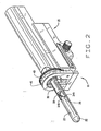
- the redirecting guide 10 consists of guide body 30, needle set channel 31, needle set insertion point 32, and needle set exit point 33, front body guide extensions 34A and 34B, imaging cutout 35.
- needle set channel 31 may be provided with enlarged bend channel 37.
- the redirecting guide 10 may be provided with insertable metal tube 38.
- the redirecting guide may contain one or more pathways may be used for insertion of the biopsy needle kit.
- the redirecting guide may be comprised of a movable device such that the opening through which the needle kit exits may be moved relative to the opening into which the biopsy needle kit is placed.
- the redirecting guide may straighten a previously curved biopsy needle kit such that the biopsy needle kit re-curve when leaving the redirecting guide.
- positioning assembly 11 is comprised of rotational adjustment collar 40, fixed collar 41, longitudinal slides 42 and longitudinal position controller 43.
- targeting software system 12 is comprised of transverse image display 212, Sagittal Image display 213, longitudinal projected needle path 51 and transverse projected needle path 52, in addition to various controls.
- flexible needle set 13 consists of flexible stylet 60 and flexible cannula 61.
- Stylet 60 may be affixed to stylet hub 76, with cannula 61 affixed to cannula hub 77. Further, cannula 61 may be provided with depth markings 79.
- the preferred Flexible stylet 60 consists of tip 62, extended specimen notch 63 and stylet body 64.
- an alternative preferred Flexible stylet 60 consists of tip 62 and segmented specimen notches 72a and 72b.
- Alternative embodiments of flexible stylet 60 as seen in FIGS. 13 and 14 , contain bending notches 70 and tiered specimen notch 71.
- cannula 61 consists of cutting tip 65, cannula body 66 and cannula sheath 81.
- the cannula sheath may have beveled edges.
- a portion of the body of flexible cannula 61 has been removed.
- cannula body 66 may be provided with spiral cut 82 to facilitate bending.
- cannula body 66 may be provided with non-spiral cut portion 83 at cutting tip 65, to facilitate the straight entry of the cannula into the tissue.
- cannula body 66 may be provided with non-spiral cut portions 83 interspersed with spiral cuts 82.
- flexible cannula 61 consists of a cutting tip inserted into the flexible cannula body.
- both the stylet cannula can be made from a range of flexible materials, including combinations of one or more materials, to facilitate the bendability. This may include traditional materials used in medical devices, such as stainless steel, as well as materials such an nitinol®.
- the cannula design may mirror the stylet, such that portion or portions of the metal cannula tube are removed to create a metal component which has a metal cutting tip, a long spine consisting of only a portion of the cannula wall in the flexible part of the cannula and then the full tubular cannula.
- the machine cannula may be partially or wholly incased in a cannula sheath, which may be plastic or some other material.
- FIG. 21 shows a biopsy being performed using the standard method, using an end-fire ultrasound probe with a biopsy needle channel.
- the probe is inserted into the rectum, and then angled upward until the probe tip is pointed at the desired portion of the prostate.
- a needle set is then inserted through the biopsy needle channel guide into the prostate 2.
- side view transrectal probe 19 is mounted on the cradle 15 of a stabilizer 16.
- Redirecting guide 10 is also mounted on the cradle 15, such that guide body 30 sits atop probe tip 22.
- front body extensions 34a and 34b partially wrap around probe tip 22 to help maintain the guide body 30 on the probe tip 22.
- the cradle 15 is moved forward, with the probe tip 22 inserted into patient's rectum 1.
- Probe tip 22 is generating ultrasound images, which are displayed on monitor 20. The physician uses this image to insure that the entirety of prostate 2 is viewable by probe imaging window 23. Once the probe tip 22 is correctly positioned, the physician locks in place cradle 15.
- the biopsy planning process is illustrated in FIG. 5 .
- a representative display of the biopsy information to the user is shown in FIG 4 .
- the process begins with the planning software obtaining a set of volumetric data 101.
- the volumetric data consists of two sets of sampled images. One set is of longitudinal images sampled at a regular angular spacing, and the other is a set of transverse images sampled at regular depth spacing. If only one of the two sets is available, one may be interpolated from the other.
- the physician starts the planning process by pressing button 203 to satisfy step 102 of FIG 4 .
- the planning system overlays a series of lines 51 a, b, c, etc. and dots 52 a, b, c, etc. on the images in panes 212 and 213.
- These lines and dots represent the available needle paths selectable with controls 40 and 43, and show where the needle intersects with image planes.
- Each line and dot combination is labeled with a coordinate 50 corresponding to a unique pair of setting for controls 40 and 43.
- the user can review the stored images using controls 210 and 211 to change the image viewed. For 104, the user can "simulate" the effect of controls 40 and 43 using on-screen controls 201 and 202 to adjust the selected needle path.
- the current path is displayed by changing the color of the appropriate line and dot (51 and 52, respectively).
- the user adds a specific needle path to the biopsy plan (105) by selecting button 208. Each time a path is selected, a record is placed into needle path coordinates display window 204 showing the coordinates of the path.
- the user may also remove a specific path from the plan by selecting button 207. When the plan is complete, the user clicks on the button 206 to send the planning process (106).
- the physician or technician may then proceed with the biopsy procedure, to complete the series of precision located biopsy's to be taken through the usage of this instrument. For example, as can be noted in FIG. 6 , once a biopsy procedure has been completed, the physician then determines whether any more biopsies are needed, and where the biopsy locations may be determined. This can be seen at 301. If no additional biopsies are required, this is the end of the procedure. If additional biopsies are considered as needed, the physician then adjusts the redirection of the guide 10, and the longitudinal controller 40, to mass the desired biopsy coordinates, as provided upon the scanner. This can be noted at 302. Then, the user inserts a needle set 13 into the channel 32, to prepare for additional biopsies.
- the physician then inserts the needle into the patient, moving the needle in and out to adjust for depth, as determined by the scanner, as can be seen at 304. Then, the physician can determine if the needle tip is at the correct depth, at 305. If it is not, then the physician may move the needle and adjust its depth further. If it is, the physician then fires the needle of the biopsy instrument, as at 306. Then the physician removes the needle set 13 from the patient, having taken the biopsy as required. Then, the tissue sample is removed from the biopsy needle notch, for further analysis by the lab. This can be noted at 308. When this is completed, this concludes the conduct of biopsies upon the patient
- preplanned biopsy selection menu 209 allows the user to select a pre-determined needle pattern, typically 9-12 needle paths, without having to select each needle path manually.
- the needle paths generated could need to be adjusted for the specific size of the organ.
- the size of the organ can be input by various means.
- the planning process allows the physician to modify the needle paths as needed and to approve that they are correct.
- Projected needle paths 51 a, 51 b, etc include needle path location registry 50, which indicate the horizontal and rotational position of the needle path in reference to the probe.
- the physician rotates redirecting guide 10 using rotational adjustment collar 40, and then advances the redirecting guide using longitudinal position controller 40, both of which have position information which correlates to the needle path location registry 50.
- the physician inserts flexible needle set 13 into needle set insertion point 32 and into needle set channel 31.
- the needle set 13 is redirected at an angle away from the axis of probe tip 22. Needle set 13 exits needle set exit point 33.
- the physician is able to see the needle set in the ultrasound image as it exits exit point 33, allowing the physician to insure that the needle set 13 is in the path marked by projected needle path 51a.
- the physician monitors the depth of the needle set 13 as it is pushed through the rectum wall and into the prostate 2. Once the desired depth is reached, the physician stops inserting the needle set 13. Using a standard biopsy gun, the needle set 13 is "fired". This causes flexible stylet 60 to rapidly advance a short distance, such that tissue from the prostate two prolapses into extended specimen notch 63.
- Almost instantaneously flexible cannula 61 quickly advances, also under spring loaded pressure or other motivational means, which serves to sever and capture the tissue that had prolapsed into the extended specimen notch 63. Because the extended specimen notch 63 extends to the point where flexible needle set 13 is bent in needle set channel bend, the stylet arid cannula are able to fire without the two pieces binding together, allowing the specimen to be effectively captured.
- the physician then removes the flexible needle set 13 with the captured specimen. The specimen is removed from the flexible needle set, and the physician then resets the redirecting guide to the coordinates of the next saved projected needle path 51b. The process is repeated until the physician has captured all of the samples as planned using the targeting software system 12.
- FIG. 23 provides a side cut-away view of the redirecting guide with a flexible needle set inserted and extending out of the guide such that the extended specimen notch is bent by the needle set channel bend.
- the invention is used to plan and perform a targeted treatment of an organ or tissue mass.
- the process begins with the planning software obtaining a set of volumetric data.
- the planning system overlays a series of needle path lines and needle path dots on the images in panes 212 and 213, which represent the available needle paths with coordinates that match the coordinates on rotational adjustment collar 40 and longitudinal position controller 43 of positioning assembly 11.
- the user selects specific needle paths, which are saved the treatment plan. Preplanned treatment selections allow the user to select a pre-determined needle pattern without having to select each needle path manually.
- the physician rotates redirecting guide using rotational adjustment collar, and then advances the redirecting guide using longitudinal position controller, both of which have position information which correlates to the needle path location registry.
- the physician then inserts a flexible needle set or treatment delivery means into needle set insertion point 32 and into needle set channel 31.
- the needle set or treatment delivery method is redirected at an angle away from the axis of probe tip 22.
- Needle set 13 exits needle set exit point 33. Because of imaging cutout 35, the physician is able to see the needle set or treatment delivery method in the ultrasound image as it exits exit point 33.
- the physician monitors the depth of the needle set or treatment delivery method as it is pushed into the targeted organ or tissue mass.
- the physician is able to undertake the preferred activity. This may include using the delivery means to inject a solid, gas or liquid material or other treatment apparatus into the targeted organ or tissue mass. Further, the physician may insert an organism into the targeted organ or tissue mass. The material may be deposited and left in the targeted organ or tissue mass. Further, material previously deposited may be removed. The use of the deposited material may be as a treatment, a marker, or other uses. Further, the delivery means may be used to apply energy to a targeted organ or tissue mass, including but not limited to heat, cold, light and radiation. Once the treatment or marking is delivered, the physician then removes the flexible needle set or treatment delivery method, and then resets the redirecting guide to the coordinates of the next saved projected needle path. The physician has the option of saving the image of the treatment needle in the targeted organ or tissue mass, to record the location of the treatment as delivered. The process is repeated until the physician has treated or marked all of the targeted areas of the organ or tissue mass.
Landscapes
- Health & Medical Sciences (AREA)
- Life Sciences & Earth Sciences (AREA)
- Surgery (AREA)
- Animal Behavior & Ethology (AREA)
- Public Health (AREA)
- Engineering & Computer Science (AREA)
- Biomedical Technology (AREA)
- Heart & Thoracic Surgery (AREA)
- Medical Informatics (AREA)
- Molecular Biology (AREA)
- Veterinary Medicine (AREA)
- General Health & Medical Sciences (AREA)
- Pathology (AREA)
- Nuclear Medicine, Radiotherapy & Molecular Imaging (AREA)
- Biodiversity & Conservation Biology (AREA)
- Ultra Sonic Daignosis Equipment (AREA)
- Investigating Or Analysing Biological Materials (AREA)
- Radiation-Therapy Devices (AREA)
- Medicines Containing Antibodies Or Antigens For Use As Internal Diagnostic Agents (AREA)
- Nuclear Medicine (AREA)
Claims (24)
- Gezieltes Biopsie-Abgabesystem, umfassend eine zurückleitende Führung (10) mit einem Führungskörper (30) und einem Nadelsatzkanal (31), einem flexiblen Biopsie-Nadelsatz (13), der in dem Nadelsatzkanal (31) der zurückleitenden Führung (10) zur Vornahme von Biopsien eingesteckt werden kann, ferner mit einem Ultraschallsystem (17), das ein Ultraschallsystem CPU (18) hat, einer Ultraschallsonde (19) und einem Monitor (20), wobei die Ultraschallsonde (19) eine Sondenspitze (22) und ein Sondenbildfenster (23) aufweist, die so geartet sind, daß der Führungskörper (30) der zurückleitenden Führung (10) oben auf der Sondenspitze (22) sitzt und in unterschiedliche Drehstellungen bewegt werden kann sowie unterschiedliche Längsstellungen in Bezug auf die Sondenspitze (22), und wobei die Sondenspitze (22) Ultraschallbilder einer Zielzellmasse erzeugt, die auf dem Monitor (20) gezeigt werden, ferner umfassend eine Anordnung (11) zur Steuerung der Drehstellung und Längsstellung des Führungskörpers (30) der zurückleitenden Führung (10) in Bezug auf die Sondenspitze (22), und ein gezieltes Softwaresystem (12, 18) mit einem Querformat-Display (212) und einem sagittalen Bilddisplay (213), wobei mehrere Nadelwege sowohl auf das Querformat-Display (212) als auch das sagittale Bilddisplay (213) gerichtet sind und jeder Nadelweg durch eine entsprechende Drehstellung und Längsstellung des Führungskörpers (30) der zurückleitenden Führung (10) in Bezug auf die Sondenspitze (22), wie durch die Anordnung (11) gesteuert, definiert wird.
- System nach Anspruch 1, dadurch gekennzeichnet, daß ein entsprechender Nadelweg als Linie in der Bildebene des Bilddisplays (212, 213) gezeigt wird.
- System nach Anspruch 1, dadurch gekennzeichnet, daß ein entsprechender Nadelweg als grafisches Element, beispielsweise als Kreis, in der Bildebene des Bilddisplays (212, 213) gezeigt wird.
- System nach Anspruch 3, dadurch gekennzeichnet, daß das grafische Element dort dargestellt wird, wo der entsprechende Nadelweg die Bildebene des Bilddisplays (212, 213) durchstößt.
- System nach einem der Ansprüche 1 bis 4, dadurch gekennzeichnet, daß die projizierten Nadelwege (51 a, 52a) ein Nadelweg-Ortregister (50) aufweisen, die die waagerechte und Drehlage des Nadelweges mit Bezug auf die Ultraschallsonde (19) anzeigt, und daß die Anordnung (11) mit einer Lageinformation versehen ist, die mit dem Nadelweg-Ortregister (50) korreliert.
- System nach einem der Ansprüche 1 bis 5, dadurch gekennzeichnet, daß der flexible Biopsie-Nadelsatz (13) eine flexible Sonde (60) und eine flexible Kanüle (61) aufweist.
- System nach Anspruch 6, dadurch gekennzeichnet, daß die Sonde (60) und die Kanüle (61) mit einem vorgeformten Bogen angefertigt sind.
- System nach Anspruch 6 oder 7, dadurch gekennzeichnet, daß die Sonde (60) und die Kanüle (61) aus einem flexiblem Material, beispielsweise Nitinol® oder Plastik, angefertigt sind.
- System nach einem der Ansprüche 6 bis 8, dadurch gekennzeichnet, daß die Sonde (60) und / oder die Kanüle (61) aus zwei oder mehr Abschnitten zusammengebaut sind, von denen jeder aus Materialien mit unterschiedlichen Eigenschaften besteht, um die Flexibilität zu verbessern.
- System nach einem der Ansprüche 6 bis 9, dadurch gekennzeichnet, daß die Sonde (60) erste und zweite Nocken (72a, 72b) aufweist, die mit Abstand nebeneinander liegen.
- System nach einem der vorhergehenden Ansprüche, dadurch gekennzeichnet, daß die zurückleitende Führung (10) des weiteren eine Öffnung (32) aufweist, in die der Biopsie-Nadelsatz (13) eingesteckt werden kann, des weiteren eine Öffnung (33), durch die ein Teil des Biopsie-Nadelsatzes (13) austritt, ferner eine Einrichtung zum Zurückleiten des Biopsie-Nadelsatzes (13), die wie eine Kanalbiegung (36) ausgebildet ist, so daß derjenige Teil des Biopsie-Nadelsatzes (13), der aus der zurückleitenden Führung (10) ausgetreten ist, in Bezug auf den Teil des Biopsie-Nadelsatzes (13), der nicht in die Führung (10) eingesteckt worden ist, gekrümmt ist.
- System nach Anspruch 11, dadurch gekennzeichnet, daß die Einrichtung der Zurückleitung des Biopsie-Nadelsatzes (13) den vorher gerade gerichteten Biopsie-Nadelsatz (13) körperlich biegt.
- System nach Anspruch 11 oder 12, dadurch gekennzeichnet, daß die Zurückleitungs-Einrichtung einen vorher gebogenen Biopsie-Nadelsatz (13) so gerade richtet, daß der Biopsie-Nadelsatz (13) sich wieder krümmt, sobald er die Zurückleitungs-Einrichtung verläßt.
- System nach einem der Ansprüche 11 bis 13, dadurch gekennzeichnet, daß die Zurückleitungs-Einrichtung einen oder mehr statische, gewinkelte oder gekrümmte Kanäle aufweist.
- System nach einem der Ansprüche 11 bis 14, dadurch gekennzeichnet, daß die Zurückleitungs-Einrichtung eine bewegliche Vorrichtung aufweist derart, daß die Öffnung (33), durch die der Nadelsatz (13) austritt, in Bezug auf die Öffnung (32), in die der Biopsie-Nadelsatz (13) plaziert wird, bewegt werden kann.
- System nach einem der vorhergehenden Ansprüche, dadurch gekennzeichnet, daß die zurückleitende Führung (10) wahlweise zugeordnet oder in Bezug auf die Sonde (19) gedreht wird, und zwar in Übereinstimmung mit den geplanten Biopsien eines Biopsieplans.
- System nach einem der vorhergehenden Ansprüche, dadurch gekennzeichnet, daß die zurückleitende Führung (10) mit Abgrenzungen oder anderen Zeichen der Lage in Bezug auf die Sonde (19) versehen ist.
- System nach einem der vorhergehenden Ansprüche, dadurch gekennzeichnet, daß das gezielte Software-System (12, 18) die Lage der Biopsie-Probe aufzeichnet.
- System nach einem der vorhergehenden Ansprüche, dadurch gekennzeichnet, daß das gezielte Software-System (12, 18) einen Biopsie-Plan aufweist, der die bevorzugten Stellen der Biopsien in Bezug auf die Sonde (19) spezifiziert.
- System nach einem der Ansprüche 6 bis 10, dadurch gekennzeichnet, daß der Querschnitt der Sonde (60) für einen Teil der Länge der Sonde reduziert ist, um dadurch ihre Flexibilität zu verbessern.
- System nach einem der Ansprüche 6 bis 10, dadurch gekennzeichnet, daß der Querschnitt der Sonde (60) zwischen Zonen wechselt, in denen der Querschnitt reduziert ist, und Zonen, in denen der Querschnitt nicht reduziert ist, um die Flexibilität der Sonde (60) zu verbessern.
- System nach einem der Ansprüche 6 bis 10, dadurch gekennzeichnet, daß die Kanüle (61) zur Verbesserung der Flexibilität spiralförmig geschnitten ist.
- System nach einem der Ansprüche 6 bis 10, dadurch gekennzeichnet, daß der Körper der Kanüle (61) spiralförmig geschnitten ist, jedoch die Spitze des Kanülenkörpers nicht.
- System nach einem der Ansprüche 6 bis 10, dadurch gekennzeichnet, daß der Körper der Kanüle (61) zwischen spiralförmig geschnittenen Teilen und nicht spiralförmig geschnittenen Teilen wechselt.
Applications Claiming Priority (2)
| Application Number | Priority Date | Filing Date | Title |
|---|---|---|---|
| US10/842,652 US20050159676A1 (en) | 2003-08-13 | 2004-05-10 | Targeted biopsy delivery system |
| PCT/US2005/015431 WO2005110255A2 (en) | 2004-05-10 | 2005-05-04 | Targeted biopsy delivery system |
Publications (3)
| Publication Number | Publication Date |
|---|---|
| EP1765178A2 EP1765178A2 (de) | 2007-03-28 |
| EP1765178A4 EP1765178A4 (de) | 2009-04-08 |
| EP1765178B1 true EP1765178B1 (de) | 2011-04-13 |
Family
ID=35394638
Family Applications (1)
| Application Number | Title | Priority Date | Filing Date |
|---|---|---|---|
| EP05744025A Expired - Lifetime EP1765178B1 (de) | 2004-05-10 | 2005-05-04 | Gezieltes biopsie-abgabesystem |
Country Status (11)
| Country | Link |
|---|---|
| US (4) | US20050159676A1 (de) |
| EP (1) | EP1765178B1 (de) |
| JP (1) | JP4837658B2 (de) |
| KR (1) | KR20070028402A (de) |
| CN (1) | CN101132736B (de) |
| AT (1) | ATE505136T1 (de) |
| AU (1) | AU2005244164B2 (de) |
| CA (1) | CA2566231C (de) |
| DE (1) | DE602005027462D1 (de) |
| ES (1) | ES2365476T3 (de) |
| WO (1) | WO2005110255A2 (de) |
Families Citing this family (158)
| Publication number | Priority date | Publication date | Assignee | Title |
|---|---|---|---|---|
| US20050159676A1 (en) * | 2003-08-13 | 2005-07-21 | Taylor James D. | Targeted biopsy delivery system |
| US8070750B2 (en) | 2004-03-05 | 2011-12-06 | Depuy Mitek, Inc. | Tunnel notcher and guidewire delivery device |
| US8795195B2 (en) * | 2004-11-29 | 2014-08-05 | Senorx, Inc. | Graphical user interface for tissue biopsy system |
| EP1858418B1 (de) * | 2005-02-28 | 2017-08-16 | Robarts Research Institute | System zur durchführung einer biopsie eines zielvolumens und computervorrichtung zu ihrer planung |
| US7831293B2 (en) * | 2005-05-10 | 2010-11-09 | Advanced Clinical Solutions, Inc. | Method of defining a biological target for treatment |
| US8784336B2 (en) | 2005-08-24 | 2014-07-22 | C. R. Bard, Inc. | Stylet apparatuses and methods of manufacture |
| US8425418B2 (en) * | 2006-05-18 | 2013-04-23 | Eigen, Llc | Method of ultrasonic imaging and biopsy of the prostate |
| WO2008017051A2 (en) | 2006-08-02 | 2008-02-07 | Inneroptic Technology Inc. | System and method of providing real-time dynamic imagery of a medical procedure site using multiple modalities |
| US8064664B2 (en) * | 2006-10-18 | 2011-11-22 | Eigen, Inc. | Alignment method for registering medical images |
| US8388546B2 (en) | 2006-10-23 | 2013-03-05 | Bard Access Systems, Inc. | Method of locating the tip of a central venous catheter |
| US7794407B2 (en) | 2006-10-23 | 2010-09-14 | Bard Access Systems, Inc. | Method of locating the tip of a central venous catheter |
| US7804989B2 (en) * | 2006-10-30 | 2010-09-28 | Eigen, Inc. | Object recognition system for medical imaging |
| US20080161687A1 (en) * | 2006-12-29 | 2008-07-03 | Suri Jasjit S | Repeat biopsy system |
| US9232959B2 (en) | 2007-01-02 | 2016-01-12 | Aquabeam, Llc | Multi fluid tissue resection methods and devices |
| US12290277B2 (en) | 2007-01-02 | 2025-05-06 | Aquabeam, Llc | Tissue resection with pressure sensing |
| US8175350B2 (en) | 2007-01-15 | 2012-05-08 | Eigen, Inc. | Method for tissue culture extraction |
| US7856130B2 (en) * | 2007-03-28 | 2010-12-21 | Eigen, Inc. | Object recognition system for medical imaging |
| US7832114B2 (en) * | 2007-04-04 | 2010-11-16 | Eigen, Llc | Tracker holder assembly |
| US8979803B2 (en) * | 2007-04-05 | 2015-03-17 | Allan J. Darr | Stylet for bilumenal flexible medical device |
| US20100152663A1 (en) * | 2007-04-05 | 2010-06-17 | Darr Allan J | Stylet for bilumenal flexible medical device |
| US20090048515A1 (en) * | 2007-08-14 | 2009-02-19 | Suri Jasjit S | Biopsy planning system |
| US8088072B2 (en) | 2007-10-12 | 2012-01-03 | Gynesonics, Inc. | Methods and systems for controlled deployment of needles in tissue |
| US8571277B2 (en) | 2007-10-18 | 2013-10-29 | Eigen, Llc | Image interpolation for medical imaging |
| US7942829B2 (en) * | 2007-11-06 | 2011-05-17 | Eigen, Inc. | Biopsy planning and display apparatus |
| US20090227874A1 (en) * | 2007-11-09 | 2009-09-10 | Eigen, Inc. | Holder assembly for a medical imaging instrument |
| US8781555B2 (en) | 2007-11-26 | 2014-07-15 | C. R. Bard, Inc. | System for placement of a catheter including a signal-generating stylet |
| ES2557084T3 (es) | 2007-11-26 | 2016-01-21 | C. R. Bard, Inc. | Sistema integrado para la colocación intravascular de un catéter |
| US10524691B2 (en) | 2007-11-26 | 2020-01-07 | C. R. Bard, Inc. | Needle assembly including an aligned magnetic element |
| US9521961B2 (en) | 2007-11-26 | 2016-12-20 | C. R. Bard, Inc. | Systems and methods for guiding a medical instrument |
| US8450290B2 (en) * | 2007-11-26 | 2013-05-28 | Enzon Pharmaceuticals, Inc. | Methods for treating androgen receptor dependent disorders including cancers |
| US10751509B2 (en) | 2007-11-26 | 2020-08-25 | C. R. Bard, Inc. | Iconic representations for guidance of an indwelling medical device |
| US9492097B2 (en) | 2007-11-26 | 2016-11-15 | C. R. Bard, Inc. | Needle length determination and calibration for insertion guidance system |
| US10449330B2 (en) | 2007-11-26 | 2019-10-22 | C. R. Bard, Inc. | Magnetic element-equipped needle assemblies |
| US9649048B2 (en) | 2007-11-26 | 2017-05-16 | C. R. Bard, Inc. | Systems and methods for breaching a sterile field for intravascular placement of a catheter |
| US8849382B2 (en) | 2007-11-26 | 2014-09-30 | C. R. Bard, Inc. | Apparatus and display methods relating to intravascular placement of a catheter |
| JP2009153831A (ja) * | 2007-12-27 | 2009-07-16 | Ge Medical Systems Global Technology Co Llc | 穿刺ガイドの取付構造、超音波プローブ及び超音波診断装置 |
| WO2009094646A2 (en) | 2008-01-24 | 2009-07-30 | The University Of North Carolina At Chapel Hill | Methods, systems, and computer readable media for image guided ablation |
| US20090216250A1 (en) * | 2008-02-27 | 2009-08-27 | Ralph Zipper | Device and Method for Carrying Material Through Tissue |
| JP5506702B2 (ja) | 2008-03-06 | 2014-05-28 | アクアビーム エルエルシー | 流体流れ内を伝達される光学エネルギーによる組織切除および焼灼 |
| US8340379B2 (en) | 2008-03-07 | 2012-12-25 | Inneroptic Technology, Inc. | Systems and methods for displaying guidance data based on updated deformable imaging data |
| US8864681B2 (en) * | 2008-04-23 | 2014-10-21 | Devicor Medical Products, Inc. | Biopsy devices |
| US20100076305A1 (en) * | 2008-06-25 | 2010-03-25 | Deutsches Krebsforschungszentrum Stiftung Des Offentlichen Rechts | Method, system and computer program product for targeting of a target with an elongate instrument |
| SE532456C2 (sv) * | 2008-08-01 | 2010-01-26 | Christer Dahlstrand Ab | Anordning vid provtagningsnålar |
| ES2525525T3 (es) | 2008-08-22 | 2014-12-26 | C.R. Bard, Inc. | Conjunto de catéter que incluye conjuntos de sensor de ECG y magnético |
| US9782565B2 (en) | 2008-10-01 | 2017-10-10 | Covidien Lp | Endoscopic ultrasound-guided biliary access system |
| US11298113B2 (en) | 2008-10-01 | 2022-04-12 | Covidien Lp | Device for needle biopsy with integrated needle protection |
| US9186128B2 (en) | 2008-10-01 | 2015-11-17 | Covidien Lp | Needle biopsy device |
| US9332973B2 (en) | 2008-10-01 | 2016-05-10 | Covidien Lp | Needle biopsy device with exchangeable needle and integrated needle protection |
| US8968210B2 (en) | 2008-10-01 | 2015-03-03 | Covidien LLP | Device for needle biopsy with integrated needle protection |
| US8437833B2 (en) | 2008-10-07 | 2013-05-07 | Bard Access Systems, Inc. | Percutaneous magnetic gastrostomy |
| JP2012508068A (ja) | 2008-11-06 | 2012-04-05 | エヌエックスセラ インコーポレイテッド | 前立腺組織の治療のためのシステム及び方法 |
| CN102271605B (zh) | 2008-11-06 | 2015-12-02 | 恩克斯特拉公司 | 用于治疗前列腺组织的系统和方法 |
| US8372065B2 (en) | 2008-11-06 | 2013-02-12 | Nxthera, Inc. | Systems and methods for treatment of BPH |
| US8388611B2 (en) * | 2009-01-14 | 2013-03-05 | Nxthera, Inc. | Systems and methods for treatment of prostatic tissue |
| US20100179416A1 (en) * | 2009-01-14 | 2010-07-15 | Michael Hoey | Medical Systems and Methods |
| US8554307B2 (en) | 2010-04-12 | 2013-10-08 | Inneroptic Technology, Inc. | Image annotation in image-guided medical procedures |
| US8690776B2 (en) | 2009-02-17 | 2014-04-08 | Inneroptic Technology, Inc. | Systems, methods, apparatuses, and computer-readable media for image guided surgery |
| US8641621B2 (en) | 2009-02-17 | 2014-02-04 | Inneroptic Technology, Inc. | Systems, methods, apparatuses, and computer-readable media for image management in image-guided medical procedures |
| US11464578B2 (en) | 2009-02-17 | 2022-10-11 | Inneroptic Technology, Inc. | Systems, methods, apparatuses, and computer-readable media for image management in image-guided medical procedures |
| US10524822B2 (en) | 2009-03-06 | 2020-01-07 | Procept Biorobotics Corporation | Image-guided eye surgery apparatus |
| US9833277B2 (en) | 2009-04-27 | 2017-12-05 | Nxthera, Inc. | Systems and methods for prostate treatment |
| US9532724B2 (en) | 2009-06-12 | 2017-01-03 | Bard Access Systems, Inc. | Apparatus and method for catheter navigation using endovascular energy mapping |
| US9339206B2 (en) | 2009-06-12 | 2016-05-17 | Bard Access Systems, Inc. | Adaptor for endovascular electrocardiography |
| WO2011017665A2 (en) * | 2009-08-07 | 2011-02-10 | Thayer Intellectual Property, Inc. | Systems and methods for treatment of compressed nerves |
| US8753364B2 (en) * | 2009-08-07 | 2014-06-17 | Thayer Intellectual Property, Inc. | Systems and methods for treatment of compressed nerves |
| US8652157B2 (en) | 2009-08-07 | 2014-02-18 | Thayer Intellectual Property, Inc. | Systems and methods for treatment of compressed nerves |
| EP2464407A4 (de) | 2009-08-10 | 2014-04-02 | Bard Access Systems Inc | Vorrichtungen und verfahren für endovaskuläre elektrographie |
| CN102665541B (zh) | 2009-09-29 | 2016-01-13 | C·R·巴德股份有限公司 | 与用于导管的血管内放置的设备一起使用的探针 |
| KR101109197B1 (ko) | 2009-12-17 | 2012-01-30 | 국립암센터 | 바늘 투입장치 |
| WO2011097312A1 (en) | 2010-02-02 | 2011-08-11 | C.R. Bard, Inc. | Apparatus and method for catheter navigation and tip location |
| AU2011230568B2 (en) | 2010-03-25 | 2016-02-18 | Boston Scientific Scimed, Inc. | Systems and methods for prostate treatment |
| EP2912999B1 (de) | 2010-05-28 | 2022-06-29 | C. R. Bard, Inc. | Vorrichtung zur Verwendung mit einem Nadeleinsatz-Führungssystem |
| CN103037761B (zh) | 2010-05-28 | 2016-11-02 | C·R·巴德股份有限公司 | 用于针和医疗部件的插入引导系统 |
| US8758256B2 (en) | 2010-07-12 | 2014-06-24 | Best Medical International, Inc. | Apparatus for brachytherapy that uses a scanning probe for treatment of malignant tissue |
| US9044216B2 (en) * | 2010-07-12 | 2015-06-02 | Best Medical International, Inc. | Biopsy needle assembly |
| JP2012042310A (ja) * | 2010-08-18 | 2012-03-01 | Sony Corp | 生理活性物質採取装置及び生体情報取得方法 |
| JP5482564B2 (ja) * | 2010-08-18 | 2014-05-07 | ソニー株式会社 | 生理活性物質採取装置 |
| JP5845260B2 (ja) | 2010-08-20 | 2016-01-20 | シー・アール・バード・インコーポレーテッドC R Bard Incorporated | Ecg支援カテーテル先端配置の再確認 |
| CN103153201B (zh) | 2010-10-05 | 2016-06-29 | 斯恩蒂斯有限公司 | 具有柔性针的骨髓收获装置 |
| US20120095371A1 (en) * | 2010-10-18 | 2012-04-19 | CardioSonic Ltd. | Ultrasound transducer and cooling thereof |
| WO2012058461A1 (en) | 2010-10-29 | 2012-05-03 | C.R.Bard, Inc. | Bioimpedance-assisted placement of a medical device |
| US11612377B2 (en) * | 2010-12-16 | 2023-03-28 | Best Medical International, Inc. | Image guided surgical methodology and system employing patient movement detection and correction |
| US9414816B2 (en) * | 2011-06-23 | 2016-08-16 | Devicor Medical Products, Inc. | Introducer for biopsy device |
| CN102894994A (zh) * | 2011-07-25 | 2013-01-30 | 滕学东 | 超声探头保护膜 |
| EP2755614B1 (de) | 2011-09-13 | 2017-11-01 | Nxthera, Inc. | Systeme zur prostatabehandlung |
| FR2983397B1 (fr) * | 2011-12-06 | 2014-07-04 | Univ Paris Curie | Dispositif d'assistance au positionnement d'un instrument medical relativement a un organe interne d'un patient et procede de commande d'un tel dispositif |
| US8670816B2 (en) | 2012-01-30 | 2014-03-11 | Inneroptic Technology, Inc. | Multiple medical device guidance |
| US20130225996A1 (en) * | 2012-02-28 | 2013-08-29 | Spiration, Inc. | Lung biopsy needle |
| CN104203078B (zh) | 2012-02-29 | 2018-04-20 | 普罗赛普特生物机器人公司 | 自动化图像引导的组织切除和处理 |
| EP4530528A3 (de) | 2012-04-03 | 2025-06-18 | Boston Scientific Scimed, Inc. | Induktionsspulendampferzeuger |
| CA2869976C (en) * | 2012-04-10 | 2020-03-10 | The Johns Hopkins University | Cohesive robot-ultrasound probe for prostate biopsy |
| US10357304B2 (en) | 2012-04-18 | 2019-07-23 | CardioSonic Ltd. | Tissue treatment |
| US9131922B2 (en) | 2013-01-29 | 2015-09-15 | Eigen, Inc. | Calibration for 3D reconstruction of medical images from a sequence of 2D images |
| TWI592141B (zh) * | 2013-03-07 | 2017-07-21 | 國立陽明大學 | 超音波探頭結構 |
| AU2014236335A1 (en) | 2013-03-14 | 2015-10-15 | Nxthera Inc. | Systems and methods for treating prostate cancer |
| US10314559B2 (en) | 2013-03-14 | 2019-06-11 | Inneroptic Technology, Inc. | Medical device guidance |
| US20160038127A1 (en) * | 2013-03-15 | 2016-02-11 | R. Hashimshony Engineering Ltd. | Biopsy Probe and Use Thereof |
| US10206664B2 (en) * | 2013-11-13 | 2019-02-19 | Cook Medical Technologies Llc | Spiral cut biopsy cannula |
| US9968395B2 (en) | 2013-12-10 | 2018-05-15 | Nxthera, Inc. | Systems and methods for treating the prostate |
| JP6422975B2 (ja) | 2013-12-10 | 2018-11-14 | エヌエックスセラ インコーポレイテッド | 蒸気焼灼システム及び方法 |
| CN105979868B (zh) | 2014-02-06 | 2020-03-10 | C·R·巴德股份有限公司 | 用于血管内装置的导向和放置的系统和方法 |
| US9149260B2 (en) | 2014-02-28 | 2015-10-06 | 3DBiopsy LLC | Biopsy needle assembly |
| US12144519B2 (en) * | 2014-04-03 | 2024-11-19 | Corbin Clinical Resources, Llc | Transperineal prostate biopsy and treatment methods |
| US10743909B2 (en) * | 2014-04-03 | 2020-08-18 | Corbin Clinical Resources, Llc | Transperineal prostate biopsy device, systems, and methods of use |
| US10064681B2 (en) * | 2014-04-03 | 2018-09-04 | Corbin Clinical Resources, Llc | Method, system, and device for planning and performing, guided and free-handed transperineal prostate biopsies |
| CN103919577A (zh) * | 2014-04-13 | 2014-07-16 | 哈尔滨理工大学 | 悬臂式前列腺活检机器人 |
| US11033296B2 (en) * | 2014-08-23 | 2021-06-15 | Intuitive Surgical Operations, Inc. | Systems and methods for dynamic trajectory control |
| KR102542848B1 (ko) * | 2014-08-23 | 2023-06-14 | 인튜어티브 서지컬 오퍼레이션즈 인코포레이티드 | 영상 안내 절차에서의 병리학적 데이터의 표시를 위한 시스템 및 방법 |
| CN104224092B (zh) * | 2014-09-19 | 2016-08-17 | 珠海普生医疗科技有限公司 | 内窥镜先端部结构 |
| US9901406B2 (en) | 2014-10-02 | 2018-02-27 | Inneroptic Technology, Inc. | Affected region display associated with a medical device |
| US10314563B2 (en) | 2014-11-26 | 2019-06-11 | Devicor Medical Products, Inc. | Graphical user interface for biopsy device |
| US10188467B2 (en) | 2014-12-12 | 2019-01-29 | Inneroptic Technology, Inc. | Surgical guidance intersection display |
| WO2016103094A1 (en) * | 2014-12-24 | 2016-06-30 | Koninklijke Philips N.V. | Needle trajectory prediction for target biopsy |
| US10973584B2 (en) | 2015-01-19 | 2021-04-13 | Bard Access Systems, Inc. | Device and method for vascular access |
| WO2016123498A1 (en) | 2015-01-29 | 2016-08-04 | Nxthera, Inc. | Vapor ablation systems and methods |
| CN104688278A (zh) * | 2015-03-11 | 2015-06-10 | 毕文志 | 一种骨肿瘤活检穿刺针 |
| EP3294171B1 (de) | 2015-05-13 | 2020-07-29 | Nxthera, Inc. | Systeme zur behandlung der blase mit kondensierbarem dampf |
| CN104939800B (zh) * | 2015-06-16 | 2017-04-05 | 西南医科大学附属医院 | 一种肠胃自动检测手术仪 |
| WO2016210325A1 (en) | 2015-06-26 | 2016-12-29 | C.R. Bard, Inc. | Connector interface for ecg-based catheter positioning system |
| US10849650B2 (en) | 2015-07-07 | 2020-12-01 | Eigen Health Services, Llc | Transperineal needle guidance |
| US9949700B2 (en) | 2015-07-22 | 2018-04-24 | Inneroptic Technology, Inc. | Medical device approaches |
| US10716544B2 (en) | 2015-10-08 | 2020-07-21 | Zmk Medical Technologies Inc. | System for 3D multi-parametric ultrasound imaging |
| CN105147335B (zh) * | 2015-10-13 | 2017-11-28 | 拜耳斯特医疗机器人技术(天津)有限公司 | 一种超声探头旋转装置及其使用方法和应用 |
| US11000207B2 (en) | 2016-01-29 | 2021-05-11 | C. R. Bard, Inc. | Multiple coil system for tracking a medical device |
| US9675319B1 (en) | 2016-02-17 | 2017-06-13 | Inneroptic Technology, Inc. | Loupe display |
| JP2019523661A (ja) * | 2016-05-25 | 2019-08-29 | スリーディーバイオプシー,インコーポレーテッド | 生検針構造 |
| CN106361396B (zh) * | 2016-09-27 | 2018-11-23 | 熊力 | 一种肝脏血管夹闭装置 |
| JP2020500082A (ja) | 2016-10-10 | 2020-01-09 | スリーディーバイオプシー,インコーポレーテッド | 生検アクチュエータアセンブリ |
| US10751034B2 (en) | 2016-10-17 | 2020-08-25 | The Johna Hopkins University | Geometric biopsy plan optimization |
| US10278778B2 (en) | 2016-10-27 | 2019-05-07 | Inneroptic Technology, Inc. | Medical device navigation using a virtual 3D space |
| WO2018089523A1 (en) | 2016-11-11 | 2018-05-17 | Gynesonics, Inc. | Controlled treatment of tissue and dynamic interaction with, and comparison of, tissue and/or treatment data |
| US11246640B2 (en) | 2016-12-21 | 2022-02-15 | Boston Scientific Scimed, Inc. | Vapor ablation systems and methods |
| EP3565481A4 (de) * | 2017-01-06 | 2020-10-07 | Sorek Medical Systems, Ltd. | Kernbiopsiesystem zur aufbewahrung und konservierung mehrerer gewebeproben |
| WO2018129466A1 (en) | 2017-01-06 | 2018-07-12 | Nxthera, Inc. | Transperineal vapor ablation systems and methods |
| MA49562A (fr) | 2017-07-14 | 2020-05-20 | Amgen Inc | Système d'insertion-rétractation d'aiguille présentant un système à ressort en double torsion |
| US11259879B2 (en) | 2017-08-01 | 2022-03-01 | Inneroptic Technology, Inc. | Selective transparency to assist medical device navigation |
| US11484365B2 (en) | 2018-01-23 | 2022-11-01 | Inneroptic Technology, Inc. | Medical image guidance |
| US10912542B2 (en) * | 2018-03-14 | 2021-02-09 | Spiration, Inc. | Catheter assembly with offset device for tissue sampling |
| US20190282218A1 (en) * | 2018-03-14 | 2019-09-19 | SPIRATION, INC., d/b/a OLYMPUS RESPIRATORY AMERICA | Catheter Assembly With Offset Device For Tissue Sampling |
| CN108888315B (zh) * | 2018-04-17 | 2024-11-19 | 建湖县人民医院 | 一种通用型数字化经会阴前列腺穿刺支架 |
| WO2019222856A1 (en) | 2018-05-24 | 2019-11-28 | Nureva Inc. | Method, apparatus and computer-readable media to manage semi-constant (persistent) sound sources in microphone pickup/focus zones |
| AU2019275406B2 (en) | 2018-05-24 | 2024-11-14 | Celanese Eva Performance Polymers Llc | Implantable device for sustained release of a macromolecular drug compound |
| WO2019226519A1 (en) | 2018-05-24 | 2019-11-28 | Celanese EVA Performance Polymers Corporation | Implantable device for sustained release of a macromolecular drug compound |
| CN109011030B (zh) * | 2018-08-08 | 2021-02-09 | 长沙理工大学 | 自动注射仪器针头的位置检测矫正方法及装置 |
| EP3852622B1 (de) | 2018-10-16 | 2025-04-02 | Bard Access Systems, Inc. | Sicherheitsausgerüstete verbindungssysteme und verfahren dafür zur herstellung von elektrischen verbindungen |
| PL4193932T3 (pl) * | 2018-12-10 | 2024-11-12 | Devicor Medical Products, Inc. | System biopsji z igłą o końcowym wysuwie |
| KR102747176B1 (ko) * | 2018-12-11 | 2024-12-27 | 삼성메디슨 주식회사 | 초음파 영상 장치, 그 제어 방법, 및 컴퓨터 프로그램 제품 |
| CN113473916A (zh) | 2019-01-30 | 2021-10-01 | 巴德阿克塞斯系统股份有限公司 | 用于跟踪医疗装置的系统和方法 |
| EP3965861B1 (de) * | 2019-05-10 | 2024-11-27 | Merit Medical Systems, Inc. | Drainagekatheteraustauschsystem |
| WO2021186342A1 (en) * | 2020-03-16 | 2021-09-23 | Robin Medical Inc. | Guidance system for interventional devices with curved shape |
| CA3199285A1 (en) | 2020-11-23 | 2022-05-27 | Willet WHITMORE | Transperineal puncture device guide |
| GB2616235B (en) * | 2020-12-10 | 2025-03-12 | Nat Univ Hospital Singapore Pte Ltd | Balloon-anchored flexible needle and catheter for biopsy |
| US20220211263A1 (en) * | 2021-03-23 | 2022-07-07 | Axcess Instruments Inc. | Multi-piece access port imaging systems |
| WO2022231930A1 (en) | 2021-04-26 | 2022-11-03 | Celanese Eva Performance Polymers Llc | Implantable device for sustained release of a macromolecular drug compound |
| CN115462837B (zh) * | 2022-07-01 | 2025-08-29 | 海斯凯尔(山东)医学科技有限公司 | 影像记录方法、装置及活检取样设备 |
| CN116458972B (zh) * | 2023-03-31 | 2023-12-01 | 拜斯特医疗科技(北京)有限公司 | 用于活体穿刺的超声探头标定定位装置 |
| CN116509627B (zh) * | 2023-04-23 | 2025-07-25 | 哈尔滨工业大学 | 一种耳科手术机器人内耳注射采样执行器 |
| JP2025043144A (ja) * | 2023-09-15 | 2025-03-28 | 富士フイルム株式会社 | バイオプシ装置及びプログラム |
Family Cites Families (48)
| Publication number | Priority date | Publication date | Assignee | Title |
|---|---|---|---|---|
| US60493A (en) * | 1866-12-18 | elder | ||
| US5095910A (en) * | 1990-04-18 | 1992-03-17 | Advanced Technology Laboratories, Inc. | Ultrasonic imaging of biopsy needle |
| US5149598A (en) * | 1991-07-11 | 1992-09-22 | Acr Electronics, Inc. | Battery arrangement |
| JPH07171150A (ja) * | 1993-12-20 | 1995-07-11 | Fuji Photo Optical Co Ltd | 体内挿入型超音波検査装置 |
| US5873828A (en) * | 1994-02-18 | 1999-02-23 | Olympus Optical Co., Ltd. | Ultrasonic diagnosis and treatment system |
| US5398690A (en) * | 1994-08-03 | 1995-03-21 | Batten; Bobby G. | Slaved biopsy device, analysis apparatus, and process |
| US5833627A (en) * | 1995-04-13 | 1998-11-10 | United States Surgical Corporation | Image-guided biopsy apparatus and methods of use |
| US5660185A (en) * | 1995-04-13 | 1997-08-26 | Neovision Corporation | Image-guided biopsy apparatus with enhanced imaging and methods |
| US6256529B1 (en) * | 1995-07-26 | 2001-07-03 | Burdette Medical Systems, Inc. | Virtual reality 3D visualization for surgical procedures |
| US6283951B1 (en) * | 1996-10-11 | 2001-09-04 | Transvascular, Inc. | Systems and methods for delivering drugs to selected locations within the body |
| US5843050A (en) * | 1995-11-13 | 1998-12-01 | Micro Therapeutics, Inc. | Microcatheter |
| US6203524B1 (en) * | 1997-02-10 | 2001-03-20 | Emx, Inc. | Surgical and pharmaceutical site access guide and methods |
| US5885258A (en) * | 1996-02-23 | 1999-03-23 | Memory Medical Systems, Inc. | Medical instrument with slotted memory metal tube |
| US6167296A (en) * | 1996-06-28 | 2000-12-26 | The Board Of Trustees Of The Leland Stanford Junior University | Method for volumetric image navigation |
| FR2757823B1 (fr) * | 1996-12-26 | 1999-03-12 | Aerospatiale | Nacelle de turboreacteur a ecoulement laminaire |
| US5931787A (en) * | 1997-02-11 | 1999-08-03 | Tetrad Corporation | Sheath and methods of ultrasonic guidance for biopsy and catheter insertion |
| US6021342A (en) * | 1997-06-30 | 2000-02-01 | Neorad A/S | Apparatus for assisting percutaneous computed tomography-guided surgical activity |
| US6171249B1 (en) * | 1997-10-14 | 2001-01-09 | Circon Corporation | Ultrasound guided therapeutic and diagnostic device |
| JP4125814B2 (ja) * | 1998-03-04 | 2008-07-30 | Hoya株式会社 | 超音波内視鏡 |
| JPH11276422A (ja) * | 1998-03-31 | 1999-10-12 | Fuji Photo Optical Co Ltd | 超音波内視鏡 |
| US6261234B1 (en) * | 1998-05-07 | 2001-07-17 | Diasonics Ultrasound, Inc. | Method and apparatus for ultrasound imaging with biplane instrument guidance |
| US6390973B1 (en) * | 1998-06-25 | 2002-05-21 | Asahi Kogaku Kogyo Kabushiki Kaisha | Endoscope for ultrasonic examination and surgical treatment associated thereto |
| US6056700A (en) * | 1998-10-13 | 2000-05-02 | Emx, Inc. | Biopsy marker assembly and method of use |
| FR2788956B1 (fr) * | 1999-01-28 | 2001-03-30 | Seb Sa | Ustensile culinaire a manche de prehension amovible |
| JP3634655B2 (ja) * | 1999-02-09 | 2005-03-30 | ペンタックス株式会社 | 内視鏡用生検鉗子 |
| US6409666B1 (en) * | 1999-04-15 | 2002-06-25 | Asahi Kogaku Kogyo Kabushiki Kaisha | Tip end of ultrasonic endoscope |
| CN2383525Y (zh) * | 1999-07-16 | 2000-06-21 | 常州市第一人民医院 | 用活检针导入的拆装式胸腔引流管 |
| US6749560B1 (en) * | 1999-10-26 | 2004-06-15 | Circon Corporation | Endoscope shaft with slotted tube |
| US6485411B1 (en) * | 2000-04-12 | 2002-11-26 | Circon Corporation | Endoscope shaft with superelastic alloy spiral frame and braid |
| US7201722B2 (en) * | 2000-04-18 | 2007-04-10 | Allegiance Corporation | Bone biopsy instrument having improved sample retention |
| CA2312142A1 (en) * | 2000-06-22 | 2001-12-22 | An-Go-Gen Inc. | Injection system for gene delivery |
| US7494494B2 (en) * | 2000-08-30 | 2009-02-24 | Johns Hopkins University | Controllable motorized device for percutaneous needle placement in soft tissue target and methods and systems related thereto |
| US6569105B1 (en) * | 2000-09-14 | 2003-05-27 | Syntheon, Llc | Rotatable and deflectable biopsy forceps |
| US7033374B2 (en) * | 2000-09-26 | 2006-04-25 | Microvention, Inc. | Microcoil vaso-occlusive device with multi-axis secondary configuration |
| WO2002069808A2 (en) * | 2000-11-06 | 2002-09-12 | Suros Surgical Systems, Inc. | Biopsy apparatus |
| US6419641B1 (en) * | 2000-11-28 | 2002-07-16 | Promex, Llc | Flexible tip medical instrument |
| IL140494A0 (en) * | 2000-12-22 | 2002-02-10 | Pneumatic control system for a biopsy device | |
| ITSV20010008A1 (it) * | 2001-03-05 | 2002-09-05 | Esaote Spa | Dispositivo guida-ago in particolare per sonde ecografiche e combinazione di sonda ecografica e detto dispositivo guida-ago |
| US6623448B2 (en) * | 2001-03-30 | 2003-09-23 | Advanced Cardiovascular Systems, Inc. | Steerable drug delivery device |
| JP2003126093A (ja) * | 2001-10-23 | 2003-05-07 | Olympus Optical Co Ltd | 超音波診断装置 |
| US6689067B2 (en) * | 2001-11-28 | 2004-02-10 | Siemens Corporate Research, Inc. | Method and apparatus for ultrasound guidance of needle biopsies |
| KR20030058423A (ko) * | 2001-12-31 | 2003-07-07 | 주식회사 메디슨 | 중재적 초음파를 사용하는 3 차원 초음파 진단 시스템에서검침 도구의 관찰 및 이의 대상체로의 진입을 용이하게하기 위한 방법 및 장치 |
| CN2540160Y (zh) * | 2002-04-03 | 2003-03-19 | 黎维忱 | 快速切割活检针 |
| WO2004006789A1 (en) * | 2002-07-12 | 2004-01-22 | Cook Urological, Incorporated | Flexible cannula shaft |
| US6884219B1 (en) * | 2002-10-17 | 2005-04-26 | Rick L. Pruter | Method and disposable apparatus for guiding needles with an endocavity medical imaging device |
| US7351205B2 (en) * | 2003-01-03 | 2008-04-01 | Civco Medical Instruments Co., Inc. | Shallow angle needle guide apparatus and method |
| WO2005014079A2 (en) * | 2003-08-07 | 2005-02-17 | Johns Hopkins University | Transcavital needle insertion device |
| US20050159676A1 (en) | 2003-08-13 | 2005-07-21 | Taylor James D. | Targeted biopsy delivery system |
-
2004
- 2004-05-10 US US10/842,652 patent/US20050159676A1/en not_active Abandoned
-
2005
- 2005-05-04 ES ES05744025T patent/ES2365476T3/es not_active Expired - Lifetime
- 2005-05-04 EP EP05744025A patent/EP1765178B1/de not_active Expired - Lifetime
- 2005-05-04 DE DE602005027462T patent/DE602005027462D1/de not_active Expired - Lifetime
- 2005-05-04 KR KR1020067025865A patent/KR20070028402A/ko not_active Withdrawn
- 2005-05-04 AU AU2005244164A patent/AU2005244164B2/en not_active Ceased
- 2005-05-04 CA CA2566231A patent/CA2566231C/en not_active Expired - Lifetime
- 2005-05-04 CN CN200580020326XA patent/CN101132736B/zh not_active Expired - Fee Related
- 2005-05-04 AT AT05744025T patent/ATE505136T1/de not_active IP Right Cessation
- 2005-05-04 JP JP2007513204A patent/JP4837658B2/ja not_active Expired - Lifetime
- 2005-05-04 WO PCT/US2005/015431 patent/WO2005110255A2/en not_active Ceased
-
2007
- 2007-08-23 US US11/895,228 patent/US7833168B2/en not_active Expired - Lifetime
-
2008
- 2008-06-17 US US12/214,163 patent/US20090054807A1/en not_active Abandoned
-
2010
- 2010-12-15 US US12/968,870 patent/US8317724B2/en not_active Expired - Fee Related
Also Published As
| Publication number | Publication date |
|---|---|
| US20050159676A1 (en) | 2005-07-21 |
| WO2005110255A2 (en) | 2005-11-24 |
| AU2005244164B2 (en) | 2011-02-03 |
| EP1765178A4 (de) | 2009-04-08 |
| JP4837658B2 (ja) | 2011-12-14 |
| EP1765178A2 (de) | 2007-03-28 |
| KR20070028402A (ko) | 2007-03-12 |
| ES2365476T3 (es) | 2011-10-06 |
| AU2005244164A1 (en) | 2005-11-24 |
| US8317724B2 (en) | 2012-11-27 |
| JP2007536063A (ja) | 2007-12-13 |
| HK1117014A1 (en) | 2009-01-09 |
| ATE505136T1 (de) | 2011-04-15 |
| CA2566231A1 (en) | 2005-11-24 |
| WO2005110255A3 (en) | 2007-02-15 |
| CA2566231C (en) | 2013-12-31 |
| US7833168B2 (en) | 2010-11-16 |
| CN101132736B (zh) | 2011-06-15 |
| US20070293787A1 (en) | 2007-12-20 |
| US20090054807A1 (en) | 2009-02-26 |
| US20110144492A1 (en) | 2011-06-16 |
| DE602005027462D1 (de) | 2011-05-26 |
| CN101132736A (zh) | 2008-02-27 |
Similar Documents
| Publication | Publication Date | Title |
|---|---|---|
| CA2566231C (en) | Targeted biopsy delivery system | |
| US9044216B2 (en) | Biopsy needle assembly | |
| EP0822785B1 (de) | Gebogenes chirurgisches instrument und verfahren zum darstellen einer gebogenen bahn für stereotaktische chirurgie | |
| US11612377B2 (en) | Image guided surgical methodology and system employing patient movement detection and correction | |
| US20040133124A1 (en) | Flexible biopsy needle | |
| US20110009748A1 (en) | Transperineal prostate biopsy system and methods | |
| JP7442003B2 (ja) | 内視鏡用穿刺針 | |
| EP3277192B1 (de) | Vorrichtungen für ultraschallbildgebung | |
| HK1117014B (en) | Targeted biopsy delivery system | |
| WO2016145259A1 (en) | Devices and methods for analysis of tissues | |
| WO2002062228A1 (en) | Biopsy apparatus and method | |
| CN210185620U (zh) | 一种脊柱转移肿瘤穿刺通道及设备 | |
| US20230134815A1 (en) | Guidance system for interventional devices with curved shape | |
| WO1996010953A1 (en) | Specification of amendment a percutaneous multicannula guide for biopsies and other percutaneous interventions |
Legal Events
| Date | Code | Title | Description |
|---|---|---|---|
| PUAI | Public reference made under article 153(3) epc to a published international application that has entered the european phase |
Free format text: ORIGINAL CODE: 0009012 |
|
| 17P | Request for examination filed |
Effective date: 20061211 |
|
| AK | Designated contracting states |
Kind code of ref document: A2 Designated state(s): AT BE BG CH CY CZ DE DK EE ES FI FR GB GR HU IE IS IT LI LT LU MC NL PL PT RO SE SI SK TR |
|
| AX | Request for extension of the european patent |
Extension state: AL BA HR LV MK YU |
|
| DAX | Request for extension of the european patent (deleted) | ||
| A4 | Supplementary search report drawn up and despatched |
Effective date: 20090309 |
|
| RIC1 | Information provided on ipc code assigned before grant |
Ipc: A61B 17/34 20060101ALI20090303BHEP Ipc: A61B 1/00 20060101ALI20090303BHEP Ipc: A61B 17/122 20060101ALI20090303BHEP Ipc: A61B 8/14 20060101ALI20090303BHEP Ipc: A61B 10/00 20060101AFI20070220BHEP Ipc: A61B 8/00 20060101ALI20090303BHEP |
|
| 17Q | First examination report despatched |
Effective date: 20090721 |
|
| GRAP | Despatch of communication of intention to grant a patent |
Free format text: ORIGINAL CODE: EPIDOSNIGR1 |
|
| GRAS | Grant fee paid |
Free format text: ORIGINAL CODE: EPIDOSNIGR3 |
|
| GRAA | (expected) grant |
Free format text: ORIGINAL CODE: 0009210 |
|
| AK | Designated contracting states |
Kind code of ref document: B1 Designated state(s): AT BE BG CH CY CZ DE DK EE ES FI FR GB GR HU IE IS IT LI LT LU MC NL PL PT RO SE SI SK TR |
|
| REG | Reference to a national code |
Ref country code: GB Ref legal event code: FG4D |
|
| REG | Reference to a national code |
Ref country code: CH Ref legal event code: EP |
|
| REG | Reference to a national code |
Ref country code: IE Ref legal event code: FG4D |
|
| REF | Corresponds to: |
Ref document number: 602005027462 Country of ref document: DE Date of ref document: 20110526 Kind code of ref document: P |
|
| REG | Reference to a national code |
Ref country code: DE Ref legal event code: R096 Ref document number: 602005027462 Country of ref document: DE Effective date: 20110526 |
|
| REG | Reference to a national code |
Ref country code: NL Ref legal event code: VDEP Effective date: 20110413 |
|
| LTIE | Lt: invalidation of european patent or patent extension |
Effective date: 20110413 |
|
| REG | Reference to a national code |
Ref country code: ES Ref legal event code: FG2A Ref document number: 2365476 Country of ref document: ES Kind code of ref document: T3 Effective date: 20111006 |
|
| PG25 | Lapsed in a contracting state [announced via postgrant information from national office to epo] |
Ref country code: SE Free format text: LAPSE BECAUSE OF FAILURE TO SUBMIT A TRANSLATION OF THE DESCRIPTION OR TO PAY THE FEE WITHIN THE PRESCRIBED TIME-LIMIT Effective date: 20110413 Ref country code: LT Free format text: LAPSE BECAUSE OF FAILURE TO SUBMIT A TRANSLATION OF THE DESCRIPTION OR TO PAY THE FEE WITHIN THE PRESCRIBED TIME-LIMIT Effective date: 20110413 Ref country code: PT Free format text: LAPSE BECAUSE OF FAILURE TO SUBMIT A TRANSLATION OF THE DESCRIPTION OR TO PAY THE FEE WITHIN THE PRESCRIBED TIME-LIMIT Effective date: 20110816 |
|
| PG25 | Lapsed in a contracting state [announced via postgrant information from national office to epo] |
Ref country code: BE Free format text: LAPSE BECAUSE OF FAILURE TO SUBMIT A TRANSLATION OF THE DESCRIPTION OR TO PAY THE FEE WITHIN THE PRESCRIBED TIME-LIMIT Effective date: 20110413 Ref country code: FI Free format text: LAPSE BECAUSE OF FAILURE TO SUBMIT A TRANSLATION OF THE DESCRIPTION OR TO PAY THE FEE WITHIN THE PRESCRIBED TIME-LIMIT Effective date: 20110413 Ref country code: GR Free format text: LAPSE BECAUSE OF FAILURE TO SUBMIT A TRANSLATION OF THE DESCRIPTION OR TO PAY THE FEE WITHIN THE PRESCRIBED TIME-LIMIT Effective date: 20110714 Ref country code: AT Free format text: LAPSE BECAUSE OF FAILURE TO SUBMIT A TRANSLATION OF THE DESCRIPTION OR TO PAY THE FEE WITHIN THE PRESCRIBED TIME-LIMIT Effective date: 20110413 Ref country code: IS Free format text: LAPSE BECAUSE OF FAILURE TO SUBMIT A TRANSLATION OF THE DESCRIPTION OR TO PAY THE FEE WITHIN THE PRESCRIBED TIME-LIMIT Effective date: 20110813 Ref country code: CY Free format text: LAPSE BECAUSE OF FAILURE TO SUBMIT A TRANSLATION OF THE DESCRIPTION OR TO PAY THE FEE WITHIN THE PRESCRIBED TIME-LIMIT Effective date: 20110413 Ref country code: SI Free format text: LAPSE BECAUSE OF FAILURE TO SUBMIT A TRANSLATION OF THE DESCRIPTION OR TO PAY THE FEE WITHIN THE PRESCRIBED TIME-LIMIT Effective date: 20110413 |
|
| PG25 | Lapsed in a contracting state [announced via postgrant information from national office to epo] |
Ref country code: MC Free format text: LAPSE BECAUSE OF NON-PAYMENT OF DUE FEES Effective date: 20110531 Ref country code: NL Free format text: LAPSE BECAUSE OF FAILURE TO SUBMIT A TRANSLATION OF THE DESCRIPTION OR TO PAY THE FEE WITHIN THE PRESCRIBED TIME-LIMIT Effective date: 20110413 |
|
| REG | Reference to a national code |
Ref country code: CH Ref legal event code: PL |
|
| PG25 | Lapsed in a contracting state [announced via postgrant information from national office to epo] |
Ref country code: LI Free format text: LAPSE BECAUSE OF NON-PAYMENT OF DUE FEES Effective date: 20110531 Ref country code: CZ Free format text: LAPSE BECAUSE OF FAILURE TO SUBMIT A TRANSLATION OF THE DESCRIPTION OR TO PAY THE FEE WITHIN THE PRESCRIBED TIME-LIMIT Effective date: 20110413 Ref country code: EE Free format text: LAPSE BECAUSE OF FAILURE TO SUBMIT A TRANSLATION OF THE DESCRIPTION OR TO PAY THE FEE WITHIN THE PRESCRIBED TIME-LIMIT Effective date: 20110413 Ref country code: CH Free format text: LAPSE BECAUSE OF NON-PAYMENT OF DUE FEES Effective date: 20110531 |
|
| PLBE | No opposition filed within time limit |
Free format text: ORIGINAL CODE: 0009261 |
|
| STAA | Information on the status of an ep patent application or granted ep patent |
Free format text: STATUS: NO OPPOSITION FILED WITHIN TIME LIMIT |
|
| PG25 | Lapsed in a contracting state [announced via postgrant information from national office to epo] |
Ref country code: RO Free format text: LAPSE BECAUSE OF FAILURE TO SUBMIT A TRANSLATION OF THE DESCRIPTION OR TO PAY THE FEE WITHIN THE PRESCRIBED TIME-LIMIT Effective date: 20110413 Ref country code: SK Free format text: LAPSE BECAUSE OF FAILURE TO SUBMIT A TRANSLATION OF THE DESCRIPTION OR TO PAY THE FEE WITHIN THE PRESCRIBED TIME-LIMIT Effective date: 20110413 Ref country code: PL Free format text: LAPSE BECAUSE OF FAILURE TO SUBMIT A TRANSLATION OF THE DESCRIPTION OR TO PAY THE FEE WITHIN THE PRESCRIBED TIME-LIMIT Effective date: 20110413 Ref country code: DK Free format text: LAPSE BECAUSE OF FAILURE TO SUBMIT A TRANSLATION OF THE DESCRIPTION OR TO PAY THE FEE WITHIN THE PRESCRIBED TIME-LIMIT Effective date: 20110413 |
|
| REG | Reference to a national code |
Ref country code: IE Ref legal event code: MM4A |
|
| REG | Reference to a national code |
Ref country code: FR Ref legal event code: ST Effective date: 20120210 |
|
| 26N | No opposition filed |
Effective date: 20120116 |
|
| PG25 | Lapsed in a contracting state [announced via postgrant information from national office to epo] |
Ref country code: IE Free format text: LAPSE BECAUSE OF NON-PAYMENT OF DUE FEES Effective date: 20110504 Ref country code: FR Free format text: LAPSE BECAUSE OF NON-PAYMENT OF DUE FEES Effective date: 20110614 |
|
| REG | Reference to a national code |
Ref country code: DE Ref legal event code: R097 Ref document number: 602005027462 Country of ref document: DE Effective date: 20120116 |
|
| PG25 | Lapsed in a contracting state [announced via postgrant information from national office to epo] |
Ref country code: LU Free format text: LAPSE BECAUSE OF NON-PAYMENT OF DUE FEES Effective date: 20110504 |
|
| PG25 | Lapsed in a contracting state [announced via postgrant information from national office to epo] |
Ref country code: BG Free format text: LAPSE BECAUSE OF FAILURE TO SUBMIT A TRANSLATION OF THE DESCRIPTION OR TO PAY THE FEE WITHIN THE PRESCRIBED TIME-LIMIT Effective date: 20110713 |
|
| PG25 | Lapsed in a contracting state [announced via postgrant information from national office to epo] |
Ref country code: TR Free format text: LAPSE BECAUSE OF FAILURE TO SUBMIT A TRANSLATION OF THE DESCRIPTION OR TO PAY THE FEE WITHIN THE PRESCRIBED TIME-LIMIT Effective date: 20110413 |
|
| PG25 | Lapsed in a contracting state [announced via postgrant information from national office to epo] |
Ref country code: HU Free format text: LAPSE BECAUSE OF FAILURE TO SUBMIT A TRANSLATION OF THE DESCRIPTION OR TO PAY THE FEE WITHIN THE PRESCRIBED TIME-LIMIT Effective date: 20110413 |
|
| REG | Reference to a national code |
Ref country code: FR Ref legal event code: PLFP Year of fee payment: 12 |
|
| REG | Reference to a national code |
Ref country code: FR Ref legal event code: PLFP Year of fee payment: 13 |
|
| REG | Reference to a national code |
Ref country code: FR Ref legal event code: PLFP Year of fee payment: 14 |
|
| PG25 | Lapsed in a contracting state [announced via postgrant information from national office to epo] |
Ref country code: FR Free format text: LAPSE BECAUSE OF NON-PAYMENT OF DUE FEES Effective date: 20110614 |
|
| PGRI | Patent reinstated in contracting state [announced from national office to epo] |
Ref country code: FR Effective date: 20120405 |
|
| P01 | Opt-out of the competence of the unified patent court (upc) registered |
Effective date: 20230522 |
|
| REG | Reference to a national code |
Ref country code: DE Ref legal event code: R082 Ref document number: 602005027462 Country of ref document: DE Representative=s name: KANDLBINDER, MARKUS, DIPL.-PHYS., DE |
|
| PGFP | Annual fee paid to national office [announced via postgrant information from national office to epo] |
Ref country code: GB Payment date: 20240522 Year of fee payment: 20 |
|
| PGFP | Annual fee paid to national office [announced via postgrant information from national office to epo] |
Ref country code: ES Payment date: 20240614 Year of fee payment: 20 |
|
| PGFP | Annual fee paid to national office [announced via postgrant information from national office to epo] |
Ref country code: FR Payment date: 20240516 Year of fee payment: 20 |
|
| PGFP | Annual fee paid to national office [announced via postgrant information from national office to epo] |
Ref country code: IT Payment date: 20240531 Year of fee payment: 20 |
|
| PGFP | Annual fee paid to national office [announced via postgrant information from national office to epo] |
Ref country code: DE Payment date: 20240725 Year of fee payment: 20 |
|
| REG | Reference to a national code |
Ref country code: DE Ref legal event code: R071 Ref document number: 602005027462 Country of ref document: DE |
|
| REG | Reference to a national code |
Ref country code: GB Ref legal event code: PE20 Expiry date: 20250503 |
|
| REG | Reference to a national code |
Ref country code: ES Ref legal event code: FD2A Effective date: 20250529 |
|
| PG25 | Lapsed in a contracting state [announced via postgrant information from national office to epo] |
Ref country code: GB Free format text: LAPSE BECAUSE OF EXPIRATION OF PROTECTION Effective date: 20250503 Ref country code: ES Free format text: LAPSE BECAUSE OF EXPIRATION OF PROTECTION Effective date: 20250505 |