EP1806364A2 - Neuartige Anti-IGF-IR-Antikörper und ihre Anwendungen - Google Patents
Neuartige Anti-IGF-IR-Antikörper und ihre Anwendungen Download PDFInfo
- Publication number
- EP1806364A2 EP1806364A2 EP06126430A EP06126430A EP1806364A2 EP 1806364 A2 EP1806364 A2 EP 1806364A2 EP 06126430 A EP06126430 A EP 06126430A EP 06126430 A EP06126430 A EP 06126430A EP 1806364 A2 EP1806364 A2 EP 1806364A2
- Authority
- EP
- European Patent Office
- Prior art keywords
- antibody
- igf
- receptor
- seq
- sequence
- Prior art date
- Legal status (The legal status is an assumption and is not a legal conclusion. Google has not performed a legal analysis and makes no representation as to the accuracy of the status listed.)
- Granted
Links
Images










Classifications
-
- C—CHEMISTRY; METALLURGY
- C07—ORGANIC CHEMISTRY
- C07K—PEPTIDES
- C07K16/00—Immunoglobulins [IG], e.g. monoclonal or polyclonal antibodies
- C07K16/18—Immunoglobulins [IG], e.g. monoclonal or polyclonal antibodies against material from animals or humans
- C07K16/28—Immunoglobulins [IG], e.g. monoclonal or polyclonal antibodies against material from animals or humans against receptors, cell surface antigens or cell surface determinants
- C07K16/2863—Immunoglobulins [IG], e.g. monoclonal or polyclonal antibodies against material from animals or humans against receptors, cell surface antigens or cell surface determinants against receptors for growth factors, growth regulators
-
- A—HUMAN NECESSITIES
- A61—MEDICAL OR VETERINARY SCIENCE; HYGIENE
- A61K—PREPARATIONS FOR MEDICAL, DENTAL OR TOILETRY PURPOSES
- A61K39/00—Medicinal preparations containing antigens or antibodies
- A61K39/395—Antibodies; Immunoglobulins; Immune serum, e.g. antilymphocytic serum
-
- A—HUMAN NECESSITIES
- A61—MEDICAL OR VETERINARY SCIENCE; HYGIENE
- A61P—SPECIFIC THERAPEUTIC ACTIVITY OF CHEMICAL COMPOUNDS OR MEDICINAL PREPARATIONS
- A61P17/00—Drugs for dermatological disorders
-
- A—HUMAN NECESSITIES
- A61—MEDICAL OR VETERINARY SCIENCE; HYGIENE
- A61P—SPECIFIC THERAPEUTIC ACTIVITY OF CHEMICAL COMPOUNDS OR MEDICINAL PREPARATIONS
- A61P17/00—Drugs for dermatological disorders
- A61P17/06—Antipsoriatics
-
- A—HUMAN NECESSITIES
- A61—MEDICAL OR VETERINARY SCIENCE; HYGIENE
- A61P—SPECIFIC THERAPEUTIC ACTIVITY OF CHEMICAL COMPOUNDS OR MEDICINAL PREPARATIONS
- A61P35/00—Antineoplastic agents
-
- A—HUMAN NECESSITIES
- A61—MEDICAL OR VETERINARY SCIENCE; HYGIENE
- A61P—SPECIFIC THERAPEUTIC ACTIVITY OF CHEMICAL COMPOUNDS OR MEDICINAL PREPARATIONS
- A61P43/00—Drugs for specific purposes, not provided for in groups A61P1/00-A61P41/00
-
- A—HUMAN NECESSITIES
- A61—MEDICAL OR VETERINARY SCIENCE; HYGIENE
- A61K—PREPARATIONS FOR MEDICAL, DENTAL OR TOILETRY PURPOSES
- A61K39/00—Medicinal preparations containing antigens or antibodies
- A61K2039/505—Medicinal preparations containing antigens or antibodies comprising antibodies
-
- A—HUMAN NECESSITIES
- A61—MEDICAL OR VETERINARY SCIENCE; HYGIENE
- A61K—PREPARATIONS FOR MEDICAL, DENTAL OR TOILETRY PURPOSES
- A61K39/00—Medicinal preparations containing antigens or antibodies
- A61K2039/505—Medicinal preparations containing antigens or antibodies comprising antibodies
- A61K2039/507—Comprising a combination of two or more separate antibodies
-
- C—CHEMISTRY; METALLURGY
- C07—ORGANIC CHEMISTRY
- C07K—PEPTIDES
- C07K2317/00—Immunoglobulins specific features
- C07K2317/50—Immunoglobulins specific features characterized by immunoglobulin fragments
- C07K2317/56—Immunoglobulins specific features characterized by immunoglobulin fragments variable (Fv) region, i.e. VH and/or VL
- C07K2317/565—Complementarity determining region [CDR]
-
- C—CHEMISTRY; METALLURGY
- C07—ORGANIC CHEMISTRY
- C07K—PEPTIDES
- C07K2319/00—Fusion polypeptide
Definitions
- the present invention relates to novel antibodies capable of binding specifically to the human IGF-IR insulin-like growth factor receptor I and / or specifically capable of inhibiting the tyrosine kinase activity of said IGF-IR receptor, in particular monoclonal antibodies of murine, chimeric and humanized origin, as well as the amino acid and nucleic acid sequences encoding these antibodies.
- the invention also includes the use of these antibodies as a medicament for the prophylactic and / or therapeutic treatment of cancers overexpressing IGF-IR or any pathology related to overexpression of said receptor as well as in diagnostic methods or kits. of diseases related to overexpression of the IGF-IR receptor.
- the invention further comprises products and / or compositions comprising such antibodies in association with antibodies and / or anti-EGFR compounds and / or anticancer or toxin-conjugated agents and their use for prevention and / or control. treatment of certain cancers.
- IGF-IR insulin-like growth factor receptor
- IGF-IR insulin-like growth factor receptor
- IR insulin receptor
- IGF-IR is a glycoprotein with a molecular weight of approximately 350 000. It is a heterotetrameric receptor, each half of which - linked by disulfide bridges - is composed of an extracellular ⁇ subunit and a transmembrane ⁇ subunit. (see Figure 1).
- IGF-IR binds IGF I and IGF II with very high affinity (Kd # 1 nM) but is also able to bind to insulin with a 100 to 1000 fold lower affinity. Conversely, IR binds insulin with a very high affinity while IGFs bind to the insulin receptor only with a 100-fold lower affinity.
- the tyrosine kinase domain of IGF-IR and IR show a very strong sequence homology, whereas the zones of weaker homology concern respectively the cysteine-rich region located on the ⁇ subunit and the C-terminal part of the subunit ⁇ .
- the sequence differences observed in the ⁇ subunit are located in the ligand binding zone and are therefore responsible for the relative affinities of IGF-IR and IR for IGFs and insulin respectively.
- the differences in the C-terminal part of the ⁇ subunit result in a divergence in the signaling pathways of the two receptors; IGF-IR mediates mitogenic effects, differentiation and anti-apoptosis, whereas activation of IR mainly results in metabolic pathway effects ( Baserga et al., Biochim. Biophys. Acta, 1332: F105-126, 1997 ; Baserga R., Exp. Cell. Res., 253: 1-6, 1999 ).
- Cytoplasmic tyrosine kinase proteins are activated by binding of the ligand to the extracellular domain of the receptor. Kinase activation in turn stimulates various intracellular substrates, including IRS-1, IRS-2, Shc and Grb 10 ( Peruzzi F. et al., J. Cancer Res. Clin. Oncol., 125: 166-173, 1999 ).
- the two major IGF-IR substrates are IRS and Shc mediating, by the activation of many downstream effectors, most of the growth and differentiation effects related to the binding of IGFs to this receptor ( Figure 2).
- the availability of substrates may therefore dictate the ultimate biological effect related to activation of IGF-IR.
- IRS-1 predominates cells tend to proliferate and transform.
- IGF-IR in addition to its mitogenic and anti-apoptotic properties, IGF-IR appears to be required for the establishment and maintenance of a transformed phenotype.
- overexpression or constitutive activation of IGF-IR leads in a wide variety of cells to cell-independent growth in media lacking fetal calf serum, and tumor formation in nude mice. This is not in itself a unique property since a wide variety of overexpressed gene products can transform cells, including a number of growth factor receptors.
- IGF-IR is expressed in a wide variety of tumors and tumor lines, and IGFs enhance tumor growth via their attachment to IGF-IR.
- Other arguments in favor of the role of IGF-IR in carcinogenesis come from studies using murine monoclonal antibodies against the receptor or negative dominant IGF-IR.
- murine monoclonal antibodies directed against IGF-IR inhibit the proliferation of numerous cell lines in culture and the growth of tumor cells in vivo ( Arteaga, C. et al., Cancer Res., 49: 6237-6241, 1989 ; Li et al., Biochem. Biophys. Res. Com: 196: 92-98, 1993 ; Zia F et al., J. Cell.
- the object of the present invention is to provide a murine monoclonal antibody, preferably a chimeric or humanized antibody, which will specifically and with high affinity recognize IGF-IR.
- This antibody will not or will not interact with the insulin IR receptor. Its binding will inhibit in vitro the growth of tumors expressing IGF-IR by interacting primarily with the signal transduction pathways activated during IGF1 / IGF-IR and IGF2 / IGF-IR interactions.
- This antibody should be active in vivo on all types of tumors expressing IGF-IR including estrogen-dependent breast tumors and prostate tumors, which is not the case for monoclonal antibodies (noted as mAbs or ACM) anti-IGF-IR currently available.
- ⁇ IR3 which is a reference in the field of IGF-IR, totally inhibits the growth of estrogen-dependent breast tumors (MCF-7) in vitro but has no effect on the corresponding model in vivo ( Artega C. et al., J. Clin. Invest. 84: 1418-1423, 1989 ).
- MCF-7 estrogen-dependent breast tumors
- scFv-Fc fragment derived from monoclonal 1H7 is only weakly active on the MCF-7 breast tumor and totally inactive on an independent androgenic prostate tumor ( Li SL et al., Cancer Immunol. Immunother., 49: 243-252, 2000 ).
- C7C10 chimeric antibody
- h7C10 humanized form 1 and h7C10 humanized form 2 derived from murine monoclonal antibody 7C10, recognizing IGF-IR and meeting all the criteria set out above, ie non-recognition of the insulin receptor, in vitro blockade of IGF1 and / or IGF2-induced proliferation but also in vivo inhibition the growth of various tumors expressing IGF-IR, including osteosarcoma and non-small cell lung tumors, and more particularly the dependent estrogen breast tumor MCF-7 and an independent androgenic prostate tumor DU-145 .
- the intensity of inhibition of the tumor growth of MCF-7 cells in vivo by the 7C10 antibody is comparable to or even significantly greater than that observed with tamoxifen, one of the reference compounds. in the treatment of estrogen-dependent breast tumors.
- these antibodies inhibit the tyrosine phosphorylation of the beta chain of IGF-IR and IRS1, the first substrate of the receptor.
- these antibodies induce the internalization of said receptor and its degradation contrary to what is usually observed with natural ligands which allow the rapid recycling of the receptor to the cell surface.
- These antibodies could be characterized by their peptide and nucleic sequence, in particular by the sequence of their complementarity determining regions (CDRs) for IGF-IR.
- the subject of the present invention is an isolated antibody, or one of its functional fragments, said antibody or one of its fragments being capable of binding specifically to the human factor receptor.
- kinase of said IGF-IR receptor characterized in that it comprises a light chain comprising at least one CDR region determining the complementarity chosen from the CDRs of amino acid sequence SEQ ID Nos.
- polypeptides polypeptide sequences, peptides and proteins attached to the antibody compounds or their sequence are interchangeable.
- the invention does not relate to antibodies in natural form, that is to say that they are not taken in their natural environment but that they could be isolated or obtained by purification from from natural sources, or obtained by genetic recombination, or by chemical synthesis, and they may then include non-natural amino acids as will be described later.
- CDR region or CDR is meant the hypervariable regions of immunoglobulin heavy and light chains as defined by Kabat et al. (Kabat et al., Sequences of Proteins of Immunological Interest, 5 th Ed., US Department of Health and Human Services, NIH, 1991, and later editions). There are 3 heavy chain CDRs and 3 light chain CDRs.
- the term CDR or CDRs is used herein to designate, as appropriate, one or more or all of these regions which contain the majority of the amino acid residues responsible for the affinity binding of the antibody for the antigen or epitope that it recognizes.
- percent identity between two nucleic acid or amino acid sequences in the sense of the present invention, it is meant to designate a percentage of nucleotides or identical amino acid residues between the two sequences to be compared, obtained after the best alignment (optimal alignment), this percentage being purely statistical and the differences between the two sequences being randomly distributed over their entire length.
- Sequence comparisons between two sequences nucleic acid or amino acid are traditionally produced by comparing these sequences after optimally aligning them, said comparison being possible by segment or by "comparison window”.
- the optimal alignment of the sequences for the comparison can be realized, besides manually, by means of the algorithm of local homology of Smith and Waterman (1981) [Ad. App. Math.
- the percentage identity between two nucleic acid or amino acid sequences is determined by comparing these two optimally aligned sequences in which the nucleic acid or amino acid sequence to be compared may comprise additions or deletions. relative to the reference sequence for optimal alignment between these two sequences.
- the percentage of identity is calculated by determining the number of identical positions for which the nucleotide or amino acid residue is identical between the two sequences, by dividing this number of identical positions by the total number of positions in the comparison window. and multiplying the result obtained by 100 to obtain the percentage identity between these two sequences.
- BLAST 2 sequences Tatusova et al., "Blast 2 sequences - a new tool for comparative protein and nucleotide sequences", FEMS Microbiol Lett. 174: 247-250 ) available on the website http://www.nebi.nlm.nih.gov/gorf/b12.html, the parameters used being those given by default (in particular for the parameters "open gap penalty”: 5, and “extension gap penalty: 2, the chosen matrix being for example the matrix "BLOSUM 62" proposed by the program), the percentage of identity between the two sequences to be compared being calculated directly by the program.
- amino acid sequence having at least 80%, preferably 85%, 90%, 95% and 98% identity with a reference amino acid sequence preferred are those having, with respect to the reference sequence, certain modifications, in particular deletion, addition or substitution of at least one amino acid, truncation or elongation.
- substitutions in which the substituted amino acids are replaced by "equivalent” amino acids are preferred.
- substitutions are intended herein to designate any amino acid capable of being substituted for one of the amino acids of the basic structure without, however, essentially modifying the biological activities of the corresponding antibodies and as they will be defined by following, especially in the examples.
- Leucine can thus be replaced by valine or isoleucine, aspartic acid with glutamine acid, glutamine with asparagine, arginine with lysine, etc., the reverse substitutions being naturally conceivable in the same way. conditions.
- the antibodies according to the present invention are preferably specific monoclonal antibodies, in particular of murine origin, chimeric or humanized that can be obtained according to the standard methods well known to those skilled in the art.
- the monoclonal antibodies according to the invention can be obtained for example from the cell of an animal immunized against the IGF-IR receptor, or one of its fragments comprising the epitope specifically recognized by said monoclonal antibodies according to the invention.
- Said IGF-IR receptor, or one of its fragments may in particular be produced according to the usual procedures, by genetic recombination from a nucleic acid sequence contained in the sequence of the cDNA coding for the IGF-IR receptor. or by peptide synthesis from an amino acid sequence included in the peptide sequence of the IGF-IR receptor.
- the monoclonal antibodies according to the invention may, for example, be purified on an affinity column on which the IGF-IR receptor has previously been immobilized or one of its fragments containing the epitope specifically recognized by said monoclonal antibodies according to the invention. More particularly, said monoclonal antibodies may be purified by chromatography on protein A and / or G, followed or not by ion exchange chromatography aimed at eliminating residual protein contaminants, as well as DNA and LPS, itself followed. or not by Sepharose gel exclusion chromatography to remove potential aggregates due to the presence of dimers or other multimers. Even more preferably, all of these techniques can be used simultaneously or successively.
- Antibodies according to the present invention also include chimeric or humanized antibodies.
- chimeric antibody an antibody which contains a naturally occurring variable (light chain and heavy chain) derived from an antibody of a given species in association with the light chain and heavy chain constant regions of an antibody of a heterologous species to said given species.
- the antibodies or their chimeric-like fragments according to the invention can be prepared using genetic recombination techniques.
- the chimeric antibody may be made by cloning a recombinant DNA comprising a promoter and a sequence coding for the variable region of a non-human monoclonal antibody, in particular murine monoclonal antibody, according to the invention and a sequence coding for the constant region. human antibody.
- a chimeric antibody of the invention encoded by such a recombinant gene will for example be a mouse-human chimera, the specificity of this antibody being determined by the variable region derived from murine DNA and its isotype determined by the constant region derived from the murine DNA. Human DNA.
- humanized antibodies is meant an antibody which contains CDRs regions derived from an antibody of non-human origin, the other parts of the antibody molecule being derived from one (or more) human antibodies.
- some of the skeletal segment residues (referred to as FR) can be modified to maintain binding affinity ( Jones et al., Nature, 321: 522-525, 1986 ; Verhoeyen et al., Science, 239: 1534-1536, 1988 ; Riechmann et al., Nature, 332: 323-327, 1988 ).
- humanized antibodies according to the invention or their fragments may be prepared by techniques known to those skilled in the art (as for example those described in the documents Singer et al., J. Immun. 150: 2844-2857, 1992 ; Mountain et al., Biotechnol. Broom. Eng. Rev., 10: 1-142, 1992 ; or Bebbington et al., Bio / Technology, 10: 169-175, 1992 ).
- Such humanized antibodies according to the invention are preferred for use in in vitro diagnostic methods , or in vivo prophylactic and / or therapeutic treatment .
- an antibody fragment such as fragments Fv, scFv (sc for single chain), Fab, F (ab ') 2 , Fab', scFv -Fc or diabodies, or any fragment whose half-life duration would have been increased by chemical modification, such as the addition of poly (alkylene) glycol such as poly (ethylene) glycol (“PEGylation") (pegylated fragments called Fv PEG, scFv-PEG, Fab-PEG, (Fab ') 2 -PEG or Fab'-PEG) ("PEG” for Poly (Ethylene) Glycol), or by incorporation into a liposome, said fragments having at least one of CDRs of sequence SEQ ID No.
- poly (alkylene) glycol such as poly (ethylene) glycol (“PEGylation") (pegylated fragments called Fv PEG, scFv-PEG, Fab-PEG, (Fab ') 2 -PEG or
- said functional fragments will comprise or comprise a partial sequence of the heavy or light variable chain of the antibody from which they are derived, said partial sequence being sufficient to retain the same binding specificity as the antibody from which it is derived and sufficient affinity, preferably less than 1/100, more preferably at least 1/10 that of the antibody from which it is derived, vis-à-vis the IGF-IR receptor.
- Such a functional fragment will comprise at least 5 amino acids, preferably 10, 15, 25, 50 and 100 consecutive amino acids of the sequence of the antibody from which it is derived.
- these functional fragments will be fragments of the Fv, scFv, Fab, F (ab ') 2 , F (ab'), scFv-Fc or diabodies type, which generally have the same binding specificity as the antibody for which they are present. are from.
- antibody fragments of the invention can be obtained from antibodies as previously described by methods such as digestion with enzymes, such as pepsin or papain and / or by cleavage of disulfide bridges. by chemical reduction.
- the antibody fragments included in the present invention may be obtained by genetic recombination techniques well known to those skilled in the art or by peptide synthesis using, for example, automatic peptide synthesizers such as than those provided by Applied Biosystems, etc.
- the invention comprises the antibodies, or their functional fragments, according to the present invention, in particular chimeric or humanized, obtained by genetic recombination or by chemical synthesis.
- the subject of the invention is an antibody, or one of its functional fragments, according to the invention, characterized in that it comprises a heavy chain comprising at least one CDR of sequence SEQ ID No. Or a sequence having at least 80% identity after optimal alignment with the sequence SEQ ID No. 12.
- CDRH3 the third CDR of the heavy chain (CDRH3) has greater size variability (large diversity mainly due to the mechanisms of arrangement of the genes that give rise to it). It can be as short as 2 amino acids while the longest known size is 26. Functionally, CDRH3 plays a role in determining the specificity of the antibody ( Segal et al., PNAS 71: 4298-4302, 1974 ; Amit et al., Science, 233: 747-753, 1986 ; Chothia et al., J. Mol. Biol., 196: 901-917, 1987 ; , Chothia et al., Nature 342: 877-883, 1989 ; Caton et al., J.
- the present invention relates to an antibody, or a functional fragment thereof, according to the invention, characterized in that it comprises a heavy chain comprising at least two of the three CDRs or the three CDRs of sequence SEQ ID Nos. 8, 10 and 12, or at least two of three CDRs or three sequence CDRs respectively having at least 80% identity after optimal alignment with SEQ ID No. 8, 10 and 12.
- the subject of the invention is an antibody, or one of its functional fragments, according to the invention, characterized in that it comprises a light chain comprising at least one CDR chosen from CDRs. of sequence SEQ ID No. 2, 4 or 6, or a CDR whose sequence has at least 80% identity after optimal alignment with the sequence SEQ ID No. 2, 4 or 6.
- the subject of the invention is an antibody, or one of its functional fragments according to the invention, characterized in that it comprises a light chain comprising at least two of the three CDRs or the three Sequence CDRs SEQ ID Nos. 2, 4 and 6, or at least two of three CDRs or three sequence CDRs having respectively at least 80% identity after optimal alignment with the sequence SEQ ID No. 2, 4 and 6.
- the antibody, or one of its functional fragments according to the invention is characterized in that it comprises a heavy chain comprising the three CDRs of sequence SEQ ID Nos. 8, 10 and 12, or three sequence CDRs having respectively at least 80% identity after optimal alignment with the sequence SEQ ID No. 8, 10 and 12 and in that it further comprises a light chain comprising the three Sequence CDRs SEQ ID Nos. 2, 4 and 6, or three sequence CDRs having respectively at least 80% identity after optimal alignment with the sequence SEQ ID No. 2, 4 and 6.
- the subject of the present invention is an antibody, or one of its functional fragments, according to the invention, characterized in that it does not settle or that it does not settle significantly at human IR receptor of insulin.
- said functional fragments according to the present invention will be chosen from Fv, scFv, Fab, (Fab ') 2 , Fab', scFv-Fc or diabodies fragments, or any functional fragment whose half life has been increased by a chemical modification, in particular by PEGylation, or by incorporation into a liposome.
- the invention relates to a murine hybridoma capable of secreting a monoclonal antibody according to the present invention, in particular the hybridoma of murine origin as deposited at the National Microorganism Culture Center (CNCM) (Pasteur Institute, Paris, France) on September 19, 2001 under number I-2717.
- CNCM National Microorganism Culture Center
- the present invention relates to a murine antibody, or a functional fragment thereof, according to the invention, characterized in that said antibody comprises a light sequence chain comprising the amino acid sequence SEQ ID No. 54, or a sequence having at least 80% identity after optimal alignment with the sequence SEQ ID No. 54, and / or comprising a sequence heavy chain comprising the amino acid sequence SEQ ID No. 69, or a sequence having at least 80% identity after optimal alignment with the sequence SEQ ID No. 69.
- the present invention relates to a chimeric antibody, or a functional fragment thereof, according to the invention, characterized in that said antibody further comprises the derived light chain and heavy chain constant regions.
- the present invention relates to a humanized antibody, or a functional fragment thereof, according to the invention, characterized in that said antibody comprises a light chain and / or a heavy chain in which the segments of backbone FR1 to FR4 (as defined hereinafter in Examples 12 and 13, in Tables 5 and 6) of said light chain and / or heavy chain are respectively derived from backbone segments FR1 to FR4 of light chain and / or of heavy chain of human antibodies.
- the humanized antibody, or one of its functional fragments, according to the present invention is characterized in that said humanized antibody comprises a light chain comprising the amino acid sequence SEQ ID No. 61 or 65, or a sequence having at least 80% identity after optimal alignment with the sequence SEQ ID No. 61 or 65, and / or comprising a heavy chain comprising the amino acid sequence SEQ ID No. 75, 79 or 83, or a sequence having at least 80% identity after optimal alignment with the sequence SEQ ID No. 75, 79 or 83.
- the humanized antibody, or one of its functional fragments, according to the invention is characterized in that said humanized antibody comprises a light chain comprising the amino acid sequence SEQ ID No. 65, and in that it comprises a sequence heavy chain comprising the amino acid sequence SEQ ID No. 79 or 83, preferably SEQ ID No. 83.
- nucleic acid nucleic or nucleic acid sequence, polynucleotide, oligonucleotide, polynucleotide sequence, nucleotide sequence, terms which will be used indifferently in the present description, is meant to designate a precise sequence of nucleotides, modified or otherwise, to define a fragment or a region of a nucleic acid, with or without unnatural nucleotides, and which may correspond to both a double-stranded DNA, a single-stranded DNA and transcripts of said DNAs.
- nucleotide sequences in their natural chromosomal environment i.e., in the natural state.
- sequences that have been isolated and / or purified that is to say that they were taken directly or indirectly, for example by copy, their environment having been at least partially modified. It is thus also meant here to designate isolated nucleic acids obtained by genetic recombination by means of, for example, host cells or obtained by chemical synthesis.
- nucleic sequences having an identity percentage of at least 80%, preferably 85%, 90%, 95% and 98%, after optimal alignment with a sequence of preference are meant nucleic sequences having, relative to the reference nucleic sequence, certain modifications such as in particular a deletion, a truncation, an elongation, a chimeric fusion and / or a substitution, in particular punctual. These are preferably sequences whose sequences encode the same amino acid sequences as the reference sequence, this linked to the degeneracy of the genetic code, or complementary sequences that are likely to hybridize specifically with the sequences. reference preferably under conditions of high stringency especially as defined below.
- Hybridization under high stringency conditions means that the temperature and ionic strength conditions are chosen such that they allow the maintenance of hybridization between two complementary DNA fragments.
- conditions of high stringency of the hybridization step for the purpose of defining the polynucleotide fragments described above are advantageously as follows.
- DNA-DNA or DNA-RNA hybridization is carried out in two steps: (1) prehybridization at 42 ° C for 3 hours in phosphate buffer (20 mM, pH 7.5) containing 5 x SSC (1 x SSC corresponds to one solution 0.15 M NaCl + 0.015 M sodium citrate), 50% formamide, 7% sodium dodecyl sulfate (SDS), 10 x Denhardt's, 5% dextran sulfate and 1% salmon sperm DNA; (2) actual hybridization for 20 hours at a temperature dependent on the size of the probe (ie: 42 ° C, for a probe of size> 100 nucleotides) followed by 2 washes of 20 minutes at 20 ° C in 2 x SSC + 2% SDS, 1 wash for 20 minutes at 20 ° C in 0.1 x SSC + 0.1% SDS.
- the last wash is carried out in 0.1 x SSC + 0.1% SDS for 30 minutes at 60 ° C for a probe of size> 100 nucleotides.
- the high stringency hybridization conditions described above for a polynucleotide of defined size may be adapted by those skilled in the art for oligonucleotides of larger or smaller size, according to the teaching of Sambrook et al., (1989, Molecular Cloning: A Laboratory Manual, 2nd Ed.) Cold Spring Harbor ).
- the invention also relates to a vector comprising a nucleic acid according to the present invention.
- the invention particularly relates to the cloning and / or expression vectors which contain a nucleotide sequence according to the invention.
- the vectors according to the invention preferably comprise elements which allow the expression and / or the secretion of the nucleotide sequences in a specific host cell.
- the vector must then include a promoter, translation initiation and termination signals, as well as appropriate transcriptional regulatory regions. It must be able to be stably maintained in the host cell and may possibly have particular signals that specify the secretion of the translated protein.
- These different elements are chosen and optimized by those skilled in the art depending on the cellular host used.
- the nucleotide sequences according to the invention can be inserted into vectors with autonomous replication within the chosen host, or be integrative vectors of the chosen host.
- Such vectors are prepared by methods commonly used by those skilled in the art, and the resulting clones can be introduced into a suitable host. by standard methods, such as lipofection, electroporation, heat shock, or chemical methods.
- the vectors according to the invention are for example vectors of plasmid or viral origin. They are useful for transforming host cells in order to clone or express the nucleotide sequences according to the invention.
- the invention also includes host cells transformed with or comprising a vector according to the invention.
- the cellular host may be chosen from prokaryotic or eukaryotic systems, for example bacterial cells, but also yeast cells or animal cells, in particular mammalian cells. Insect cells or plant cells can also be used.
- the invention also relates to animals, except humans, which comprise at least one transformed cell according to the invention.
- the cells transformed according to the invention can be used in processes for preparing recombinant polypeptides according to the invention.
- the methods for preparing a polypeptide according to the invention in recombinant form, characterized in that they implement a vector and / or a cell transformed with a vector according to the invention are themselves included in the present invention.
- a cell transformed with a vector according to the invention is cultured under conditions which allow the expression of said polypeptide and said recombinant peptide is recovered.
- the cellular host may be selected from prokaryotic or eukaryotic systems.
- a vector according to the invention carrying such a sequence can therefore be advantageously used for the production of recombinant proteins, intended to be secreted. Indeed, the purification of these recombinant proteins of interest will be facilitated by the fact that they are present in the supernatant of the cell culture rather than inside the host cells.
- the polypeptides according to the invention can also be prepared by chemical synthesis. Such a preparation method is also an object of the invention.
- the person skilled in the art knows the chemical synthesis processes, for example the techniques implementing solid phases (see in particular Steward et al., 1984, Solid phase peptides synthesis, Pierce Chem. Company, Rockford, 111, 2nd ed., (1984) )) or techniques using partial solid phases, by fragment condensation or by conventional solution synthesis.
- the polypeptides obtained by chemical synthesis and which may comprise corresponding non-natural amino acids are also included in the invention.
- the antibodies, or one of their functional fragments, obtainable by a process according to the invention are also included in the present invention.
- the present invention relates to an antibody according to the invention as described above, characterized in that it is, in addition, capable of binding specifically to the human growth factor receptor.
- growth factors are small proteins involved in regulating the proliferation and differentiation of normal cells. Some of these growth factors also play an important role in initiating and maintaining cellular transformation, which can function as autocrine or paracrine factors. This is particularly the case, in addition to the IGF1 described above, EGF epidermal growth factor (EGF for "Epidermal Growth Factor”) which seems particularly involved in the appearance of the tumor phenotype, the progression of tumors and the generation of metastases.
- EGF epidermal growth factor EGF epidermal growth factor
- EGF and IGF1 exert their action via their respective receptor here called EGFR and IGF-IR.
- these are receptors Membranes with tyrosine kinase activity whose overexpression is described in many cancers. It should be noted, however, that the interaction of these two receptors is not clearly established and that the studies carried out by various teams in this regard give conflicting results as to the collaboration of these two receptors.
- the inventors have, in a first step, thus demonstrated that a co-inhibition of the binding of IGF1 and / or IGF2 on the IGF-IR receptor and the fixation of EGF on the EGFR receptor provided a significant synergistic action of these two actions against tumor growth in vivo in nude mice bearing a tumor expressing these two receptors.
- the two growth factors EGF and IGF1 act synergistically in the transformation of normal cells into tumor cells and / or in the growth and / or proliferation of tumor cells for certain tumors, in particular for those overexpressing the two EGFR and IGF-IR receptors and / or having an over-activation of the transduction signal mediated by these two receptors, in particular at the level of the activity tyrosine kinase of these receptors.
- the invention relates to an antibody as described above, characterized in that it consists of a bispecific antibody comprising a second motif specifically inhibiting the binding of EGF to EGFR. and / or specifically inhibiting the tyrosine kinase activity of said EGFR receptor.
- second motif is understood to mean above especially an amino acid sequence comprising a fragment capable of binding specifically to EGFR, in particular a CDR region of a variable chain of an anti-human antibody. EGFR, or one of the fragments of this CDR region of sufficient length to exert this specific binding, or several CDR regions of an anti-EGFR antibody.
- Bispecific or bifunctional antibodies are a second generation of monoclonal antibodies in which two different variable regions are combined in the same molecule ( Hollinger and Bohlen 1999 Cancer and metastasis rev. 18: 411-419 ). Their utility has been demonstrated both in the field of diagnosis and in the field of therapy by their ability to recruit new effector functions or to target several molecules on the surface of tumor cells.
- These antibodies can be obtained by chemical methods ( Glennie MJ et al. J. Immunol. 139, 2367-2375 ; Repp R. et al. 1995 J. Hemat. 377-382 ) or somatic ( Staerz UD and Bevan MJ 1986 PNAS 83, 1453-1457 ; Suresh MR et al. 1986 Method Enzymol.
- bispecific antibodies can be constructed as whole IgGs, as bispecific Fab'2, as Fab'PEG or as diabodies or as bispecific scFv but also as a tetravalent bispecific antibody or two binding sites are present for each antigen targeted ( Park et al. 2000 Mol. Immunol. 37 (18): 1123-30 ) or its fragments as described above.
- bispecific antibodies In addition to the economic advantage that the production and administration of a bispecific antibody is less expensive than the production of two specific antibodies, the use of such bispecific antibodies has the advantage of reducing the toxicity of the treatment. Indeed, the use of a bispecific antibody makes it possible to reduce the overall quantity of circulating antibodies and, consequently, the possible toxicity.
- the bispecific antibody is a bivalent or tetravalent antibody.
- the advantage of using a bispecific tetravalent antibody is that it has a greater avidity compared to a bivalent antibody because of the presence of two binding sites for each target, respectively IGF-IR and EGFR in the present invention.
- said second motif is selected from the Fv, Fab, (Fab ') 2 , Fab', scFv, scFv-Fc fragments and diabodies, or any form whose half-life was increased as pegylated fragments such as Fv-PEG, scFv-PEG, Fab-PEG, (Fab ') 2 -PEG or Fab'-PEG.
- said second anti-EGFR motif is derived from mouse monoclonal antibody 225, its chimeric mouse-human C225 derivative, or a humanized antibody derived from this antibody 225.
- the subject of the invention is an antibody, or one of its functional fragments, according to the invention, as a medicament, preferably a humanized antibody as defined above.
- antibody it is necessary to understand, for the remainder of the present description, both an anti-IGF-IR antibody and a bispecific anti-IGF-IR / EGFR antibody.
- the invention also relates to a pharmaceutical composition
- a pharmaceutical composition comprising as active principle a compound consisting of an antibody, or one of its functional fragments, according to the invention, preferably supplemented with an excipient and / or a pharmaceutically acceptable vehicle. acceptable.
- the present invention also relates to a pharmaceutical composition as described above which comprises a second compound chosen from compounds capable of specifically inhibiting the binding of EGF to the human growth factor receptor. of the EGFR epidermis and / or capable of specifically inhibiting the tyrosine kinase activity of said EGFR receptor.
- said second compound is selected from isolated anti-EGFR antibodies, or their functional fragments, capable of competitively inhibiting the binding of EGF to EGFR.
- said anti-EGFR antibody is chosen from monoclonal, chimeric or humanized anti-EGFR antibodies, or their functional fragments.
- said functional fragments of the anti-EGFR antibody are chosen from Fv, Fab, (Fab ') 2, Fab', scFv-Fc fragments and diabodies, or any fragment whose half-life has been increased. like pegylated fragments.
- Said antibody may consist, even more preferably, of the mouse monoclonal antibody 225, its chimeric mouse-human C225 derivative (also called IMC-C225) or a humanized antibody derived from this antibody 225.
- Another complementary embodiment of the invention consists of a composition as described above which further comprises, as a combination product for simultaneous, separate or spread use over time, a cytotoxic / cytostatic agent and / or an inhibitor of tyrosine kinase activity respectively of IGF-I and / or EGF receptors.
- the composition according to the invention considerably increases the effectiveness of cancer treatment.
- the therapeutic effect of the anti-IGF-IR antibody according to the invention is unexpectedly potentiated by the administration of a cytotoxic agent.
- Another major subsequent advantage produced by a composition according to the invention concerns the possibility of using lower effective doses of active ingredient, which makes it possible to avoid or reduce the risks of occurrence of side effects, in particular the effect of the cytotoxic agent.
- this composition according to the invention would make it possible to achieve the desired therapeutic effect more rapidly.
- said composition as a combination product according to the invention is characterized in that said cytotoxic / cytostatic agent is chosen from agents interacting with DNA, antimetabolites, topoisomerase I or II inhibitors, or the spindle inhibitors or stabilizers or any agent that can be used in chemotherapy.
- cytotoxic / cytostatic agents for each of the classes of cytotoxic agents mentioned above, are for example cited in the 2001 edition of VIDAL, on the page devoted to the compounds attached to the oncology and hematology column "Cytotoxic", these compounds
- the cytotoxic drugs cited by reference to this document are cited here as preferred cytotoxic agents.
- said composition as a combination product according to the invention is characterized in that said cytotoxic agent is chemically coupled to said antibody for simultaneous use.
- said composition according to the invention is characterized in that said cytotoxic / cytostatic agent is chosen from inhibitors or spindle stabilizers, preferably vinorelbine and / or vinflunine and / or vincristine.
- said inhibitor of IGF-I and / or EGF receptor tyrosine kinase activity is selected from the group consisting of naturally derived agents, dianilinophthalimides, pyrazolo- or pyrrolo-pyridopyrimidines or quinazilines.
- Such inhibiting agents are well known to those skilled in the art and described in the literature ( Ciardiello F., Drugs 2000, Suppl. 1, 25-32 ).
- EGFR inhibitors may consist of, without limitation, the anti-EGFR (ImClone Systems Incorporated), ABX-EGF (Abgenix / Cell Genesys), EMG-7200 (Merck KgaA) or anti-EGFR monoclonal antibodies C225 and 22Mab.
- the composition as described above may also comprise another antibody compound directed against the extracellular domain of the HER2 / neu receptor, as a combination product for simultaneous, separate or spread over time, intended for the prevention and treatment of cancer, especially cancers overexpressing said HER2 / neu receptor and the IGF-IR and / or EGFR receptor, such as breast cancer.
- another antibody compound directed against the extracellular domain of the HER2 / neu receptor as a combination product for simultaneous, separate or spread over time, intended for the prevention and treatment of cancer, especially cancers overexpressing said HER2 / neu receptor and the IGF-IR and / or EGFR receptor, such as breast cancer.
- said anti-HER2 / neu antibody of the composition according to the invention is the antibody called Trastuzumab (also called Herceptin).
- the invention is, in another aspect, relating to a composition characterized in that at least one of said antibodies, or one of their functional fragment, is conjugated with a cellular toxin and / or a radioelement.
- said toxin or said radioelement is capable of inhibiting at least one cellular activity of cells expressing the IGF-IR and / or EGFR receptor, more preferably capable of preventing the growth or proliferation of said cell, in particular of completely inactivate said cell.
- said toxin is an enterobacterial toxin, especially exotoxin A of Pseudomonas.
- the preferred radioelements (or radioisotopes) conjugated to the antibodies used for the therapy are radioisotopes emitting gamma rays and preferably Iodine 131 , Yttrium 90 , Gold 199 , Palladium 100 , Copper 67 , Bismuth 217 and Antimony 211 . Radioisotopes emitting beta and alpha rays can also be used for therapy.
- toxin or radioelement conjugated to at least one antibody, or one of their functional fragment is meant any means for linking said toxin or said radioelement to said at least one antibody, in particular by covalent coupling between the two compounds, with or without introduction of binding molecule.
- Another form of coupling may consist in the use of a bi-functional ion chelator.
- chelates derived from EDTA (ethylenediamine-tetraacetic acid) or DTPA (diethylenetriamine-pentaacetic acid) which have been developed for binding metals, in particular radioactive metals, and immunoglobulins.
- DTPA and its derivatives can be substituted by different groups on the carbon chain in order to increase the stability and rigidity of the ligand-metal complex (Krejcarek et al (1977), Brechbiel et al (1991), Gansow (1991); patent US 4,831,175 ).
- DTPA diethylenetriamine-pentaacetic acid
- its derivatives which have long been widely used in medicine and biology either in their free form or in the form of a metal ion complex, have the remarkable characteristic of forming stable chelates with metal ions and coupled with proteins of therapeutic or diagnostic interest such as antibodies for the development of radioimmunoconjugates in cancer therapy (Meases et al., (1984), Gansow et al (1990)) .
- said at least one antibody forming said conjugate according to the invention is chosen from among its functional fragments, in particular fragments having their Fc component amputated, such as scFv fragments.
- the present invention further comprises the use of the composition according to the invention for the preparation of a medicament.
- the invention relates to the use of an antibody, or a functional fragment thereof, and / or a composition for the preparation of a medicament for the prevention of or in the treatment of a disease induced by overexpression and / or abnormal activation of the IGF-IR and / or EGFR receptor, and / or related to hyperactivation of the signal transduction pathway mediated by the interaction of 1-IGF1 or IGF2 with IGF-IR and / or EGF with EGFR and / or HER2 / neu.
- said use according to the invention is characterized in that the administration of said drug does not induce or few side effects related to an inhibition of the IR insulin receptor, that is to say to a inhibition of the interaction of the IR receptor with its natural ligands due to the presence of said drug, in particular by a competitive inhibition linked to the fixing of said drug on the IR.
- the present invention further comprises the use of an antibody, or a functional fragment thereof, preferably humanized, and / or a composition according to the invention for the preparation of a medicament for inhibiting transformation. from normal cells to tumor cells, preferably IGF, in particular IGF1 and / or IGF2, dependent and / or dependent EGF and / or HER2 / neu dependent.
- IGF in particular IGF1 and / or IGF2, dependent and / or dependent EGF and / or HER2 / neu dependent.
- the present invention also relates to the use of an antibody, or one of its functional fragments, preferably humanized, and / or a composition according to the invention for the preparation of a medicament intended to inhibit the growth and / or proliferation of tumor cells, preferably IGF, in particular IGF1 and / or dependent IGF2 and / or dependent EGF and / or estrogen, dependent and / or HER2 / neu dependent.
- IGF in particular IGF1 and / or dependent IGF2 and / or dependent EGF and / or estrogen, dependent and / or HER2 / neu dependent.
- the subject of the present invention is the use of an antibody, or one of its functional fragments, preferably humanized, and / or of a composition according to the invention, for the preparation of a medicament for the prevention or treatment of cancer, preferably expressing IGF-IR and / or EGFR, and / or cancer preferably exhibiting hyperactivation of the signal transduction pathway mediated by the interaction of IGF1 or IGF2 with IGF-IR, such as overexpression of IRS1 and / or EGF with EGFR.
- the subject of the present invention is also the use of an antibody, or one of its functional fragments, preferably humanized, and / or of a composition according to the invention, for the preparation of a medicament intended for the prevention or treatment of psoriasis, psoriasis whose epidermal hyperproliferation may be related to expression or overexpression of IGF-IR and / or EGFR, and / or hyperactivation of the transduction pathway of signal mediated by the interaction of IGF-IR with its natural ligands ( Wraight CJ et al. Nat. Biotechnol., 2000, 18 (5): 521-526 . Reversal of epidermal hyperproliferation in psoriasis by insulin-like growth factor receptor antisense oligonucleotides) and / or EGFR with its natural ligands.
- prostate cancer Of the cancers that can be prevented and / or treated, prostate cancer, osteosarcomas, lung cancer, breast cancer, endometrial cancer or colon cancer, or any other cancer overexpressing IGF are preferred. -IR.
- the present invention relates to a method for diagnosing, preferably in vitro, diseases related to overexpression or under-expression, preferably overexpression of the IGF-IR and / or EGFR receptor from a biological sample whose abnormal presence in IGF-IR and / or EGFR receptor is suspected, characterized in that said biological sample is brought into contact with an antibody, or one of its functional fragments, according to the invention, said antibody can be, if necessary, marked.
- said diseases linked by overexpression of the IGF-IR and / or EGFR receptor in said diagnostic method will be cancers.
- the antibody, or a functional fragment thereof may be in the form of an immunoconjugate or labeled antibody to provide a detectable and / or quantifiable signal.
- the labeled antibodies according to the invention or their functional fragments include, for example, so-called immunoconjugated antibodies which may be conjugated for example with enzymes such as peroxidase, alkaline phosphatase, ⁇ -D-galactosidase, glucose oxidase or glucose. amylase, carbonic anhydrase, acetylcholinesterase, lysozyme, malate dehydrogenase or glucose-6 phosphate dehydrogenase or by a molecule such as biotin, digoxigenin or 5-bromo-deoxyuridine.
- enzymes such as peroxidase, alkaline phosphatase, ⁇ -D-galactosidase, glucose oxidase or glucose.
- amylase carbonic anhydrase, acetylcholinesterase, lysozyme, malate dehydrogenase or glucose-6 phosphate dehydrogenase or by a molecule such as
- Fluorescent markers may also be conjugated to the antibodies or their functional fragments according to the invention and include, in particular, fluorescein and its derivatives, fluorochrome, rhodamine and its derivatives, GFP (GFP for "Green Fluorescent Protein”), dansyl, umbelliferone etc.
- fluorescein and its derivatives include fluorochrome, rhodamine and its derivatives, GFP (GFP for "Green Fluorescent Protein"
- dansyl dansyl
- umbelliferone etc.
- the antibodies of the invention or their functional fragments can be prepared by methods known to those skilled in the art.
- Conjugates having fluorescein labels may be prepared by reaction with isothiocyanate.
- conjugates may also include chemiluminescent labels such as luminol and dioxetanes, bioluminescent markers such as luciferase and luciferin, or radioactive labels such as Iodine 123 , Iodine 125 , Iodine 126 , Iodine 133 , Bromine 77 , Technetium 99m , Indium 111 , Indium 113m , Gallium 67 , Gallium 68 , Ruthenium 95 , Ruthenium 97 , Ruthenium 103 , Ruthenium 105 , Mercury 107 , Mercury 203 , Rhenium 99m , Rhenium 101 , Rhenium 105 , Scandium 47 , Tellurium 121m , Tellurium 122m , Tellurium 125m , Thulium 165 , Thulium 167 , Thulium 168 , Fluor 18 , Yttrium 199 .
- chemiluminescent labels such
- Iodine 131 Methods known to those skilled in the art for coupling therapeutic radioisotopes to antibodies either directly or via a chelating agent such as EDTA, DTPA mentioned above can be used for radioelements used in diagnostics. It is also possible to mention here the labeling with Na [I 125 ] by the chloramine T method. Hunter WM and Greenwood FC (1962) Nature 194: 495 ] or with Technetium 99m by the technique of Crockford et al. (patent US 4,424,200 ) or attached via DTPA as described by Hnatowich (patent US 4,479,930 ).
- the antibodies, or their functional fragments, according to the invention may be used in a process for the detection and / or quantification of the IGF-IR and / or EGFR receptor in a biological sample, for the monitoring the effectiveness of a prophylactic and / or therapeutic treatment of a cancer IGF and / or dependent EGF or psoriasis.
- the antibodies, or their functional fragments, according to the invention can be advantageously used in any situation where the expression of the IGF-IR and / or EGFR receptor must be observed qualitatively and / or quantitatively.
- the biological sample consists of a biological fluid, such as serum, whole blood, cells, a tissue sample, or biopsies of human origin.
- a biological fluid such as serum, whole blood, cells, a tissue sample, or biopsies of human origin.
- any conventional procedure or test may be implemented to perform such detection and / or assay.
- Said test may be a competition or sandwich test, or any test known to those skilled in the art depending on the formation of an antibody-antigen-type immune complex.
- the antibody or one of its functional fragments can be immobilized or labeled. This immobilization can be performed on many supports known to those skilled in the art. These supports may include glass, polystyrene, polypropylene, polyethylene, dextran, nylon, or natural or modified celluloses. These supports can be either soluble or insoluble.
- a preferred method involves immunoenzymatic processes according to the ELISA technique, by immunofluorescence, or radioimmunoassay (RIA) or equivalent.
- the invention further relates to the use of a composition as a combination product according to the invention for the preparation of a medicament for prevention or treatment of cancer, especially cancers for which said cytotoxic agent or said anti-HER2 / neu antibody is generally prescribed and, in particular, for which cancers, the tumor cells express or overexpress the IGF-IR and / or EGFR receptor.
- the subject of the invention is also the use of an antibody according to the invention for the preparation of a medicinal product intended for the specific targeting of a biologically active compound to cells expressing or overexpressing the IGF-IR receptor and / or EGFR.
- biologically active compound is intended to mean any compound capable of modulating, in particular of inhibiting, cell activity, in particular their growth, proliferation, transcription or gene translation.
- the subject of the invention is also an in vivo diagnostic reagent comprising an antibody according to the invention, or one of its functional fragments, preferably labeled, in particular radiolabelled, and its use in medical imaging, in particular for the detection of cancer related to the expression or overexpression by a cell of the IGF-IR and / or EGFR receptor.
- the invention also relates to a composition as a combination product or an anti-IGF-IR and / or EGFR / toxin or radioelement conjugate, according to the invention, as a medicament.
- composition as a combination product or said conjugate according to the invention will be supplemented with an excipient and / or a pharmaceutically acceptable vehicle.
- the term "pharmaceutically acceptable carrier” is intended to denote a compound or combination of compounds used in a pharmaceutical composition that does not cause side reactions and that makes it possible, for example, to facilitate the administration of the active compound or compounds. increase its life and / or effectiveness in the body, increase its solubility in solution or improve its conservation.
- pharmaceutically acceptable vehicles are well known and will be adapted by those skilled in the art depending on the nature and mode of administration of the selected active compound (s).
- these compounds will be administered systemically, in particular intravenously, intramuscularly, intradermally, intraperitoneally or subcutaneously, or orally. More preferably, the composition comprising the antibodies according to the invention will be administered several times, spread over time.
- dosages and optimal dosage forms can be determined according to the criteria generally taken into account in the establishment of a treatment adapted to a patient such as the age or the body weight of the patient, the severity of his general condition, tolerance to treatment and side effects noted.
- mice were injected twice, subcutaneously with 8 ⁇ g of recombinant IGF-IR.
- the mice were stimulated by intravenous injection of 3 ⁇ g of the recombinant receptor.
- the hybridoma supernatants were screened by ELISA, on plates sensitized by recombinant IGF-IR. Hybridomas with positive supernatants were stored and amplified prior to FACScan testing to verify that the produced antibodies were also capable of recognizing native IGF-IR.
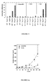
- FIGS. 3A to 3C show a standard histogram obtained with the supernatant of the hybridoma 7C10 (FIG. 3C) compared with a single cell + secondary antibody labeling (FIG. 3A) or with a labeling using a control isotype (FIG. 3B).
- FIG. 4A shows a total recovery of the histograms 1, 2, 3 respectively corresponding to the uninfected cells + secondary antibodies (1), to the uninfected cells labeled with the ⁇ IR3 + secondary antibodies (2) and to the uninfected cells labeled with a anti-IR antibody + secondary antibodies (3).
- FIG. 4B shows a stain of cells infected with a bacullovirus expressing IGF-IR.
- the ⁇ cIR3, used as a positive control mark well as expected cells (peak 2), while the anti-IR (peak 3) is superimposed on the peak of single cells.
- FIG. 4C it is shown that the anti-IR marks well as expected the Sf9 cells expressing the IR (peak 3), but unexpectedly, the ⁇ IR3 described in the literature as specific for IGF- IR, also seems to recognize IR (peak 2).
- the last two screens for MAb selection were to verify that the MAb was able to inhibit IGF-1-induced cell proliferation in vitro and in vivo on the MCF-7 cell line.
- MCF-7 cells were seeded, released into fetal calf serum, and then incubated in the presence of increasing concentrations of IGF-1 (1 to 50 ng / ml) in the presence or absence of 7C10 antibody to be tested added to a final concentration of 10 ⁇ g / ml.
- the commercial mAb ⁇ IR3 was introduced as a positive control and the mAb 7G3 (isolated parallel to 7C10 and weakly recognizing the native receptor (MFI at FACS 50 compared to 200 for ACM 7C10)) as isotype control .
- Cell proliferation is estimated by ⁇ -meter monitoring of tritiated thymidine incorporation by cells. The results are expressed in proliferation index.
- IGF1 is able to stimulate the proliferation of MCF-7 cells in a dose-dependent manner.
- MAb ⁇ IR3, used as a positive control completely inhibits IGF-1-induced proliferation of MCF-7 cells.
- mAb 7C10 is able to significantly inhibit MCF-7 cell growth induced by IGF-1.
- the mAb 7G3 used as an isotypic control proves to be well, as expected, without any effect on the cell tumor growth in vitro of the MCF-7 cell.
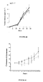
- mice received a slow-release subcutaneous estrogen implant, which is essential for tumor uptake in a murine model. Twenty four hours after implantation of the estrogens, 5.10 6 MCF-7 cells are grafted on the right flank of the mouse subcutaneously. Five days after this cell transplant tumors are measurable and batches of 6 mice are randomly constituted. The treatment of the mice is carried out twice a week for 5 to 6 weeks at a dose of 250 ⁇ g / dose / mouse. In the control group, the mice are treated in the same way with a murine control isotype.
- the results presented in FIG. 6A show a very significant inhibition of the tumor growth induced by the 7C10 antibody.
- 7C10 was compared to the compound tamoxifen commonly used for the treatment of breast carcinoma in locally advanced and advanced forms. metastatic and in the context of the prevention of recurrences (see VIDAL 2000, 1975-1976 pages ).
- E2 In hormone-dependent breast cancer, there is a significant correlation between estrogen receptor (ER) and IGF-IR ( Surmacz E. et al., Breast Cancer Res. Treat., Feb., 47 (3): 255-267, 1998 ). Moreover, it seems that estrogens (E2) act synergistically with IGFI (sometimes called IGF-I or IGFI) to stimulate cell proliferation. It has been shown that treatment with E2 increases the mRNA level of IGF-IR by approximately 10-fold, as well as the level of expression of the protein ( Lee AV et al., Mol. Endocrinol., May, 13 (5): 787-796, 1999 ). This increase results in a significant increase in phosphorylation of IGF-IR. In addition, E2 significantly stimulate the expression of IRS-1 ("IRS-1" for "Insulin Receptor Substrate-1”) which is one of the substrates of phosphorylated IGF-IR.
- IRS-1 IRS-1
- Tamoxifen has been widely used for many years in hormone therapy for the treatment of patients with E2-dependent breast cancer ( Forbes JF, Semin. Oncol., Feb., 24 (1.Suppl.1): S1-5-S1-19, 1997 ). This molecule competes with estradiol and inhibits the attachment of estradiol to its receptor ( Jordan VC, Breast Cancer Res. Treat., 31 (1): 41-52, 1994 ). Tamoxifen has also been shown to inhibit IGF-IR dependent proliferation by inhibiting receptor expression and phosphorylation ( Guvakova MA et al., Cancer Res., July 1, 57 (13): 2606-2610, 1997 ). All of these data seem to indicate that IGF-IR is an important mediator of the proliferation induced by the E2 / ER interaction.
- MCF-7 cells were implanted in sc (subcutaneous) in nude mice, 24 hours after implantation in these same mice of an extended-release estradiol granule (0.72 mg / tablet released over 60 days), essential for the establishment of any human E2 dependent tumor in this animal species. Five days after this implantation, the tumors are measured and groups of 6 mice are constituted.
- mice with ACM 7C10 also receive an injection of 3% HPC PBS daily.
- 7C10 was tested in vivo in an independent androgenic DU145 tumor tumor model (also noted DU-145), in a SKES-1 osteosarcoma model and in a non-small cell lung tumor model A549.
- the protocol is comparable to that described above for MCF-7 and the results shown in FIGS. 8A to 8C show a significant activity of this ACM in the 3 tumor models.
- the activity observed in the prostate tumor model is particularly noteworthy since the single scFv chain of ACM 1H7 is inactive in an independent androgenic prostate tumor model (Li et al., 2000). ).
- Navelbine is a chemotherapy compound indicated for non-small cell lung cancer and metastatic breast cancer.
- the comparative study of 7C10 and navelbine and the possible synergy between the two products was studied on the A549 tumor model.
- 5.10 6 A549 cells were grafted subcutaneously on the right flank of the mouse.
- Five days after the cell transplant tumors are measurable and treatments with mAb and / or navelbine are started.
- the dose of mAb is always 250 ⁇ g / dose / mouse, twice a week, intraperitoneally.
- navelbine it will be administered at the maximum dose tolerated by the mouse, ie 10 mg / kg, intraperitoneally.
- three injections will be performed 7 days apart. In co-administrations, both products are mixed before injection.
- IGF-IR is overexpressed by many tumors but it has been described moreover that in a large part of breast and colon cancers in particular, the proliferation signal is given to this receptor via IGF2 (sometimes noted IGF-II or IGFII). It is therefore essential to ensure that the AcM 7C10 is also able to inhibit IGF2 growth induced on the MCF-7 tumor in vitro.
- IGF2 sometimes noted IGF-II or IGFII
- the chimeric form of mAb 7C10 and purified humanized form 1 were tested in vitro in the MCF-7 model as described above.
- the results presented respectively in FIGS. 11 and 12 show that these two forms perfectly preserved their properties of inhibiting the IGF1 growth induced by the MCF-7 tumor.
- the IGF1 growth inhibition activity induced in vitro on the MCF-7 line should be a translation of an inhibition of IGF1-mediated signal transduction when attaching the mAb 7C10 to the receptor.
- MCF-7 cells were incubated with or without IGF1, in the presence or absence of the antibodies to be tested. After a short incubation period, the cells were lysed, the ⁇ chain immunoprecipitated and the phosphorylation of this subunit estimated using an antiphosphotyrosine kinase antibody.
- the results presented in FIG. 13 show that the binding of 7C10 or of h7C10 significantly inhibits the phosphorylation of the ⁇ subunit of IGF-IR in contrast to an irrelevant murine (9G4) or human (noted IgG1 in the scheme).
- the inhibition of signal transduction described above in paragraph b) is the primary mechanism of action involved in the biological activity of the 7C10 and 7H2HM antibodies.
- the 7H2HM antibody isotype IgG1
- ADCC Antibody Dependent Cellular Cytotoxicity
- NK Natural Killer cells from the peripheral blood of human donors are placed in the presence of A549 or MCF-7 cells previously incubated for 4 hours with 10 ⁇ g of 7H2HM antibody per 5 ⁇ 10 5 cells and, labeled at 51 Cr (50 ⁇ g).
- herceptin (noted in Figures 32A and 32B-4D5) is used as a positive test of experience.
- Figures 32A-32D show that, as expected, the herceptin induces significant ADCC on both A549 and MCF-7 cells (see Figures 32A and 32B, respectively).
- 7H2HM is also capable of inducing ADCC on A549 cells (see Figure 32C), but this phenomenon is of lesser amplitude on MCF-7 cells (see Figure 32D).
- FIG. 33B shows the effect of IGF1 on the entry into the cycle and the growth of MCF-7 cells, compared with the entry into the cycle and the growth of MCF-7 cells in the absence of IGF1 (see FIG. Figure 33A).
- the humanized 7H2HM antibody was compared to 7C10 in the non-small cell lung tumor model A549. This experiment was carried out exactly as described above except the dose of antibody which is 125 ⁇ g / dose twice a week instead of 250 ⁇ g / dose twice a week and because of the unavailability of large quantities. from 7H2HM.
- the 9G4 antibody was used as an isotype control for 7C10 and an irrelevant human immunoglobulin of IgG1 isotype (hereinafter referred to as HIgG1) was used as a control for the humanized 7H2HM antibody.
- Figure 34A shows that there are no significant differences between the 9G4 and HIgG1 control curves. As expected, a significant inhibition of tumor growth is observed with the murine 7C10 antibody. Regarding the humanized antibody 7H2HM, the observed activity is exactly the same intensity as that observed with its murine counterpart. This data, added to the observations described above in vitro, indicates that the humanization did not modify the properties of the antibody generated. On the other hand, in mouse xenograft models, the activity of the humanized antibody appears to be integrally related to a mechanism of inhibition of signal transduction. Indeed if an ADCC share was involved in the inhibition of tumor growth in Nude mice, a difference should be observed between the activity of murine and humanized antibodies.
- Example 4 The protocol described in Example 4 was repeated in order to reproduce the results obtained with 7C10 with its humanized counterpart: the antibody 7H2HM.
- FIGS. 35A and 35B show that, as in the case of 7C10, a significant synergy is demonstrated between the humanized antibody 7H2HM and navelbine.
- IGF-IR is capable of conferring protection against apoptosis when overexpressed on the surface of cells.
- the antibodies 7C10 and 7H2HM were capable of potentiating a compound that is active in chemotherapy.
- the MCF-7 cells are seeded at 2 ⁇ 10 4 / cm 2 in a petri dish and cultured for 24 h in RPMI without phenol red supplemented with 10% fetal calf serum (FCS). The cells are then washed twice with PBS and resuspended in medium at 0% FCS. An adaptation time of 10 minutes at 37 ° C. is left before the addition of the antibodies to 10 ⁇ g / ml. After an additional 10 minutes at 37 ° C, recombinant IGF-I (Sigma) was added to the culture medium at a final concentration of 50 ng / ml. The cells are again left at 37 ° C for one hour to allow binding of antibodies and IGF-I. Finally, doxorubicin (Sigma) is added to the culture medium at 2 ⁇ g / ml and the cells are incubated for 24 hours at 37 ° C.
- FCS fetal calf serum
- Cell viability analysis is performed by flow cytometric analysis after labeling with annexin V-FITC (20 min, 4 ° C) and DAPI (2 ⁇ g / ml). The percentage of dead cells considered is the Annexin + / DAPI + labeled population.
- the 5C2 antibody is used as a control isotype.
- Example 7 Cloning strategy of the genes coding for the variable regions of the heavy and light chains of the monoclonal antibody (mAb) 7C10
- the cDNA: mRNA hybrid thus obtained was used for the PCR amplification of the genes encoding the heavy and light chains of the mAb 7C10.
- the PCRs were performed using a combination of oligonucleotides specific for heavy and light chains (Kappa) of mouse immunoglobulins.
- the primers corresponding to the 5 'ends hybridize in the region corresponding to the signal peptides (Table 2 for heavy chains, Table 3 for light chains). These primers were compiled from a large number of mouse antibody sequences found in the databases ( Jones ST et al., Bio / Technology 9: 88-89, 1991 ).
- the primers corresponding to the 3 'ends hybridize in the constant regions of the heavy chains (CH1 domain of the IgG1 subclass, not far from the VC junction, MHC-1 primer Table 4) and light (Kappa domain not far from the junction VC, MKC primer Table 4).
- MHV mouse immunoglobulin heavy chain
- MHV-2 5 'ATGGGATGGAGCTRTATCATSYTCTT 3' (SEQ ID No.
- MHV-3 5 'ATGAAGWTGTGGTTAAACTGGGTTTT 3' (SEQ ID No. 15)
- MHV-4 5 'ATGRACTTTGGGYTCAGCTTGRT 3' (SEQ ID No. 16)
- MHV-5 5 'ATGGACTCCAGGCTCAATTTAGTTTT 3' (SEQ ID No. 17)
- MHV-6 5 'ATGGCTGTCYTRGSGCTRCTCTTCTG 3' (SEQ ID No. 18)
- MHV-7 5 'ATGGRATOOAOCKOORTCTTTMTCTT 3' (SEQ ID No. 19)
- MHV-8 5 'ATGAGAGTGCTGATTCTTTTGTG 3' (SEQ ID No.
- MHV-9 5 'ATGGMTTGGGTGTGGAMCTTGCTATT 3'
- MHV-10 5 'ATGGGCAGACTTACATTCTCATTCCT 3'
- MHV-11 5 'ATGGATTTTGGGCTGATTTTTTTTTATTG 3'
- MHV-12 5 'ATGATGGTGTTAAGTCTTCTGTACCT 3' (SEQ ID No. 24)
- R A / G
- Y T / C
- W A / T
- K T / G
- M A / C
- S C / G.
- MKV-1 5 'ATGAAGTTGCCTGTTAGGCTGTTGGTGCT 3' (SEQ ID No. 25)
- MKV-2 5 'ATGGAGWCAGACACACTCCTGYTATGGGT 3' (SEQ ID No. 26)
- MKV-3 5 'ATGAGTGTGCTCACTCAGGTCCT 3' (SEQ ID No. 27)
- MKV-4 5 'ATGAGGRCCCCTGCTCAGWTTYTTGG 3' (SEQ ID No. 28)
- MKV-5 5 'ATGGATTTWCAGGTGCAGATTWTCAGCTT 3' (SEQ ID No. 29)
- MKV-5A 5 'ATGGATTTWCARGTGCAGATTWTCAGCTT 3' (SEQ ID No.
- MKV-6 5 'ATGAGGTKCYYTGYTSAGYTYCTGRG 3' (SEQ ID No. 31)
- MKV-7 5 'ATGGGCWTCAAGATGGAGTCACA 3'
- MKV-8 5 'ATGTGGGGAYCTKTTTYCMMTTTTTCAAT 3'
- MKV-9 5 'ATGGTRTCCWCASCTCAGTTCCTT 3'
- MKV-10 5 'ATGTATATATGTTTGTTGTCTATTTC 3' (SEQ ID No. 35)
- MKV-11 5 'ATGGAAGCCCCAGCTCAGCTTCTCTT 3' (SEQ ID No.
- MKV-12A 5 'ATGRAGTYWCAGACCCAGGTCTTYRT 3' (SEQ ID No. 37)
- MKV-12B 5 'ATGGAGACACATTCTCAGGTCTTTGT 3' (SEQ ID No. 38)
- MKV-13 5 'ATGGATTCACAGGCCCAGGTTCTTAT 3' (SEQ ID No. 39)
- Example 8 Immunoglobulin sequences cloned from mouse hybridoma 7C10
- PCR products corresponding to heavy (VH) and light (VL) variable regions were cloned using "pGEM®-T Easy Vector Systems" (Promega).
- PCR products were obtained with the MKC primer in combination with the MKV1 and MKV2 primers.
- PCR products were obtained with MHC-1 primer in combination with primers MHV8 and MHV12.
- In-depth sequencing of PCR products cloned into the pGEM-T easy vectors revealed two different sequences for the light chain and a single sequence for the heavy chain.
- the resulting DNA sequence is characteristic of a functional Ig variable region. This new sequence is thus presumed to be that encoding 7C10 VL.
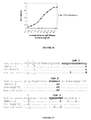
- the DNA sequences (SEQ ID Nos. 48 and 50) and amino acids (SEQ ID No. 49) of the cDNA coding for 7C10 VL are shown in FIG.
- the gene encoding this light chain is derived from an aberrant mRNA transcript that is present in all standard tumor-derived fusion partners original MOPC-21 of which Sp2 / Oag14 mouse myeloma was used which was used to produce the 7C10 hybridoma.
- This sequence comprises an aberrant recombination between the V and J genes (deletion of four nucleotide bases leading to a change of frame of reading) and a mutation of the invariable cysteine at position 23 in tyrosine.
- the DNA sequences obtained with these two oligos are identical, apart from the sequence encoded by the oligo itself. This sequence is a new sequence coding for a functional heavy chain presumed to be that of the monoclonal antibody 7C10.
- the DNA sequences (SEQ ID Nos. 51 and 53) and amino acids (SEQ ID No. 52) of the cDNA coding for 7C10 VH are shown in FIG.
- Chimeric antibody 7C10 was constructed to have mouse 7C10 VL and VH regions connected to the human kappa and gamma-1 constant regions, respectively. Oligos were used to modify the 5 'and 3' ends of the sequences flanking the DNA coding for 7C10 VL and VH to allow their cloning into mammalian cell expression vectors. These vectors use the strong HCMV promoter to efficiently transcribe the heavy and light chains of the chimeric 7C10 antibody. On the other hand, these vectors also contain the SV40 origin of replication allowing efficient replication of the DNA and consequently transient expression of the proteins in cos cells.
- Both plasmids containing the DNA encoding the chimeric 7C10 antibody were cotransfected into cos-7 cells (ATCC number CRL-1651) to study transient expression of the recombinant antibody. After 72 hours of incubation, the culture medium was removed, centrifuged to remove cell debris and analyzed by ELISA technique for production of human IgG1 (see Example 16) and recognition of the IGF-1 receptor (see Example 17).
- the ELISA tests for measuring human IgG1 / Kappa concentrations showed that the expression of the chimeric 7C10 antibody in the cos7 cells was between 300 and 500 ng / ml, which is comparable to the values obtained with the majority of the antibodies.
- the IGF-1 receptor recognition ELISA tests show that the chimeric antibody recognizes it specifically and with good relative avidity (see FIGS. 3A, 3B and 3C). This provides functional proof that the good VH and VL of the 7C10 antibody have been identified. In addition, this chimeric form of 7C10 appears to be an indispensable tool for evaluating the affinity of humanized forms.
- Example 12 Humanization process by CDR-grafting of the variable region of the light chain of the antibody 7C10 (7C10 VL)
- 7C10 VL As a preliminary step to humanization by CDR-grafting, the amino acid sequence of 7C10 VL was first compared to all the mouse VL sequences present in the Kabat database (Internet address: ftp: / /ftp.ebi.ac.uk/pub/database/kabat/fasta_format/, last data update dates from 1999). 7C10 VL has thus been identified as belonging to subgroup II of Kappa light chains as defined by Kabat et al. (In Sequences of Proteins of Immunological Interest (5th edn.), NIH Publication No. 91-3242, US Department of Health and Human Services, Public Health Service, National Institutes of Health, Bethesda, 1991 ).
- VL regions of mouse monoclonal antibodies having sequence identity of up to 95% have been identified (DRB1-4.3 (SEQ ID No. 55): 95% and C94-5B11'CL (SEQ ID No. 56) 95%, see Figure 17).
- DRB1-4.3 SEQ ID No. 55
- C94-5B11'CL SEQ ID No. 56
- Figure 17 the amino acid sequence of 7C10 VL was aligned with the consensus sequence of mouse kappa subgroup II (SEQ ID No. 57) as defined by Kabat (see Figure 17).
- the human-derived kappa VL region with the highest possible homology with 7C10 VL was searched for.
- the amino acid sequence of 7C10 VL mouse kappa was compared with all human kappa VL sequences present in the Kabat database.
- Mouse 7C10 VL had the highest sequence homology with human kappa VL regions of subgroup II as defined by Kabat et al. (1991).
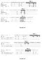
- VH regions of monoclonal antibodies of human origin have been identified having a sequence identity of up to 75.9% (GM607 (SEQ ID No. 58), see Figure 18) on all 112 amino acids composing the variable region.
- GM607 A germline of human origin, DPK15 / A19 (SEQ ID No. 59), having a sequence identity of 76% (see Figure 18) was also identified. GM607 (Klobeck et al., 1984). GM607 was therefore chosen as the recipient human sequence of CDRs (according to the definition of Kabat) of 7C10 VL of mice. By comparing the GM607 sequences with that of the consensus sequence of human subgroup II (SEQ ID No. 60) (Fig. 18), no particular residues in the framework regions (Rch) could be identified, thereby indicating that GM607 was a good candidate for CDR-grafting.
- the next step in the humanization process was to join the mouse 7C10 VL CDRs to the framework regions (Rch) of the selected human light chain, GM607 (Klobeck et al., 1984).
- the molecular model of mouse Fv regions of 7C10 is particularly useful in the choice of mouse residues to be conserved because they may play a role either in maintaining the three-dimensional structure of the molecule (canonical structure of the CDRs, interface VH / VL, etc.) or in antigen binding.
- Rchs each difference between mouse (7C10 VL) and human (GM607) amino acids was carefully examined (see Table 5).
- residues specific to the mouse 7C10 VL sequence that had been identified were taken into account if necessary.
- the second column (#) indicates the position of the acid residue amine in the regular sequence
- the third column (FR or CDR) was performed to easily identify backbone segments (FR1, FR2, FR3 and FR4) and CDR segments (CDR1, CDR2 and CDR3) ("CDR" for "Complementary-Determining Region") with the three CDRs separating the four FRs
- the fourth column (mouse light chain 7C10) represents the amino acid sequence (SEQ ID No. 54) of the V L region of mouse antibody 7C10
- the fifth column (DPK15 / A19 human germline) represents the amino acid sequence (SEQ ID No.
- the sixth column represents the amino acid sequence (SEQ ID No. 58) of the V L region of the human GM 607 antibody
- the seventh and eighth columns (7C10 L reshaped humans 1 and 2) represent the amino acid sequences of humanized 7C10 VL antibodies 1 and 2 (respectively SEQ ID Nos. 61 and 65).
- "*" indicates the parts of the canonical structure of the CDR loop as defined by Chothia et al. (Nature, 342, 877-883, 1989 ).
- Example 13 Humanization process by CDR-grafting of the variable region of the heavy chain of the antibody 7C10 (7C10 VH)
- Residue 17 (Kabat numbering), Thr for the consensus sequence of subgroup I (A) and Ser in 7C10 VH, is located on the surface of the molecule on the interface side with the constant region. This residue does not seem important.
- the residue 27 (Kabat numbering), Asp for the consensus sequence of subgroup I (A) and Tyr in 7C10 VH, is a canonical residue for CDR 1. Tyr at this position is not uncommon and is probably critical for the maintenance of CDR 1 in its correct conformation.
- the residue 84 (Kabat numbering), Thr for the consensus sequence of subgroup I (A) and Asn in 7C10 VH. Asn, was found 93 times in mouse VH and 3 times in human VH. According to the molecular model, it is a surface residue far from the paratope.
- AN03'CL represents the heavy chain sequence of a mouse antibody (access number in the Kabat databank is P001289).
- VH region of human origin having the highest possible homology with 7C10 VH was searched.
- amino acid sequence of mouse 7C10 VH was compared with all human VH sequences present in the Kabat database.
- Mouse 7C10 VH had the highest sequence homology with the human VH regions of subgroup II as defined by Kabat et al. (1991).
- VH regions of monoclonal antibodies of human origin have been identified having a sequence identity of up to 67.3% (human VH FUR1'CL (SEQ ID No. 73, see FIG.
- variable gene i.e., excluding CDR3 and J region
- a germline of human origin 4.22 VH IV (Sanz et al., 1989), having a sequence identity of 68.4 %, according to the same criteria as for VH FUR1'CL, was also identified (human Germ-line (SEQ ID No. 74), see Fig.
- the sequence encoded by the germ line 4.22 VH IV was chosen as sequence human recipient of CDRs (according to the definition of Kabat) of mouse 7C10 VH rather than VH FUR1'CL because by comparing the sequences of 4.22 VH IV and VH FUR1'CL with that of the consensus sequence of human subgroup II (human Kabat sg II (SEQ ID No. 72), see Figure 23 and Table 6), no atypical residues in the framework regions (Rch) could be identified for 4.22 VH IV while the presence of two atypical residues (Gln and Arg at positions 81 and 82A according to Kabat nomenclature, respectively) were identified in the sequence encoded by VH FUR1'CL.
- the next step in the humanization process was to join the mouse 7C10 VH CDRs to the framework regions (Rch) of the human 4.22 VH IV germline (Sanz et al., 1989).
- the molecular model of mouse Fv regions of 7C10 is particularly useful in the choice of mouse residues to be conserved because they may play a role either in maintaining the three-dimensional structure of the molecule (canonical structure of the CDRs, VH / VL interface, etc.) or in antigen binding (paratope membership).
- each difference between mouse (7C10 VH) and human (4.22 VH IV) amino acids was carefully examined (see Table 6).
- residues specific to the mouse 7C10 VH sequence that had been identified were taken into account if necessary.
- the second column (FR or CDR) was performed to easily identify backbone segments (FR1, FR2, FR3 and FR4) and CDR segments (CDR1, CDR2 and CDR3) with the three CDRs separating the four FRs;
- the third column (mouse heavy chain 7C10) represents the amino acid sequence (SEQ ID No. 69) of the V H region of mouse antibody 7C10;
- the fourth column (4.22 VH IV germline) represents the amino acid sequence of the 4.22 VH IV gene (Sanz et al., 1989) (SEQ ID No. 74);
- the fifth column (FUR1'CL VH human, kabat accession number N020619) represents the amino acid sequence (SEQ ID No.
- IgM K antilamine B of human origin (Mariette et al., 1993); the sixth, seventh and eighth columns (reshaped human 7C10H 1, 2 and 3) represent the amino acid sequences of the V H region of human 7C10 reshaped respectively for the versions 1 (SEQ ID No. 75), 2 (SEQ ID No. 79) and 3 (SEQ ID No. 83).
- "*" indicates the parts of the canonical structure of the CDR loop as defined by Chothia et al. (1989).
- Example 14 Construction of genes encoding humanized versions 1 of 7C10 VL and VH by oligonucleotide assembly.
- the genes (leader peptide + VDJ variable regions for VH or VJ for VK) encoding the humanized variable regions were synthesized by solid phase assembly on streptavidin-labeled magnetic beads.
- the genes encoding humanized 7C10 VH (445 base pairs) and humanized 7C10 VL (433 base pairs) are constructed by fusing two DNA fragments through the presence of a KpnI restriction site present in both sequences and located about half of the gene (at 200 and 245 nucleotides to the 5 'end of the gene for VL and VH, respectively).
- the two fragments which are fused together are themselves assembled by an assembly technique which consists in using phosphorylated oligonucleotides (approximately 30-35 mer) hybridized two by two (one sense oligo and the other antisense, on a homology d about 50%) so that they overlap during elongation.
- a first 5'-biotinylated oligonucleotide is attached to the magnetic beads and the pairs of phosphorylated oligonucleotides are added one by one.
- the phosphodiester bond between the juxtaposed phosphorylated oligonucleotides is carried out by the enzyme T4 DNA ligase.
- the genes thus synthesized de novo can be directly cloned (by digestion with restriction enzymes compatible with the chosen expression vector) or amplified by PCR to obtain more material in the preamble to directional cloning by enzymatic digestion.
- the gene sequence thus constructed by de novo assembly is then verified by automatic sequencing of the DNA.
- oligonucleotides whose concentration was adjusted to 100 ⁇ M were ordered from MWG Biotech (see the sequences of the oligonucleotides used in Table 7 for the construction of humanized 7C10 VL, and Table 8 for the construction of humanized 7C10 VH).
- the oligonucleotides were hybridized in pairs (an equimolar mixture, 500 pmol, of a trace and an antisense oligo in the T4 DNA ligase buffer was heated at 95 ° C for 5 minutes then allowed to cool on the bench to room temperature) according to a scheme described in Table 9.
- the first biotinylated oligonucleotide is attached to streptavidin-labeled magnetic beads (Dynabeads M-280 Streptavidin, Dynal product No. 112-05).
- streptavidin-labeled magnetic beads Dynabeads M-280 Streptavidin, Dynal product No. 112-05
- 1X TE buffer 100X Tris-EDTA buffer: 1M Tris-HCl, pH 8, 0.1 M EDTA, Sigma T-9285
- 500 pmol of the biotinylated oligonucleotide was added in 15 mM NaCl solution.
- the beads are washed twice with the wash buffer (10 mM Tris-HCl pH 7.6, 10 mM EDTA and 50 mM NaCl) and the hybridized oligonucleotide pairs are then added one by one.
- wash buffer 10 mM Tris-HCl pH 7.6, 10 mM EDTA and 50 mM NaCl
- Each addition of a pair of oligonucleotides is heated at 95 ° C for 5 min and then allowed to cool on the bench to room temperature. Once the ambient temperature is reached, 2 ⁇ l of T4 DNA ligase at 10 U / ⁇ l (Biolabs) are added and incubated for 20 min at 37 ° C. The beads are then washed (wash buffer) and the following oligonucleotide pairs are added so on.
- the last unpaired oligo (antisense) is assembled as follows. 5 ⁇ l of oligo (500 pmol) and 43 ⁇ l of T4 DNA ligase buffer are added to the decanted beads and the mixture is then heated at 95 ° C. for 5 minutes and allowed to cool on the bench to room temperature. Once the ambient temperature is reached, 2 ⁇ l of T4 DNA ligase are added and incubated at 37 ° C. for 20 minutes. The beads are then washed twice with the wash buffer and then twice with 1X TE buffer.
- Example 15 Construction of genes encoding humanized versions 2 of 7C10 VL and 7C10 VH and 3 of 7C10 VH by site-directed mutagenesis.
- the humanized version 2 of 7C10 VH was obtained by site-directed mutation of residues 48 and 67 (according to the Kabat nomenclature) of version 1. This site-directed mutagenesis was carried out using Stratagene's QuikChange TM Site-directed mutagenesis system. (kit # 200518) according to the protocol described by the manufacturer. The construction was done in two stages, at first the residue 48 on the version 1 was transferred using the pair of primers 7C10Hhumanisé1 QCM48 sense and antisense (see Table 10) and in a second time this version mutated at residue 48 was itself mutated at residue 67 using the sense and antisense pair of primers 7C10Hhumanized1QCI67 (see Table 10).
- the humanized version 3 of 7C10 VH was obtained by directed mutation of residues 30 and 71 (according to the Kabat nomenclature) of version 2 also using the QuikChange TM system. This construction was done in two stages. Firstly, the residue on version 2 was mutated with the primers 7C10HhumanizedQCT30 sense and antisense (see Table 10). In a second step, this mutated version at residue 30 was itself mutated at residue 71 using the sense and antisense pair of primed 7C10Hhumanized1V67QCR71 primers (see Table 10).
- the humanized version 2 of 7C10 VL was obtained by directed mutation of residue 2 (according to the Kabat nomenclature) of version 1 using the QuikChange TM system. Residue 2 on version 1 was mutated using the sense and antisense humanized 7C10L primer pair (see Table 10).
- ⁇ b> TABLE 10 List of oligonucleotides used for QuikChange TM system-directed mutagenesis of stratagen ⁇ / b> 7C10Hhumanisé1QCT30. meaning 5'-CTGGTTACTCCATCAGCGGTGGTTATTTATG (SEQ ID No. 147) 7C10Hhumanisé1QCT30.
- antisense 5'-CATAAATAACCACCGCTGATGGAGTAACCAG (SEQ ID No. 148) 7C10Hhumanisé1QCM48. meaning 5'-GGGACTGGAGTGGATCGGGTATATCAGCTAC (SEQ ID No. 149) 7C10Hhumanisé1QCM48.
- antisense 5 '-GTAGCTGATATACCCGATCCACTCCAGTCCC (SEQ ID No.150) 7C10Hhumanized1QCI 67. meaning 5'-TCCCTCAAGGATCGAGTCACCATATCACGTG (SEQ ID No. 151) 7C10HhumanisélQCI67.
- antisense 5'-CACGTGATATGGTGACTCGATCCTTGAGGGA (SEQ ID No.
- Mammalian expression vectors containing the chimeric or humanized versions of the heavy and light chains of the 7C10 antibody were tested in cos7 cells for transient expression of recombinant 7C10 antibodies.
- the DNA was introduced into the cos cells by electroporation using a Biorad instrument (Gene Pulsar).
- the DNA (10 ⁇ g of each vector) is added to aliquots of 0.8 ml of cos cells at a concentration of 1 ⁇ 10 7 cells per ml in PBS buffer (without Ca ++ and Mg ++). A pulsation of 1,900 volts and a capacity of 25 ⁇ F was delivered.
- the transfected cos cells are then added to 8 ml of DMEM medium containing 5% calf serum and incubated at 37 ° C for 72 hours. The supernatant is then harvested, centrifuged to remove cell debris and tested by ELISA for the measurement of its recombinant human IgG1 / Kappa 7C10 antibody concentration.
- Example 17 ELISA Method for Measuring the Levels of Human IgG1 / Kappa Recombinant Antibodies Present in the Supernatant of the Cos Transfectants
- the supernatants produced by transient expression in cos7 cells were tested for the presence of human IgG1 / Kappa type 7C10 antibodies.
- human IgG1 / Kappa immunoglobulin 96-well ELISA plates (Maxisorb, Nunc) were scored with polyclonal goat anti-human IgG antibody (specific for the Fc gamma fragment, Jackson Immuno-Research Laboratories Inc. . # 109-005-098).
- Supernatants of cos cells were serially diluted and added to the listed wells.
- Example 18 ELISA Method for Determining the Recognition Activity of Human IgG1 / Kappa-type 7C10 Recombinant Antibodies on the IGF-1 Receptor (IGF-IR)
- the cos7 culture supernatants were tested for their ability to recognize IGF-1 R by an ELISA method.
- 96-well ELISA plates (Dynex Immulon 2HB) were scored with 100 ⁇ l per well of a PBS solution containing 0.31 ng / ⁇ l of IGF-1R (Human Insulin-like Growth Factor I soluble Receptor, R & D Systems, # 391-GR) by incubation overnight at 4 ° C. After washing with PBS containing 0.05% Tween 20, the plates were saturated by the addition of a solution of PBS containing 0.5% gelatin and incubation at 37 ° C for 1 hour.
- IGF-1R Human Insulin-like Growth Factor I soluble Receptor
- the TMB substrate KPL # 50-76-04 was added. After 10 minutes of incubation the reaction was stopped by the addition of 1M sulfuric acid and the optical density read at 450 nm.
- FIG. 28 shows the results of an IGF-1R recognition ELISA assay (see Example 18) from supernatants of cos7 cells whose human IgG1 / Kappa concentration had previously been determined by ELISA (see Example 17). .
- the titration curves of the four recombinant antibodies tested overlap perfectly, indicating that their relative affinities for IGF-IR are very similar. It is therefore concluded that the humanized 1 tonne of 7C10, composed of the humanized light chain 1 (1 single mouse residue present in the framework regions) in combination with the humanized heavy chain 1 (4 mouse residues present in the framework regions). specifically recognizes the IGF-1 receptor and has an affinity very similar to that of the chimeric antibody (variable regions of mice).
- FIG. 29 shows the results of the IGF-IR recognition ELISA assay (see Example 18) from supernatants of cos7 cells whose human IgG1 / Kappa concentration had previously been determined by ELISA (see Example 17).
- the two humanized versions 1 and 2 of the light chain were successively associated with humanized 7C10 VH 1.
- the titration curves of the two combinations are superimposed indicating that the mutation of residue 2 of the light chain, which has been changed from a valine in the humanized version 1 for an isoleucine in humanized form 2, apparently has no influence on the relative recognition affinity of the IGF1 receptor.
- the humanized form 2 of the 7C10 light chain thus constitutes a version where no mouse residues (excluding CDRs) have been preserved.
- This version, completely humanized, represents the preferred version of 7C10 VL.
- FIG. 30 shows the results of the IGF-1R recognition ELISA assay from supernatants of cos7 cells whose human IgG1 / Kappa concentration had previously been determined by ELISA (see Example 17).
- the titration curves are very similar and overlap substantially with the reference curve corresponding to the chimeric antibody, indicating that the three humanized versions 1, 2 and 3 of 7C10 VH give the same relative affinity for IGF-1R when they are associated with humanized 7C10 VL 2.
- Example 20 Expression of EGFR and IGF-IR on the surface of A549 cells
- a marking for FACS reading of these cells was performed with murine ACM 7C10 anti-IGF-IR respectively (FIG. 37B) and murine ACM 225 anti-EGFR (FIG. 37D).
- the cells were saturated for 30 min at 4 ° C. with a solution of PBS 10% FCS (fetal calf serum), washed and then incubated for 30 min at 4 ° C. with the ACM of interest. After 3 new washings, the FITC-coupled anti-species secondary antibody (fluorosene isothiocyanate) is added. After 30 min of incubation, the FACS (“Fluorescence Activated Cells Sorter") reading is performed at 520 nm (excitation 488 nm).
- FIGS. 37A to 37D show that the A549 cells have on their surface a comparable number of EGF and IGFI receptors. In both cases the population is homogeneous with respect to the distribution of each of the receptors. The specificity of the labeling is confirmed by the use of isotypic control ( Figure 37C). These results validate the use of the A549 cell as a model for the study of a synergistic action on two IGF-IR and EGFR receptors and for the study of a collaboration of these two receptors.
- Example 21 Synergistic action of an anti-IGF-IR ACM and an anti-EGFR ACM coadministered in vivo, in the nude mouse as part of an antitumor treatment.
- nude mice are sc grafted with 5 ⁇ 10 6 A549 cells. Five days after the cell transplant, the tumors are measured and a homogeneous lot of mice in terms of tumor volume is constituted. From this batch groups of 6 mice are randomly generated. These mice will be treated intraperitoneally (ip), twice a week with each ACM 7C10 and 225 individually at the dose of 250 ⁇ g / mouse or with the two ACMs in co-administration. ACM 9G4 is administered as an isotype control experiment.
- Example 22 Study of the antitumor activity of murine antibodies 7C10 and 225 coadministered in mice implanted in orthotopic with A549 cells.
- Example 23 7C10 and 7H2HM inhibits tyrosine phosphorylation of the ⁇ chain of IGF-IR and IRS-I
- MCF7 cells are cultured for 24 hours at 5 ⁇ 10 4 cells / cm 2 (75 cm 2 dishes, COSTAR) in 20 ml of RPMI without phenol red added with 5 mM glutamine, penicillin / streptomycin (respectively 100 U / 100 ⁇ g / ml) and 10% fetal calf serum. After three washes in PBS, the cells were incubated for 12 hours in medium (RPMI) without phenol red, devoid of fetal calf serum and supplemented with 5 mM glutamine, penicillin / streptomycin, bovine serum albumin at 0.5 ⁇ g / ml. ml (Sigma A-8022) and transferrin at 5 ⁇ g / ml (Sigma T8158).
- the cells were first incubated at 37 ° C for 2 minutes with blocking antibodies (10 ⁇ g / ml) and then IGF-I (Sigma 13769, 50 ng / ml) was added to two more minutes. The reaction was stopped by aspiration of the incubation medium and the dishes were deposited on ice. The cells were solubilized by addition of 0.5 ml of lysis buffer (50 mM tris-HCL pH 7.5, 150 mM NaCl, 1% Nonidet P40, 0.5% sodium deoxycholate), supplemented with protease inhibitors.
- lysis buffer 50 mM tris-HCL pH 7.5, 150 mM NaCl, 1% Nonidet P40, 0.5% sodium deoxycholate
- the agarose beads were washed twice with 1 ml of lysis buffer, twice with Wash Buffer 1 (50 mM Tris-HCL pH 7.5, 500 mM NaCl, 0.1% Nonidet P40; 05% sodium deoxycholate (Boehringer 1,332,597), supplemented with protease inhibitors and phosphatase inhibitors) and once with wash buffer 2 (50 mM Tris-HCL, 0.1% Nonidet P40, 0.05 % sodium deoxycholate (Boehringer Ref .: 1 332 597), supplemented with protease inhibitors and phosphatase inhibitors 1/100).
- Wash Buffer 1 50 mM Tris-HCL pH 7.5, 500 mM NaCl, 0.1% Nonidet P40; 05% sodium deoxycholate (Boehringer 1,332,597), supplemented with protease inhibitors and phosphatase inhibitors
- wash buffer 2 50 mM Tris-HCL, 0.
- the immunoprecipitates were resuspended in Laemmli buffer, heated at 100 ° C for 5 minutes. The supernatants were analyzed by SDS electrophoresis of polyacrylamide (8% Novex EC6015). Proteins were transferred to nitrocellulose membrane followed by either an immunoblot with anti-phosphotyrosine antibodies conjugated to HRP (upstate Biotechnology 4G10) or anti-beta chain of IGF-IR or anti-IRS-1 (Santa Cruz Ref .: sc 8038) followed by an anti- rabbit conjugated with HRP. Fingerprints were revealed by chemiluminiscence (Amersham RPN 2209) followed by radioautography on Kodak X-mat AR films.
- Figure 40A shows unstimulated MCF7 cells (0) or stimulated with either IGF-1 (50 ng / ml) alone (0 + IGF-1) or combined with monoclonal or humanized anti-IGF-IR antibodies (10 ⁇ g / ml).
- the 9G4 or hIgG1 antibodies are murine or human immunoglobulins of IgG1 isotype used as a negative control of the experiment.
- the beta chains of IGF-IR were immunoprecipitated and blotted with anti-tyrosine phosphorylated antibodies. The results obtained show that the monoclonal or humanized anti-IGF-IR 7C10, 1H7 and 7H2HM antibodies inhibit the tyrosine phosphorylation of the beta chain of IGF-IR.
- Figure 40B shows unstimulated MCF7 cells (0) or stimulated with either IGF-1 (50 ng / ml) alone (0 + IGF-1) or combined with monoclonal or humanized anti-IGF-IR antibodies (10 ⁇ g / ml).
- the 9G4 or hIgG1 antibodies are murine or human immunoglobulins of IgG1 isotype used as a negative control of the experiment.
- IRS-1 was immunoprecipitated and blotted with anti-tyrosine phosphorylated antibodies. The results obtained show that the monoclonal antibodies 7C10, 7H2HM and 1H7 inhibit the phosphorylation of tyrosine of IRS-1.
- MCF7 and A549 cells were suspended at 1.10 7 cells / ml in PBS with 10% fetal calf serum (FACS buffer). 1.10 6 cells were incubated for 30 minutes at 37 ° C. with the monoclonal antibodies at 10 ⁇ g / ml (7C10, 7G3, 9G4) or at 20 ⁇ g / ml for 7H2HM. After washing, the cells were labeled at 4 ° C for 30 minutes with a biotinylated anti-IGF-IR (12B1 monoclonal antibody) and finally incubated at 4 ° C for 30 minutes with streptavidin-488 alexa Fluor ® conjugate. The cells were analyzed by FACScan (Becton-Dickinson, Enembogegem, Belgium) with Cellquest software after debris removal.
- FACS buffer 10% fetal calf serum
- Figure 41 shows the A549 cells without staining (peak 1), A549 cells incubated with 7C10 or 7H2HM (2nd peak) and A549 cells incubated with a mouse IgG1 or irrelevant rat (3rd peak). A decrease in the surface expression of IGF-IR by the cells is observed by two when the cells have been previously incubated with 7C10 or 7H2HM.
- Example 25 7C10 and 7H2HM induces degradation of IGF-IR
- MCF-7 cells were cultured for 24 hours at 10 ⁇ 10 4 cells / cm 2 (75 cm 2 , Costar) in 15 ml of complete medium. Then the cultures were washed three times with PBS and incubated for 12 hours with serum-free medium. Then, the cells were incubated with cycloheximide at 25 ⁇ g / ml alone or with 10 ⁇ g / ml of monoclonal antibody 7C10, 9G4, 7G3 or IGF-I (50 ng / ml). In some experiments, before incubation with the monoclonal antibodies, the cells were treated for 1 hour at 37 ° C with MG-132 (10 ⁇ M, Calbiochem 474791) to inhibit proteasome activities.
- FIG. 42A Western blot analysis (FIG. 42A) of the integrity of IGF-IR shows that 7C10 and 7H2HM induce the degradation of the receptor whereas the natural ligand does not cause any degradation of the latter. No degradation of the receptor is observed with 9G4, irrelevant antibody used as isotypic control.
- Figure 42B shows that the degradation is inhibited by a proteasome inhibitor MG132 (incubation period of 2 hours).
Landscapes
- Health & Medical Sciences (AREA)
- Chemical & Material Sciences (AREA)
- Life Sciences & Earth Sciences (AREA)
- Medicinal Chemistry (AREA)
- General Health & Medical Sciences (AREA)
- Organic Chemistry (AREA)
- Pharmacology & Pharmacy (AREA)
- Animal Behavior & Ethology (AREA)
- Veterinary Medicine (AREA)
- Public Health (AREA)
- Bioinformatics & Cheminformatics (AREA)
- Engineering & Computer Science (AREA)
- Nuclear Medicine, Radiotherapy & Molecular Imaging (AREA)
- General Chemical & Material Sciences (AREA)
- Chemical Kinetics & Catalysis (AREA)
- Immunology (AREA)
- Dermatology (AREA)
- Proteomics, Peptides & Aminoacids (AREA)
- Molecular Biology (AREA)
- Biophysics (AREA)
- Biochemistry (AREA)
- Mycology (AREA)
- Microbiology (AREA)
- Genetics & Genomics (AREA)
- Epidemiology (AREA)
- Peptides Or Proteins (AREA)
- Medicines Containing Antibodies Or Antigens For Use As Internal Diagnostic Agents (AREA)
- Medicines That Contain Protein Lipid Enzymes And Other Medicines (AREA)
- Preparation Of Compounds By Using Micro-Organisms (AREA)
- Micro-Organisms Or Cultivation Processes Thereof (AREA)
- Pharmaceuticals Containing Other Organic And Inorganic Compounds (AREA)
- Medicines Containing Material From Animals Or Micro-Organisms (AREA)
- Measuring Or Testing Involving Enzymes Or Micro-Organisms (AREA)
Applications Claiming Priority (4)
| Application Number | Priority Date | Filing Date | Title |
|---|---|---|---|
| FR0200653A FR2834990A1 (fr) | 2002-01-18 | 2002-01-18 | Nouveaux anticorps anti-igf-ir et leurs applications |
| FR0200654A FR2834900B1 (fr) | 2002-01-18 | 2002-01-18 | Nouvelles compositions a activite anti-igf-ir et anti-egfr et leurs applications |
| FR0205753A FR2834991B1 (fr) | 2002-01-18 | 2002-05-07 | Nouveaux anticorps anti-igf-ir et leurs applications |
| EP03712270A EP1461359B1 (de) | 2002-01-18 | 2003-01-20 | Antikörper gegen igf-ir und ihre verwendungen |
Related Parent Applications (2)
| Application Number | Title | Priority Date | Filing Date |
|---|---|---|---|
| EP03712270A Division EP1461359B1 (de) | 2002-01-18 | 2003-01-20 | Antikörper gegen igf-ir und ihre verwendungen |
| EP03712270.2 Division | 2003-01-20 |
Publications (3)
| Publication Number | Publication Date |
|---|---|
| EP1806364A2 true EP1806364A2 (de) | 2007-07-11 |
| EP1806364A3 EP1806364A3 (de) | 2010-09-08 |
| EP1806364B1 EP1806364B1 (de) | 2013-07-31 |
Family
ID=27248849
Family Applications (3)
| Application Number | Title | Priority Date | Filing Date |
|---|---|---|---|
| EP06126429A Expired - Lifetime EP1798242B1 (de) | 2002-01-18 | 2003-01-20 | Anti-IGF-IF-Antikörper und ihre Anwendungen |
| EP03712270A Expired - Lifetime EP1461359B1 (de) | 2002-01-18 | 2003-01-20 | Antikörper gegen igf-ir und ihre verwendungen |
| EP06126430.5A Expired - Lifetime EP1806364B1 (de) | 2002-01-18 | 2003-01-20 | Neuartige Anti-IGF-IR-Antikörper und ihre Anwendungen |
Family Applications Before (2)
| Application Number | Title | Priority Date | Filing Date |
|---|---|---|---|
| EP06126429A Expired - Lifetime EP1798242B1 (de) | 2002-01-18 | 2003-01-20 | Anti-IGF-IF-Antikörper und ihre Anwendungen |
| EP03712270A Expired - Lifetime EP1461359B1 (de) | 2002-01-18 | 2003-01-20 | Antikörper gegen igf-ir und ihre verwendungen |
Country Status (16)
| Country | Link |
|---|---|
| EP (3) | EP1798242B1 (de) |
| JP (2) | JP4606739B2 (de) |
| CN (1) | CN100410275C (de) |
| AT (1) | ATE357460T1 (de) |
| AU (2) | AU2003216748B2 (de) |
| BR (1) | BRPI0306973B8 (de) |
| CA (1) | CA2473039C (de) |
| CY (1) | CY1106662T1 (de) |
| DE (1) | DE60312639T2 (de) |
| DK (1) | DK1461359T3 (de) |
| ES (2) | ES2283759T3 (de) |
| MX (1) | MXPA04006980A (de) |
| PT (1) | PT1461359E (de) |
| SI (1) | SI1461359T1 (de) |
| WO (1) | WO2003059951A2 (de) |
| ZA (1) | ZA200405402B (de) |
Cited By (2)
| Publication number | Priority date | Publication date | Assignee | Title |
|---|---|---|---|---|
| WO2015035332A1 (en) * | 2013-09-09 | 2015-03-12 | Canimguide Therapeutics Ab | Immune system modulators |
| US10881709B2 (en) | 2015-03-06 | 2021-01-05 | Canimguide Therapeutics Ab | Immune system modulators and compositions |
Families Citing this family (150)
| Publication number | Priority date | Publication date | Assignee | Title |
|---|---|---|---|---|
| DE10201182A1 (de) | 2002-01-14 | 2003-07-24 | Cfs Gmbh Kempten | Positioniersystem |
| US7553485B2 (en) | 2002-01-18 | 2009-06-30 | Pierre Fabre Medicament | Anti-IGF-IR and/or anti-insulin/IGF-I hybrid receptors antibodies and uses thereof |
| EP1506286B1 (de) | 2002-05-24 | 2014-03-19 | Merck Sharp & Dohme Corp. | Neutralisierung eines menschlichen anti-igfr-antikörpers |
| US8034904B2 (en) | 2002-06-14 | 2011-10-11 | Immunogen Inc. | Anti-IGF-I receptor antibody |
| US7538195B2 (en) | 2002-06-14 | 2009-05-26 | Immunogen Inc. | Anti-IGF-I receptor antibody |
| US8663650B2 (en) | 2003-02-21 | 2014-03-04 | Ac Immune Sa | Methods and compositions comprising supramolecular constructs |
| ES2383014T3 (es) | 2003-04-02 | 2012-06-15 | F. Hoffmann-La Roche Ag | Anticuerpos contra el factor I de crecimiento similar a insulina y usos de los mismos |
| CA2524305C (en) | 2003-05-01 | 2015-12-08 | Imclone Systems Incorporated | Fully human antibodies directed against the human insulin-like growth factor-1 receptor |
| AR046071A1 (es) | 2003-07-10 | 2005-11-23 | Hoffmann La Roche | Anticuerpos contra el receptor i del factor de crecimiento de tipo insulinico y los usos de los mismos |
| DE602004029581D1 (de) | 2003-08-13 | 2010-11-25 | Pfizer Prod Inc | Modifizierte humane igf-1r antikörper |
| US7326567B2 (en) | 2003-11-12 | 2008-02-05 | Schering Corporation | Plasmid system for multigene expression |
| TW200526684A (en) | 2003-11-21 | 2005-08-16 | Schering Corp | Anti-IGFR1 antibody therapeutic combinations |
| WO2005058967A2 (en) * | 2003-12-16 | 2005-06-30 | Pierre Fabre Medicament | Novel anti-insulin/igf-i hybrid receptor or anti-insulin/igf-i hybrid receptor and igf-ir antibodies and uses thereof |
| EP1740615B1 (de) * | 2004-03-31 | 2014-11-05 | Genentech, Inc. | Humanisierte anti-tgf-beta-antikörper |
| KR20080019733A (ko) | 2004-07-16 | 2008-03-04 | 화이자 프로덕츠 인코포레이티드 | 항-아이지에프-1알 항체를 사용하는 비-혈액학적악성종양에 대한 조합 치료 |
| AU2011236042B2 (en) * | 2004-07-29 | 2013-09-05 | Pierre Fabre Medicament | Novel anti-IGF-IR antibodies and uses thereof |
| FR2873699B1 (fr) | 2004-07-29 | 2009-08-21 | Pierre Fabre Medicament Sa | Nouveaux anticorps anti igf ir rt leurs utilisations |
| PT1828249E (pt) | 2004-12-03 | 2011-02-25 | Schering Corp | Biomarcadores para a pré-selecção de pacientes para terapêutica anti-igf1r |
| MY146381A (en) | 2004-12-22 | 2012-08-15 | Amgen Inc | Compositions and methods relating relating to anti-igf-1 receptor antibodies |
| JP4875064B2 (ja) * | 2005-04-15 | 2012-02-15 | シェーリング コーポレイション | 癌を処置または予防するための方法および組成物 |
| CA2612449A1 (en) | 2005-06-17 | 2006-12-28 | Imclone Systems Incorporated | Receptor antagonists for treatment of metastatic bone cancer |
| WO2007000328A1 (en) * | 2005-06-27 | 2007-01-04 | Istituto Di Ricerche Di Biologia Molecolare P Angeletti Spa | Antibodies that bind to an epitope on insulin-like growth factor 1 receptor and uses thereof |
| FR2888850B1 (fr) * | 2005-07-22 | 2013-01-11 | Pf Medicament | Nouveaux anticorps anti-igf-ir et leurs applications |
| EA017812B1 (ru) * | 2005-12-08 | 2013-03-29 | Медарекс, Инк. | Выделенное моноклональное антитело или его антигенсвязывающий участок, которые связываются с протеинтирозинкиназой 7 ( ptk7) человека, и их применение |
| AP2008004569A0 (en) | 2006-02-03 | 2008-08-31 | Imclone Systems Inc | IGR-IR antagonists as adjuvants for treatment of prostrate cancer |
| GB0603041D0 (en) | 2006-02-15 | 2006-03-29 | Angeletti P Ist Richerche Bio | Therapeutic compounds |
| WO2007093008A1 (en) * | 2006-02-17 | 2007-08-23 | Adelaide Research & Innovation Pty Ltd | Antibodies to insulin-like growth factor i receptor |
| US7846724B2 (en) | 2006-04-11 | 2010-12-07 | Hoffmann-La Roche Inc. | Method for selecting CHO cell for production of glycosylated antibodies |
| NZ573187A (en) * | 2006-06-02 | 2011-11-25 | Pfizer Prod Inc | Circulating tumor cell assay for predicting efficacy of 1gf-1r antagonist therapy |
| WO2008005469A2 (en) | 2006-06-30 | 2008-01-10 | Schering Corporation | Igfbp2 biomarker |
| FR2906533B1 (fr) | 2006-09-28 | 2013-02-22 | Pf Medicament | Procede de generation d'anticorps actifs contre un antigene de resistance,anticorps obtenus par ledit procede et leurs utilisations |
| EP2091565A2 (de) * | 2006-11-29 | 2009-08-26 | Genentech, Inc. | Verfahren zur diagnose und behandlung von gliomen |
| EP2104501B1 (de) * | 2006-12-13 | 2014-03-12 | Merck Sharp & Dohme Corp. | Verfahren zur krebsbehandlung mit igf1r-inhibitoren |
| JP4611444B2 (ja) | 2007-01-10 | 2011-01-12 | イステイチユート・デイ・リチエルケ・デイ・ビオロジア・モレコラーレ・ピ・アンジエレツテイ・エツセ・ピー・アー | ポリ(adp−リボース)ポリメラーゼ(parp)阻害剤としてのアミド置換インダゾール |
| EP2559771A3 (de) | 2007-05-17 | 2013-06-12 | Bristol-Myers Squibb Company | Biomarker und Verfahren zur Bestimmung der Empfindlichkeit gegen Rezeptor-1-Modulatoren des vaskulären Insulinwachstumsfaktors |
| JP2010528583A (ja) * | 2007-06-11 | 2010-08-26 | エーシー イミューン ソシエテ アノニム | アミロイドβに対するヒト化抗体 |
| ES2529174T3 (es) * | 2007-06-12 | 2015-02-17 | Ac Immune S.A. | Anticuerpos humanizados para amiloide beta |
| PE20090368A1 (es) | 2007-06-19 | 2009-04-28 | Boehringer Ingelheim Int | Anticuerpos anti-igf |
| CA2694990A1 (en) * | 2007-07-31 | 2009-02-05 | Merck Sharp & Dohme Corp. | Igf-1r specific antibodies useful in the detection and diagnosis of cellular proliferative disorders |
| EP2205280B1 (de) | 2007-09-27 | 2019-09-04 | Amgen Inc. | Pharmazeutische formulierungen |
| US8796206B2 (en) | 2007-11-15 | 2014-08-05 | Amgen Inc. | Aqueous formulation of erythropoiesis stimulating protein stabilised by antioxidants for parenteral administration |
| CN102164960A (zh) * | 2008-09-26 | 2011-08-24 | 罗氏格黎卡特股份公司 | 双特异性抗-egfr/抗-igf-1r抗体 |
| NZ597692A (en) | 2008-12-12 | 2013-08-30 | Boehringer Ingelheim Int | Anti-IGF antibodies |
| US20100247484A1 (en) | 2009-03-31 | 2010-09-30 | Heinrich Barchet | Combination therapy of an afucosylated antibody and one or more of the cytokines gm csf, m csf and/or il3 |
| US20100316639A1 (en) | 2009-06-16 | 2010-12-16 | Genentech, Inc. | Biomarkers for igf-1r inhibitor therapy |
| US8470297B1 (en) | 2009-09-10 | 2013-06-25 | Merck Sharp & Dohme Corp. | FDG-pet evaluation of Ewing's sarcoma sensitivity |
| WO2011050333A1 (en) | 2009-10-23 | 2011-04-28 | Amgen Inc. | Vial adapter and system |
| SI2493922T1 (sl) | 2009-10-26 | 2017-06-30 | F. Hoffmann-La Roche Ag | Postopek za proizvodnjo glikoziliranega imunoglobulina |
| EP2494070A2 (de) | 2009-10-30 | 2012-09-05 | Bristol-Myers Squibb Company | Verfahren zur krebsbehandlung bei patienten mit igf-1r-hemmer-resistenz |
| WO2011066200A1 (en) | 2009-11-30 | 2011-06-03 | Merck Sharp & Dohme Corp. | Methods for the identification and treatment of patients sensitive to anti igf-1r inhibition therapy |
| WO2011083391A2 (en) | 2010-01-05 | 2011-07-14 | Pfizer Inc. | Biomarkers for anti-igf-ir cancer therapy |
| SG186094A1 (en) | 2010-06-07 | 2013-01-30 | Amgen Inc | Drug delivery device |
| JP2013532971A (ja) | 2010-06-25 | 2013-08-22 | バッシボディ アクスイェ セルスカプ | ホモ二量体タンパク質コンストラクト |
| MX2013008833A (es) | 2011-02-02 | 2013-12-06 | Amgen Inc | Metodos y composiciones relacionadas con la inhibicion de receptor del factor de crecimiento similar a la insulina 1 (igf-1r). |
| AU2012236573B2 (en) | 2011-03-31 | 2016-06-02 | Amgen Inc. | Vial adapter and system |
| HUE042822T2 (hu) | 2011-04-20 | 2019-07-29 | Amgen Inc | Automata befecskendezõ szerkezet |
| EP2766497A1 (de) | 2011-10-13 | 2014-08-20 | Bristol-Myers Squibb Company | Verfahren zur auswahl und behandlung von krebs bei patienten mit igf-1r/ir-hemmern |
| PL3045187T3 (pl) | 2011-10-14 | 2019-09-30 | Amgen Inc. | Wstrzykiwacz i sposób montażu |
| WO2013071056A2 (en) | 2011-11-11 | 2013-05-16 | Duke University | Combination drug therapy for the treatment of solid tumors |
| US8980259B2 (en) | 2012-07-20 | 2015-03-17 | Novartis Ag | Combination therapy |
| CN102793922A (zh) * | 2012-08-14 | 2012-11-28 | 四川汇宇制药有限公司 | 一种治疗哺乳动物体内肿瘤疾病的方法 |
| ES2860954T3 (es) | 2012-11-21 | 2021-10-05 | Amgen Inc | Dispositivo de administración de fármacos |
| US20140255413A1 (en) | 2013-03-07 | 2014-09-11 | Boehringer Ingelheim International Gmbh | Combination therapy for neoplasia treatment |
| TWI614041B (zh) | 2013-03-15 | 2018-02-11 | 安美基公司 | 用於注射器之匣盒 |
| US10646664B2 (en) | 2013-03-15 | 2020-05-12 | Amgen Inc. | Body contour adaptable autoinjector device |
| JP6336564B2 (ja) | 2013-03-15 | 2018-06-06 | アムゲン・インコーポレーテッド | 薬物カセット、自動注入器、および自動注入器システム |
| WO2014149357A1 (en) | 2013-03-22 | 2014-09-25 | Amgen Inc. | Injector and method of assembly |
| CN103319597B (zh) * | 2013-05-13 | 2014-10-22 | 杭州德同生物技术有限公司 | 一种抗胰岛素样生长因子-1受体抗体及其编码基因和应用 |
| CN103319598B (zh) * | 2013-05-13 | 2014-10-22 | 杭州德同生物技术有限公司 | 抗胰岛素样生长因子-1受体抗体及其编码基因和应用 |
| SG11201602876WA (en) | 2013-10-24 | 2016-05-30 | Amgen Inc | Injector and method of assembly |
| CA3168888A1 (en) | 2013-10-24 | 2015-04-30 | Amgen Inc. | Drug delivery system with temperature-sensitive control |
| US10994112B2 (en) | 2014-02-05 | 2021-05-04 | Amgen Inc. | Drug delivery system with electromagnetic field generator |
| AU2015256082C1 (en) | 2014-05-07 | 2020-09-10 | Amgen Inc. | Autoinjector with shock reducing elements |
| CN111840696B (zh) | 2014-06-03 | 2022-12-20 | 安姆根有限公司 | 可控制药物递送系统和使用方法 |
| EP3206739B1 (de) | 2014-10-14 | 2021-12-01 | Amgen Inc. | Vorrichtung zur injektion von medikamenten mit visuellen indikatoren und audio-indikatoren |
| EP3848072A1 (de) | 2014-12-19 | 2021-07-14 | Amgen Inc. | Medikamentenabgabevorrichtung mit näherungssensor |
| US11357916B2 (en) | 2014-12-19 | 2022-06-14 | Amgen Inc. | Drug delivery device with live button or user interface field |
| JP6484345B2 (ja) | 2015-02-17 | 2019-03-20 | アムジエン・インコーポレーテツド | 固定及び/または戻りが真空によって支援された薬物送達装置 |
| WO2016138434A1 (en) | 2015-02-27 | 2016-09-01 | Amgen Inc. | Drug delivery device having a needle guard mechanism with a tunable threshold of resistance to needle guard movement |
| WO2017039786A1 (en) | 2015-09-02 | 2017-03-09 | Amgen Inc. | Syringe assembly adapter for a syringe |
| JP7082568B2 (ja) | 2015-12-09 | 2022-06-08 | アムジエン・インコーポレーテツド | 信号伝達キャップ付き自動注射器 |
| US11154661B2 (en) | 2016-01-06 | 2021-10-26 | Amgen Inc. | Auto-injector with signaling electronics |
| IL260030B2 (en) | 2016-01-08 | 2025-12-01 | Nykode Therapeutics ASA | Therapeutic anticancer neoepitope vaccine |
| DK3429663T3 (da) | 2016-03-15 | 2020-09-28 | Amgen Inc | Reduktion af sandsynligheden for glasbrud i anordninger til indgivelse af lægemidler |
| WO2017189089A1 (en) | 2016-04-29 | 2017-11-02 | Amgen Inc. | Drug delivery device with messaging label |
| US11389588B2 (en) | 2016-05-02 | 2022-07-19 | Amgen Inc. | Syringe adapter and guide for filling an on-body injector |
| AU2017263558B2 (en) | 2016-05-13 | 2022-12-22 | Amgen Inc. | Vial sleeve assembly |
| WO2017200989A1 (en) | 2016-05-16 | 2017-11-23 | Amgen Inc. | Data encryption in medical devices with limited computational capability |
| US11541176B2 (en) | 2016-06-03 | 2023-01-03 | Amgen Inc. | Impact testing apparatuses and methods for drug delivery devices |
| WO2017222951A1 (en) | 2016-06-23 | 2017-12-28 | Merck Sharp & Dohme Corp. | 3-aryl and heteroaryl substituted 5-trifluoromethyl oxadiazoles as histone deacetylase 6 (hdac6) inhibitors |
| EP3478342B1 (de) | 2016-07-01 | 2025-03-12 | Amgen Inc. | Arzneimittelabgabevorrichtung mit minimiertem teilebruchrisiko nach aufprallereignissen |
| WO2018034784A1 (en) | 2016-08-17 | 2018-02-22 | Amgen Inc. | Drug delivery device with placement detection |
| US20200261643A1 (en) | 2016-10-25 | 2020-08-20 | Amgen Inc. | On-body injector |
| AU2018210301A1 (en) | 2017-01-17 | 2019-08-01 | Amgen Inc. | Injection devices and related methods of use and assembly |
| CA3052204A1 (en) | 2017-02-17 | 2018-08-23 | Amgen Inc. | Insertion mechanism for drug delivery device |
| WO2018151890A1 (en) | 2017-02-17 | 2018-08-23 | Amgen Inc. | Drug delivery device with sterile fluid flowpath and related method of assembly |
| EP3592403B1 (de) | 2017-03-06 | 2025-08-20 | Amgen Inc. | Wirkstofffreisetzungsvorrichtung mit aktivierungsverhinderungsfunktion |
| US11571511B2 (en) | 2017-03-07 | 2023-02-07 | Amgen Inc. | Insertion mechanism and method of inserting a needle of a drug delivery device |
| AU2018230486B2 (en) | 2017-03-09 | 2023-05-11 | Amgen Inc. | Insertion mechanism for drug delivery device |
| ES3023742T3 (en) | 2017-03-28 | 2025-06-03 | Amgen Inc | Plunger rod and syringe assembly system |
| EP3624820A1 (de) * | 2017-04-21 | 2020-03-25 | H. Hoffnabb-La Roche Ag | Verwendung von klk5-antagonisten zur behandlung einer krankheit |
| JP7200134B2 (ja) | 2017-06-08 | 2023-01-06 | アムジエン・インコーポレーテツド | トルク駆動式薬物送達デバイス |
| US11590294B2 (en) | 2017-06-08 | 2023-02-28 | Amgen Inc. | Syringe assembly for a drug delivery device and method of assembly |
| JP7195276B2 (ja) | 2017-06-22 | 2022-12-23 | アムジエン・インコーポレーテツド | デバイス起動による衝突/衝撃の低減 |
| WO2018237225A1 (en) | 2017-06-23 | 2018-12-27 | Amgen Inc. | Electronic drug delivery device comprising a cap activated by a switch assembly |
| IL270784B2 (en) | 2017-07-14 | 2023-11-01 | Amgen Inc | Needle insertion-extraction system with a double torsion spring system |
| JP7649105B2 (ja) | 2017-07-21 | 2025-03-19 | アムジエン・インコーポレーテツド | 薬物容器のためのガス透過性シーリング部材及び組立方法 |
| MA49676A (fr) | 2017-07-25 | 2020-06-03 | Amgen Inc | Dispositif d'administration de médicament doté d'un système d'accès à un récipient et procédé d'assemblage associé |
| US11617837B2 (en) | 2017-07-25 | 2023-04-04 | Amgen Inc. | Drug delivery device with gear module and related method of assembly |
| MA49838A (fr) | 2017-08-09 | 2020-06-17 | Amgen Inc | Systèm de administration de médicaments avec pression hydraulique-pneumatique de chambre |
| EP3668567B1 (de) | 2017-08-18 | 2026-02-18 | Amgen Inc. | Tragbarer injektor mit sterilem klebepatch |
| US11103636B2 (en) | 2017-08-22 | 2021-08-31 | Amgen Inc. | Needle insertion mechanism for drug delivery device |
| WO2019070472A1 (en) | 2017-10-04 | 2019-04-11 | Amgen Inc. | FLOW ADAPTER FOR MEDICATION DELIVERY DEVICE |
| ES2971450T3 (es) | 2017-10-06 | 2024-06-05 | Amgen Inc | Dispositivo de administración de fármacos con conjunto de enclavamiento y procedimiento de montaje correspondiente |
| US11464903B2 (en) | 2017-10-09 | 2022-10-11 | Amgen Inc. | Drug delivery device with drive assembly and related method of assembly |
| IL273582B2 (en) | 2017-11-03 | 2024-12-01 | Amgen Inc | Systems and approaches for sterilizing a drug delivery device |
| EP3707075A1 (de) | 2017-11-06 | 2020-09-16 | Amgen Inc. | Füllabschlussanordnungen und zugehörige verfahren |
| EP3706830B1 (de) | 2017-11-06 | 2024-08-07 | Amgen Inc. | Arzneimittelabgabevorrichtung mit positionierungs- und durchflussmessung |
| MA50557A (fr) | 2017-11-10 | 2020-09-16 | Amgen Inc | Pistons pour dispositifs d'administration de médicament |
| MX2020005066A (es) | 2017-11-16 | 2020-08-20 | Amgen Inc | Autoinyector con deteccion de detencion y punto final. |
| JP7370969B2 (ja) | 2017-11-16 | 2023-10-30 | アムジエン・インコーポレーテツド | 薬物送達デバイスの扉ラッチ機構 |
| US10835685B2 (en) | 2018-05-30 | 2020-11-17 | Amgen Inc. | Thermal spring release mechanism for a drug delivery device |
| US11083840B2 (en) | 2018-06-01 | 2021-08-10 | Amgen Inc. | Modular fluid path assemblies for drug delivery devices |
| US12303677B2 (en) | 2018-07-24 | 2025-05-20 | Amgen Inc. | Hybrid drug delivery devices with optional grip portion and related method of preparation |
| MA53375A (fr) | 2018-07-24 | 2021-06-02 | Amgen Inc | Dispositifs d'administration pour l'administration de médicaments |
| WO2020023336A1 (en) | 2018-07-24 | 2020-01-30 | Amgen Inc. | Hybrid drug delivery devices with grip portion |
| CA3103681A1 (en) | 2018-07-24 | 2020-01-30 | Amgen Inc. | Delivery devices for administering drugs |
| CA3103105A1 (en) | 2018-07-31 | 2020-02-06 | Amgen Inc. | Fluid path assembly for a drug delivery device |
| US11911484B2 (en) | 2018-08-02 | 2024-02-27 | Dyne Therapeutics, Inc. | Muscle targeting complexes and uses thereof for treating myotonic dystrophy |
| US12018087B2 (en) | 2018-08-02 | 2024-06-25 | Dyne Therapeutics, Inc. | Muscle-targeting complexes comprising an anti-transferrin receptor antibody linked to an oligonucleotide and methods of delivering oligonucleotide to a subject |
| US12097263B2 (en) | 2018-08-02 | 2024-09-24 | Dyne Therapeutics, Inc. | Muscle targeting complexes and uses thereof for treating myotonic dystrophy |
| US12370264B1 (en) | 2018-08-02 | 2025-07-29 | Dyne Therapeutics, Inc. | Complexes comprising an anti-transferrin receptor antibody linked to an oligonucleotide and method of delivering oligonucleotide to a subject |
| US20210346601A1 (en) | 2018-09-24 | 2021-11-11 | Amgen Inc. | Interventional dosing systems and methods |
| CA3110371A1 (en) | 2018-09-28 | 2020-04-02 | Amgen Inc. | Muscle wire escapement activation assembly for a drug delivery device |
| EP3860685A1 (de) | 2018-10-02 | 2021-08-11 | Amgen Inc. | Injektionssysteme zur wirkstoffabgabe mit übertragung der inneren kraft |
| US12151089B2 (en) | 2018-10-05 | 2024-11-26 | Amgen Inc. | Drug delivery device having dose indicator |
| WO2020081480A1 (en) | 2018-10-15 | 2020-04-23 | Amgen Inc. | Platform assembly process for drug delivery device |
| TW202541859A (zh) | 2018-10-15 | 2025-11-01 | 美商安進公司 | 具有阻尼機構之藥物遞送裝置及自動注射器 |
| EP3873563A1 (de) | 2018-11-01 | 2021-09-08 | Amgen Inc. | Vorrichtung zur verabreichung von medikamenten mit partiellem einzug des wirkstoffabgabeelements |
| TWI831847B (zh) | 2018-11-01 | 2024-02-11 | 美商安進公司 | 部分針頭縮回之藥物遞送裝置及其操作方法 |
| MA54048A (fr) | 2018-11-01 | 2022-02-09 | Amgen Inc | Dispositifs d'administration de médicament avec rétraction partielle de l'organe d'administration de médicament |
| AU2020263289B2 (en) | 2019-04-24 | 2025-05-22 | Amgen Inc. | Syringe sterilization verification assemblies and methods |
| WO2021041067A2 (en) | 2019-08-23 | 2021-03-04 | Amgen Inc. | Drug delivery device with configurable needle shield engagement components and related methods |
| AU2021206234A1 (en) * | 2020-01-10 | 2022-09-01 | Dyne Therapeutics, Inc. | Muscle targeting complexes and uses thereof for treating myotonic dystrophy |
| JP7780691B2 (ja) * | 2020-01-10 | 2025-12-05 | ダイン セラピューティクス,インコーポレーテッド | ジストロフィン異常症を処置するための筋標的化複合体およびそれらの使用 |
| JP2024523779A (ja) | 2021-05-21 | 2024-07-02 | アムジェン インコーポレイテッド | 薬物容器用の充填レシピを最適化する方法 |
| US11969475B2 (en) | 2021-07-09 | 2024-04-30 | Dyne Therapeutics, Inc. | Muscle targeting complexes and uses thereof for treating facioscapulohumeral muscular dystrophy |
| US11633498B2 (en) | 2021-07-09 | 2023-04-25 | Dyne Therapeutics, Inc. | Muscle targeting complexes and uses thereof for treating myotonic dystrophy |
| IL316143A (en) | 2022-04-15 | 2024-12-01 | Dyne Therapeutics Inc | Muscle targeting complexes and formulations for the treatment of myotonic dystrophy |
| WO2026030152A1 (en) | 2024-07-29 | 2026-02-05 | Amgen Inc. | System and method for assessing transferability of a fill recipe |
Family Cites Families (3)
| Publication number | Priority date | Publication date | Assignee | Title |
|---|---|---|---|---|
| JPH11507535A (ja) * | 1995-06-07 | 1999-07-06 | イムクローン システムズ インコーポレイテッド | 腫瘍の成長を抑制する抗体および抗体フラグメント類 |
| BR0110927A (pt) * | 2000-05-19 | 2003-03-11 | Scancell Ltd | Anticorpo, ácido nucléico, vetor, célula, método de fabricar um anticorpo, composição farmacêutica, uso de um anticorpo ou de um ácido nucleico, e, método para o tratamento ou profilaxia do câncer |
| ME00502B (me) * | 2001-01-05 | 2011-10-10 | Amgen Fremont Inc | Antitjela za insulinu sličan receptor faktora i rasta |
-
2003
- 2003-01-20 SI SI200330732T patent/SI1461359T1/sl unknown
- 2003-01-20 JP JP2003560053A patent/JP4606739B2/ja not_active Expired - Lifetime
- 2003-01-20 EP EP06126429A patent/EP1798242B1/de not_active Expired - Lifetime
- 2003-01-20 DK DK03712270T patent/DK1461359T3/da active
- 2003-01-20 DE DE60312639T patent/DE60312639T2/de not_active Expired - Lifetime
- 2003-01-20 CA CA2473039A patent/CA2473039C/fr not_active Expired - Lifetime
- 2003-01-20 AU AU2003216748A patent/AU2003216748B2/en not_active Expired
- 2003-01-20 CN CNB038024489A patent/CN100410275C/zh not_active Expired - Lifetime
- 2003-01-20 AT AT03712270T patent/ATE357460T1/de active
- 2003-01-20 EP EP03712270A patent/EP1461359B1/de not_active Expired - Lifetime
- 2003-01-20 EP EP06126430.5A patent/EP1806364B1/de not_active Expired - Lifetime
- 2003-01-20 WO PCT/FR2003/000178 patent/WO2003059951A2/fr not_active Ceased
- 2003-01-20 ES ES03712270T patent/ES2283759T3/es not_active Expired - Lifetime
- 2003-01-20 PT PT03712270T patent/PT1461359E/pt unknown
- 2003-01-20 MX MXPA04006980A patent/MXPA04006980A/es active IP Right Grant
- 2003-01-20 BR BRPI0306973A patent/BRPI0306973B8/pt not_active IP Right Cessation
- 2003-01-20 ES ES06126430T patent/ES2427964T3/es not_active Expired - Lifetime
-
2004
- 2004-07-07 ZA ZA2004/05402A patent/ZA200405402B/en unknown
-
2007
- 2007-06-15 CY CY20071100801T patent/CY1106662T1/el unknown
-
2009
- 2009-04-24 AU AU2009201674A patent/AU2009201674B2/en not_active Expired
-
2010
- 2010-05-31 JP JP2010124341A patent/JP2010233575A/ja active Pending
Non-Patent Citations (1)
| Title |
|---|
| LI S.L. ET AL., CANCER IMMUNOL. IMMUNOTHER., vol. 49, 2000, pages 243 - 252 |
Cited By (7)
| Publication number | Priority date | Publication date | Assignee | Title |
|---|---|---|---|---|
| WO2015035332A1 (en) * | 2013-09-09 | 2015-03-12 | Canimguide Therapeutics Ab | Immune system modulators |
| KR20160054531A (ko) * | 2013-09-09 | 2016-05-16 | 캐님가이드 테라퓨틱스 에이비 | 면역계 조절제 |
| US9657059B2 (en) | 2013-09-09 | 2017-05-23 | Canimguide Therapeutics Ab | Immune system modulators |
| AU2014317884B2 (en) * | 2013-09-09 | 2017-09-07 | Canimguide Therapeutics Ab | Immune system modulators |
| US10301356B2 (en) | 2013-09-09 | 2019-05-28 | Canimguide Therapeutics Ab | Immune system modulators |
| EP3656391A1 (de) * | 2013-09-09 | 2020-05-27 | CanImGuide Therapeutics AB | Immunsystemmodulatoren |
| US10881709B2 (en) | 2015-03-06 | 2021-01-05 | Canimguide Therapeutics Ab | Immune system modulators and compositions |
Also Published As
Similar Documents
| Publication | Publication Date | Title |
|---|---|---|
| EP1798242B1 (de) | Anti-IGF-IF-Antikörper und ihre Anwendungen | |
| US7914784B2 (en) | Anti-IGF-IR antibodies and uses thereof | |
| FR2888850A1 (fr) | Nouveaux anticorps anti-igf-ir et leurs applications | |
| CN107667120B (zh) | 抗muc16抗体及其应用 | |
| CN111201240B (zh) | 特异性地结合muc1的抗体及其用途 | |
| TWI640535B (zh) | 抗-cd79b抗體及免疫共軛物及使用方法 | |
| US8808999B2 (en) | Anti-IGF-IR and/or anti-insulin/IGF-I hybrid receptors antibodies and uses thereof | |
| FR2873699A1 (fr) | Nouveaux anticorps anti igf ir rt leurs utilisations | |
| TW200819464A (en) | Anti-NKG2A antibodies and uses thereof | |
| TW201132352A (en) | Anti-GCC antibody molecules and related compositions and methods | |
| JP2016102109A (ja) | ヒト抗ミュラーホルモンii型レセプタ(amhr−ii)に対するモノクローナル抗体とそのフラグメント | |
| CN101939336A (zh) | Axl抗体 | |
| TW200918089A (en) | Humanized anti-CD79b antibodies and immunoconjugates and methods of use | |
| US20080063639A1 (en) | Method for the treatment of psoriasis comprising novel anti-IGF-IR antibodies | |
| FR2909092A1 (fr) | Nouveaux anticorps anti-proliferation | |
| FR2834991A1 (fr) | Nouveaux anticorps anti-igf-ir et leurs applications | |
| FR2834990A1 (fr) | Nouveaux anticorps anti-igf-ir et leurs applications | |
| FR2834900A1 (fr) | Nouvelles compositions a activite anti-igf-ir et anti-egfr et leurs applications | |
| TW202408584A (zh) | 抗體-藥物結合物與抗SIRPα抗體之組合 | |
| HK1078590B (en) | Novel anti-igf-ir antibodies and uses thereof | |
| TW200813092A (en) | Anti-TAT226 antibodies and immunoconjugates |
Legal Events
| Date | Code | Title | Description |
|---|---|---|---|
| PUAI | Public reference made under article 153(3) epc to a published international application that has entered the european phase |
Free format text: ORIGINAL CODE: 0009012 |
|
| 17P | Request for examination filed |
Effective date: 20061218 |
|
| AC | Divisional application: reference to earlier application |
Ref document number: 1461359 Country of ref document: EP Kind code of ref document: P |
|
| AK | Designated contracting states |
Kind code of ref document: A2 Designated state(s): AT BE BG CH CY CZ DE DK EE ES FI FR GB GR HU IE IT LI LU MC NL PT SE SI SK TR |
|
| RIN1 | Information on inventor provided before grant (corrected) |
Inventor name: CORVAIA, NATHALIE,RESIDENCE L'ARC EN CIEL Inventor name: LEGER, OLIVIER Inventor name: GOETSCH, LILIANE |
|
| PUAL | Search report despatched |
Free format text: ORIGINAL CODE: 0009013 |
|
| AK | Designated contracting states |
Kind code of ref document: A3 Designated state(s): AT BE BG CH CY CZ DE DK EE ES FI FR GB GR HU IE IT LI LU MC NL PT SE SI SK TR |
|
| AKX | Designation fees paid |
Designated state(s): AT BE BG CH CY CZ DE DK EE ES FI FR GB GR HU IE IT LI LU MC NL PT SE SI SK TR |
|
| 17Q | First examination report despatched |
Effective date: 20111128 |
|
| GRAP | Despatch of communication of intention to grant a patent |
Free format text: ORIGINAL CODE: EPIDOSNIGR1 |
|
| RIN1 | Information on inventor provided before grant (corrected) |
Inventor name: LEGER, OLIVIER Inventor name: GOETSCH, LILIANE Inventor name: CORVAIA, NATHALIE RESIDENCE L'ARC EN CIEL |
|
| GRAS | Grant fee paid |
Free format text: ORIGINAL CODE: EPIDOSNIGR3 |
|
| GRAA | (expected) grant |
Free format text: ORIGINAL CODE: 0009210 |
|
| AC | Divisional application: reference to earlier application |
Ref document number: 1461359 Country of ref document: EP Kind code of ref document: P |
|
| AK | Designated contracting states |
Kind code of ref document: B1 Designated state(s): AT BE BG CH CY CZ DE DK EE ES FI FR GB GR HU IE IT LI LU MC NL PT SE SI SK TR |
|
| REG | Reference to a national code |
Ref country code: GB Ref legal event code: FG4D Free format text: NOT ENGLISH Ref country code: CH Ref legal event code: EP |
|
| REG | Reference to a national code |
Ref country code: AT Ref legal event code: REF Ref document number: 624610 Country of ref document: AT Kind code of ref document: T Effective date: 20130815 |
|
| REG | Reference to a national code |
Ref country code: IE Ref legal event code: FG4D Free format text: LANGUAGE OF EP DOCUMENT: FRENCH |
|
| REG | Reference to a national code |
Ref country code: DE Ref legal event code: R096 Ref document number: 60344632 Country of ref document: DE Effective date: 20130926 |
|
| REG | Reference to a national code |
Ref country code: CH Ref legal event code: NV Representative=s name: MICHELI AND CIE SA, CH |
|
| REG | Reference to a national code |
Ref country code: ES Ref legal event code: FG2A Ref document number: 2427964 Country of ref document: ES Kind code of ref document: T3 Effective date: 20131105 |
|
| REG | Reference to a national code |
Ref country code: AT Ref legal event code: MK05 Ref document number: 624610 Country of ref document: AT Kind code of ref document: T Effective date: 20130731 |
|
| REG | Reference to a national code |
Ref country code: NL Ref legal event code: VDEP Effective date: 20130731 |
|
| PG25 | Lapsed in a contracting state [announced via postgrant information from national office to epo] |
Ref country code: AT Free format text: LAPSE BECAUSE OF FAILURE TO SUBMIT A TRANSLATION OF THE DESCRIPTION OR TO PAY THE FEE WITHIN THE PRESCRIBED TIME-LIMIT Effective date: 20130731 Ref country code: PT Free format text: LAPSE BECAUSE OF FAILURE TO SUBMIT A TRANSLATION OF THE DESCRIPTION OR TO PAY THE FEE WITHIN THE PRESCRIBED TIME-LIMIT Effective date: 20131202 Ref country code: CY Free format text: LAPSE BECAUSE OF FAILURE TO SUBMIT A TRANSLATION OF THE DESCRIPTION OR TO PAY THE FEE WITHIN THE PRESCRIBED TIME-LIMIT Effective date: 20130717 Ref country code: SE Free format text: LAPSE BECAUSE OF FAILURE TO SUBMIT A TRANSLATION OF THE DESCRIPTION OR TO PAY THE FEE WITHIN THE PRESCRIBED TIME-LIMIT Effective date: 20130731 |
|
| PG25 | Lapsed in a contracting state [announced via postgrant information from national office to epo] |
Ref country code: SI Free format text: LAPSE BECAUSE OF FAILURE TO SUBMIT A TRANSLATION OF THE DESCRIPTION OR TO PAY THE FEE WITHIN THE PRESCRIBED TIME-LIMIT Effective date: 20130731 Ref country code: GR Free format text: LAPSE BECAUSE OF FAILURE TO SUBMIT A TRANSLATION OF THE DESCRIPTION OR TO PAY THE FEE WITHIN THE PRESCRIBED TIME-LIMIT Effective date: 20131101 Ref country code: FI Free format text: LAPSE BECAUSE OF FAILURE TO SUBMIT A TRANSLATION OF THE DESCRIPTION OR TO PAY THE FEE WITHIN THE PRESCRIBED TIME-LIMIT Effective date: 20130731 Ref country code: NL Free format text: LAPSE BECAUSE OF FAILURE TO SUBMIT A TRANSLATION OF THE DESCRIPTION OR TO PAY THE FEE WITHIN THE PRESCRIBED TIME-LIMIT Effective date: 20130731 |
|
| PG25 | Lapsed in a contracting state [announced via postgrant information from national office to epo] |
Ref country code: CY Free format text: LAPSE BECAUSE OF FAILURE TO SUBMIT A TRANSLATION OF THE DESCRIPTION OR TO PAY THE FEE WITHIN THE PRESCRIBED TIME-LIMIT Effective date: 20130731 |
|
| PG25 | Lapsed in a contracting state [announced via postgrant information from national office to epo] |
Ref country code: DK Free format text: LAPSE BECAUSE OF FAILURE TO SUBMIT A TRANSLATION OF THE DESCRIPTION OR TO PAY THE FEE WITHIN THE PRESCRIBED TIME-LIMIT Effective date: 20130731 Ref country code: EE Free format text: LAPSE BECAUSE OF FAILURE TO SUBMIT A TRANSLATION OF THE DESCRIPTION OR TO PAY THE FEE WITHIN THE PRESCRIBED TIME-LIMIT Effective date: 20130731 Ref country code: SK Free format text: LAPSE BECAUSE OF FAILURE TO SUBMIT A TRANSLATION OF THE DESCRIPTION OR TO PAY THE FEE WITHIN THE PRESCRIBED TIME-LIMIT Effective date: 20130731 Ref country code: CZ Free format text: LAPSE BECAUSE OF FAILURE TO SUBMIT A TRANSLATION OF THE DESCRIPTION OR TO PAY THE FEE WITHIN THE PRESCRIBED TIME-LIMIT Effective date: 20130731 |
|
| PLBE | No opposition filed within time limit |
Free format text: ORIGINAL CODE: 0009261 |
|
| STAA | Information on the status of an ep patent application or granted ep patent |
Free format text: STATUS: NO OPPOSITION FILED WITHIN TIME LIMIT |
|
| 26N | No opposition filed |
Effective date: 20140502 |
|
| REG | Reference to a national code |
Ref country code: DE Ref legal event code: R097 Ref document number: 60344632 Country of ref document: DE Effective date: 20140502 |
|
| REG | Reference to a national code |
Ref country code: HU Ref legal event code: AG4A Ref document number: E019925 Country of ref document: HU |
|
| PG25 | Lapsed in a contracting state [announced via postgrant information from national office to epo] |
Ref country code: MC Free format text: LAPSE BECAUSE OF FAILURE TO SUBMIT A TRANSLATION OF THE DESCRIPTION OR TO PAY THE FEE WITHIN THE PRESCRIBED TIME-LIMIT Effective date: 20130731 |
|
| PGFP | Annual fee paid to national office [announced via postgrant information from national office to epo] |
Ref country code: IE Payment date: 20141217 Year of fee payment: 13 Ref country code: LU Payment date: 20141218 Year of fee payment: 13 |
|
| PGFP | Annual fee paid to national office [announced via postgrant information from national office to epo] |
Ref country code: HU Payment date: 20141231 Year of fee payment: 13 Ref country code: CH Payment date: 20150121 Year of fee payment: 13 |
|
| PGFP | Annual fee paid to national office [announced via postgrant information from national office to epo] |
Ref country code: TR Payment date: 20150112 Year of fee payment: 13 |
|
| PGFP | Annual fee paid to national office [announced via postgrant information from national office to epo] |
Ref country code: BE Payment date: 20150128 Year of fee payment: 13 |
|
| REG | Reference to a national code |
Ref country code: FR Ref legal event code: PLFP Year of fee payment: 14 |
|
| PG25 | Lapsed in a contracting state [announced via postgrant information from national office to epo] |
Ref country code: BG Free format text: LAPSE BECAUSE OF FAILURE TO SUBMIT A TRANSLATION OF THE DESCRIPTION OR TO PAY THE FEE WITHIN THE PRESCRIBED TIME-LIMIT Effective date: 20130731 Ref country code: BE Free format text: LAPSE BECAUSE OF NON-PAYMENT OF DUE FEES Effective date: 20160131 |
|
| PG25 | Lapsed in a contracting state [announced via postgrant information from national office to epo] |
Ref country code: LU Free format text: LAPSE BECAUSE OF NON-PAYMENT OF DUE FEES Effective date: 20160120 |
|
| REG | Reference to a national code |
Ref country code: CH Ref legal event code: PL |
|
| PG25 | Lapsed in a contracting state [announced via postgrant information from national office to epo] |
Ref country code: CH Free format text: LAPSE BECAUSE OF NON-PAYMENT OF DUE FEES Effective date: 20160131 Ref country code: HU Free format text: LAPSE BECAUSE OF NON-PAYMENT OF DUE FEES Effective date: 20160121 Ref country code: LI Free format text: LAPSE BECAUSE OF NON-PAYMENT OF DUE FEES Effective date: 20160131 |
|
| REG | Reference to a national code |
Ref country code: IE Ref legal event code: MM4A |
|
| PG25 | Lapsed in a contracting state [announced via postgrant information from national office to epo] |
Ref country code: IE Free format text: LAPSE BECAUSE OF NON-PAYMENT OF DUE FEES Effective date: 20160120 |
|
| REG | Reference to a national code |
Ref country code: FR Ref legal event code: PLFP Year of fee payment: 15 |
|
| REG | Reference to a national code |
Ref country code: FR Ref legal event code: PLFP Year of fee payment: 16 |
|
| PGFP | Annual fee paid to national office [announced via postgrant information from national office to epo] |
Ref country code: GB Payment date: 20220120 Year of fee payment: 20 Ref country code: DE Payment date: 20220114 Year of fee payment: 20 |
|
| PGFP | Annual fee paid to national office [announced via postgrant information from national office to epo] |
Ref country code: IT Payment date: 20220111 Year of fee payment: 20 Ref country code: FR Payment date: 20220131 Year of fee payment: 20 Ref country code: ES Payment date: 20220203 Year of fee payment: 20 |
|
| REG | Reference to a national code |
Ref country code: DE Ref legal event code: R071 Ref document number: 60344632 Country of ref document: DE |
|
| REG | Reference to a national code |
Ref country code: GB Ref legal event code: PE20 Expiry date: 20230119 |
|
| REG | Reference to a national code |
Ref country code: ES Ref legal event code: FD2A Effective date: 20230428 |
|
| PG25 | Lapsed in a contracting state [announced via postgrant information from national office to epo] |
Ref country code: GB Free format text: LAPSE BECAUSE OF EXPIRATION OF PROTECTION Effective date: 20230119 |
|
| PG25 | Lapsed in a contracting state [announced via postgrant information from national office to epo] |
Ref country code: ES Free format text: LAPSE BECAUSE OF EXPIRATION OF PROTECTION Effective date: 20230121 |
|
| PG25 | Lapsed in a contracting state [announced via postgrant information from national office to epo] |
Ref country code: TR Free format text: LAPSE BECAUSE OF NON-PAYMENT OF DUE FEES Effective date: 20160120 |