EP1854884A2 - Immununterdrückung durch Blockierung des T-Zellencostimulierungssignales 2 (B7/CD28 Interaktion) - Google Patents
Immununterdrückung durch Blockierung des T-Zellencostimulierungssignales 2 (B7/CD28 Interaktion) Download PDFInfo
- Publication number
- EP1854884A2 EP1854884A2 EP06077284A EP06077284A EP1854884A2 EP 1854884 A2 EP1854884 A2 EP 1854884A2 EP 06077284 A EP06077284 A EP 06077284A EP 06077284 A EP06077284 A EP 06077284A EP 1854884 A2 EP1854884 A2 EP 1854884A2
- Authority
- EP
- European Patent Office
- Prior art keywords
- cell
- cells
- recipient
- protein
- ctla
- Prior art date
- Legal status (The legal status is an assumption and is not a legal conclusion. Google has not performed a legal analysis and makes no representation as to the accuracy of the status listed.)
- Withdrawn
Links
Images





Classifications
-
- C—CHEMISTRY; METALLURGY
- C07—ORGANIC CHEMISTRY
- C07K—PEPTIDES
- C07K14/00—Peptides having more than 20 amino acids; Gastrins; Somatostatins; Melanotropins; Derivatives thereof
- C07K14/435—Peptides having more than 20 amino acids; Gastrins; Somatostatins; Melanotropins; Derivatives thereof from animals; from humans
- C07K14/705—Receptors; Cell surface antigens; Cell surface determinants
- C07K14/70596—Molecules with a "CD"-designation not provided for elsewhere
-
- A—HUMAN NECESSITIES
- A01—AGRICULTURE; FORESTRY; ANIMAL HUSBANDRY; HUNTING; TRAPPING; FISHING
- A01K—ANIMAL HUSBANDRY; AVICULTURE; APICULTURE; PISCICULTURE; FISHING; REARING OR BREEDING ANIMALS, NOT OTHERWISE PROVIDED FOR; NEW BREEDS OF ANIMALS
- A01K67/00—Rearing or breeding animals, not otherwise provided for; New or modified breeds of animals
- A01K67/027—New or modified breeds of vertebrates
- A01K67/0275—Genetically modified vertebrates, e.g. transgenic
-
- A—HUMAN NECESSITIES
- A61—MEDICAL OR VETERINARY SCIENCE; HYGIENE
- A61K—PREPARATIONS FOR MEDICAL, DENTAL OR TOILETRY PURPOSES
- A61K39/00—Medicinal preparations containing antigens or antibodies
- A61K39/0005—Vertebrate antigens
- A61K39/001—Preparations to induce tolerance to non-self, e.g. prior to transplantation
-
- A—HUMAN NECESSITIES
- A61—MEDICAL OR VETERINARY SCIENCE; HYGIENE
- A61P—SPECIFIC THERAPEUTIC ACTIVITY OF CHEMICAL COMPOUNDS OR MEDICINAL PREPARATIONS
- A61P37/00—Drugs for immunological or allergic disorders
- A61P37/02—Immunomodulators
- A61P37/06—Immunosuppressants, e.g. drugs for graft rejection
-
- A—HUMAN NECESSITIES
- A61—MEDICAL OR VETERINARY SCIENCE; HYGIENE
- A61P—SPECIFIC THERAPEUTIC ACTIVITY OF CHEMICAL COMPOUNDS OR MEDICINAL PREPARATIONS
- A61P43/00—Drugs for specific purposes, not provided for in groups A61P1/00-A61P41/00
-
- C—CHEMISTRY; METALLURGY
- C07—ORGANIC CHEMISTRY
- C07K—PEPTIDES
- C07K14/00—Peptides having more than 20 amino acids; Gastrins; Somatostatins; Melanotropins; Derivatives thereof
- C07K14/435—Peptides having more than 20 amino acids; Gastrins; Somatostatins; Melanotropins; Derivatives thereof from animals; from humans
- C07K14/705—Receptors; Cell surface antigens; Cell surface determinants
- C07K14/70503—Immunoglobulin superfamily
- C07K14/70521—CD28, CD152
-
- C—CHEMISTRY; METALLURGY
- C07—ORGANIC CHEMISTRY
- C07K—PEPTIDES
- C07K14/00—Peptides having more than 20 amino acids; Gastrins; Somatostatins; Melanotropins; Derivatives thereof
- C07K14/435—Peptides having more than 20 amino acids; Gastrins; Somatostatins; Melanotropins; Derivatives thereof from animals; from humans
- C07K14/705—Receptors; Cell surface antigens; Cell surface determinants
- C07K14/70503—Immunoglobulin superfamily
- C07K14/70532—B7 molecules, e.g. CD80, CD86
-
- C—CHEMISTRY; METALLURGY
- C07—ORGANIC CHEMISTRY
- C07K—PEPTIDES
- C07K16/00—Immunoglobulins [IG], e.g. monoclonal or polyclonal antibodies
- C07K16/18—Immunoglobulins [IG], e.g. monoclonal or polyclonal antibodies against material from animals or humans
- C07K16/28—Immunoglobulins [IG], e.g. monoclonal or polyclonal antibodies against material from animals or humans against receptors, cell surface antigens or cell surface determinants
- C07K16/2803—Immunoglobulins [IG], e.g. monoclonal or polyclonal antibodies against material from animals or humans against receptors, cell surface antigens or cell surface determinants against the immunoglobulin superfamily
- C07K16/2818—Immunoglobulins [IG], e.g. monoclonal or polyclonal antibodies against material from animals or humans against receptors, cell surface antigens or cell surface determinants against the immunoglobulin superfamily against CD28 or CD152
-
- A—HUMAN NECESSITIES
- A01—AGRICULTURE; FORESTRY; ANIMAL HUSBANDRY; HUNTING; TRAPPING; FISHING
- A01K—ANIMAL HUSBANDRY; AVICULTURE; APICULTURE; PISCICULTURE; FISHING; REARING OR BREEDING ANIMALS, NOT OTHERWISE PROVIDED FOR; NEW BREEDS OF ANIMALS
- A01K2217/00—Genetically modified animals
- A01K2217/05—Animals comprising random inserted nucleic acids (transgenic)
-
- A—HUMAN NECESSITIES
- A61—MEDICAL OR VETERINARY SCIENCE; HYGIENE
- A61K—PREPARATIONS FOR MEDICAL, DENTAL OR TOILETRY PURPOSES
- A61K39/00—Medicinal preparations containing antigens or antibodies
- A61K2039/51—Medicinal preparations containing antigens or antibodies comprising whole cells, viruses or DNA/RNA
- A61K2039/515—Animal cells
- A61K2039/5154—Antigen presenting cells [APCs], e.g. dendritic cells or macrophages
-
- A—HUMAN NECESSITIES
- A61—MEDICAL OR VETERINARY SCIENCE; HYGIENE
- A61K—PREPARATIONS FOR MEDICAL, DENTAL OR TOILETRY PURPOSES
- A61K39/00—Medicinal preparations containing antigens or antibodies
- A61K2039/51—Medicinal preparations containing antigens or antibodies comprising whole cells, viruses or DNA/RNA
- A61K2039/515—Animal cells
- A61K2039/5156—Animal cells expressing foreign proteins
-
- A—HUMAN NECESSITIES
- A61—MEDICAL OR VETERINARY SCIENCE; HYGIENE
- A61K—PREPARATIONS FOR MEDICAL, DENTAL OR TOILETRY PURPOSES
- A61K38/00—Medicinal preparations containing peptides
-
- A—HUMAN NECESSITIES
- A61—MEDICAL OR VETERINARY SCIENCE; HYGIENE
- A61K—PREPARATIONS FOR MEDICAL, DENTAL OR TOILETRY PURPOSES
- A61K48/00—Medicinal preparations containing genetic material which is inserted into cells of the living body to treat genetic diseases; Gene therapy
-
- C—CHEMISTRY; METALLURGY
- C07—ORGANIC CHEMISTRY
- C07K—PEPTIDES
- C07K2317/00—Immunoglobulins specific features
- C07K2317/60—Immunoglobulins specific features characterized by non-natural combinations of immunoglobulin fragments
- C07K2317/62—Immunoglobulins specific features characterized by non-natural combinations of immunoglobulin fragments comprising only variable region components
- C07K2317/622—Single chain antibody (scFv)
-
- C—CHEMISTRY; METALLURGY
- C07—ORGANIC CHEMISTRY
- C07K—PEPTIDES
- C07K2319/00—Fusion polypeptide
-
- C—CHEMISTRY; METALLURGY
- C07—ORGANIC CHEMISTRY
- C07K—PEPTIDES
- C07K2319/00—Fusion polypeptide
- C07K2319/30—Non-immunoglobulin-derived peptide or protein having an immunoglobulin constant or Fc region, or a fragment thereof, attached thereto
Definitions
- This invention relates to the suppression of xenograft rejection.
- Porcine donor organs are thought to be suitable candidates because pigs are anatomically and physiologically similar to humans and are in abundant supply. Porcine organs are rejected rapidly upon revascularisation, however, by a humoral process called hyperacute rejection (HAR). This is caused by naturally-occurring antibodies in the recipient which recognise and cross-react with antigens on the endothelial cells (ECs) of the xenograft. This recognition triggers the complement cascade which in turn leads to rejection.
- HAR hyperacute rejection
- European patent 0495852 suggests that membrane-bound regulators of host complement should be expressed on the xenograft in order to prevent the complete activation of complement in the organ recipient. This approach has been developed and applied in order to produce transgenic animals with organs designed to survive hyperacute rejection [ eg . refs 1 & 2].
- DXR delayed xenograft rejection
- HAR and DXR are followed by the host T lymphocyte-mediated response.
- the direct pathway involves interactions between T-cells and MHC molecules on xenogeneic donor cells, whereas the indirect pathway involves the presentation of processed xenoantigens by host APCs in the context of MHC class II.
- the indirect T-cell response is much stronger against xenoantigens than against alloantigens [3], which contrasts with findings for the direct pathway [4], indicating that both the direct and indirect human T-cell responses against xenoantigens must be suppressed if xenotransplantation is to be effective.
- T-cell activation requires two separate signals. Delivery of signal 1 alone induces a refractory state ("anergy”), defined as the inability to produce IL-2 after subsequent antigenic exposure. For full activation to occur, the cell must be co-stimulated with signal 2.
- signal I is provided by the interaction of the TCR/CD4 complex with either allogeneic MHC or antigenic peptide complexed with self MHC;
- signal 2 is supplied by the interaction between B7 molecules (B7.1 and B7.2, also known as CD80 and CD86, respectively) on the antigen-presenting cell (APC) and CD28 on the T-cell
- mAbs Monoclonal antibodies have played a key role in studying T-cell activation.
- Signal 1 can be supplied by mAbs directed against the TCR/CD3 complex, and mAbs against CD28 can provide signal 2.
- T-cells can be activated by two suitable mAbs, even in the absence of APC. Activation can also be prevented, rather than provided, using mAbs.
- Signal 2 can be blocked, for instance, using mAbs which block either B7 or CD28.
- CTLA-4 is a natural negative regulator of T-cell activation, and B7 binding to CTLA-4 on an activated T-cell antagonises the co-stimulatory signal provided by the B7/CD28 interaction.
- Soluble forms of CTLA-4 consisting of the extracellular domains of CTLA-4 linked to the constant domain of an antibody, have been constructed [8,9] to block T-cell activation.
- These molecules (“CTLA4-Ig” or "CTLA4-Fc”) behave in a similar way to anti-B7 antibodies and have been used in vitro and in vivo to prevent the co-stimulatory functions of B7 and thus promote tolerance [10].
- CTLA4-Ig Targeting the B7/CD28 interaction to prevent T cell sensitisation to graft antigens in vivo has been shown to be an effective strategy to enhance graft survival.
- prolonged survival has been obtained in various allograft models [ eg. 11] and in a human-to-murine islet xenograft model [12].
- CTLA4-Ig administration caused full tolerance against the xenoantigens by rendering direct-reactive T cells anergic.
- it as an object of the invention to inhibit T-cell-mediated rejection of xenotransplanted organs by preventing the organ recipient's T-cells from mounting an immune response against the organ. More specifically, it is an object to prevent this immune response by inducing anergy in the recipient's T-cells which recognise the xenotransplanted organ, resulting in xenograft-specific T-cell tolerance.
- the invention provides methods and biological reagents for inhibiting T-cell mediated rejection of a xenotransplanted organ by blocking the delivery of co-stimulatory signal 2 in order to prevent the activation of xenoreactive T-cells in the recipient.
- the first aspect is a first aspect.
- co-stimulation by signal 2 is prevented by administration to the organ recipient of a soluble form of CTLA-4 from the xenogeneic donor organism. If, for instance, a pig organ (donor) were being transplanted into a human (recipient), a soluble form of porcine CTLA-4 (see below) would be administered to the human.
- CTLA-4 from one organism eg . pig
- B7 from another organism
- the highest avidity is found for allogeneic B7.
- soluble CTLA-4 from the donor organism can thus bind to both recipient B7 (on normal cells) and donor B7 (on xenotransplanted cells), it preferentially binds B7 on the xenograft. This results in xenograft-specific immunosuppression, unlike the administration of CTLA-4 from the recipient organism, which would tend to lead to systemic immunosuppression.
- co-stimulatory signal 2 is not delivered to the T-cell of the recipient. Xenoreactive recipient T-cells are therefore rendered anergic.
- the invention thus provides a method of inducing xenotransplant tolerance in an organ recipient, comprising the administration to said recipient of a soluble form of the CTLA-4 protein from the xenogeneic donor organism.
- the soluble form of CTLA-4 preferably comprises a fragment of the CTLA-4 from the donor organism which retains the ability to bind B7. This fragment is preferably the complete extracellular domain of CTLA-4.
- the soluble protein further comprises the constant domain of an immunoglobulin (eg . the C ⁇ 1 chain of IgG1).
- an immunoglobulin eg . the C ⁇ 1 chain of IgG1.
- this is from the recipient organism, in order to prevent an immune response against this portion of the molecule.
- the soluble protein could comprise the extracellular domain of porcine CTLA-4 fused to a human C ⁇ 1 sequence.
- the invention also provides the use of a soluble form of xenogeneic CTLA-4 in the preparation of a medicament for inducing xenograft tolerance in an organ recipient.
- the protein may be administered before, during, or after the xenotransplantation.
- Pre-xenotransplantation administration is most useful when the recipient is undergoing a pre-transplantation immunisation programme involving exposure to xenogeneic cells.
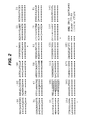
- the invention provides a protein comprising the amino acid sequence shown in Figure 2 as SEQ ID:1, which is CTLA-4 cloned from porcine cells. This is the preferred form of CTLA-4 for use in the invention.
- the extracellular domain of this protein is also shown in Figure 2.
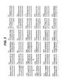
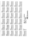
- the invention also provides nucleic acid which encodes protein SEQ ID:1 (or fragments thereof). This preferably comprises the nucleotide sequence shown in Figure 3 as SEQ ID:2.
- the invention provides a vector comprising the nucleic acid of the invention, and a cell transformed with such a vector.
- co-stimulation by signal 2 is antagonised by expressing a ligand for CTLA-4 on the xenogeneic donor cells.
- This ligand binds to CTLA-4 on activated T-cells of the recipient and antagonises the co-stimulatory signal provided by the interaction between donor B7 and recipient CD28. This renders xenoreactive T-cells anergic.
- the invention thus provides a membrane-associated protein which can bind to CTLA-4.
- This will typically be a chimeric protein (ie. a protein produced by combining regions of different proteins into a single protein) comprising a CTLA-4-binding region and a membrane-association region.
- the protein will thus be a fusion protein
- membrane-associated protein it is meant that the protein is attached to a cell membrane such that its extracellular domain can bind to CTLA-4.
- the protein might comprise a transmembrane sequence from a membrane protein, for instance, or a GPI anchor.
- a preferred transmembrane sequence is that of CD4 or CD8.
- the protein might include a sequence which enables it to associate extracellularly with a membrane protein without the protein itself being inserted into the cell membrane.
- the protein may also be desirable for the protein to comprise the cytoplasmic domain which is usually associated with said transmembrane regions (eg . the CD8 cytoplasmic domain), such that the protein is targeted to the cell membrane.
- the protein may also be desirable for the protein to comprise the extracellular sequences immediately juxtaposed with the cell membrane (eg . CD4 domains 3 and 4) in order to separate the CTLA-4-binding domain from the cell membrane.
- Synthetic linkers such as glycine linkers, can be used for the same purpose.
- the CTLA-4-binding domain of the protein preferably comprises an antibody with specificity for CTLA-4. This is preferably a single chain antibody (sFv). It is preferably specific for the CTLA-4 of a recipient organism.
- sFv single chain antibody
- a protein of the second aspect can comprise a single chain antibody fused via a linker to the transmembrane and cytoplasmic domains of CD8.
- the invention also provides nucleic acid which encodes a protein of the second aspect.
- the invention provides a vector comprising said nucleic acid of the invention, and a cell transformed with said vector.
- the invention also provides a delivery system comprising nucleic acid, and/or vector according to the second aspect of the invention, and means to deliver this material to a target cell.
- the invention provides a cell which expresses a protein of the second aspect on its surface, preferably such that the protein can bind to available CTLA-4.
- the cell preferably also expresses MHC (class I or class II) on its surface.
- MHC class I or class II
- the cell of the invention is a donor professional APC. Because of the antagonistic signal provided by the anti-CTLA-4 protein, however, these professional APC behave functionally as B7-negative cells.
- the invention also provides biological tissue comprising such a cell.
- the invention further provides an animal comprising a cell and/or biological tissue according to the second aspect.
- the invention also provides a process for rendering biological tissue suitable for xenotransplantation, comprising the step of treating said biological tissue such that it expresses one or more proteins according to the second aspect on the surface of its cells.
- the invention also provides a method of transplantation comprising the step of transplanting biological tissue according to the invention from a donor animal (eg . a pig) into a xenogeneic recipient animal ( eg . a human).
- a donor animal eg . a pig
- a xenogeneic recipient animal eg . a human
- the cells of the invention are suitable for pre-transplantation administration. This results in tolerance being induced in recipient T-cells before the xenograft itself is transplanted. Whilst the cells used in such pre-transplantation regimes should preferably express MHC class II, it will be appreciated that the cells need not be professional APCs.
- the invention provides protein or nucleic acid according to the second aspect for use as a medicament.
- the invention also provides the use of protein, nucleic acid, a vector, or a delivery system according to the second aspect in the manufacture of a formulation for administration to a xenotransplant recipient or donor.
- co-stimulation by signal 2 is prevented by expressing recipient organism MHC class II on the surface of the cells of the xenogeneic donor organ. If, for instance, a pig organ (donor) were being transplanted into a human (recipient), the pig organ would express human MHC class II.
- the donor cells By expressing recipient MHC class II on the cells of the xenogeneic donor, the donor cells will present xenoantigens to a recipient T-cell in the context of self MHC class II. If the donor cells do not express B7, or if B7 is blocked, the xenoreactive recipient T-cell will not receive co-stimulatory signal 2 and will become anergic before the recipient's APCs have an opportunity to present the xenoantigens themselves.
- the invention thus provides a cell which expresses on its surface MHC class II of a different organism.
- this is a porcine cell expressing human MHC class II on its surface.
- the MHC class II is preferably of the HLA-DR family.
- the MHC class II is preferably constitutively expressed on the surface of the cells.
- the MHC class II is preferably tissue-typed for maximum compatibility with the particular recipient. This will typically involve, for instance, ensuring that the HLA-DR expressed on the xenogeneic donor cell should match the HLA-DR of the particular recipient.
- the cell should also express one or more of the following three proteins, each of which has an important role in antigen processing: invariant chain, HLA-DM ⁇ and HLA-DM ⁇ .
- the cell preferably does not express co-stimulatory molecules (eg . B7) on its surface.
- co-stimulatory molecules eg . B7
- the donor cell is not a professional APC. It may, however, be a transfected non-immunogenic APC, such as an immature dendritic cell, which may be B7 + .
- the invention also provides biological tissue comprising a cell according to the third aspect.
- the invention further provides an animal comprising a cell and/or biological tissue according to the third aspect.
- the invention also provides a process for rendering biological tissue suitable for xenotransplantation, comprising the step of treating said biological tissue such that it expresses xenogeneic MHC class II on the surface of its cells.
- this process comprises the steps of isolating non-immunogenic cells (ie. cells which cannot provide a co-stimulatory signal, such as 87-negative cells) from a xenogeneic organism and transfecting these cells with HLA-DR.
- the HLA-DR is preferably tissue-typed for a specific recipient.
- the cells may also be transfected with other proteins necessary for efficient antigen processing.
- suitable non-immunogenic cells include renal tubular epithelial cells, which are 87-negative and have been shown to induce tolerance in rodent models of allogeneic transplantation.
- the invention also provides a method of transplantation comprising the step of transplanting biological tissue according to the third aspect from a donor animal (eg . a pig) into a xenogeneic recipient animal ( eg . a human).
- a donor animal eg . a pig
- a xenogeneic recipient animal eg . a human
- the cells of the invention are suitable for pre-transplantation administration. This results in tolerance being induced in recipient T-cells before the xenograft itself is transplanted.
- the invention provides a cell according to the third aspect for use as a medicament.
- the invention also provides the use of a cell or of biological tissue according to the third aspect in the manufacture of a formulation for administering to a xenotransplant recipient.
- the invention also provides the use of xenogeneic MHC class II, or nucleic acid encoding xenogeneic MHC class II, in the preparation of a formulation for administering to a xenotransplant donor.
- nucleic acid includes both DNA and RNA, although modified and synthetic nucleic acids are also included,
- the nucleic acid may be synthetic (eg . PNA), or may have modified inter-nucleotide linkages (eg . phosphorothioates).
- the term includes both sense and antisense nucleic acid sequences, as well as double-stranded sequences.
- the nucleic acid comprises sequences suitable for the regulation of expression of protein according to the invention.
- This expression can preferably be controlled, such as cell-specific control, inducible control, or temporal control.
- vector signifies a molecule which is capable of transferring nucleic acid to a host cell, and numerous suitable vectors are known in the art
- the vector is suitable for the production of a transgenic animal.
- Vectors suitable for the generation of transgenic pigs are described in references 13, 14, 15, 16 & 17.
- delivery system refers to means for delivering genetic material to a target cell.
- certain vectors as described above may also function as suitable delivery systems.
- certain delivery systems may also inherently be vectors, but this is not always the case.
- a viral vector can also function as a delivery system, whereas a liposomal delivery system is not a vector.
- the delivery system may be viral or non-viral.
- Non-viral systems, such as liposomes avoid some of the difficulties associated with virus-based systems, such as the expense of scaled production, poor persistence of expression, and concerns about safety.
- the delivery system is suitable for use in gene therapy. Numerous appropriate delivery systems are known in the art.
- the delivery system will be targeted so that molecules according to the invention are taken up by cells suitable for xenotransplantation, or cells which have been transplanted. More preferably the delivery system will be specific for these cells.
- the delivery system may be targeted to a specific organ, such as the heart or the kidney, or to a specific cell type, such as endothelial cells or professional APC.
- the delivery system may, for example, be a receptor-mediated delivery system, being targeted to receptors found on target cells.
- the delivery system may be targeted to receptors found on heart cells, preferably to receptors found exclusively on heart cells, or it may be targeted to receptors found on endothelial cells, preferably to receptors found exclusively on endothelial cells.
- the delivery system is preferably suitable for the generation of a transgenic animal.
- the delivery system may be targeted to a gamete, a zygote, or an embryonic stem cell.
- the vectors and delivery systems of the invention can be used to transfect cells to produce cells according to the invention.
- the transfection can occur in vivo or ex vivo.
- biological tissue includes collections of cells, tissues, and organs. Accordingly the definition includes, for example, fibroblasts, a cornea, nervous tissue, a heart, a liver, or a kidney.
- the animal is preferably suitable for the production of organs for xenotransplantation and/or cells of the invention (eg . cells for pre-xenotransplant administration to xenograft recipients).
- the animal is a mammal, and more preferably it is a transgenic pig or a transgenic sheep.
- the animal might be treated whilst alive such that it comprises transgenic biological tissue (ie . treated by gene therapy).
- a live animal is transfected with a vector according to the invention in order to produce a transgenic animal.
- a vector according to the invention could be specifically delivered to the heart of a pig to produce biological tissue suitable for xenotransplantation.
- the animal might be born as a transgenic animal.
- Many suitable approaches for generating such transgenic animals are known in the art [ eg . refs. 18, 19, 20].
- direct manipulation of the zygote or early embryo, by microinjection of DNA for instance is well known, as is the in vitro manipulation of pluripotent cells such as embryonic stem cells.
- Retroviral infection of early embryos has proved successful in a range of species, and adenoviral infection of zona-free eggs has been reported.
- Transgenesis and cloning of sheep by nuclear transfer has also been described (eg. WO97/07668 ).
- said biological tissue may be so rendered either in vivo or ex vivo.
- an animal organ may be in vivo transfected with a vector according to the invention, or an organ could be transfected ex vivo before transplantation or in vivo after transplantation.
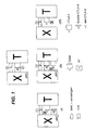
- Figure 1 illustrates the three aspects of the invention.
- X represents a xenogeneic cell (or, in the indirect activation pathway, a xenoantigen-presenting recipient APC), and
- T represents a recipient T-cell.
- the delivery of co-stimulatory signal 2 is prevented by using a soluble form of CTLA-4.
- anti-CTLA-4 is used to antagonise signal 2.
- X expresses recipient MHC-II, but does not express B7.
- Figure 2 shows the amino acid sequence of pCTLA-4 (SEQ ID NO:1).
- Figure 5 shows the results of flow cytometric analysis of hCTLA4-Ig ( ⁇ & ⁇ ) and pCTLA4-Ig ( ⁇ & ⁇ ) binding to human fibroblasts transfected with either human B7 (lower two lines) or porcine B7 (upper two lines).
- Figure 6 shows the selective inhibition of proliferation by pCTLA4-Ig ( ⁇ & ⁇ ) compared to hCTLA4-Ig ( ⁇ & ⁇ ) when co-stimulated by human B7 ( ⁇ & ⁇ ) or porcine B7 ( ⁇ & ⁇ ).
- Figure 7 shows the inhibition of human CD4 + T cell proliferation by hCTLA4-Ig ( ⁇ ) or pCTLA4-Ig ( ⁇ ) when human (7A) or porcine (7B) cells expressing MHC-class II were used as stimulators in a five day mixed leukocyte reaction.
- Figure 8 shows the nucleotide sequence of an anti-human CTLA-4 sFv. The inferred protein sequence is shown in Figure 9.
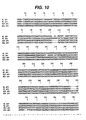
- Figure 10 shows the nucleotide sequences of four anti-murine CTLA-4 sFv. The inferred protein sequences are shown in Figure 11.
- the heavy and light chains are linked by a serine-glycine linker as indicated in Figures 9 and 11..
- Figure 12 shows the construct encoding the soluble Ig-fusion of the CTLA-4-specific sFv.
- Figure 13 shows the inhibition of T cell proliferation by cells expressing either an anti-hCTLA-4 sFv ( ⁇ ) or a control sFv ( ⁇ ).
- Figure 14 shows construct encoding the membrane-bound form of the anti-CTLA-4 sFv.
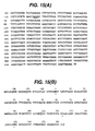
- Figure 15 shows (A) the nucleotide sequence and (B) the amino acid sequence of human CTLA-4. The start codon is underlined. At position -21, the sequence differs from GenBank sequence L15006, and at position 110 the sequence differs from both L15006 and M74363.
- Figure 16 shows the sequence of cloned human CD8 ⁇ .
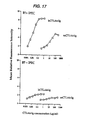
- Figure 17 shows the binding of human and murine CTLA4-Ig to IPEC, in order to define clones as B7-negative or B7-positive.
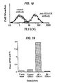
- Figure 18 shows the binding to transfected cells of HLA-DR-specific mAb L243.
- Figure 19 shows the proliferation by human T-cells to HLA-DR-1 transfected IPEC.
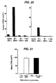
- Figure 20 shows the results of a human T-cell proliferation assay following 2 days of incubation HLA-DR-1 transfected cells. The X-axis indicates the stimulator cells used in the second step of the proliferation assay.
- the black bars show results with CD4 T-cells which were primed with B7-positive transfectants; the white bars (hardly visible) show results after priming with B7-negative transfectants.
- the first graph shows results with cells harvested on day 3; the second graph shows results from a harvest on the sixth day.
- Figure 21 shows the proliferation of an APC-dependent, HLA-DR-1 restricted T-cell line raised against IPEC. The stimulator population is indicated on the X-axis.
- Porcine CTLA-4 (“pCTLA4") was cloned from PHA-activated pig T cells. RNA was prepared using standard techniques and pCTLA4 was amplified by PCR using primers: 5' -TTGAAGCTTAGCCATGGCTTGCTCTGGA- 3' (5' primer) 5' -TAATGAATTCTCAATTGATGGGAATAAAATAAG-3' (3' primer)
- the resulting 700bp fragment was sub-cloned into EcoR I/ Hind III digested pBluescript, and the nucleotide sequence was determined using the standard T3 and T7 primers. The sequence of a single clone is shown in figure 3, which also shows a comparison with the human and bovine CTLA-4 sequences.
- the predicted amino acid sequence of pCTLA4 is shown in figure 2, with a comparison with that of human and cattle. Of significance is the predicted amino acid difference at residue 97, which is important in B7 binding, being part of the conserved hexapeptide motif MYPPPY. In pCTLA4, residue 97 is leucine (giving LYPPPY), whereas other species have methionine (although leucine has also been found in bovine CD28 [21]). This important amino acid difference is believed to be of key importance to the advantageous differential binding of pCTLA4 to human and pig B7.
- the extracellular domain of pCTLA4 was amplified using the 5' primer described above and 5'-CGGTT CTGCAG CACCACCGGAGCCACC ATCAGAATCTGGGCATGGTTCTGGATCAATGAC-3'
- the chimeric pCTLA4-Ig DNA sequence was released from pBluescript as a Hind III/ Bst XI fragment and was sub-cloned into the expression vector pHOOK-3 TM (Invitrogen). This was used to transfect DAP.3 or CHO-K1 cells using standard calcium phosphate precipitation. G418-resistant cells were separated using the CaptureTec TM system. These transfected cells were grown in tissue culture flasks until confluent, at which point the medium was changed, and the cells were kept in culture for a further 3 days. At this stage the medium was harvested and perfused through a protein G column. pCTLA-4-Ig was eluted under acid conditions. The concentration of the eluted protein was calculated using ELISA with an antibody directed against human IgG 1 and a standard human IgG 1 myeloma protein.
- the binding characteristics of pCTLA4-Ig were compared to those of human CTLA4-Ig using flow cytometric analysis with human fibroblasts transfected with either human B7-1 or porcine B7-2. For these experiments, the concentration of pig and human CTLA4-Ig were equivalent as assessed by ELISA. As illustrated in figure 5, human and porcine CTLA4-Ig appeared to have similar binding characteristics on human cells expressing porcine B7. Unlike human CTLA4-Ig, however, pCTLA4-Ig failed to bind human- B7, implying that pCTLA4-Ig binds preferentially to porcine B7 and is useful as a species-specific reagent.
- pCTLA4-Ig was used to inhibit human T cell proliferative responses to a variety of stimulators.
- co-stimulation of the T cell response was provided by either porcine or human B7, expressed either by transfection or naturally on professional APCs.
- hCTLA4-Ig inhibited all proliferative responses ( Figure 6, ⁇ & ⁇ ).
- pCTLA4-Ig only fully inhibited the response when stimulators expressed porcine B7 ( ⁇ ); the proliferative response to cells expressing human B7 was only minimally affected ( ⁇ ).
- pCTLA4-Ig failed to have a significant inhibitory effect on the proliferative responses to human cells expressing MHC class II and human B7 but did inhibit the response to porcine stimulators (figure 7).
- binding characteristics of pCTLA4-Ig to both human and porcine B7-family molecules may be compared to those of hCTLA4-Ig, for example using the following tests:
- a phage display library containing 10 12 semi-synthetic variable sequences was screened using human or murine CTLA4-1g and a control human IgG1 myeloma protein.
- the sFv from a phage displaying differential binding to the human CTLA4-Ig protein after 4 rounds of screening were isolated and purified using standard techniques.
- the nucleotide and inferred amino acid sequences are shown in figures 8, 9, 10, and 11.
- the sFv were amplified by PCR using specific primers based on the nucleotide sequences. The distal portions of the primers were based on sequence within pHOOK1. The 5' primer contained an Apa I site and the 3' primer contained a Sal I site, both of which were predicted to be unique. The resulting sFv were sub-cloned into pBluescript for sequencing to determine faithful amplification.
- the Apa I/ Sal I fragments were then sub-cloned into pHOOK1, where it is flanked upstream by an in-frame signal sequence from the murine Ig ⁇ -chain and a haemaggluttinin A epitope sequence, and downstream by two in-frame myc sequences and a transmembrane sequence from the PDGF receptor.
- the myc sequences from pHOOK1 were amplified by PCR using the 5' primer 5' -GAGCTGAAACGG GCGGCCGC AGAAC-3', which contains a Not I site (underlined) and the 3' primer 5'-CTGGC CTGCAG CATTCAGATCC-3', which introduced a Pst I site (underlined).
- the resulting 113 base pair fragment was sub-cloned into Not I/ Pst I digested pBluescript.
- the sFv was released from pHOOK1 as an EcoR I/ Not I fragment, and was ligated into EcoR I/ Pst I digested pBluescript-IgG1, along with the Not I/ Pst I PCR product [ Figure 12].
- This construct encodes a soluble Ig-fusion of the CTLA-4-specific sFv.
- the construct was sub-cloned into pHOOK3 as a Hind III/ BstX I fragment.
- the pHOOK construct was transfected into cells already expressing HLA-DR molecules and human B7.
- Cells expressing the anti-CTLA4-sFv construct on the cell surface were identified by flow cytometric analysis using hCTLA4-Ig. These cells were cloned by limiting dilution and were used as stimulators of T cell proliferation in 5 day cultures. The results of one experiment are shown in figure 13.
- Cells expressing the anti-hCTLA4 sFv failed to stimulate T cell proliferation ( ⁇ ), whereas those expressing a control sFv stimulated proliferation in the same way as normal cells ( ⁇ ).
- the EcoR I/ BamH I fragment from pBluescript was sub-cloned into the expression vector pH ⁇ Apr-1-neo or the sister vector pH ⁇ Apr-1-gpt. These were transfected into cells already expressing HLA-DR molecules and B7 and selected as described above for the pHOOK construct.
- CTLA-4 on by activated T-cells is only transient so, to test the functional characteristics of the anti-CTLA4-sFv, chimeric constructs comprising the DNA sequences encoding the extracellular domains of human or murine CTLA4 and the transmembrane/cytoplasmic sequences of human CD8 were made. Cells expressing these constructs can be used for the study of the anti-CTLA4-sFv protein.
- RNA from PHA-activated human T cells was prepared using standard techniques.
- hCTLA4 was amplified PCR using primers: 5'-TTCAAAGCTTCAGGATCCTGAAAGGTTTTG-3' introducing a Hind III site (5' primer) 5' -TAATGAATTCTCAATTGATGGGAATAAAATAAG-3' introducing an EcoR I site (3' primer)
- the resulting fragment was sub cloned into Hind III lEcoR I digested pBluescript and the nucleotide sequence determined using standard T3 and T7 primers.
- the sequence of a single clone is shown in figure 15. This differed by a single base (position 439) from GenBank-listed sequences for human CTLA-4.
- the predicted amino acid sequence of hCTLA4 is also shown.
- hCTLA-4 The extracellular domain of hCTLA-4 was amplified using 5' primer described above and: 5'-GATGTAGATATCACAGGCGAA GTCGAC ACCACCGGAGCC ACCAATTACATAAATCTGGGCTCCGTTGCCTATGCCC-3'
- the resulting fragment was sub cloned into Hind III/ EcoR I digested pBluescript and the nucleotide sequence determined using standard T3 and T7 primers. The sequence of a single clone is shown in figure 16. This clone differed from the sequence deposited with GenBank at four positions, although none of these were within the region that was subsequently amplified.
- TM and C domains of hCD8 were amplified using the 3' primer described above and the following 5' primer: 5'-CATAGGCAACGGAGCCCAGATTTATGTAATT GGTGGCTCCGGTGGT GTCGAC TTCGCCTGTGATATCTACATC-3'
- CTLA-4 The extracellular domain of human CTLA-4 was cut from pBluescript as an EcoR I/ Sal I fragment, and the TM-IC domain of CD8 cut as a Sal I/ BamH I fragment. Together they were ligated back into EcoR I/ BamH I digested pBluescript. The whole CTLA-4-CD8 chimera was then removed as a single EcoR I fragment and was sub-cloned into a number of expression vectors for expression into the human T cell leukaemia line J6.
- the cell-surface anti-CTLA-4 proteins may be further characterised by the following functional tests:
- PEC immortalised porcine aortic endothelial cells
- IPEC immortalised porcine aortic endothelial cells
- B7-negative clones were identified by flow cytometric screening with hCTLA4-Ig and mCTLA4-Ig [see figure 17]. These were then transfected with cDNAs encoding HLA-DRA and DR.B1*0101 in the plasmid expression vectors pcExV1-gpt and pH ⁇ Apr-1neo, and cells were placed under selection with MXH and G418.
- B7-positive IPEC controls were generated similarly [4].
- IPEC transfectants expressing the murine MHC class II molecule I-A b were also generated for experiments in mice.
- MHC class II-positive cells underwent several rounds of fluorescence activated cell sorting before being cloned by limiting dilution.
- a second batch of transfectants was prepared in exactly the same way, but with additional transfected cDNAs encoding HLA-DMA and HLA-DMB and p31Ii (invariant chain) in the expression vector pCMU.
- T-cells Human T-cells were purified using standard protocols [3]. For primary proliferation assays, T-cells were incubated for 5 days with fixed numbers of irradiated stimulator cells, before addition of 1 ⁇ Ci 3 H-thymidine sixteen hours prior to harvesting onto glass fibre filters. The filters were read in a scintillation counter.
- T-cells incubated with B7-positive IPEC mounted an anti-DR1 proliferative response in the secondary step with the kinetics of a primed secondary immune response (maximal at three days).
- T-cells incubated with B7-negative IPEC in the primary step became tolerant to DR1 and failed to mount a response on subsequent exposure to DR1-expressed on B7-positive IPEC [figure 20].
- CD4 + T-cells from a DR1-expressing individual were purified according to standard procedures. In primary proliferation assays, they proliferated to B7-positive IPEC transfected with HLA-DR1, indicating that the DR1 can perform as a restriction element for pig-peptide-specific T-cells. Assays comparing the proliferative response to B7-positive and 87-negative DR1+ transfectants are being performed.
- Two step anergy induction assays may also be performed to demonstrate that DR1-transfected, B7-negative pig cells induce anergy in HLA-DR-restricted human T-cells.
- a human T-cell line against wild type IPEC was raised from human PBMC.
- the proliferative response of this line was dependent on the presence of human APC and inhibitable by antibodies against HLA-DR, indicating that the line had indirect specificity for processed porcine xenoantigens presented by human APC.
- porcine pancreatic islet cells may be transplanted under the kidney capsule of streptozotocin-treated diabetic mice. Islet xenografts, being non vascular, are rejected solely by T-cells. Porcine islets are prepared from the pancreas of pigs under terminal anaesthesia, and their survival in the recipients assessed by maintenance of normoglycaemia. Mice are injected intravenously with B7-negative, I-A b -expressing IPEC before transplantation of pig islets.
- This strategy can be combined with other aspects of the invention to tolerise the direct pathway of T-cell recognition, to ensure that rejection via the direct pathway does not occur.
- nephrectomy of the islet-carrying kidney is performed before re-transplantation (under the capsule of the surviving kidney), of identical or third party porcine islets.
Landscapes
- Health & Medical Sciences (AREA)
- Life Sciences & Earth Sciences (AREA)
- Chemical & Material Sciences (AREA)
- Immunology (AREA)
- Organic Chemistry (AREA)
- General Health & Medical Sciences (AREA)
- Medicinal Chemistry (AREA)
- Genetics & Genomics (AREA)
- Biophysics (AREA)
- Biochemistry (AREA)
- Molecular Biology (AREA)
- Zoology (AREA)
- Proteomics, Peptides & Aminoacids (AREA)
- Veterinary Medicine (AREA)
- Animal Behavior & Ethology (AREA)
- Gastroenterology & Hepatology (AREA)
- Cell Biology (AREA)
- Toxicology (AREA)
- Engineering & Computer Science (AREA)
- Environmental Sciences (AREA)
- Pharmacology & Pharmacy (AREA)
- Public Health (AREA)
- Transplantation (AREA)
- Bioinformatics & Cheminformatics (AREA)
- Biodiversity & Conservation Biology (AREA)
- Animal Husbandry (AREA)
- Microbiology (AREA)
- Mycology (AREA)
- Epidemiology (AREA)
- Biotechnology (AREA)
- Chemical Kinetics & Catalysis (AREA)
- General Chemical & Material Sciences (AREA)
- Nuclear Medicine, Radiotherapy & Molecular Imaging (AREA)
- Micro-Organisms Or Cultivation Processes Thereof (AREA)
- Peptides Or Proteins (AREA)
- Medicines Containing Material From Animals Or Micro-Organisms (AREA)
- Pharmaceuticals Containing Other Organic And Inorganic Compounds (AREA)
- Medicines That Contain Protein Lipid Enzymes And Other Medicines (AREA)
- Devices Affording Protection Of Roads Or Walls For Sound Insulation (AREA)
- Medicines Containing Antibodies Or Antigens For Use As Internal Diagnostic Agents (AREA)
Applications Claiming Priority (2)
| Application Number | Priority Date | Filing Date | Title |
|---|---|---|---|
| GBGB9809280.2A GB9809280D0 (en) | 1998-04-30 | 1998-04-30 | Immunosupression |
| EP99919418A EP1073737B1 (de) | 1998-04-30 | 1999-04-30 | Immununterdrückung durch blockierung des t-zellencostimulierungssignales 2 (b7/cd28 interaktion) |
Related Parent Applications (1)
| Application Number | Title | Priority Date | Filing Date |
|---|---|---|---|
| EP99919418A Division EP1073737B1 (de) | 1998-04-30 | 1999-04-30 | Immununterdrückung durch blockierung des t-zellencostimulierungssignales 2 (b7/cd28 interaktion) |
Publications (2)
| Publication Number | Publication Date |
|---|---|
| EP1854884A2 true EP1854884A2 (de) | 2007-11-14 |
| EP1854884A3 EP1854884A3 (de) | 2009-07-29 |
Family
ID=10831274
Family Applications (2)
| Application Number | Title | Priority Date | Filing Date |
|---|---|---|---|
| EP06077284A Withdrawn EP1854884A3 (de) | 1998-04-30 | 1999-04-30 | Immununterdrückung durch Blockierung des T-Zellencostimulierungssignales 2 (B7/CD28 Interaktion) |
| EP99919418A Expired - Lifetime EP1073737B1 (de) | 1998-04-30 | 1999-04-30 | Immununterdrückung durch blockierung des t-zellencostimulierungssignales 2 (b7/cd28 interaktion) |
Family Applications After (1)
| Application Number | Title | Priority Date | Filing Date |
|---|---|---|---|
| EP99919418A Expired - Lifetime EP1073737B1 (de) | 1998-04-30 | 1999-04-30 | Immununterdrückung durch blockierung des t-zellencostimulierungssignales 2 (b7/cd28 interaktion) |
Country Status (11)
| Country | Link |
|---|---|
| US (3) | US7432344B1 (de) |
| EP (2) | EP1854884A3 (de) |
| JP (2) | JP2002513568A (de) |
| AT (1) | ATE355373T1 (de) |
| AU (1) | AU772239B2 (de) |
| CA (1) | CA2326671C (de) |
| DE (1) | DE69935318T2 (de) |
| ES (1) | ES2284251T3 (de) |
| GB (1) | GB9809280D0 (de) |
| NZ (2) | NZ507706A (de) |
| WO (1) | WO1999057266A2 (de) |
Families Citing this family (33)
| Publication number | Priority date | Publication date | Assignee | Title |
|---|---|---|---|---|
| GB9809280D0 (en) * | 1998-04-30 | 1998-07-01 | Rpms Technology Ltd | Immunosupression |
| US7030228B1 (en) | 1999-11-15 | 2006-04-18 | Miltenyi Biotec Gmbh | Antigen-binding fragments specific for dendritic cells, compositions and methods of use thereof antigens recognized thereby and cells obtained thereby |
| AU2001255398A1 (en) * | 2000-04-12 | 2001-10-30 | Genetics Institute Inc. | Surface-bound antigen binding portions of antibodies that bind to ctla-4 and cd28 and uses therefor |
| CN101255192A (zh) * | 2000-05-26 | 2008-09-03 | 布里斯托尔-迈尔斯斯奎布公司 | 可溶性ctla4突变体分子及其应用 |
| US7094874B2 (en) | 2000-05-26 | 2006-08-22 | Bristol-Myers Squibb Co. | Soluble CTLA4 mutant molecules |
| US7754208B2 (en) | 2001-01-17 | 2010-07-13 | Trubion Pharmaceuticals, Inc. | Binding domain-immunoglobulin fusion proteins |
| US20030133939A1 (en) | 2001-01-17 | 2003-07-17 | Genecraft, Inc. | Binding domain-immunoglobulin fusion proteins |
| RU2420537C2 (ru) * | 2001-01-17 | 2011-06-10 | Трабьон Фармасьютикалз Инк. | Слитые белки связывающий домен-иммуноглобулин |
| ITRM20010408A1 (it) * | 2001-07-10 | 2003-01-10 | Univ Napoli Federico Ii | Mini-anticorpo umano citotossico per cellule tumorali che esprimono il recettore erbb2. |
| US7601351B1 (en) | 2002-06-26 | 2009-10-13 | Human Genome Sciences, Inc. | Antibodies against protective antigen |
| EP1539235A2 (de) | 2002-07-01 | 2005-06-15 | Human Genome Sciences, Inc. | Spezifisch an reg iv bindende antikörper |
| CA2533259C (en) * | 2003-07-21 | 2014-01-28 | Lifecell Corporation | Acellular tissue matrices made from galactose .alpha.-1,3-galactose-deficient tissue |
| AU2005223617A1 (en) | 2004-03-17 | 2005-09-29 | Revivicor, Inc. | Tissue products from animals lacking functional alpha 1,3 galactosyl transferase |
| CA2958259C (en) | 2004-10-22 | 2020-06-30 | Revivicor, Inc. | Ungulates with genetically modified immune systems |
| RU2423381C2 (ru) | 2005-07-25 | 2011-07-10 | Трабьон Фармасьютикалз, Инк. | Снижение количества в-клеток с использованием cd37-специфических и cd20-специфических связывающих молекул |
| EP3138403A1 (de) | 2005-08-09 | 2017-03-08 | Revivicor, Inc. | Ctla4-ig exprimierende transgene huftiere und verwendungen davon |
| NZ573646A (en) | 2006-06-12 | 2012-04-27 | Wyeth Llc | Single-chain multivalent binding proteins with effector function |
| RU2531754C2 (ru) | 2008-04-11 | 2014-10-27 | ЭМЕРДЖЕНТ ПРОДАКТ ДИВЕЛОПМЕНТ СИЭТЛ,ЭлЭлСи,US | Связывающееся с cd37 иммунотерапевтическое средство и его комбинация с бифункциональным химиотерапевтическим средством |
| DK2348827T3 (en) | 2008-10-27 | 2015-07-20 | Revivicor Inc | IMMUNICIPLY COMPROMATED PETS |
| ES2767881T3 (es) | 2009-08-14 | 2020-06-18 | Revivicor Inc | Cerdos multi-transgénicos para el tratamiento de diabetes |
| US9420770B2 (en) | 2009-12-01 | 2016-08-23 | Indiana University Research & Technology Corporation | Methods of modulating thrombocytopenia and modified transgenic pigs |
| CN106432474A (zh) | 2010-03-12 | 2017-02-22 | 艾伯维生物医疗股份有限公司 | Ctla4蛋白和其用途 |
| ES2680636T3 (es) | 2011-02-14 | 2018-09-10 | Revivicor Inc. | Cerdos genéticamente modificados para xenotrasplante de xenoinjertos vascularizados y derivados de los mismos |
| US20140115728A1 (en) | 2012-10-24 | 2014-04-24 | A. Joseph Tector | Double knockout (gt/cmah-ko) pigs, organs and tissues |
| US9944689B2 (en) * | 2013-03-07 | 2018-04-17 | The General Hospital Corporation | Human CTLA4 mutants and use thereof |
| WO2015066668A1 (en) | 2013-11-04 | 2015-05-07 | Lifecell Corporation | Methods of removing alpha-galactose |
| TWI617319B (zh) * | 2015-09-01 | 2018-03-11 | 免疫功坊股份有限公司 | 用以治療病理性血栓的融合蛋白 |
| WO2017053469A2 (en) | 2015-09-21 | 2017-03-30 | Aptevo Research And Development Llc | Cd3 binding polypeptides |
| KR102746223B1 (ko) | 2017-04-20 | 2024-12-27 | 이제네시스, 인크. | 유전자 변형 동물의 생성 방법 |
| WO2019168897A2 (en) | 2018-02-28 | 2019-09-06 | Dana-Farber Cancer Institute, Inc. | Methods for treating cancer using combinations of anti-btnl2 and immune checkpoint blockade agents |
| CN111214646B (zh) * | 2019-12-30 | 2023-08-01 | 中山大学·深圳 | Pd-l1/ctla-4在制备免疫抑制剂中的应用 |
| US20220211018A1 (en) | 2020-11-20 | 2022-07-07 | Revivicor, Inc. | Multi-transgenic pigs with growth hormone receptor knockout for xenotransplantation |
| CA3232376A1 (en) | 2021-09-20 | 2023-03-23 | Maria KOKKINAKI | Multitransgenic pigs comprising ten genetic modifications for xenotransplantation |
Family Cites Families (5)
| Publication number | Priority date | Publication date | Assignee | Title |
|---|---|---|---|---|
| JPH10501815A (ja) * | 1994-06-07 | 1998-02-17 | リージェンツ・オブ・ザ・ユニバーシティ・オブ・ミネソタ | 抗原特異的t細胞応答の阻害方法 |
| CA2222055A1 (en) * | 1995-05-23 | 1996-11-28 | Morphosys Gesellschaft Fur Proteinoptimierung Mbh | Multimeric proteins |
| EP0892643B2 (de) * | 1996-03-20 | 2009-09-02 | Bristol-Myers Squibb Company | Verfahren zur inhibierung der immunreaktion durch blockierung der gp39/cd40 und ctla4/cd28/b7 routen und zusammensetzung zu deren verwendung |
| US6165476A (en) * | 1997-07-10 | 2000-12-26 | Beth Israel Deaconess Medical Center | Fusion proteins with an immunoglobulin hinge region linker |
| GB9809280D0 (en) * | 1998-04-30 | 1998-07-01 | Rpms Technology Ltd | Immunosupression |
-
1998
- 1998-04-30 GB GBGB9809280.2A patent/GB9809280D0/en not_active Ceased
-
1999
- 1999-04-30 EP EP06077284A patent/EP1854884A3/de not_active Withdrawn
- 1999-04-30 JP JP2000547221A patent/JP2002513568A/ja active Pending
- 1999-04-30 AT AT99919418T patent/ATE355373T1/de not_active IP Right Cessation
- 1999-04-30 ES ES99919418T patent/ES2284251T3/es not_active Expired - Lifetime
- 1999-04-30 DE DE69935318T patent/DE69935318T2/de not_active Expired - Lifetime
- 1999-04-30 EP EP99919418A patent/EP1073737B1/de not_active Expired - Lifetime
- 1999-04-30 NZ NZ507706A patent/NZ507706A/en not_active IP Right Cessation
- 1999-04-30 AU AU37213/99A patent/AU772239B2/en not_active Expired
- 1999-04-30 NZ NZ527297A patent/NZ527297A/xx active IP Right Revival
- 1999-04-30 WO PCT/GB1999/001350 patent/WO1999057266A2/en not_active Ceased
- 1999-04-30 US US09/674,462 patent/US7432344B1/en not_active Expired - Fee Related
- 1999-04-30 CA CA2326671A patent/CA2326671C/en not_active Expired - Lifetime
-
2008
- 2008-09-04 US US12/231,608 patent/US8097598B2/en not_active Expired - Fee Related
-
2011
- 2011-04-22 JP JP2011096600A patent/JP5706225B2/ja not_active Expired - Lifetime
- 2011-12-12 US US13/316,791 patent/US9090708B2/en not_active Expired - Fee Related
Also Published As
| Publication number | Publication date |
|---|---|
| US7432344B1 (en) | 2008-10-07 |
| JP5706225B2 (ja) | 2015-04-22 |
| EP1073737B1 (de) | 2007-02-28 |
| ATE355373T1 (de) | 2006-03-15 |
| DE69935318D1 (de) | 2007-04-12 |
| JP2002513568A (ja) | 2002-05-14 |
| JP2011144205A (ja) | 2011-07-28 |
| CA2326671C (en) | 2015-12-15 |
| EP1073737A2 (de) | 2001-02-07 |
| EP1854884A3 (de) | 2009-07-29 |
| ES2284251T3 (es) | 2007-11-01 |
| AU772239B2 (en) | 2004-04-22 |
| US8097598B2 (en) | 2012-01-17 |
| US9090708B2 (en) | 2015-07-28 |
| US20130078266A1 (en) | 2013-03-28 |
| CA2326671A1 (en) | 1999-11-11 |
| WO1999057266A3 (en) | 2000-04-20 |
| NZ527297A (en) | 2004-12-24 |
| US20100146648A1 (en) | 2010-06-10 |
| GB9809280D0 (en) | 1998-07-01 |
| NZ507706A (en) | 2004-01-30 |
| AU3721399A (en) | 1999-11-23 |
| WO1999057266A2 (en) | 1999-11-11 |
| DE69935318T2 (de) | 2007-11-15 |
Similar Documents
| Publication | Publication Date | Title |
|---|---|---|
| US8097598B2 (en) | Porcine CTLA-4 for xenograft-specific immunosuppression | |
| US6319709B1 (en) | Tumor cells with increased immunogenicity and uses therefor | |
| US7459544B2 (en) | Nucleic acids encoding B7-2 fusion proteins | |
| US5861310A (en) | Tumor cells modified to express B7-2 with increased immunogenicity and uses therefor | |
| EP0711345A1 (de) | B7-2: ctl a4/cd 28 "gegen rezepter" | |
| JP4236925B2 (ja) | 免疫応答に関与する新規ポリペプチド | |
| US6824779B1 (en) | Methods for inhibiting the interaction of B7-2 with its natural ligand | |
| AU776618B2 (en) | Improvement of tolerance to a xenograft | |
| US6723705B1 (en) | Tumor cells modified to express B7-2 with increased immunogenicity and uses therefor | |
| NZ535708A (en) | Immunosuppression by blocking T cell co-stimulation signal 2 (B7/CD28 interaction) | |
| AU772061B2 (en) | B7-2:CTL A4/CD 28 counter receptor | |
| HK1160665A (en) | Polypeptides involved in immune response |
Legal Events
| Date | Code | Title | Description |
|---|---|---|---|
| PUAI | Public reference made under article 153(3) epc to a published international application that has entered the european phase |
Free format text: ORIGINAL CODE: 0009012 |
|
| AC | Divisional application: reference to earlier application |
Ref document number: 1073737 Country of ref document: EP Kind code of ref document: P |
|
| AK | Designated contracting states |
Kind code of ref document: A2 Designated state(s): AT BE CH CY DE DK ES FI FR GB GR IE IT LI LU MC NL PT SE |
|
| PUAL | Search report despatched |
Free format text: ORIGINAL CODE: 0009013 |
|
| AK | Designated contracting states |
Kind code of ref document: A3 Designated state(s): AT BE CH CY DE DK ES FI FR GB GR IE IT LI LU MC NL PT SE |
|
| 17P | Request for examination filed |
Effective date: 20100105 |
|
| 17Q | First examination report despatched |
Effective date: 20100204 |
|
| AKX | Designation fees paid |
Designated state(s): AT BE CH CY DE DK ES FI FR GB GR IE IT LI LU MC NL PT SE |
|
| STAA | Information on the status of an ep patent application or granted ep patent |
Free format text: STATUS: THE APPLICATION IS DEEMED TO BE WITHDRAWN |
|
| 18D | Application deemed to be withdrawn |
Effective date: 20100817 |