EP1877428B1 - Fluoreszentes chlorotoxinkonjugat und verfahren zur intraoperativen sichtbarmachung von krebs - Google Patents
Fluoreszentes chlorotoxinkonjugat und verfahren zur intraoperativen sichtbarmachung von krebs Download PDFInfo
- Publication number
- EP1877428B1 EP1877428B1 EP06739100A EP06739100A EP1877428B1 EP 1877428 B1 EP1877428 B1 EP 1877428B1 EP 06739100 A EP06739100 A EP 06739100A EP 06739100 A EP06739100 A EP 06739100A EP 1877428 B1 EP1877428 B1 EP 1877428B1
- Authority
- EP
- European Patent Office
- Prior art keywords
- chlorotoxin
- conjugate
- cancers
- ctx
- binding
- Prior art date
- Legal status (The legal status is an assumption and is not a legal conclusion. Google has not performed a legal analysis and makes no representation as to the accuracy of the status listed.)
- Expired - Lifetime
Links
Images









Classifications
-
- A—HUMAN NECESSITIES
- A61—MEDICAL OR VETERINARY SCIENCE; HYGIENE
- A61K—PREPARATIONS FOR MEDICAL, DENTAL OR TOILETRY PURPOSES
- A61K49/00—Preparations for testing in vivo
- A61K49/001—Preparation for luminescence or biological staining
- A61K49/0013—Luminescence
- A61K49/0017—Fluorescence in vivo
- A61K49/0019—Fluorescence in vivo characterised by the fluorescent group, e.g. oligomeric, polymeric or dendritic molecules
- A61K49/0021—Fluorescence in vivo characterised by the fluorescent group, e.g. oligomeric, polymeric or dendritic molecules the fluorescent group being a small organic molecule
- A61K49/0041—Xanthene dyes, used in vivo, e.g. administered to a mice, e.g. rhodamines, rose Bengal
-
- A—HUMAN NECESSITIES
- A61—MEDICAL OR VETERINARY SCIENCE; HYGIENE
- A61B—DIAGNOSIS; SURGERY; IDENTIFICATION
- A61B17/00—Surgical instruments, devices or methods
- A61B17/22—Implements for squeezing-off ulcers or the like on inner organs of the body; Implements for scraping-out cavities of body organs, e.g. bones; for invasive removal or destruction of calculus using mechanical vibrations; for removing obstructions in blood vessels, not otherwise provided for
-
- A—HUMAN NECESSITIES
- A61—MEDICAL OR VETERINARY SCIENCE; HYGIENE
- A61K—PREPARATIONS FOR MEDICAL, DENTAL OR TOILETRY PURPOSES
- A61K47/00—Medicinal preparations characterised by the non-active ingredients used, e.g. carriers or inert additives; Targeting or modifying agents chemically bound to the active ingredient
- A61K47/50—Medicinal preparations characterised by the non-active ingredients used, e.g. carriers or inert additives; Targeting or modifying agents chemically bound to the active ingredient the non-active ingredient being chemically bound to the active ingredient, e.g. polymer-drug conjugates
- A61K47/51—Medicinal preparations characterised by the non-active ingredients used, e.g. carriers or inert additives; Targeting or modifying agents chemically bound to the active ingredient the non-active ingredient being chemically bound to the active ingredient, e.g. polymer-drug conjugates the non-active ingredient being a modifying agent
- A61K47/62—Medicinal preparations characterised by the non-active ingredients used, e.g. carriers or inert additives; Targeting or modifying agents chemically bound to the active ingredient the non-active ingredient being chemically bound to the active ingredient, e.g. polymer-drug conjugates the non-active ingredient being a modifying agent the modifying agent being a protein, peptide or polyamino acid
- A61K47/64—Drug-peptide, drug-protein or drug-polyamino acid conjugates, i.e. the modifying agent being a peptide, protein or polyamino acid which is covalently bonded or complexed to a therapeutically active agent
- A61K47/6415—Toxins or lectins, e.g. clostridial toxins or Pseudomonas exotoxins
-
- A—HUMAN NECESSITIES
- A61—MEDICAL OR VETERINARY SCIENCE; HYGIENE
- A61K—PREPARATIONS FOR MEDICAL, DENTAL OR TOILETRY PURPOSES
- A61K49/00—Preparations for testing in vivo
- A61K49/001—Preparation for luminescence or biological staining
- A61K49/0013—Luminescence
- A61K49/0017—Fluorescence in vivo
- A61K49/0019—Fluorescence in vivo characterised by the fluorescent group, e.g. oligomeric, polymeric or dendritic molecules
- A61K49/0021—Fluorescence in vivo characterised by the fluorescent group, e.g. oligomeric, polymeric or dendritic molecules the fluorescent group being a small organic molecule
- A61K49/0032—Methine dyes, e.g. cyanine dyes
-
- A—HUMAN NECESSITIES
- A61—MEDICAL OR VETERINARY SCIENCE; HYGIENE
- A61K—PREPARATIONS FOR MEDICAL, DENTAL OR TOILETRY PURPOSES
- A61K49/00—Preparations for testing in vivo
- A61K49/001—Preparation for luminescence or biological staining
- A61K49/0013—Luminescence
- A61K49/0017—Fluorescence in vivo
- A61K49/005—Fluorescence in vivo characterised by the carrier molecule carrying the fluorescent agent
- A61K49/0056—Peptides, proteins, polyamino acids
-
- A—HUMAN NECESSITIES
- A61—MEDICAL OR VETERINARY SCIENCE; HYGIENE
- A61P—SPECIFIC THERAPEUTIC ACTIVITY OF CHEMICAL COMPOUNDS OR MEDICINAL PREPARATIONS
- A61P35/00—Antineoplastic agents
-
- C—CHEMISTRY; METALLURGY
- C07—ORGANIC CHEMISTRY
- C07K—PEPTIDES
- C07K14/00—Peptides having more than 20 amino acids; Gastrins; Somatostatins; Melanotropins; Derivatives thereof
- C07K14/435—Peptides having more than 20 amino acids; Gastrins; Somatostatins; Melanotropins; Derivatives thereof from animals; from humans
- C07K14/43504—Peptides having more than 20 amino acids; Gastrins; Somatostatins; Melanotropins; Derivatives thereof from animals; from humans from invertebrates
- C07K14/43513—Peptides having more than 20 amino acids; Gastrins; Somatostatins; Melanotropins; Derivatives thereof from animals; from humans from invertebrates from arachnidae
- C07K14/43522—Peptides having more than 20 amino acids; Gastrins; Somatostatins; Melanotropins; Derivatives thereof from animals; from humans from invertebrates from arachnidae from scorpions
-
- G—PHYSICS
- G01—MEASURING; TESTING
- G01N—INVESTIGATING OR ANALYSING MATERIALS BY DETERMINING THEIR CHEMICAL OR PHYSICAL PROPERTIES
- G01N33/00—Investigating or analysing materials by specific methods not covered by groups G01N1/00 - G01N31/00
- G01N33/48—Biological material, e.g. blood, urine; Haemocytometers
- G01N33/50—Chemical analysis of biological material, e.g. blood, urine; Testing involving biospecific ligand binding methods; Immunological testing
- G01N33/53—Immunoassay; Biospecific binding assay; Materials therefor
- G01N33/575—Immunoassay; Biospecific binding assay; Materials therefor for cancer
- G01N33/5758—Immunoassay; Biospecific binding assay; Materials therefor for cancer involving compounds serving as markers for tumours, cancers or neoplasias, e.g. cellular determinants, receptors, heat shock/stress proteins, A-protein, oligosaccharides or metabolites
- G01N33/5759—Immunoassay; Biospecific binding assay; Materials therefor for cancer involving compounds serving as markers for tumours, cancers or neoplasias, e.g. cellular determinants, receptors, heat shock/stress proteins, A-protein, oligosaccharides or metabolites involving compounds localised on the membrane of tumour or cancer cells
-
- A—HUMAN NECESSITIES
- A61—MEDICAL OR VETERINARY SCIENCE; HYGIENE
- A61K—PREPARATIONS FOR MEDICAL, DENTAL OR TOILETRY PURPOSES
- A61K38/00—Medicinal preparations containing peptides
- A61K38/16—Peptides having more than 20 amino acids; Gastrins; Somatostatins; Melanotropins; Derivatives thereof
-
- A—HUMAN NECESSITIES
- A61—MEDICAL OR VETERINARY SCIENCE; HYGIENE
- A61K—PREPARATIONS FOR MEDICAL, DENTAL OR TOILETRY PURPOSES
- A61K49/00—Preparations for testing in vivo
- A61K49/001—Preparation for luminescence or biological staining
- A61K49/0013—Luminescence
- A61K49/0017—Fluorescence in vivo
-
- A—HUMAN NECESSITIES
- A61—MEDICAL OR VETERINARY SCIENCE; HYGIENE
- A61K—PREPARATIONS FOR MEDICAL, DENTAL OR TOILETRY PURPOSES
- A61K49/00—Preparations for testing in vivo
- A61K49/001—Preparation for luminescence or biological staining
- A61K49/0013—Luminescence
- A61K49/0017—Fluorescence in vivo
- A61K49/005—Fluorescence in vivo characterised by the carrier molecule carrying the fluorescent agent
-
- A—HUMAN NECESSITIES
- A61—MEDICAL OR VETERINARY SCIENCE; HYGIENE
- A61K—PREPARATIONS FOR MEDICAL, DENTAL OR TOILETRY PURPOSES
- A61K51/00—Preparations containing radioactive substances for use in therapy or testing in vivo
- A61K51/02—Preparations containing radioactive substances for use in therapy or testing in vivo characterised by the carrier, i.e. characterised by the agent or material covalently linked or complexing the radioactive nucleus
- A61K51/04—Organic compounds
- A61K51/08—Peptides, e.g. proteins, carriers being peptides, polyamino acids, proteins
Definitions
- Matrix metalloproteinase is highly expressed in many common human malignancies including glioma, melanoma, sarcoma, breast cancer, colon adenocarcinoma, prostate cancer, neuroectodermal tumors, and ovarian cancer. MMP-2 and other MMP family members are believed to contribute to cancer invasion by proteolytic degrading adjacent non-neoplastic tissue.
- chlorotoxin is a 36 amino acid peptide that was initially characterized as a ligand that blocked reconstituted chloride channels. It was thought that CTX inhibited glioma cell migration by blocking glioma-specific chloride channels, but this activity was subsequently attributed to CTX-mediated inhibition of MMP-2. Chlorotoxin binds preferentially to glioma cells compared to normal brain. A radiopharmaceutical bound to a synthetic chlorotoxin, 131 I.TM-601, was recently approved for Phase I/II clinical trials for brain cancer therapy based on an acceptable safety profile in preclinical studies.
- Cyanine compounds are molecular beacons that emit light in the near infrared (NIR) spectrum. Because light at this wavelength is minimally absorbed by water or haemoglobin, NIR beacons are well-suited for intra-operative imaging.
- NIR beacons are well-suited for intra-operative imaging.
- Previous attempts to image brain tumours by NIR have focussed on targeting the probe to inflammatory microglia around the tumour or utilizing probes that require proteolytic cleavage for activation. The former approach is problematic because the presence of microglia correlates poorly, if at all, with margins of many bran tumours.
- dexamethasone which is a potent inhibitor of microglial activation.
- US 5,905,027 describes a method of diagnosing and treating gliomas.
- WO 00/62807 describes diagnosis and treatment of neuroectodermal tumours.
- the present invention provides a chlorotoxin conjugate (as defined in the claims detectable by fluorescence imaging that allows for intra-operative visualization of cancerous tissues.
- the chlorotoxin conjugate of the invention is for in vivo imaging, and includes one or more Cy5.5 moieties covalently coupled to a chlorotoxin.
- the fluorescent chlorotoxin conjugate has a prolonged half-life in vitro.
- the fluorescent chlorotoxin conjugate includes one or more Cy5.5 moieties covalently coupled to a chlorotoxin.
- the conjugate has a sufficiently long half-life to enable biophotonic imaging of cancer from about 4 to about 14 days after administration of the chlorotoxin conjugate.
- compositions that include the chlorotoxin conjugate are provided.
- the composition is suitable for administration to a human and includes pharmaceutically acceptable carrier.
- the composition includes a pharmacologically effective amount of a chlorotoxin conjugate.
- the invention provides in vitro methods for using the chlorotoxin conjugate to detect tissues of interest, as defined in the claims.
- an in vitro method for differentiating foci of cancers that express chlorotoxin binding sites from non-neoplastic tissue includes the steps of:
- an in vitro method for detecting cancers that express chlorotoxin binding sites includes the steps of:
- an in vitro method for detecting cells expressing matrix metalloproteinase (MMP-2) protein complex includes the steps of:
- an in vitro method for determining the location in a patient of cancer cells that express chlorotoxin binding sites is provided, as defined in the claims.
- the invention provides for treating a cancer that expresses chlorotoxin binding sites in a patient, with an effective amount of a pharmaceutical composition comprising the chlorotoxin conjugate of the invention and a pharmaceutically acceptable carrier.
- the invention provides for treating a cancer that expresses chlorotoxin binding sites, with an effective amount of a pharmaceutical composition comprising the chlorotoxin conjugate of the invention and a pharmaceutically acceptable carrier.
- the invention provides for treating a tumor expressing matrix metalloproteinase (MMP-2) protein complex, with an effective amount of a pharmaceutical composition comprising the chlorotoxin conjugate of the invention and a pharmaceutically acceptable carrier.
- MMP-2 matrix metalloproteinase
- the invention provides for inhibiting invasive activity of cells that express chlorotoxin binding sites, with an effective amount of a pharmaceutical composition comprising the chlorotoxin conjugate of the invention and a pharmaceutically acceptable carrier.
- the present invention provides a chlorotoxin conjugate as defined in the claims detectable by fluorescence imaging that allows for intra-operative visualization of cancerous tissues, and uses of the chlorotoxin conjugate.
- the chlorotoxin conjugates allows for intra-operative visualization of cancerous tissues.
- the chlorotoxin is a targeting agent that directs the conjugate to a tissue of interest.
- the chlorotoxin conjugate of the invention includes one or more Cy5.5 moieties covalently coupled to a chlorotoxin.
- the chlorotoxin may be native chlorotoxin, synthetic chlorotoxin, or recombinant chlorotoxin. Chlorotoxin fragments and variants having chlorotoxin binding activity (e.g., specificity and affinity to cancerous tissues) may also be used.
- red or near infrared emitting fluorescent moiety refers to a fluorescent moiety having a fluorescence emission maximum greater than about 600 nm.
- Fluorescent chlorotoxin conjugates having shorter wavelength (e.g., from about 500 to about 600 nm) emitting fluorescent moieties are useful in histochemical imaging. These conjugates may be useful less for in vivo imaging in humans and animals where longer wavelength (e.g., greater than about 600 nm) emitting fluorescent moieties are preferred.
- Fluorescent moieties may be derived from fluorescent compounds characterized by emission wavelength maxima greater than about 600 nm to avoid autofluorescence, emission that travels through millimeters to one centimeter of tissue/blood/fluids, emission that is not absorbed by hemoglobin, other blood components, or proteins in human or animal tissue.
- the fluorescent moiety is covalently coupled to the chlorotoxin to allow for the visualization of the conjugate by fluorescence imaging.
- the fluorescent moiety is derived from a fluorescent compound. Suitable fluorescent compounds are those that can be covalently coupled to a chlorotoxin without substantially adversely affecting the targeting and binding function of the chlorotoxin conjugate. Similarly, suitable fluorescent compounds retain their fluorescent properties after conjugation to the chlorotoxin.
- the Cy5.5 fluorescent moiety is a cyanine moiety.
- Cyanine compounds are characterized by their relative high extinction coefficients and favorable fluorescence quantum yields.
- the fluorescence emission wavelength maximum for a cyanine compound varies as a function of the cyanine structure. Depending on the particular cyanine compound, the fluorescence emission wavelength maxima can vary from the green (about 490 nm) to the near infrared (about 740 nm). In the practice of the methods of the invention, cyanine compounds having fluorescence emission maxima in the far red (about 650 nm) to the near infrared (about 750 nm) are preferred. At these emission wavelengths, background fluorescence from the local environment is minimal and tissues of interest are relatively transparent.
- excitation and fluorescence emission visualization is maximized and relatively greater amounts of tissue targeted by the conjugate of the invention can be observed compared to other conjugates utilizing fluorescent compounds having emission at shorter wavelengths (less than 600 nm).
- Cyanines include the CYDYE fluors commercially available from GE Healthcare under the designation Cy2 (506 nm); Cy2 (506 nm); Cy3 (570 mn); Cy3B (572 nm); Cy3.5 (596 nm); Cy5 (670 nm); Cy5.5 (675 nm); and Cy7 (694 nm) (emission maxima in parentheses).
- the cyanine compound is Cy5.5.
- the preparation of a cyanine-chlorotoxin conjugate as defined in the claims is described in Example 1.
- ALEXA FLUOR is the designation for a family of fluorophores that are characterized by their relative high extinction coefficients and favorable fluorescence quantum yields.
- the fluorescence emission wavelength maximum for a sulfonated xanthene compound varies as a function of the compound's structure. Depending on the particular sulfonated xanthene compound, the fluorescence emission wavelength maxima can vary from the green (about 450 nm) to the near infrared (about 780 nm). In the practice of the methods of the invention, ALEXA FLUOR compounds having fluorescence emission maxima in the far red (about 650 nm) to the near infrared (about 750 nm) are preferred.
- Sulfonated xanthene compounds include ALEXA FLUORS, such as ALEXA FLUOR 350 (442 nm), ALEXA FLUOR 405 (421 nm), ALEXA FLUOR 488 (539 nm), ALEXA FLUOR 500 (525 nm), ALEXA FLUOR 514 (540 nm), ALEXA FLUOR 532 (554 nm), ALEXA FLUOR 546 (575 nm), ALEXA FLUOR 555 (565 nm), ALEXA FLUOR 568 (603 nm), ALEXA FLUOR 594 (617 nm), ALEXA FLUOR 610 (628 nm), ALEXA FLUOR 633 (647 nm), ALEXA FLUOR 635 (645 nm), ALEXA FLUOR 647 (668 nm), ALEXA FLUOR 660 (690 nm), ALEXA FLUOR 680
- Sulfonated xanthene-chlorotoxin conjugates can be prepared in manner analogous to that described for cyanine conjugation in Example 1 and as further described in The Handbook - A Guide to Fluorescent Probes and Labeling Technologies, Richard P. Haugland (Molecular Probes, Inc., a subsidiary of Invitrogen Corp.).
- Suitable fluorescent compounds include a functional group that renders the compound chemically reactive toward the chlorotoxin.
- Suitable functional groups include the N-hydroxysuccinimide (NHS) group for covalent coupling to amine groups, the maleimide group for covalent coupling to thiol groups, and the hydrazide group for covalent coupling to aldehyde groups.
- the fluorescent compound useful in preparing the conjugate of the invention includes a single reactive functional group (e.g., mono-NHS ester). It will be appreciated that other conjugating chemistries are suitable for making the chlorotoxin conjugate of the present invention.
- Suitable conjugates of the invention include from about 1 to about 3 Cy5.5 moieties/chlorotoxin. In one embodiment, the conjugate include about 1 Cy5.5 moiety.
- Example 1 The preparation of a representative chlorotoxin conjugate of the invention is described in Example 1.
- Example 2 In vitro imaging using a representative chlorotoxin conjugate of the invention is describe in Example 2.
- Example 3 In vivo imaging using a representative chlorotoxin conjugate of the invention is described in Example 3.
- the involvement of MMP-2 in chlorotoxin binding is described in Example 4.
- the invention provides a fluorescent chlorotoxin conjugate as defined in the claims having a prolonged half-life in vivo.
- the fluorescent chlorotoxin conjugate includes one or more Cy5.5 moieties covalently coupled to a chlorotoxin.
- the conjugate has a sufficiently long half-life to enable biophotonic imaging of cancer from about 4 to about 14 days after administration of the chlorotoxin conjugate.
- compositions that include the chlorotoxin conjugate are described herein.
- the composition is suitable for administration to a human and animal subjects and includes pharmaceutically acceptable carrier.
- the composition includes a pharmacologically effective amount of a chlorotoxin conjugate.
- An effective amount can be routinely determined by established procedures.
- An effective amount is an amount sufficient to occupy chlorotoxin binding sites in cancer cells, but low enough to minimize non-specific binding to non-neoplastic tissues.
- An effective amount optimizes signal-to-noise ratio for intra-operative imaging.
- the specification describes uses of the chlorotoxin conjugate to detect and treat tissues of interest.
- the invention provides for detecting a tissue using the chlorotoxin conjugates.
- the chlorotoxin conjugates of the invention target and are bound by chlorotoxin binding sites. It will be appreciated that chlorotoxin binding sites may take two forms: sites that bind chlorotoxin and sites that bind the chlorotoxin conjugates of the invention. It will be appreciated that chlorotoxin binding sites may be distinct from chlorotoxin conjugate binding sites.
- an in vitro method for differentiating foci of cancers that express chlorotoxin binding sites from non-neoplastic tissue includes the steps of:
- an in vitro method for detecting cancers that express chlorotoxin binding sites includes the steps of:
- an in vitro method for detecting cells expressing matrix metalloproteinase (MMP-2) protein complex includes the steps of:
- the matrix metalloproteinase (mump-2) protein complex may be membrane bound.
- an in vitro method for determining the location in a patient of cancer cells that express chlorotoxin binding sites is provided.
- the imaging methods of the invention for detection of cancer foci is applicable to mouse and other animal models of cancer as well as to veterinary practice.
- the invention provides for treating a tissue using the chlorotoxin conjugates.
- the invention provides for treating a cancer that expresses chlorotoxin binding sites in a patient, with an effective amount of a pharmaceutical composition comprising the chlorotoxin conjugate of the invention and a pharmaceutically acceptable carrier.
- the invention provides for treating a cancer that expresses chlorotoxin binding sites, with an effective amount of a pharmaceutical composition comprising the chlorotoxin conjugate of the invention and a pharmaceutically acceptable carrier.
- the invention provides for treading a tumor expressing matrix metalloproteinase (MMP-2) protein complex, with an effective amount of a pharmaceutical composition comprising the chlorotoxin conjugate of the invention and a pharmaceutically acceptable carrier.
- MMP-2 matrix metalloproteinase
- the invention provides for inhibiting invasive activity of cells that express chlorotoxin binding sites, with an effective amount of a pharmaceutical composition comprising the chlorotoxin conjugate of the invention and a pharmaceutically acceptable carrier.
- the therapeutic uses of the invention are applicable to human and animal subjects in need of such treatment.
- the chlorotoxin conjugates of the invention are useful for detecting and treating various cancers (e.g., prostate cancer, sarcomas, hematological malignancies, and leukemias) and various neuroectodermal tumors (e.g., glioma, meningioma, ependymonas, medulloblastoma, neuroblastoma, glioblastoma, ganglioma, pheochromocytoma, melanoma, Ewing's sarcoma, small cell lung carcinoma, and metastatic brain tumors).
- cancers e.g., prostate cancer, sarcomas, hematological malignancies, and leukemias
- various neuroectodermal tumors e.g., glioma, meningioma, ependymonas, medulloblastoma, neuroblastoma, glioblastoma, gan
- malignant cancers include gliomas, astrocytomas medulloblastomas, choroids plexus carcinomas, ependymomas, other brain tumors, neuroblastoma, head and neck cancer, lung cancer, breast cancer, intestinal cancer, pancreatic cancer, liver cancer, kidney cancer, sarcomas (over 30 types), osteosarcoma, rhabdomyosarcoma, Ewing's sarcoma, carcinomas; melanomas, ovarian cancer, cervical cancer, lymphoma, thyroid cancer, anal cancer, colo-rectal cancer, endometrial cancer, germ cell tumors, laryngeal cancer, multiple myeloma, prostate cancer, retinoblastoma, gastric cancer, testicular cancer, and Wilm's tumor.
- the fluorescent chlorotoxin conjugate of the invention may include other useful agents.
- Other useful agents include diagnostic agents and therapeutic agents.
- Suitable diagnostic agents include agents that provide for the detection of the nanoparticle by methods other than fluorescence imaging.
- Other suitable diagnostic agents include radiolabels (e.g., radio isotopically labeled compounds) such as 125 I, 14 C, and 31 P, among others.
- Suitable therapeutic agents include cytotoxic agents.
- Representative therapeutic agents include chemotherapeutic agents such as methotrexate, docetaxel, cisplatin, and etoposide, among others.
- Other therapeutic agents include nucleic acid molecules (e.g., DNAs and RNAs, such as siRNAs) for specific cancers and diseases.
- the present invention provides a chlorotoxin conjugate (CTX:Cy5.5) that enables real time biophotonic imaging of malignant cancer foci as small as 1.5 mm.
- CX:Cy5.5 chlorotoxin conjugate
- the same agent clearly delineated malignant prostate cancer cells from normal prostate at single cell resolution in human surgical biopsy samples, establishing the relevance of the mouse studies to human disease. See Examples 2 and 3. All tissues sent to pathology tagged as cancer based on CTX:Cy5.5 signal were cancerous and all adjacent normal tissues were histologically normal. The resolution of cancer foci from normal tissue under simulated operating conditions wasaki.
- CTX Chlorotoxin
- Chlorotoxin was originally thought to bind to a chloride channel and early studies indicated that it impaired transmembrane chloride flux.
- the use of a chloride channel toxin that crosses the blood brain barrier would obviously be problematic for clinical application of CTX-based probes.
- Recent work has determined that chlorotoxin does not bind to chloride ion channels and that the original report was scientifically flawed
- specific binding of CTX:Cy5.5 to non-neoplastic brain tissue was minimal.
- no neurologic or behavioral deficits and no neuropathology in brains of mice that had been exposed to CTX:Cy5.5 were observed. This is consistent with the absence of neurotoxicity in other studies, including one that involved direct injection of CTX into mouse brain. No pathologic changes in other organs and no alteration of electrolytes, blood counts, or kidney and liver function tests were observed.
- the chlorotoxin conjugate of the invention enables detection and monitoring of a variety of cancers in mouse models.
- This approach adds particular value to genetically engineered mouse (GEM) models of human cancers, which in contrast to xenograft or mutagenic models of cancer, develop in situ cancers as a result of genetic manipulation that recapitulates human disease.
- GEM mice A particular advantage of GEM mice is that tumors can be studied from early to advanced stages to better understand the evolution of growth, angiogenesis, local invasion, metastases, drug response and drug resistance.
- the present invention provides a chlorotoxin conjugate (CTX:Cy5.5) that enabled real-time biophotonic imaging of xenografted gliomas and sarcomas.
- the chlorotoxin conjugate also effectively distinguished autochthanous medulloblastoma, prostate, and intestinal cancers from normal tissues in genetically engineered mice (GEM). Micrometastases to lung and lymph nodes were readily detected. Human tumor specimens showed perfect correlation between CTX:Cy5.5 binding and rests of prostate cancer cells.
- a representative chlorotoxin conjugate of the invention, CTX:Cy5.5 was prepared by conjugating Cy5.5 to chlorotoxin.
- the conjugate was synthesized using a mixture of CTX (Alomone Labs, Israel, 250 ul of 2 mg/ml in 50 mM bicarbonate buffer, pH 8.5) and Cy5.5-NHS ester (Amersham Biosciences, Sweden, 43 ul of 10 mg/ml in anhydrous dimethylformamide). Conjugation was performed in dark at room temperature for 1 hour. Unconjugated dye was removed by dialysis against PBS (3 times) using Slide-A-Lyzer (Pierce Biotechnology, IL) membrane (Mr, cutoff, 3500) for 9 hours at 4 °C. Samples were diluted with PBS to produce 1 and 10 uM of CTX solution and filtered with a 0.2 um syringe filter before use.
- CTX bicinchoninic acid
- 9L/lacZ gliosarcoma cells ATCC, VA
- HFF primary human foreskin fibroblast
- 2 x 10 5 cells were seeded on sterile cover slips 36 hrs prior to labeling and confocal microscopy.
- Cells were cultured with 1 ml of CTX:CY5.5 conjugate (1um) for 2 hours in a 37°C humidified incubator maintained at 5% CO 2 . Cover slips were washed 2 times in cell culture medium and 2 times in PBS buffer.
- cell membranes were stained with 1 ⁇ M solution of FM 1-43FX (Molecular Probes, OR) for 20 min in dark at room temperature, washed 2 times in PBS and fixed in 4% paraformaldehyde. Cellular nuclei were stained with 4',6-diamidino-2-phenyindole (DAPI, Sigma Aldrich, MO).
- DAPI 4',6-diamidino-2-phenyindole
- Confocal images were acquired using a DeltaVision SA3.1 Wide-Field Deconvolution Microscope (Applied Precision, WA) equipped with DAPI, TRITC, and Cy5 filters. Image processing was performed using SoftWoRX (Applied Precision, WA).
- Subcutaneous xenografts were established in nu/nu (nude) mice using 9L, a rat gliosarcoma cell line (ATCC), and RH30, a rhabdomyosarcoma cell line. The xenografts were established using 1 million 9L or RH30 cells suspended in serum free media and matrigel at a 1:1 ratio. Intracranial xenografts were established by stereotaxic injection of 1 million 9L cells suspended in 10 ⁇ l PBS into the brain 3 mm lateral and posterior to the bregma.
- ND2:SmoA1 medulloblastoma mice, TRAMP prostate cancer mice and Apc 1638N intestinal adenoma and adenocarcinoma mice were as previously described. See, Fodde, R., et al., A targeted chain-termination mutation in the mouse Apc gene results in multiple intestinal tumors. Proc. Natl. Acad. Sci. U.S.A., 1994. 91(19): p. 8969-73 ; Greenberg, N.M., et al., Prostate cancer in a transgenic mouse. Proc. Natl. Acad. Sci. U.S.A., 1995. 92(8): p.
- CTX:Cy5.5 (0.1 - 0.2 ml of 10-20 ⁇ M) was administered intravenously by tail vein injection.
- Biophotonic images were obtained on the Xenogen IVIS system (Alameda, CA). Mice were anesthetized with 1 to 2.5% isoflurane (Abbott Labs, IL) before they were placed in the imaging chamber and imaged at various time points postinjection. Relevant organs and tumors were dissected from some of the animals and fluorescence image were obtained immediately following dissection.
- time course experiments all images were captured using identical system settings and fluorescence emission was normalized to photons per second per centimeter squared per steradian (p/s/cm 2 /sr). All experiments were repeated multiple times and representative images are shown in the figures.
- FIGURE 2E Histologic analysis confirmed glioma invasion in brain of xenograft mice, but not controls.
- Specific binding of CTX:Cy5.5 was six-fold lower when mice were pre-treated with the MMP-2 blocker, 1,10-phenanthroline indicating that imaging reflected binding of CTX:Cy5.5 to tumor cells rather than simply measuring increased blood flow in neovascularized tumors ( FIGURE 2F ).
- CTX:Cy5.5 Non-invasive imaging of medulloblastoma through cranial bone.
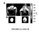
- CTX:Cy5.5 was tested in a genetically precise autochthonous mouse model of medulloblastoma. In these mice, medulloblastomas arise in the cerebellum due to transgenic expression of constitutively active smoothened, a mediator of sonic hedgehog activity. In these mice, tumors were detected through intact skull and scalp and the level of NIR emission correlated with the size of each tumor ( FIGURES 3A-D and not shown). Because these tumors arise in the absence of surgical disruption of the blood brain barrier, it was concluded that CTX:Cy5.5 enters mouse medulloblastoma tissue at concentrations suitable for real time imaging and that intracranial tumors can be readily detected noninvasively.
- Prostate tissue imaging Normal and cancerous human prostate tissue samples were collected and handled in accordance with Human Subjects IRB approved protocols. Sections were incubated with 1 ⁇ M CTX:Cy5.5 conjugate in 5% normal goat serum buffer for 45 minutes. Unbound conjugate was reduced by washing slides 3 times for 5minutes in PBS buffer. Signal was detected by fluorescence microscopy and correlated to adjacent sections stained with hematoxylin and eosin (H&E).
- CTX:Cy5.5 binding to sections of surgically-derived human prostate cancer was compared to non-neoplastic prostate epithelium. No specific binding was observed in normal prostate tissue, whereas high levels of CTX:Cy5.5 were observed in islands of cancer cells ( FIGURE 4A ). Adjacent sections revealed perfect correlation between rests of cancer cells detected by CTX:Cy5.5 and standard histologic staining ( FIGURE 4A ).
- CTX:Cy5.5 binding to human prostate cancer sections lead to evaluation of the conjugate in a GEM model of prostate cancer.
- CTX:Cy5.5 illuminated the primary prostate cancer as well as lung and lymphatic metastases ( FIGURES 4B-D ).
- CTX:Cy5.5 binding correlated with the histological presence of neoplastic cells in the primary and distant organs ( FIGURE 4C ). Microscopic foci of cancer cells in lymphatic channels and lymph nodes were easily detected ( FIGURES 4D-E ).
- CTX:Cy5.5 binding in adenocarcinoma and sarcoma was evaluated.
- FAP familial adenomatous polyposis
- Apc 1638N mice intestinal neoplasms could not be resolved by noninvasive imaging because of autofluorescence of the intestinal contents.
- Ex vivo analysis of intestine that was free of intestinal contents showed extremely delineation of 3-5 mm diameter cancer nodules and no appreciable signal from otherwise normal intestine ( FIGURES 5A-C ).
- Microscopic analyses of the rhabdomyosarcoma xenograft showed that virtually every cancer cell was brightly illuminated by CTX:Cy5.5 ( FIGURES 5D-F ).
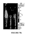
- CTX and Cy5.5 have previously been administered to human patients in clinical trials, but the pharmacokinetic properties of the conjugate have not previously been studied. Unbound conjugates were fairly evenly distributed through the mouse body during the first 24-96 hours, with the exception that NIR emission from kidney was much higher than any other organ. Confocal microscopy showed that CTX:Cy5.5 was concentrated in the renal collecting system as expected for a compound that is primarily excreted in urine. No other organ showed specific binding of CTX:Cy5.5.
- CTX was previously reported to reduce locomotor activity in mice. In this study, mice that were injected with CTX were indistinguishable from control mice in open field observation. To further assess potential CTX toxicity at doses used for optical imaging, complete necropsy was performed on mice two weeks after exposure to CTX. Brain, heart, lungs, kidneys, liver, spleen, and skin were indistinguishable from control mice (not shown). Laboratory evaluation showed no alteration in hematocrit, platelet count, white blood cell count, electrolytes, liver function or kidney function (not shown). At doses used for optical imaging, no toxicity could be detected in mice.
- MMP-2 The involvement of MMP-2 in chlorotoxin conjugate binding is described in this example.
- MMP-2 activity was analyzed by gelatinase assays. Gelatinase assays also detect MMP-9 activity. These analyses showed that all tumors that bound CTX:Cy5.5 express MMP-2 and MMP-9 and that autochthanous tumors in genetically engineered medulloblastoma and prostate cancer models also express MMP-2, but not MMP-9 (see FIGURE 11 ). Non-neoplastic tissue from mice did not show MMP-2 or MMP-9 activity. This experiment showed that MMP-2 was expressed in tissues that bound CTX:Cy5.5, but not in non-neoplastic tissues that showed only background CTX:Cy5.5 binding.
- MMP-2 reduced CTX:Cy5.5 binding in vitro and in vivo experiments suggesting that MMP-2 may be important for CTX:Cy5.5 binding (see FIGURE 2F and not shown).
- FIGURE 2F To further, definitively, determine whether MMP-2 was involved in CTX:Cy5.5 binding, a genetic approach was taken.
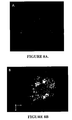
- MCF-7 cells which minimally express MMP-2 and minimally bind CTX:Cy5.5 (see FIGURES 11 and 8 ) were transfected with a plasmid that encodes MMP-2. Following transfection, cells expressed high levels of MMP-2 as shown in green in FIGURE 8 .
- MCF-7 cells transfected with MMP-2 plasmid but not those transfected with control plasmid bound high levels of CTX:Cy5.5 and this signal highly localized with MMP-2 staining. This clearly demonstrates that MMP-2 is important for CTX:Cy5.5 binding.
- CTX:Cy5.5 binding in MCF7 and HeLa cells was compared under the following three conditions.
- Condition (1) CTX:Cy5.5 was added directly to MCF7 or HeLa media and incubated for 24 hours under conditions that resulted in high CTX:Cy5.5 binding to 9L or U87 cells. There was minimal staining of MCF7 or HeLa cells under these conditions.
- Condition (2) the same amount of CTX:Cy5.5 was added to media of 9L or U87 cells, incubated for 24 hours, then transferred to MCF7 or HeLa cells.
- CTX:Cy5.5 When conditioned media from either 9L or U87 cells was used for staining, both MCF7 and HeLa cells were brightly stained by CTX:Cy5.5 (see FIGURE 13 ). Because the amount of MMP2 does not change in MCF7 or HeLa cells due to the presence of conditioned media, CTX:Cy5.5 ultimately binds to a target other than MMP-2. There are two major possibilities that are currently being studied: (1) MMP-2 enzymatically alters CTX:Cy5.5 so that it gains affinity for another cellular target; or (2) MMP-2 enzymatically alters molecule(s) in MCF7 or HeLa cells so that they gain affinity for CTX:Cy5.5.
- MMP-2 was shown as important for CTX:Cy5.5 binding, but that it may not be the ultimate cellular target of CTX:Cy5.5.
- CTX:Cy5.5 wa observed to bind to a rapidly migrating band on Western analyses that was consistent with a phospholipid band. This raised the possibility that CTX:Cy5.5 bound to a phospholipid.
- Sphingomyelin and phosphatidylcholine are present in all cells and only a subset of MCF7 and HeLa cells treated with CTX:Cy5.5 conditioned media from 9L and U87 cells showed high levels of signal.
Landscapes
- Health & Medical Sciences (AREA)
- Life Sciences & Earth Sciences (AREA)
- Engineering & Computer Science (AREA)
- General Health & Medical Sciences (AREA)
- Chemical & Material Sciences (AREA)
- Veterinary Medicine (AREA)
- Public Health (AREA)
- Animal Behavior & Ethology (AREA)
- Biomedical Technology (AREA)
- Epidemiology (AREA)
- Proteomics, Peptides & Aminoacids (AREA)
- Molecular Biology (AREA)
- Medicinal Chemistry (AREA)
- Organic Chemistry (AREA)
- Insects & Arthropods (AREA)
- Toxicology (AREA)
- Bioinformatics & Cheminformatics (AREA)
- Biochemistry (AREA)
- Pharmacology & Pharmacy (AREA)
- Immunology (AREA)
- Surgery (AREA)
- Genetics & Genomics (AREA)
- Tropical Medicine & Parasitology (AREA)
- Gastroenterology & Hepatology (AREA)
- Nuclear Medicine, Radiotherapy & Molecular Imaging (AREA)
- Zoology (AREA)
- Biophysics (AREA)
- Urology & Nephrology (AREA)
- Hematology (AREA)
- Cell Biology (AREA)
- Heart & Thoracic Surgery (AREA)
- Orthopedic Medicine & Surgery (AREA)
- Medical Informatics (AREA)
- Vascular Medicine (AREA)
- Food Science & Technology (AREA)
- Biotechnology (AREA)
- Microbiology (AREA)
- General Chemical & Material Sciences (AREA)
- Chemical Kinetics & Catalysis (AREA)
- Physics & Mathematics (AREA)
Claims (16)
- Chlorotoxin-Konjugat, bestehend aus einer oder mehreren Cy5.5-Einheiten, welche kovalent an ein Chlorotoxin gekuppelt sind.
- Konjugat nach Anspruch 1, wobei das Chlorotoxin aus der Gruppe ausgewählt ist, welche aus nativem Chlorotoxin, synthetischem Chlorotoxin und rekombinantem Chlorotoxin besteht.
- Konjugat nach Anspruch 1, wobei das Konjugat 1 bis 3 Cy5.5-Einheiten umfasst.
- Konjugat nach Anspruch 1, wobei das Konjugat 1 Cy5.5-Einheit umfasst.
- Zusammensetzung, umfassend ein Chlorotoxin-Konjugat gemäß einem vorstehenden Anspruch, wobei das Konjugat zur Verabreichung an einen Menschen geeignet ist.
- In-vitro-Verfahren zum Unterscheiden von Krebsherden, welche Chlorotoxin-Bindungsstellen exprimieren, von nicht-neoplastischem Gewebe, umfassend:(a) das Inkontaktbringen eines Gewebes von Interesse mit einem Chlorotoxin-Konjugat gemäß einem der Ansprüche 1 bis 4 und mit Affinität und Spezifität für Zellen, welche Chlorotoxin-Bindungsstellen exprimieren;(b) das Messen des Ausmaßes der Bindung des Chlorotoxin-Konjugats;wobei ein erhöhtes Ausmaß der Bindung relativ zu normalem Gewebe anzeigt, dass das Gewebe neoplastisch ist.
- In-vitro-Verfahren zum Nachweisen von Krebsarten, welche Chlorotoxin-Bindungsstellen exprimieren, umfassend:(a) das Inkontaktbringen eines Gewebes von Interesse mit einem Chlorotoxin-Konjugat gemäß einem der Ansprüche 1 bis 4 und mit Affinität und Spezifität für Zellen, welche Chlorotoxin-Bindungsstellen exprimieren; und(b) das Messen des Ausmaßes der Bindung des Chlorotoxin-Konjugats;wobei ein erhöhtes Ausmaß der Bindung relativ zu normalem Gewebe anzeigt, dass das Gewebe neoplastisch ist.
- In-vitro-Verfahren zum Nachweisen von Zellen, welche Matrixmetallproteinase (MMP-2)-Proteinkomplex exprimieren, umfassend:(a) das Inkontaktbringen eines Gewebes von Interesse mit einem Chlorotoxin-Konjugat gemäß einem der Ansprüche 1 bis 4 und mit Affinität und Spezifität für Matrixmetallproteinase (MMP-2)-Proteinkomplex; und(b) das Messen des Ausmaßes der Bindung des Chlorotoxin-Konjugats;wobei ein erhöhtes Ausmaß der Bindung relativ zu normalem Gewebe die Gegenwart eines Tumors, welcher Matrixmetallproteinase (MMP-2)-Proteinkomplex exprimiert, anzeigt.
- In-vitro-Verfahren zum Bestimmen des Ortes von Krebszellen, welche Chlorotoxin-Bindungsstellen exprimieren, in einem Patienten, umfassend:das Messen durch Fluoreszenz-Bildgebung des Ausmaßes der Bindung eines Chlorotoxin-Konjugats gemäß einem der Ansprüche 1 bis 4 in einer Probe, welche von dem Patienten intra-operativ erhalten wird, um den Ort von Krebszellen, welche Chlorotoxin-Bindungsstellen exprimieren, zu bestimmen,wobei ein erhöhtes Ausmaß der Bindung relativ zu normalem Gewebe die Gegenwart von Krebszellen, welche Chlorotoxin-Bindungsstellen exprimieren, anzeigt.
- Chlorotoxin-Konjugat gemäß einem der Ansprüche 1 bis 4 oder Zusammensetzung gemäß Anspruch 5 zur Verwendung in der Behandlung eines Krebses, welcher Chlorotoxin-Bindungsstellen exprimiert.
- Chlorotoxin-Konjugat gemäß einem der Ansprüche 1 bis 4 oder Zusammensetzung gemäß Anspruch 5 zur Verwendung in der Behandlung eines Tumors, welcher Matrixmetallproteinase (MMP-2)-Proteinkomplex exprimiert.
- Chlorotoxin-Konjugat gemäß einem der Ansprüche 1 bis 4 oder Zusammensetzung gemäß Anspruch 5 zur Verwendung bei der Inhibierung von invasiver Aktivität von Zellen, welche Chlorotoxin-Bindungsstellen exprimieren.
- Chlorotoxin-Konjugat gemäß einem der Ansprüche 1 bis 4 oder Zusammensetzung gemäß Anspruch 5 zur Verwendung beim:(i) Unterscheiden von Krebsherden, welche Chlorotoxin-Bindungsstellen exprimieren, von nicht-neoplastischem Gewebe;(ii) Nachweisen von Krebsarten, welche Chlorotoxin-Bindungsstellen exprimieren; oder(iii) intra-operativen Bestimmen des Ortes von Krebszellen, welche Chlorotoxin-Bindungsstellen exprimieren, in einem Patienten.
- Verfahren nach den Ansprüchen 6, 7 oder 9 oder Chlorotoxin-Konjugat oder Zusammensetzung zur Verwendung gemäß den Ansprüchen 10, 12 oder 13, wobei der Krebs, welcher Chlorotoxin-Bindungsstellen exprimiert, aus Gliomen, Astrozytomen, Medulloblastomen, Plexus choroideus-Karzinomen, Ependymozytomen, Gehirntumoren, Neuroblastomen, Kopf- und Halskrebs, Lungenkrebs, Brustkrebs, Darmkrebs, Bauchspeicheldrüsenkrebs, Leberkrebs, Nierenkrebs, Sarkomen, Osteosarkomen, Rhabdomyosarkomen, Ewing-Sarkom, Karzinomen, Melanomen, Eierstockkrebs, Gebärmutterhalskrebs, Lymphomen, Schilddrüsenkrebs, Afterkrebs, kolorektalem Krebs, Endometriumkrebs, Keimzelltumoren, Kehlkopfkrebs, multiplen Myelomen, Prostatakrebs, Retinoblastomen, Magenkrebs, Hodenkrebs und Wilm-Tumor ausgewählt ist.
- Verfahren nach den Ansprüchen 6, 7 oder 9 oder Chlorotoxin-Konjugat oder Zusammensetzung zur Verwendung gemäß den Ansprüchen 10, 12 oder 13, wobei die Krebsart, welche Chlorotoxin-Bindungsstellen exprimiert, aus Gliomen, Medulloblastomen, Sarkomen, Prostatakrebs und Darmkrebs ausgewählt ist.
- Chlorotoxin-Konjugat gemäß einem der Ansprüche 1 bis 4 oder Zusammensetzung gemäß Anspruch 5 zur Verwendung beim Nachweisen von Zellen, welche Matrixmetallproteinase (MMP-2)-Proteinkomplex exprimieren.
Applications Claiming Priority (2)
| Application Number | Priority Date | Filing Date | Title |
|---|---|---|---|
| US67428005P | 2005-04-22 | 2005-04-22 | |
| PCT/US2006/010170 WO2006115633A2 (en) | 2005-04-22 | 2006-03-20 | Cyanine-chlorotoxin conjugate and method for intra-operative fluorescent visualization of cancer |
Publications (2)
| Publication Number | Publication Date |
|---|---|
| EP1877428A2 EP1877428A2 (de) | 2008-01-16 |
| EP1877428B1 true EP1877428B1 (de) | 2011-03-16 |
Family
ID=36609637
Family Applications (1)
| Application Number | Title | Priority Date | Filing Date |
|---|---|---|---|
| EP06739100A Expired - Lifetime EP1877428B1 (de) | 2005-04-22 | 2006-03-20 | Fluoreszentes chlorotoxinkonjugat und verfahren zur intraoperativen sichtbarmachung von krebs |
Country Status (6)
| Country | Link |
|---|---|
| US (3) | US20070154965A1 (de) |
| EP (1) | EP1877428B1 (de) |
| AT (1) | ATE502048T1 (de) |
| DE (1) | DE602006020710D1 (de) |
| ES (1) | ES2360519T3 (de) |
| WO (1) | WO2006115633A2 (de) |
Families Citing this family (24)
| Publication number | Priority date | Publication date | Assignee | Title |
|---|---|---|---|---|
| US5905027A (en) * | 1995-12-27 | 1999-05-18 | Uab Research Foundation | Method of diagnosing and treating gliomas |
| US20060088899A1 (en) * | 2002-05-31 | 2006-04-27 | Alvarez Vernon L | Combination chemotherapy with chlorotoxin |
| DE602006020710D1 (de) * | 2005-04-22 | 2011-04-28 | Fred Hutchinson Cancer Res Foundation | Fluoreszentes chlorotoxinkonjugat und verfahren zur intraoperativen sichtbarmachung von krebs |
| CA2616147A1 (en) | 2006-02-24 | 2007-09-20 | Mallinckrodt Inc. | Process for using optical agents |
| US20090098574A1 (en) * | 2006-04-25 | 2009-04-16 | Centre National De La Recherche Scientifique (Cnrs) | Functionalization of gold nanoparticles with oriented proteins, application to the high-density labelling of cell membranes |
| ATE551073T1 (de) * | 2007-05-16 | 2012-04-15 | Ge Healthcare As | Markierte hgf-bindende peptide für bildgebung |
| CA2694974C (en) * | 2007-07-30 | 2016-11-08 | University Of Maryland, Baltimore | Methods and devices for the detection of biofilm |
| EP2173385A1 (de) * | 2007-08-07 | 2010-04-14 | Transmolecular, Inc. | Chlorotoxine als wirkstoffträger |
| US20100215575A1 (en) * | 2007-10-12 | 2010-08-26 | Transmolecular, Inc. | Systemic Administration of Chlorotoxin Agents for the Diagnosis and Treatment of Tumors |
| EP3115469B1 (de) * | 2007-11-19 | 2020-04-29 | Celera Corporation | Lungenkrebsmarker und verwendungen davon |
| WO2009126835A2 (en) * | 2008-04-09 | 2009-10-15 | University Of Washington Techtransfer Invention Licensing | Magnetic nanoparticle and method for imaging t cells |
| JP5497006B2 (ja) * | 2008-04-14 | 2014-05-21 | ザ ジェネラル ホスピタル コーポレイション | 膵管腺癌の検出および治療のためのプレクチン−1標的化剤 |
| CA2724384A1 (en) | 2008-05-15 | 2009-11-19 | Transmolecular, Inc. | Treatment of metastatic tumors |
| WO2010028013A2 (en) * | 2008-09-02 | 2010-03-11 | University Of Maryland, Baltimore | In vivo biofilm infection diagnosis and treatment |
| CN102844044B (zh) | 2010-02-04 | 2016-10-26 | 摩尔弗泰克有限公司 | 氯毒素多肽和结合物及其应用 |
| EP3165533B1 (de) * | 2010-05-11 | 2020-04-08 | Fred Hutchinson Cancer Research Center | Chlorotoxin-varianten und -konjugate sowie verfahren zu deren verwendung |
| WO2013003507A1 (en) * | 2011-06-27 | 2013-01-03 | Morphotek, Inc. | Multifunctional agents |
| WO2013166487A1 (en) | 2012-05-04 | 2013-11-07 | Yale University | Highly penetrative nanocarriers for treatment of cns disease |
| CA2913029A1 (en) | 2012-12-10 | 2014-06-19 | Fred Hutchinson Cancer Research Center | Lipocalin fusion partners |
| US9784730B2 (en) | 2013-03-21 | 2017-10-10 | University Of Washington Through Its Center For Commercialization | Nanoparticle for targeting brain tumors and delivery of O6-benzylguanine |
| US11559580B1 (en) | 2013-09-17 | 2023-01-24 | Blaze Bioscience, Inc. | Tissue-homing peptide conjugates and methods of use thereof |
| BR112016005802A2 (pt) * | 2013-09-17 | 2017-09-12 | Blaze Bioscience Inc | conjugados de clorotoxina e métodos de seu uso |
| WO2017180789A2 (en) | 2016-04-12 | 2017-10-19 | Blaze Bioscience, Inc. | Methods of treatment using chlorotoxin conjugates |
| US12048732B2 (en) | 2016-04-15 | 2024-07-30 | Blaze Bioscience, Inc. | Methods of treating breast cancer |
Family Cites Families (44)
| Publication number | Priority date | Publication date | Assignee | Title |
|---|---|---|---|---|
| DE3407582A1 (de) | 1984-03-01 | 1985-09-05 | Dornier System Gmbh, 7990 Friedrichshafen | Schaltungsanordnung fuer einen regelkreis |
| US6667156B2 (en) * | 1995-12-27 | 2003-12-23 | Uab Research Foundation | Diagnosis and treatment of neuroectodermal tumors |
| US5905027A (en) * | 1995-12-27 | 1999-05-18 | Uab Research Foundation | Method of diagnosing and treating gliomas |
| US6130101A (en) * | 1997-09-23 | 2000-10-10 | Molecular Probes, Inc. | Sulfonated xanthene derivatives |
| US6703381B1 (en) * | 1998-08-14 | 2004-03-09 | Nobex Corporation | Methods for delivery therapeutic compounds across the blood-brain barrier |
| CA2384429A1 (en) * | 1999-09-14 | 2001-03-22 | Michael K. Bahr | Magnetic nanoparticles having biochemical activity, method for the production thereof and their use |
| WO2001037721A2 (en) * | 1999-11-22 | 2001-05-31 | The Research Foundation Of State University Of New York | Magnetic nanoparticles for selective therapy |
| DE10020376A1 (de) * | 2000-04-26 | 2001-11-08 | Inst Zelltechnologie E V | Dynamische Marker |
| US20030021810A1 (en) | 2001-06-26 | 2003-01-30 | Sontheimer Harald W. | Chlorotoxin inhibition of cell invasion, cancer metastasis, angiogenesis and tissue remodeling |
| US6972326B2 (en) * | 2001-12-03 | 2005-12-06 | Molecular Probes, Inc. | Labeling of immobilized proteins using dipyrrometheneboron difluoride dyes |
| US20060088899A1 (en) * | 2002-05-31 | 2006-04-27 | Alvarez Vernon L | Combination chemotherapy with chlorotoxin |
| AU2003237347B2 (en) | 2002-05-31 | 2009-07-23 | Transmolecular Inc. | Treatment of cell proliferative disorders with chlorotoxin |
| US7560160B2 (en) * | 2002-11-25 | 2009-07-14 | Materials Modification, Inc. | Multifunctional particulate material, fluid, and composition |
| US20040101822A1 (en) * | 2002-11-26 | 2004-05-27 | Ulrich Wiesner | Fluorescent silica-based nanoparticles |
| DE10328251A1 (de) | 2003-06-24 | 2005-01-13 | Toximed Gmbh | Pharmazeutischer Wirkstoff |
| WO2005053611A2 (en) | 2003-11-26 | 2005-06-16 | Transmolecular, Inc. | Treatment of phosphatidylinositol phospholipid disorders |
| AU2005232616A1 (en) * | 2004-04-06 | 2005-10-27 | Transmolecular, Inc. | Diagnosis and treatment of myeloid and lymphoid cell cancers |
| US20080153746A1 (en) | 2004-04-06 | 2008-06-26 | Transmolecular, Inc. | Diagnosis and Treatment of Myeloid and Lymphoid Cell Cancers |
| GB0422901D0 (en) | 2004-10-14 | 2004-11-17 | Ares Trading Sa | Lipocalin protein |
| GB0504767D0 (en) | 2005-03-08 | 2005-04-13 | Ares Trading Sa | Lipocalin protein |
| US7462446B2 (en) * | 2005-03-18 | 2008-12-09 | University Of Washington | Magnetic nanoparticle compositions and methods |
| DE602006020710D1 (de) | 2005-04-22 | 2011-04-28 | Fred Hutchinson Cancer Res Foundation | Fluoreszentes chlorotoxinkonjugat und verfahren zur intraoperativen sichtbarmachung von krebs |
| AU2005335491B2 (en) * | 2005-08-18 | 2010-11-25 | Ambrx, Inc. | Compositions of tRNA and uses thereof |
| CA2647907A1 (en) | 2006-03-31 | 2007-10-18 | Transmolecular, Inc. | Use of tm-601 for the diagnosis and treatment of tumors |
| US8106013B2 (en) * | 2006-05-19 | 2012-01-31 | Georgia Tech Research Corporation | ABC transporter ligand GATX1 |
| CN100564517C (zh) | 2006-09-21 | 2009-12-02 | 武汉大学 | 一种蝎抗神经胶质瘤肽及其制备方法和应用 |
| CN101003788A (zh) | 2006-09-21 | 2007-07-25 | 武汉大学 | 一种蝎抗肿瘤转移肽及其制备方法和应用 |
| PE20081140A1 (es) | 2006-10-25 | 2008-09-22 | Amgen Inc | Agentes terapeuticos a base de peptidos derivados de toxinas |
| US20090004105A1 (en) * | 2007-06-27 | 2009-01-01 | Zhen Cheng | Molecular imaging of matrix metalloproteinase expression using labeled chlorotoxin |
| EP2173385A1 (de) | 2007-08-07 | 2010-04-14 | Transmolecular, Inc. | Chlorotoxine als wirkstoffträger |
| US20100215575A1 (en) * | 2007-10-12 | 2010-08-26 | Transmolecular, Inc. | Systemic Administration of Chlorotoxin Agents for the Diagnosis and Treatment of Tumors |
| US8470607B2 (en) * | 2008-03-20 | 2013-06-25 | Morphotek, Inc. | Inhibition of angiogenesis |
| CN101270158B (zh) | 2008-04-30 | 2010-12-22 | 武汉大学 | 一种靶向抗神经胶质瘤蛋白及制备方法和用途 |
| CA2724384A1 (en) * | 2008-05-15 | 2009-11-19 | Transmolecular, Inc. | Treatment of metastatic tumors |
| DK3381933T3 (da) | 2008-06-24 | 2020-08-03 | Technische Universität München | Muteiner af hngal og relaterede proteiner med affinitet til et givet mål |
| US20100098637A1 (en) * | 2008-09-23 | 2010-04-22 | The Regents Of The University Of Michigan | Dye-loaded nanoparticle |
| CN101381405B (zh) | 2008-09-24 | 2011-07-27 | 武汉摩尔生物科技有限公司 | 基因工程肿瘤靶向kct-w1多肽及制备方法和用途 |
| CN106519588B (zh) | 2009-11-09 | 2019-08-20 | 华盛顿大学商业化中心 | 官能化发色聚合物点及其生物共轭体 |
| CN101921769B (zh) | 2010-01-11 | 2012-07-25 | 山西大学 | 一种重组腺病毒及其制备方法和应用 |
| WO2011094671A2 (en) | 2010-01-29 | 2011-08-04 | The Uab Research Foundation | N-terminally conjugated polypeptides for targeted therapy and diagnosis |
| CN102844044B (zh) * | 2010-02-04 | 2016-10-26 | 摩尔弗泰克有限公司 | 氯毒素多肽和结合物及其应用 |
| CN101824084A (zh) | 2010-04-08 | 2010-09-08 | 山西大学 | 靶向性抗胶质瘤蛋白质及其应用 |
| EP3165533B1 (de) | 2010-05-11 | 2020-04-08 | Fred Hutchinson Cancer Research Center | Chlorotoxin-varianten und -konjugate sowie verfahren zu deren verwendung |
| EP3299386A1 (de) | 2010-08-16 | 2018-03-28 | Pieris Pharmaceuticals GmbH | Bindungsproteine für hepcidin |
-
2006
- 2006-03-20 DE DE602006020710T patent/DE602006020710D1/de not_active Expired - Lifetime
- 2006-03-20 EP EP06739100A patent/EP1877428B1/de not_active Expired - Lifetime
- 2006-03-20 ES ES06739100T patent/ES2360519T3/es not_active Expired - Lifetime
- 2006-03-20 WO PCT/US2006/010170 patent/WO2006115633A2/en not_active Ceased
- 2006-03-20 AT AT06739100T patent/ATE502048T1/de active
- 2006-11-14 US US11/600,004 patent/US20070154965A1/en not_active Abandoned
-
2007
- 2007-08-31 US US11/897,721 patent/US8778310B2/en active Active
-
2014
- 2014-05-08 US US14/273,374 patent/US20140241993A1/en not_active Abandoned
Non-Patent Citations (3)
| Title |
|---|
| Annual Report 2004 by the "Joint Institute for Nanoscience", Eds. D. B. Baer, C. T. Campbell, November 2005. "Smart Superparamagnetic Imaging Probes for Brain Tumor Research", J. Gunn. * |
| EGLETON R. D., DAVIS T. P.: "Development of Neuropeptide Drugs that Cross the Blood-Brain Barrier", J AM SOC EXP NEUROTHERAPEUTICS, vol. 2, January 2005 (2005-01-01), pages 44 - 53, XP025343798 * |
| TATIKOLOV A S; COSTA S M B: "Complexation of polymethine dyes with human serum albumin: a spectroscopic study", BIOPHYSICAL CHEMISTRY, vol. 107, 1 January 2004 (2004-01-01), pages 33 - 49, XP002381135 * |
Also Published As
| Publication number | Publication date |
|---|---|
| WO2006115633A3 (en) | 2006-12-28 |
| WO2006115633A2 (en) | 2006-11-02 |
| US20070154965A1 (en) | 2007-07-05 |
| US8778310B2 (en) | 2014-07-15 |
| US20140241993A1 (en) | 2014-08-28 |
| US20080279780A1 (en) | 2008-11-13 |
| DE602006020710D1 (de) | 2011-04-28 |
| EP1877428A2 (de) | 2008-01-16 |
| ES2360519T3 (es) | 2011-06-06 |
| ATE502048T1 (de) | 2011-04-15 |
Similar Documents
| Publication | Publication Date | Title |
|---|---|---|
| US8778310B2 (en) | Fluorescent chlorotoxin conjugate and method for intra-operative visualization of cancer | |
| Veiseh et al. | Tumor paint: a chlorotoxin: Cy5. 5 bioconjugate for intraoperative visualization of cancer foci | |
| Kularatne et al. | Evaluation of novel prostate-specific membrane antigen-targeted near-infrared imaging agent for fluorescence-guided surgery of prostate cancer | |
| Steinmetz et al. | Intravital imaging of human prostate cancer using viral nanoparticles targeted to gastrin‐releasing peptide receptors | |
| US10746741B2 (en) | CA IX—NIR dyes and their uses | |
| R. Stroud et al. | In vivo bio-imaging using chlorotoxin-based conjugates | |
| CN107206108B (zh) | 术中成像 | |
| Boogerd et al. | Fluorescence-guided tumor detection with a novel anti-EpCAM targeted antibody fragment: preclinical validation | |
| He et al. | A novel peptide probe for imaging and targeted delivery of liposomal doxorubicin to lung tumor | |
| JP2004501664A (ja) | 標的化薬剤送達のための腫瘍組織を浸潤する細胞特異的な内部移行ペプチドの単離 | |
| Citrin et al. | In vivo tumor imaging in mice with near-infrared labeled endostatin | |
| CN114933633B (zh) | 一种特异性识别fgfr4的天然肽探针及其应用 | |
| Ni et al. | Transferrin receptor 1 targeted optical imaging for identifying glioma margin in mouse models | |
| CN110831634B (zh) | 磷脂酰肌醇蛋白聚糖-3肽试剂和方法 | |
| CN108721649A (zh) | 一种肿瘤靶向近红外荧光显像剂的设计、合成及应用 | |
| Trac et al. | MRI Detection of Lymph Node Metastasis through Molecular Targeting of C–C Chemokine Receptor Type 2 and Monocyte Hitchhiking | |
| Bahng et al. | In vivo targeting of c-Met using a non-standard macrocyclic peptide in gastric carcinoma | |
| Liu et al. | Near-infrared fluorescent peptides with high tumor selectivity: novel probes for image-guided surgical resection of orthotopic glioma | |
| Hou et al. | In vivo self-assembled bispecific fluorescence probe for early detection of bladder cancer and metastasis | |
| Gazzi et al. | DOTA‐Based Plectin‐1 Targeted Contrast Agent Enables Detection of Pancreatic Cancer in Human Tissue | |
| Chirizzi et al. | Next‐generation agents for fluorescence‐guided glioblastoma surgery | |
| Li et al. | Near‐Infrared Organic Room Temperature Phosphorescent Probes Targeting Fibroblast Activation Protein for Surgical Navigation of Liver Cancer | |
| Guo et al. | A fluorescent,[18F]-Positron-Emitting agent for imaging prostate-specific membrane antigen allows genetic reporting in adoptively transferred, genetically modified cells | |
| Chopra | ZW800-1, a zwitterionic near-infrared fluorophore, and its cyclic RGD peptide derivative cyclo-(RGDyK)-ZW800-1 | |
| US11555821B2 (en) | CA IX-NIR dyes and their uses |
Legal Events
| Date | Code | Title | Description |
|---|---|---|---|
| PUAI | Public reference made under article 153(3) epc to a published international application that has entered the european phase |
Free format text: ORIGINAL CODE: 0009012 |
|
| 17P | Request for examination filed |
Effective date: 20071120 |
|
| AK | Designated contracting states |
Kind code of ref document: A2 Designated state(s): AT BE BG CH CY CZ DE DK EE ES FI FR GB GR HU IE IS IT LI LT LU LV MC NL PL PT RO SE SI SK TR |
|
| DAX | Request for extension of the european patent (deleted) | ||
| RIN1 | Information on inventor provided before grant (corrected) |
Inventor name: GABIKIAN, PATRICK Inventor name: OLSON, JAMES Inventor name: VEISEH, MANDANA Inventor name: SZE, RAYMOND Inventor name: BAHRAMI, S-BAHRAM Inventor name: ZHANG, MIQIN Inventor name: VEISEH, OMID Inventor name: ELLENBOGEN, RICHARD |
|
| 17Q | First examination report despatched |
Effective date: 20080718 |
|
| GRAP | Despatch of communication of intention to grant a patent |
Free format text: ORIGINAL CODE: EPIDOSNIGR1 |
|
| GRAS | Grant fee paid |
Free format text: ORIGINAL CODE: EPIDOSNIGR3 |
|
| GRAA | (expected) grant |
Free format text: ORIGINAL CODE: 0009210 |
|
| RIN1 | Information on inventor provided before grant (corrected) |
Inventor name: VEISEH, MANDANA Inventor name: OLSON, JAMES Inventor name: BAHRAMI, S-BAHRAM Inventor name: GABIKIAN, PATRIK Inventor name: ELLENBOGEN, RICHARD Inventor name: SZE, RAYMOND Inventor name: VEISEH, OMID Inventor name: ZHANG, MIQIN |
|
| AK | Designated contracting states |
Kind code of ref document: B1 Designated state(s): AT BE BG CH CY CZ DE DK EE ES FI FR GB GR HU IE IS IT LI LT LU LV MC NL PL PT RO SE SI SK TR |
|
| REG | Reference to a national code |
Ref country code: GB Ref legal event code: FG4D |
|
| BECN | Be: change of holder's name |
Owner name: FRED HUTCHINSON CANCER RESEARCH CENTER Effective date: 20110316 Owner name: UNIVERSITY OF WASHINGTON Effective date: 20110316 |
|
| REG | Reference to a national code |
Ref country code: CH Ref legal event code: EP |
|
| RAP2 | Party data changed (patent owner data changed or rights of a patent transferred) |
Owner name: UNIVERSITY OF WASHINGTON Owner name: FRED HUTCHINSON CANCER RESEARCH CENTER |
|
| REG | Reference to a national code |
Ref country code: IE Ref legal event code: FG4D |
|
| REF | Corresponds to: |
Ref document number: 602006020710 Country of ref document: DE Date of ref document: 20110428 Kind code of ref document: P |
|
| REG | Reference to a national code |
Ref country code: DE Ref legal event code: R096 Ref document number: 602006020710 Country of ref document: DE Effective date: 20110428 |
|
| REG | Reference to a national code |
Ref country code: CH Ref legal event code: NV Representative=s name: RENTSCH & PARTNER |
|
| REG | Reference to a national code |
Ref country code: NL Ref legal event code: T3 |
|
| REG | Reference to a national code |
Ref country code: ES Ref legal event code: FG2A Ref document number: 2360519 Country of ref document: ES Kind code of ref document: T3 Effective date: 20110606 |
|
| REG | Reference to a national code |
Ref country code: CH Ref legal event code: PFA Owner name: FRED HUTCHINSON CANCER RESEARCH CENTER Free format text: FRED HUTCHINSON CANCER RESEARCH CENTER#1100 FAIRWAY AVENUE NORTH#SEATTLE, WA 98109-1024 (US) $ UNIVERSITY OF WASHINGTON#4311 11TH AVENUE N.E., SUITE 500#SEATTLE, WA 98105-4608 (US) -TRANSFER TO- FRED HUTCHINSON CANCER RESEARCH CENTER#1100 FAIRWAY AVENUE NORTH#SEATTLE, WA 98109-1024 (US) $ UNIVERSITY OF WASHINGTON#4311 11TH AVENUE N.E., SUITE 500#SEATTLE, WA 98105-4608 (US) |
|
| PG25 | Lapsed in a contracting state [announced via postgrant information from national office to epo] |
Ref country code: GR Free format text: LAPSE BECAUSE OF FAILURE TO SUBMIT A TRANSLATION OF THE DESCRIPTION OR TO PAY THE FEE WITHIN THE PRESCRIBED TIME-LIMIT Effective date: 20110617 Ref country code: SE Free format text: LAPSE BECAUSE OF FAILURE TO SUBMIT A TRANSLATION OF THE DESCRIPTION OR TO PAY THE FEE WITHIN THE PRESCRIBED TIME-LIMIT Effective date: 20110316 Ref country code: LT Free format text: LAPSE BECAUSE OF FAILURE TO SUBMIT A TRANSLATION OF THE DESCRIPTION OR TO PAY THE FEE WITHIN THE PRESCRIBED TIME-LIMIT Effective date: 20110316 Ref country code: LV Free format text: LAPSE BECAUSE OF FAILURE TO SUBMIT A TRANSLATION OF THE DESCRIPTION OR TO PAY THE FEE WITHIN THE PRESCRIBED TIME-LIMIT Effective date: 20110316 |
|
| LTIE | Lt: invalidation of european patent or patent extension |
Effective date: 20110316 |
|
| PG25 | Lapsed in a contracting state [announced via postgrant information from national office to epo] |
Ref country code: SI Free format text: LAPSE BECAUSE OF FAILURE TO SUBMIT A TRANSLATION OF THE DESCRIPTION OR TO PAY THE FEE WITHIN THE PRESCRIBED TIME-LIMIT Effective date: 20110316 Ref country code: FI Free format text: LAPSE BECAUSE OF FAILURE TO SUBMIT A TRANSLATION OF THE DESCRIPTION OR TO PAY THE FEE WITHIN THE PRESCRIBED TIME-LIMIT Effective date: 20110316 Ref country code: CY Free format text: LAPSE BECAUSE OF FAILURE TO SUBMIT A TRANSLATION OF THE DESCRIPTION OR TO PAY THE FEE WITHIN THE PRESCRIBED TIME-LIMIT Effective date: 20110316 Ref country code: BG Free format text: LAPSE BECAUSE OF FAILURE TO SUBMIT A TRANSLATION OF THE DESCRIPTION OR TO PAY THE FEE WITHIN THE PRESCRIBED TIME-LIMIT Effective date: 20110616 |
|
| REG | Reference to a national code |
Ref country code: DE Ref legal event code: R081 Ref document number: 602006020710 Country of ref document: DE Owner name: FRED HUTCHINSON CANCER RESEARCH CENTER, 98109-, US Free format text: FORMER OWNER: FRED HUTCHINSON CANCER RESEARCH, UNIVERSITY OF WASHINGTON, , US Effective date: 20110905 Ref country code: DE Ref legal event code: R081 Ref document number: 602006020710 Country of ref document: DE Owner name: UNIVERSITY OF WASHINGTON, SEATTLE, US Free format text: FORMER OWNER: FRED HUTCHINSON CANCER RESEARCH, UNIVERSITY OF WASHINGTON, , US Effective date: 20110905 Ref country code: DE Ref legal event code: R081 Ref document number: 602006020710 Country of ref document: DE Owner name: FRED HUTCHINSON CANCER RESEARCH CENTER, US Free format text: FORMER OWNER: FRED HUTCHINSON CANCER RESEARCH, UNIVERSITY OF WASHINGTON, , US Effective date: 20110905 Ref country code: DE Ref legal event code: R081 Ref document number: 602006020710 Country of ref document: DE Owner name: UNIVERSITY OF WASHINGTON, US Free format text: FORMER OWNER: FRED HUTCHINSON CANCER RESEARCH, UNIVERSITY OF WASHINGTON, , US Effective date: 20110905 Ref country code: DE Ref legal event code: R081 Ref document number: 602006020710 Country of ref document: DE Owner name: UNIVERSITY OF WASHINGTON, SEATTLE, US Free format text: FORMER OWNERS: FRED HUTCHINSON CANCER RESEARCH FOUNDATION, SEATTLE, WASH., US; UNIVERSITY OF WASHINGTON, SEATTLE, WASH., US Effective date: 20110905 Ref country code: DE Ref legal event code: R081 Ref document number: 602006020710 Country of ref document: DE Owner name: FRED HUTCHINSON CANCER RESEARCH CENTER, 98109-, US Free format text: FORMER OWNERS: FRED HUTCHINSON CANCER RESEARCH FOUNDATION, SEATTLE, WASH., US; UNIVERSITY OF WASHINGTON, SEATTLE, WASH., US Effective date: 20110905 |
|
| PG25 | Lapsed in a contracting state [announced via postgrant information from national office to epo] |
Ref country code: EE Free format text: LAPSE BECAUSE OF FAILURE TO SUBMIT A TRANSLATION OF THE DESCRIPTION OR TO PAY THE FEE WITHIN THE PRESCRIBED TIME-LIMIT Effective date: 20110316 Ref country code: PT Free format text: LAPSE BECAUSE OF FAILURE TO SUBMIT A TRANSLATION OF THE DESCRIPTION OR TO PAY THE FEE WITHIN THE PRESCRIBED TIME-LIMIT Effective date: 20110718 Ref country code: MC Free format text: LAPSE BECAUSE OF NON-PAYMENT OF DUE FEES Effective date: 20110331 |
|
| PG25 | Lapsed in a contracting state [announced via postgrant information from national office to epo] |
Ref country code: IS Free format text: LAPSE BECAUSE OF FAILURE TO SUBMIT A TRANSLATION OF THE DESCRIPTION OR TO PAY THE FEE WITHIN THE PRESCRIBED TIME-LIMIT Effective date: 20110716 Ref country code: RO Free format text: LAPSE BECAUSE OF FAILURE TO SUBMIT A TRANSLATION OF THE DESCRIPTION OR TO PAY THE FEE WITHIN THE PRESCRIBED TIME-LIMIT Effective date: 20110316 Ref country code: SK Free format text: LAPSE BECAUSE OF FAILURE TO SUBMIT A TRANSLATION OF THE DESCRIPTION OR TO PAY THE FEE WITHIN THE PRESCRIBED TIME-LIMIT Effective date: 20110316 Ref country code: CZ Free format text: LAPSE BECAUSE OF FAILURE TO SUBMIT A TRANSLATION OF THE DESCRIPTION OR TO PAY THE FEE WITHIN THE PRESCRIBED TIME-LIMIT Effective date: 20110316 |
|
| PLBE | No opposition filed within time limit |
Free format text: ORIGINAL CODE: 0009261 |
|
| STAA | Information on the status of an ep patent application or granted ep patent |
Free format text: STATUS: NO OPPOSITION FILED WITHIN TIME LIMIT |
|
| 26N | No opposition filed |
Effective date: 20111219 |
|
| PG25 | Lapsed in a contracting state [announced via postgrant information from national office to epo] |
Ref country code: DK Free format text: LAPSE BECAUSE OF FAILURE TO SUBMIT A TRANSLATION OF THE DESCRIPTION OR TO PAY THE FEE WITHIN THE PRESCRIBED TIME-LIMIT Effective date: 20110316 Ref country code: PL Free format text: LAPSE BECAUSE OF FAILURE TO SUBMIT A TRANSLATION OF THE DESCRIPTION OR TO PAY THE FEE WITHIN THE PRESCRIBED TIME-LIMIT Effective date: 20110316 |
|
| REG | Reference to a national code |
Ref country code: DE Ref legal event code: R097 Ref document number: 602006020710 Country of ref document: DE Effective date: 20111219 |
|
| PG25 | Lapsed in a contracting state [announced via postgrant information from national office to epo] |
Ref country code: LU Free format text: LAPSE BECAUSE OF NON-PAYMENT OF DUE FEES Effective date: 20110320 |
|
| PG25 | Lapsed in a contracting state [announced via postgrant information from national office to epo] |
Ref country code: TR Free format text: LAPSE BECAUSE OF FAILURE TO SUBMIT A TRANSLATION OF THE DESCRIPTION OR TO PAY THE FEE WITHIN THE PRESCRIBED TIME-LIMIT Effective date: 20110316 |
|
| PG25 | Lapsed in a contracting state [announced via postgrant information from national office to epo] |
Ref country code: HU Free format text: LAPSE BECAUSE OF FAILURE TO SUBMIT A TRANSLATION OF THE DESCRIPTION OR TO PAY THE FEE WITHIN THE PRESCRIBED TIME-LIMIT Effective date: 20110316 |
|
| REG | Reference to a national code |
Ref country code: FR Ref legal event code: PLFP Year of fee payment: 10 |
|
| REG | Reference to a national code |
Ref country code: FR Ref legal event code: PLFP Year of fee payment: 11 |
|
| REG | Reference to a national code |
Ref country code: FR Ref legal event code: PLFP Year of fee payment: 12 |
|
| REG | Reference to a national code |
Ref country code: CH Ref legal event code: PCAR Free format text: NEW ADDRESS: BELLERIVESTRASSE 203 POSTFACH, 8034 ZUERICH (CH) |
|
| REG | Reference to a national code |
Ref country code: FR Ref legal event code: PLFP Year of fee payment: 13 |
|
| REG | Reference to a national code |
Ref country code: NL Ref legal event code: PD Owner name: FRED HUTCHINSON CANCER CENTER; US Free format text: DETAILS ASSIGNMENT: CHANGE OF OWNER(S), MERGE; FORMER OWNER NAME: UNIVERSITY OF WASHINGTON Effective date: 20220916 |
|
| REG | Reference to a national code |
Ref country code: GB Ref legal event code: 732E Free format text: REGISTERED BETWEEN 20220908 AND 20220914 |
|
| REG | Reference to a national code |
Ref country code: BE Ref legal event code: PD Owner name: UNIVERSITY OF WASHINGTON; US Free format text: DETAILS ASSIGNMENT: CHANGE OF OWNER(S), MERGE; FORMER OWNER NAME: FRED HUTCHINSON CANCER RESEARCH CENTER Effective date: 20220913 Ref country code: BE Ref legal event code: HC Owner name: UNIVERSITY OF WASHINGTON; US Free format text: DETAILS ASSIGNMENT: CHANGE OF OWNER(S), CHANGE OF OWNER(S) NAME; FORMER OWNER NAME: SEATTLE CANCER CARE ALLIANCE Effective date: 20220913 |
|
| REG | Reference to a national code |
Ref country code: AT Ref legal event code: PC Ref document number: 502048 Country of ref document: AT Kind code of ref document: T Owner name: FRED HUTCHINSON CANCER CENTER, US Effective date: 20221122 Ref country code: AT Ref legal event code: PC Ref document number: 502048 Country of ref document: AT Kind code of ref document: T Owner name: UNIVERSITY OF WASHINGTON, US Effective date: 20221122 |
|
| REG | Reference to a national code |
Ref country code: ES Ref legal event code: PC2A Owner name: FRED HUTCHINSON CANCER CENTER Effective date: 20230125 |
|
| REG | Reference to a national code |
Ref country code: DE Ref legal event code: R081 Ref document number: 602006020710 Country of ref document: DE Owner name: UNIVERSITY OF WASHINGTON, SEATTLE, US Free format text: FORMER OWNERS: FRED HUTCHINSON CANCER RESEARCH CENTER, 98109-1024 SEATTLE, WA, US; UNIVERSITY OF WASHINGTON, SEATTLE, WASH., US Ref country code: DE Ref legal event code: R081 Ref document number: 602006020710 Country of ref document: DE Owner name: FRED HUTCHINSON CANCER CENTER, SEATTLE, US Free format text: FORMER OWNERS: FRED HUTCHINSON CANCER RESEARCH CENTER, 98109-1024 SEATTLE, WA, US; UNIVERSITY OF WASHINGTON, SEATTLE, WASH., US |
|
| P01 | Opt-out of the competence of the unified patent court (upc) registered |
Effective date: 20230519 |
|
| PGFP | Annual fee paid to national office [announced via postgrant information from national office to epo] |
Ref country code: NL Payment date: 20240320 Year of fee payment: 19 Ref country code: IE Payment date: 20240321 Year of fee payment: 19 |
|
| PGFP | Annual fee paid to national office [announced via postgrant information from national office to epo] |
Ref country code: AT Payment date: 20240321 Year of fee payment: 19 |
|
| PGFP | Annual fee paid to national office [announced via postgrant information from national office to epo] |
Ref country code: DE Payment date: 20240320 Year of fee payment: 19 Ref country code: GB Payment date: 20240320 Year of fee payment: 19 |
|
| PGFP | Annual fee paid to national office [announced via postgrant information from national office to epo] |
Ref country code: IT Payment date: 20240329 Year of fee payment: 19 Ref country code: FR Payment date: 20240321 Year of fee payment: 19 Ref country code: BE Payment date: 20240320 Year of fee payment: 19 |
|
| PGFP | Annual fee paid to national office [announced via postgrant information from national office to epo] |
Ref country code: CH Payment date: 20240401 Year of fee payment: 19 |
|
| PGFP | Annual fee paid to national office [announced via postgrant information from national office to epo] |
Ref country code: ES Payment date: 20240426 Year of fee payment: 19 |
|
| REG | Reference to a national code |
Ref country code: DE Ref legal event code: R119 Ref document number: 602006020710 Country of ref document: DE |
|
| REG | Reference to a national code |
Ref country code: CH Ref legal event code: H13 Free format text: ST27 STATUS EVENT CODE: U-0-0-H10-H13 (AS PROVIDED BY THE NATIONAL OFFICE) Effective date: 20251023 |
|
| REG | Reference to a national code |
Ref country code: NL Ref legal event code: MM Effective date: 20250401 |
|
| REG | Reference to a national code |
Ref country code: AT Ref legal event code: MM01 Ref document number: 502048 Country of ref document: AT Kind code of ref document: T Effective date: 20250320 |
|
| GBPC | Gb: european patent ceased through non-payment of renewal fee |
Effective date: 20250320 |
|
| REG | Reference to a national code |
Ref country code: BE Ref legal event code: MM Effective date: 20250331 |
|
| PG25 | Lapsed in a contracting state [announced via postgrant information from national office to epo] |
Ref country code: NL Free format text: LAPSE BECAUSE OF NON-PAYMENT OF DUE FEES Effective date: 20250401 |
|
| PG25 | Lapsed in a contracting state [announced via postgrant information from national office to epo] |
Ref country code: DE Free format text: LAPSE BECAUSE OF NON-PAYMENT OF DUE FEES Effective date: 20251001 |
|
| PG25 | Lapsed in a contracting state [announced via postgrant information from national office to epo] |
Ref country code: GB Free format text: LAPSE BECAUSE OF NON-PAYMENT OF DUE FEES Effective date: 20250320 |
|
| PG25 | Lapsed in a contracting state [announced via postgrant information from national office to epo] |
Ref country code: AT Free format text: LAPSE BECAUSE OF NON-PAYMENT OF DUE FEES Effective date: 20250320 |
|
| PG25 | Lapsed in a contracting state [announced via postgrant information from national office to epo] |
Ref country code: FR Free format text: LAPSE BECAUSE OF NON-PAYMENT OF DUE FEES Effective date: 20250331 Ref country code: IT Free format text: LAPSE BECAUSE OF NON-PAYMENT OF DUE FEES Effective date: 20250320 |
|
| PG25 | Lapsed in a contracting state [announced via postgrant information from national office to epo] |
Ref country code: BE Free format text: LAPSE BECAUSE OF NON-PAYMENT OF DUE FEES Effective date: 20250331 |
|
| PG25 | Lapsed in a contracting state [announced via postgrant information from national office to epo] |
Ref country code: CH Free format text: LAPSE BECAUSE OF NON-PAYMENT OF DUE FEES Effective date: 20250331 |
|
| PG25 | Lapsed in a contracting state [announced via postgrant information from national office to epo] |
Ref country code: IE Free format text: LAPSE BECAUSE OF NON-PAYMENT OF DUE FEES Effective date: 20250320 |
|
| REG | Reference to a national code |
Ref country code: ES Ref legal event code: FD2A Effective date: 20260430 |