EP1925663A1 - Two-steps enzyme method for preparing 7-aminocephalosporanic acid - Google Patents

Two-steps enzyme method for preparing 7-aminocephalosporanic acid Download PDF

Info

Publication number
EP1925663A1
EP1925663A1 EP06775270A EP06775270A EP1925663A1 EP 1925663 A1 EP1925663 A1 EP 1925663A1 EP 06775270 A EP06775270 A EP 06775270A EP 06775270 A EP06775270 A EP 06775270A EP 1925663 A1 EP1925663 A1 EP 1925663A1
Authority
EP
European Patent Office
Prior art keywords
amino acid
aca
glutaryl
acid oxidase
sequence
Prior art date
Legal status (The legal status is an assumption and is not a legal conclusion. Google has not performed a legal analysis and makes no representation as to the accuracy of the status listed.)
Withdrawn
Application number
EP06775270A
Other languages
German (de)
French (fr)
Other versions
EP1925663A4 (en
Inventor
Jun Wang
Waikei Tsang
Hongkin Yap
Junmin Chen
Yaulung Siu
Supyin Tsang
Current Assignee (The listed assignees may be inaccurate. Google has not performed a legal analysis and makes no representation or warranty as to the accuracy of the list.)
GeneHarbor (Hong Kong) Technologies Ltd
Original Assignee
Bioright Worldwide Co Ltd
Priority date (The priority date is an assumption and is not a legal conclusion. Google has not performed a legal analysis and makes no representation as to the accuracy of the date listed.)
Filing date
Publication date
Application filed by Bioright Worldwide Co Ltd filed Critical Bioright Worldwide Co Ltd
Publication of EP1925663A1 publication Critical patent/EP1925663A1/en
Publication of EP1925663A4 publication Critical patent/EP1925663A4/en
Withdrawn legal-status Critical Current

Links

Images

Classifications

    • CCHEMISTRY; METALLURGY
    • C12BIOCHEMISTRY; BEER; SPIRITS; WINE; VINEGAR; MICROBIOLOGY; ENZYMOLOGY; MUTATION OR GENETIC ENGINEERING
    • C12PFERMENTATION OR ENZYME-USING PROCESSES TO SYNTHESISE A DESIRED CHEMICAL COMPOUND OR COMPOSITION OR TO SEPARATE OPTICAL ISOMERS FROM A RACEMIC MIXTURE
    • C12P35/00Preparation of compounds having a 5-thia-1-azabicyclo [4.2.0] octane ring system, e.g. cephalosporin
    • C12P35/02Preparation of compounds having a 5-thia-1-azabicyclo [4.2.0] octane ring system, e.g. cephalosporin by desacylation of the substituent in the 7 position
    • CCHEMISTRY; METALLURGY
    • C12BIOCHEMISTRY; BEER; SPIRITS; WINE; VINEGAR; MICROBIOLOGY; ENZYMOLOGY; MUTATION OR GENETIC ENGINEERING
    • C12NMICROORGANISMS OR ENZYMES; COMPOSITIONS THEREOF; PROPAGATING, PRESERVING, OR MAINTAINING MICROORGANISMS; MUTATION OR GENETIC ENGINEERING; CULTURE MEDIA
    • C12N9/00Enzymes; Proenzymes; Compositions thereof; Processes for preparing, activating, inhibiting, separating or purifying enzymes
    • C12N9/0004Oxidoreductases (1.)
    • C12N9/0012Oxidoreductases (1.) acting on nitrogen containing compounds as donors (1.4, 1.5, 1.6, 1.7)
    • C12N9/0014Oxidoreductases (1.) acting on nitrogen containing compounds as donors (1.4, 1.5, 1.6, 1.7) acting on the CH-NH2 group of donors (1.4)
    • C12N9/0022Oxidoreductases (1.) acting on nitrogen containing compounds as donors (1.4, 1.5, 1.6, 1.7) acting on the CH-NH2 group of donors (1.4) with oxygen as acceptor (1.4.3)
    • C12N9/0024D-Amino acid oxidase (1.4.3.3)
    • CCHEMISTRY; METALLURGY
    • C12BIOCHEMISTRY; BEER; SPIRITS; WINE; VINEGAR; MICROBIOLOGY; ENZYMOLOGY; MUTATION OR GENETIC ENGINEERING
    • C12PFERMENTATION OR ENZYME-USING PROCESSES TO SYNTHESISE A DESIRED CHEMICAL COMPOUND OR COMPOSITION OR TO SEPARATE OPTICAL ISOMERS FROM A RACEMIC MIXTURE
    • C12P35/00Preparation of compounds having a 5-thia-1-azabicyclo [4.2.0] octane ring system, e.g. cephalosporin
    • C12P35/06Cephalosporin C; Derivatives thereof

Definitions

  • the present invention relates to biotechnology, and more specifically, relates to a two-step enzyme method for preparing 7-aminocephalosporanic acid.
  • CPC cephalosporin C
  • the chemical process uses many chemical reagents that are highly toxic and heavily pollute the environment and is low in conversion rate and high in cost. Enzyme methods offer attractive alternative for production of fine chemicals without using toxic reagents and are high in conversion rate.
  • the bioconversion of CPC to 7-ACA is conducted in two steps ( Fig. 1 ): (1) CPC is first oxidized by D-amino acid oxidase to glutaryl-7-ACA; (2) glutaryl-7-ACA is in turn converted to 7-ACA by glutaryl-7-ACA acylase.
  • D-amino acid oxidase and glutaryl-7-ACA acylase are major obstacles for large scale production of 7-ACA.
  • Current reports on the production level of D-amino acid oxidase is low, about 2,300 U/L fermentation medium ( Pollegioni, L. et al., 1997, J. Biotechnol. 58, 115-123 ) and 800 U/L fermentation medium ( Molla, G. et al., 1998, Protein Exp. Purif. 14, 289-294 ).
  • the production level of glutaryl-7-ACA acylase production level is also low, about 129-2,500 U/L fermentation medium ( Ishiye, M. and Niwa, M., 1992, Biochim.
  • the object of the invention is to provide a simple, inexpensive and high yield two-step enzyme method for the preparation of 7-ACA.
  • the invention provides a method to prepare 7-ACA from CPC, which comprises oxidation of CPC to glutaryl-7-ACA by D-amino acid oxidase (first step reaction) and conversion of glutaryl-7-ACA to 7-ACA by glutaryl-7-ACA acylase (second step reaction).
  • said D-amino acid oxidase is a purified D-amino acid oxidase, with the amino acid sequence of Trigonopsis variabilis D-amino acid oxidase mutant as in Sequence 2.
  • said first step reaction does not require addition of hydrogen peroxide
  • said D-amino acid oxidase is expressed by the expression vector pHS-GHA with the DNA sequence as in Sequence 3.
  • the sequential purification of D-amino acid oxidase comprises DEAE-cellulose ion exchange chromatography and ammonium sulphate precipitation.
  • first step and second step reactions there is no addition of ⁇ -lactamase inhibitors selected from ascorbic acid, 3-amino-1,2,3-triazole, sodium perborate, and sodium azide; in said first step reaction, there is no addition of catalase inhibitors selected from sodium sulbactam, clavulanic acid, boric acid and their derivatives; in said second step reaction, there is no addition of catalase.
  • ⁇ -lactamase inhibitors selected from ascorbic acid, 3-amino-1,2,3-triazole, sodium perborate, and sodium azide
  • catalase inhibitors selected from sodium sulbactam, clavulanic acid, boric acid and their derivatives
  • second step reaction there is no addition of catalase.
  • said D-amino acid oxidase is immobilized, or said glutaryl-7-ACA acylase is immobilized, or both said D-amino acid oxidase and glutaryl-7-ACA acylase are immobilized.
  • said glutaryl-7-ACA acylase is glutaryl-7-ACA acylase of Pseudomonas sp.
  • SE83 said glutaryl-7-ACA acylase of Pseudomonas sp. SE83 is expressed by the expression vector pT7-kan-ACY with the DNA sequence as in Sequence 4.
  • the merits of the invention comprise: (1) the purified D-amino acid oxidase mutant, with the amino acid sequence as in Sequence 2 in the invention, possesses a specific activity of 105% higher than that of parent D-amino acid oxidase; (2) the fermentation products of the expression vectors pHS-GHA and pT7-kan-ACY of the invention do not contain ⁇ -lactamase, therefore no need to add exogenous ⁇ -lactamase inhibitor, which saves production cost and simplifies the manufacturing procedures; (3) the D-amino acid oxidase produced from the invention contains little or no catalase and therefore there is no need to add exogenous catalase inhibitor; the level of ⁇ -ketoadipyl-7-ACA is low and therefore there is no need to add exogenous hydrogen peroxide and catalase, which also reduces production cost and simplifies the manufacturing procedures; (4) the molar conversion rate of the two-step enzyme method in the invention can reach 93% or above, about 12% higher than that of
  • PCR mixture for amplifying pRSET-A fragment devoid of ampicillin resistance gene contained: 50ng pRSET-A (Invitrogen), 0.4 ⁇ M VET-F, 0.4 ⁇ M VET-R, 50 ⁇ M dATP, 50 ⁇ M dTTP, 50 ⁇ M dCTP, 50 ⁇ M dGTP, 20mM Tris-HCl (pH8.8), 10mM KCl, 10mM (NH 4 ) 2 SO 4 , 2mM MgSO 4 , 0.1% Triton X-100, 2.5U Pfu DNA polymerase (Promega). The volume of the mixture was made up to 50 ⁇ L with sterile deionized water.
  • PCR profile was as follows: 94°C, 1 min 35 cycles 94°C, 5 min ⁇ 50°C, 1 min ⁇ 72°C, 10 min 72°C, 4 min
  • PCR mixture for amplifying kanamycin resistance gene from plasmid pET-28b contained: 50ng pET-28b (Novagen), 0.4 ⁇ M KAN-F, 0.4 ⁇ M KAN-R, 50 ⁇ M dATP, 50 ⁇ M dTTP, 50 ⁇ M dCTP, 50 ⁇ M dGTP, 20mM Tris-HCl (pH8.8), 10mM KCl, 10mM (NH 4 ) 2 SO 4 , 2mM MgSO 4 , 0.1% Triton X-100, 2.5U Pfu DNA polymerase (Promega). The volume of the mixture was made up to 50 ⁇ L with sterile deionized water.
  • PCR profile was as follows: 94°C, 1 min 35 cycles 94°C, 5 min ⁇ 50°C, 1 min ⁇ 72°C, 10 min 72°C, 4 min
  • the two PCR products (pRSET-A fragment devoid of ampicillin resistance gene, 2,036bp in size; kanamycin resistance gene, 816bp in size) were resolved in 0.8% agarose, purified and ligated to generate plasmid pRSET-kan.
  • the plasmid was used to transform competent E. coli BL21(DE3)pLysS (Novagen), spread onto LB (1% sodium chloride, 1% peptone, 0.5% yeast extract) agar containing 50 ⁇ g/mL kanamycin and incubated at 37°C overnight. Plasmid was extracted in accordance with Molecular Cloning-A Laboratory Manual (Sambrook, J. et al., 1989, CSHL Press ).
  • PCR was performed using pGEMT-Easy (Promega) as template to generate a 755bp PCR product.
  • PCR mixture contained 50ng pGEMT-Easy (Promega), 0.4 ⁇ M RBS-NdeI, 0.4 ⁇ M RBS-AlwNI, 50 ⁇ M dATP, 50 ⁇ M dTTP, 50 ⁇ M dCTP, 50 ⁇ M dGTP, 20mM Tris-HCl (pH8.8), 10mM KCl, 10mM (NH 4 ) 2 SO 4 , 2mM MgSO 4 , 0.1% Triton X-100, 2.5U Pfu DNA polymerase (Promega). The volume of the mixture was made up to 50 ⁇ L with sterile deionized water.
  • PCR profile was as follows: 94°C, 1 min 35 cycles 94°C, 5 min ⁇ 50°C, 1 min ⁇ 72°C, 10 min 72°C, 4 min
  • the PCR product (755bp) contains NdeI restriction site and ribosome binding site at the 5' end and AlwNI restriction site at the 3' end.
  • the PCR product was resolved in 0.8% agarose, purified and digested by NdeI and AlwNI and then ligated with NdeI/AlwNI restricted pRSETA (Invitrogen) to generate pRSET-lac.
  • the plasmid was used to transform competent E. coli BL21(DE3)pLysS (Novagen), spread onto LB (1% sodium chloride, 1% peptone, 0.5% yeast extract) agar containing 100 ⁇ g/mL ampicillin and incubated at 37°C overnight. Plasmid was extracted in accordance with Molecular Cloning-A Laboratory Manual (Sambrook, J. et al., 1989, CSHL Press).
  • Vectors pRSET-lac and pRSET-kan were cut with AlwNI and EcoRI and resolved in 0.8% agarose, purified and ligated, generating pRSET-lac-kan.
  • the plasmid was used to transform competent E. coli BL21(DE3)pLysS (Novagen), spread onto LB agar containing 50 ⁇ g/mL kanamycin and incubated at 37°C overnight. Plasmid was extracted in accordance with Molecular Cloning-A Laboratory Manual (Sambrook, J. et al., 1989, CSHL Press ).
  • Thermoanaerobacterium saccharolyticum glucose isomerase gene GenBank L09699
  • PCR was performed using Thermoanaerobacterium saccharolyticum (purchased from ATCC, USA) DNA as template to generate a 1,336bp PCR product. PCR mixture contained 50ng T.
  • saccharolyticum DNA 0.4 ⁇ M GI-NdeI, 0.4 ⁇ M GI-EcoRI, 50 ⁇ M dATP, 50 ⁇ M dTTP, 50 ⁇ M dCTP, 50 ⁇ M dGTP, 20mM Tris-HCl (pH8.8), 10mM KCl, 10mM (NH 4 ) 2 SO 4 , 2mM MgSO 4 , 0.1% Triton X-100, 2.5U Platinum Taq High Fidelity DNA polymerase (Invitrogen). The volume of the mixture was made up to 50 ⁇ L with sterile deionized water.
  • PCR profile was as follows: 94°C, 1 min 35 cycles 95°C, 5 min ⁇ 50°C, 1 min ⁇ 72°C, 10 min 72°C, 3 min
  • the PCR product (1,336bp) contains NdeI restriction site at the 5' end and EcoRI restriction site at the 3' end.
  • the PCR product was resolved in 0.8% agarose, purified and ligated to pGEMT-Easy (Promega) by TA cloning, generating pGEMT-Easy-GI.
  • the plasmid was used to transform competent E. coli DH5 ⁇ (Invitrogen), spread onto LB agar containing 100 ⁇ g/mL ampicillin and incubated at 37°C overnight. Plasmid was extracted in accordance with Molecular Cloning-A Laboratory Manual (Sambrook, J. et al., 1989, CSHL Press ).
  • Vector pGEMT-Easy-GI was cut by NdeI and EcoRI and resolved in 0.8% agarose, purified and ligated to NdeI/EcoRI-digested pRSET-lac-kan, generating pRSET-lac-GI-kan.
  • the plasmid was used to transform competent E. coli BL21(DE3)pLysS (Novagen), spread onto LB agar containing 50 ⁇ g/mL kanamycin and incubated at 37°C overnight. Plasmid was extracted in accordance with Molecular Cloning-A Laboratory Manual (Sambrook, J. et al., 1989, CSHL Press ).
  • PCR gene assembly was performed as described by Kikuchi, M. et al., 1999, Gene 236:159-167 , with modifications.
  • PCR mixture contained 20ng each primer, 50 ⁇ M dATP, 50 ⁇ M dTTP, 50 ⁇ M dCTP, 50 ⁇ M dGTP, 20mM Tris-HCl (pH8.8), 10mM KCl, 10mM (NH 4 ) 2 SO 4 , 2mM MgSO 4 , 0.1% Triton X-100, 2.5U Pfu DNA polymerase (Promega). The volume of the mixture was made up to 50 ⁇ L with sterile deionized water.
  • PCR profile was as follows: 94°C, 1.5 min 30 cycles 95°C, 4 min ⁇ 50°C, 1.5 min ⁇ 72°C, 10 min 72°C, 5 min
  • the PCR product (580bp) contains AscI restriction site at the 5' end and EcoRI restriction site at the 3' end.
  • the PCR product was resolved in 0.8% agarose, cut with AscI and EcoRI and ligated to AscI/EcoRI-digested pRSET-lac-GI-kan, generating pRSET-lac-GI-hok/sok-kan.
  • the plasmid was used to transform competent E. coli BL21(DE3)pLysS (Novagen), spread onto LB agar containing 50 ⁇ g/mL kanamycin and incubated at 37°C overnight. Plasmid was extracted in accordance with Molecular Cloning-A Laboratory Manual (Sambrook, J.
  • T. variabilis D-amino acid oxidase gene was synthesized using the above primers, Pfu DNA polymerase (Promega) and plasmid pJL ( Yang, Y. L. et al. CN1371999A ) as template. Plasmid pJL contains T. variabilis FA10 D-amino acid oxidase gene ( Li, W. et al., Acta Microbiologica Sinica 31:251-253, 1991 ).
  • PCR mixture contained 40ng pJL, 0.4 ⁇ M 5'-NdeI, 0.4 ⁇ M 3'-BglII, 50 ⁇ M dATP, 50 ⁇ M dTTP, 50 ⁇ M dCTP, 50 ⁇ M dGTP, 20mM Tris-HCl (pH8.8), 10mM KCl, 10mM (NH 4 ) 2 SO 4 , 2mM MgSO 4 , 0.1% Triton X-100, 2.5U Pfu DNA polymerase. The volume of the mixture was made up to 50 ⁇ L with sterile deionized water.
  • PCR profile was as follows: 94°C, 1 min 10 cycles 94°C, 1 min 25 cycles 94°C, 5 min ⁇ 50°C, 1 min ⁇ 60°C, 1 min ⁇ 72°C, 10 min 72°C, 2 min 72°C, 2 min
  • the PCR product (1,098bp) contains NdeI restriction site at the 5' end and BglII restriction site at the 3' end.
  • the PCR product was resolved in 1% agarose, purified and digested by NdeI and BglII and then ligated with 2.9kb NdeI/BglII restricted pRSET-A (Invitrogen), generating pRSET-A-DAO.
  • the plasmid was used to transform competent E. coli BL21(DE3)pLysS (Novagen), spread onto LB agar containing ampicillin and incubated at 37°C overnight. Plasmid was extracted in accordance with Molecular Cloning-A Laboratory Manual (Sambrook, J. et al., 1989, CSHL Press ) and sequenced. The DNA fragment was confirmed as T. variabilis D-amino acid oxidase gene as Sequence 5 and the translated amino acid sequence as Sequence 6.
  • Recombinant D-amino acid oxidase GHA was constructed by site-directed mutagenesis, which was based on the procedures in PCR Protocols (Ed. John M.S. Bartlett and David Stirling, Totowa, N.J.: Humana Press, 2003 ).
  • Primers A and B are a pair of outer primer.
  • Primer A contains NdeI restriction site, with a portion of nucleotides overlapped with the 5'-end of the D-amino acid oxidase gene;
  • primer B contains BglII restriction site, with a portion of nucleotides overlapped with the 3'-end of the D-amino acid oxidase gene.
  • Primers C1 and D are inner primers.
  • Primer C1 converts the 53rd amino acid residue of wild-type D-amino acid oxidase from threonine (Thr) to proline (Pro).
  • Primer D contains a portion of nucleotides overlapped with primer C1.
  • PCR was performed, using pRSET-A-DAO as template, to synthesize fragment 1 (primers A and D) and fragment 2 (primers B and C1).
  • PCR mixture contained: 20ng pRSET-A-DAO, 20mM Tris-HCl (pH8.8), 10mM KCl, 10mM (NH 4 ) 2 SO 4 , 2mM MgSO 4 , 0.1% Triton X-100, 0.4 ⁇ M primer A and 0.4 ⁇ M primer D (for synthesizing fragment 1) or 0.4 ⁇ M primer B and 0.4 ⁇ M primer Cl (for synthesizing fragment 2), 50 ⁇ M dATP, 50 ⁇ M dTTP, 50 ⁇ M dCTP, 50 ⁇ M dGTP, 1.5U Pfu DNA polymerase. The volume of the mixture was made up to 50 ⁇ L with sterile deionized water.
  • PCR profile was as follows: 94°C, 1 min 30 cycles 94°C, 2 min ⁇ 53°C, 1 min ⁇ 72°C, 10 min 72°C, 1 min
  • the amplified fragment 1 and fragment 2 were resolved in and purified from 1% agarose, and used to generate full-length D-amino acid oxidase GHA gene.
  • PCR mixture for synthesizing the full-length gene contained: 20ng fragment 1, 20ng fragment 2, 20mM Tris-HCl (pH8.8), 10mM KCl, 10mM (NH 4 ) 2 SO 4 , 2mM MgSO 4 , 0.1% Triton X-100, 0.4 ⁇ M primer A and 0.4 ⁇ M primer B, 50 ⁇ M dATP, 50 ⁇ M dTTP, 50 ⁇ M dCTP, 50 ⁇ M dGTP, 1.5U Pfu DNA polymerase. The volume of the mixture was made up to 50 ⁇ L with sterile deionized water.
  • PCR profile was as follows: 94°C, 1 min 35 cycles 94°C, 2 min ⁇ 53°C, 1 min ⁇ 72°C, 10 min 72°C, 2 min
  • the full-length recombinant D-amino acid oxidase GHA gene fragment was obtained, cut with NdeI and BglII and ligated with pRSET-kan, generating pRSET-kan-DAOGHA.
  • the plasmid was used to transform competent E. coli BL21(DE3)pLysS, spread onto LB agar containing kanamycin and incubated at 37°C overnight.. Plasmid was extracted, and the insert was sequenced and confirmed as D-amino acid oxidase mutant GHA as Sequence 1 and the translated amino acid sequence as Sequence 2.
  • Vector pRSET-kan-DAOGHA was cut by NdeI and BglII to release a DNA fragment (1,074bp, containing D-amino acid oxidase GHA) and resolved in 0.8% agarose, purified and ligated to the large fragment of NdeI/BglII-digested pRSET-lac-GI-hok/sok-kan, generating pHS-GHA.
  • the plasmid was used to transform competent E. coli BL21(DE3)pLysS (Novagen), generating clone BL-HS-GHA, spread onto LB agar containing 50 ⁇ g/mL kanamycin and incubated at 37°C overnight.
  • Plasmid was extracted in accordance with Molecular Cloning-A Laboratory Manual (Sambrook, J. et al., 1989, CSHL Press) and sequenced (Sequence 3). Nucleotide variations were observed in some nucleotides: 1390 (C ⁇ G); 1535 (missed a T); 1826(A ⁇ T); 1848(C ⁇ T); 2501(G ⁇ T); 2577(T ⁇ A); 3882(C ⁇ T).
  • primers were synthesized based on the sequence of known glutaryl-7-ACA acylase gene of Pseudomonas sp. SE83 ( Matsuda, A. et al., 1987, J. Bacteriol. 169, 5821-5826 ).
  • PCR was performed using Pseudomonas sp. SE83 DNA as template to generate a 1,676bp PCR product.
  • PCR mixture contained 50ng Pseudomonas sp. SE83 DNA, 0.4 ⁇ M NdeI-ACY, 0.4 ⁇ M BglII-ACY, 50 ⁇ M dATP, 50 ⁇ M dTTP, 50 ⁇ M dCTP, 50 ⁇ M dGTP, 20mM Tris-HCl (pH8.8), 10mM KCl, 10mM (NH 4 ) 2 SO 4 , 2mM MgSO 4 , 0.1% Triton X-100, 2.5U Pfu DNA polymerase (Promega). The volume of the mixture was made up to 50 ⁇ L with sterile deionized water.
  • PCR profile was as follows: 94°C, 1 min 35 cycles 95°C, 5 min ⁇ 50°C, 1 min ⁇ 72°C, 10 min 72°C, 3 min
  • the PCR product (1,676bp) contains NdeI restriction site at the 5' end and BglII restriction site at the 3' end.
  • the PCR product was resolved in 0.8% agarose, cut with NdeI and BgIII and ligated to NdeI/BglII-digested pRSET-kan, generating pT7-kan-ACY.
  • the plasmid was used to transform competent E. coli BL21(DE3)pLysS (Novagen), generating clone BL-T7K-ACY, spread onto LB agar containing 50 ⁇ g/mL kanamycin and incubated at 37°C overnight.
  • a single colony of clone BL-HS-GHA (Example 7) was picked from LB agar containing 50 ⁇ g/mL kanamycin and grown in 2x5mL LB medium containing 50 ⁇ g/mL kanamycin at 37°C for 8 hours (shaker at 250rpm). The culture was then inoculated to 2x50mL seed medium containing 100 ⁇ g/mL kanamycin and 40 ⁇ g/mL chloramphenicol, incubated at 30°C for 16 hours (shaker at 400rpm).
  • a 50mL seed medium contained: Corn steep liquor 1 4mL Corn steep liquor 2 4mL Yeast extract 0.2g Ammonium sulphate 0.075g Disodium hydrogen phosphate 0.25g Potassium dihydrogen phosphate 0.04g Sodium chloride 0.075g
  • 2L fermentation medium contained:
  • the sterilized glucose and magnesium sulphate were added to the 2L fermentor prior to fermentation.
  • the fermentation was held at 35°C. In the first 6 hours, the pH value rose from 6.9 to 7.2 and the feed was started (50mL/hour). The fermentation was proceeded for another 26 hours at controlled conditions (the pH value was maintained at 7.2 by 5N potassium hydroxide; dissolved oxygen pO 2 was not greater than 0.5%).
  • Fermentation was performed as in Example 9. The cells were collected by centrifugation at 4°C (5,000g, 8min.), and supernatant was discarded. The wet weight of cell pellet was 220g and it was resuspended in 600mL sodium phosphate buffer (50mM, pH7.5). The cells were lysed by grinding in a dynomill (DYNO-MILL TYP KDL, 0.2mm glass beads, WA Bachofen). Cell suspension was injected into dynomill at 50mL/min. and washed by 800mL sodium phosphate buffer (50mM, pH7.5). The cell lysate was heat treated at 55°C for 30min. in a water bath, and centrifuged (10,000g, 30min.).
  • Supernatant was partial purified D-amino acid oxidase mutant GHA.
  • the purity and concentration of the target protein was analyzed by SDS-PAGE ( Fig. 5 ).
  • partial purified D-amino acid oxidase mutant GHA constituted about 40% of the total soluble protein.
  • Partial purified D-amino acid oxidase mutant GHA was prepared as in Example 10 and glycerol was added to final concentration of 10% and the pH value was adjusted to 8 by 5N sodium hydroxide. The mixture was centrifuged (13,000g, 30min.) and the supernatant was collected. DEAE-cellulose ion exchange resin (Sigma, D-0909) was prepared in accordance with the manufacturer's instructions. The partial purified D-amino acid oxidase mutant GHA was mixed with DEAE-cellulose ion exchange resin at a ratio of 1mL (GHA) : 0.5mL (resin) and stirred at 4°C for 5 hours (100rpm) and then filtered (Buchner filter funnel, 120mm P1).
  • the DEAE-cellulose ion exchange resin was washed by 40mM sodium dihydrogen phosphate (with 10% glycerol) by three bed volumes, followed by 400mM sodium dihydrogen phosphate by two bed volumes to elute D-amino acid oxidase mutant GHA.
  • Ammonium sulphate was added (262g/L eluted D-amino acid oxidase mutant GHA), stirred at room temperature for 15min. and then centrifuged (13,000g, 15min.). Supernatant was discarded and the protein pellet was dissolved in 10mM sodium dihydrogen phosphate (pH7.5).
  • the purity and level of the target protein was analyzed by SDS-PAGE ( Fig. 5 ). As shown in the figure, partial purified D-amino acid oxidase mutant GHA constituted about 90% of the total soluble protein.
  • Purified D-amino acid oxidase mutant GHA was prepared as in Example 11 and diluted 10 times by sodium phosphate buffer (50mM, pH7.5). Reaction mixture contained 2mL 150mM CPC sodium and 2mL diluted purified D-amino acid oxidase mutant GHA, stirred (450rpm) at 37°C and the pH value was maintained at 7.5 by 5N sodium hydroxide. Aliquots (100 ⁇ L) were withdrawn at different time points (0, 15, 30, 45 min., Fig.
  • 10g wet Sephabeads HA was mixed with 30mL potassium dihydrogen phosphate (100mM, pH8) and stirred (300rpm) at room temperature for 15min., then the pH value was adjusted to 8 by 5N sodium hydroxide.
  • the matrix was filtered and washed by 40mL potassium phosphate buffer (20mM, pH8) for 5min. with stirring and filtered.
  • the matrix was added with 40mL 2% glutaraldehyde and stirred (300rpm) at room temperature for 1 hour.
  • the matrix was filtered and washed with 40mL potassium phosphate (20mM, pH8) for 5min. and filtered. The washing was repeated 5 times and the matrix was activated.
  • Enzyme immobilization The activated matrix was mixed with purified D-amino acid oxidase mutant GHA (10g activated matrix to 100mL purified D-amino acid oxidase mutant GHA), stirred (300rpm) at room temperature for 1min. The pH value was adjusted to 8 by 1N sodium hydroxide and stirred for another 18 hours. The matrix was filtered and washed by 40mL potassium phosphate buffer (20mM, pH8) with stirring for 2min. and filtered. The matrix was washed by 40mL sodium chloride (0.5M sodium chloride dissolved in 20mM potassium phosphate buffer, pH8) for 20min. with stirring and filtered. The washing was repeated until the eluate contained protein less than 0.1mg/mL.
  • the matrix was washed by 40mL potassium phosphate buffer (20mM, pH8) with stirring for 2min. and filtered.
  • the total immobilized enzyme generated was 115g.
  • the activity of the immobilized D-amino acid oxidase mutant GHA was determined as in Example 12, with 4g immobilized D-amino acid oxidase mutant GHA in a reaction volume of 200mL of 75mM CPC sodium.
  • the activity of the immobilized recombinant D-amino acid oxidase GHA was 77U/g wet matrix.
  • a single colony of clone BL-T7K-ACY (Example 8) was picked from LB agar containing 50 ⁇ g/mL kanamycin and grown in 2 ⁇ 5mL LB medium containing 50 ⁇ g/mL kanamycin at 37°C for 8 hours (shaker at 250rpm). An aliquot of culture was inoculated to 2 ⁇ 50mL seed medium, incubated at 30°C for 16 hours (shaker at 400rpm).
  • a 50mL seed medium contained: Yeast extract 0.35g Disodium hydrogen phosphate 0.35g Potassium dihydrogen phosphate 0.35g Dipotassium hydrogen phosphate 0.48g Ammonium sulphate 0.06g Ammonium chloride 0.01g Glycerol 0.5mL Calcium chloride 0.00055g
  • 2L fermentation medium contained: Yeast extract 14g Disodium hydrogen phosphate 14g Potassium dihydrogen phosphate 14g Dipotassium hydrogen phosphate 19.2g Ammonium sulphate 2.4g Ammonium chloride 0.4g Glycerol 20mL Calcium chloride 0.022g
  • magnesium sulphate 1g was dissolved in 20mL distilled water, sterilized by autoclaving; 0.14g zinc chloride was dissolved in 20mL distilled water, sterilized by autoclaving.
  • the sterilized magnesium sulphate and zinc chloride were added to the 2L fermentor prior to fermentation.
  • magnesium sulphate 0.34g was dissolved in 20mL distilled water, sterilized by autoclaving and added to sterilized 700mL feed.
  • Kanamycin was added to final concentration of 50 ⁇ g/mL, 2mL antifoam was added.
  • the fermentation was held at 30°C. In the first 4 hours, the pH value was raised from 6.9 to 7.2 by 5N potassium hydroxide. Feeding was started when the OD 600 reached 4 at a feed rate of 60mL/hour. The pH value was raised to 7.4 by 5N potassium hydroxide in the following 4 hours. IPTG (Sigma) was added to final concentration of 0.1mM when OD 600 reached 8. The fermentation was proceeded for another 26 hours at controlled conditions (the pH value was maintained at 7.4 by 5N potassium hydroxide; dissolved oxygen pO 2 at 30%).
  • Fermentation was performed as in Example 14. The cells were collected by centrifugation at 4°C (5,000g, 8min.), and supernatant was discarded. The wet weight of cell pellet was 130g and it was resuspended in 400mL sodium phosphate buffer (50mM, pH8). The cells were lysed by grinding in the dynomill (DYNO-MILL TYP KDL, 0.2mm glass beads, WA Bachofen). Cell suspension was injected into dynomill at 50mL/min. and washed by 600mL sodium phosphate buffer (50mM, pH8). The cell lysate was heat treated at 55°C for 15min. in a water bath, centrifuged (10,000g, 30min.).
  • Supernatant was partial purified glutaryl-7-ACA acylase of Pseudomonas sp. SE83.
  • the purity and concentration of the target protein was analyzed by SDS-PAGE ( Fig. 7 ).
  • partial purified glutaryl-7-ACA acylase of Pseudomonas sp. SE83 constituted about 40% of the total soluble protein.
  • the procedures were performed in accordance with Binder, R. et al., 1994, Appl. Environ. Microbiol. 60, 1805-1809 , with modifications.
  • the partially purified glutaryl-7-ACA acylase of Pseudomonas sp. SE83 (Example 15) was diluted 10 times by sodium phosphate buffer (50mM, pH8) and mixed with same volume of 150mM glutaryl-7-ACA (preparation as described in Shibuya, Y. et al., 1981, Agric. Biol. Chem. 45, 1561-1567 ) at 37°C with continuously stirring (450rpm) and the pH value was maintained at 8 by 5N sodium hydroxide.
  • Glutaryl-7-ACA acylase of Pseudomonas sp. SE83 was performed as in Example 15. Preparation of immobilized glutaryl-7-ACA acylase of Pseudomonas sp. SE83 was performed in accordance with the description from Röhm (Germany) with modifications. A 100mL partially purified glutaryl-7-ACA acylase of Pseudomonas sp. SE83 was mixed with 10g Eupergit C250L wet matrix, stirred (300rpm) at room temperature for 72 hours. The matrix was filtered and washed by 100mL distilled water at room temperature with stirring (300rpm) for 2min. and finally filtered by No.3 sand funnel.
  • the washing was repeated until the eluate contained protein less than 0.1mg/mL.
  • the total immobilized glutaryl-7-ACA acylase of Pseudomonas sp. SE83 generated was 80g.
  • the activity of the immobilized glutaryl-7-ACA acylase of Pseudomonas sp. SE83 was determined as in Example 16, with 6g immobilized glutaryl-7-ACA acylase of Pseudomonas sp. SE83 in a reaction volume of 200mL of 75mM glutaryl-7-ACA.
  • the activity of the immobilized glutaryl-7-ACA acylase of Pseudomonas sp. SE83 was 50U/g wet matrix.
  • Immobilized D-amino acid oxidase mutant GHA was prepared (40g) as in Example 13 and added to 75mM CPC sodium solution (1L), stirred (250rpm) at room temperature for 1 hour with supply of pure oxygen at a rate of 0.3m 3 /hour. The pH value was maintained at 7.5 by 3M ammonia. The reaction mixture was filtered and immobilized glutaryl-7-ACA acylase of Pseudomonas sp. SE83 (50g, prepared as in Example 17) was added, stirred (250rpm) at room temperature for 1 hour. The pH value was maintained at 8 by 3M ammonia. The level of 7-ACA was analyzed by HPLC as in Example 16. The HPLC chromatogram was shown in Fig. 9 .
  • the whole conversion took 120 min.: after the first 60 min., most of the CPC was converted to glutaryl-7-ACA by D-amino acid oxidase mutant GHA ( Fig. 9 , GL-7-ACA, 60 min. peak). After another 60 min., most of the glutaryl-7-ACA was converted to 7-ACA by glutaryl-7-ACA acylase of Pseudomonas sp. SE83 ( Fig. 9 , 7-ACA, 120 min. peak). According to the HPLC data, the conversion rate of CPC to glutaryl-7-ACA was 97.96%; the conversion rate of glutaryl-7-ACA to 7-ACA was 95.78%. The conversion rate of CPC to 7-ACA by the two step enzyme method was 93.83%.

Landscapes

  • Chemical & Material Sciences (AREA)
  • Organic Chemistry (AREA)
  • Life Sciences & Earth Sciences (AREA)
  • Zoology (AREA)
  • Engineering & Computer Science (AREA)
  • Wood Science & Technology (AREA)
  • Health & Medical Sciences (AREA)
  • Genetics & Genomics (AREA)
  • Bioinformatics & Cheminformatics (AREA)
  • General Engineering & Computer Science (AREA)
  • Biochemistry (AREA)
  • Microbiology (AREA)
  • Biotechnology (AREA)
  • General Health & Medical Sciences (AREA)
  • General Chemical & Material Sciences (AREA)
  • Chemical Kinetics & Catalysis (AREA)
  • Medicinal Chemistry (AREA)
  • Molecular Biology (AREA)
  • Proteomics, Peptides & Aminoacids (AREA)
  • Biomedical Technology (AREA)
  • Enzymes And Modification Thereof (AREA)
  • Micro-Organisms Or Cultivation Processes Thereof (AREA)
  • Preparation Of Compounds By Using Micro-Organisms (AREA)

Abstract

The present invention discloses a two-step enzyme method for preparing 7-aminocephalosporanic acid from cephalosporin C, wherein D-amino acid oxidase used is purified D-amino acid oxidase mutant of yeast Trigonopsis variabilis, having a specific activity of 105% higher than that of parent D-amino acid oxidase. The method has no need of addition of hydrogen peroxide, β-lactamase inhibitor, catalase inhibitor, catalase and the like commonly used in the prior art. The productivity of the method can reach more than 93%. Thus, the method is simple, low in cost and high in productivity.

Description

    Field of the Invention
  • The present invention relates to biotechnology, and more specifically, relates to a two-step enzyme method for preparing 7-aminocephalosporanic acid.
  • Background of the Invention
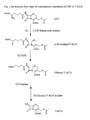
  • The core of many semi-synthetic cephalosporins, 7-aminocephalosporanic acid (7-ACA), can be manufactured chemically from cephalosporin C (CPC). The chemical process uses many chemical reagents that are highly toxic and heavily pollute the environment and is low in conversion rate and high in cost. Enzyme methods offer attractive alternative for production of fine chemicals without using toxic reagents and are high in conversion rate. The bioconversion of CPC to 7-ACA is conducted in two steps (Fig. 1): (1) CPC is first oxidized by D-amino acid oxidase to glutaryl-7-ACA; (2) glutaryl-7-ACA is in turn converted to 7-ACA by glutaryl-7-ACA acylase.
  • One of the major obstacles for large scale production of 7-ACA is the low yield and high cost of production of D-amino acid oxidase and glutaryl-7-ACA acylase. Current reports on the production level of D-amino acid oxidase is low, about 2,300 U/L fermentation medium (Pollegioni, L. et al., 1997, J. Biotechnol. 58, 115-123) and 800 U/L fermentation medium (Molla, G. et al., 1998, Protein Exp. Purif. 14, 289-294). The production level of glutaryl-7-ACA acylase production level is also low, about 129-2,500 U/L fermentation medium (Ishiye, M. and Niwa, M., 1992, Biochim. Biophys. Acta 1132, 233-239; Yang, Y. L. et al., 2001, CN1301813A ; Xu, G. and Zhu, M. 2003, CN1428424A ). Therefore, it is critical to produce these two enzymes for industrial production of 7-ACA at low cost.
  • Another obstacle for large scale production of 7-ACA resides in that existing manufacturing procedures are complex and expensive. For example, the procedures of related products by Roche Diagnostics (CC2 Twin Enzyme Process: D-AOD, product number: 1462865; Gl-Ac, product number: 1464213, Roche Diagnostics) are complicated (Fig. 1). Besides the reactions catalyzed by D-amino acid oxidase and glutaryl-7-ACA acylase, extra steps are needed: (1) due to the impurity of the D-amino acid oxidase used, a high proportion of α-ketoadipyl-7-ACA is not converted to glutaryl-7-ACA after the oxidation, thus an exogenous addition of hydrogen peroxide is required to complete the conversion; and (2) exogenous addition of catalase is required to degrade remaining hydrogen peroxide, for hydrogen peroxide can inactivate D-amino acid oxidase and oxidize CPC and glutaryl-7-ACA, thus reducing the final yield of 7-ACA.
  • In addition, a method to prepare 7-ACA has been published in CN1104255 . In that method, since the expression vector used contained ampicillin resistance gene, the fermentation product contains β-lactamase, which significantly reduces the yield of 7-ACA and β-lactamase inhibitor is therefore required in the process. Furthermore, the host cell employed in that method produces catalase, which degrades hydrogen peroxide, therefore exogenous additions of hydrogen peroxide and catalase inhibitor are required. Consequently, the manufacturing procedures are complex and highly cost.
  • Summary of the Invention
  • The object of the invention is to provide a simple, inexpensive and high yield two-step enzyme method for the preparation of 7-ACA.
  • To achieve the above object, the invention provides a method to prepare 7-ACA from CPC, which comprises oxidation of CPC to glutaryl-7-ACA by D-amino acid oxidase (first step reaction) and conversion of glutaryl-7-ACA to 7-ACA by glutaryl-7-ACA acylase (second step reaction). More specifically, said D-amino acid oxidase is a purified D-amino acid oxidase, with the amino acid sequence of Trigonopsis variabilis D-amino acid oxidase mutant as in Sequence 2.
  • Preferably, said first step reaction does not require addition of hydrogen peroxide, more preferably, said D-amino acid oxidase is expressed by the expression vector pHS-GHA with the DNA sequence as in Sequence 3. The sequential purification of D-amino acid oxidase comprises DEAE-cellulose ion exchange chromatography and ammonium sulphate precipitation. More preferably, in said first step and second step reactions, there is no addition of β-lactamase inhibitors selected from ascorbic acid, 3-amino-1,2,3-triazole, sodium perborate, and sodium azide; in said first step reaction, there is no addition of catalase inhibitors selected from sodium sulbactam, clavulanic acid, boric acid and their derivatives; in said second step reaction, there is no addition of catalase.
  • Preferably, said D-amino acid oxidase is immobilized, or said glutaryl-7-ACA acylase is immobilized, or both said D-amino acid oxidase and glutaryl-7-ACA acylase are immobilized.
  • More preferably, said glutaryl-7-ACA acylase is glutaryl-7-ACA acylase of Pseudomonas sp. SE83, said glutaryl-7-ACA acylase of Pseudomonas sp. SE83 is expressed by the expression vector pT7-kan-ACY with the DNA sequence as in Sequence 4.
  • The merits of the invention comprise: (1) the purified D-amino acid oxidase mutant, with the amino acid sequence as in Sequence 2 in the invention, possesses a specific activity of 105% higher than that of parent D-amino acid oxidase; (2) the fermentation products of the expression vectors pHS-GHA and pT7-kan-ACY of the invention do not contain β-lactamase, therefore no need to add exogenous β-lactamase inhibitor, which saves production cost and simplifies the manufacturing procedures; (3) the D-amino acid oxidase produced from the invention contains little or no catalase and therefore there is no need to add exogenous catalase inhibitor; the level of α-ketoadipyl-7-ACA is low and therefore there is no need to add exogenous hydrogen peroxide and catalase, which also reduces production cost and simplifies the manufacturing procedures; (4) the molar conversion rate of the two-step enzyme method in the invention can reach 93% or above, about 12% higher than that of the products of Roche Diagnostics (molar conversion rate is about 82%).
  • Brief Description of the Drawings
    • Fig. 1 shows the reaction flow chart of current procedure of conversion of CPC to 7-ACA.
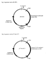
    • Fig. 2 shows expression vector pHS-GHA.
    • Fig. 3 shows expression vector pT7-kan-ACY.
    • Fig. 4 shows expression vector pRSET-lac-GI-hok/sok-kan.
    • Fig. 5 shows SDS-PAGE of D-amino acid oxidase GHA. Lane 1: BenchMark Pre-Stained Protein Ladder (Invitrogen), sizes of the proteins are in kDa; lane 2: partially purified D-amino acid oxidase GHA; lane 3: purified D-amino acid oxidase GHA.
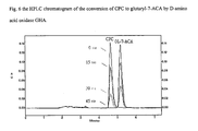
    • Fig. 6 shows the HPLC chromatogram of the conversion of CPC to glutaryl-7-ACA by D-amino acid oxidase GHA.
    • Fig. 7 shows SDS-PAGE of glutaryl-7-ACA acylase of Pseudomonas sp. SE83. Lane 1: BenchMark Pre-Stained Protein Ladder (Invitrogen), sizes of the proteins are in kDa; lane 2: partially purified glutaryl-7-ACA acylase of Pseudomonas sp. SE83.
    • Fig. 8 shows the HPLC chromatogram of the conversion of glutaryl-7-ACA to 7-ACA by glutaryl-7-ACA acylase of Pseudomonas sp. SE83.
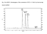
    • Fig. 9 shows the HPLC chromatogram of the conversion of CPC to 7-ACA by the two-step enzyme method.
    Example 1 Construction of expression vector pRSET-kan
  • The following primers were synthesized based on the sequence of pRSET-A (purchased from Invitrogen):
    • VET-F
      5'-CTGTCAGACCAAGTTTACTCATATATACTTTAG-3'
    • VET-R
      5'-ACTCTTCCTTTTTCAATATTATTGAAGC-3'
  • The following primers were synthesized based on the sequence of pET-28b (purchased from Novagen):
    • KAN-F
      5'-ATGAGCCATATTCAACGGGAAAC-3'
    • KAN-R
      5'-TTAGAAAAACTCATCGAGCATCAAATG-3'
  • PCR mixture for amplifying pRSET-A fragment devoid of ampicillin resistance gene contained: 50ng pRSET-A (Invitrogen), 0.4µM VET-F, 0.4µM VET-R, 50µM dATP, 50µM dTTP, 50µM dCTP, 50µM dGTP, 20mM Tris-HCl (pH8.8), 10mM KCl, 10mM (NH4)2SO4, 2mM MgSO4, 0.1% Triton X-100, 2.5U Pfu DNA polymerase (Promega). The volume of the mixture was made up to 50µL with sterile deionized water.
  • PCR profile was as follows:
       94°C, 1 min 35 cycles
          94°C, 5 min →50°C, 1 min →→→→→ 72°C, 10 min
       72°C, 4 min
  • PCR mixture for amplifying kanamycin resistance gene from plasmid pET-28b contained: 50ng pET-28b (Novagen), 0.4µM KAN-F, 0.4µM KAN-R, 50µM dATP, 50µM dTTP, 50µM dCTP, 50µM dGTP, 20mM Tris-HCl (pH8.8), 10mM KCl, 10mM (NH4)2SO4, 2mM MgSO4, 0.1% Triton X-100, 2.5U Pfu DNA polymerase (Promega). The volume of the mixture was made up to 50µL with sterile deionized water.
  • PCR profile was as follows:
       94°C, 1 min 35 cycles
          94°C, 5 min →50°C, 1 min →→→→→ 72°C, 10 min
       72°C, 4 min
  • The two PCR products (pRSET-A fragment devoid of ampicillin resistance gene, 2,036bp in size; kanamycin resistance gene, 816bp in size) were resolved in 0.8% agarose, purified and ligated to generate plasmid pRSET-kan. The plasmid was used to transform competent E. coli BL21(DE3)pLysS (Novagen), spread onto LB (1% sodium chloride, 1% peptone, 0.5% yeast extract) agar containing 50µg/mL kanamycin and incubated at 37°C overnight. Plasmid was extracted in accordance with Molecular Cloning-A Laboratory Manual (Sambrook, J. et al., 1989, CSHL Press).
  • Example 2 Construction of vector pRSET-lac-kan
  • The following primers were synthesized based on the sequence of pGEMT-Easy (Promega):
    • RBS-NdeI
      5'-CATATGTATATCTCCTTCTTGTGTGAAATTG-3'
  • (NdeI restriction site is underlined and ribosome binding site is marked by broken underline);
    • RBS-AlwNI
      5'-CAGTGGCTGCTGCCAGTGGCGATAAGTC-3'
  • (AlwNI restriction site is underlined).
  • PCR was performed using pGEMT-Easy (Promega) as template to generate a 755bp PCR product. PCR mixture contained 50ng pGEMT-Easy (Promega), 0.4µM RBS-NdeI, 0.4µM RBS-AlwNI, 50µM dATP, 50µM dTTP, 50µM dCTP, 50µM dGTP, 20mM Tris-HCl (pH8.8), 10mM KCl, 10mM (NH4)2SO4, 2mM MgSO4, 0.1% Triton X-100, 2.5U Pfu DNA polymerase (Promega). The volume of the mixture was made up to 50µL with sterile deionized water.
  • PCR profile was as follows:
       94°C, 1 min 35 cycles
          94°C, 5 min →50°C, 1 min →→→→→ 72°C, 10 min
       72°C, 4 min
  • The PCR product (755bp) contains NdeI restriction site and ribosome binding site at the 5' end and AlwNI restriction site at the 3' end. The PCR product was resolved in 0.8% agarose, purified and digested by NdeI and AlwNI and then ligated with NdeI/AlwNI restricted pRSETA (Invitrogen) to generate pRSET-lac. The plasmid was used to transform competent E. coli BL21(DE3)pLysS (Novagen), spread onto LB (1% sodium chloride, 1% peptone, 0.5% yeast extract) agar containing 100µg/mL ampicillin and incubated at 37°C overnight. Plasmid was extracted in accordance with Molecular Cloning-A Laboratory Manual (Sambrook, J. et al., 1989, CSHL Press).
  • Vectors pRSET-lac and pRSET-kan were cut with AlwNI and EcoRI and resolved in 0.8% agarose, purified and ligated, generating pRSET-lac-kan. The plasmid was used to transform competent E. coli BL21(DE3)pLysS (Novagen), spread onto LB agar containing 50µg/mL kanamycin and incubated at 37°C overnight. Plasmid was extracted in accordance with Molecular Cloning-A Laboratory Manual (Sambrook, J. et al., 1989, CSHL Press).
  • Example 3 Construction of vector pGEMT-Easy-GI
  • The following primers were synthesized based on the sequence of known Thermoanaerobacterium saccharolyticum glucose isomerase gene (GenBank L09699).
    • GI-NdeI
      5'-CATATGAATAAATATTTTGAGAACGTATCTAAAATA-3'
    • (NdeI restriction site is underlined);
      GI-EcoRI
      5'-GATATCTTAAGGCGCGCCTTATTCTGCAAAC-3'
  • (EcoRI restriction site is underlined and AscI restriction site is double underlined). PCR was performed using Thermoanaerobacterium saccharolyticum (purchased from ATCC, USA) DNA as template to generate a 1,336bp PCR product. PCR mixture contained 50ng T. saccharolyticum DNA, 0.4µM GI-NdeI, 0.4µM GI-EcoRI, 50µM dATP, 50µM dTTP, 50µM dCTP, 50µM dGTP, 20mM Tris-HCl (pH8.8), 10mM KCl, 10mM (NH4)2SO4, 2mM MgSO4, 0.1% Triton X-100, 2.5U Platinum Taq High Fidelity DNA polymerase (Invitrogen). The volume of the mixture was made up to 50µL with sterile deionized water.
  • PCR profile was as follows:
       94°C, 1 min 35 cycles
          95°C, 5 min →50°C, 1 min →→→→→ 72°C, 10 min
       72°C, 3 min
  • The PCR product (1,336bp) contains NdeI restriction site at the 5' end and EcoRI restriction site at the 3' end. The PCR product was resolved in 0.8% agarose, purified and ligated to pGEMT-Easy (Promega) by TA cloning, generating pGEMT-Easy-GI. The plasmid was used to transform competent E. coli DH5α (Invitrogen), spread onto LB agar containing 100µg/mL ampicillin and incubated at 37°C overnight. Plasmid was extracted in accordance with Molecular Cloning-A Laboratory Manual (Sambrook, J. et al., 1989, CSHL Press).
  • Example 4 Construction of vector pRSET-lac-GI-hok/sok-kan (Fig. 4)
  • Vector pGEMT-Easy-GI was cut by NdeI and EcoRI and resolved in 0.8% agarose, purified and ligated to NdeI/EcoRI-digested pRSET-lac-kan, generating pRSET-lac-GI-kan. The plasmid was used to transform competent E. coli BL21(DE3)pLysS (Novagen), spread onto LB agar containing 50µg/mL kanamycin and incubated at 37°C overnight. Plasmid was extracted in accordance with Molecular Cloning-A Laboratory Manual (Sambrook, J. et al., 1989, CSHL Press).
  • Ten primers were synthesized based on known hok/sok gene sequence (GenBank X05813) (Table 1). PCR gene assembly was performed as described by Kikuchi, M. et al., 1999, Gene 236:159-167, with modifications. PCR mixture contained 20ng each primer, 50µM dATP, 50µM dTTP, 50µM dCTP, 50µM dGTP, 20mM Tris-HCl (pH8.8), 10mM KCl, 10mM (NH4)2SO4, 2mM MgSO4, 0.1% Triton X-100, 2.5U Pfu DNA polymerase (Promega). The volume of the mixture was made up to 50µL with sterile deionized water.
  • PCR profile was as follows:
       94°C, 1.5 min 30 cycles
          95°C, 4 min →50°C, 1.5 min →→→→→ 72°C, 10 min
       72°C, 5 min
  • The PCR product (580bp) contains AscI restriction site at the 5' end and EcoRI restriction site at the 3' end. The PCR product was resolved in 0.8% agarose, cut with AscI and EcoRI and ligated to AscI/EcoRI-digested pRSET-lac-GI-kan, generating pRSET-lac-GI-hok/sok-kan. The plasmid was used to transform competent E. coli BL21(DE3)pLysS (Novagen), spread onto LB agar containing 50µg/mL kanamycin and incubated at 37°C overnight. Plasmid was extracted in accordance with Molecular Cloning-A Laboratory Manual (Sambrook, J. et al., 1989, CSHL Press) and sequenced as in Sequence 3 in the Sequence Listing. Nucleotide variations were observed in some nucleotides: 1368 (C→G); 1513 (missed a T); 1804(A→T); 1826(C→T); 2479(G→T); 2555(T→A); 3860(C→T). Table 1
    Number Primer sequence
    1 5'-ttggcgcgccttaagatatcaacaaactccgggaggcagcgtgatgcggcaacaatcacacggatttcccgtgaa-3'
    2 5'-catatacctgcacgctgaccacactcactttccctgaaaataatccgctcattcagaccgttcacgggaaatccgtgtga-3'
    3 5'-ggtcagcgtgcaggtatatgggctatgatgtgcccggcgcttgaggctttctgcctcatgacgtgaaggtggtttgttgc-3'
    4 5'-cgtggtggttaatgaaaattaacttactacggggctatcttctttctgccacacaacacggcaacaaaccaccttcacgt-3'
    5 5'-aattttcattaaccaccacgaggcatccctatgtctagtccacatcaggatagcctcttaccgcgctttgcgcaaggaga-3'
    6 5'-tgagacacacgatcaacacacaccagacaagggaacttcgtggtagtttcatggccttcttctccttgcgcaaagcgcgg-3'
    7 5'-tgtgttgatcgtgtgtctcacactgttgatattcacttatctgacacgaaaatcgctgtgcgagattcgttacagagacg-3'
    8 5'-cgcctccaggttgctacttaccggattcgtaagccatgaaagccgccacctccctgtgtccgtctctgtaacgaatctcg-3'
    9 5'-taagtagcaacctggaggcgggcgcaggcccgccttttcaggactgatgctggtctgactactgaagcgcctttataaag-3'
    10 5'-cggaattcacaacatcagcaaggagaaaggggctaccggcgaaccagcagcccctttataaaggcgcttcagt-3'
  • Example 5 Construction of vector pRSET-A-DAO with recombinant D-amino acid oxidase
  • Primers were synthesized based on published Trigonopsis variabilis D-amino acid oxidase gene (Gonzalez, F. J., Montes, J., Martin, F., Lopez, M. C., Ferminan, E., Catalan, J., Galan, M. A., Dominguez, A. Molecular cloning of TvDAO1, a gene encoding a D-amino acid oxidase from Trigonopsis variabilis and its expression in Saccharomyces cerevisiae and Kluyveromyces lactis. Yeast 13:1399-1408, 1997).
    • 5'-NdeI (incorporation of NdeI restriction site)
    • 5'-TAGGGCTGACATATGGCTAAAATCGTTGTTATTGGTGC-3'
    • 3'-BgIII (incorporation of BglII restriction site)
    • 5'-TAGGGCTGAAGATCTCTAAAGGTTTGGACGAGTAAGAGC-3'
  • T. variabilis D-amino acid oxidase gene was synthesized using the above primers, Pfu DNA polymerase (Promega) and plasmid pJL ( Yang, Y. L. et al. CN1371999A ) as template. Plasmid pJL contains T. variabilis FA10 D-amino acid oxidase gene (Li, W. et al., Acta Microbiologica Sinica 31:251-253, 1991). PCR mixture contained 40ng pJL, 0.4µM 5'-NdeI, 0.4µM 3'-BglII, 50µM dATP, 50µM dTTP, 50µM dCTP, 50µM dGTP, 20mM Tris-HCl (pH8.8), 10mM KCl, 10mM (NH4)2SO4, 2mM MgSO4, 0.1% Triton X-100, 2.5U Pfu DNA polymerase. The volume of the mixture was made up to 50µL with sterile deionized water.
  • PCR profile was as follows:
       94°C, 1 min 10 cycles 94°C, 1 min 25 cycles
          94°C, 5 min →50°C, 1 min →→→→→ 60°C, 1 min →→→→→ 72°C, 10 min
       72°C, 2 min 72°C, 2 min
  • The PCR product (1,098bp) contains NdeI restriction site at the 5' end and BglII restriction site at the 3' end. The PCR product was resolved in 1% agarose, purified and digested by NdeI and BglII and then ligated with 2.9kb NdeI/BglII restricted pRSET-A (Invitrogen), generating pRSET-A-DAO. The plasmid was used to transform competent E. coli BL21(DE3)pLysS (Novagen), spread onto LB agar containing ampicillin and incubated at 37°C overnight. Plasmid was extracted in accordance with Molecular Cloning-A Laboratory Manual (Sambrook, J. et al., 1989, CSHL Press) and sequenced. The DNA fragment was confirmed as T. variabilis D-amino acid oxidase gene as Sequence 5 and the translated amino acid sequence as Sequence 6.
  • Example 6 Construction of expression vector with recombinant D-amino acid oxidase GHA
  • Recombinant D-amino acid oxidase GHA was constructed by site-directed mutagenesis, which was based on the procedures in PCR Protocols (Ed. John M.S. Bartlett and David Stirling, Totowa, N.J.: Humana Press, 2003).
  • Primers were synthesized in accordance with the sequence of cloned T. variabilis D-amino acid oxidase (Sequence 5):
    • Primer A
      5'-TAGGGCTGACATATGGCTAAAATCGTTGTTATTG-3'
    • Primer B
      5'-TAGGGCTGAAGATCTCTAAAGGTTTGGACGAG-3'
    • Primer C 1
      5'-GCAGGTGCCAACTGGCTCCCGTTTTACGATGGAGGCAAG-3'
    • Primer D
      5'-GAGCCAGTTGGCACCTGCCCAAGG-3'
  • Primers A and B are a pair of outer primer. Primer A contains NdeI restriction site, with a portion of nucleotides overlapped with the 5'-end of the D-amino acid oxidase gene; primer B contains BglII restriction site, with a portion of nucleotides overlapped with the 3'-end of the D-amino acid oxidase gene. Primers C1 and D are inner primers. Primer C1 converts the 53rd amino acid residue of wild-type D-amino acid oxidase from threonine (Thr) to proline (Pro). Primer D contains a portion of nucleotides overlapped with primer C1.
  • PCR was performed, using pRSET-A-DAO as template, to synthesize fragment 1 (primers A and D) and fragment 2 (primers B and C1). PCR mixture contained: 20ng pRSET-A-DAO, 20mM Tris-HCl (pH8.8), 10mM KCl, 10mM (NH4)2SO4, 2mM MgSO4, 0.1% Triton X-100, 0.4µM primer A and 0.4µM primer D (for synthesizing fragment 1) or 0.4µM primer B and 0.4µM primer Cl (for synthesizing fragment 2), 50µM dATP, 50µM dTTP, 50µM dCTP, 50µM dGTP, 1.5U Pfu DNA polymerase. The volume of the mixture was made up to 50µL with sterile deionized water.
  • PCR profile was as follows:
       94°C, 1 min 30 cycles
          94°C, 2 min →53°C, 1 min →→→→→ 72°C, 10 min
       72°C, 1 min
  • The amplified fragment 1 and fragment 2 were resolved in and purified from 1% agarose, and used to generate full-length D-amino acid oxidase GHA gene. PCR mixture for synthesizing the full-length gene contained: 20ng fragment 1, 20ng fragment 2, 20mM Tris-HCl (pH8.8), 10mM KCl, 10mM (NH4)2SO4, 2mM MgSO4, 0.1% Triton X-100, 0.4µM primer A and 0.4µM primer B, 50µM dATP, 50µM dTTP, 50µM dCTP, 50µM dGTP, 1.5U Pfu DNA polymerase. The volume of the mixture was made up to 50µL with sterile deionized water.
  • PCR profile was as follows:
       94°C, 1 min 35 cycles
          94°C, 2 min →53°C, 1 min →→→→→ 72°C, 10 min
       72°C, 2 min
  • The full-length recombinant D-amino acid oxidase GHA gene fragment was obtained, cut with NdeI and BglII and ligated with pRSET-kan, generating pRSET-kan-DAOGHA. The plasmid was used to transform competent E. coli BL21(DE3)pLysS, spread onto LB agar containing kanamycin and incubated at 37°C overnight.. Plasmid was extracted, and the insert was sequenced and confirmed as D-amino acid oxidase mutant GHA as Sequence 1 and the translated amino acid sequence as Sequence 2.
  • Example 7 Construction of vector pHS-GHA (Fig. 2)
  • Vector pRSET-kan-DAOGHA was cut by NdeI and BglII to release a DNA fragment (1,074bp, containing D-amino acid oxidase GHA) and resolved in 0.8% agarose, purified and ligated to the large fragment of NdeI/BglII-digested pRSET-lac-GI-hok/sok-kan, generating pHS-GHA. The plasmid was used to transform competent E. coli BL21(DE3)pLysS (Novagen), generating clone BL-HS-GHA, spread onto LB agar containing 50µg/mL kanamycin and incubated at 37°C overnight. Plasmid was extracted in accordance with Molecular Cloning-A Laboratory Manual (Sambrook, J. et al., 1989, CSHL Press) and sequenced (Sequence 3). Nucleotide variations were observed in some nucleotides: 1390 (C→G); 1535 (missed a T); 1826(A→T); 1848(C→T); 2501(G→T); 2577(T→A); 3882(C→T).
  • Example 8 Construction of vector pT7-kan-ACY (Fig. 3)
  • The following primers were synthesized based on the sequence of known glutaryl-7-ACA acylase gene of Pseudomonas sp. SE83 (Matsuda, A. et al., 1987, J. Bacteriol. 169, 5821-5826).
    • NdeI-ACY
      5'-CATATGAACGCTCCCGTCCCCGTCCC-3'
    • (NdeI restriction site is underlined);
      BglII-ACY
      5'-AGATCTTCAGATGGTGAAGCGGGCAC-3'
  • (BglII restriction site is underlined).
  • PCR was performed using Pseudomonas sp. SE83 DNA as template to generate a 1,676bp PCR product. PCR mixture contained 50ng Pseudomonas sp. SE83 DNA, 0.4µM NdeI-ACY, 0.4µM BglII-ACY, 50µM dATP, 50µM dTTP, 50µM dCTP, 50µM dGTP, 20mM Tris-HCl (pH8.8), 10mM KCl, 10mM (NH4)2SO4, 2mM MgSO4, 0.1% Triton X-100, 2.5U Pfu DNA polymerase (Promega). The volume of the mixture was made up to 50µL with sterile deionized water.
  • PCR profile was as follows:
       94°C, 1 min 35 cycles
          95°C, 5 min →50°C, 1 min →→→→→ 72°C, 10 min
       72°C, 3 min
  • The PCR product (1,676bp) contains NdeI restriction site at the 5' end and BglII restriction site at the 3' end. The PCR product was resolved in 0.8% agarose, cut with NdeI and BgIII and ligated to NdeI/BglII-digested pRSET-kan, generating pT7-kan-ACY. The plasmid was used to transform competent E. coli BL21(DE3)pLysS (Novagen), generating clone BL-T7K-ACY, spread onto LB agar containing 50µg/mL kanamycin and incubated at 37°C overnight. Plasmid was extracted in accordance with Molecular Cloning-A Laboratory Manual (Sambrook, J. et al., 1989, CSHL Press) and sequenced (Sequence 4). Nucleotide variations were observed in four nucleotides: 2260 (G→T); 2336 (T→A); 3641(C→T); 4117(G→C).
  • Example 9 Medium and fermentation of D-amino acid oxidase GHA
  • A single colony of clone BL-HS-GHA (Example 7) was picked from LB agar containing 50µg/mL kanamycin and grown in 2x5mL LB medium containing 50µg/mL kanamycin at 37°C for 8 hours (shaker at 250rpm). The culture was then inoculated to 2x50mL seed medium containing 100µg/mL kanamycin and 40µg/mL chloramphenicol, incubated at 30°C for 16 hours (shaker at 400rpm).
  • Preparation of corn steep liquor 1:
  • Dissolved 300g corn steep liquor (purchased from North China Pharmaceutical Kangxin Co., Ltd.) in 300mL distilled water and then centrifuged (5,000g, 8min.) to harvest the supernatant as corn steep liquor 1. The pellet was kept for later use.
  • Preparation of corn steep liquor 2:
  • Dissolved the above mentioned pellet in 600mL distilled water and then centrifuged (5,000g, 8min.) to harvest the supernatant as corn steep liquor 2.
  • A 50mL seed medium contained:
    Corn steep liquor 1 4mL
    Corn steep liquor 2 4mL
    Yeast extract 0.2g
    Ammonium sulphate 0.075g
    Disodium hydrogen phosphate 0.25g
    Potassium dihydrogen phosphate 0.04g
    Sodium chloride 0.075g
  • They were dissolved in 50mL distilled water and adjusted to pH 7.15 by 10N sodium hydroxide and sterilized by autoclaving.
  • After overnight incubation, the total 100mL seed culture was inoculated to a 2L fermentor (BIOENGINEERING, Benchtop Fermentor, KLF2000) containing 50µg/mL kanamycin.
  • 2L fermentation medium contained:
  • Corn steep liquor 1 160mL
    Corn steep liquor 2 160mL
    Yeast extract 8g
    Ammonium sulphate 3g
    Disodium hydrogen phosphate 10g
    Potassium dihydrogen phosphate 1g
    Sodium chloride 3g
  • They were dissolved in 1.9L distilled water and adjusted to pH 7.15 by 10N sodium hydroxide and sterilized by autoclaving in the 2L fermentor (BIOENGINEERING, Benchtop Fermentor, KLF2000).
  • 12.5g glucose was dissolved in 50mL distilled water, sterilized by autoclaving; 1.25g magnesium sulphate was dissolved in 50mL distilled water, sterilized by autoclaving.
  • The sterilized glucose and magnesium sulphate were added to the 2L fermentor prior to fermentation.
  • Preparation of feed:
    • Corn steep liquor 1 (250mL) and corn steep liquor 2 (250mL) were mixed and adjusted to pH 7.25 by 10N sodium hydroxide and sterilized by autoclaving.
    • 2.25g ammonium sulphate, 7.56g disodium hydrogen phosphate, 1.2g potassium dihydrogen phosphate, 2.25g sodium chloride was dissolved in 60mL distilled water and sterilized by autoclaving.
    • 15g yeast extract was dissolved in 100mL distilled water and sterilized by autoclaving.
    • 70g glucose was dissolved in 140mL distilled water and sterilized by autoclaving.
    • 30mL glycerol was mixed with 10mL distilled water and sterilized by autoclaving.
    • 20g magnesium sulphate was dissolved in 30mL distilled water and sterilized by autoclaving.
  • All solutions were mixed and kanamycin was added to final concentration of 50µg/mL, 2mL antifoam was added.
  • The fermentation was held at 35°C. In the first 6 hours, the pH value rose from 6.9 to 7.2 and the feed was started (50mL/hour). The fermentation was proceeded for another 26 hours at controlled conditions (the pH value was maintained at 7.2 by 5N potassium hydroxide; dissolved oxygen pO2 was not greater than 0.5%).
  • Example 10 Partial purification of D-amino acid oxidase mutant GHA
  • Fermentation was performed as in Example 9. The cells were collected by centrifugation at 4°C (5,000g, 8min.), and supernatant was discarded. The wet weight of cell pellet was 220g and it was resuspended in 600mL sodium phosphate buffer (50mM, pH7.5). The cells were lysed by grinding in a dynomill (DYNO-MILL TYP KDL, 0.2mm glass beads, WA Bachofen). Cell suspension was injected into dynomill at 50mL/min. and washed by 800mL sodium phosphate buffer (50mM, pH7.5). The cell lysate was heat treated at 55°C for 30min. in a water bath, and centrifuged (10,000g, 30min.). Supernatant was partial purified D-amino acid oxidase mutant GHA. The purity and concentration of the target protein was analyzed by SDS-PAGE (Fig. 5). As shown in the figure, partial purified D-amino acid oxidase mutant GHA constituted about 40% of the total soluble protein.
  • Example 11 Purification of D-amino acid mutant GHA
  • Partial purified D-amino acid oxidase mutant GHA was prepared as in Example 10 and glycerol was added to final concentration of 10% and the pH value was adjusted to 8 by 5N sodium hydroxide. The mixture was centrifuged (13,000g, 30min.) and the supernatant was collected. DEAE-cellulose ion exchange resin (Sigma, D-0909) was prepared in accordance with the manufacturer's instructions. The partial purified D-amino acid oxidase mutant GHA was mixed with DEAE-cellulose ion exchange resin at a ratio of 1mL (GHA) : 0.5mL (resin) and stirred at 4°C for 5 hours (100rpm) and then filtered (Buchner filter funnel, 120mm P1). The DEAE-cellulose ion exchange resin was washed by 40mM sodium dihydrogen phosphate (with 10% glycerol) by three bed volumes, followed by 400mM sodium dihydrogen phosphate by two bed volumes to elute D-amino acid oxidase mutant GHA. Ammonium sulphate was added (262g/L eluted D-amino acid oxidase mutant GHA), stirred at room temperature for 15min. and then centrifuged (13,000g, 15min.). Supernatant was discarded and the protein pellet was dissolved in 10mM sodium dihydrogen phosphate (pH7.5). The purity and level of the target protein was analyzed by SDS-PAGE (Fig. 5). As shown in the figure, partial purified D-amino acid oxidase mutant GHA constituted about 90% of the total soluble protein.
  • Example 12 Determination of activity of D-amino acid oxidase mutant GHA
  • The procedures were performed in accordance with Isogai, T., et al, J. Biochem. [Tokyo] 108:1063-1069, 1990, with modifications. Purified D-amino acid oxidase mutant GHA was prepared as in Example 11 and diluted 10 times by sodium phosphate buffer (50mM, pH7.5). Reaction mixture contained 2mL 150mM CPC sodium and 2mL diluted purified D-amino acid oxidase mutant GHA, stirred (450rpm) at 37°C and the pH value was maintained at 7.5 by 5N sodium hydroxide. Aliquots (100µL) were withdrawn at different time points (0, 15, 30, 45 min., Fig. 6), mixed with 10µL 3% hydrogen peroxide, followed by addition of 50µL 10% trichloroacetic acid to stop the reaction. The mixture was centrifuged (10,000g, 3min) and 10µL of supernatant was mixed with 990µL HPLC mobile phase (50mM potassium phosphate, pH7; 5% acetonitrile), then analyzed by HPLC. HPLC column: Diamonsil™ C18, 250 × 4.6mm (Diam Company, Beijing); column temperature: 30°C; flow rate: 1mL/min; scanning: 260nm UV. One unit of enzyme activity was defined as the amount of enzyme that converted 1 µmole of CPC to glutary-7-ACA per min under the above reaction condition. The total activity of the D-amino acid oxidase mutant GHA was 95,607U, which was 35,410U/L fermentation medium.
  • Example 13 Preparation of immobilized D-amino acid oxidase mutant GHA
  • Purification of D-amino acid oxidase mutant GHA was performed as in Example 11. Preparation of immobilized D-amino acid oxidase was performed in accordance with the description from Resindion S. R. L. (Italy) with modifications.
  • Activation of the matrix: 10g wet Sephabeads HA was mixed with 30mL potassium dihydrogen phosphate (100mM, pH8) and stirred (300rpm) at room temperature for 15min., then the pH value was adjusted to 8 by 5N sodium hydroxide. The matrix was filtered and washed by 40mL potassium phosphate buffer (20mM, pH8) for 5min. with stirring and filtered. The matrix was added with 40mL 2% glutaraldehyde and stirred (300rpm) at room temperature for 1 hour. The matrix was filtered and washed with 40mL potassium phosphate (20mM, pH8) for 5min. and filtered. The washing was repeated 5 times and the matrix was activated.
  • Enzyme immobilization: The activated matrix was mixed with purified D-amino acid oxidase mutant GHA (10g activated matrix to 100mL purified D-amino acid oxidase mutant GHA), stirred (300rpm) at room temperature for 1min. The pH value was adjusted to 8 by 1N sodium hydroxide and stirred for another 18 hours. The matrix was filtered and washed by 40mL potassium phosphate buffer (20mM, pH8) with stirring for 2min. and filtered. The matrix was washed by 40mL sodium chloride (0.5M sodium chloride dissolved in 20mM potassium phosphate buffer, pH8) for 20min. with stirring and filtered. The washing was repeated until the eluate contained protein less than 0.1mg/mL. The matrix was washed by 40mL potassium phosphate buffer (20mM, pH8) with stirring for 2min. and filtered. The total immobilized enzyme generated was 115g. The activity of the immobilized D-amino acid oxidase mutant GHA was determined as in Example 12, with 4g immobilized D-amino acid oxidase mutant GHA in a reaction volume of 200mL of 75mM CPC sodium. The activity of the immobilized recombinant D-amino acid oxidase GHA was 77U/g wet matrix.
  • Example 14 Medium and fermentation of glutaryl-7-ACA acylase of Pseudomonas sp. SE83
  • A single colony of clone BL-T7K-ACY (Example 8) was picked from LB agar containing 50µg/mL kanamycin and grown in 2×5mL LB medium containing 50µg/mL kanamycin at 37°C for 8 hours (shaker at 250rpm). An aliquot of culture was inoculated to 2×50mL seed medium, incubated at 30°C for 16 hours (shaker at 400rpm).
  • A 50mL seed medium contained:
    Yeast extract 0.35g
    Disodium hydrogen phosphate 0.35g
    Potassium dihydrogen phosphate 0.35g
    Dipotassium hydrogen phosphate 0.48g
    Ammonium sulphate 0.06g
    Ammonium chloride 0.01g
    Glycerol 0.5mL
    Calcium chloride 0.00055g
  • They were dissolved in 50mL distilled water and sterilized by autoclaving.
  • After overnight incubation, the total 100mL seed culture was inoculated to a 2L fermentor (BIOENGINEERING, Benchtop Fermentor, KLF2000) containing 50µg/mL kanamycin.
  • 2L fermentation medium contained:
    Yeast extract 14g
    Disodium hydrogen phosphate 14g
    Potassium dihydrogen phosphate 14g
    Dipotassium hydrogen phosphate 19.2g
    Ammonium sulphate 2.4g
    Ammonium chloride 0.4g
    Glycerol 20mL
    Calcium chloride 0.022g
  • They were dissolved in 2L distilled water and sterilized by autoclaving in the 2L fermentor (BIOENGINEERING, Benchtop Fermentor, KLF2000).
  • 1g magnesium sulphate was dissolved in 20mL distilled water, sterilized by autoclaving; 0.14g zinc chloride was dissolved in 20mL distilled water, sterilized by autoclaving.
  • The sterilized magnesium sulphate and zinc chloride were added to the 2L fermentor prior to fermentation.
  • Preparation of feed:
    Yeast extract 14g
    Disodium hydrogen phosphate 4.9g
    Potassium dihydrogen phosphate 4.9g
    Dipotassium hydrogen phosphate 6.4g
    Ammonium sulphate 0.84g
    Ammonium chloride 0.14g
    Glycerol 175mL
    Calcium chloride 0.008g
  • They were dissolved in 700mL distilled water, sterilized by autoclaving.
  • 0.34g magnesium sulphate was dissolved in 20mL distilled water, sterilized by autoclaving and added to sterilized 700mL feed.
  • Kanamycin was added to final concentration of 50µg/mL, 2mL antifoam was added.
  • The fermentation was held at 30°C. In the first 4 hours, the pH value was raised from 6.9 to 7.2 by 5N potassium hydroxide. Feeding was started when the OD600 reached 4 at a feed rate of 60mL/hour. The pH value was raised to 7.4 by 5N potassium hydroxide in the following 4 hours. IPTG (Sigma) was added to final concentration of 0.1mM when OD600 reached 8. The fermentation was proceeded for another 26 hours at controlled conditions (the pH value was maintained at 7.4 by 5N potassium hydroxide; dissolved oxygen pO2 at 30%).
  • Example 15 Partial purification of glutaryl-7-ACA acylase of Pseudomonas sp. SE83
  • Fermentation was performed as in Example 14. The cells were collected by centrifugation at 4°C (5,000g, 8min.), and supernatant was discarded. The wet weight of cell pellet was 130g and it was resuspended in 400mL sodium phosphate buffer (50mM, pH8). The cells were lysed by grinding in the dynomill (DYNO-MILL TYP KDL, 0.2mm glass beads, WA Bachofen). Cell suspension was injected into dynomill at 50mL/min. and washed by 600mL sodium phosphate buffer (50mM, pH8). The cell lysate was heat treated at 55°C for 15min. in a water bath, centrifuged (10,000g, 30min.). Supernatant was partial purified glutaryl-7-ACA acylase of Pseudomonas sp. SE83. The purity and concentration of the target protein was analyzed by SDS-PAGE (Fig. 7). As shown in the figure, partial purified glutaryl-7-ACA acylase of Pseudomonas sp. SE83 constituted about 40% of the total soluble protein.
  • Example 16 Determination of activity of glutaryl-7-ACA acylase of Pseudomonas sp. SE83
  • The procedures were performed in accordance with Binder, R. et al., 1994, Appl. Environ. Microbiol. 60, 1805-1809, with modifications. The partially purified glutaryl-7-ACA acylase of Pseudomonas sp. SE83 (Example 15) was diluted 10 times by sodium phosphate buffer (50mM, pH8) and mixed with same volume of 150mM glutaryl-7-ACA (preparation as described in Shibuya, Y. et al., 1981, Agric. Biol. Chem. 45, 1561-1567) at 37°C with continuously stirring (450rpm) and the pH value was maintained at 8 by 5N sodium hydroxide. Aliquots (60µL) were withdrawn at different time points (0, 15, 30, 45 min., Fig. 8) and mixed with 30µL 10% trichloroacetic acid to stop the reaction. The mixture was centrifuged (10,000g, 3min.) and 10µL of supernatant was mixed with 990µL HPLC mobile phase (50mM potassium phosphate, pH7; 5% acetonitrile), then analyzed by HPLC. HPLC conditions were as described in Example 12. One unit of enzyme activity was defined as the amount of enzyme that converted 1 µmole of glutaryl-7-ACA to 7-ACA per min under the above reaction condition. The total activity of the glutaryl-7-ACA acylase of Pseudomonas sp. SE83 was 24,822U, about 9,570U/L fermentation medium.
  • Example 17 Preparation of immobilized glutaryl-7-ACA acylase of Pseudomonas sp. SE83
  • Glutaryl-7-ACA acylase of Pseudomonas sp. SE83 was performed as in Example 15. Preparation of immobilized glutaryl-7-ACA acylase of Pseudomonas sp. SE83 was performed in accordance with the description from Röhm (Germany) with modifications. A 100mL partially purified glutaryl-7-ACA acylase of Pseudomonas sp. SE83 was mixed with 10g Eupergit C250L wet matrix, stirred (300rpm) at room temperature for 72 hours. The matrix was filtered and washed by 100mL distilled water at room temperature with stirring (300rpm) for 2min. and finally filtered by No.3 sand funnel. The washing was repeated until the eluate contained protein less than 0.1mg/mL. The total immobilized glutaryl-7-ACA acylase of Pseudomonas sp. SE83 generated was 80g. The activity of the immobilized glutaryl-7-ACA acylase of Pseudomonas sp. SE83 was determined as in Example 16, with 6g immobilized glutaryl-7-ACA acylase of Pseudomonas sp. SE83 in a reaction volume of 200mL of 75mM glutaryl-7-ACA. The activity of the immobilized glutaryl-7-ACA acylase of Pseudomonas sp. SE83 was 50U/g wet matrix.
  • Example 18 Two-step enzymatic conversion of CPC to 7-ACA
  • Immobilized D-amino acid oxidase mutant GHA was prepared (40g) as in Example 13 and added to 75mM CPC sodium solution (1L), stirred (250rpm) at room temperature for 1 hour with supply of pure oxygen at a rate of 0.3m3/hour. The pH value was maintained at 7.5 by 3M ammonia. The reaction mixture was filtered and immobilized glutaryl-7-ACA acylase of Pseudomonas sp. SE83 (50g, prepared as in Example 17) was added, stirred (250rpm) at room temperature for 1 hour. The pH value was maintained at 8 by 3M ammonia. The level of 7-ACA was analyzed by HPLC as in Example 16. The HPLC chromatogram was shown in Fig. 9. As shown in Fig. 9, the whole conversion took 120 min.: after the first 60 min., most of the CPC was converted to glutaryl-7-ACA by D-amino acid oxidase mutant GHA (Fig. 9, GL-7-ACA, 60 min. peak). After another 60 min., most of the glutaryl-7-ACA was converted to 7-ACA by glutaryl-7-ACA acylase of Pseudomonas sp. SE83 (Fig. 9, 7-ACA, 120 min. peak). According to the HPLC data, the conversion rate of CPC to glutaryl-7-ACA was 97.96%; the conversion rate of glutaryl-7-ACA to 7-ACA was 95.78%. The conversion rate of CPC to 7-ACA by the two step enzyme method was 93.83%.
  • This invention is not limited by the detailed description provided in the Examples above. Various modifications can be made by those skilled in the field and these modifications should be regarded as within the scope of the claims of the invention.
Claims (10)

  1. A method to prepare 7-ACA from CPC, which comprises the conversion of CPC to glutaryl-7-ACA by D-amino acid oxidase (first step reaction) and the conversion of glutaryl-7-ACA to 7-ACA by glutaryl-7-ACA acylase (second step reaction), wherein said D-amino acid oxidase is a purified Trigonopsis variabilis D-amino acid oxidase mutant with the amino acid sequence as in Sequence 2.
  2. The method according to claim 1, in said first step reaction, hydrogen peroxide is not added.
  3. The method according to claim 2, said D-amino acid oxidase is expressed from expression vector pHS-GHA with the DNA sequence as in Sequence 3.
  4. The method according to claim 3, said D-amino acid oxidase is purified by sequential purifications comprising DEAE-cellulose ion exchange resin purification and ammonium sulphate precipitation.
  5. The method according to claim 4, in said first step and second step reactions, β-lactamase inhibitors selected from ascorbic acid, 3-amino-1,2,3-triazole, sodium perborate, and sodium azide is not added.
  6. The method according to claim 5, in said first step reaction, catalase inhibitors selected from sodium sulbactam, clavulanic acid, boric acid and their derivatives is not added.
  7. The method according to claim 6, in said second step reaction, catalase is not added.
  8. The method according to any of the claims 1-7, said D-amino acid oxidase is immobilized, or said glutaryl-7-ACA acylase is immobilized.
  9. The method according to claim 8, said glutaryl-7-ACA acylase is glutaryl-7-ACA acylase of Pseudomonas sp. SE83.
  10. The method according to claim 9, said glutaryl-7-ACA acylase of Pseudomonas sp. SE83 is expressed by the expression vector pT7-kan-ACY with the DNA sequence as in Sequence 4.
EP06775270A 2005-08-08 2006-08-02 Two-steps enzyme method for preparing 7-aminocephalosporanic acid Withdrawn EP1925663A4 (en)

Applications Claiming Priority (2)

Application Number Priority Date Filing Date Title
CN2005100899653A CN1912130B (en) 2005-08-08 2005-08-08 Two-step enzyme method for preparing 7-aminocephalosporanic acid
PCT/CN2006/001940 WO2007016861A1 (en) 2005-08-08 2006-08-02 Two-steps enzyme method for preparing 7-aminocephalosporanic acid

Publications (2)

Publication Number Publication Date
EP1925663A1 true EP1925663A1 (en) 2008-05-28
EP1925663A4 EP1925663A4 (en) 2009-11-11

Family

ID=37721186

Family Applications (1)

Application Number Title Priority Date Filing Date
EP06775270A Withdrawn EP1925663A4 (en) 2005-08-08 2006-08-02 Two-steps enzyme method for preparing 7-aminocephalosporanic acid

Country Status (4)

Country Link
US (1) US8003358B2 (en)
EP (1) EP1925663A4 (en)
CN (1) CN1912130B (en)
WO (1) WO2007016861A1 (en)

Cited By (1)

* Cited by examiner, † Cited by third party
Publication number Priority date Publication date Assignee Title
EP2132323A4 (en) * 2007-03-08 2010-05-05 Richmond Chemical Corp Compositions of variant biocatalysts for preparing enantiopure amino acids

Family Cites Families (10)

* Cited by examiner, † Cited by third party
Publication number Priority date Publication date Assignee Title
CN1033164C (en) * 1993-08-11 1996-10-30 东北制药总厂 Refining method of 7-aminocephalosporanic acid
CN1065278C (en) 1994-08-27 2001-05-02 中国科学院上海植物生理研究所 Method for preparation of 7-amino cephaloalkyl acid by one step and two enzyme method
ES2131018B1 (en) * 1997-09-25 2000-04-01 Antibioticos Sau AN ENZYMATIC PROCEDURE FOR PREPARING 7BETA- (4-CARBOXYBUTANAMIDE) CEPHALOSPORANIC ACID USING THE MODIFIED TRIGONOPSIS VARIABILIS ENZYME D-AMINO ACID PRODUCED IN ESCHERICHIA COLI.
ATE226954T1 (en) * 1998-03-27 2002-11-15 Dsm Nv METHOD FOR THE FERMENTATIVE PRODUCTION OF CEPHALOSPORIN
CN1301813A (en) 1999-12-29 2001-07-04 中国科学院上海植物生理研究所 Engineering fungus containing glutaryl-7-amino-cephalo phytanic acid acylated enzyme gene
CN1151261C (en) 2001-02-28 2004-05-26 中国科学院上海植物生理研究所 Double gene coexpression plasmid, construction method and application thereof
EP1344830A1 (en) * 2002-03-12 2003-09-17 Bioferma Murcia, S.A. A process for the preparation of cephalosporan acid derivatives from cephalosporin C
CN1428424A (en) 2002-12-30 2003-07-09 湖南福来格生物技术有限公司 Preparation method of glutaryl-7-aminocefaphytanic acid acyltransferase
CN100342014C (en) * 2004-04-08 2007-10-10 百瑞全球有限公司 recombinant D-amino acid oxidase
CN100386440C (en) * 2004-04-08 2008-05-07 百瑞全球有限公司 Use of recommbined D-amino acid oxidase

Cited By (1)

* Cited by examiner, † Cited by third party
Publication number Priority date Publication date Assignee Title
EP2132323A4 (en) * 2007-03-08 2010-05-05 Richmond Chemical Corp Compositions of variant biocatalysts for preparing enantiopure amino acids

Also Published As

Publication number Publication date
US8003358B2 (en) 2011-08-23
US20100112633A1 (en) 2010-05-06
WO2007016861A1 (en) 2007-02-15
CN1912130A (en) 2007-02-14
CN1912130B (en) 2011-06-15
EP1925663A4 (en) 2009-11-11

Similar Documents

Publication Publication Date Title
Pilone et al. D-amino acid oxidase as an industrial biocatalyst
JP3072101B2 (en) 7-A new method for biochemical production of ADCA
CN117535256A (en) Carbonyl reductase and application thereof in synthesis of vitronectin
CN112980906B (en) Enzyme composition for preparing beta-nicotinamide mononucleotide and application thereof
JP5627546B2 (en) Method for enantioselective enzymatic reduction of secodione derivatives
CN109055324B (en) Improved ketoreductase and application thereof
EP1188838A1 (en) Method for producing 7-aminodesacetoxycephalosporanic acid (7-adca)
CN112852912B (en) A kind of method for synthesizing 7-aminodeacetoxy cephalosporanic acid
EP1748073B1 (en) Recombinant d-amino acid oxidase
CN113444702A (en) Enone reductase mutant and application thereof
US7399624B2 (en) Cephalosporin C acylases
EP1925663A1 (en) Two-steps enzyme method for preparing 7-aminocephalosporanic acid
US9404139B2 (en) Mutated cephalosporin hydroxylase and its application in deacetylcephalosporanic acid synthesis
CN115975964A (en) A highly active keto-pantoate lactone reductase mutant and its coding gene and application
CN115261342A (en) Burkholderia BJQ 0011-derived ester synthetase JFN _18195, encoding gene and application thereof
WO2008107782A2 (en) Modified hydroxylase and its applications
CN114990138B (en) Gene, expression vector, bacterial strain and application thereof in improving cephalosporin C yield
EP1925670A1 (en) Escherichia coli expression vector and the uses thereof
CN114807066B (en) Ketone reductase mutant
CN111254170A (en) Method for preparing (S) -1,2,3, 4-tetrahydroisoquinoline-3-formic acid by multi-enzyme coupling
CN119144591A (en) Cephalosporin C acylase mutant with substrate specificity and application thereof
CN109897872A (en) Enzyme process prepares (2S, 3S)-N- tertbutyloxycarbonyl -3- amino -1- chlorine-2-hydroxyl -4- phenyl butane
US20230175023A1 (en) Engineered biosynthetic pathways for production of 3,4-dihydroxybenzoic acid by fermentation
CN121294597A (en) Penicillin G acylase used in the synthesis of penicillin V
CN117467629A (en) Method for producing baodanone by using whole cell transformation of genetically engineered bacteria and application

Legal Events

Date Code Title Description
PUAI Public reference made under article 153(3) epc to a published international application that has entered the european phase

Free format text: ORIGINAL CODE: 0009012

17P Request for examination filed

Effective date: 20080310

AK Designated contracting states

Kind code of ref document: A1

Designated state(s): AT BE BG CH CY CZ DE DK EE ES FI FR GB GR HU IE IS IT LI LT LU LV MC NL PL PT RO SE SI SK TR

RAP1 Party data changed (applicant data changed or rights of an application transferred)

Owner name: GENEHARBOR (HONG KONG) TECHNOLOGIES LIMITED

RA4 Supplementary search report drawn up and despatched (corrected)

Effective date: 20090914

A4 Supplementary search report drawn up and despatched

Effective date: 20090914

STAA Information on the status of an ep patent application or granted ep patent

Free format text: STATUS: THE APPLICATION IS DEEMED TO BE WITHDRAWN

18D Application deemed to be withdrawn

Effective date: 20091212