EP1945767B1 - Identifizierung und charakterisierung von funktionsblockierenden anti-ed-b-fibronektin antikörpern - Google Patents
Identifizierung und charakterisierung von funktionsblockierenden anti-ed-b-fibronektin antikörpern Download PDFInfo
- Publication number
- EP1945767B1 EP1945767B1 EP05811334A EP05811334A EP1945767B1 EP 1945767 B1 EP1945767 B1 EP 1945767B1 EP 05811334 A EP05811334 A EP 05811334A EP 05811334 A EP05811334 A EP 05811334A EP 1945767 B1 EP1945767 B1 EP 1945767B1
- Authority
- EP
- European Patent Office
- Prior art keywords
- antibody
- region
- domain
- antibody fragment
- human
- Prior art date
- Legal status (The legal status is an assumption and is not a legal conclusion. Google has not performed a legal analysis and makes no representation as to the accuracy of the status listed.)
- Expired - Lifetime
Links
Images


Classifications
-
- C—CHEMISTRY; METALLURGY
- C07—ORGANIC CHEMISTRY
- C07K—PEPTIDES
- C07K16/00—Immunoglobulins [IG], e.g. monoclonal or polyclonal antibodies
- C07K16/18—Immunoglobulins [IG], e.g. monoclonal or polyclonal antibodies against material from animals or humans
-
- A—HUMAN NECESSITIES
- A61—MEDICAL OR VETERINARY SCIENCE; HYGIENE
- A61P—SPECIFIC THERAPEUTIC ACTIVITY OF CHEMICAL COMPOUNDS OR MEDICINAL PREPARATIONS
- A61P1/00—Drugs for disorders of the alimentary tract or the digestive system
- A61P1/04—Drugs for disorders of the alimentary tract or the digestive system for ulcers, gastritis or reflux esophagitis, e.g. antacids, inhibitors of acid secretion, mucosal protectants
-
- A—HUMAN NECESSITIES
- A61—MEDICAL OR VETERINARY SCIENCE; HYGIENE
- A61P—SPECIFIC THERAPEUTIC ACTIVITY OF CHEMICAL COMPOUNDS OR MEDICINAL PREPARATIONS
- A61P15/00—Drugs for genital or sexual disorders; Contraceptives
-
- A—HUMAN NECESSITIES
- A61—MEDICAL OR VETERINARY SCIENCE; HYGIENE
- A61P—SPECIFIC THERAPEUTIC ACTIVITY OF CHEMICAL COMPOUNDS OR MEDICINAL PREPARATIONS
- A61P17/00—Drugs for dermatological disorders
- A61P17/02—Drugs for dermatological disorders for treating wounds, ulcers, burns, scars, keloids, or the like
-
- A—HUMAN NECESSITIES
- A61—MEDICAL OR VETERINARY SCIENCE; HYGIENE
- A61P—SPECIFIC THERAPEUTIC ACTIVITY OF CHEMICAL COMPOUNDS OR MEDICINAL PREPARATIONS
- A61P17/00—Drugs for dermatological disorders
- A61P17/06—Antipsoriatics
-
- A—HUMAN NECESSITIES
- A61—MEDICAL OR VETERINARY SCIENCE; HYGIENE
- A61P—SPECIFIC THERAPEUTIC ACTIVITY OF CHEMICAL COMPOUNDS OR MEDICINAL PREPARATIONS
- A61P19/00—Drugs for skeletal disorders
- A61P19/02—Drugs for skeletal disorders for joint disorders, e.g. arthritis, arthrosis
-
- A—HUMAN NECESSITIES
- A61—MEDICAL OR VETERINARY SCIENCE; HYGIENE
- A61P—SPECIFIC THERAPEUTIC ACTIVITY OF CHEMICAL COMPOUNDS OR MEDICINAL PREPARATIONS
- A61P21/00—Drugs for disorders of the muscular or neuromuscular system
-
- A—HUMAN NECESSITIES
- A61—MEDICAL OR VETERINARY SCIENCE; HYGIENE
- A61P—SPECIFIC THERAPEUTIC ACTIVITY OF CHEMICAL COMPOUNDS OR MEDICINAL PREPARATIONS
- A61P25/00—Drugs for disorders of the nervous system
-
- A—HUMAN NECESSITIES
- A61—MEDICAL OR VETERINARY SCIENCE; HYGIENE
- A61P—SPECIFIC THERAPEUTIC ACTIVITY OF CHEMICAL COMPOUNDS OR MEDICINAL PREPARATIONS
- A61P27/00—Drugs for disorders of the senses
- A61P27/02—Ophthalmic agents
-
- A—HUMAN NECESSITIES
- A61—MEDICAL OR VETERINARY SCIENCE; HYGIENE
- A61P—SPECIFIC THERAPEUTIC ACTIVITY OF CHEMICAL COMPOUNDS OR MEDICINAL PREPARATIONS
- A61P29/00—Non-central analgesic, antipyretic or antiinflammatory agents, e.g. antirheumatic agents; Non-steroidal antiinflammatory drugs [NSAID]
-
- A—HUMAN NECESSITIES
- A61—MEDICAL OR VETERINARY SCIENCE; HYGIENE
- A61P—SPECIFIC THERAPEUTIC ACTIVITY OF CHEMICAL COMPOUNDS OR MEDICINAL PREPARATIONS
- A61P35/00—Antineoplastic agents
-
- A—HUMAN NECESSITIES
- A61—MEDICAL OR VETERINARY SCIENCE; HYGIENE
- A61P—SPECIFIC THERAPEUTIC ACTIVITY OF CHEMICAL COMPOUNDS OR MEDICINAL PREPARATIONS
- A61P9/00—Drugs for disorders of the cardiovascular system
- A61P9/10—Drugs for disorders of the cardiovascular system for treating ischaemic or atherosclerotic diseases, e.g. antianginal drugs, coronary vasodilators, drugs for myocardial infarction, retinopathy, cerebrovascula insufficiency, renal arteriosclerosis
-
- A—HUMAN NECESSITIES
- A61—MEDICAL OR VETERINARY SCIENCE; HYGIENE
- A61K—PREPARATIONS FOR MEDICAL, DENTAL OR TOILETRY PURPOSES
- A61K39/00—Medicinal preparations containing antigens or antibodies
- A61K2039/505—Medicinal preparations containing antigens or antibodies comprising antibodies
-
- C—CHEMISTRY; METALLURGY
- C07—ORGANIC CHEMISTRY
- C07K—PEPTIDES
- C07K2317/00—Immunoglobulins specific features
- C07K2317/50—Immunoglobulins specific features characterized by immunoglobulin fragments
- C07K2317/55—Fab or Fab'
-
- C—CHEMISTRY; METALLURGY
- C07—ORGANIC CHEMISTRY
- C07K—PEPTIDES
- C07K2317/00—Immunoglobulins specific features
- C07K2317/50—Immunoglobulins specific features characterized by immunoglobulin fragments
- C07K2317/56—Immunoglobulins specific features characterized by immunoglobulin fragments variable (Fv) region, i.e. VH and/or VL
-
- C—CHEMISTRY; METALLURGY
- C07—ORGANIC CHEMISTRY
- C07K—PEPTIDES
- C07K2317/00—Immunoglobulins specific features
- C07K2317/50—Immunoglobulins specific features characterized by immunoglobulin fragments
- C07K2317/56—Immunoglobulins specific features characterized by immunoglobulin fragments variable (Fv) region, i.e. VH and/or VL
- C07K2317/565—Complementarity determining region [CDR]
-
- C—CHEMISTRY; METALLURGY
- C07—ORGANIC CHEMISTRY
- C07K—PEPTIDES
- C07K2317/00—Immunoglobulins specific features
- C07K2317/70—Immunoglobulins specific features characterized by effect upon binding to a cell or to an antigen
- C07K2317/76—Antagonist effect on antigen, e.g. neutralization or inhibition of binding
-
- C—CHEMISTRY; METALLURGY
- C07—ORGANIC CHEMISTRY
- C07K—PEPTIDES
- C07K2317/00—Immunoglobulins specific features
- C07K2317/90—Immunoglobulins specific features characterized by (pharmaco)kinetic aspects or by stability of the immunoglobulin
- C07K2317/92—Affinity (KD), association rate (Ka), dissociation rate (Kd) or EC50 value
Definitions
- the invention relates to recombinant antibodies or antibody fragments which can bind to the ED-B isoform of fibronectin and block their function. Furthermore, the diagnostic and pharmaceutical application of the polypeptides of the invention is disclosed.
- ECM extracellular matrix
- Tumor-induced ECM appears to be the optimal environment for tumor growth and tumor angiogenesis (1-4).
- Fibronectins are an important class of matrix glycoproteins. Their main role is to allow the attachment of cells to a variety of different extracellular matrices. The presence of fibronectins on the surface of untransformed cells in culture, as well as their absence in transformed cells, led to the identification of fibronectins as important adhesion proteins. They interact with many different other molecules, such as collagen, heparan sulfate proteoglycans, and fibrin, thereby regulating the cell shape and structure of the cytoskeleton. Furthermore, they are responsible for cell migration and cell differentiation during embryogenesis. They are also important in wound healing, enabling the migration of macrophages and other immune cells into the affected area, and in the formation of blood clots by allowing the attachment of platelets to damaged regions of the blood vessels.
- Fibronectins are dimers of two similar peptides, each chain being about 60-70 nm in length. At least 20 different fibronectin chains have been identified, all of which are generated by alternative splicing of the RNA transcript of a single fibronectin gene.
- Fibronectins are high molecular weight, adhesion-promoting glycoproteins that play an important role in the development of the blood vessel system. Furthermore, fibronectins chemotactically act on endothelial cells, modulate the action of growth factors, and assist endothelial cell elongation during angiogenesis (6-9). Analysis of proteolytic fibronectin cleavage products indicates that the polypeptides consist of six strongly folded domains, each domain containing repetition of so-called repeats whose similarities in their amino acid sequence permit classification into three types (Type I, II , III). The central region of both chains from the dimer consists of a section of so-called type III repeats, which are on average 90 amino acids long (10). Structural studies have shown that each type III repeat consists of seven beta strands folded into two antiparallel sheets, with short loop regions exposed as potential protein-protein interaction sites (11).
- the type III repeats allow fibronectins to act as adhesion molecules that interact with cell surface molecules, the so-called "integrins".
- integrin was first used in 1987 in (12) to describe a related group of heterodimers To describe cell surface molecules that act as mediators between the extracellular matrix and the intracellular cytoskeleton, thus inducing cell adhesion and migration. These heterodimeric receptors thus "integrate” or mediate signals from the extracellular milieu with specific cellular functions.
- 17 beta subunits are known that can specifically and noncovalently interact with more than 20 alpha subunits to form more than 20 distinct families (13).
- sequence RGDS located in the tenth repeat of type III fibronectin mediates the interaction of fibronectin with at least 8 different integrins.
- at least four integrins can interact specifically with fibronectin in an RGDS-independent manner (13).
- the group of repeat sequences of type III comprises not only the III7, III8, III9 and III10 sequences but also the repeats EIIIB and EIIIA (ED-B and ED-A).
- ED-B fibronectin is undetectable in normal adult tissue (only exception: proliferating endometrium), whereas it is highly expressed in fetal tissue and tumor growth, in addition to stromal local expression of ED-B.
- ED-B fibronectin localizes perivascularly to blood vessels that have newly formed during angiogenesis. For this reason, ED-B fibronectin is a specific marker protein for the process of (tumor) angiogenesis (14).
- the ED-B domain is a highly conserved, complete type III homology building block consisting of 91 amino acids. The degree of homology between humans and rats is 100%, between humans and chickens 96%. Very little is known about the function of the ED-B domain in the literature. A few publications (15-17) speculate about a general adhesion-mediating effect for different cells. A specific effect on endothelial cells has not yet been shown.
- Recombinant ED-B was shown to be specific pro-angiogenic activity in vitro : (i) the proliferation of bFGF-stimulated human dermal microvascular endothelial cells (HDMVEc) is enhanced by recombinant ED-B, (ii) the protein mediates the adhesion of HDMVEC and (iii) recombinant ED-B stimulates the Invasion and differentiation (tube formation) of HDMVEc in collagen gels.
- HDMVEc bFGF-stimulated human dermal microvascular endothelial cells
- ED-B-mediated effects could be specifically blocked by synthetic peptides derived from the ED-B domain.
- the peptide sequences thus represent the binding region for an ED-B specific receptor on the endothelial cell surface, which was identified by affinity chromatography as the ⁇ 2 ⁇ 1 integrin.
- the specific interaction between the ⁇ 2 ⁇ 1 integrin and the ED-B domain has not previously been described in the literature.
- integrin receptors with partially overlapping properties are expressed by endothelial cells (Lit .: DG Stupack and DA Cheresh, SciSTKE, 2002 Feb 12; 2002 ). These expression patterns indicate that different integrins (eg, alphavbeta3 and alpha5beta1) mediate similar biological phenomena (adhesion, migration, and survival) and therefore represent redundant systems for the endothelial cell that ensure their behavior and survival.
- alpha2beta1 interacts with its natural ligands, the collagens. The blockade of this interaction can lead to an anti-angiogenic effect ( Y Funahashi et al. Cancer Res. 62: 6116-6123, 2002 ).
- Menrad et al. Molecular and functional characterization of ED-B fibronectin selective function blocking antibodies and the evaluation of phage angiomics for target identification and validation "HUMAN ANTIBODIES, Vol. 13, No. 1-2, 2004, page 33 . 11TH INTERNATIONAL CONFERENCE ON HUMAN ANTIBODIES AND HYBRIDOMAS; DUBLIN; IRELAND; OCTOBER 06-08, 2004 ISSN: 1093-2607 ) very generally describes that high affinity ED-B fibronectin-function blocking antibody fragments have been successfully identified. However, it is not described how such antibody fragments can be obtained and selected. Also, no antigen binding sites are indicated. Menrad et al. Nor does it disclose whether the antibodies bind to the ED-B domains of ED-B fibronketin. Also, no antibody fragment concentrations are indicated. Also, only the first signs of action were found here. Antibody production can not be deduced from this article.
- An object of the present invention was therefore to provide functionally-blocking binding molecules, such as antibodies, which specifically block receptor binding sites of the ED-B domain. These binding molecules have an anti-angiogenic effect on (tumor) endothelial cells. In contrast to the relatively broadly expressed ⁇ 2 ⁇ 1 integrin, the ED-B domain is an ideal and specific target molecule for such binding molecules.
- ED-B The structure of ED-B makes the development of monoclonal and polyclonal antibodies difficult, as ED-B is expected to have low immunogenicity in vivo .
- the Antibody BC-1 J. Cell Biol. 108 (1989), 1139-1148 ) with a cryptic epitope of fibronectin present only in the presence of ED-B and therefore not directly with ED-B.
- the antibody L19 Tarli et al. Blood 94 (1999), 192-198 and WO 01/62800 ), which was generated by using recombinant ED-B as an immunogen, is again biologically inactive, ie it can not know cell adhesion to ED-B significantly.
- ED-B function blocking antibody MOR03255 which strongly inhibit the adhesion of cells to ED-B and induce a significant reduction in tumor growth in vivo .
- the blockade of ED-B caused by the binding molecules according to the invention can not be compensated for by compensatory mechanisms of the collagen-integrin interaction.
- HuCAL ® GOLD antibody library a library of about 1.6 x 10E10 different antibodies in the Fab fragment format, the starting (from the HuCAL consensus sequences WO 97/08320 ; Knappik A, Ge L, Honegger A, Pack P, Fischer M, Wellnhofer G, Hoess A, Wolle J, Plunckthun A, and Vimekas B (2000) J. Mol. Biol.
- antibody in the context of the present invention encompasses polyclonal, monoclonal, chimeric, humanized or human antibodies as well as recombinant antibodies, for example single-chain antibodies, or antigen-binding antibody fragments, for example monovalent antibody fragments such as Fab fragments or scFv fragments, or divalent antibody fragments such as about F (ab ') 2 fragments.
- the antibodies contain one or more antigen-binding sites, which the o.g. Conditions, i. specific binding to the ED-B domain and inhibition of the interaction between the ED-B domain and its receptor, in particular its receptor on endothelial cells.
- antigen-binding properties are preferably achieved by combining a VH and VL region, these regions being composed of so-called framework regions (FR1, FR2, FR3 and FR4) and the CDR regions mediating antigen-binding (H-CDR1, H-CDR2, H- CDR3 for the VH region and L-CDR1, L-CDR2, L-CDR3 for the VL region).
- the antibodies or antibody fragments according to the invention preferably have an affinity for the ED-B domain corresponding to a co-value ⁇ 1 ⁇ M, preferably ⁇ 100 nM, more preferably ⁇ 10 nM, even more preferably ⁇ 1 nM and most preferably ⁇ 0.1 nM, wherein the affinity as indicated by a test on BIAcore ® system in the examples can be determined.
- the antibody or antibody fragments according to the invention are characterized in that they bind specifically to the ED-B domain of fibronectin, ie it binds with a significantly lower affinity to other fibronectin domains, in particular to the fibronectin domain 6 (FN6) or / and the fibronectin domains 7 8-9 (7-8-9).
- the antibodies or antibody fragments according to the invention and the fibronectin bind ED-B domain with at least the factor 2, in particular by at least the factor 5 and more preferably by at least the factor 10 higher affinity than at FN6 and / or 7-8-9, such as can be determined by the binding test given in the examples.
- the antibodies or antibody fragments according to the invention exhibit in vitro an inhibition of the adhesion of recombinant ED-B to HDMVEc cells, preferably an inhibition of at least 50% and more preferably of at least 75% in a concentration of 10 ⁇ g / ml.
- the antibodies or antibody fragments of the present invention exhibit in vivo inhibition of the growth of a tumor produced by implantation of F9 teratocarcinoma cells (ATCC CRL-1720) in experimental animals such as nude mice.
- changes in the L-CDR3 region or / and in the VH region are preferably changes in the H-CDR2 region.
- the antibodies or antibody fragments of the invention that bind specifically to the ED-B domain of fibronectin and inhibit its interaction with its receptor are characterized by encoding the VH region of SEQ ID NO. 75 (MOR 03251) and the VL region encodes SEQ ID NO. 77 (MOR 03251) or the H-CDR1, H-CDR2 and H-CDR3 region from said VH region and the L-CDR1, L-CDR2 and L-CDR3 regions from said VL region, wherein the antibody or antibody fragment determines the ED-B domain with a KD value of 0.1 nM or lower binder and the KD value is determined by surface plasmon resonance, or characterized in that they the VH region encodes SEQ ID NO.
- said Antibody or antibody fragment binds the ED-B domain with a KD value of 0.1 nM or lower and the KD value is determined by surface plasmon resonance.
- polypeptide of the invention may be used for therapeutic or diagnostic purposes, eg for in vitro or in vivo diagnosis.
- the polypeptide of the invention may be in the form of a conjugate with a therapeutic agent, for example selected from radio- or chemotherapeutic agents, e.g. low molecular weight or biological cytostatic or cytotoxic agents present.
- a therapeutic agent for example selected from radio- or chemotherapeutic agents, e.g. low molecular weight or biological cytostatic or cytotoxic agents present.
- the conjugation of the therapeutic agent to the polypeptide may be by known methods, preferably via covalent coupling to reactive amino, carboxy, hydroxy or / and thiol groups of the protein, optionally using homo- or heterobifunctional linkers, by known methods.
- polypeptide may also be in the form of a fusion protein which, in addition to the antibody, eg in the form of an IgG molecule or a fragment thereof, contains a cytokine fused thereto, eg IL2, II12 or TNFa polypeptide.
- the fusion protein may be present in the form of a bispecific antibody, wherein, in addition to the binding to the ED-B domain, binding to a further antigen is preferred.
- bispecific antibodies binding domains against chelators for diagnostically or / and therapeutically relevant radionuclides, eg ⁇ , ⁇ or ⁇ emitters such as 90 Y, diagnostic NIR (near infrared) dyes, therapeutically active dyes, surface molecules on immunological effector cells (eg NK cells, cytotoxic T cells or NKT cells), angiogenesis-relevant integrins, in particular binding domains which block their function (eg ⁇ 1 , ⁇ 3 , ⁇ 1 ⁇ 3 , ⁇ 2 ⁇ 1 , ⁇ 2 ⁇ 2 ), inactivating anti-VEGF binding domains and inactivating Binding domains against VEGF receptor 1, 2 and 3.
- chelators for diagnostically or / and therapeutically relevant radionuclides eg ⁇ , ⁇ or ⁇ emitters such as 90 Y
- diagnostic NIR (near infrared) dyes eg NK cells, cytotoxic T cells or NKT cells
- angiogenesis-relevant integrins
- the polypeptide may be in the form of a conjugate of a diagnostically detectable label group, eg, a marker group for in vitro or in vivo diagnostics.
- a diagnostically detectable label group eg, a marker group for in vitro or in vivo diagnostics.
- tag groups are radioactive tag groups, NMR, dye, enzyme and fluorescence (eg near infrared fluorescence) tagging groups.
- the polypeptide is preferably formulated as a pharmaceutical composition containing as active ingredient the polypeptide of the invention, optionally further active ingredients and pharmacologically conventional carriers, excipients and / or diluents.
- the pharmaceutical composition contains the active ingredient in a therapeutically effective dose which can be readily determined by a person skilled in the art by in vitro experiments, for example on suitable cell cultures, or in animal models.
- the administration of the composition is preferably by injection or infusion, but other modes of administration are contemplated. Preferably, an intravenous and / or subcutaneous administration takes place.
- the dose of the drug administered depends on the nature and severity of the disease, as well as the condition of the patient being treated.
- the therapeutic composition is administered several times over a longer period of, for example, at least 2-4 weeks.
- known methods of administering antibodies or antibody conjugates such as in U.S. Pat Ferarra, N. et al., Nature Reviews Drug Discovery, Volume 3, May 2004, 391-400 and Salgaller, ML, Current Opinion in Molecular Therapeutics 2003 5 (6): 657-667 , or referred to existing pharmaceutical antibody administration protocols such as rituximab, CAMPATH, Remicade, etc.
- Another object of the invention is a diagnostic Composition
- a diagnostic Composition comprising as diagnostic reagent a polypeptide according to the invention.
- the diagnostic composition may contain other diagnostically customary reagents, carriers, excipients and / or diluents.
- the diagnostic composition contains the polypeptide in an amount sufficient to enable diagnostic detection in the particular format, eg, in an in vivo or in vitro diagnostic format. Reference is made in this connection to known methods for in vivo and in vitro diagnostics using labeled antibodies.
- the pharmaceutical and diagnostic compositions can be used for the therapy and diagnosis of all diseases associated with ED-B expression, for example, stromal and / or perivascular ED-B expression.
- diseases are hyperproliferative diseases, e.g. Diseases associated with angiogenesis, especially cancer, ocular fundus diseases, hypertrophic scars, etc., disorders associated with malfunction of myofibroblasts, e.g. Endometriosis, arteriosclerotic plaques, etc. or inflammatory diseases, e.g. Psoriasis, Crohn's disease, rheumatoid arthritis or multiple sclerosis.
- the pharmaceutical or diagnostic composition of the invention may contain one or more active polypeptides, e.g. a combination of polypeptides that bind to different regions of the ED-B domain.
- active polypeptides e.g. a combination of polypeptides that bind to different regions of the ED-B domain.
- the compositions are suitable for use in human and veterinary medicine.
- a further subject of the invention is a nucleic acid which codes for a polypeptide or fusion polypeptide according to the invention.
- This nucleic acid may be single-stranded or double-stranded DNA or RNA.
- the nucleic acid is preferably in operative association with an expression control sequence that is capable of expression in a suitable manner Host cell or a suitable host organism allows.
- the nucleic acid may be on a vector suitable for introduction into a host cell or host organism.
- the vector may be, for example, a prokaryotic vector suitable for introducing prokaryotic cells, eg a plasmid or bacteriophage.
- the vector may also be a eukaryotic vector suitable for introduction into eukaryotic host cells or host organisms, eg a plasmid, an artificial chromosome or a viral vector.
- a eukaryotic vector suitable for introduction into eukaryotic host cells or host organisms, eg a plasmid, an artificial chromosome or a viral vector.
- Suitable vectors are for example the expert from Sambrook et al. (1989), Molecular Cloning, A Laboratory Manual, Cold Spring Harbor Laboratory Press , and Ausubel et al. (1989), Current Protocols in Molecular Biology, John Wiley and Sons , known.
- Yet another object of the invention is a cell, e.g. a prokaryotic cell or a eukaryotic cell, such as a human cell transformed with a nucleic acid or a vector of the invention.
- Another object of the invention is a non-human organism, e.g. a transgenic animal transformed with a nucleic acid or a vector according to the invention.
- transformation in the context of the present invention includes all possibilities for introducing foreign nucleic acids into a cell or an organism including transfection or infection.
- the polypeptide of the invention can be made by culturing a cell or a non-human organism according to the invention under conditions in which expression of the polypeptide takes place, and then the expressed polypeptide, e.g. from the cell, the culture medium, the organism or excretion products of the organism.
- fibronectin domain ED-B (His) 6 fibronectin domain 6 (6FN), fibronectin domain 7-8-9 and domain 7-ED-B-8-9 were prepared according to standard procedures (eg Sambrook et al. Molecular Cloning, A Laboratory Manual, Cold Spring Harbor Press).
- Mouse antibodies to purified ED-B receptor were also prepared by standard methods by administering to a mouse 50 ⁇ g of purified receptor in Freund's complete adjuvant, 3 weeks later, 25 ⁇ g of purified receptor in Freund's incomplete adjuvant and 3 weeks later all applications in incomplete adjuvant were administered intraperitoneally. Fourteen days after the third immunization, blood was then withdrawn via the retroorbital sinus. Plasma fibronectin was purchased from Sigma.
- the selection was carried out according to the HuCAL ® Gold Phage selection protocol.
- the HuCAL ® Gold library has been divided into six framework-specific pools.
- the ED-B specific phages were enriched in recombinant protein immobilized in 96-well plates (Maxisorb, Nunc, Rochester, NY, USA) in three consecutive rounds of selection.
- A is the plate with 1 ug / well of ED-B (His 6) was coated (10 .mu.g / ml in Ampuwa ®) for 2 h at 37 ° C.
- the plate with ED-B was in the same concentration in PBS (pH 7.2 with 1 mM MgCl 2 / 1mM CaCl2) overnight at 4 ° C.
- the plates were washed with 5% skimmed milk powder in PBS, 0.05% Tween20 (Sigma, St. Louis, MO, USA) (blocking buffer) and incubating phage with 1 x 10 13 HuCAL ® Gold, which had been preincubated with a blocking buffer volume.
- the phage were additionally pre-incubated with 0.5 ⁇ g / ml fibronectin domain 6 to reduce the selection of binding molecules specific for the conserved structure of a type 3 domain repeat of fibronectin.
- the HuCAL ® Gold phage were amplified in 2 x TY medium with 34 ug / ml chloramphenicol and 1% glucose (2x TY-CG). After infection with a helper phage (VCSM13) at 37 ° C and an OD 600 of about 0.5, centrifugation and resuspension in 2x TY-CG / 50 ⁇ g / ml kanamycin, the cells were induced with 0.25 mM IPTG and overnight 22 ° C cultivated. The phages were precipitated from the supernatant with polyethylene glycol ( Ausubel et al. (1998), Current Protocols of Microbiology ), resuspended in PBS and used for subsequent rounds of selection.
- VCSM13 helper phage
- the Fab encoding inserts of the selected HuCAL ® Gold phages were subcloned into the expression vector pMORPHx9-FS.
- the plasmid DNA preparation of the selected HuCAL ® Gold clones was digested with the restriction enzymes xball / EcoRI, the Fab-encoding insert was excised (ompA-VL and phoA-Fd).
- the Fab-encoding insert was excised (ompA-VL and phoA-Fd).
- this Vector-expressed Fab molecules contain two C-terminal labels (FLAG TM and Strep-tagII) for detection and purification.
- the expression of the Fab fragments encoded on pMORPHx9-FS in E. coli TG-1 F cells was carried out in shake flask cultures with 0.75 l 2xTY medium and 34 ⁇ g / ml chloramphenicol. After induction with 0.75 mM IPTG, the cells were cultured for 16 h at 30 ° C. Alternatively, Fab clones obtained from the second maturation pool 2 were induced with 0.1 mM IPTG and then cultured at 22 ° C. Periplasmic extracts of cell pellets were prepared by osmotic shock, and the Fab fragments were isolated by Strep-Tactin ® chromatography (IBA, Göttingen, Germany). The apparent molecular weights were determined by size exclusion chromatography (SEC) with calibration standards. The concentrations were determined by UV spectrophotometry.
- 96-well Maxisorb ELISA plates were coated with 100 ⁇ l ED-B solution (10 ⁇ g / ml in coating buffer (PBS pH 7.4, 1 mM CaCl 2 , 1 mM MgCl 2 ) overnight at 4 ° C crude lysates or purified Fab molecules were added, non-binding Fab molecules were removed by washing five times with washing buffer (PBS pH 7.4, 0.05% Tween20) . The Fab fragments were removed by incubation with anti-human Fab. Antibody-peroxidase conjugates (Dianova), followed by development with a soluble peroxidase substrate (Roche) and measurement at 370 nm. The clones expressing for ED-B specific Fab molecules were compared by a positive ELISA signal on immobilized ED-B no or only a small signal at 6FN identified.
- coating buffer PBS pH 7.4, 1 mM CaCl 2 , 1 mM MgCl 2
- the Fab fragments were
- the coating with ED-B (2.5 ⁇ g / ml) was performed essentially as described above.
- the Fab fragments were incubated for 4 h at 37 ° C. and a concentration of 50 ⁇ g / ml in human plasma (German Red Cross, lot 9985550). After incubation, Fab ELISA ELISA molecules were assayed for concentrations of 5.0, 2.5, 1.25, 0.62, 0.31, 0.156, 0.078 and 0.039 ⁇ g / ml in PBS containing 1% bovine serum albumin diluted to determine the linear range of signal intensity. Functional Fab molecules were determined with the anti-Flag M2 antibody (Sigma F3165) and a secondary anti-mouse antibody-alkaline phosphatase conjugate followed by development with Attophos (Roche) and by measurement at 535 nm.
- HDMVEc Human dermal microvascular endothelial cells isolated from juvenile foreskin were grown in EGM MV medium (Clonetics Inc.) with the addition of 10% fetal calf serum, 2 mM glutamine, 20 ⁇ g / ml heparin, and 3 ng / ml bFGF in vials , coated with 0.1% gelatin (Sigma), cultured. For adhesion tests, cells that had no longer been cultured as passage 8 were used.
- ED-B (10 ⁇ g / ml) or plasma fibronectin (2.5 ⁇ g / ml) was incubated in PBS pH 7.4, 0.9 mM CaCl 2 , 0.5 mM MgCl 2 in 96-well ELISA plates overnight immobilized at 4 ° C and blocked with 1% BSA in PBS pH 7.4, 0.9 mM CaCl 2 , 0.5 mM MgCl 2 for 1 h at 37 ° C.
- the cells were labeled with 0.15 ⁇ g / ml calcein for 30 min at 37 ° C and washed once in adhesion medium (MCDB 131, 2 mM glutamine, 0.1% BSA, 20 ⁇ g / ml heparin). 1 x 10 5 cells were incubated with the respective indicated antibody concentration, diluted in adhesion medium (final volume 100 ⁇ l) for 20 min at 37 ° C. The cell suspension became that with ED-B After washing the cells twice in PBS pH 7.4, 0.9 mM CaCl 2 , 0.5 mM MgCl 2 , adherent cells were detected in a plate fluorimeter (excitation 485 nm, emission 530 nm) nm).
- CDR regions were optimized by cassette mutagenesis using trinucleotide directed mutagenesis.
- Fab fragments from the expression vector pMORPHx9 were cloned into the phagemid vector pMORPH-23 using the EcoRI / XbaI restriction sites.
- two consecutive maturation steps were performed for each maturation pool.
- an antibody fragment phage library was generated in which the L-CDR3 region of the parent clones varies by repertoire of 7 x 10 8 (pool 1) and 3.8 x 10 8 (pool 2) single light chain CDR sequences has been.
- affinity improved derivatives from the first maturation step were then subjected to a second round of maturation by replacing the H-CDR-2 region with a library of diversified elements. In this way, two phage libraries were generated, each with 1 ⁇ 10 8 individual clones.
- the affinity maturation libraries were generated by transformation into E. coli TOP10F.
- the phages were prepared as previously described. Selection for Fab fragments with improved affinity was performed under stringent conditions for three rounds at the first maturation and for two rounds at the second step.
- EDC 1-ethyl-3- (3-dimethylaminopropyl) carbodiimide
- NHS N-hydroxysuccinimide
- RU resonance units
- the coating with ED-B for the first batch was after the conditions known from the biological assay to present ED-B in a conformation that allows the adhesion of HDMVEc cells to the immobilized antigen.
- the phage were additionally blocked with an excess of 6FN to avoid selection of binding molecules that cross-react with the fibronectin domain 6.
- HuCAL Fab molecules were found from 1315 primary hits that meet the criteria of binding to ED-B and not to 6FN. Furthermore, the antibodies were tested for their ability to bind to a recombinantly expressed fibronectin comprising domains 7, 8 and 9 with and without the inserted ED-B domain in their natural position (see FIG. 3 ). All antibodies tested showed specific recognition for the 7-ED-B-8-9 construct, but did not react with the corresponding construct without the ED-B domain.
- the antibodies identified as specific in the ELISA were purified and tested for their ability to block the adhesion of HDMVEc cells to ED-B coated plates.
- the adhesion of HDMVEc cells in the presence of ED-B specific Fab fragments was tested in two concentrations as compared to a non-specific HuCAL control Fab molecule and mouse positive control sera.
- FIG. 4 A representative selection of the six most potent Fab molecules is in FIG. 4 shown. At a concentration of 25 ⁇ g / ml, cell adhesion was inhibited by 50% to nearly 100%. At a concentration of 0.4 ⁇ g / ml showed only few Fab molecules, eg MOR02610, MOR02616, MOR02715 and MOR02718, a reproducible inhibition of cell adhesion.
- the functionally-blocking Fab antibody fragments were immunohistochemically characterized for their localization on F9 mouse teratocarcinoma cells and human IRM30 neuroblastoma xenograft cryosections. All antibodies tested showed vascular localization on both tissue sections but no staining of vessels for normal tissue (eg normal and sclerotic liver and skin).
- FIG. 5B and 5D a perivascular abluminal staining of vessels, proliferating endothelium and tumor stroma by MOR02616 is shown. An accumulation of ED-B in the vicinity of neovascular structures of exponentially growing tumors, but not on normal vessels, can be seen. No staining was visible with a negative control antibody to lysozyme at the same concentration.
- Figures 5A and 5C ).
- the properties of twelve selected binding molecules were determined by BIAcore analysis (see Table 1).
- the affinities (K on ) ranged from 15 to 500 nM.
- the affinity of the bivalent L19 derivative AP39 (a biologically inactive reference antibody against ED-B) was determined to be 2.4 nM.
- Table 1 also shows the CDR sequences (L-CDR3 and H-CDR3 only) and the framework combinations. At least two independent measurements were made with Fab molecules from different approaches to protein expression and purification. The binding rate (K on ) and the dissociation rate (K off ) of the Fab molecules are given in separate columns.
- the affinity maturations of the Fab molecules were performed in two steps. First, the L-CDR3 sequence was diversified and then exchanged for H-CDR2 sequence of the improved Fab molecules obtained in the first step.
- the L-CDR3 library 1 with a diversity level of 7 x 10 8 elements contained the four starting Fab molecules MOR02610, MOR02616, MOR02619 and MOR02622.
- the L-CDR3 library 2 with a diversity level of 3.8 x 10 8 elements contained only derivatives of MOR02715 and MOR02718.
- the Fab molecules were combined in groups according to their affinity and biological activity in the adhesion test.
- Panning I was performed with ED-B immobilized on Maxisorb plates for three rounds as previously indicated.
- Panning II was selected on biotinylated ED-B in solution.
- the phage antigen complex was trapped by streptavidin-coated particles in the first round and on neutravidin plates in the two subsequent rounds. Stringent conditions were used in the selection procedure.
- the screening for optimized Fab molecules was carried out by determining the K off values in the BlAcore system. A total of 11 derivatives of library 1 were characterized. The 5 highest affinity derivatives are given in Table 2. All improved clones were derivatives of MOR02619 with an up to 7-fold affinity enhancement. The clone MOR03075 found an affinity of 2.4 nM.
- the Fab molecules shown in Tables 2a and 2b were selected for subsequent H-CDR2 maturation in two pools.
- pool 1 five Fab molecules with a similar activity but different L-CDR3 regions were diversified into H-CDR2 (library size of 7 x 10 7 elements).
- pool 2 two Fab molecules were selected.
- the libraries were selected separately as described above. Increased affinity binding molecules were enriched in solution under stringent conditions by two rounds of panning.
- H-CDR2 sequences of the corresponding clones and the affinity data are shown in Table 3a.
- Table 3a ⁇ / b> MOR0 Parental clones
- MOR03243 3055 EIHRGGYTQYNPSLKS 2,9E + 06 1.8E-03 0.7 ⁇ 0.2
- MOR03245 3069 VIHKWGFTNYNPSLKS 3.5E + 06 1.6E-03 0.7 ⁇ 0.3
- MOR03246 3075 YIHKYGWTKYNPSLKS 4.8E + 06 3.0E-03 0.6 ⁇ 0.1
- Table 3b shows H-CDR2 amino acid sequences of the selected clones from pool 2 as well as the corresponding affinity data.
- Table 3b ⁇ / b> MOR0 Parental clones Sequence HCDR2 kon [1 / Ms] koff [1 / s] KD [nM] STDEV MOR03251 3062 VISNMSYTIYYADSVKG Amounted to 3.3e + 06 2.0E-04 0.07 0.04 MOR03252 3062 VISNYSWHIYYADSVKG 3.1e + 08 6,1E-05 0.03 0.03 MOR03253 3062 courage VISNMGFEIYYADSVKG 1.6E + 08 2.0E-04 0.13 0.05 MOR03255 3062 VISNRSSYIYYADSVKG 4,9E + 06 8,6E-05 0.03 0.03 MOR03257 3062 VISNRGNYIYYADSVKG 5,3E + 06 3.0E-04 0.08 0.06 MOR03258 3064 VISNQSNYIYYY
- the thermal stability of Fab molecules in human plasma was tested.
- the Fab molecules were incubated with human plasma for 4 h at 37 ° C, without a significant loss of immunoreactivity determined by ELISA ( FIG. 9 ) was found.
- All-selected Fab molecules are capable of blocking the interaction of a receptor on human microvascular endothelial cells with ED-B in a dose-dependent manner, a property that does not have known ED-B antibody L19. Since the adhesion of HDMVEc to the extracellular matrix is one of the early steps in neoangiogenesis, the blockage of this process by the binding molecules of the invention is a therapeutic agent for inhibiting the formation of new vessels and inhibiting tumor growth.
- F9 teratocarcinoma cells (10 6 / mouse in Matrigel with / without 100 ⁇ g Fab MOR 03255) were sc. Implanted into the flank of nude mice. After 3 days animals were treated with 100 ⁇ g MOR03255 / mouse iv for 7 consecutive days. Both treatment groups show tumor growth inhibition in the range of 42-64% compared to solvent control. A summary of the test results is in FIG. 10 shown.
Landscapes
- Health & Medical Sciences (AREA)
- Chemical & Material Sciences (AREA)
- Organic Chemistry (AREA)
- General Health & Medical Sciences (AREA)
- Medicinal Chemistry (AREA)
- Life Sciences & Earth Sciences (AREA)
- Veterinary Medicine (AREA)
- Public Health (AREA)
- Animal Behavior & Ethology (AREA)
- Chemical Kinetics & Catalysis (AREA)
- General Chemical & Material Sciences (AREA)
- Nuclear Medicine, Radiotherapy & Molecular Imaging (AREA)
- Pharmacology & Pharmacy (AREA)
- Bioinformatics & Cheminformatics (AREA)
- Engineering & Computer Science (AREA)
- Rheumatology (AREA)
- Neurology (AREA)
- Dermatology (AREA)
- Physical Education & Sports Medicine (AREA)
- Orthopedic Medicine & Surgery (AREA)
- Immunology (AREA)
- Biomedical Technology (AREA)
- Biophysics (AREA)
- Ophthalmology & Optometry (AREA)
- Neurosurgery (AREA)
- Urology & Nephrology (AREA)
- Pain & Pain Management (AREA)
- Vascular Medicine (AREA)
- Cardiology (AREA)
- Endocrinology (AREA)
- Reproductive Health (AREA)
- Biochemistry (AREA)
- Heart & Thoracic Surgery (AREA)
- Genetics & Genomics (AREA)
- Molecular Biology (AREA)
- Proteomics, Peptides & Aminoacids (AREA)
- Peptides Or Proteins (AREA)
- Medicines Containing Antibodies Or Antigens For Use As Internal Diagnostic Agents (AREA)
- Micro-Organisms Or Cultivation Processes Thereof (AREA)
- Medicines That Contain Protein Lipid Enzymes And Other Medicines (AREA)
Description
- Die Erfindung betrifft rekombinante Antikörper oder Antikörperfragmente, die an die ED-B-Isoform von Fibronektin binden und deren Funktion blockieren können. Weiterhin wird die diagnostische und pharmazeutische Anwendung der erfindungsgemäßen Polypeptide offenbart.
- Während der Tumorentstehung und Progression wird die extrazelluläre Matrix (ECM), in welcher der Tumor wächst, durch proteolytischen Abbau von bereits existierender ECM umgebaut. Dieser Prozess generiert eine vom Tumor induzierte Matrix, die sich von der im normalen Gewebe vorkommenden ECM unterscheidet. Die tumorinduzierte ECM scheint die optimale Umgebung für das Tumorwachstum- und die Tumorangiogenese zu sein (1-4).
- Bei der Tumorangiogenese bilden sich neue Blutgefäße aus bereits existierenden Gefäßen. Dieser Prozess setzt die Proteolyse von ECM, das zielgerichtete Wachstum und Differenzierung von Endothelzellen in neue Gefäßstrukturen voraus, die essentiell für das weitere Tumorwachstum sind (5).
- Fibronektine sind eine wichtige Klasse von Matrix-Glykoproteinen. Ihre Hauptrolle besteht darin, das Anhaften von Zellen an eine Vielzahl verschiedener extrazellulärer Matrizes zu ermöglichen. Das Vorhandensein von Fibronektinen auf der Oberfläche von nicht-transformierten Zellen in Kultur sowie deren Abwesenheit bei transformierten Zellen führte zur Identifizierung von Fibronektinen als wichtigen Adhäsionsproteinen. Sie wechselwirken mit zahlreichen verschiedenen anderen Molekülen, z.B. Collagen, Heparansulfat-Proteoglykanen und Fibrin, und regulieren damit die Zellform und den Aufbau des Zytoskeletts. Weiterhin sind sie verantwortlich für Zellmigration und Zelldifferenzierung während der Embryogenese. Sie sind außerdem wichtig für die Wundheilung, bei der sie die Wanderung von Makrophagen und anderen Immunzellen in das betroffene Gebiet ermöglichen, und bei der Bildung von Blutgerinnseln, indem sie das Anheften von Blutplättchen an beschädigte Regionen der Blutgefäße ermöglichen.
- Fibronektine sind Dimere zweier ähnlicher Peptide, wobei jede Kette ungefähr 60-70 nm lang ist. Wenigstens 20 verschiedene Fibronektin-Ketten sind identifiziert worden, von denen alle durch alternatives Spleißen des RNA-Transkripts eines einzigen Fibronektin-Gens erzeugt werden.
- Fibronektine sind hochmolekulare, adhäsionsvermittelnde Glykoproteine, die eine wichtige Rolle bei der Entwicklung des Blutgefäßsystems spielen. Weiterhin wirken Fibronektine chemotaktisch auf Endothelzellen, modulieren die Wirkung von Wachstumsfaktoren und unterstützen das Längenwachstum von Endothelzellen während der Angiogenese (6-9). Eine Analyse vpm proteolytischen Fibronektin-Spaltprodukten zeigt, dass die Polypeptide aus sechs stark gefalteten Domänen bestehen, von denen jede Domäne wiederung so genannte Wiederholungssequenzen ("repeats") enthält, deren Ähnlichkeiten hinsichtlich ihrer Aminosäuresequenz eine Klassifikation in drei Typen erlauben (Typ I, II, III). Die zentrale Region beider Ketten aus dem Dimer besteht aus einem Abschnitt von so genannten Typ-III-Repeats, die durchschnittlich 90 Aminosäuren lang sind (10). Strukturelle Studien haben ergeben, dass jeder Typ-III-Repeat aus sieben beta-Strängen besteht, die in zwei antiparallele Faltblätter gefaltet sind, wobei kurze Loop-Regionen als potentielle Protein-Protein-Wechselwirkungsstellen exponiert sind (11).
- Die Typ-III-Repeats ermöglichen es, dass Fibronektine als Adhäsionsmoleküle fungieren, die mit Zelloberflächenmolekülen, den so genannten "Integrinen" interagieren. Der Begriff des Integrins wurde 1987 erstmals in (12) verwendet, um eine verwandte Gruppe von heterodimeren Zelloberflächenmolekülen zu beschreiben, die als Vermittler zwischen der extrazellulären Matrix und dem intrazellulären Zytoskelett fungieren und so die Zelladhäsion und Migration induzieren. Diese heterodimeren Rezeptoren "integrieren" oder vermitteln also Signale vom extrazellulären Milieu mit spezifischen zellulären Funktionen. Bis heute sind 17 beta-Untereinheiten bekannt, die mit mehr als 20 alpha-Untereinheiten spezifisch und nicht kovalent interagieren können, um so mehr als 20 verschiedene Familien zu bilden (13). Insbesondere vermittelt die Sequenz RGDS, die sich im zehnten Repeat vom Typ III des Fibronektins befindet (III-10), die Wechselwirkung von Fibronektin mit wenigstens 8 verschiedenen Integrinen. Darüber hinaus wurde gezeigt, dass mindestens vier Integrine in einer RGDS-unabhängigen Weise spezifisch mit Fibronektin interagieren können (13). Die Gruppe der Wiederholungssequenzen vom Typ III umfasst neben den III7-, III8-, III9- und III10-Sequenzen auch die Repeats EIIIB und EIIIA (ED-B und ED-A).
- Das ED-B-Fibronektin ist in normalem Gewebe von Erwachsenen (einzige Ausnahme: proliferierendes Endometrium) nicht nachweisbar, während es im fötalen Gewebe und beim Tumorwachstum stark exprimiert wird, neben einer stromalen lokalen Expression von ED-B. Darüber hinaus lokalisiert ED-B-Fibronektin perivaskulär um Blutgefäße, die sich während der Angiogenese neu gebildet haben. Aus diesem Grund ist ED-B-Fibronektin ein spezifisches Markerprotein für den Prozess der (Tumor-)Angiogenese (14).
- Die ED-B-Domäne ist ein hochkonservierter, kompletter Typ-III-Homologiebaustein, bestehend aus 91 Aminosäuren. Der Homologiegrad zwischen Mensch und Ratte beträgt 100 %, zwischen Mensch und Huhn 96 %. Über die Funktion der ED-B-Domäne ist in der Literatur sehr wenig bekannt. Einige wenige Publikationen (15-17) spekulieren über eine generelle adhäsionsvermittelnde Wirkung für verschiedene Zellen. Eine spezifische Wirkung auf Endothelzellen wurde bisher noch nicht gezeigt.
- In
WO 02/20563 - Weiterhin konnten diese ED-B-vermittelten Effekte durch synthetische Peptide, die aus der ED-B-Domäne abgeleitet wurden, spezifisch blockiert werden. Die Peptidsequenzen stellen somit die Bindungsregion für einen ED-B-spezifischen Rezeptor auf der Endothelzelloberfläche dar, der mittels Affinitätschromatographie als das α2β1-Integrin identifiziert wurde. Die spezifische Interaktion zwischen dem α2β1-Integrin und der ED-B-Domäne war bisher in der Literatur nicht beschrieben.
- Ferner werden in
WO 02/20563 - Eine Vielzahl von Integrinrezeptoren mit teilweise überlappenden Eigenschaften werden von Endothelzellen exprimiert (Lit.: DG Stupack und DA Cheresh, SciSTKE, 2002 Feb 12; 2002). Diese Expressionsmuster zeigen, dass verschiedene Integrine (z.B. alphavbeta3 und alpha5beta1) ähnliche biologische Phänomene (Adhäsion, Migration und Überleben) vermitteln und daher redundante Systeme für die Endothelzelle darstellen, die ihr Verhalten und Überleben absichern. Bisher war bekannt, dass alpha2beta1 mit seinen natürlichen Liganden, den Kollagenen, interagiert. Die Blockade dieser Interaktion kann zu einer anti-angiogenen Wirkung führen (Y Funahashi et al. Cancer Res. 62:6116-6123, 2002).
- Menrad et al. (Molecular and functional characterization of ED-B fibronectin selective function blocking antibodies and the evaluation of phage angiomics for target identification and validation" HUMAN ANTIBODIES, Bd. 13, Nr. 1-2, 2004, Seite 33, 11TH INTERNATIONAL CONFERENCE ON HUMAN ANTIBODIES AND HYBRIDOMAS; DUBLIN; IRELAND; OCTOBER 06-08, 2004 ISSN: 1093-2607) beschreibt sehr allgemein, dass hochaffine ED-B Fibronektin funktionsblockierende Antikörperfragmente erfolgreich identifiziert wurden. Es wird jedoch nicht beschrieben, wie derartige Antikörperfragmente erhalten und selektiert werden können. Auch werden keine Antigenbindungsstellen angegeben. Menrad et al. offenbart auch nicht, ob die Antikörper an die ED-B Domände von ED-B Fibronketin binden. Ebenfalls sind keine Antikörperfragment-Konzentrationen angegeben. Auch wurden hier nur erste Wirkanzeichen festgestellt. Eine Antikörperherstellung kann aus diesem Artikel nicht hergeleitet werden.
- Eine Aufgabe der vorliegenden Erfindung war daher die Bereitstellung von funktionsblockierenden Bindemolekülen, wie z.B. Antikörpern, die Rezeptorbindungsstellen der ED-B-Domäne spezifisch blockieren. Diese Bindemoleküle besitzen eine anti-angiogene Wirkung auf (Tumor-) Endothelzellen. Im Gegensatz zum relativ breit exprimierten α2β1-Integrin stellt die ED-B-Domäne ein ideales und spezifisches Zielmolekül für solche Bindemoleküle dar.
- Die Struktur von ED-B macht die Entwicklung von monoklonalen und polyklonalen Antikörpern schwierig, da damit zu rechnen ist, dass ED-B eine geringe Immunogenität in vivo hat. So reagiert der Antikörper BC-1 (J. Cell Biol. 108 (1989), 1139-1148) mit einem kryptischen Epitop von Fibronektin, das nur in Anwesenheit von ED-B vorliegt und daher nicht direkt mit ED-B. Der Antikörper L19 (Tarli et al. Blood 94 (1999), 192-198 und
WO 01/62800 - Überraschenderweise wurde nun gefunden, dass funktionsblockierende ED-B-Bindemoleküle hergestellt werden können, wie etwa der ED-B-funktionsblockierende Antikörper MOR03255, die die Adhäsion von Zellen an ED-B stark hemmen und in vivo eine signifikante Verringerung des Tumorwachstums herbeiführen. Die durch die erfindungsgemäßen Bindemoleküle bewirkte Blockade von ED-B kann offensichtlich nicht durch kompensatorische Mechanismen der Kollagen-Integrin Interaktion ausgeglichen werden.
- Mittels der HuCAL®-GOLD-Antikörperbibliothek, einer Bibliothek mit etwa 1,6 x 10E10 unterschiedlichen Antikörpern im Fab-Fragmentformat, die ausgehend von den HuCAL-Konsensussequenzen (
WO 97/08320 WO 01/05950 - Ein erster Aspekt der Erfindung ist somit ein Antikörper oder Antikörperfragment, das
- (i)spezifisch an die ED-B-Domäne von Fibronektin bindet und
- (ii)die Interaktion zwischen der ED-B-Domäne und ihrem Rezeptor inhibiert.
- Der Begriff "Antikörper" im Sinne der vorliegenden Erfindung umfasst polyklonale, monoklonale, chimäre, humanisierte oder humane Antikörper sowie rekombinante Antikörper, z.B. einzelkettige Antikörper, oder Antigenbindende Antikörperfragmente, z.B monovalente Antikörperfragmente wie etwa Fab-Fragmente oder scFv-Fragmente, oder divalente Antikörperfragmente wie etwa F(ab')2-Fragmente.
- Erfindungswesentlich ist in diesem Zusammenhang, dass die Antikörper eine oder mehrere Antigenbindungsstellen enthalten, welche die o.g. Voraussetzungen, d.h. spezifische Bindung an die ED-B-Domäne und Inhibition der Interaktion zwischen der ED-B-Domäne und ihrem Rezeptor, insbesondere ihrem Rezeptor auf Endothelzellen, erfüllen. Diese Antigenbindungseigenschaften werden vorzugsweise durch Kombination einer VH- und VL-Region erreicht, wobei diese Regionen aus so genannten Gerüstregionen (FR1, FR2, FR3 und FR4) sowie den die Antigenbindung vermittelnden CDR-Regionen (H-CDR1, H-CDR2, H-CDR3 für die VH-Region und L-CDR1, L-CDR2, L-CDR3 für die VL-Region) aufgebaut sind.
- Die erfindungsgemäßen Antikörper oder Antikörperfragmente besitzen vorzugsweise eine Affinität für die ED-B-Domäne entsprechend einem Ko-Wert ≤ 1 µM, vorzugsweise ≤ 100 nM, besonders bevorzugt ≤ 10 nM, noch mehr bevorzugt ≤ 1 nM und am meisten bevorzugt ≤ 0,1 nM, wobei die Affinität wie in den Beispielen angegeben durch einen Test am BIAcore® System ermittelt werden kann.
- Das erfindungsgemäßen Antikörper oder Antikörperfragmente zeichnen sich aus, dass sie spezifisch an die ED-B-Domäne von Fibronektin bindet, d.h. es bindet mit einer signifikant geringeren Affinität an andere Fibronektindomänen, insbesondere an die Fibronektindomäne 6 (FN6) oder/und die Fibronektindomänen 7-8-9 (7-8-9). In einer bevorzugten Ausführungsform binden die erfindungsgemäßen Antikörper oder Antikörperfragmente und die Fibronektin ED-B-Domäne mit um mindestens den Faktor 2, insbesondere um mindestens den Faktor 5 und besonders bevorzugt um mindestens den Faktor 10 höheren Affinität als an FN6 oder/und 7-8-9, wie etwa durch den in den Beispielen angegebenen Bindetest ermittelt werden kann.
- In einer weiteren bevorzugten Ausführungsform zeigen die erfindungsgemäßen Antikörper oder Antikörperfragmente in vitro eine Hemmung der Adhäsion von rekombinantem ED-B an HDMVEc-Zellen zeigt, vorzugsweise eine Hemmung von mindestens 50 % und besonders bevorzugt von mindestens 75 % in einer Konzentration von jeweils 10 µg/ml.
- Darüber hinaus ist es bevorzugt, dass die erfindungsgemäße Antikörper oder Antikörperfragmente in vivo eine Hemmung des Wachstums eines durch Implantation von F9-Teratokarzinomzellen (ATCC CRL-1720) in Versuchstieren, beispielsweise Nacktmäusen, erzeugten Tumors zeigt.
- Bevorzugt sind in der VL-Region Veränderungen der L-CDR3-Region oder/und in der VH-Region Veränderungen der H-CDR2-Region.
- Die erfindungsgemäßen Antikörper oder Antikörperfragmente, die spezifisch an die ED-B Domäne von Fibronektin binden und ihre Interaktion mit ihrem Rezeptor inhibieren, sind dadurch gekennzeichnet, dass sie die VH-Region kodiert von SEQ ID NO. 75 (MOR 03251) und die VL-Region kodiert von SEQ ID NO. 77 (MOR 03251) oder die H-CDR1-, H-CDR2- und H-CDR3- Region aus dieser VH Region und die L-CDR1-, L-CDR2- und L-CDR3-Region aus dieser VL Region aufweisen, wobei der Antikörper oder das Antikörperfragment die ED-B-Domäne mit einem KD-Wert von 0,1 nM oder niedriger binder und der KD-Wert durch Oberflächenplasmonenresonanz ermittelt wird,
oder dadurch gekennzeichnet sind, dass sie
die VH-Region kodiert von SEQ ID NO. 79 (MOR 03252) und die VL-Region kodiert von SEQ ID NO. 77 (MOR 03252) oder die H-CDR1-, H-CDR2- und H-CDR3- Region aus dieser VH Region und die L-CDR1-, L-CDR2- und L-CDR3- Region aus dieser VL Region aufweisen, wobei der Antikörper oder das Antikörperfragment die ED-B-Domäne mit einem KD-Wert von 0,1 nM oder niedriger bindet und der KD-Wert durch Oberflächenplasmonenresonanz ermittelt wird,
oder dadurch gekennzeichnet sind, dass sie die VH-Region kodiert von SEQ ID NO. 83 (MOR 03255) und die VL-Region kodiert von SEQ ID NO. 77 (MOR 03255) oder die H-CDR1-, H-CDR2- und H-CDR3- Region aus dieser VH Region und die L-CDR1-, L-CDR2- und L-CDR3- Region aus dieser VL Region aufweisen, wobei der Antikörper oder das Antikörperfragment die ED-B-Domäne mit einem KD-Wert von 0,1 nM oder niedriger bindet und der KD-Wert durch Oberflächenplasmonenresonanz ermittelt wird,
oder dadurch gekennzeichnet sind, dass sie die VH-Region kodiert von SEQ ID NO. 85 (MOR 03257) und die VL-Region kodiert von SEQ ID NO. 77 (MOR 03257) oder die H-CDR1-, H-CDR2- und H-CDR3- Region aus dieser VH Region und die L-CDR1-, L-CDR2- und L-CDR3- Region aus dieser VL Region aufweisen, wobei der Antikörper oder das Antiktirperfragment die ED-B-Domäne mit einem KD-Wert von 0,1 nM oder niedriger bindet und der KD-Wert durch Oberflächenplasmonenresonanz ermittelt wird,
oder dadurch gekennzeichnet sind, dass sie die VH-Region kodiert von SEQ ID NO. 87 (MOR 03258) und die VL- Region kodiert von SEQ ID NO. 59 (MOR 03258) oder die H-CDR1-, H-CDR2- und H-CDR3- Region aus dieser VH Region und die L-COR1-, L-CDR2 und L-CDR3- Region aus dieser VL Region aufweisen, wobei der Antikörper oder das Antikörperfragment die ED-B-Domäne mit einem KD-Wert von 0,1 nM oder niedriger bindet und der KD-Wert durch Oberflächenplasmonenresonanz ermittelt wird. - Der Aufbau der VH- und VL-Ketten der erfindungsgemäßen Polypeptide ist wie folgt:
- VH-Kette:
- Die Framework 1-Region reicht von nt 1-75 (also 25aa), das letzte Codon (aa) von FR1 ist immer: TCC (Ser).
- Die "COR1-Region" kann zwei verschiedene Längen aufweisen, entweder 30 nt (10 aa) (möglich in Familien VH1A, VH1B, 3, 4 und 5); oder 36 nt (12 aa) (möglich in Familien VH2, VH4 und VH6).
- Die Framework 2-Region weist immer 33 nt (also 11 aa) auf, das erste Codon (aa) von FR2 ist immer: TGG (Trp), die letzten beiden Codons sind immer CTC.GAG (LeuGlu).
- Die "CDR2-Region" kann drei verschiedene Längen aufweisen, entweder 57 nt (19 aa) (möglich in Familien VH2 und VH4); 60 nt (20 aa) (möglich in Familien VH1A, VH1B, 3 und 5) oder 63 nt (21 aa) (möglich in Familie VH6).
- Die Framework 3-Region weist immer 96 nt (also 32 aa) auf, das dritte Codon (aa) von FR3 ist immer: ACC (Thr), die letzten fünf Codons sind immer TAT.TAT.TGC.GCG.CGT (Tyr Tyr Cys Ala Arg).
- Die "CDR3-Region" kann mehrere verschiedene Längen aufweisen, von 12-69 nt (4-23 aa) (in allen Familien VH2 und VH4); 60 nt (20 aa) (möglich in Familien VH1A, VH1B, 3 und 5) oder 63 nt (21 aa) (möglich in Familie VH6).
- Die Framework 4-Region weist immer 33 nt (also 11 aa) und ist in allen 7 Familien identisch: TGG.GGC.CAA.GGC.ACC.CTG.GTG.ACG.GTT. AGC.TCA
- VL kappa-Kette:
- Die Framework 1-Region reicht von nt 1-69 (also 23 aa), das letzte Codon (aa) von FR1 ist immer: TGC (Cys).
- Die "CDR1-Region" kann vier verschiedene Längen aufweisen, entweder 24 nt (8 aa) (möglich in Familien Vk1 und Vk3); 27 nt (9 aa) (möglich in Familie Vk3); 39 nt (13 aa) (möglich in Familie Vk2); oder 42 nt (14 aa) (möglich in Familie Vk4); erstes Codon (aa) immer AGA (Arg), vorletztes Codon (aa) von CDR1-Region immer: CTG (Leu).
- Die Framework 2-Region weist immer 33 nt (also 11 aa) auf, die ersten beiden Codons (aa) von FR2 sind immer: TGG.TAC (Trp Tyr), das vorletzte Codon ist immer CCG (Pro).
- Die "CDR2-Region" weist immer 33 nt (also 11 aa) auf, wobei die ersten drei Codons (aa) immer CTA.TTA.ATT (Leu Leu Ile) sind.
- Die Framework 3-Region weist immer 96 nt (also 32 aa) auf, die ersten drei Codons (aa) von FR3 sind immer: GGG.GTC.CCG (Gly Val Pro), die letzten drei Codons sind immer TAT.TAT.TGC (Tyr Tyr Cys).
- Die "CDR3-Region" weist immer 24 nt (also 8 aa) auf, wobei das zweite Codon (aa) immer CAG (Gln) ist.
- Die Framework 4-Region weist immer 39 nt (also 13 aa) und ist in allen vier Familien identisch: ACC.TTT.GGC.CAG.GGT.ACG.AAA.GTT.GAA. ATT.AAA.CGT.ACG.
- VL lambda-Kette:
- Die Framework 1-Region reicht von nt 1-66 (also 22 aa), das letzte Codon (aa) von FR1 ist immer: TGT (Cys).
- Die "CDR1-Region" kann drei verschiedene Längen aufweisen, entweder 33 nt (11 aa) (möglich in Familie VI3); 39 nt (13 aa) (möglich in Familie VI1); oder 42 nt (14 aa) (möglich in Familien VI1 und VI2).
- Die Framework 2-Region weist immer 33 nt (also 11 aa) auf, die ersten beiden Codons (aa) von FR2 sind immer: TGG.TAC (Trp Tyr), das drittletzte Codon ist immer GCG (Ala).
- Die "CDR2-Region" weist immer 33 nt (also 11 aa) auf, wobei das dritte Codon (aa) immer ATT (Ile) ist, und die letzten drei Codons immer CGT.CCC.TCA (Arg Pro Ser) sind.
- Die Framework 3-Region weist immer 96 nt (also 32 aa) auf, wobei das erste Codon (aa) von FR3 immer: GGC (Gly) ist und die letzten vier Codons immer GAT.TAT.TAT.TGC (Asp Tyr-Tyr Cys) sind.
- Die "CDR3-Region" kann drei verschiedene Langen aufweisen, entweder 24 nt (8 aa); 27 nt (9 aa); oder 30 nt (10 aa).
- Die Framework 4-Region weist immer 39 nt (also 13 aa) und ist in allen 3 Familie identisch: GTG.TTT.GGC.GGC.GGC.ACG.AAG.TTA.ACC. GTT.CTT. GGC.CAG.
- Das erfindungsgemäße Polypeptid kann für therapeutische oder diagnostische Zwecke, z.B für eine in vitro oder in vivo Diagnose verwendet werden.
- Für therapeutische Anwendungen kann das erfindungsgemäße Polypeptid in Form eines Konjugats mit einem therapeutischen Wirkstoff, beispielsweise ausgewählt aus Radio- oder Chemotherapeutika, z.B. niedermolekularen oder biologischen zytostatischen oder zytotoxischen Wirkstoffen, vorliegen. Die Konjugation des therapeutischen Wirkstoffs an das Polypeptid kann nach bekannten Methoden, vorzugsweise über eine kovalente Kopplung an reaktive Amino-, Carboxy-, Hydroxy- oder/und Thiolgruppen des Proteins, gegebenenfalls unter Verwendung von homo- oder heterobifunktionellen Linke, nach bekannten Methoden erfolgen.
- Darüber hinaus kann das Polypeptid auch in Form eines Fusionsproteins vorliegen, welches neben dem Antikörper, z.B. in Form eines IgG-Moleküls oder eines Fragments davon, ein daran fusioniertes Cytokin, z.B. IL2, II12 oder TNFa-Polypeptid, enthält. Weiterhin kann das Fusionsprotein auf in Form eines bispezifischen Antikörpers vorliegen, wobei neben der Bindung an die ED-B-Domäne eine Bindung an ein weiteres Antigen, bevorzugt ist. Weitere Antikörperspezifitäten bispezifischer Antikörper sind Bindedomänen gegen Chelatoren für diagnostisch oder/und therapeutisch relevante Radionuklide, z.B. α-, β- oder γ-Strahler wie etwa 90Y, diagnostische NIR (Nahinfrarot)-Farbstoffe, therapeutisch wirksame Farbstoffe, Oberflächenmoleküle auf immunologischen Effektorzellen (z.B. NK-Zellen, cytotoxische T-Zellen oder NKT-Zellen), angiogenese-relevante Integrine, insbesondere Bindedomänen, die deren Funktion blockieren (z.B. α1, β3, α1ß3, α2β1, α2β2), inaktivierende anti VEGF-Bindedomänen und inaktivierende Bindedomänen gegen VEGF-Rezeptor 1, 2 und 3.
- Für diagnostische Anwendungen kann das Polypeptid in Form eines Konjugats einer diagnostisch nachweisbaren Markierungsgruppe, z.B. einer Markierungsgruppe für eine in vitro oder eine in vivo Diagnostik, vorliegen. Beispiele für Markierungsgruppen sind radioaktive Markierungsgruppen, NMR-, Farbstoff-, Enzym- und Fluoreszenz (z.B. Fluoreszenz im Nahinfrarotbereich)-Markierungsgruppen.
- Für therapeutische Anwendungen wird das Polypeptid vorzugsweise als pharmazeutische Zusammensetzung formuliert, die als Wirkstoff das erfindungsgemäße Polypeptid, gegebenenfalls weitere Wirkstoffe sowie pharmakologisch übliche Träger, Hilfsstoffe oder/und Verdünnungsmittel enthält. Die pharmazeutische Zusammensetzung enthält den Wirkstoff in einer therapeutisch wirksamen Dosis, die von einem Fachmann auf einfache Weise durch in vitro Versuche, beispielsweise an geeigneten Zellkulturen, oder in Tiermodellen bestimmt werden kann. Die Verabreichung der Zusammensetzung erfolgt vorzugsweise durch Injektion oder Infusion, aber auch andere Arten der Verabreichung sind denkbar. Bevorzugt erfolgt eine intravenöse oder/und subkutane Verabreichung. Die Dosis des verabreichten Wirkstoffs hängt von der Art und Schwere der Erkrankung sowie dem Zustand des zu behandelnden Patienten ab. Bevorzugt wird die therapeutische Zusammensetzung mehrmals über einen längeren Zeitraum von beispielsweise mindestens 2-4 Wochen verabreicht. Es wird in diesem Zusammenhang auf bekannte Verfahren zur Verabreichung von Antikörpern oder Antikörperkonjugaten, wie etwa in Ferarra, N. et al., Nature Reviews Drug Discovery, Volume 3, Mai 2004, 391-400 und Salgaller, M.L., Current Opinion in Molecular Therapeutics 2003 5(6):657-667, beschrieben, oder auf existierende Verabreichungsprotokolle für pharmazeutische Antikörper, wie Rituximab, CAMPATH, Remicade etc., verwiesen.
- Ein weiterer Gegenstand der Erfindung eine diagnostische Zusammensetzung, umfassend als diagnostisches Reagenz ein erfindungsgemäßes Polypeptid. Daneben kann die diagnostische Zusammensetzung noch weitere diagnostisch übliche Reagenzien, Träger, Hilfsstoffe oder/und Verdünnungsmittel enthalten. Die diagnostische Zusammensetzung enthält das Polypeptid in einer ausreichenden Menge, um einen diagnostischen Nachweis in dem jeweiligen Format, z.B. in einem in vivo oder in vitro Diagnostikformat zu ermöglichen. Es wird in diesem Zusammenhang auf bekannte Verfahren zur in vivo und in vitro Diagnostik unter Verwendung von markierten Antikörpern verwiesen.
- Die pharmazeutische und diagnostische Zusammensetzung können zur Therapie und Diagnose aller Erkrankungen, die mit einer ED-B-Expression, beispielsweise einer stromalen oder/und perivaskulären ED-B-Expression einhergehen. Beispiele für solche Erkrankungen sind hyperproliferative Erkrankungen, z.B. Erkrankungen, die mit Angiogenese assoziiert sind, insbesondere Krebs, Augenhintergrunderkrankungen, hypertrophe Narben etc., Erkrankungen, die mit einer Fehlfunktion von Myofibroblasten assoziiert sind, z.B. Endometriose, arteroisklerotische Plaques etc. oder entzündliche Erkrankungen, z.B. Psoriasis, Morbus Crohn, rheumatoide Arthritis oder Multiple Sklerose.
- Die erfindungsgemäße pharmazeutische oder diagnostische Zusammensetzung kann ein oder mehrere wirksame Polypeptide enthalten, z.B. eine Kombination von Polypeptiden, die an unterschiedliche Bereiche der ED-B-Domäne binden. Die Zusammensetzungen sind zur Anwendung in der Human- und in der Veterinärmedizin geeignet.
- Ein weiterer Gegenstand der Erfindung ist eine Nukleinsäure, die für ein erfindungsgemäßes Polypeptid bzw. Fusionspolypeptid kodiert. Diese Nukleinsäure kann eine einzelsträngige oder doppelsträngige DNA oder eine RNA sein. Die Nukleinsäure ist vorzugsweise in operativer Verknüpfung mit einer Expressionskontrollsequenz, die eine Expression in einer geeigneten Wirtszelle bzw. einem geeigneten Wirtsorganismus ermöglicht. Die Nukleinsäure kann auf einem Vektor vorliegen, der zur Einführung in eine Wirtszelle oder einen Wirtsorganismus geeignet ist. Der Vektor kann beispielsweise ein prokaryontischer Vektor, geeignet zur Einführung prokaryontische Zellen, z.B. ein Plasmid oder Bakteriophage, sein. Andererseits kann der Vektor auch eine eukaryontischer Vektor, geeignet zur Einführung in eukaryontische Wirtzellen bzw. Wirtsorganismen, z.B. ein Plasmid, ein artifizielles Chromosom oder ein viraler Vektor, sein. Geeignete Vektoren sind dem Fachmann beispielsweise aus Sambrook et al. (1989), Molecular Cloning, A Laboratory Manual, Cold Spring Harbor Laboratory Press, und Ausubel et al. (1989), Current Protocols in Molecular Biology, John Wiley and Sons, bekannt.
- Noch ein weiterer Gegenstand der Erfindung ist eine Zelle, z.B. eine prokaryontische Zelle oder eine eukaryontische Zellen, wie etwa eine humane Zelle, die mit einer erfindungsgemäßen Nukleinsäure oder einem erfindungsgemäßen Vektor transformiert ist. Ein weiterer Gegenstand der Erfindung ist ein nicht-humaner Organismus, z.B. ein transgenes Tier, das mit einer erfindungsgemäßen Nukleinsäure oder einem erfindungsgemäßen Vektor transformiert ist. Der Begriff "Transformation" im Rahmen der vorliegenden Erfindung beinhaltet dabei sämtliche Möglichkeiten zum Einbringen von fremden Nukleinsäuren in eine Zelle oder einen Organismus einschließlich Transfektion oder Infektion.
- Das erfindungsgemäße Polypeptid kann durch Kultivierung einer erfindungsgemäßen Zelle oder eines erfindungsgemäßen nicht-humanen Organismus unter Bedingungen erfolgen, bei denen eine Expression des Polypeptids stattfindet, und anschließend das exprimierte Polypeptid, z.B. aus der Zelle, dem Kulturmedium, dem Organismus oder Ausscheidungsprodukten des Organismus, gewonnen wird.
- Weiterhin soll die Erfindung durch die nachfolgenden Figuren und Beispiele erläutert werden:
-
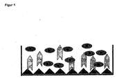
Figur 1 zeigt die schematische Durchführung eines Inhibitionstests zur Identifizierung funktionell aktiver Antikörper.
Menschliche Endothelzellen, die aus Mikrogefäßen der Haut gewonnen wurden (human dermal microvessel endothelial cells, HDMVEc) wurden zusammen mit ED-B-spezifischen Fab-Antikörperfragmenten inkubiert. Die Anzahl der gebundenen Zellen wird mittels Kristallviolett-Färbung sichtbar gemacht und im Photometer gemessen. Hohe Farbintensität bedeutet viele adhärierte Zellen und kein bindungsblockierender Antikörper. Geringe Farbintensität bedeutet wenig Adhäsion und ein bindungsblockierender Antikörper. -
Figur 2 zeigt ein Screening-Schema zur Identifizierung ED-B-Fibronektinfunktionsblockierender Antikörper mittel Fab-Fragment Display.
In einem ersten Schritt wird eine Panning-Prozedur mit rekombinantem ED-B durchgeführt. Die auf diese Weise erhaltenen Antikörperfragmente werden einem Spezifitätstest (ELISA), einem funktionellen Adhäsionstest sowie einer immunhistochemischen Untersuchung unterzogen. Danach wird die Affinität der diese Tests erfüllenden Antikörper bestimmt. Anschließend werden zur Verbesserung der Affinität Veränderungen in den CDR-Domänen durchgeführt, wobei Affinitätsmaturation 1 eine Veränderung der L-CDR3-Domäne und Affinitätsmaturation 2 eine Veränderung der H-CDR2-Domäne bedeutet.
Nachdem die durch Affinitätsmaturation erhaltenen Antikörper zuvor genannten Tests durchlaufen haben, wird anschließend die in vivo Wirksamkeit getestet. -
Figur 3 zeigt die Ergebnisse eines Spezifitätstests im ELISA-Format mit den durch Panning mit rekombinantem ED-B erzeugten Antikörper-Fab-Fragmenten.
ED-B bedeutet die Bindung an rekombinantes ED-B, 6-FN die Bindung an die rekombinante Fibronektin-Domäne 6, Domäne 7-8-9 bedeutet die Bindung an die rekombinanten Fibronektin-Domänen 7-8-9 (ohne ED-B) und Domäne 7-EDB-8-9 bedeutet die Bindung an die rekombinanten Fibronektin-Domänen 7-8-9 mit ED-B. Das Verhältnis von ED-B zu 6-FN bzw. 7-8-9 ergibt die Spezifität des untersuchten Antikörpers. AP-39 bedeutet ein kovalentes Dimer des biologisch inaktiven anti-ED-B scFv Antikörpers L19. -
Figur 4 zeigt das Ergebnis eines Adhäsionstests mit den untersuchten Antikörpern.
Es wurde die Hemmung der Adhäsion von HDMVEc-Zellen an ED-B beschichtete Platten durch die angegebenen Fab-Fragmente bei Konzentrationen 25 µg/ml bzw. 0,4 µg/ml Fab untersucht. Die angegebenen Werte sind das Ergebnis einer Dreifach-Bestimmung. -
Figur 5 zeigt das immunhistochemische Reaktionsmuster funktionsblockierender Anti-ED-B-Fab-Fragmente am Beispiel von MOR 02616. In denFiguren 5A und5C sind die Ergebnisse mit einer Negativkontrolle (negative HuCAL-Fab) und in denFiguren 5B und5D die Ergebnisse mit dem Antikörper MOR 02616 an humanen Neuroblastomzellen-IRM Xenotransplantaten (Figuren 5A und 5B ) sowie an murinen F9-Teratokarzinomzellen (Figuren 5C und 5D ) gezeigt.
Cryoschnitte der Zellen (Dicke: 10 µm) wurden luftgetrocknet und anschließend für 10 min in eiskaltem Aceton fixiert. Danach wurden die Schnitte in PBS gewaschen und für 60 min mit 2 µg/ml MOR 02616 bzw. Negativkontrolle bei Raumtemperatur inkubiert. Als Sekundärantikörper wurde ein Peroxidase-markiertes polyklonales Ziege anti Human F(ab)2 Antiserum (1:100 in PBS verdünnt, Dianova) in Kombination mit Diaminobenzidin (Sigma Chemicals) als chromogenes Substrat verwendet. -
Figur 6 zeigt die Ergebnisse eines Spezifitätstests im ELISA-Format mit den durch Maturierung optimierten Antikörper Fab-Fragmenten.
Die Versuchsdurchführung war wie inFigur 3 beschrieben. -
Figur 7 zeigt das immunhistochemische Reaktionsmuster des durch Maturierung optimierten Antikörper Fab-Fragments MOR 03255 an Cryostatschnitten von F9-Teratokarzinomzellen (Figur 7A) und an SKMEL-28 Human-Melanomzell-Xenotransplantaten in der Nacktmaus (Figur 7B).
Die Versuchsdurchführung war wie inFigur 5 beschrieben. -
Figur 8 zeigt das Ergebnis eines Adhäsionstests mit den erfindungsgemäßen Antikörper Fab-Fragmenten gegenüber den Vergleichsantikörpern L19 und Ly6 0.3.
Die Versuchsdurchführung war ähnlich wie inFigur 4 beschrieben. Die Antikörperkonzentrationen waren 1 µl/ml, 10 µl/ml bzw. 20 µl/ml. -
Figur 9 zeigt die Stabilität der erfindungsgemäßen Antikörper-Fab-Fragmente nach Inkubation für 4 h in Humanplasma bzw. PBS bei 37 °C.
Die Immunreaktivität wurde im ELISA-Test mit Konzentrationen im Bereich von 0,039 bis 5 µg/ml bestimmt. Als 100 %-Wert wurde das Signal mit 0,62 µg/ml in Humanplasma ohne Inkubation definiert. -
Figur 10 zeigt die therapeutische Wirksamkeit von Anti-ED-B-funktionsblockierenden Fab-Antikörperfragmenten am Beispiel von MOR 03255. -
Figur 11 zeigt die für die VH- und VL-Regionen der erfindungsgemäßen Antikörper kodierenden Nukleotidseqenzen. - Aufgereinigte rekombinante Fibronektin-Domäne ED-B (His)6, Fibronektin-Domäne 6 (6FN), Fibronektin-Domäne 7-8-9 und Domäne 7-ED-B-8-9 wurden nach Standardprozeduren (z.B. Sambrook et al., Molecular Cloning. A Laboratory Manual, Cold Spring Harbour Press) hergestellt.
- Mausantikörper gegen gereinigten ED-B-Rezeptor wurden ebenfalls nach Standardverfahren hergestellt, indem einer Maus 50 µg gereinigter Rezeptor in Freund's Komplett-Adjuvanz verabreicht wurden, 3 Wochen später 25 µg gereinigter Rezeptor in Freund's Inkomplett-Adjuvanz verabreicht wurden und weitere 3 Wochen später alle Applikationen in Inkomplett-Adjuvanz intraperitoneal verabreicht wurden. 14 Tage nach der 3. Immunisierung wurde dann Blut über den Retroorbitalsinus entnommen.
Plasmafibronektin wurde von Sigma bezogen. - Die Selektion wurde nach dem HuCAL® Gold Phage Selektionsprotokoll durchgeführt. Die HuCAL® Gold-Bibliothek wurde in sechs Frameworkspezifische Pools unterteilt. Die ED-B spezifischen Phagen wurden in drei aufeinander folgenden Selektionsrunden an rekombinantem Protein immobilisiert in 96-Loch-Platten (Maxisorb, Nunc, Rochester , NY, USA) angereichert. Für das Panning A wurde die Platte mit 1 µg/Loch ED-B (His6) (10 µg/ml in Ampuwa®) für 2 h bei 37 °C beschichtet. Für das Panning B wurde die Platte mit ED-B in derselben Konzentration in PBS (pH 7,2 mit 1 mM MgCl2/1 mM CaCl2) über Nacht bei 4 °C beschichtet. Die Platten wurden mit 5 % Magermilchpulver in PBS, 0,05 % Tween20 (Sigma, St. Louis, MO, USA) (Blockierungspuffer) blockiert und mit 1 x 10¹³ HuCAL® Gold Phagen inkubiert, die mit einem Volumen Blockierungspuffer vorinkubiert worden waren. Für das Panning B wurden die Phagen zusätzlich mit 0,5 µg/ml Fibronektin-Domäne 6 vorinkubiert, um die Selektion von Bindemolekülen zu verringern, die spezifisch für die konservierte Struktur eines Typ3 Domänen-Repeats von Fibronektin sind. Nach einer Inkubation mit den Phagen für 2 h bei Raumtemperatur wurden die Löcher 5x mit PBS, 0,05 % Tween20 und 5x mit PBS gewaschen. Die verbleibenden Phagen wurden 20 mM Dithiothreitol (DTT) in 10mM Tris/HCl (pH 8,0) für 10 min bei Raumtemperatur eluiert. Anschließend erfolgte eine Inkubation mit E.coli TG-1 (Stratagene, OD600 = 0,5) zur Infektion. Danach wurden die Löcher mit E.coli TG-1 als zusätzlichem Elutionsschritt inkubiert.
- Die HuCAL® Gold Phagen wurden in 2x TY-Medium mit 34 µg/ml Chloramphenicol und 1 % Glucose (2x TY-CG) amplifiziert. Nach Infektion mit einem Helferphagen (VCSM13) bei 37 °C und einer OD600 von etwa 0,5, Zentrifugation und Resuspendierung in 2x TY-CG/50 µg/ml Kanamycin wurden die Zellen mit 0,25 mM IPTG induziert und über Nacht bei 22 °C kultiviert. Die Phagen wurden mit Polyethylenglycol aus dem Überstand präzipitiert (Ausubel et al. (1998), Current Protocols of Microbiology), in PBS resuspendiert und für nachfolgende Selektionsrunden verwendet.
- Die Fab-kodierenden Insertionen der selektierten HuCAL® Gold Phagen wurden in den Expressionsvektor pMORPHx9-FS subkloniert. Die Plasmid-DNA-Präparation der selektierten HuCAL® Gold Klone wurde mit den Restriktionsenzymen Xball/EcoRI gespalten, wobei die Fab-kodierende Insertion herausgeschnitten wurde (ompA-VL und phoA-Fd). In diesem Vektor exprimierte Fab-Moleküle enthalten zwei C-terminale Markierungen (FLAG™ und Strep-tagII) zum Nachweis und zur Aufreinigung.
- Die Expression der auf pMORPHx9-FS-kodierten Fab-Fragmente in E.coli TG-1 F-Zellen wurde in Schüttelflaschenkulturen mit 0,75 I 2xTY-Medium und 34 µg/ml Chloramphenicol durchgeführt. Nach Induktion mit 0,75 mM IPTG wurden die Zellen für 16 h bei 30 °C kultiviert. Alternativ wurden Fab-Klone, die aus dem zweiten Maturationspool 2 erhalten worden waren, mit 0,1 mM IPTG induziert und anschließend bei 22 °C kultiviert. Periplasmatische Extrakte von Zellpellets wurden durch osmotischen Schock hergestellt und die Fab-Fragmente wurden durch strep-Tactin®-Chromatographie (IBA, Göttingen, Deutschland) isoliert. Die apparenten Molekulargewichte wurden durch Größenausschlusschromatographie (SEC) mit Kalibrierungsstandards bestimmt. Die Konzentrationen wurden durch UV-Spektrophotometrie ermittelt.
- 96-Loch-Maxisorb ELISA-Platten wurden mit 100 µl ED-B-Lösung (10 µg/ml in Beschichtungspuffer (PBS pH 7,4, 1 mM CaCl2, 1 mM MgCl2) über Nacht bei 4 °C beschichtet. Es wurden rohe Lysate oder aufgereinigte Fab-Moleküle zugegeben, nicht bindende Fab-Moleküle wurden durch fünfmaliges Waschen mit Waschpuffer (PBS pH 7,4, 0,05 % Tween20) entfernt. Die Fab-Fragmente wurden durch Inkubation mit Anti-human-Fab-Antikörper-Peroxidase-Konjugaten (Dianova), gefolgt von Entwicklung mit einem löslichen Peroxidasesubstrat (Roche) und Messung bei 370 nm nachgewiesen. Die für ED-B spezifische Fab-Moleküle exprimierenden Klone wurden durch ein positives ELISA-Signal auf immobilisiertem ED-B gegenüber keinem oder nur einem geringen Signal bei 6FN identifiziert.
- Die Beschichtung mit ED-B (2,5 µg/ml) wurde im Wesentlichen wie oben beschrieben durchgeführt. Die Fab-Fragmente wurden für 4 h bei 37 °C und einer Konzentration von 50 µg/ml in humanem Plasma (Deutsches Rotes Kreuz; Charge 9985550) inkubiert. Nach Inkubation wurden die Fab-Moleküle für den ELISA-Test auf die Konzentrationen von 5,0, 2,5, 1,25, 0,62, 0,31, 0,156, 0,078 und 0,039 µg/ml in PBS mit 1 % Rinderserumalbumin verdünnt, um den linearen Bereich der Signalintensität zu bestimmen. Funktionelle Fab-Moleküle wurden mit dem anti-Flag M2-Antikörper (Sigma F3165) und einem sekundären anti-Maus-Antikörper-Alkalische Phosphatase-Konjugat, gefolgt durch Entwicklung mit Attophos (Roche) und durch Messung bein 535 nm bestimmt.
- Humane dermale mikrovaskuläre Endothelialzellen (HDMVEc), isoliert aus juveniler Vorhaut, wurden in EGM-MV-Medium (Clonetics Inc.) unter Zusatz von 10 % fötalem Kälberserum, 2 mM Glutamin, 20 µg/ml Heparin und 3 ng/ml bFGF in Gefäßen, beschichtet mit 0,1 % Gelatine (Sigma), kultiviert. Für Adhäsionstests wurden Zellen verwendet, die nicht länger als Passage 8 kultiviert worden waren. ED-B (10 µg/ml) oder Plasmafibronektin (2,5 µg/ml) wurde in PBS pH 7,4, 0,9 mM CaCl2, 0,5 mM MgCl2 in 96-loch-ELISA-Platten über Nacht bei 4 °C immobilisiert und mit 1 % RSA in PBS pH 7,4, 0,9 mM CaCl2, 0,5 mM MgCl2 für 1 h bei 37 °C blockiert. Die Zellen wurden mit 0,15 µg/ml Calcein für 30 min bei 37 °C markiert und einmal in Adhäsionsmedium (MCDB 131, 2 mM Glutamin, 0,1 % RSA, 20 µg/ml Heparin) gewaschen. 1 x 10⁵ Zellen wurden mit der jeweils angegebenen Antikörperkonzentration, verdünnt in Adhäsionsmedium (Endvolumen 100 µl) für 20 min bei 37 °C inkubiert. Die Zellsuspension wurde zu den mit ED-B beschichteten Platten für 2 h bei 37 °C gegeben: Nach zweimaligem Waschen der Zellen in PBS pH 7,4, 0,9 mM CaCl2, 0,5 mM MgCl2 wurden adhärente Zellen in einem Plattenfluorimeter nachgewiesen (Anregung 485 nm; Emission 530 nm).
- Zur Kontrolle wurde (A) die Zellbindung an Ligand-beschichteten Platten ohne Zugabe von spezifischen Antikörper und (B) die Zelladhäsion an Löcher ohne Ligandenbeschichtung bestimmt, wobei der bei der letztgenannten Kontrolle erhaltene Wert als Minimum der Zelladhäsion im Test definiert wurde.
- Zur Erhöhung der Affinität und biologischen Aktivität von ausgewählten Antikörperfragmenten wurden CDR-Regionen durch Kassettenmutagenese unter Verwendung von Trinukleotid-gerichteter Mutagenese optimiert.
- Hierzu wurden Fab-Fragmente aus dem Expressionsvektor pMORPHx9 in den Phagemidvektor pMORPH-23 unter Verwendung der EcoRI/XbaI Restriktionsstellen kloniert. Zur Optimierung der Affinität der ausgewählten Fab-Fragmente wurden zwei aufeinanderfolgende Maturierungsschritte für jeden Maturierungspool durchgeführt. Im ersten Schritt wurde eine Antikörperfragment-Phagenbibliothek erzeugt, in der die L-CDR3-Region der Ausgangsklone durch ein Repertoire von 7 x 10⁸ (Pool 1) und 3,8 x 10⁸ (Pool 2) einzelnen Leichtketten-CDR-Sequenzen variiert wurde. In oder Affinität verbesserte Derivate aus dem ersten Maturierungsschritt wurden anschließend einer zweiten Maturierungsrunde ausgesetzt, indem die H-CDR-2-Region durch eine Bibliothek von diversifizierten Elementen ersetzt wurde. Auf diese Weise wurden zwei Phagenbibliotheken mit jeweils 1 x 10⁸ einzelnen Klonen erzeugt.
- Die Affinitätsmaturierungsbibliotheken wurden durch Transformation in E.coli TOP10F erzeugt. Die Phagen wurden wie zuvor beschrieben hergestellt. Die Selektion auf Fab-Fragmente mit verbesserter Affinität wurde unter stringenten Bedingungen für drei Runden bei der ersten Maturierung und für zwei Runden beim zweiten Schritt durchgeführt.
- Zur Bestimmung der KD-Werte wurden Monomerfraktionen (mindestens 80 % Monomergehalt, bestimmt durch analytische SEC; Superdex75, Amersham Pharmacia) von Fab-Fragmenten verwendet. F1-Chips (Biacore, Schweden) wurden mit ca. 200 RU ED-B (30µg/ml, 10 mM Acetatpuffer, pH 4,0) und Referenz-Flusszellen mit einer entsprechenden Menge von Humanserumalbumin (20 µg/ml, 10 mM Acetatpuffer, pH 4,5) unter Verwendung von Standard EDC-NHS-Aminkopplungschemie beschichtet. Die Antigendichte wurde für die Fab-Charakterisierung der zweiten Maturierung auf ca. 100 RU verringert. Die Regenerierung erfolgte mit 10 mM HCl. Alle kinetischen Messungen erfolgten in PBS-Puffer (136 mM NaCl, 2,7 mM KCI, 10 mM Na2HPO4, 1,76 mM KH2PO4 pH 7,4) mit einer Flussrate von 20 µl/min unter Verwendung eines Fab-Konzentrationsbereichs von 1,5-500 nM. Die Injektionzeit war jeweils 1 min. Alle Sensogramme wurden unter Verwendung der BIA-Evaluierungssoftware 3.1 ausgewertet.
- Abkürzungen: EDC: 1-Ethyl-3-(3-dimethylaminopropyl)carbodiimid, NHS: N-Hydroxysuccinimid, RU: Resonanzeinheiten
- Es wurden zwei unterschiedliche Panning-Ansätze durchgeführt. Die Beschichtung mit ED-B für den ersten Ansatz (Panning A) wurde nach den aus dem biologischen Tests bekannten Bedingungen durchgeführt, um ED-B in einer Konformation zu präsentieren, welche die Adhäsion von HDMVEc-Zellen an das immobilisierte Antigen erlaubt. Beim zweiten Panning (B) wurden die Phagen zusätzlich mit einem Überschuss an 6FN blockiert, um eine Selektion von Bindemolekülen zu vermeiden, die mit der Fibronektin-Domäne 6 kreuzreagieren.
- Insgesamt wurden 23 unterschiedliche HuCAL-Fab-Moleküle aus 1315 Primärtreffern gefunden, welche die Kriterien der Bindung an ED-B und nicht an 6FN erfüllen. Weiterhin wurden die Antikörper auf ihre Fähigkeit zur Bindung an ein rekombinant exprimiertes Fibronektin, umfassend die Domänen 7, 8 und 9 mit und ohne der inserierten ED-B-Domäne in ihrer natürlichen Anordnung getestet (siehe
Figur 3 ). Alle getesteten Antikörper zeigten eine spezifische Erkennung für das 7-ED-B-8-9-Konstrukt, reagierten jedoch nicht mit dem entsprechenden Konstrukt ohne die ED-B-Domäne. - Die im ELISA als spezifisch identifizierten Antikörper wurden aufgereinigt und auf ihre Fähigkeit zur Blockierung der Adhäsion von HDMVEc-Zellen an mit ED-B beschichtete Platten getestet. Es wurde die Adhäsion von HDMVEc-Zellen in Gegenwart von ED-B spezifischen Fab-Fragmenten in zwei Konzentrationen im Vergleich zu einem unspezifischen HuCAL-Kontroll-Fab-Molekül und positiven Mauskontrollseren getestet.
- Zwölf der 23 in Abschnitt 2.1 beschriebenen Fab-Moleküle zeigten eine Hemmung der Zelladhäsion an immobilisiertes ED-B. Eine repräsentative Auswahl der sechs wirksamsten Fab-Moleküle ist in
Figur 4 gezeigt. Bei einer Konzentration von 25 µg/ml wurde die Zelladhäsion um 50 % bis nahezu 100 % gehemmt. Bei einer Konzentration von 0,4 µg/ml zeigten nur wenige Fab-Moleküle, z.B. MOR02610, MOR02616, MOR02715 und MOR02718 eine reproduzierbare Hemmung der Zelladhäsion. - Die funktionsblockierenden Fab-Antikörperfragmente wurden immunhistochemisch hinsichtlich ihrer Lokalisierung auf F9-Maus-Teratokarzimomzellen und humanen IRM30 Neuroblastom-Xenotransplantat-Cryoschnitten charakterisiert. Alle getesteten Antikörper zeigten eine vaskuläre Lokalisierung auf beiden Gewebeschnitten, aber keine Anfärbung von Gefäßen auf Normalgewebe (z.B. normale und sklerotische Leber und Haut). In Figur
Figur 5B und5D ist eine perivaskuläre abluminale Anfärbung von Gefäßen, proliferierendem Endothel und Tumorstroma durch MOR02616 gezeigt. Eine Ansammlung von ED-B in der Umgebung neovaskulärer Strukturen von exponentiell wachsenden Tumoren, aber nicht auf normalen Gefäßen, ist zu erkennen. Mit einem negativen Kontrollantikörper gegen Lysozym bei derselben Konzentration war keine Anfärbung sichtbar (Figuren 5A und5C ). - Die Eigenschaften von zwölf ausgewählten Bindemolekülen wurde durch BIAcore-Analyse bestimmt (siehe Tabelle 1). Die Affinitäten (Kon) lagen im Bereich von 15 bis 500 nM. Die Affinität des bivalenten L19-Derivats AP39 (eines biologisch inaktiven Vergleichsantikörpers gegen ED-B) wurde mit 2,4 nM bestimmt. Tabelle 1 zeigt auch die CDR-Sequenzen (nur L-CDR3 und H-CDR3) und die Framework-Kombinationen. Es wurden mindestens zwei unabhängige Messungen mit Fab-Molekülen aus unterschiedlichen Ansätzen zur Proteinexpression und -aufreinigung durchgeführt. Die Bindungsrate (Kon) und die Abdissoziationsrate (Koff) der Fab-Molekole ist in separaten Spalten angegeben.
Tabelle 1 Clone VH H-CDR3 VL L-CDR3 kon
[1/Ms]koff
[1/s]KD
[nM]AP 39 VH3 PFPY--FDY K3 QQTGRI--PP 6,6E+05 1,5E-03 2,4 ±0,6 MOR02610 VH1B SPVYYKYDY L1 QSYDKTSSTY 1,3E+06 9,1E-02 75 ±19 MOR02611 VH1B GLYYR-FAS L2 AAATG---GW 1,5E+05 5,1E-02 332 ±23 MOR02613 VH3 YVNG--FDI L2 QTYAKKDYSL 7,8E+05 1,3E-01 164 ±14 MOR02614 VH3 A-----YDV L1 AMFSP---EG 2,8E+05 4,2E-02 160 ±6 MOR02616 VH3 VIVL--FDY K3 LQKYSI--PF 5,4E+05 2.8E-02 59±25 MOR02618 VH3 NYWV--FAY L3 QSYDNFNDSV 4,3E+05 2,0E-01 477 ± 177 MOR02M9 VH4 F-----FDV L1 QSWDGAS-TG 7,8E+05 1,2E-02 15,4±0,6 MOR02622 VH3 GLVT--FDN L2 SSWTHSFTDY 2,4E+05 5,1E-03 21,3±1.7 MOR02715 VH3 NKVG--FDV L2 QAWDNQGMKY 8,9E±05 4,7E-02 53,7 ±1,7. MOR02718 VH3 GWF---FAH k3 FQYSSV--PL 4,5E±05 3,9E-02 85±12,5 MOR02721 VH5 YH----GAF L1 QAYTTG--SI 4,3E+05 2,4E-02 56±11,6 MOR02722 VH3 F-----IAS K2 QQYSNF--PF 1,5E+05 1,4E-02 94 ± 2.3 - Die Affinitätsmaturierungen der Fab-Moleküle wurden in zwei Schritten durchgeführt. Zunächst wurde die L-CDR3-Sequenz diversifiziert und anschließend H-CDR2-Sequenz der im ersten Schritt erhaltenen, verbesserten Fab-Moleküle ausgetauscht.
- Es wurden zwei Affinitätsmaturationsbibliotheken für jeden Schritt erzeugt. Die Optimierung wurde in zwei separaten Pools durchgeführt. Die L-CDR3-Bibliothek 1 mit einem Diversitätsgrad von 7 x 10⁸ Elementen enthielt die vier Ausgangs-Fab-Moleküle MOR02610, MOR02616, MOR02619 und MOR02622. Die L-CDR3-Bibiliothek 2 mit einem Diversitätsgrad von 3,8 x 10⁸ Elementen enthielt nur Derivate von MOR02715 und MOR02718. Die Fab-Moleküle wurden dabei jeweils entsprechend ihrer Affinität und biologischen Aktivität im Adhäsionstest in Gruppen zusammengefasst.
- Beide Bibiliotheken wurden während der Selektitinsprozedur separat gehalte. Es wurden dabei zwei Panning-Strategien durchgeführt. Das Panning I erfolgte mit ED-B immobilisiert an Maxisorb-Platten für drei Runden, wie zuvor angegeben. Beim Panning II erfolgte eine Selektion an biotinyliertes ED-B in Lösung. Dabei wurde der Phage-Antigenkomplex durch Streptavidin-beschichtete Partikel in der ersten Runde und an Neutravidin-Platten in den zwei nachfolgenden Runden abgefangen. Im Rahmen der Selektionsprozedur wurden stringente Bedingungen verwendet.
- Das Screening auf optimierte Fab-Moleküle erfolgte durch Bestimmung der Koff-Werte in BlAcore-System. Insgesamt wurden 11 Derivate der Bibiliothek 1 charakterisiert. Die 5 Derivate mit der höchsten Affinität sind in Tabelle 2 angegeben. Bei allen verbesserten Klonen handelte es sich dabei um Derivate von MOR02619 mit einer bis zu 7fachen Affinitätserhöhung. Beim Klon MOR03075 wurde eine Affinität von 2,4 nM gefunden.
- Aus der Bibiliothek 2 wurden 2 Derivate von MOR02715 ausführlich analysiert. Für den Klon MOR03062 wurde eine 15fache Affinitätserhöhung gefunden, so dass eine Monomeraffinität von 2,4 nM erhalten wurde. Die Veränderungen in der Aminosäuresequenz der L-CDR3-Region ist in den Tabellen 2a und 2b gezeigt.
Tabelle 2a Panning Pool 1 MOR0 Stammklon LCDR3 kon
[1/Ms]koff
[1/s]KD
[nm]x fache Verbesserung gegenüber Stammklon kon koff KD AP 39-Biotin QQTGRI -- PP 6,6E + 05 1,49E - 003 2,4 ± 0,8 2619 QSWDGAS-TG 7,8E + 05 1,2E -02 15,4 ± 0,6 fest 3055 2619 QAWTRAHRYP 1,8E+08 5,1E , 03 3,0 ± 0,5 2,2 2,6 5,4 gelöst 3066 2619 SSYD-TQVTR 1,1E ± 06 4,7E - 03 4,2 ± 0,6 1,4 2,8 3,8 gelöst 3075 2619 QSWDP-RSFT 1,1E + 08 2,6E -03 2,4 ± 0,3 1,4 5.0 6,8 gelöst 3069 2619 WTGM - - SYHF 1,2E + 06 4,1E - 03 3.310,2 1,5 3,2 4,8 gelöst 3071 2619 LAYIQS-KGH 1,9E+06 5,7E - 03 3,1±0,8 2,3 2,3 5,1 Tabelle 2b Panning Pool 2 MOR0 Stammklon LCDR3 kon
[1/Ms]koff
[1/s]KD
[nm]× fache Verbesserung gegenüber Stammklon kon koff KD AP 39-Biotin QQTGRI--PP 6,75E + 05 1,49E -03 2,4 ± 0,8 2715 QAWDNQGMKY 9,9E + 05 4,7E-02 53,7 ± 1,7 fest 3064 2715 SWDLLAPSV 1,5E + 06 1,4E -02 10 ± 3,5 1,7 3,5 5,3 fest 3062 2715 QSWDLSVHQV 3,5E+ 0,6 1,1 E-02 3,4 ± 0,8 4,0 4,2 15,8 - Die Spezifität für ED-B (bestimmt durch ELISA, Immunhistochemie und Zelladhäsionstest) aller Derivate aus der ersten Maturierungsrunde waren unverändert.
- Die in Tabellen 2a und 2b gezeigten Fab-Moleküle wurden für eine anschließende H-CDR2-Maturierung in zwei Pools ausgewählt. In Pool 1 wurden fünf Fab-Moleküle mit einer ähnlichen Aktivität, aber unterschiedlichen L-CDR3-Regionen einer Diversifizierung in H-CDR2 unterzogen (Bibiliotheksgröße von 7 x 10⁷ Elementen). Für Pool 2 (Bibiliotheksgröße von 8,5 x 10⁷ Elementen) wurden zwei Fab-Moleküle ausgewählt. Die Bibliotheken wurden separat wie oben beschrieben selektioniert. Bindemoleküle mit erhöhter Affinität wurden durch zwei Panningrunden in Lösung unter stringenten Bedingungen angereichert.
- Aus Pool 1 wurden drei Fab-Moleküle ausgewählt. Für die Derivate MOR03243 und MOR03245 wurden Affinitäten von etwa 0,7 nM gefunden. Für das Derivat MOR03246 wurde eine Affinität von 0,6 nM gefunden.
- Die H-CDR2-Sequenzen der entsprechenden Klone und die Affinitätsdaten sind in Tabelle 3a gezeigt.
Tabelle 3a MOR0 Parental Clone Sequenz HCDR2 kon [1/Ms] koff [1/s] KD [nM] MOR03243 3055 EIHRGGYTQYNPSLKS 2,9E+06 1,8E-03 0,7 ± 0.2 MOR03245 3069 VIHKWGFTNYNPSLKS 3,5E+06 1,6E-03 0,7 ± 0,3 MOR03246 3075 YIHKYGWTKYNPSLKS 4,8E+06 3,0E-03 0,6 ± 0,1 - Auch aus Pool 2 konnten Derivate mit verbesserten Eigenschaften selektioniert werden. Im Vergleich zu den Stammklonen waren die Affinitäten der Derivate um einen Faktor von 26 bis zu 250fach verbessert. Die Affinitäten der Klone MOR03255 und MOR03258 wurden mit 30 pM bzw. 40 pM bestimmt.
- In Tabelle 3b sind H-CDR2-Aminosäuresequenzen der selektierten Klone aus Pool 2 sowie die entsprechenden Affinitätsdaten gezeigt.
Tabelle 3b MOR0 Parental Clone Sequenz HCDR2 kon [1/Ms] koff [1/s] KD [nM] STDEV MOR03251 3062 VISNMSYTIYYADSVKG 3,3E+06 2,0E-04 0,07 0,04 MOR03252 3062 VISNYSWHIYYADSVKG 3,1E+08 6,1E-05 0,03 0,03 MOR03253 3062 Mut VISNMGFEIYYADSVKG 1,6E+08 2,0E-04 0,13 0,05 MOR03255 3062 VISNRSSYIYYADSVKG 4,9E+06 8,6E-05 0,03 0,03 MOR03257 3062 VISNRGNYIYYADSVKG 5,3E+06 3,0E-04 0,08 0,06 MOR03258 3064 VISNQSNYIYYADSVKG 2,7E+06 8,8E-05 0,04 0,02 - Um zu testen, ob die durch Affinitätsmaturierung erhaltenen Antikörper immer noch für ED-B spezifisch sind, wurden Spezifitätstests im ELISA-Format (
Figur 6 ), immunhistochemische Untersuchungen (Figur 7 ) und Adhäsionstests (Figur 8 ) durchgeführt. In allen drei Tests wurde gefunden, dass die Spezifität der affinitätsmaturierten Fab-Moleküle für ED-B unverändert blieb. AusFigur 8 ist weiterhin ersichtlich, dass sowohl die Stammklone wie MOR 02715 als auch die durch Affinitätsmaturierung erzeugten Klone wie MOR 03062, MOR 03255, MOR 03064 und MOR 03259 eine signifikant höhere biologische Aktivität (Adhäsionshemmung) als der bekannte Antikörper L19 aufweisen. - Zusätzlich wurde die thermische Stabilität der Fab-Moleküle in Humanplasma getestet. Hierzu wurden die Fab-Moleküle mit humanem Plasma für 4 h bei 37°C inkubiert, ohne dass ein signifikanter Verlust an Immunreaktivität bestimmt durch ELISA (
Figur 9 ) gefunden wurde. - Alle-selektierten Fab-Moleküle sind in der Lage, die Wechselwirkung eines Rezeptors auf humanen mikrovaskulären Endothelzellen an ED-B auf dosisabhängige Weise zu blockieren, eine Eigenschaft, der bekannte ED-B-Antikörper L19 nicht aufweist. Da die Adhäsion von HDMVEc an die extrazelluäre Matrix einer der frühen Schritte in der Neoangiogenese ist, stellt die Blockierung dieses Vorgangs durch die erfindungsgemäßen Bindemoleküle ein therapeutisches Mittel zur Hemmung der Bildung von neuen Gefäßen und zur Hemmung des Tumorwachstums dar.
- F9-Teratokarzinomazellen (106/Maus in Matrigel mit/ohne 100µg Fab MOR 03255) wurden sc. in die Flanke von Nacktmäusen implantiert. Nach 3 Tagen wurden die Tiere mit jeweils 100 µg MOR03255/Maus i.v. für 7 aufeinanderfolgendeTage behandelt. Beide Behandlungsgruppen zeigen im Vergleich zur Lösungsmittelkontrolle eine.Wachstumshemmung des Tumors im Bereich von 42-64 %. Eine Zusammenfassung der Versuchsergebnisse ist in
Figur 10 dargestellt. -
- (1)Brown LF, Guidi AJ, Schnitt SJ, Van De Water L, Iruela-Arispe ML, Yeo T-K, Tognazzi K, Dvorak HF (1999) Clin. Cancer Res. 5: 1041-1056
- (2)Folkman J (1995) Nature (Med.) 1: 27-31
- (3)Risau W und Lemmon V (1988) Develop. Biol. 125: 441-450
- (4)Van Den Hoff A (1988) Adv. Cancer Res. 50: 159-196
- (5)Folkman J und Shing Y (1992) J. Biol. Chem. 267: 10931-10934
- (6)George EL, Baldwin HS, Hynes RO (1997) Blood 90: 3073-3081
- (7)Bowersox JC und Sorgente N (1982) Cancer Res. 42: 2547-2551
- (8)Madri JA, Pratt BM, Tucker AM (1988) J. Cell Biol. 106: 1375- 1384
- (9)Nicosia RF, Bonnano E, Smith M (1993) J. Cell. Physiol. 154: 654-661
- (10)Kornblihtt AR, Vibe-Pedersen K und Baralle FE, 1983. Isolation and characterization of cDNA clones for human and bovine fibronectins. Proc. Natl. Acad. Sci. USA, 80, 3218-22
- (11)Leahy DJ, Hendrickson WA, Aukhil I und Erickson HP. 1992. Structure of fibronectin type III domain from tenascin phased by MAS analysis of the selenomethionyl protein. Science, 258, 987-91
- (12)Hynes R.O., 1987, Cell 48, 549-550
- (13)Plow E.F. et al. 2000, J. Biol. Chem., 275, 21785-21788
- (14)Castellani P, Viale G, Dorcaratto A, Nicolo G, Kaczmarek J, Querze G, Zardi L (1994) Int. J. Cancer 59: 612-618
- (15)Hashimoto-Uoshima M, Yan YZ, Schneider G, Aukhil I (1997) J. Cell Science 110: 227-2280
- (16)Chen W and Culp LA (1996) Exp. Cell Res. 223: 9-19
- (17)Chen W and Culp LA (1998) Clin. Exp. Metastasis 16: 30-42
-
- <110> Schering Aktiengesellschaft
- <120> Identifizierung und charakterisierung von funktionsblockierenden anti-ED-B-Fibronektin Antikörpern
- <130> 33042P DE (WWHC)
- <140>
<141> - <160> 90
- <170> PatentIn ver. 2.1
- <210> 1
<211> 354
<212> DNA
<213> human - <220>
<221> CDS
<222> (1)..(354)
<223> Antikörper-VH-Region; MOR02610 - <400> 1

- <210> 2
<211> 118
<212> PRT
<213> human - <400> 2


- <210> 3
<211> 336
<212> DNA
<213> human - <220>
<221> CDS
<222> (1) .. (336)
<223> Antikörper-VL-Region; MOR02610 - <400> 3

- <210> 4
<211> 112
<212> PRT
<213> human - <400> 4

- <210> 5
<211> 351
<212> DNA
<213> human - <220>
<221> CDS
<222> (1)..(351)
<223> Antikörper-VH-Region; MOR02611 - <400> 5

- <210> 6
<211> 117
<212> PRT
<213> human - <400> 6

- <210> 7
<211> 327
<212> DNA
<213> human - <220>
<221> CDS
<222> (1)..(327)
<223> Antikörper-VL-Region; MOR02611 - <400> 7


- <210> 8
<211> 109
<212> PRT
<213> human - <400> 8

- <210> 9
<211> 348
<212> DNA
<213> human - <220>
<221> CDS
<222> (1)..(348)
<223> Antikörper-VH-Region; MOR02613 - <400> 9


- <210> 10
<211> 116
<212> PRT
<213> human - <400> 10

- <210> 11
<211> 339
<212> DNA
<213> human - <220>
<221> CDS
<222> (1)..(339)
<223> Antikörper-VL-Region; MOR02613 - <400> 11


- <210> 12
<211> 113
<212> PRT
<213> human - <400> 12

- <210> 13
<211> 339
<212> DNA
<213> human - <220>
<221> CDS
<222> (1) .. (339)
<223> Antikörper-VH-Region; MOR02614 - <400> 13


- <210> 14
<211> 113
<212> PRT
<213> human - <400> 14

- <210> 15
<211> 327
<212> DNA
<213> human - <220>
<221> CDS
<222> (1)..(327)
<223> Antikörper-VL-Region; MOR02614 - <400> 15

- <210> 16
<211> 109
<212> PRT
<213> human - <400> 16

- <210> 17
<211> 348
<212> DNA
<213> human - <220>
<221> CDS
<222> (1)..(348)
<223> Antikörper-VH-Region; MOR02616 - <400> 17

- <210> 18
<211> 116
<212> PRT
<213> human - <400> 18

- <210> 19
<211> 327
<212> DNA
<213> human - <220>
<221> CDS
<222> (1)..(327)
<223> Antikörper-VL-Region; MOR02616 - <400> 19

- <210> 20
<211> 109
<212> PRT
<213> human - <400> 20

- <210> 20
<211> 109
<212> PRT
<213> human - <400> 20

- <211> 348
<212> DNA
<213> human - <220>
<221> CDS
<222> (1)..(348)
<223> Antikörper-VH-Region; MOR02618 - <400> 21

- <210> 22
<211> 116
<212> PRT
<213> human - <400> 22


- <210> 23
<211> 330
<212> DNA
<213> human - <220>
<221> CDS
<222> (1)..(330)
<223> Antikörper-VL-Region; MOR02618 - <400> 23

- <210> 24
<211> 110
<212> PRT
<213> human - <400> 24


- <210> 25
<211> 333
<212> DNA
<213> human - <220>
<221> CDS
<222> (1)..(333)
<223> Antikörper-VH-Region; MOR02619 - <400> 25

- <210> 26
<211> 111
<212> PRT
<213> human - <400> 26


- <210> 27
<211> 333
<212> DNA
<213> human - <220>
<221> CDS
<222> (1)..(333)
<223> Antikörper-VL-Region; MOR02619 - <400> 27

- <210> 28
<211> 111
<212> PRT
<213> human - <400> 28


- <210> 29
<211> 348
<212> DNA
<213> human - <220>
<221> CDS
<222> (1)..(348)
<223> Antikörper-VH-Region; MOR02622 - <400> 29

- <210> 30
<211> 116
<212> PRT
<213> human - <400> 30


- <210> 31
<211> 339
<212> DNA
<213> human - <220>
<221> CDS
<222> (1)..(339)
<223> Antikörper-VL-Region; MOR02622 - <400> 31

- <210> 32
<211> 113
<212> PRT
<213> human - <400> 32


- <210> 33
<211> 348
<212> DNA
<213> human - <220>
<221> CDS
<222> (1)..(348)
<223> Antikörper-VH-Region; MOR02715 - <400> 33

- <210> 34
<211> 116
<212> PRT
<213> human - <400> 34

- <210> 35
<211> 339
<212> DNA
<213> human - <220>
<221> CDS
<222> (1)..(339)
<223> Antikörper-VL-Region; MOR02715 - <400> 35


- <210> 36
<211> 113
<212> PRT
<213> human - <400> 36

- <210> 37
<211> 345
<212> DNA
<213> human - <220>
<221> CDS
<222> (1)..(345)
<223> Antikörper-VH-Region; MOR02718 - <400> 37


- <210> 38
<211> 115
<212> PRT
<213> human - <400> 38

- <210> 39
<211> 327
<212> DNA
<213> human - <220>
<221> CDS
<222> (1)..(327)
<223> Antikörper-VL-Region; MOR02718 - <400> 39


- <210> 40
<211> 109
<212> PRT
<213> human - <400> 40

- <210> 41
<211> 342
<212> DNA
<213> human - <220>
<221> CDS
<222> (1)..(342)
<223> Antikörper-VH-Region; MOR02721 - <400> 41


- <210> 42
<211> 114
<212> PRT
<213> human - <400> 42

- <210> 43
<211> 342
<212> DNA
<213> human - <220>
<221> CDS
<222> (1)..(342)
<223> Antikörper-VL-Region; MOR02721 - <400> 43


- <210> 44
<211> 114
<212> PRT
<213> human - <400> 44

- <210> 45
<211> 339
<212> DNA
<213> human - <220>
<221> CDS
<222> (1)..(339)
<223> Antikörper-VH-Region; MOR02722 - <400> 45

- <210> 46
<211> 113
<212> PRT
<213> human - <400> 46

- <210> 47
<211> 342
<212> DNA
<213> human - <220>
<221> CDS
<222> (1)..(342)
<223> Antikörper-VL-Region; MOR02722 - <400> 47

- <210> 48
<211> 114
<212> PRT
<213> human - <400> 48


- <210> 49
<211> 336
<212> DNA
<213> human - <220>
<221> CDS
<222> (1)..(336)
<223> Antikörper-VL-Region; MOR03055 - <400> 49

- <210> 50
<211> 112
<212> PRT
<213> human - <400> 50


- <210> 51
<211> 333
<212> DNA
<213> human - <220>
<221> CDS
<222> (1)..(333)
<223> Antikörper-VL-Region; MOR03066 - <400> 51

- <210> 52
<211> 111
<212> PRT
<213> human - <400> 52


- <210> 53
<211> 333
<212> DNA
<213> human - <220>
<221> CDS
<222> (1)..(333)
<223> Antikörper-VL-Region; MOR03075 - <400> 53

- <210> 54
<211> 111
<212> PRT
<213> human - <400> 54
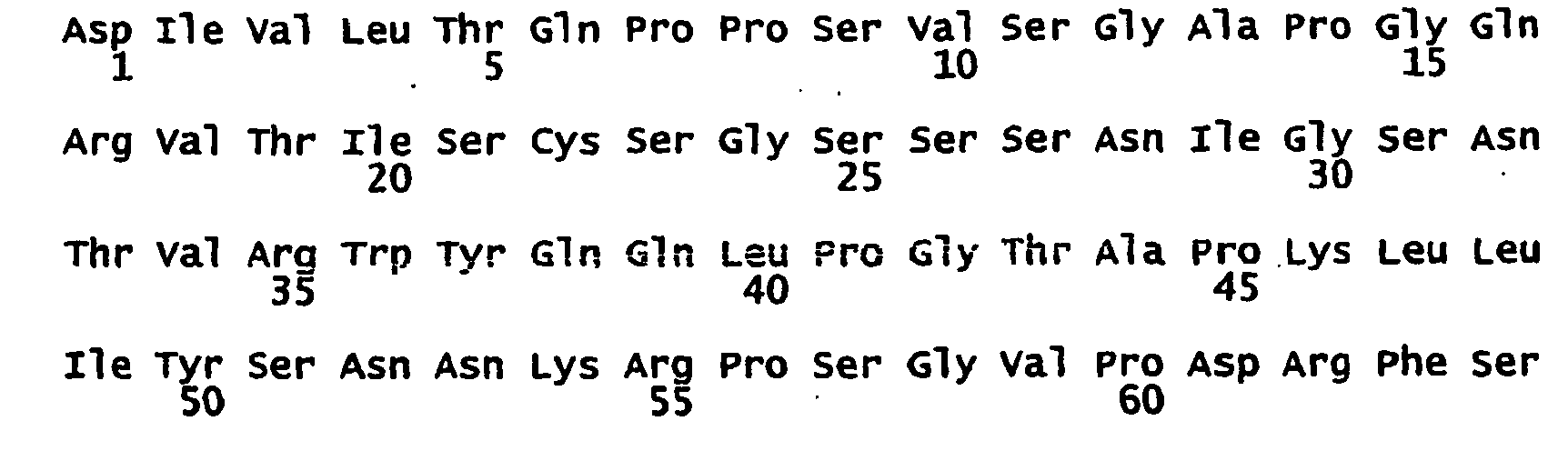

- <210> 55
<211> 330
<212> DNA
<213> human - <220>
<221> CDS
<222> (1)..(330)
<223> Antikörper-VL-Region; MOR03069 - <400> 55

- <210> 56
<211> 110
<212> PRT
<213> human - <400> 56


- <210> 57
<211> 333
<212> DNA
<213> human - <220>
<221> CDS
<222> (1)..(333)
<223> Antikörper-VL-Region; MOR03071 - <400> 57

- <210> 58
<211> 111
<212> PRT
<213> human - <400> 58


- <210> 59
<211> 339
<212> DNA
<213> human - <220>
<221> CDS
<222> (1)..(339)
<223> Antikörper-VL-Region; MOR03064 - <400> 59

- <210> 60
<211> 113
<212> PRT
<213> human - <400> 60


- <210> 61
<211> 339
<212> DNA
<213> human - <220>
<221> CDS
<222> (1)..(339)
<223> Antikörper-VL-Region; MOR03062 - <400> 61

- <210> 62
<211> 113
<212> PRT
<213> human - <400> 62

- <210> 63
<211> 336
<212> DNA
<213> human - <220>
<221> CDS
<222> (1)..(336)
<223> Derivat von MOR03055: Antikörper-VH-Region; MOR03243 - <400> 63

- <210> 64
<211> 112
<212> PRT
<213> human - <400> 64

- <210> 65
<211> 336
<212> DNA
<213> human - <220>
<221> CDS
<222> (1)..(336)
<223> Derivat von MOR03055: Antikörper-VL-Region; MOR03243 - <400> 65


- <210> 66
<211> 112
<212> PRT
<213> human - <400> 66
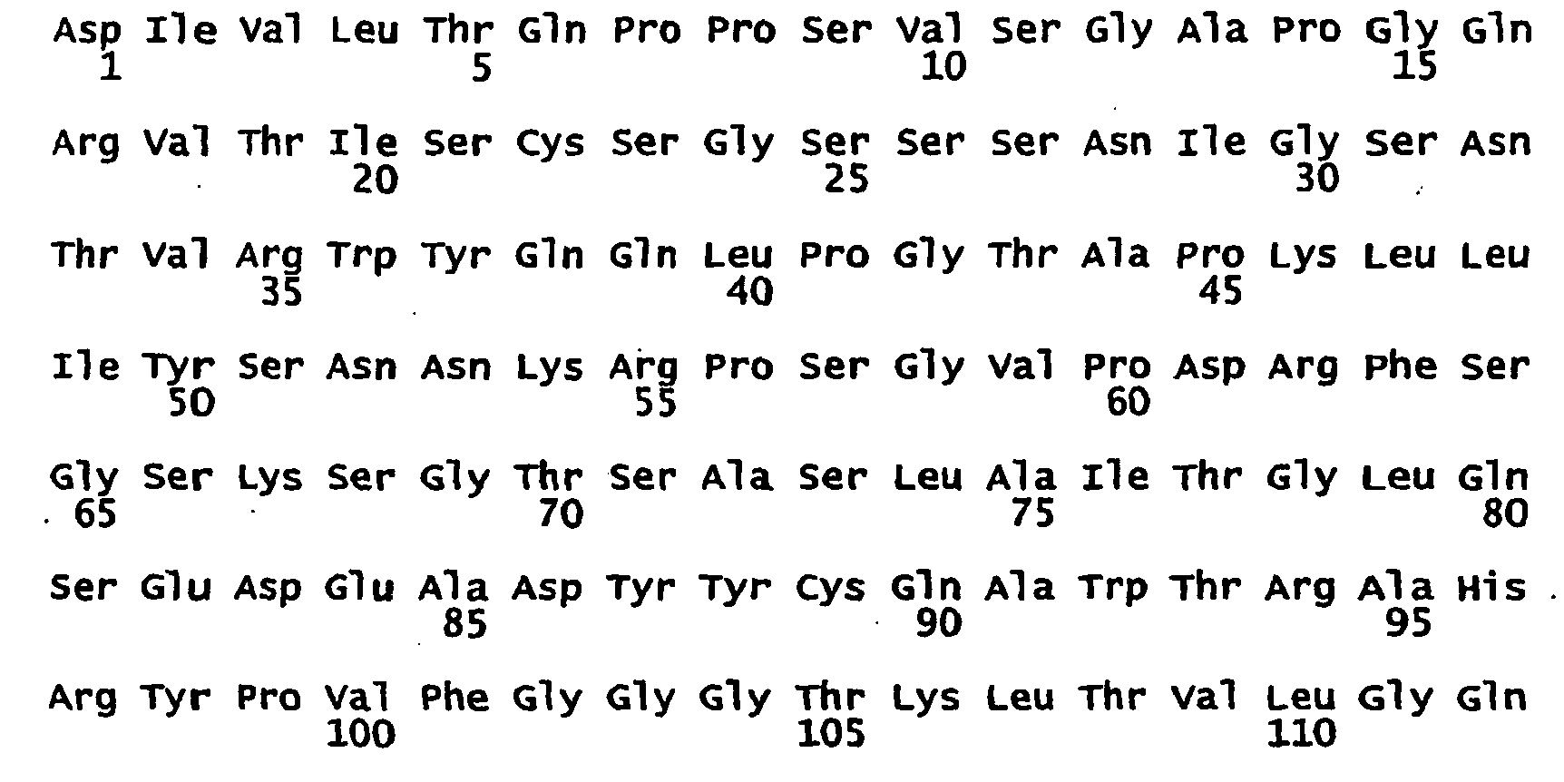
- <210> 67
<211> 336
<212> DNA
<213> human - <220>
<221> CDS
<222> (1)..(336)
<223> Derivat von MOR03069; Antikörper-vH-Region; MOR03245 - <400> 67


- <210> 68
<211> 112
<212> PRT
<213> human - <400> 68

- <210> 69
<211> 330
<212> DNA
<213> human - <220>
<221> CDS
<222> (1)..(330)
<223> Derivat von MOR03069; Antikörper-VL-Region; MOR03245 - <400> 69


- <210> 70
<211> 110
<212> PRT
<213> human - <400> 70

- <210> 71
<211> 354
<212> DNA
<213> human - <220>
<221> CDS
<222> (1) .. (354)
<223> Derivat von MOR03075; Antikörper-VH-Region; MOR03246 - <400> 71


- <210> 72
<211> 118
<212> PRT
<213> human - <400> 72

- <210> 73
<211> 333
<212> DNA
<213> human - <220>
<221> CDS
<222> (1)..(333)
<223> Derivat von MOR03075; Antikörper-VL-Region; MOR03246 - <400> 73


- <210> 74
<211> 111
<212> PRT
<213> human - <400> 74

- <210> 75
<211> 348
<212> DNA
<213> human - <220>
<221> CDS
<222> (1)..(348)
<223> Derivat von MOR03062; Antikörper-VH-Region; MOR03251 - <400> 75


- <210> 76
<211> 116
<212> PRT
<213> human - <400> 76

- <210> 77
<211> 339
<212> DNA
<213> human - <220>
<221> CDS
<222> (1)..(339)
<223> Derivat von MOR03062; Antikörper-VL-Region; MOR03251 - <400> 77

- <210> 78
<211> 113
<212> PRT
<213> human - <400> 78

- <210> 79
<211> 348
<212> DNA
<213> human - <220>
<221> CDS
<222> (1)..(348)
<223> Antikörper-VH-Region; MOR03252 - <400> 79

- <210> 80
<211> 116
<212> PRT
<213> human - <400> 80


- <210> 81
<211> 348
<212> DNA
<213> human - <220>
<221> CDS
<222> (1) .. (348)
<223> Antikörper-VH-Region; MOR03253 - <400> 81

- <210> 82
<211> 116
<212> PRT
<213> human - <400> 82


- <210> 83
<211> 348
<212> DNA
<213> human - <220>
<221> CDS
<222> (1)..(348)
<223> Antikörper-VH-Region; MOR03255 - <400> 83

- <210> 84
<211> 116
<212> PRT
<213> human - <400> 84


- <210> 85
<211> 348
<212> DNA
<213> human - <220>
<221> CDS
<222> (1)..(348)
<223> Antikörper-VH-Region; MOR03257 - <400> 85

- <210> 86
<211> 116
<212> PRT
<213> human - <210> 86
<211> 116
<212> PRT
<213> human - <400> 86

- <210> 87
<211> 348
<212> DNA
<213> human - <220>
<221> CDS
<222> (1)..(348)
<223> Derivat von MOR03075; Antikörper-VH-Region; MOR03258 - <400> 87


- <210> 88
<211> 116
<212> PRT
<213> human - <400> 88

- <210> 89
<211> 354
<212> DNA
<213> human - <220>
<221> CDS
<222> (1)..(354)
<223> Derivat von MOR03075; Antikörper-VL-Region; MOR03258 - <400> 89


- <210> 90
<211> 118
<212> PRT
<213> human - <400> 90

Claims (19)
- Antikörper oder Antikörperfragment,
der bzw. das spezifisch an die ED-B Domäne von Fibronektin bindet
dadurch gekennzeichnet,
dass er bzw. es die VH-Region kodiert von SEQ ID NO. 75 (MOR 03251) und die VL-Region kodiert von SEQ ID NO. 77 (MOR 03251) oder die H-CDR1-, H-CDR2- und H-CDR3- Region aus dieser VH Region und die L-CDR1-, L-CDR2- und L-CDR3- Region aus dieser VL Region aufweist, wobei der Antikörper oder das Antikörperfragment die ED-B-Domäne mit einem KD-Wert von 0,1 nM oder niedriger bindet und der KD-Wert durch Oberflächenplasmonenresonanz ermittelt wird, und die Interaktion zwischen der ED-B-Domäne und ihrem Rezeptor inhibiert. - Antikörper oder Antikörperfragment,
der bzw. das spezifisch an die ED-B Domäne von Fibronektin bindet,
dadurch gekennzeichnet,
dass er bzw. es die VH-Region kodiert von SEQ ID NO. 79 (MOR 03252) und die VL-Region kodiert von SEQ ID NO. 77 (MOR 03252) oder die H-CDR1-, H-CDR2- und H-CDR3- Region aus dieser VH Region und die L-CDR1-, L-CDR2- und L-CDR3- Region aus dieser VL Region aufweist, wobei der Antikörper oder das Antikörperfragment die ED-B-Domäne mit einem KD-Wert von 0,1 nM oder niedriger bindet und der KD-Wert durch Oberflächenplasmonenresonanz ermittelt wird, und die Interaktion zwischen der ED-B-Domäne und ihrem Rezeptor inhibiert. - Antikörper oder Antikörperfragment,
der bzw. das spezifisch an die ED-B Domäne von Fibronektin bindet"
dadurch gekennzeichnet,
dass er bzw. es die VH-Region kodiert von SEQ ID NO. 83 (MOR 03255) und die VL-Region kodiert von SEQ ID NO. 77 (MOR 03255) oder die H-CDR1-, H-CDR2- und H-CDR3- Region aus dieser VH Region und die L-CDR1-, L-CDR2- und L-CDR3- Region aus dieser VL Region aufweist, wobei der Antikörper oder das Antikörperfragment die ED-B-Domäne mit einem KD-Wert von 0,1 nM oder niedriger bindet und der KD-Wert durch Oberflächenplasmonenresonanz ermittelt wird, und die Interaktion zwischen der ED-B-Domäne und ihrem Rezeptor inhibiert. - Antikörper oder Antikörperfragment,
der bzw. das spezifisch an die ED-B Domäne von Fibronektin bindet,
dadurch gekennzeichnet,
dass er bzw. es die VH-Region kodiert von SEQ ID NO. 85 (MOR 03257) und die VL-Region kodiert von SEQ ID NO. 77 (MOR 03257) oder die H-CDR1-, H-CDR2- und H-CDR3- Region aus dieser VH Region und die L-CDR1-, L-CDR2- und L-CDR3- Region aus dieser VL Region aufweist, wobei der Antikörper oder das Antikörperfragment die ED-8-Domäne mit einem KO-Wert von 0,1 nM oder niedriger bindet und der KD-Wert durch Oberflächenplasmonenresonanz ermittelt wird, und die Interaktion zwischen der ED-B-Domäne und ihrem Rezeptor inhibiert. - Antikörper oder Antikörperfragment,
der bzw. das spezifisch an die ED-B Domäne von Fibronektin bindet,
dadurch gekennzeichnet,
dass er bzw. es die VH-Region kodiert von SEQ ID NO. 87 (MOR 03258) und die VL-Region kodiert von SEQ ID NO. 59 (MOR 03258) oder die H-COR1-, H-CDR2- und H-CDR3- Region aus dieser VH Region und die L-CDR1-, L-CDR2- und L-CDR3- Region aus dieser VL Region aufweist, wobei der Antikörper oder das Antikörperfragment die ED-B-Domäne mit einem KD-Wert von 0,1 nM oder niedriger bindet und der KD-Wert durch Oberflächenplasmonenresonanz ermittelt wird, und die Interaktion zwischen der ED-B-Domäne und ihrem Rezeptor inhibiert. - Antikörper oder Antikörperfragment nach einem der Ansprüche 1 bis 5 in Form eines Konjugats mit einem therapeutischen Wirkstoff.
- Antikörper oder Antikörperfragment nach Anspruch 6,
dadurch gekennzeichnet,
dass der therapeutische Wirkstoff ausgewählt ist aus Radio- und Chemotherapeutika. - Antikörper oder Antikörperfragment nach einem der Ansprüche 1 bis 5 in Form eines Fusionsproteins.
- Antikörper oder Antikörperfragment nach einem der Ansprüche 1 bis 5 in Form eines Konjugats mit einer diagnostisch nachweisbaren Markierungsgruppe.
- Pharmazeutische Zusammensetzung,
dadurch gekennzeichnet,
dass sie als Wirkstoff einen Antikörper oder Antikörperfragment nach einem der Ansprüche 1 bis 8 sowie pharmakologisch übliche Träger, Hilfsstoffe oder/und Verdünnungsmittel enthält. - Diagnostische Zusammensetzung, welche als diagnostisches Reagenz einen Antikörper oder Antikörperfragment nach einem der Ansprüche 1 bis 5, oder 9 enthält.
- Nukleinsäure, die für ein Antikörper oder Antikörperfragment nach einem der Ansprüche 1 bis 5 oder 8 kodiert.
- Nukleinsäure nach Anspruch 12 in operativer Verknüpfung mit einer Expressionskontrollsequenz.
- Vektor,
dadurch gekennzeichnet,
dass er eine Nukleinsäure nach einem der Ansprüche 12 bis 13 enthält. - Isolierte Zelle,
dadurch gekennzeichnet,
dass sie mit einer Nukleinsäure nach einem der Ansprüche 12 bis 13 oder einem Vektor nach Anspruch 14 transformiert ist. - Nicht-humaner Organismus,
dadurch gekennzeichnet,
dass er mit einer Nukleinsäure nach einem der Ansprüche 12 bis 13 oder einem Vektor nach Anspruch 14 transformiert ist. - Verfahren zur Herstellung eines Antikörpers oder Antikörperfragments nach einem der Ansprüche 1 bis 5,
dadurch gekennzeichnet,
dass man eine Zelle nach Anspruch 15 oder einen nicht-humanen Organismus nach Anspruch 16 unter Bedingungen kultiviert, bei denen eine Expression des Antikörpers oder Antikörperfragments erfolgt, und der exprimierte Antikörper oder Antikörperfragment gewonnen wird. - Verwendung eines Antikörpers oder Antikörperfragments nach einem der Ansprüche 1 bis 8 zur Herstellung eines Arzneimittels zur Prävention oder Behandlung von Erkrankungen die mit einer ED-B Expression einhergehen und die ausgewählt sind aus hyperproliferativen Erkrankungen, Erkrankungen, die mit einer Fehlfunktion von Myofibroblasten assoziiert sind, und entzündlichen Erkrankungen.
- Verwendung eines Antikörpers oder Antikörperfragments nach einem der Ansprüche 1 bis 9 zur Herstellung eines Nachweisreagenzes zur Diagnose von Erkrankungen die mit einer ED-B Expression einhergehen und die ausgewählt sind aus hyperproliferativen Erkrankungen, Erkrankungen, die mit einer Fehlfunktion von Myofibroblasten assoziiert sind, und entzündlichen Erkrankungen, oder einer Prädisposition dafür.
Applications Claiming Priority (1)
| Application Number | Priority Date | Filing Date | Title |
|---|---|---|---|
| PCT/EP2005/012185 WO2007054120A1 (de) | 2005-11-09 | 2005-11-09 | Identifizierung und charakterisierung von funktionsblockierenden anti-ed-b-fibronektin antikörpern |
Publications (2)
| Publication Number | Publication Date |
|---|---|
| EP1945767A1 EP1945767A1 (de) | 2008-07-23 |
| EP1945767B1 true EP1945767B1 (de) | 2010-04-07 |
Family
ID=36170357
Family Applications (1)
| Application Number | Title | Priority Date | Filing Date |
|---|---|---|---|
| EP05811334A Expired - Lifetime EP1945767B1 (de) | 2005-11-09 | 2005-11-09 | Identifizierung und charakterisierung von funktionsblockierenden anti-ed-b-fibronektin antikörpern |
Country Status (8)
| Country | Link |
|---|---|
| EP (1) | EP1945767B1 (de) |
| JP (1) | JP2009514540A (de) |
| CN (1) | CN101356271A (de) |
| AT (1) | ATE463512T1 (de) |
| BR (1) | BRPI0520677A2 (de) |
| CA (1) | CA2628552A1 (de) |
| DE (1) | DE502005009389D1 (de) |
| WO (1) | WO2007054120A1 (de) |
Families Citing this family (23)
| Publication number | Priority date | Publication date | Assignee | Title |
|---|---|---|---|---|
| DE19932688B4 (de) | 1999-07-13 | 2009-10-08 | Scil Proteins Gmbh | Design von Beta-Faltblatt-Proteinen des gamma-II-kristallins antikörperähnlichen |
| DE10324447A1 (de) | 2003-05-28 | 2004-12-30 | Scil Proteins Gmbh | Generierung künstlicher Bindungsproteine auf der Grundlage von Ubiquitin |
| EP1925664A1 (de) | 2006-11-15 | 2008-05-28 | Scil proteins GmbH | Künstliche Bindungsproteine auf Grundlage einer modifizierten alpha-Helix Region von Ubiquitin |
| JP2011517314A (ja) | 2008-02-14 | 2011-06-02 | ブリストル−マイヤーズ スクイブ カンパニー | Egfrに結合する操作されたタンパク質に基づく標的化された治療薬 |
| TWI496582B (zh) | 2008-11-24 | 2015-08-21 | 必治妥美雅史谷比公司 | 雙重專一性之egfr/igfir結合分子 |
| KR101436222B1 (ko) | 2009-12-14 | 2014-09-01 | 쉴 프로테인즈 게엠베하 | 리간드 결합능력을 가진 헤테로-다량체 변형 유비퀴틴 단백질을 확인하는 방법 |
| US9562089B2 (en) | 2010-05-26 | 2017-02-07 | Bristol-Myers Squibb Company | Fibronectin based scaffold proteins having improved stability |
| WO2012171541A1 (en) | 2011-06-15 | 2012-12-20 | Scil Proteins Gmbh | Human fusion proteins comprising interferons and hetero-dimeric modified ubiquitin proteins |
| EP2721152B1 (de) | 2011-06-15 | 2019-03-27 | Navigo Proteins GmbH | Dimere bindeproteine auf basis modifizierter ubiquitine |
| WO2013186329A1 (en) | 2012-06-13 | 2013-12-19 | Scil Proteins Gmbh | Human fusion proteins comprising single chain tnfalpha and targeting domains |
| WO2014094799A1 (en) | 2012-12-19 | 2014-06-26 | Scil-Proteins-Gmbh | Ubiquitin moieties as a means for prolonging serum half-life |
| EP3253785B1 (de) | 2015-02-06 | 2019-04-10 | Navigo Proteins Gmbh | Neuartige egfr-bindende proteine |
| CN107922483B (zh) | 2015-07-16 | 2021-07-30 | 纳维格蛋白质有限公司 | 新型免疫球蛋白结合蛋白及其在亲和纯化中的用途 |
| WO2017013136A1 (en) | 2015-07-20 | 2017-01-26 | Scil Proteins Gmbh | Novel binding proteins based on di-ubiquitin muteins and methods for generation |
| WO2017084017A1 (zh) * | 2015-11-16 | 2017-05-26 | 合肥立方制药股份有限公司 | Ed-b蛋白在诊断组织增生中的应用 |
| AU2017260274A1 (en) | 2016-05-04 | 2018-11-01 | Navigo Proteins Gmbh | Targeted compounds for the site-specific coupling of chemical moieties comprising a peptide linker |
| CA3030208A1 (en) | 2016-08-11 | 2018-02-15 | Navigo Proteins Gmbh | Novel alkaline stable immunoglobulin binding proteins |
| WO2018217351A1 (en) | 2017-05-24 | 2018-11-29 | Tets Viktor Veniaminovich | Methods for treating and preventing diseases |
| US11192943B2 (en) | 2017-09-30 | 2021-12-07 | Hefei Lifeon Pharmaceutical Co., Ltd. | Protein binding to fibronectin B domain |
| US12535491B2 (en) | 2018-04-30 | 2026-01-27 | Viktor Veniaminovich Tets | Vaccine comprising protein-targeted (PT) extracellular DNA isolated from bacterial matrix |
| WO2020006357A1 (en) | 2018-06-29 | 2020-01-02 | Tets Viktor Veniaminovich | Methods for diagnosis and treatment of type 1 diabetes |
| WO2020010214A1 (en) | 2018-07-03 | 2020-01-09 | Tets Viktor Veniaminovich | Method for assessing and inhibiting aging and means of determining biological age |
| CA3123986A1 (en) | 2018-12-18 | 2020-06-25 | Navigo Proteins Gmbh | Novel folr1 specific binding proteins for cancer diagnosis and treatment |
Family Cites Families (6)
| Publication number | Priority date | Publication date | Assignee | Title |
|---|---|---|---|---|
| ATE219517T1 (de) | 1995-08-18 | 2002-07-15 | Morphosys Ag | Protein-/(poly)peptidbibliotheken |
| TWI259837B (en) * | 1998-05-11 | 2006-08-11 | Eidgenossische Tech Hochscule | Specific binding molecules for scintigraphy, conjugates containing them and therapeutic method for treatment of angiogenesis |
| EP1990409A3 (de) | 1999-07-20 | 2011-05-18 | MorphoSys AG | Bakteriophage |
| CA2395567A1 (en) * | 2000-01-12 | 2001-07-19 | Light Sciences Corporation | Novel treatment for eye disease |
| AU4243201A (en) | 2000-02-24 | 2001-09-03 | Eidgenoess Tech Hochschule | Antibody specific for the ed-b domain of fibronectin, conjugates comprising saidantibody, and their use for the detection and treatment of angiogenesis |
| PL364358A1 (en) | 2000-09-07 | 2004-12-13 | Schering Ag | Receptor in the edb fibronectin domain (ii) |
-
2005
- 2005-11-09 EP EP05811334A patent/EP1945767B1/de not_active Expired - Lifetime
- 2005-11-09 JP JP2008539258A patent/JP2009514540A/ja not_active Withdrawn
- 2005-11-09 DE DE502005009389T patent/DE502005009389D1/de not_active Expired - Lifetime
- 2005-11-09 BR BRPI0520677-4A patent/BRPI0520677A2/pt not_active IP Right Cessation
- 2005-11-09 AT AT05811334T patent/ATE463512T1/de not_active IP Right Cessation
- 2005-11-09 WO PCT/EP2005/012185 patent/WO2007054120A1/de not_active Ceased
- 2005-11-09 CN CNA2005800524909A patent/CN101356271A/zh active Pending
- 2005-11-09 CA CA002628552A patent/CA2628552A1/en not_active Abandoned
Also Published As
| Publication number | Publication date |
|---|---|
| CN101356271A (zh) | 2009-01-28 |
| EP1945767A1 (de) | 2008-07-23 |
| DE502005009389D1 (de) | 2010-05-20 |
| ATE463512T1 (de) | 2010-04-15 |
| BRPI0520677A2 (pt) | 2009-05-19 |
| JP2009514540A (ja) | 2009-04-09 |
| WO2007054120A1 (de) | 2007-05-18 |
| CA2628552A1 (en) | 2007-05-18 |
Similar Documents
| Publication | Publication Date | Title |
|---|---|---|
| EP1945767A1 (de) | Identifizierung und charakterisierung von funktionsblockierenden anti-ed-b-fibronektin antikörpern | |
| DE69934515T2 (de) | Humanisierte monoklonale inetegrin antikörper | |
| KR101078459B1 (ko) | 항 수초 관련 당단백질 항체 | |
| JP2023002706A (ja) | 抗pdl1抗体、活性化可能抗pdl1抗体、およびその使用方法 | |
| AU2009245354C1 (en) | Antibodies to receptor of advanced glycation end products (RAGE) and uses thereof | |
| DE60012500T2 (de) | Antikörper und antikörperfragmente mit spezifität für tgfbeta1 | |
| DE69611766T2 (de) | Spezifische Bindungspartner für menschliches TGF-beta, Materialien und Verfahren | |
| DE69829891T2 (de) | Anti-VEGF Antikörper | |
| DE60226036T9 (de) | ANTIKÖRPER, DER DAS GM1-GANGLIOSID-GEBUNDENE AMYLOID-b-PROTEIN ERKENNT, UND DNA, DIE FÜR DIESEN ANTIKÖRPER CODIERT | |
| DE69934241T2 (de) | Agonistische antikörper gegen tie2 | |
| JP5951498B2 (ja) | 網膜神経線維層変性の治療に使用するためのrgmaタンパク質に対するモノクローナル抗体 | |
| CN104661677A (zh) | 抗Jagged 1/Jagged 2交叉反应抗体、可活化的抗Jagged抗体及其使用方法 | |
| CN106163556A (zh) | 基质金属蛋白酶底物和其它可切割部分及其使用方法 | |
| DE212016000236U1 (de) | Humaner Endothelinrezeptor bindender Antikörper und dessen Verwendung | |
| DE69828154T2 (de) | Anti-alphavbeta3 humanizierte monoklonale antikörper | |
| CN116249547B (zh) | 脑特异性血管生成抑制剂1(bai1)抗体及其用途 | |
| US20070031418A1 (en) | Methods for treating lower motor neuron diseases and compositions containing the same | |
| DE60226253T2 (de) | Humanisierter antikörper gegen fibroblastenwachstumsfaktor 8 und fragment des antikörpers | |
| US8017115B2 (en) | Therapeutical use of anti-myelin associated glycoprotein (MAG) antibodies | |
| US7785591B2 (en) | Identification and characterization of function-blocking anti-ED-B-fibronectin antibodies | |
| DE102004050101A1 (de) | Identifizierung und Charakterisierung von funktionsblockierenden anti-ED-B-Fibronektin Antikörpern | |
| RU2303461C2 (ru) | Антитела против миелин-ассоциированного гликопротеина (mag) | |
| KR20080068108A (ko) | 작용-차단성 항-ed-b-피브로넥틴 항체의 동정 및 특징화 | |
| LV13485B (en) | Identification and improvement of edb-fibronectin function blocking antibodies | |
| NZ544443A (en) | A antibody characterised in that it binds specifically to ED-B domain of fibronectin and inhibits the interaction with its receptor |
Legal Events
| Date | Code | Title | Description |
|---|---|---|---|
| PUAI | Public reference made under article 153(3) epc to a published international application that has entered the european phase |
Free format text: ORIGINAL CODE: 0009012 |
|
| 17P | Request for examination filed |
Effective date: 20080507 |
|
| AK | Designated contracting states |
Kind code of ref document: A1 Designated state(s): AT BE BG CH CY CZ DE DK EE ES FI FR GB GR HU IE IS IT LI LT LU LV MC NL PL PT RO SE SI SK TR |
|
| RIN1 | Information on inventor provided before grant (corrected) |
Inventor name: PRASSLER, JOSEF Inventor name: MENRAD, ANDREAS Inventor name: WEIDMANN, ARMIN |
|
| 17Q | First examination report despatched |
Effective date: 20081211 |
|
| GRAP | Despatch of communication of intention to grant a patent |
Free format text: ORIGINAL CODE: EPIDOSNIGR1 |
|
| RIC1 | Information provided on ipc code assigned before grant |
Ipc: G01N 33/563 20060101ALI20090915BHEP Ipc: C07K 16/18 20060101AFI20090915BHEP Ipc: C07K 16/30 20060101ALI20090915BHEP Ipc: G01N 33/574 20060101ALI20090915BHEP Ipc: A61P 35/00 20060101ALI20090915BHEP Ipc: A61K 39/395 20060101ALI20090915BHEP Ipc: C07K 14/78 20060101ALN20090915BHEP |
|
| DAX | Request for extension of the european patent (deleted) | ||
| RAP1 | Party data changed (applicant data changed or rights of an application transferred) |
Owner name: MORPHOSYS AG |
|
| GRAS | Grant fee paid |
Free format text: ORIGINAL CODE: EPIDOSNIGR3 |
|
| GRAA | (expected) grant |
Free format text: ORIGINAL CODE: 0009210 |
|
| AK | Designated contracting states |
Kind code of ref document: B1 Designated state(s): AT BE BG CH CY CZ DE DK EE ES FI FR GB GR HU IE IS IT LI LT LU LV MC NL PL PT RO SE SI SK TR |
|
| REG | Reference to a national code |
Ref country code: GB Ref legal event code: FG4D Free format text: NOT ENGLISH |
|
| REG | Reference to a national code |
Ref country code: CH Ref legal event code: EP |
|
| REG | Reference to a national code |
Ref country code: IE Ref legal event code: FG4D Free format text: LANGUAGE OF EP DOCUMENT: GERMAN |
|
| REF | Corresponds to: |
Ref document number: 502005009389 Country of ref document: DE Date of ref document: 20100520 Kind code of ref document: P |
|
| REG | Reference to a national code |
Ref country code: NL Ref legal event code: VDEP Effective date: 20100407 |
|
| PG25 | Lapsed in a contracting state [announced via postgrant information from national office to epo] |
Ref country code: SI Free format text: LAPSE BECAUSE OF FAILURE TO SUBMIT A TRANSLATION OF THE DESCRIPTION OR TO PAY THE FEE WITHIN THE PRESCRIBED TIME-LIMIT Effective date: 20100407 |
|
| LTIE | Lt: invalidation of european patent or patent extension |
Effective date: 20100407 |
|
| REG | Reference to a national code |
Ref country code: IE Ref legal event code: FD4D |
|
| PG25 | Lapsed in a contracting state [announced via postgrant information from national office to epo] |
Ref country code: SE Free format text: LAPSE BECAUSE OF FAILURE TO SUBMIT A TRANSLATION OF THE DESCRIPTION OR TO PAY THE FEE WITHIN THE PRESCRIBED TIME-LIMIT Effective date: 20100407 Ref country code: NL Free format text: LAPSE BECAUSE OF FAILURE TO SUBMIT A TRANSLATION OF THE DESCRIPTION OR TO PAY THE FEE WITHIN THE PRESCRIBED TIME-LIMIT Effective date: 20100407 Ref country code: ES Free format text: LAPSE BECAUSE OF FAILURE TO SUBMIT A TRANSLATION OF THE DESCRIPTION OR TO PAY THE FEE WITHIN THE PRESCRIBED TIME-LIMIT Effective date: 20100718 Ref country code: LT Free format text: LAPSE BECAUSE OF FAILURE TO SUBMIT A TRANSLATION OF THE DESCRIPTION OR TO PAY THE FEE WITHIN THE PRESCRIBED TIME-LIMIT Effective date: 20100407 |
|
| PG25 | Lapsed in a contracting state [announced via postgrant information from national office to epo] |
Ref country code: IS Free format text: LAPSE BECAUSE OF FAILURE TO SUBMIT A TRANSLATION OF THE DESCRIPTION OR TO PAY THE FEE WITHIN THE PRESCRIBED TIME-LIMIT Effective date: 20100807 Ref country code: FI Free format text: LAPSE BECAUSE OF FAILURE TO SUBMIT A TRANSLATION OF THE DESCRIPTION OR TO PAY THE FEE WITHIN THE PRESCRIBED TIME-LIMIT Effective date: 20100407 Ref country code: LV Free format text: LAPSE BECAUSE OF FAILURE TO SUBMIT A TRANSLATION OF THE DESCRIPTION OR TO PAY THE FEE WITHIN THE PRESCRIBED TIME-LIMIT Effective date: 20100407 |
|
| PG25 | Lapsed in a contracting state [announced via postgrant information from national office to epo] |
Ref country code: GR Free format text: LAPSE BECAUSE OF FAILURE TO SUBMIT A TRANSLATION OF THE DESCRIPTION OR TO PAY THE FEE WITHIN THE PRESCRIBED TIME-LIMIT Effective date: 20100708 Ref country code: CY Free format text: LAPSE BECAUSE OF FAILURE TO SUBMIT A TRANSLATION OF THE DESCRIPTION OR TO PAY THE FEE WITHIN THE PRESCRIBED TIME-LIMIT Effective date: 20100526 Ref country code: PL Free format text: LAPSE BECAUSE OF FAILURE TO SUBMIT A TRANSLATION OF THE DESCRIPTION OR TO PAY THE FEE WITHIN THE PRESCRIBED TIME-LIMIT Effective date: 20100407 |
|
| PG25 | Lapsed in a contracting state [announced via postgrant information from national office to epo] |
Ref country code: EE Free format text: LAPSE BECAUSE OF FAILURE TO SUBMIT A TRANSLATION OF THE DESCRIPTION OR TO PAY THE FEE WITHIN THE PRESCRIBED TIME-LIMIT Effective date: 20100407 Ref country code: DK Free format text: LAPSE BECAUSE OF FAILURE TO SUBMIT A TRANSLATION OF THE DESCRIPTION OR TO PAY THE FEE WITHIN THE PRESCRIBED TIME-LIMIT Effective date: 20100407 Ref country code: PT Free format text: LAPSE BECAUSE OF FAILURE TO SUBMIT A TRANSLATION OF THE DESCRIPTION OR TO PAY THE FEE WITHIN THE PRESCRIBED TIME-LIMIT Effective date: 20100809 Ref country code: IE Free format text: LAPSE BECAUSE OF FAILURE TO SUBMIT A TRANSLATION OF THE DESCRIPTION OR TO PAY THE FEE WITHIN THE PRESCRIBED TIME-LIMIT Effective date: 20100407 |
|
| PLBE | No opposition filed within time limit |
Free format text: ORIGINAL CODE: 0009261 |
|
| STAA | Information on the status of an ep patent application or granted ep patent |
Free format text: STATUS: NO OPPOSITION FILED WITHIN TIME LIMIT |
|
| PG25 | Lapsed in a contracting state [announced via postgrant information from national office to epo] |
Ref country code: RO Free format text: LAPSE BECAUSE OF FAILURE TO SUBMIT A TRANSLATION OF THE DESCRIPTION OR TO PAY THE FEE WITHIN THE PRESCRIBED TIME-LIMIT Effective date: 20100407 Ref country code: SK Free format text: LAPSE BECAUSE OF FAILURE TO SUBMIT A TRANSLATION OF THE DESCRIPTION OR TO PAY THE FEE WITHIN THE PRESCRIBED TIME-LIMIT Effective date: 20100407 Ref country code: CZ Free format text: LAPSE BECAUSE OF FAILURE TO SUBMIT A TRANSLATION OF THE DESCRIPTION OR TO PAY THE FEE WITHIN THE PRESCRIBED TIME-LIMIT Effective date: 20100407 |
|
| 26N | No opposition filed |
Effective date: 20110110 |
|
| PG25 | Lapsed in a contracting state [announced via postgrant information from national office to epo] |
Ref country code: IT Free format text: LAPSE BECAUSE OF FAILURE TO SUBMIT A TRANSLATION OF THE DESCRIPTION OR TO PAY THE FEE WITHIN THE PRESCRIBED TIME-LIMIT Effective date: 20100407 |
|
| BERE | Be: lapsed |
Owner name: MORPHOSYS A.G. Effective date: 20101130 |
|
| PG25 | Lapsed in a contracting state [announced via postgrant information from national office to epo] |
Ref country code: MC Free format text: LAPSE BECAUSE OF NON-PAYMENT OF DUE FEES Effective date: 20101130 |
|
| REG | Reference to a national code |
Ref country code: CH Ref legal event code: PL |
|
| GBPC | Gb: european patent ceased through non-payment of renewal fee |
Effective date: 20101109 |
|
| PG25 | Lapsed in a contracting state [announced via postgrant information from national office to epo] |
Ref country code: LI Free format text: LAPSE BECAUSE OF NON-PAYMENT OF DUE FEES Effective date: 20101130 Ref country code: CH Free format text: LAPSE BECAUSE OF NON-PAYMENT OF DUE FEES Effective date: 20101130 |
|
| REG | Reference to a national code |
Ref country code: FR Ref legal event code: ST Effective date: 20110801 |
|
| PG25 | Lapsed in a contracting state [announced via postgrant information from national office to epo] |
Ref country code: BE Free format text: LAPSE BECAUSE OF NON-PAYMENT OF DUE FEES Effective date: 20101130 |
|
| REG | Reference to a national code |
Ref country code: DE Ref legal event code: R119 Ref document number: 502005009389 Country of ref document: DE Effective date: 20110601 Ref country code: DE Ref legal event code: R119 Ref document number: 502005009389 Country of ref document: DE Effective date: 20110531 |
|
| PG25 | Lapsed in a contracting state [announced via postgrant information from national office to epo] |
Ref country code: FR Free format text: LAPSE BECAUSE OF NON-PAYMENT OF DUE FEES Effective date: 20101130 |
|
| PG25 | Lapsed in a contracting state [announced via postgrant information from national office to epo] |
Ref country code: GB Free format text: LAPSE BECAUSE OF NON-PAYMENT OF DUE FEES Effective date: 20101109 |
|
| REG | Reference to a national code |
Ref country code: AT Ref legal event code: MM01 Ref document number: 463512 Country of ref document: AT Kind code of ref document: T Effective date: 20101109 |
|
| PG25 | Lapsed in a contracting state [announced via postgrant information from national office to epo] |
Ref country code: AT Free format text: LAPSE BECAUSE OF NON-PAYMENT OF DUE FEES Effective date: 20101109 |
|
| PG25 | Lapsed in a contracting state [announced via postgrant information from national office to epo] |
Ref country code: HU Free format text: LAPSE BECAUSE OF FAILURE TO SUBMIT A TRANSLATION OF THE DESCRIPTION OR TO PAY THE FEE WITHIN THE PRESCRIBED TIME-LIMIT Effective date: 20101008 Ref country code: LU Free format text: LAPSE BECAUSE OF NON-PAYMENT OF DUE FEES Effective date: 20101109 Ref country code: BG Free format text: LAPSE BECAUSE OF FAILURE TO SUBMIT A TRANSLATION OF THE DESCRIPTION OR TO PAY THE FEE WITHIN THE PRESCRIBED TIME-LIMIT Effective date: 20100407 |
|
| PG25 | Lapsed in a contracting state [announced via postgrant information from national office to epo] |
Ref country code: TR Free format text: LAPSE BECAUSE OF FAILURE TO SUBMIT A TRANSLATION OF THE DESCRIPTION OR TO PAY THE FEE WITHIN THE PRESCRIBED TIME-LIMIT Effective date: 20100407 |
|
| PG25 | Lapsed in a contracting state [announced via postgrant information from national office to epo] |
Ref country code: DE Free format text: LAPSE BECAUSE OF NON-PAYMENT OF DUE FEES Effective date: 20110531 |
|
| PG25 | Lapsed in a contracting state [announced via postgrant information from national office to epo] |
Ref country code: BG Free format text: LAPSE BECAUSE OF FAILURE TO SUBMIT A TRANSLATION OF THE DESCRIPTION OR TO PAY THE FEE WITHIN THE PRESCRIBED TIME-LIMIT Effective date: 20100707 |