EP1960432B1 - Anticorps anti-ICAM induisant l'apoptose. - Google Patents
Anticorps anti-ICAM induisant l'apoptose. Download PDFInfo
- Publication number
- EP1960432B1 EP1960432B1 EP06829616.9A EP06829616A EP1960432B1 EP 1960432 B1 EP1960432 B1 EP 1960432B1 EP 06829616 A EP06829616 A EP 06829616A EP 1960432 B1 EP1960432 B1 EP 1960432B1
- Authority
- EP
- European Patent Office
- Prior art keywords
- cancer
- cells
- igg
- binding
- cell
- Prior art date
- Legal status (The legal status is an assumption and is not a legal conclusion. Google has not performed a legal analysis and makes no representation as to the accuracy of the status listed.)
- Active
Links
Images







Classifications
-
- A—HUMAN NECESSITIES
- A61—MEDICAL OR VETERINARY SCIENCE; HYGIENE
- A61K—PREPARATIONS FOR MEDICAL, DENTAL OR TOILETRY PURPOSES
- A61K39/00—Medicinal preparations containing antigens or antibodies
- A61K39/395—Antibodies; Immunoglobulins; Immune serum, e.g. antilymphocytic serum
-
- C—CHEMISTRY; METALLURGY
- C07—ORGANIC CHEMISTRY
- C07K—PEPTIDES
- C07K16/00—Immunoglobulins [IG], e.g. monoclonal or polyclonal antibodies
- C07K16/18—Immunoglobulins [IG], e.g. monoclonal or polyclonal antibodies against material from animals or humans
- C07K16/28—Immunoglobulins [IG], e.g. monoclonal or polyclonal antibodies against material from animals or humans against receptors, cell surface antigens or cell surface determinants
- C07K16/2803—Immunoglobulins [IG], e.g. monoclonal or polyclonal antibodies against material from animals or humans against receptors, cell surface antigens or cell surface determinants against the immunoglobulin superfamily
- C07K16/2821—Immunoglobulins [IG], e.g. monoclonal or polyclonal antibodies against material from animals or humans against receptors, cell surface antigens or cell surface determinants against the immunoglobulin superfamily against ICAM molecules, e.g. CD50, CD54, CD102
-
- C—CHEMISTRY; METALLURGY
- C07—ORGANIC CHEMISTRY
- C07K—PEPTIDES
- C07K16/00—Immunoglobulins [IG], e.g. monoclonal or polyclonal antibodies
- C07K16/18—Immunoglobulins [IG], e.g. monoclonal or polyclonal antibodies against material from animals or humans
- C07K16/28—Immunoglobulins [IG], e.g. monoclonal or polyclonal antibodies against material from animals or humans against receptors, cell surface antigens or cell surface determinants
- C07K16/2803—Immunoglobulins [IG], e.g. monoclonal or polyclonal antibodies against material from animals or humans against receptors, cell surface antigens or cell surface determinants against the immunoglobulin superfamily
-
- A—HUMAN NECESSITIES
- A61—MEDICAL OR VETERINARY SCIENCE; HYGIENE
- A61P—SPECIFIC THERAPEUTIC ACTIVITY OF CHEMICAL COMPOUNDS OR MEDICINAL PREPARATIONS
- A61P19/00—Drugs for skeletal disorders
- A61P19/02—Drugs for skeletal disorders for joint disorders, e.g. arthritis, arthrosis
-
- A—HUMAN NECESSITIES
- A61—MEDICAL OR VETERINARY SCIENCE; HYGIENE
- A61P—SPECIFIC THERAPEUTIC ACTIVITY OF CHEMICAL COMPOUNDS OR MEDICINAL PREPARATIONS
- A61P19/00—Drugs for skeletal disorders
- A61P19/04—Drugs for skeletal disorders for non-specific disorders of the connective tissue
-
- A—HUMAN NECESSITIES
- A61—MEDICAL OR VETERINARY SCIENCE; HYGIENE
- A61P—SPECIFIC THERAPEUTIC ACTIVITY OF CHEMICAL COMPOUNDS OR MEDICINAL PREPARATIONS
- A61P29/00—Non-central analgesic, antipyretic or antiinflammatory agents, e.g. antirheumatic agents; Non-steroidal antiinflammatory drugs [NSAID]
-
- A—HUMAN NECESSITIES
- A61—MEDICAL OR VETERINARY SCIENCE; HYGIENE
- A61P—SPECIFIC THERAPEUTIC ACTIVITY OF CHEMICAL COMPOUNDS OR MEDICINAL PREPARATIONS
- A61P31/00—Antiinfectives, i.e. antibiotics, antiseptics, chemotherapeutics
-
- A—HUMAN NECESSITIES
- A61—MEDICAL OR VETERINARY SCIENCE; HYGIENE
- A61P—SPECIFIC THERAPEUTIC ACTIVITY OF CHEMICAL COMPOUNDS OR MEDICINAL PREPARATIONS
- A61P31/00—Antiinfectives, i.e. antibiotics, antiseptics, chemotherapeutics
- A61P31/04—Antibacterial agents
-
- A—HUMAN NECESSITIES
- A61—MEDICAL OR VETERINARY SCIENCE; HYGIENE
- A61P—SPECIFIC THERAPEUTIC ACTIVITY OF CHEMICAL COMPOUNDS OR MEDICINAL PREPARATIONS
- A61P31/00—Antiinfectives, i.e. antibiotics, antiseptics, chemotherapeutics
- A61P31/12—Antivirals
- A61P31/14—Antivirals for RNA viruses
- A61P31/18—Antivirals for RNA viruses for HIV
-
- A—HUMAN NECESSITIES
- A61—MEDICAL OR VETERINARY SCIENCE; HYGIENE
- A61P—SPECIFIC THERAPEUTIC ACTIVITY OF CHEMICAL COMPOUNDS OR MEDICINAL PREPARATIONS
- A61P35/00—Antineoplastic agents
-
- A—HUMAN NECESSITIES
- A61—MEDICAL OR VETERINARY SCIENCE; HYGIENE
- A61P—SPECIFIC THERAPEUTIC ACTIVITY OF CHEMICAL COMPOUNDS OR MEDICINAL PREPARATIONS
- A61P35/00—Antineoplastic agents
- A61P35/02—Antineoplastic agents specific for leukemia
-
- A—HUMAN NECESSITIES
- A61—MEDICAL OR VETERINARY SCIENCE; HYGIENE
- A61P—SPECIFIC THERAPEUTIC ACTIVITY OF CHEMICAL COMPOUNDS OR MEDICINAL PREPARATIONS
- A61P37/00—Drugs for immunological or allergic disorders
-
- A—HUMAN NECESSITIES
- A61—MEDICAL OR VETERINARY SCIENCE; HYGIENE
- A61P—SPECIFIC THERAPEUTIC ACTIVITY OF CHEMICAL COMPOUNDS OR MEDICINAL PREPARATIONS
- A61P37/00—Drugs for immunological or allergic disorders
- A61P37/02—Immunomodulators
-
- A—HUMAN NECESSITIES
- A61—MEDICAL OR VETERINARY SCIENCE; HYGIENE
- A61P—SPECIFIC THERAPEUTIC ACTIVITY OF CHEMICAL COMPOUNDS OR MEDICINAL PREPARATIONS
- A61P37/00—Drugs for immunological or allergic disorders
- A61P37/02—Immunomodulators
- A61P37/06—Immunosuppressants, e.g. drugs for graft rejection
-
- A—HUMAN NECESSITIES
- A61—MEDICAL OR VETERINARY SCIENCE; HYGIENE
- A61P—SPECIFIC THERAPEUTIC ACTIVITY OF CHEMICAL COMPOUNDS OR MEDICINAL PREPARATIONS
- A61P43/00—Drugs for specific purposes, not provided for in groups A61P1/00-A61P41/00
-
- C—CHEMISTRY; METALLURGY
- C07—ORGANIC CHEMISTRY
- C07K—PEPTIDES
- C07K16/00—Immunoglobulins [IG], e.g. monoclonal or polyclonal antibodies
- C07K16/18—Immunoglobulins [IG], e.g. monoclonal or polyclonal antibodies against material from animals or humans
- C07K16/28—Immunoglobulins [IG], e.g. monoclonal or polyclonal antibodies against material from animals or humans against receptors, cell surface antigens or cell surface determinants
-
- C—CHEMISTRY; METALLURGY
- C07—ORGANIC CHEMISTRY
- C07K—PEPTIDES
- C07K16/00—Immunoglobulins [IG], e.g. monoclonal or polyclonal antibodies
- C07K16/18—Immunoglobulins [IG], e.g. monoclonal or polyclonal antibodies against material from animals or humans
- C07K16/28—Immunoglobulins [IG], e.g. monoclonal or polyclonal antibodies against material from animals or humans against receptors, cell surface antigens or cell surface determinants
- C07K16/2803—Immunoglobulins [IG], e.g. monoclonal or polyclonal antibodies against material from animals or humans against receptors, cell surface antigens or cell surface determinants against the immunoglobulin superfamily
- C07K16/2833—Immunoglobulins [IG], e.g. monoclonal or polyclonal antibodies against material from animals or humans against receptors, cell surface antigens or cell surface determinants against the immunoglobulin superfamily against MHC-molecules, e.g. HLA-molecules
-
- C—CHEMISTRY; METALLURGY
- C07—ORGANIC CHEMISTRY
- C07K—PEPTIDES
- C07K16/00—Immunoglobulins [IG], e.g. monoclonal or polyclonal antibodies
- C07K16/42—Immunoglobulins [IG], e.g. monoclonal or polyclonal antibodies against immunoglobulins
- C07K16/4283—Immunoglobulins [IG], e.g. monoclonal or polyclonal antibodies against immunoglobulins against an allotypic or isotypic determinant on Ig
-
- A—HUMAN NECESSITIES
- A61—MEDICAL OR VETERINARY SCIENCE; HYGIENE
- A61K—PREPARATIONS FOR MEDICAL, DENTAL OR TOILETRY PURPOSES
- A61K39/00—Medicinal preparations containing antigens or antibodies
- A61K2039/505—Medicinal preparations containing antigens or antibodies comprising antibodies
-
- C—CHEMISTRY; METALLURGY
- C07—ORGANIC CHEMISTRY
- C07K—PEPTIDES
- C07K2317/00—Immunoglobulins specific features
- C07K2317/20—Immunoglobulins specific features characterized by taxonomic origin
- C07K2317/21—Immunoglobulins specific features characterized by taxonomic origin from primates, e.g. man
-
- C—CHEMISTRY; METALLURGY
- C07—ORGANIC CHEMISTRY
- C07K—PEPTIDES
- C07K2317/00—Immunoglobulins specific features
- C07K2317/60—Immunoglobulins specific features characterized by non-natural combinations of immunoglobulin fragments
- C07K2317/62—Immunoglobulins specific features characterized by non-natural combinations of immunoglobulin fragments comprising only variable region components
- C07K2317/622—Single chain antibody (scFv)
-
- C—CHEMISTRY; METALLURGY
- C07—ORGANIC CHEMISTRY
- C07K—PEPTIDES
- C07K2317/00—Immunoglobulins specific features
- C07K2317/70—Immunoglobulins specific features characterized by effect upon binding to a cell or to an antigen
- C07K2317/73—Inducing cell death, e.g. apoptosis, necrosis or inhibition of cell proliferation
-
- C—CHEMISTRY; METALLURGY
- C07—ORGANIC CHEMISTRY
- C07K—PEPTIDES
- C07K2317/00—Immunoglobulins specific features
- C07K2317/70—Immunoglobulins specific features characterized by effect upon binding to a cell or to an antigen
- C07K2317/73—Inducing cell death, e.g. apoptosis, necrosis or inhibition of cell proliferation
- C07K2317/732—Antibody-dependent cellular cytotoxicity [ADCC]
-
- C—CHEMISTRY; METALLURGY
- C07—ORGANIC CHEMISTRY
- C07K—PEPTIDES
- C07K2317/00—Immunoglobulins specific features
- C07K2317/70—Immunoglobulins specific features characterized by effect upon binding to a cell or to an antigen
- C07K2317/75—Agonist effect on antigen
-
- C—CHEMISTRY; METALLURGY
- C07—ORGANIC CHEMISTRY
- C07K—PEPTIDES
- C07K2317/00—Immunoglobulins specific features
- C07K2317/90—Immunoglobulins specific features characterized by (pharmaco)kinetic aspects or by stability of the immunoglobulin
- C07K2317/94—Stability, e.g. half-life, pH, temperature or enzyme-resistance
Definitions
- the invention relates to molecules involved in apoptosis induction, methods and pharmaceutical compositions for apoptosis induction and uses thereof.
- Antibodies have recently become the protein therapeutics of choice for targeting cancer but also for treating other indications ( Brekke et al. Nat Rev Drug Discov 2003;2:52-62 ).
- the advent of antibody engineering has provided the tools to generate human antibodies from synthetic phage libraries, displaying decreased immunogenicity and enhanced specificity and affinity due to their human nature and greater diversity ( Weiner et al. Nat Biotechnol 2005;23:556-7 ).
- Na ⁇ ve libraries are particularly attractive, as they may be used for isolation of antibodies for any specificity, including self-antigens ( Griffiths et al. Embo J 1993;12:725-34 ), independent of immunizations and reconstruction of new libraries.
- Cell surface receptors constitute by far the most successful group of antigens targeted by contemporary therapeutic drugs, including small molecule inhibitors and antibodies. Of particular interest are cell surface receptors that are uniquely expressed or that display an increased expression level on a target cell and are additionally capable of relaying death or survival signals to the cell. Such differentially expressed receptors with intrinsic signalling properties enable antibody-based targeting of microbial infected, transformed, or otherwise malfunctioning cells.
- antibodies that have the ability to induce apoptosis in a target tumour cell whilst sparing normal tissue are of particular interest.
- FDA US Food and Drug Administration
- lymphoma rituximab targeting CD20
- breast cancer trastuzumab or cetuximab targeting Her-2 and EGFR respectively.
- Anti-idiotypic immunoglobulin targeting of B cell tumours was the first monoclonal antibody therapy conducted in man ( Miller et al. N Engl J Med 1982;306:517-22 .). Destruction of tumour cells by such means of passive antibody administration ( Riechmann et al. Nature 1988;332:323-7 .), or active vaccination with the patients own tumour immunoglobulin protein ( Kwak et al. N Engl J Med 1992;327:1209-15 .), has since been demonstrated to confer tumour regression or tumour dormancy in patients with different kinds of B cell malignancies. A more recent report describes the generation of fully human anti-idiotype antibodies using transgenic mice deficient in mouse antibody production and expressing selected human antibody chain loci ( Suarez et al. Mol Immunol 2004;41:519-26 .).
- naive n-CoDeR® antibody phage library WO 2004/023140 ; Soderlind et al. Nat Biotechnol 2000;18:852-6 .
- functionality in the selected binding molecules was demonstrated by the ability of the antibodies tested to induce apoptosis in target but not in non-target cells.
- Antibody specificities identified include HLA-DR/DP (the B1 antibody of the invention) and surface IgM (the C11 antibody of the invention), as well as ICAM-1 (the B11 antibody of the invention), an adhesion molecule not previously associated with apoptosis induction. Isolated antibodies had affinities in the subnanomolar to nanomolar range, directly making them possible choices for targeted antibody therapy.
- a method of inducing apoptosis in a target cell comprising the steps:
- binding molecule is an antibody molecule having variable regions having the sequences of figures 10 .
- a method of inducing apoptosis in a target cell comprising the steps:
- a binding molecule which selectively binds to cell surface ICAM-1 and, on binding ICAM-1, induces apoptosis of the target cell and is a human antibody having the variable regions having the sequences of figure 10 .
- ICAM-1 is also designated CD54, but for the purpose of this application ICAM-1 will be used.
- Binding molecules may be derived from antibodies and based on the antibody scaffold [ Clackson T et al., Nature. 1991 Aug 15;352(6336):624-8 , Marks JD et al., J Mol Biol. 1991 Dec 5;222(3):581-97 ] that has been used extensively in many libraries, but binding molecules may also be derived form other molecular scaffolds such as the fibronectin scaffold [ Weng S et al., Proteomics. 2002 Jan;2(1):48-57 ] and the protein A scaffold [ Nord K, et al.,Nat Biotechnol 1997 Aug;15(8):772-7 , Hogbom M et al., Proc Natal Acad Sci USA. 2003 Mar 18;100(6):3191-6 ]. Each of these scaffolds may have their advantages depending on application, and the antibody scaffold, as one example, may be used advantageously for creating variability indistinguishable from natural variability.
- CDRs Complementary Determining Regions
- non-antibody binding molecules are those having scaffold structures with a high degree of stability yet allowing variability to be introduced at certain positions.
- An example of another binding molecule is a fibronectin domain and a 58 amino acids large protein A domain which tolerate variability.
- MHC major histocompatibility complex
- defensins have been identified to be similar in basic structure while still harbouring extensive sequence variability in-between the gene family members indicating that they are suitable as scaffolds for harbouring molecular diversity.
- natural ligand(s) e.g. LFA-1 in the case of ICAM-1 as a target molecule, or recombinant variants of them, may constitute specific binding molecules able to induce apoptosis in target cells.
- the binding molecule may be any molecule selectively binding cell surface ICAM-1 of a target cell and, on binding, inducing apoptosis of the target cell.
- the present screening retrieved an antibody (B11) specific for ICAM-1 - a receptor not previously associated with apoptosis and not attributed intrinsic negative signalling properties in cells.
- ICAM-1 as an apoptosis-inducing molecule was a direct result of the screening being designed to isolate specificities for all surface receptors differentially expressed between target and non-target cells, irrespective of and without prior knowledge of their respective identity. ICAM-1-induced cell death has been verified as an active apoptotic process that involved mitochondrial membrane depolarisation. Mitochondrial membrane depolarisation has been previously described for both caspase dependent and caspase independent apoptosis ( Nagy et al. J Mol Med 2003;81:757-65 .).
- the present findings further show that the epitope bound by the B11 antibody is expressed in B lymphoma tissue of different origin, and is up regulated in certain B lymphoma cells compared to resting peripheral blood leukocytes.
- B lymphoma cells also carcinoma cells expressing ICAM-1 underwent apoptosis when subjected to the ICAM-1 specific B11 antibody in vitro (see Example 6).
- ICAM-1 is involved in cell to cell adhesion and plays an important role in immune responses and inflammation through binding to its receptor LFA-1.
- Antibodies directed to ICAM-1 have been used to interfere with pathological immune responses and inflammation.
- In vivo administration of a murine anti-ICAM-1 mAb in cymologous monkeys Cosimi et al. J Immunol 1990;144:4604-12 .
- use in clinical trials in human patients with rheumatoid arthritis or patients receiving kidney transplants has also revealed no overt toxicity ( Kavanaugh et al. Arthritis Rheum 1994;37:992-9 ; Haug et al. Transplantation 1993;55:766-72 .).
- ICAM-1 targeting can lead to apoptosis demonstrates the possibility to use ICAM-1 specific binding molecules, such as antibodies for treatment of cancers of different origins provided that they express the antigen.
- B lymphoma myeloma
- myeloma Hu et al. (1993) Hybridoma 12 p661-75 ; Huang et al., (1995) Cancer Res 55 p610-6 ; Smallshaw et al., (2004) J Immunother 27 p419-24
- gastric cancer Maruo et al., (2002) Int J Cancer 100 p486-90
- breast cancer Rosette et al., (2005) Carcinogenesis 26 p943-50
- liver cancer Sun et al., (1999) J Cancer Res Clin Oncol 125 p28-34
- lung cancer Grothey et al., (1998) Br J Cancer 77 p801-7 )
- melanoma Wang et al., (2005) Int J Cancer 27 p419-24
- bladder cancer Roche et al.,
- the cell surface antigen is HLA-DR/DP.
- HLA-DR/DP is normally present on, for example, B cells and can be found up-regulated on B lymphoma cells.
- a novel human antibody (B1) directed against HLA-DR/DP that rapidly and with high potency induces apoptosis in a multitude of B-lymphoma cell lines has been identified thereby demonstrating HLA-DR/DP is linked to the induction of apoptosis in a target cell.
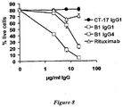
- the B1 antibody bound a multitude of B lymphoma cell lines of different origin (see Example 1 and Table 1) and was shown to induce apoptosis in HLA-DR/DP antigen expressing cells. Furthermore, the B1 antibody showed a higher potency than Rituximab when tested on the Raji B lymphoma cell line. This was particularly evident, when the IgG 4 format of the B1 antibody was used ( Figure 8 ).
- this antibody has suitable characteristics for treatment of HLA-DR/DP expressing B lymphoma cells.
- the B1 antibody may prove efficacious in eliminating activated B cells in disorders where HLA-DR/DP expressing B cells are detrimental.
- the cell surface antigen is surface IgM.
- IgM in its free form exists as a large pentameric structure, whose higher molecular weight tends to confine it within blood vessels.
- Monomeric IgM can be found on the cell walls of B lymphocytes and functions as an antibody receptor for antigen recognition.
- the C11 antibody upon binding to surface IgM, expressed on B lymphoma cells, induces apoptosis in a rapid and efficient manner (see Example 1 and Table 1).
- the C11 antibody bind to a non-polymorphic epitope expressed on B cells from different donors and is thus suitable for treatment of patients suffering from B lymphoma irrespective of B lymphoma idiotype.
- the effector molecule e.g. the anti-IgM antibody could also be any type of specific binding molecule that causes apoptosis in IgM expressing cells of different idiotypes.
- the target cell is an immune cell or epithelial cell and advantageously that immune cell is a B lymphocytes.
- the target cell is associated with a disease.
- the disease is selected from the group consisting of: cancer; autoimmune diseases including but not restricted to rheumatoid arthritis and SLE, acute and chronic inflammatory disorders, sepsis and infectious disease including but not restricted to HIV.
- the disease is a cancer selected from lymphoma (leukaemia, myeloma), gastric cancer, breast cancer, liver cancer, lung cancer, melanoma, bladder cancer, chorioid cancer, pancreatic cancer, colon cancer and prostate cancer.
- lymphoma leukaemia, myeloma
- gastric cancer breast cancer
- liver cancer lung cancer
- melanoma bladder cancer
- chorioid cancer pancreatic cancer
- colon cancer and prostate cancer prostate cancer selected from lymphoma (leukaemia, myeloma), gastric cancer, breast cancer, liver cancer, lung cancer, melanoma, bladder cancer, chorioid cancer, pancreatic cancer, colon cancer and prostate cancer.
- the antibody molecule is an IgG.
- the IgG may be any of IgG 1 , IgG 2 , IgG 3 or IgG 4 , but preferably any of IgG 1 and IgG 4 .
- the antibody molecule is preferably human.
- the binding molecule or antibody molecule of the invention has the sequence of any one of variable region sequences of Figure 10
- the binding molecule or antibody molecule has the variable region sequences of Figure 9 or functionally equivalent homologues thereof.
- the binding molecule or antibody molecule has the variable region sequences of Figure 11 or functionally equivalent homologues thereof.
- nucleic acid has the nucleotide sequence of any one of figures 9 to 11 .
- the antibody molecule of the invention in the diagnosis and/or treatment and/or prevention of a disease requiring the destruction of a target cell.
- the disease to be treated is selected from the group consisting of: cancer; autoimmune diseases including but not restricted to rheumatoid arthritis and SLE, acute and chronic inflammatory disorders, sepsis and infectious disease including but not restricted to HIV.
- the disease to be treated is cancer selected from lymphoma (leukaemia, myeloma), gastric cancer, breast cancer, liver cancer, lung cancer, melanoma, bladder cancer, chorioid cancer, pancreatic cancer, colon cancer and prostate cancer.
- lymphoma leukaemia, myeloma
- gastric cancer breast cancer
- liver cancer lung cancer
- melanoma bladder cancer
- chorioid cancer pancreatic cancer
- colon cancer and prostate cancer is cancer selected from lymphoma (leukaemia, myeloma), gastric cancer, breast cancer, liver cancer, lung cancer, melanoma, bladder cancer, chorioid cancer, pancreatic cancer, colon cancer and prostate cancer.
- the binding molecule or antibody molecule binds specifically to ICAM-1 and/or has the sequence of figure 10 and is used in relation to the diseases listed above.
- the antibody molecule binds specifically to HLA-DR/DP and/or has the sequence of figure 9 and is used in relation to the diseases of lymphoma (leukaemia, myeloma), gastric cancer, breast cancer, liver cancer, lung cancer, melanoma, bladder cancer, chorioid cancer, pancreatic cancer, colon cancer and prostate cancer.
- lymphoma leukaemia, myeloma
- gastric cancer breast cancer
- breast cancer liver cancer
- lung cancer melanoma
- bladder cancer chorioid cancer
- pancreatic cancer pancreatic cancer
- colon cancer colon cancer and prostate cancer.
- the antibody molecule binds specifically to surface IgM and/or has the sequence of figure 11 and is used in relation to the diseases of lymphoma (leukaemia, myeloma), gastric cancer, breast cancer, liver cancer, lung cancer, melanoma, bladder cancer, chorioid cancer, pancreatic cancer, colon cancer and prostate cancer.
- lymphoma leukaemia, myeloma
- gastric cancer breast cancer
- breast cancer liver cancer
- lung cancer melanoma
- bladder cancer chorioid cancer
- pancreatic cancer pancreatic cancer
- colon cancer colon cancer and prostate cancer.
- composition comprising the antibody molecule of the invention and a pharmaceutically acceptable carrier, excipient or diluent.
- antibody molecule shall be taken to refer to any one of an antibody, an antibody fragment, or antibody derivative. It is intended to embrace wildtype antibodies, synthetic antibodies, recombinant antibodies or antibody hybrids, such as, but not limited to, a single-chain modified antibody molecule produced by phage-display of immunoglobulin light and/or heavy chain variable and/or constant regions, or other immunointeractive molecules capable of binding to an antigen in an immunoassay format that is known to those skilled in the art.
- antibody fragment shall be taken to refer to any one of an antibody, an antibody fragment, or antibody derivative. It is intended to embrace wildtype antibodies (i.e. a molecule comprising four polypeptide chains), synthetic antibodies, recombinant antibodies or antibody hybrids, such as, but not limited to, a single-chain modified antibody molecule produced by phage-display of immunoglobulin light and/or heavy chain variable and/or constant regions, or other immunointeractive molecules capable of binding to an antigen in an immunoassay format that is known to those skilled in the art.
- wildtype antibodies i.e. a molecule comprising four polypeptide chains
- synthetic antibodies recombinant antibodies or antibody hybrids
- recombinant antibodies or antibody hybrids such as, but not limited to, a single-chain modified antibody molecule produced by phage-display of immunoglobulin light and/or heavy chain variable and/or constant regions, or other immunointeractive molecules capable of binding to an antigen in an immunoassay format that is known to those skilled in the art.
- antibody derivative refers to any modified antibody molecule that is capable of binding to an antigen in an immunoassay format that is known to those skilled in the art, such as a fragment of an antibody (e.g. Fab or Fv fragment), or a modified antibody molecule that is modified by the addition of one or more amino acids or other molecules to facilitate coupling the antibodies to another peptide or polypeptide, to a large carrier protein or to a solid support (e.g. the amino acids tyrosine, lysine, glutamic acid, aspartic acid, cysteine and derivatives thereof, NH 2 -acetyl groups or COOH-terminal amido groups, amongst others).
- a fragment of an antibody e.g. Fab or Fv fragment
- modified antibody molecule that is modified by the addition of one or more amino acids or other molecules to facilitate coupling the antibodies to another peptide or polypeptide, to a large carrier protein or to a solid support (e.g. the amino acids tyrosine, lysine
- ScFv molecule refers to any molecules wherein the V H and V L partner domains are linked via a flexible oligopeptide.
- nucleotide sequence or “nucleic acid” or “polynucleotide” or “oligonucleotide” are used interchangeably and refer to a heteropolymer of nucleotides or the sequence of these nucleotides. These phrases also refer to DNA or RNA of genomic or synthetic origin which may be single-stranded or doublestranded and may represent the sense or the antisense strand, to peptide nucleic acid (PNA) or to any DNA-like or RNA-like material.
- PNA peptide nucleic acid
- A is adenine
- C cytosine
- T thymine
- G guanine
- N A, C, G or T (U).
- nucleic acid segments provided by this invention may be assembled from fragments of the genome and short oligonucleotide linkers, or from a series of oligonucleotides, or from individual nucleotides, to provide a synthetic nucleic acid which is capable of being expressed in a recombinant transcriptional unit comprising regulatory elements derived from a microbial or viral operon, or a eukaryotic gene.
- polypeptide or “peptide” or “amino acid sequence” refer to an oligopeptide, peptide, polypeptide or protein sequence or fragment thereof and to naturally occurring or synthetic molecules.
- a polypeptide "fragment,” “portion,” or “segment” is a stretch of amino acid residues of at least about 5 amino acids, preferably at least about 7 amino acids, more preferably at least about 9 amino acids and most preferably at least about 17 or more amino acids.
- any polypeptide must have sufficient length to display biological and/or immunological activity.
- purified or “substantially purified” as used herein denotes that the indicated nucleic acid or polypeptide is present in the substantial absence of other biological macromolecules, e.g., polynucleotides, proteins, and the like.
- the polynucleotide or polypeptide is purified such that it constitutes at least 95% by weight, more preferably at least 99% by weight, of the indicated biological macromolecules present (but water, buffers, and other small molecules, especially molecules having a molecular weight of less than 1000 daltons, can be present).
- isolated refers to a nucleic acid or polypeptide separated from at least one other component (e.g., nucleic acid or polypeptide) present with the nucleic acid or polypeptide in its natural source.
- the nucleic acid or polypeptide is found in the presence of (if anything) only a solvent, buffer, ion, or other component normally present in a solution of the same.
- isolated and purified do not encompass nucleic acids or polypeptides present in their natural source.
- recombinant when used herein to refer to a polypeptide or protein, means that a polypeptide or protein is derived from recombinant (e.g., microbial, insect, or mammalian) expression systems.
- Microbial refers to recombinant polypeptides or proteins made in bacterial or fungal (e.g., yeast) expression systems.
- recombinant microbial defines a polypeptide or protein essentially free of native endogenous substances and unaccompanied by associated native glycosylation.
- Polypeptides or proteins expressed in most bacterial cultures e.g., Escherichia coli
- yeast will have a glycosylation pattern in general different from those expressed in mammalian cells.
- variable binding and binding selectivity indicates that the variable regions of the antibodies of the invention recognise and bind polypeptides of the invention exclusively (i.e., able to distinguish the polypeptide of the invention from other similar polypeptides despite sequence identity, homology, or similarity found in the family of polypeptides), but may also interact with other proteins (for example, Staphylococcus aureus protein A or other antibodies in ELISA techniques) through interactions with sequences outside the variable region of the antibodies, and in particular, in the constant region of the molecule.
- Screening assays to determine binding selectivity of an antibody of the invention are well known and routinely practiced in the art. For a comprehensive discussion of such assays, see Harlow et al .
- Antibodies that recognise and bind fragments of the polypeptides of the invention are also contemplated, provided that the antibodies are first and foremost selective for, as defined above, full-length polypeptides of the invention.
- antibodies of the invention that recognise fragments are those which can distinguish polypeptides from the same family of polypeptides despite inherent sequence identity, homology, or similarity found in the family of proteins.
- binding affinity includes the meaning of the strength of binding between an antibody molecule and an antigen.
- immune cell we mean any cell that is involved in a host immune or inflammatory response, including but not limited to B cells and T cells.
- epithelium is a tissue composed of a layer of cells. Epithelium can be found lining internal (e.g. endothelium, which lines the inside of blood vessels) or external (e.g. skin) free surfaces of the body.
- epithelial cells The outermost layer of our skin is composed of squamous epithelial cells, as are the mucous membranes lining the inside of mouths and body cavities. Other epithelial cells line the insides of the lungs, the gastrointestinal tract, the reproductive and urinary tracts, and make up the exocrine and endocrine glands. Functions of epithelial cells include secretion, absorption and protection. Epithelial cells sit on a basal lamina.
- Example 1 Selection and Screening (Biopanning) for apoptosis inducing antibodies. with specificity for B lymphoma associated cell surface receptors
- the cell lines used in this study were obtained from ATCC-(Manassas, VA, USA) or Deutsche Sammlung von Mikroorganismen und Zellkulturen (DSMZ) GmbH (Braunschweig, Germany) and were cultured in RPMI 1640 medium supplemented with 10 % FCS, 2 mM L-Glutamine, 10 mM HEPES and 1 mM Na pyruvate (all from Invitrogen, Carlsbad, CA, USA) unless otherwise stated.
- the Jurkat T leukaemia cell line (clone E6-1, TIB-152, ATCC), the B lymphoma cell lines DOHH-2 (ACC47, DSMZ), SC-1 (ACC558, DSMZ), WSU-NHL (ACC58, DSMZ), JVM-2 (ACC12, DSMZ), Jeko-1 (ACC553, DSMZ grown in 20 % FCS), Rec-1 (ACC 584, DSMZ), Sup-53 ( Daibata et al. Cancer 1989;64:1248-53 ), RL (CRL-2261, ATCC), Granta 519 (DSMZ), NCEB-1 ( Saltman et. al. Blood 1988;72:2026-30 ), BJAB ( Menezes et al.
- Jurkat cells were harvested by centrifugation at 300 ⁇ g for 15 min in 500 ml buckets (Corning Inc. Life Sciences, New York, USA), washed in Dulbecco's PBS (Invitrogen), and resuspended in buffer A (1 mM NaHCO 3 , 1.5 mM MgAc, pH 7.4). Cell concentration was approximately 5 ⁇ 10 7 Jurkat cells/ml (5 ⁇ 10 9 cells in 100 ml Buffer A).
- Cell disruption was achieved by hypo-osmotic shock treatment (Buffer A) on ice for 10-30 min and subsequent nitrogen cavitation in a Nitrogen cavitation bomb (Parr Instrument Company, Moline, IL, USA). Cells were kept at a constant pressure of 40 bar (4,000 kPa) for 15 min at 0 °C.
- Disrupted cells were collected in a 250 ml Sarstedt tube (Sarstedt AG & Co, Nümbrecht, Germany) containing 500 ⁇ l 0.5 M EDTA to yield a final EDTA concentration of 2.5 mM. Addition of EDTA prevents aggregation of membrane vesicles.
- the homogenate 100 ml was divided between 4 ⁇ 25 ml Beckman thick-walled rotor tubes (Beckman Coulter, Inc., Fullerton, CA, USA), which were centrifuged for 10 min at 1900 ⁇ g (4,000 rpm in an Sorvall SS34 rotor) at 4 °C to remove unbroken cells, nuclei, and heavy mitochondria.
- the crude membrane pellet was transferred to a Dounce homogeniser with the aid of a metal bar and was resuspended in 2.5 ml HES buffer (10 mM Hepes, 1 mM EDTA, 0.25 M sucrose, pH 7.4) by several careful strokes in the homogenizer.
- HES buffer 10 mM Hepes, 1 mM EDTA, 0.25 M sucrose, pH 7.4
- a membrane suspension concentration equivalent of 2 ⁇ 10 9 cells/ml containing 80 - 100 mg protein was, thus, achieved.
- Jurkat membrane vesicle preparations derived from 2 ⁇ 10 9 cells round 1 selection; 2 ⁇ 10 8 cells round 2 and 3 selections) were thawed on ice, and were mixed with the blocked phage particles. The mixture was incubated for 15 min on ice.
- Ramos cells were harvested by centrifugation at 1,200 rpm for 6 min at 4 °C. Supernatant was discarded and Ramos cells were resuspended in the milk-phage-Jurkat membrane vesicle mixture. The suspension was incubated at 10 °C under slow end-over-end rotation for 4 h.
- the cell/ cell membrane vesicle/ phage mixture was transferred to a 15 ml Falcon tube (BD Biosciences, Bedford, MA, USA), containing 0.5 ml 100 % (trypan blue stained) Ficoll-Paque PLUS (Amersham Biosciences, Uppsala, Sweden) at the bottom, and 9.5 ml overlaid 40 % (v/v) Ficoll in 2 % (w/v) BSA/PBS (Ficoll-pillar).
- the tube was centrifuged at 1,500 rpm for 10 min at 4 °C. The tube was removed from the centrifuge and the tube cap was screwed on and sealed airtight.
- the bottom "tip" of the Falcon tube containing 100 % Ficoll was chopped off using a cigar-chopper. Thus, very high-density material including membrane vesicle sheets and cell nuclei were eliminated from the tube.
- the tube cap was then carefully opened disrupting the vacuum inside the tube and allowing liquid to be expelled drop-wise through the opening at the (cut off) tube bottom.
- the harvested cell suspension was washed once in PBS to remove excess Ficoll.
- the pellet was resuspended in 1 ml PBS (not performed following final wash) and the suspension was reloaded on top a fresh Ficoll-pillar and the washing procedure was repeated (twice in rounds 2 and 3).
- Phage were eluted from cells by addition of 150 ⁇ l of 76 mM citric acid (pH 2.5) in PBS followed by incubation at room temperature for 5 min. The mixture was neutralized by addition of 200 ⁇ l of 1 M Tris-HCl, pH 7.4. Supernatants containing eluted phage were saved following pelleting of cells at 300 ⁇ g for 5 min. Further elution of phage was by resuspension and incubation of the cell pellet in 1 ml trypsin at RT for 10 min.
- the phagemid pool obtained following three rounds of selection was digested with Eag I to remove the gene III.
- the resulting vector was re-ligated.
- Vectors containing re-ligated uncut gene III fragments were linearized by digestion with Eco RI enzyme.
- the scFv vector pool thus generated was used to transform E. coli TOP10 cells essentially as described earlier ( Soderlind et al. Nat Biotechnol 2000;18:852-6 .).
- Bacteria were plated on large 500 cm 2 agar plates and individual clones were picked, transferred to 96-well plates, and expressed in TB medium by over night culture at 37 °C, 220 rpm using an automated system (Hallborn Biotechniques 2002;Suppl:30-7.). Recombinant scFv fragments were produced in TB medium containing appropriate antibiotics.
- scFv clone binding For primary screening of scFv clone binding to target Ramos cells and Jurkat non-target cells, 5,000 Ramos or Jurkat cells were incubated with either of 960 scFv clones, derived from the 3 rd round of selection and produced as described above. Cells were incubated with 0.5 ⁇ g/ml anti-6xHis mAb (R&D Systems, Minneapolis, MN, USA) and 0.7 ⁇ g/ml Cy5-conjugated Goat anti-mouse reagent (Amersham Biosciences). Cell binding was analysed in an ⁇ 200 Cellular Detection System Fluorescence Macroconfocal High Throughput Screening (FMAT) instrument (Applied Biosystems, Foster City, CA, USA).
- FMAT Cellular Detection System Fluorescence Macroconfocal High Throughput Screening
- Cells were washed by centrifugation at 300 ⁇ g for 6 min. Cells were then incubated with FITC-conjugated CD 19 mAb and PE-conjugated CD3 mAb (BD) enabling subsequent identification of target and non-target cells, respectively. Detection of scFv binding was achieved by incubation with RPE-Cy5-streptavidin (Dako Cytomation, Glostrup, Denmark) following incubation with biotinylated anti-6xHis mAb. Cells were incubated with secondary and tertiary reagents for 40 min, and 15 min respectively. All incubations were performed on ice using ice-cold solutions.
- the present study utilized a novel panning protocol to isolate antibodies that target differentially expressed antigens in their native cell surface configuration. Following three rounds of competition biopanning, using whole Ramos B lymphoma cells and membrane vesicles derived from Jurkat T leukaemia cells, recombinant phage scFv were isolated. These were converted to soluble scFv and expressed in E. coli TOP10 cells.
- scFv Recombinant scFv were incubated with target (Ramos) or non-target (Jurkat) whole cells and examined for cell binding.
- target Ramos
- Jurkat non-target whole cells
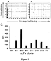
- the clones were simultaneously re-expressed and re-evaluated for target cell specificity (Ramos vs. Jurkat) by FMAT technology, as described ( Fig 1 B) . Seven different antibody genotypes were identified, as determined by their different CDRH3 and CDRL3 sequences (data not shown).
- Target and non-target cells were defined by CD19 and CD3 expression, respectively, using fluorochrome conjugated CD specific monoclonal antibodies.
- the seven genotypically unique scFv clones showed high and variable binding intensities to target Ramos cells, but no binding to the non-target Jurkat cells, as compared to the negative control scFv.
- Lipopolysaccharide levels of recombinantly produced scFv fragments were reduced using Detoxigel columns according to manufacturer's instructions (Pierce Biotechnology, Rockford, IL, USA). Remaining endotoxin levels were quantified by the LAL-amebocyte lysate assay (Cambrex Bioscience, Walkersville, MD, USA).
- scFv samples were found to contain less than 0.1 IU/ml of lipopolysaccharide.
- the chimeric anti-CD20 antibody MabtheraTM (Rituximab) was purchased from Lund University Hospital (Lund, Sweden). 2 ⁇ 10 5 B lymphoma cells (Raji or Ramos) or Jurkat T cells were incubated with serially diluted and detoxified scFv's in culture medium for 1 h on ice.
- Cells were sequentially incubated with secondary anti-6xHis mAb (5 ⁇ g/ml), and tertiary Goat Fab'2 anti-mouse Fab'2 antibody (Jackson ImmunoResearch Laboratories, Inc., West Grove, PA, USA). Intermittent washings ensured removal of excess unbound antibody reagent. Cells were incubated at 37 °C in a humidified atmosphere of 5 % CO 2 for 24 h.
- cross-linking reagent was replaced by goat Fab'2 anti-human Fc y antibody (Jackson ImmunoResearch) with minimal cross-reactivity with non-IgG antibody isotypes (to avoid unspecific cross-linking of endogenous B lymphoma associated surface immunoglobulins) and incubated, as described above, for 6 h.
- Apoptotic cells were, unless otherwise stated, detected by combined staining with Annexin V Alexa Fluor 488 (AV) and Propidium Iodide (PI) (both from Molecular Probes, Invitrogen) and subsequent flow-cytometric analysis, Cells were defined as viable (AV-/PI-), early apoptotic (AV+/ PI-) or late apoptotic/necrotic (AV+/PI+).
- AV and PI signals were recorded in the FL1 and FL2 or FL3 channels (as indicated in the text), respectively, using a FACSCalibur instrument (BD Biosciences).
- apoptosis screening assay based on sequential incubation and washing of cells with scFv and cross-linking reagent.
- the dependence on scFv clone and concentration in the apoptosis assay is demonstrated in Fig. 2A-C , where the apoptotic effect of selected scFv clone B1 is compared to the - effect of scFv clone F1 which shows no induction of apoptosis.
- Jurkat cells lacking target antigen expression did not die from apoptosis after treatment with any of the examined scFv demonstrating that apoptosis induction depended on binding to target antigen (data not shown).
- scFv-apoptosis assay we screened clones for apoptosis on Ramos and Raji B lymphoma cells. scFv-induced apoptotic effects were compared to that induced by Rituximab anti-CD20 mAb ( Fig. 2D ). Three scFv clones - B1, B11 and C11 - were identified that induced significant apoptosis on both Ramos and Raji cells ( Fig. 2D and E ). Induction of apoptosis by scFv on Raji cells correlated with binding to these cells ( Fig. 2D ), since scFv clones that failed to bind Raji cells did not induce apoptosis.
- the B1, B11 and C11 clones were transferred to fully human IgG1 antibodies. Both their specificity and functionality remained intact after reformatting, as demonstrated by their strong binding and potent cytotoxicity towards a broad panel of B lymphoma cell lines (Table 1). Notably, apoptosis induction was rapid with maximal percentage of annexin V positive apoptotic cells being reached already after three to six hours in several cell lines (Table 1 and data not shown).
- scFv antibody fragments were converted to full-length human IgG1 ⁇ format via cloning into a modified pCDNA3 vector ( Norderhaug et al. J Immunol Methods 1997;204:77-87 .), and transiently transfected into HEK293 cells using Lipofectamine 2000 reagent according to manufacturer's instructions (Invitrogen).
- Human IgG was purified from spent cultivation medium on a MabSelect protein A column (Amersham Biosciences). The purity of preparations was >98% as determined by SDS-PAGE analysis. Antibody preparations were screened and found to contain ⁇ 0.1 IU/ml endotoxin at concentrations used in the present study, and as determined by the LAL amoebocyte lysate test (Cambrex Bioscience).
- the identity of the targeted antigens was determined by immunoprecipitation of B lymphoma cell lysates.
- Cells 50-600 ⁇ 10 6 per ml lysis buffer, depending on antibody and cell line) were harvested by centrifugation, washed twice in PBS and incubated for 15 min in Lysis Buffer (25 mM Tris-HCl, pH 7.5, 150 mM NaCl, 5 mM EDTA, and Complete EDTA-free Protease inhibitor cocktail (Roche Diagnostics GmbH, Mannheim, Germany)) containing the detergent Triton X-100 (Sigma-Aldrich, St. Louis, MO, USA) at 0.5 % v/v.
- Lysis Buffer 25 mM Tris-HCl, pH 7.5, 150 mM NaCl, 5 mM EDTA, and Complete EDTA-free Protease inhibitor cocktail (Roche Diagnostics GmbH, Mannheim, Germany) containing the detergent Triton X-100 (Sigma-Al
- Protein A Sepharose 4 Fast Flow was added again and incubated for 30 min, where after the immuno-complexes were washed extensively in lysis buffer, boiled for 5 min, and finally resuspended in Sample Buffer (1X NuPAGE LDS Sample Buffer, 1X NuPAGE Sample Reducing Agent) and separated in a NuPAGE Novex 4-12 % Bis-Tris Gel (all from Invitrogen).
- gel plugs were destained and equilibrated by washing three times with 200 ⁇ l 50 % acetonitril (ACN) under agitation. After drying in a SpeedVac concentrator (Savant, Farmingdale, NY, USA) for 15 min, samples were reduced by addition of 25 ⁇ l 10 mM DTT / 100 mM NH 4 HCO 3 and incubated for 56 °C for 1 h and alkylated by addition of 25 ⁇ l 55 mM iodoacetamide /100 mM NH 4 HCO 3 followed by incubation for 45 min at room temperature.
- ACN acetonitril
- the gel pieces were dried in a SpeedVac concentrator and re-swelled and digested in 15 ⁇ l of 15 ng/ ⁇ l trypsin (Promega Corporation, Madison, WI, USA) in 25 mM NH 4 HCO 3 at 37 °C over night.
- Peptides were extracted by addition of 50 % v/v ACN / 1 % v/v TFA and 10 min incubation at RT. 1 ⁇ l of the extract was spotted onto MALDI sample plates and allowed to dry.
- Peptide masses were determined using an Applied Biosystems 4700 Maldi Workstation. The proteins were identified by peptide mass fingerprint database searching using Mascot search tools (Matrixscience, UK). Antigen specificities of clones B10, C10, and G12 were identified using similar methodology, except scFv's and anti-His coated magnetic microbeads (Miltenyi Biotec GmbH, Bergisch Gladbach, Germany) were used for immunoprecipitations.
- IgG B1 precipitated two bands of approximately 28 and 34 ⁇ kDa, respectively ( Fig. 3A , lane 1). Gel slices containing these bands were prepared and digested with trypsin and analysed by mass spectrometry identifying HLADR/DP as the target antigen.
- IgG B1 for HLA-DR/DP was verified both by western blotting and detection of HLA-DR/DP protein, using a commercially available monoclonal antibody, and by blocking of B1 IgG binding following pre-incubation of cells with a commercially available HLA-DR specific monoclonal antibody ( Fig. 3B ).
- IgG B11 and C11 defined antigens were established using similar methodology.
- IgG C11 was found to precipitate a 68 kDa protein band identified as the membrane bound form of the B cell receptor ⁇ chain ( Fig. 3A , lane 3).
- IgG B11 precipitated a 90 kDa protein band that was identified as the intercellular cell adhesion molecule-1 (ICAM-1) ( Fig. 3A , lane 2).
- IgG B11 for ICAM-1, and of C11 IgG for IgM were confirmed by MS-MS analysis, antibody blocking studies ( Fig. 3B ), and western blot analysis (data not shown) using commercially available antibodies.
- clones B10, C10, and G12 were determined, using scFv and anti-His coated magnetic microbeads for immunoprecipitation.
- the three scFv clones precipitated a protein band of 68 kDa, and MS-analysis of trypsin digested gel slices containing these bands revealed their specificity for surface IgM. Presumably, these antibodies recognize the Ramos IgM idiotype, since neither of them cross-react with peripheral blood B lymphocytes or other IgM positive B cell lines.
- Iodination of 1 mg/ml of IgG 1 B1 or IgG 1 B11 proteins with [ 125 I] NaI was performed in PBS for 10 min using Iodogen pre-coated iodination tubes (Pierce). Free [ 125 I] NaI was removed by desalting on PD-10 columns (Amersham Biosciences), yielding specific radioactivities in the range of 1000-1600 cpm per ng protein. [ 125 I] IgG 1 B1 and [ 125 I] IgG 1 B11 was used for determination of antibody affinities.
- Radioiodinated IgG B1 or IgG B11 was incubated with B lymphoma cells in DPBS-B-hIgG (DPBS-B containing 0.2 mg/ml human IgG) for 2 h on ice with intermittent mixing. Non-specific binding was determined in the presence of 0.2 mg/ml unlabeled IgG B1 or IgG B11 protein, as appropriate. Analysis was performed in triplicates.
- Kd values Antibody affinity constants (Kd values) and epitope numbers per cell were determined from Scatchard plot analysis according to Rosenthal et al. (Anal Biochem 1967;20:525-32 ), Bylund and Yamamura (Methods in Neurotransmitter Analysed. New York: Raven Press Ltd., 1990 ), and Marquardt (J. Soc. Indust. Appl. Math 1963;11:431-41 ), as previously described ( Brix et al. J. Clin. Invest. 1998;102:283-93 ).
- IgG B1 and IgG B11 binding to HLA-DR and ICAM-1 was characterised by incubating the radio-iodinated proteins with Raji or Ramos cells in the presence or absence of 0.2 mg/ml of the corresponding unlabeled IgG protein at 4 °C.
- the specific binding of [ 125 I]IgG to the cell surface was calculated by subtracting non-specific binding (binding in the presence of excess unlabeled IgG) from the total binding.
- MFI Apoptosis Induction Follicular Lymphoma DOHH-2 140 - - 100 - 90 - 480 ++ WSU-NHL 280 + 0 - 60 - 790 + SC-1 170 + 0 - 50 - 50 - RL 50 - 100 - 210 - 200 + Mantle cell Lymphoma Granta 519 370 ++ 260 + 60 + 360 +++ JVM-2 650 + 100 - 10 - 520 + Rec-1 0 - 380 - 900 - 580 + SP-53 500 ++ 90 - 360 - 740 ++ NCEB-1 750 + 340 + 10 - 430 + Jeko-1 1000 +++ 30 + 10
- Example 4 - ICAM-1 is a B lymphoma associated antigen with apoptosis inducing properties
- Ramos cells (13 x 10 6 ) were stained with CD45-PerCp-Cy5.5 mAb by incubation on ice for 45 min, washed in DPBS-B, and kept on ice until mixing with unlabeled purified PBLs.
- Buffy coats from two healthy volunteers were obtained from the Lund University Hospital. Buffy coats were diluted 1:2 in PBS and washed by centrifugation at 500 ⁇ g (1500 rpm Beckman Spinchron centrifuge) for 10 min, complete aspiration of the supernatant and resuspension in DPBS containing 1 % heat inactivated FCS (DPBS-HI). Washing was repeated twice. Red blood cells were lysed by incubation with red blood cell lysing solution (BD Biosciences) for 15 min at RT. Cells were washed by centrifugation at 60 ⁇ g (667 rpm Beckman spinchron centrifuge) for 10 min and the supernatant was carefully aspirated.
- IgG B1, B11, C11, and Rituximab antibodies were incubated with 20 ⁇ l of AF647-Fab labelling reagent for 5 min at RT.
- AF647-labeled IgG was three-fold serially diluted in DBPS-B, and diluted IgG proteins were added to the mixed Ramos/PBL cell solutions.
- Ramos cells were identified as the PerCpCy5.5 high population distinct from the B lymphocyte PerCpCy5.5 low population.
- Cryopreserved lymph node biopsies of patients with Anaplastic Large Cell B lymphoma (one patient), Centroblastic/Centrocytic B non-Hodgkin lymphoma (three patients), and B cell chronic lymphocytic leukaemia (one patient) were obtained from the Department of Pathology at Lund University (Lund, Sweden). Eight-micrometer sections of cryo-preserved tissue were fixed in acetone for 10 min at 4 °C. Endogenous biotin-binding activity was blocked by sequential treatment with Avidin and Biotin (Avidin/Biotin blocking kit, Invitrogen) for 20 min each.
- Tissues were incubated with 5 ⁇ g/ml control scFv or B11 scFv for 1 h. Following washing, sections were incubated with biotin-conjugated mouse anti-His mAb (R&D Systems) for 30 min. scFv binding was detected following treatment with ABC Complex/ HRP reagent (Dako Cytomation) for 30 min, and subsequent incubation with DAB for 5 min.
- biotin-conjugated mouse anti-His mAb R&D Systems
- Sections were photographed using a Leica DC 300F digital camera mounted on top of a Leica DMR light/fluorescence microscope.
- Mitochondrial membrane depolarisation was analysed as previously described ( Kim et al. Mol Biol Cell 2004;15:420-34 ). Briefly, antibody-treated cells were mixed with JC-1 reagent (Molecular Probes) at 5 ⁇ g/ml and incubated for 30 min at RT. Cells were washed twice in ice-cold PBS and resuspended in 300 ⁇ l PBS and analysed on a FACS Aria (BD Biosciences). The green and red fluorescence was collected through 494/518 nm (FL-1) and 595/615 nm (FL-2) bandpass filters, respectively.
- JC-1 reagent Molecular Probes
- ICAM-1 is a glycoprotein of the immunoglobulin superfamily ( Marlin et al. Cell 1987;51:813-9 ) capable of inducing bi-directional signalling ( Rothlein et al. J Immunol 1994;152:2488-95 ; Vyth-Dreese et al. Blood 1995;85:2802-12 ). ICAM-1 has not previously been demonstrated to be involved in programmed death in B lymphoma cells.
- IgG B11 induced cell death was an active process, by means other than cell membrane phosphatidyl serine translocation.
- Mitochondrial membrane depolarisation was chosen as a validation of apoptosis, since this is a common feature of caspase dependent and caspase independent apoptosis that may be monitored by cell staining with 5,5',6,6'-tetrachloro-1,1',3,3'-tetraethylbenzimidazolyl-carbocyanine iodide (JC-1 reagent).
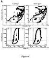
- IgG B11 was found to induce mitochondrial membrane depolarisation in CL-01 B lymphoma cells, as determined by flow-cytometric analysis following staining with JC-1 reagent ( Fig. 4A , lower panel).
- IgG B11 showed strong binding to the five lymphoma tissues ( Fig. 4B ), at intensities comparable to, or slightly lower than, the anti-HLA-DR/DP antibody IgG B1 (Table 2).
- B11 demonstrated the greatest differential binding (strongest antigen up-regulation) of the three antibodies to Ramos B lymphoma cells versus normal peripheral blood leukocytes ( Fig. 4C . and data not shown). IgG B11 binding peaked already at 0.1 ⁇ g/ml and was 3.7-fold up regulated on Ramos versus monocyte cells (MFI 654 versus 176), 8.3-fold up regulated on Ramos versus peripheral blood B lymphocytes (MFI 654 versus 78), and 23-fold up regulated compared to NK cells. Binding to other monitored peripheral blood leukocyte subsets was negative. Table 2.
- ICAM-1 is strongly expressed in B lymphoma tissue of different origin
- B-CLL c low malignant non-Hodgkin Lymphoma
- B-CLL c low malignant non-Hodgkin Lymphoma
- B-CLL d B-CLL/B-PLL
- B-CLL B- Chronic Lymphocytic Leukemia
- d B-PLL B- Pro Lymphocytic Leukemia. Increasing numbers of + indicate stronger staining.
- Example 5 Antigen distribution of B1, B11, C11 IgG 1 on tumour cell lines of various origins, as determined by flow cytometry.
- the antigen distribution of human antibody targeted antigens, mainly for B 11, on different carcinoma cell lines was investigated.
- Cells (MCF-7 and MDA MB 435S breast carcinoma, JAR and JEG-3 chorio-carcinoma, A549 lung carcinoma, TCC-SUP urinary bladder carcinoma, MDA MB 435 melanoma, HPAC, PANC-1 and BxPC-3 pancreatic carcinoma, PC-3 and DU145 prostate carcinoma, LS174 T, CaCo 2 , and Lovo colorectal carcinoma, and THP-1 monocytic leukaemia cells), were washed in PBS, and resuspended at 4 x 10 6 cells/ml in Complete Medium (200,000 cells/50 ⁇ l sample).
- B1 IgG 1 , B11 IgG 1 , C11 IgG 1 , negative control FITC-8 IgG 1 , and Rituximab anti-CD20 mAb was 3-10-fold serially diluted (10-0.1 pg/ml) in Complete Medium (50 ⁇ l/sample). Cells were incubated with either of the antibodies for 1 hour on ice, washed by resuspension in PBS/BSA 0.5 %, centrifuged at 1200 rpm for 5min, and complete aspiration of the supernatants was undertaken.
- PC-3 prostate carcinoma cells showed strong expression of ICAM-1 as demonstrated by the strong binding of B11 IgG to these cells ( Fig. 6 ).
- MCF-7 breast carcinoma, HPAC pancreatic carcinoma, and LS174 T colorectal carcinoma cells were also found to express ICAM-1 albeit at lower intensity compared to the prostate cancer cells.
- THP-1 monocytic leukaemia cells did not express ICAM-1.
- All carcinoma cell lines initially tested were found negative for CD20, HLA-DR/DP, and IgM expression as demonstrated by the lack of binding of Rituximab IgG, B1 IgG, and C11 IgG, respectively. Further studies on additional carcinoma cells lines indicated that all carcinoma cells examined were positive for ICAM-1 expression (Table 3). Table 3.
- ICAM-1 is strongly expressed in carcinoma cell lines of different origin Tumor cell type Cell line MFI Chorio-carcinoma JAR 2000 JEG-3 1600 Prostate carcinoma DU145 2200 PC-3 1500 Pancreatic carcinoma BxPC-3 2000 PANC-1 3800 Colon carcinoma CaCo2 800 Lovo 1600 Lung carcinoma A549 800 Urinary bladder carcinoma TCC-SUP 3200 Melanoma MDA MB 435 4000 Mammary carcinoma MDA MB 435S 800
- Example 6 B11 IgG 1 apoptosis induction in carcinoma cells
- B11 IgG 1 was shown in example 5 to bind strongly to carcinoma cells.
- the present example examined the apoptosis inducing properties of this antibody on carcinoma cells.
- Cells were seeded in 6 well plates with Complete Growth Medium three days before the onset of the experiment. Cells were between 50-75 % confluent at the time of the experiment. Cells were washed with ice-cold PBS and incubated with serially diluted (20- 0.02 ⁇ g/ml as indicated in the figures, in 1 ml Complete Growth Medium) B11 IgG 1 , 20 ⁇ g/ml control B1 IgG 1 , 10 ⁇ g/ml negative control IgG 1 or 10 ⁇ g/ml Trastuzumab IgG 1 as indicated at 4 °C for 1-2 hours.
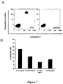
- the B11 IgG 1 was shown to induce apoptosis in the carcinoma cell lines in a specific and titratable manner ( Figure 7 ).
- Control IgG B1 which did not bind to PC-3 cells (see example 5), also did not induce apoptosis in PC-3 cells.
- Negative control IgG 1 or Trastuzumab IgG 1 were not able to induce apoptosis in DU145 or MDA MB435 cells.
- Example 7 Pharmaceutical formulations and administration.
- a further aspect of the invention provides a pharmaceutical formulation comprising a compound according to the first aspect of the invention in admixture with a pharmaceutically or veterinarily acceptable adjuvant, diluent or carrier.
- the formulation is a unit dosage containing a daily dose or unit, daily sub-dose or an appropriate fraction thereof, of the active ingredient
- the compounds of the invention will normally be administered orally or by any parenteral route, in the form of a pharmaceutical formulation comprising the active ingredient, optionally in the form of a non-toxic organic, or inorganic, acid, or base, addition salt, in a pharmaceutically acceptable dosage form.
- a pharmaceutical formulation comprising the active ingredient, optionally in the form of a non-toxic organic, or inorganic, acid, or base, addition salt, in a pharmaceutically acceptable dosage form.
- the compositions may be administered at varying doses.
- the compounds of the invention can be administered alone but will generally be administered in admixture with a suitable pharmaceutical excipient, diluent or carrier selected with regard to the intended route of administration and standard pharmaceutical practice.
- the compounds of the invention can be administered orally, buccally or sublingually in the form of tablets, capsules, ovules, elixirs, solutions or suspensions, which may contain flavouring or colouring agents, for immediate-, delayed- or controlled-release applications.
- the compounds of the invention may also be administered via intracavemosal injection.
- Such tablets may contain excipients such as microcrystalline cellulose, lactose, sodium citrate, calcium carbonate, dibasic calcium phosphate and glycine, disintegrants such as starch (preferably corn, potato or tapioca starch), sodium starch glycollate, croscarmellose sodium and certain complex silicates, and granulation binders such as polyvinylpyrrolidone, hydroxypropylmethylcellulose (HPMC), hydroxy-propylcellulose (HPC), sucrose, gelatin and acacia. Additionally, lubricating agents such as magnesium stearate, stearic acid, glyceryl behenate and talc may be included.
- Solid compositions of a similar type may also be employed as fillers in gelatin capsules.
- Preferred excipients in this regard include lactose, starch, a cellulose, milk sugar or high molecular weight polyethylene glycols.
- the compounds of the invention may be combined with various sweetening or flavouring agents, colouring matter or dyes, with emulsifying and/or suspending agents and with diluents such as water, ethanol, propylene glycol and glycerin, and combinations thereof.
- the compounds of the invention can also be administered parenterally, for example, intravenously, intra-arterially, intraperitoneally, intrathecally, intraventricularly, intrasternally, intracranially, intra-muscularly or subcutaneously, or they may be administered by infusion techniques. They are best used in the form of a sterile aqueous solution which may contain other substances, for example, enough salts or glucose to make the solution isotonic with blood.
- the aqueous solutions should be suitably buffered (preferably to a pH of from 3 to 9), if necessary.
- the preparation of suitable parenteral formulations under sterile conditions is readily accomplished by standard pharmaceutical techniques well-known to those skilled in the art.
- Formulations suitable for parenteral administration include aqueous and non-aqueous sterile injection solutions which may contain anti-oxidants, buffers, bacteriostats and solutes which render the formulation isotonic with the blood of the intended recipient; and aqueous and non-aqueous sterile suspensions which may include suspending agents and thickening agents.
- the formulations may be presented in unit-dose or multi-dose containers, for example sealed ampoules and vials, and may be stored in a freeze-dried (lyophilised) condition requiring only the addition of the sterile liquid carrier, for example water for injections, immediately prior to use.
- Extemporaneous injection solutions and suspensions may be prepared from sterile powders, granules and tablets of the kind previously described.
- the daily dosage level of the compounds of the invention will usually be from 1 mg/kg to 30 mg/kg.
- the tablets or capsules of the compound of the invention may contain a dose of active compound for administration singly or two or more at a time, as appropriate.
- the physician in any event will determine the actual dosage, which will be most suitable for any individual patient, and it will vary with the age, weight and response of the particular patient.
- the above dosages are exemplary of the average case. There can, of course, be individual instances where higher or lower dosage ranges are merited and such are within the scope of this invention.
- the compounds of the invention can also be administered intranasally or by inhalation and are conveniently delivered in the form of a dry powder inhaler or an aerosol spray presentation from a pressurised container, pump, spray or nebuliser with the use of a suitable propellant, e.g. dichlorodifluoromethane, trichlorofluoromethane, dichlorotetrafluoro-ethane, a hydrofluoroalkane such as 1,1,1,2-tetrafluoroethane (HFA 134A3 or 1,1,1,2,3,3,3-heptafluoropropane (HFA 227EA3), carbon dioxide or other suitable gas.
- a suitable propellant e.g. dichlorodifluoromethane, trichlorofluoromethane, dichlorotetrafluoro-ethane, a hydrofluoroalkane such as 1,1,1,2-tetrafluoroethane (HFA 134A3 or 1,
- the dosage unit may be determined by providing a valve to deliver a metered amount.
- the pressurised container, pump, spray or nebuliser may contain a solution or suspension of the active compound, e.g. using a mixture of ethanol and the propellant as the solvent, which may additionally contain a lubricant, e.g. sorbitan trioleate.
- a lubricant e.g. sorbitan trioleate.
- Capsules and cartridges (made, for example, from gelatin) for use in an inhaler or insufflator may be formulated to contain a powder mix of a compound of the invention and a suitable powder base such as lactose or starch.
- Aerosol or dry powder formulations are preferably arranged so that each metered dose or "puff" delivers an appropriate dose of a compound of the invention for delivery to the patient. It will be appreciated that the overall daily dose with an aerosol will vary from patient to patient, and may be administered in a single dose or, more usually, in divided doses throughout the day.
- the compounds of the invention can be administered in the form of a suppository or pessary, or they may be applied topically in the form of a lotion, solution, cream, ointment or dusting powder.
- the compounds of the invention may also be transdermally administered, for example, by the use of a skin patch. They may also be administered by the ocular route, particularly for treating diseases of the eye.
- the compounds of the invention can be formulated as micronised suspensions in isotonic, pH adjusted, sterile saline, or, preferably, as solutions in isotonic, pH adjusted, sterile saline, optionally in combination with a preservative such as a benzylalkonium chloride. Alternatively, they may be formulated in an ointment such as petrolatum.
- the compounds of the invention can be formulated as a suitable ointment containing the active compound suspended or dissolved in, for example, a mixture with one or more of the following: mineral oil, liquid petrolatum, white petrolatum, propylene glycol, polyoxyethylene polyoxypropylene compound, emulsifying wax and water.
- they can be formulated as a suitable lotion or cream, suspended or dissolved in, for example, a mixture of one or more of the following: mineral oil, sorbitan monostearate, a polyethylene glycol, liquid paraffin, polysorbate 60, cetyl esters wax, cetearyl alcohol, 2-octyldodecanol, benzyl alcohol and water.
- Formulations suitable for topical administration in the mouth include lozenges comprising the active ingredient in a flavoured basis, usually sucrose and acacia or tragacanth; pastilles comprising the active ingredient in an inert basis such as gelatin and glycerin, or sucrose and acacia; and mouth-washes comprising the active ingredient in a suitable liquid carrier.
- oral or topical administration of the compounds of the invention is the preferred route, being the most convenient.
- the drug may be administered parenterally, e.g. sublingually or buccally.
- a compound of the invention is administered as a suitably acceptable formulation in accordance with normal veterinary practice and the veterinary surgeon will determine the dosing regimen and route of administration, which will be most appropriate for a particular animal.
Landscapes
- Health & Medical Sciences (AREA)
- Chemical & Material Sciences (AREA)
- Immunology (AREA)
- Organic Chemistry (AREA)
- Life Sciences & Earth Sciences (AREA)
- Medicinal Chemistry (AREA)
- General Health & Medical Sciences (AREA)
- Veterinary Medicine (AREA)
- Pharmacology & Pharmacy (AREA)
- Animal Behavior & Ethology (AREA)
- Public Health (AREA)
- Molecular Biology (AREA)
- Chemical Kinetics & Catalysis (AREA)
- General Chemical & Material Sciences (AREA)
- Nuclear Medicine, Radiotherapy & Molecular Imaging (AREA)
- Biophysics (AREA)
- Proteomics, Peptides & Aminoacids (AREA)
- Genetics & Genomics (AREA)
- Biochemistry (AREA)
- Engineering & Computer Science (AREA)
- Bioinformatics & Cheminformatics (AREA)
- Oncology (AREA)
- Communicable Diseases (AREA)
- Physical Education & Sports Medicine (AREA)
- Virology (AREA)
- Rheumatology (AREA)
- Mycology (AREA)
- AIDS & HIV (AREA)
- Biomedical Technology (AREA)
- Microbiology (AREA)
- Transplantation (AREA)
- Epidemiology (AREA)
- Pain & Pain Management (AREA)
- Hematology (AREA)
- Tropical Medicine & Parasitology (AREA)
- Orthopedic Medicine & Surgery (AREA)
- Medicines Containing Antibodies Or Antigens For Use As Internal Diagnostic Agents (AREA)
- Peptides Or Proteins (AREA)
- Medicines That Contain Protein Lipid Enzymes And Other Medicines (AREA)
- Micro-Organisms Or Cultivation Processes Thereof (AREA)
Claims (19)
- Procédé in vitro d'induction de l'apoptose dans une cellule cible, comprenant les étapes consistant à :a. fournir une ou plusieurs cellules cibles présentant l'antigène de surface cellulaire, ICAM-I ;b. fournir une ou plusieurs molécules de liaison qui se lient de façon sélective à ICAM-I de surface cellulaire et, lors de la liaison à ICAM-I, induisent l'apoptose de la cellule cible ;c. exposer les cellules cibles de (a) aux molécules de liaisons de (b) pour induire l'apoptose dans les cellules cibles,dans lequel les molécules de liaison sont des molécules d'anticorps humain, lesdites molécules d'anticorps ayant des régions variables ayant les séquences de la figure 10.
- Molécule de liaison qui se lie de façon sélective à ICAM-I de surface cellulaire et, lors de la liaison à ICAM-I, induit l'apoptose d'une cellule cible, laquelle molécule de liaison est une molécule d'anticorps humain, ladite molécule d'anticorps ayant des régions variables ayant les séquences de la figure 10.
- Molécule de liaison selon la revendication 2, pour une utilisation dans le procédé selon la revendication 1.
- Molécule de liaison selon l'une quelconque des revendications 2 ou 3, dans laquelle la cellule cible est une cellule immunitaire ou une cellule épithéliale.
- Molécule de liaison selon la revendication 4, dans laquelle la cellule immunitaire est un lymphocyte B.
- Molécule de liaison selon l'une quelconque des revendications 2 à 5, dans laquelle la cellule cible est associée à une maladie.
- Molécule de liaison selon la revendication 6, dans laquelle la maladie est choisie dans le groupe constitué par : le cancer ; les maladies auto-immunes telles que, entre autres, la polyarthrite rhumatoïde et le lupus érythémateux disséminé (LED), les inflammations aiguës et chroniques, la sepsie et les maladies infectieuses telles que, entre autres, le VIH.
- Molécule de liaison selon la revendication 7, dans laquelle la maladie est un cancer choisi parmi le lymphome (leucémie, myélome), le cancer de l'estomac, le cancer du sein, le cancer du foie, le cancer du poumon, le mélanome, le cancer de la vessie, le cancer de la choroïde, le cancer du pancréas, le cancer du côlon et le cancer de la prostate.
- Molécule de liaison selon l'une quelconque des revendications 2 à 8, dans laquelle la molécule d'anticorps est une IgG.
- Molécule de liaison selon la revendication 9, dans laquelle l'IgG est choisie dans le groupe constitué par une IgG1, une IgG2, une IgG3 ou une IgG4.
- Acide nucléique ayant une séquence nucléotidique codant pour une molécule d'anticorps selon l'une quelconque des revendications 2 à 10.
- Acide nucléique selon la revendication 11, ayant les séquences nucléotidiques de la figure 10.
- Molécule de liaison selon l'une quelconque des revendications 2 à 10, pour une utilisation dans le diagnostic et/ou le traitement et/ou la prévention d'une maladie, le diagnostic et/ou le traitement et/ou la prévention nécessitant la destruction d'une cellule cible.
- Utilisation de la molécule de liaison selon l'une quelconque des revendications 2 à 10 dans la fabrication d'un médicament pour le traitement et/ou la prévention d'une maladie dont le diagnostic et/ou le traitement et/ou la prévention nécessitant la destruction d'une cellule cible.
- Molécule de liaison pour une utilisation selon la revendication 13 ou utilisation selon la revendication 14, dans laquelle la maladie à traiter est choisie dans le groupe constitué par : le cancer ; les maladies auto-immunes telles que, entre autres, la polyarthrite rhumatoïde et le lupus érythémateux disséminé (LED), les inflammations aiguës et chroniques, la sepsie et les maladies infectieuses telles que, entre autres, le VIH.
- Molécule de liaison pour une utilisation ou utilisation selon la revendication 15, dans laquelle la maladie à traiter est un cancer choisi parmi le lymphome (leucémie, myélome), le cancer de l'estomac, le cancer du sein, le cancer du foie, le cancer du poumon, le mélanome, le cancer de la vessie, le cancer de la choroïde, le cancer du pancréas, le cancer du côlon et le cancer de la prostate.
- Molécule de liaison pour une utilisation ou utilisation selon l'une quelconque des revendications 13 à 16, dans laquelle la molécule de liaison ou la molécule d'anticorps est telle que définie selon la revendication 1, 2 ou 3 et la maladie à traiter est un lymphome tel que défini dans la revendication 16.
- Composition pharmaceutique comprenant la molécule de liaison selon l'une quelconque des revendications 2 à 10 et un vecteur, excipient ou diluant pharmaceutiquement acceptable.
- Molécule de liaison, molécule de liaison pour une utilisation, acide nucléique, utilisation ou composition pharmaceutique selon l'une quelconque des revendications 2 à 18, la molécule de liaison se liant de façon sélective à ICAM-1 de surface cellulaire et, lors de liaison à ICAM-1, induisant l'apoptose d'une cellule cible in vitro.
Priority Applications (2)
| Application Number | Priority Date | Filing Date | Title |
|---|---|---|---|
| EP20110195698 EP2468775A1 (fr) | 2005-12-12 | 2006-12-08 | Anticorps anti-ICAM1 pour induction d'apoptose |
| SI200632069A SI1960432T1 (sl) | 2005-12-12 | 2006-12-08 | Protitelo proci icam, ki inducira apoptozo |
Applications Claiming Priority (2)
| Application Number | Priority Date | Filing Date | Title |
|---|---|---|---|
| GB0525214A GB0525214D0 (en) | 2005-12-12 | 2005-12-12 | Biological materials and uses thereof |
| PCT/EP2006/012065 WO2007068485A2 (fr) | 2005-12-12 | 2006-12-08 | Materiaux biologiques et utilisations de ceux-ci |
Related Child Applications (1)
| Application Number | Title | Priority Date | Filing Date |
|---|---|---|---|
| EP20110195698 Division-Into EP2468775A1 (fr) | 2005-12-12 | 2006-12-08 | Anticorps anti-ICAM1 pour induction d'apoptose |
Publications (2)
| Publication Number | Publication Date |
|---|---|
| EP1960432A2 EP1960432A2 (fr) | 2008-08-27 |
| EP1960432B1 true EP1960432B1 (fr) | 2016-04-06 |
Family
ID=35735917
Family Applications (2)
| Application Number | Title | Priority Date | Filing Date |
|---|---|---|---|
| EP20110195698 Ceased EP2468775A1 (fr) | 2005-12-12 | 2006-12-08 | Anticorps anti-ICAM1 pour induction d'apoptose |
| EP06829616.9A Active EP1960432B1 (fr) | 2005-12-12 | 2006-12-08 | Anticorps anti-ICAM induisant l'apoptose. |
Family Applications Before (1)
| Application Number | Title | Priority Date | Filing Date |
|---|---|---|---|
| EP20110195698 Ceased EP2468775A1 (fr) | 2005-12-12 | 2006-12-08 | Anticorps anti-ICAM1 pour induction d'apoptose |
Country Status (23)
| Country | Link |
|---|---|
| US (3) | US7943744B2 (fr) |
| EP (2) | EP2468775A1 (fr) |
| JP (3) | JP5247463B2 (fr) |
| KR (3) | KR20150023914A (fr) |
| CN (2) | CN101360760B (fr) |
| AU (1) | AU2006326267B2 (fr) |
| BR (1) | BRPI0619741A2 (fr) |
| CA (1) | CA2632983C (fr) |
| DK (1) | DK1960432T3 (fr) |
| ES (1) | ES2577290T3 (fr) |
| GB (1) | GB0525214D0 (fr) |
| HU (1) | HUE028363T2 (fr) |
| IL (1) | IL191868A (fr) |
| MY (1) | MY147438A (fr) |
| NO (1) | NO341192B1 (fr) |
| NZ (2) | NZ595644A (fr) |
| PL (1) | PL1960432T3 (fr) |
| PT (1) | PT1960432T (fr) |
| RU (1) | RU2432363C9 (fr) |
| SI (1) | SI1960432T1 (fr) |
| UA (1) | UA100664C2 (fr) |
| WO (1) | WO2007068485A2 (fr) |
| ZA (1) | ZA200804864B (fr) |
Families Citing this family (21)
| Publication number | Priority date | Publication date | Assignee | Title |
|---|---|---|---|---|
| GB0525214D0 (en) | 2005-12-12 | 2006-01-18 | Bioinvent Int Ab | Biological materials and uses thereof |
| US20100041874A1 (en) * | 2006-10-03 | 2010-02-18 | Genmab A/S | High throughput methods for characterization of antibodies |
| GB0903151D0 (en) * | 2009-02-25 | 2009-04-08 | Bioinvent Int Ab | Antibody uses and methods |
| EP2493506B1 (fr) * | 2009-10-30 | 2019-04-10 | Janssen Biotech, Inc. | Antagonistes de l'il-17a |
| KR20130009760A (ko) * | 2010-02-10 | 2013-01-23 | 이뮤노젠 아이엔씨 | Cd20 항체 및 이의 용도 |
| US9175086B2 (en) * | 2010-02-10 | 2015-11-03 | Immunogen, Inc. | CD20 antibodies and uses thereof |
| GB201011771D0 (en) * | 2010-07-13 | 2010-08-25 | Bioinvent Int Ab | Biological material and particular uses thereof |
| GB2495113A (en) * | 2011-09-29 | 2013-04-03 | Bioinvent Int Ab | Anti-ICAM-1 antibody for treating multiple-myeloma-related disorders |
| EP3559049B1 (fr) | 2011-10-28 | 2025-06-11 | Teva Pharmaceuticals Australia Pty Ltd | Produits de recombinaison de polypeptide et utilisations de ceux-ci |
| BR112015027313A2 (pt) | 2013-04-29 | 2017-09-26 | Teva Pharmaceuticals Australia Pty Ltd | anticorpos anti-cd38 e fusões ao interferon alfa-2b atenuado |
| CN105658217B (zh) | 2013-04-29 | 2021-03-19 | 约翰霍普金斯大学 | 经由自体干细胞动员的创伤愈合 |
| US11117975B2 (en) | 2013-04-29 | 2021-09-14 | Teva Pharmaceuticals Australia Pty Ltd | Anti-CD38 antibodies and fusions to attenuated interferon alpha-2B |
| US20170173005A1 (en) | 2014-03-27 | 2017-06-22 | Research Foundation Of The City University Of New York | Method for detecting or treating triple negative breast cancer |
| UA119352C2 (uk) | 2014-05-01 | 2019-06-10 | Тева Фармасьютикалз Острейліа Пті Лтд | Комбінація леналідоміду або помалідоміду і конструкції анти-cd38 антитіло-атенуйований інтерферон альфа-2b та спосіб лікування суб'єкта, який має cd38-експресуючу пухлину |
| WO2015197639A1 (fr) * | 2014-06-23 | 2015-12-30 | Vib Vzw | Déplétion de cellules spécifiques d'un tissu avec deux protéines chimères |
| US10544199B2 (en) | 2014-10-29 | 2020-01-28 | Teva Pharmaceuticals Australia Pty Ltd | Interferon alpha 2B variants |
| US9795624B2 (en) | 2015-05-04 | 2017-10-24 | Research Foundation Of The City University Of New York | Cationic polymers as co-drugs for chemotherapeutic agents |
| JP2018528257A (ja) | 2015-07-10 | 2018-09-27 | ペプティノボ・バイオファーマ・リミテッド・ライアビリティ・カンパニーPeptinovo Biopharma, LLC | 疎水性薬剤の効能を改善するための配合物 |
| US11260132B2 (en) | 2017-03-16 | 2022-03-01 | Children's Medical Center Corporation | Engineered liposomes as cancer-targeted therapeutics |
| CN107375940B (zh) * | 2017-07-18 | 2020-11-03 | 福州大学 | 以粘附因子icam-1为靶点的纳米药物制备及其应用 |
| KR102711074B1 (ko) * | 2021-10-26 | 2024-10-04 | 재단법인 아산사회복지재단 | 암 생존자 유래 항체 및 이의 용도 |
Family Cites Families (26)
| Publication number | Priority date | Publication date | Assignee | Title |
|---|---|---|---|---|
| US4816567A (en) | 1983-04-08 | 1989-03-28 | Genentech, Inc. | Recombinant immunoglobin preparations |
| ZA883159B (en) * | 1987-05-04 | 1990-01-31 | Dana Farber Cancer Inst Inc | Intercellular adhesion molecules and their binding ligands |
| DE3852374T2 (de) | 1987-11-02 | 1995-05-04 | Baylor College Medicine | Verwendung von ICAM-1 oder ihre funktionelle Derivate zur Behandlung unspezifischer Entzündungen. |
| US5530101A (en) * | 1988-12-28 | 1996-06-25 | Protein Design Labs, Inc. | Humanized immunoglobulins |
| ES2035668T3 (es) | 1989-03-09 | 1993-04-16 | Boehringer Ingelheim Pharmaceuticals Inc. | Uso de moleculas de adherencia intercelular y sus ligandos de union en el tratamiento del asma. |
| US5643872A (en) | 1989-10-23 | 1997-07-01 | Smithkline Beecham Corporation | Cyclic anti-aggregatory peptides |
| GB8928874D0 (en) * | 1989-12-21 | 1990-02-28 | Celltech Ltd | Humanised antibodies |
| US6008058A (en) | 1993-06-18 | 1999-12-28 | University Of Louisville | Cyclic peptide mixtures via side chain or backbone attachment and solid phase synthesis |
| US5695760A (en) * | 1995-04-24 | 1997-12-09 | Boehringer Inglehiem Pharmaceuticals, Inc. | Modified anti-ICAM-1 antibodies and their use in the treatment of inflammation |
| GB9701425D0 (en) | 1997-01-24 | 1997-03-12 | Bioinvent Int Ab | A method for in vitro molecular evolution of protein function |
| US6306393B1 (en) | 1997-03-24 | 2001-10-23 | Immunomedics, Inc. | Immunotherapy of B-cell malignancies using anti-CD22 antibodies |
| AU5588099A (en) * | 1998-08-28 | 2000-03-21 | Dendreon Corporation | Selective apoptosis of neoplastic cells by an hla-dr specific monoclonal antibody |
| US20030035798A1 (en) * | 2000-08-16 | 2003-02-20 | Fang Fang | Humanized antibodies |
| US6946129B1 (en) | 1999-06-08 | 2005-09-20 | Seattle Genetics, Inc. | Recombinant anti-CD40 antibody and uses thereof |
| AU2003217912A1 (en) | 2002-03-01 | 2003-09-16 | Xencor | Antibody optimization |
| GB2392723B (en) | 2002-09-04 | 2005-01-12 | Bioinvent Int Ab | Screening methods |
| EP1671129A1 (fr) | 2003-07-21 | 2006-06-21 | Angiogenetics Sweden AB | Composes et methodes pour favoriser l'angiogenese, au moyen d'un inhibiteur de la gamma secretase et en inhibant la voie de la gamma secretase |
| US20060264380A1 (en) | 2003-07-21 | 2006-11-23 | Mats Hellstrom | Compounds and Methods for Promoting Angiogenesis |
| WO2005058251A2 (fr) * | 2003-12-15 | 2005-06-30 | Dendreon Corporation | Anticorps specifiques de hla-dr, compositions et methodes associees |
| WO2005086568A2 (fr) * | 2004-01-26 | 2005-09-22 | Morphosys Ag | Anticorps anti-icam 1 humains et utilisations de ceux-ci |
| GB0525214D0 (en) * | 2005-12-12 | 2006-01-18 | Bioinvent Int Ab | Biological materials and uses thereof |
| JP4719930B2 (ja) | 2008-02-29 | 2011-07-06 | 国立大学法人信州大学 | センチネルリンパ節内転移癌細胞検出用薬物輸送剤 |
| GB0903151D0 (en) | 2009-02-25 | 2009-04-08 | Bioinvent Int Ab | Antibody uses and methods |
| GB201011771D0 (en) | 2010-07-13 | 2010-08-25 | Bioinvent Int Ab | Biological material and particular uses thereof |
| GB2495113A (en) | 2011-09-29 | 2013-04-03 | Bioinvent Int Ab | Anti-ICAM-1 antibody for treating multiple-myeloma-related disorders |
| GB201300346D0 (en) | 2013-01-09 | 2013-02-20 | Biolnvent Internat Ab | BiologIcal materials and uses thereof |
-
2005
- 2005-12-12 GB GB0525214A patent/GB0525214D0/en not_active Ceased
-
2006
- 2006-08-12 UA UAA200809108A patent/UA100664C2/uk unknown
- 2006-12-08 RU RU2008128471/10A patent/RU2432363C9/ru not_active IP Right Cessation
- 2006-12-08 BR BRPI0619741-8A patent/BRPI0619741A2/pt not_active Application Discontinuation
- 2006-12-08 NZ NZ59564406A patent/NZ595644A/xx not_active IP Right Cessation
- 2006-12-08 DK DK06829616.9T patent/DK1960432T3/en active
- 2006-12-08 KR KR20157002482A patent/KR20150023914A/ko not_active Ceased
- 2006-12-08 NZ NZ569038A patent/NZ569038A/en not_active IP Right Cessation
- 2006-12-08 CA CA2632983A patent/CA2632983C/fr not_active Expired - Fee Related
- 2006-12-08 EP EP20110195698 patent/EP2468775A1/fr not_active Ceased
- 2006-12-08 PL PL06829616.9T patent/PL1960432T3/pl unknown
- 2006-12-08 SI SI200632069A patent/SI1960432T1/sl unknown
- 2006-12-08 CN CN2006800512130A patent/CN101360760B/zh not_active Expired - Fee Related
- 2006-12-08 KR KR1020087014064A patent/KR101446508B1/ko not_active Expired - Fee Related
- 2006-12-08 KR KR1020147003908A patent/KR20140031402A/ko not_active Ceased
- 2006-12-08 CN CN2011103562628A patent/CN102517252A/zh active Pending
- 2006-12-08 JP JP2008544879A patent/JP5247463B2/ja not_active Expired - Fee Related
- 2006-12-08 AU AU2006326267A patent/AU2006326267B2/en not_active Ceased
- 2006-12-08 HU HUE06829616A patent/HUE028363T2/en unknown
- 2006-12-08 MY MYPI20082038 patent/MY147438A/en unknown
- 2006-12-08 ES ES06829616.9T patent/ES2577290T3/es active Active
- 2006-12-08 US US12/097,193 patent/US7943744B2/en not_active Expired - Fee Related
- 2006-12-08 WO PCT/EP2006/012065 patent/WO2007068485A2/fr not_active Ceased
- 2006-12-08 EP EP06829616.9A patent/EP1960432B1/fr active Active
- 2006-12-08 PT PT68296169T patent/PT1960432T/pt unknown
- 2006-12-08 ZA ZA200804864A patent/ZA200804864B/xx unknown
-
2008
- 2008-06-01 IL IL191868A patent/IL191868A/en not_active IP Right Cessation
- 2008-06-02 NO NO20082524A patent/NO341192B1/no not_active IP Right Cessation
-
2011
- 2011-03-22 US US13/053,846 patent/US8758749B2/en active Active
-
2012
- 2012-09-21 JP JP2012207965A patent/JP2013005815A/ja active Pending
-
2014
- 2014-04-11 US US14/250,898 patent/US9803013B2/en not_active Expired - Fee Related
-
2015
- 2015-12-09 JP JP2015240012A patent/JP2016039824A/ja active Pending
Non-Patent Citations (1)
| Title |
|---|
| WANG SHIXUAN ET AL: "Effect of an anti-cd54 (ICAM-1) monoclonal antibody (UV3) on the growth of human uveal melanoma cells transplanted heterotopically and orthotopically in SCID mice", INTERNATIONAL JOURNAL OF CANCER, JOHN WILEY & SONS, INC, US, vol. 118, no. 4, 1 February 2006 (2006-02-01), pages 932 - 941, XP002432680, ISSN: 0020-7136, DOI: 10.1002/IJC.21289 * |
Also Published As
Similar Documents
| Publication | Publication Date | Title |
|---|---|---|
| US8758749B2 (en) | Biological materials and uses thereof | |
| EP3901175A1 (fr) | Anticorps monoclonal anti-cd73 et son utilisation | |
| JP2021177768A (ja) | 新規pd−1免疫調節剤 | |
| KR20160024391A (ko) | 종양 성장 및 전이를 억제하기 위한 면역 조절 요법과 병용되는 세마포린-4d 억제성 분자의 용도 | |
| CN111683969A (zh) | 抗体的新型组合和用途 | |
| US20230312709A1 (en) | Antibody targeting CD47 and application thereof | |
| AU2012216655B2 (en) | Biological materials and uses thereof | |
| HK1121767B (en) | Anti-icam antibody inducing apoptosis | |
| HK1167668A (en) | Anti-icam1 antibodies for inducing apoptosis | |
| EP4265639A2 (fr) | Anticorps anti-hvem ainsi que composition et procédé associés à celui-ci | |
| HK40126705A (zh) | 抗体的新型组合和用途 | |
| CN117257935A (zh) | 一种含抗cd47抗体或其抗原结合片段的制剂及其制备方法和应用 | |
| HK40029838A (zh) | 抗体的新型组合和用途 |
Legal Events
| Date | Code | Title | Description |
|---|---|---|---|
| PUAI | Public reference made under article 153(3) epc to a published international application that has entered the european phase |
Free format text: ORIGINAL CODE: 0009012 |
|
| 17P | Request for examination filed |
Effective date: 20080313 |
|
| AK | Designated contracting states |
Kind code of ref document: A2 Designated state(s): AT BE BG CH CY CZ DE DK EE ES FI FR GB GR HU IE IS IT LI LT LU LV MC NL PL PT RO SE SI SK TR |
|
| AX | Request for extension of the european patent |
Extension state: AL BA HR MK RS |
|
| REG | Reference to a national code |
Ref country code: HK Ref legal event code: DE Ref document number: 1121767 Country of ref document: HK |
|
| 17Q | First examination report despatched |
Effective date: 20091230 |
|
| RIN1 | Information on inventor provided before grant (corrected) |
Inventor name: CARLSSON, ROLAND Inventor name: FRENDEUS, BJOERN |
|
| GRAP | Despatch of communication of intention to grant a patent |
Free format text: ORIGINAL CODE: EPIDOSNIGR1 |
|
| INTG | Intention to grant announced |
Effective date: 20150626 |
|
| GRAS | Grant fee paid |
Free format text: ORIGINAL CODE: EPIDOSNIGR3 |
|
| RIN1 | Information on inventor provided before grant (corrected) |
Inventor name: CARLSSON, ROLAND Inventor name: FRENDEUS, BJOERN |
|
| RAP1 | Party data changed (applicant data changed or rights of an application transferred) |
Owner name: BIOINVENT INTERNATIONAL AB |
|
| GRAA | (expected) grant |
Free format text: ORIGINAL CODE: 0009210 |
|
| INTG | Intention to grant announced |
Effective date: 20160212 |
|
| AK | Designated contracting states |
Kind code of ref document: B1 Designated state(s): AT BE BG CH CY CZ DE DK EE ES FI FR GB GR HU IE IS IT LI LT LU LV MC NL PL PT RO SE SI SK TR |
|
| AX | Request for extension of the european patent |
Extension state: AL BA HR MK RS |
|
| REG | Reference to a national code |
Ref country code: GB Ref legal event code: FG4D |
|
| REG | Reference to a national code |
Ref country code: AT Ref legal event code: REF Ref document number: 787729 Country of ref document: AT Kind code of ref document: T Effective date: 20160415 Ref country code: CH Ref legal event code: EP |
|
| REG | Reference to a national code |
Ref country code: IE Ref legal event code: FG4D |
|
| REG | Reference to a national code |
Ref country code: DE Ref legal event code: R096 Ref document number: 602006048568 Country of ref document: DE |
|
| REG | Reference to a national code |
Ref country code: DK Ref legal event code: T3 Effective date: 20160614 |
|
| REG | Reference to a national code |
Ref country code: CH Ref legal event code: NV Representative=s name: ISLER AND PEDRAZZINI AG, CH |
|
| REG | Reference to a national code |
Ref country code: PT Ref legal event code: SC4A Ref document number: 1960432 Country of ref document: PT Date of ref document: 20160707 Kind code of ref document: T Free format text: AVAILABILITY OF NATIONAL TRANSLATION Effective date: 20160620 |
|
| REG | Reference to a national code |
Ref country code: ES Ref legal event code: FG2A Ref document number: 2577290 Country of ref document: ES Kind code of ref document: T3 Effective date: 20160714 |
|
| REG | Reference to a national code |
Ref country code: SE Ref legal event code: TRGR |
|
| REG | Reference to a national code |
Ref country code: NL Ref legal event code: FP |
|
| REG | Reference to a national code |
Ref country code: LT Ref legal event code: MG4D |
|
| PG25 | Lapsed in a contracting state [announced via postgrant information from national office to epo] |
Ref country code: IS Free format text: LAPSE BECAUSE OF FAILURE TO SUBMIT A TRANSLATION OF THE DESCRIPTION OR TO PAY THE FEE WITHIN THE PRESCRIBED TIME-LIMIT Effective date: 20160806 Ref country code: LT Free format text: LAPSE BECAUSE OF FAILURE TO SUBMIT A TRANSLATION OF THE DESCRIPTION OR TO PAY THE FEE WITHIN THE PRESCRIBED TIME-LIMIT Effective date: 20160406 |
|
| REG | Reference to a national code |
Ref country code: FR Ref legal event code: PLFP Year of fee payment: 11 |
|
| PG25 | Lapsed in a contracting state [announced via postgrant information from national office to epo] |
Ref country code: LV Free format text: LAPSE BECAUSE OF FAILURE TO SUBMIT A TRANSLATION OF THE DESCRIPTION OR TO PAY THE FEE WITHIN THE PRESCRIBED TIME-LIMIT Effective date: 20160406 |
|
| REG | Reference to a national code |
Ref country code: SK Ref legal event code: T3 Ref document number: E 21466 Country of ref document: SK |
|
| REG | Reference to a national code |
Ref country code: HU Ref legal event code: AG4A Ref document number: E028363 Country of ref document: HU |
|
| REG | Reference to a national code |
Ref country code: DE Ref legal event code: R097 Ref document number: 602006048568 Country of ref document: DE |
|
| PG25 | Lapsed in a contracting state [announced via postgrant information from national office to epo] |
Ref country code: RO Free format text: LAPSE BECAUSE OF FAILURE TO SUBMIT A TRANSLATION OF THE DESCRIPTION OR TO PAY THE FEE WITHIN THE PRESCRIBED TIME-LIMIT Effective date: 20160406 Ref country code: EE Free format text: LAPSE BECAUSE OF FAILURE TO SUBMIT A TRANSLATION OF THE DESCRIPTION OR TO PAY THE FEE WITHIN THE PRESCRIBED TIME-LIMIT Effective date: 20160406 |
|
| REG | Reference to a national code |
Ref country code: GR Ref legal event code: EP Ref document number: 20160401366 Country of ref document: GR Effective date: 20160906 |
|
| PLBE | No opposition filed within time limit |
Free format text: ORIGINAL CODE: 0009261 |
|
| STAA | Information on the status of an ep patent application or granted ep patent |
Free format text: STATUS: NO OPPOSITION FILED WITHIN TIME LIMIT |
|
| PGFP | Annual fee paid to national office [announced via postgrant information from national office to epo] |
Ref country code: ES Payment date: 20161111 Year of fee payment: 11 |
|
| 26N | No opposition filed |
Effective date: 20170110 |
|
| REG | Reference to a national code |
Ref country code: HK Ref legal event code: GR Ref document number: 1121767 Country of ref document: HK |
|
| PG25 | Lapsed in a contracting state [announced via postgrant information from national office to epo] |
Ref country code: MC Free format text: LAPSE BECAUSE OF FAILURE TO SUBMIT A TRANSLATION OF THE DESCRIPTION OR TO PAY THE FEE WITHIN THE PRESCRIBED TIME-LIMIT Effective date: 20160406 |
|
| REG | Reference to a national code |
Ref country code: FR Ref legal event code: PLFP Year of fee payment: 12 |
|
| PGFP | Annual fee paid to national office [announced via postgrant information from national office to epo] |
Ref country code: PL Payment date: 20170915 Year of fee payment: 12 |
|
| PGFP | Annual fee paid to national office [announced via postgrant information from national office to epo] |
Ref country code: LU Payment date: 20171124 Year of fee payment: 12 |
|
| PGFP | Annual fee paid to national office [announced via postgrant information from national office to epo] |
Ref country code: CZ Payment date: 20171113 Year of fee payment: 12 Ref country code: FI Payment date: 20171211 Year of fee payment: 12 Ref country code: TR Payment date: 20171127 Year of fee payment: 12 Ref country code: SK Payment date: 20171030 Year of fee payment: 12 Ref country code: HU Payment date: 20171107 Year of fee payment: 12 Ref country code: NL Payment date: 20171213 Year of fee payment: 12 Ref country code: DK Payment date: 20171212 Year of fee payment: 12 |
|
| PGFP | Annual fee paid to national office [announced via postgrant information from national office to epo] |
Ref country code: PT Payment date: 20171211 Year of fee payment: 12 Ref country code: BE Payment date: 20171024 Year of fee payment: 12 Ref country code: GR Payment date: 20171113 Year of fee payment: 12 Ref country code: SE Payment date: 20171213 Year of fee payment: 12 Ref country code: AT Payment date: 20171128 Year of fee payment: 12 Ref country code: IE Payment date: 20171211 Year of fee payment: 12 Ref country code: SI Payment date: 20171107 Year of fee payment: 12 Ref country code: CH Payment date: 20171212 Year of fee payment: 12 |
|
| PG25 | Lapsed in a contracting state [announced via postgrant information from national office to epo] |
Ref country code: CY Free format text: LAPSE BECAUSE OF FAILURE TO SUBMIT A TRANSLATION OF THE DESCRIPTION OR TO PAY THE FEE WITHIN THE PRESCRIBED TIME-LIMIT Effective date: 20160406 |
|
| PGFP | Annual fee paid to national office [announced via postgrant information from national office to epo] |
Ref country code: IT Payment date: 20171221 Year of fee payment: 12 |
|
| PG25 | Lapsed in a contracting state [announced via postgrant information from national office to epo] |
Ref country code: BG Free format text: LAPSE BECAUSE OF FAILURE TO SUBMIT A TRANSLATION OF THE DESCRIPTION OR TO PAY THE FEE WITHIN THE PRESCRIBED TIME-LIMIT Effective date: 20160406 |
|
| REG | Reference to a national code |
Ref country code: ES Ref legal event code: FD2A Effective date: 20190702 |
|
| REG | Reference to a national code |
Ref country code: AT Ref legal event code: UEP Ref document number: 787729 Country of ref document: AT Kind code of ref document: T Effective date: 20160406 |
|
| REG | Reference to a national code |
Ref country code: DK Ref legal event code: EBP Effective date: 20181231 |
|
| REG | Reference to a national code |
Ref country code: SE Ref legal event code: EUG |
|
| PG25 | Lapsed in a contracting state [announced via postgrant information from national office to epo] |
Ref country code: PT Free format text: LAPSE BECAUSE OF NON-PAYMENT OF DUE FEES Effective date: 20190611 Ref country code: ES Free format text: LAPSE BECAUSE OF NON-PAYMENT OF DUE FEES Effective date: 20171209 Ref country code: SE Free format text: LAPSE BECAUSE OF NON-PAYMENT OF DUE FEES Effective date: 20181209 Ref country code: CZ Free format text: LAPSE BECAUSE OF NON-PAYMENT OF DUE FEES Effective date: 20181208 Ref country code: FI Free format text: LAPSE BECAUSE OF NON-PAYMENT OF DUE FEES Effective date: 20181208 |
|
| REG | Reference to a national code |
Ref country code: CH Ref legal event code: PL |
|
| REG | Reference to a national code |
Ref country code: NL Ref legal event code: MM Effective date: 20190101 |
|
| REG | Reference to a national code |
Ref country code: AT Ref legal event code: MM01 Ref document number: 787729 Country of ref document: AT Kind code of ref document: T Effective date: 20181208 |
|
| PG25 | Lapsed in a contracting state [announced via postgrant information from national office to epo] |
Ref country code: LU Free format text: LAPSE BECAUSE OF NON-PAYMENT OF DUE FEES Effective date: 20181208 |
|
| REG | Reference to a national code |
Ref country code: SK Ref legal event code: MM4A Ref document number: E 21466 Country of ref document: SK Effective date: 20181208 |
|
| REG | Reference to a national code |
Ref country code: IE Ref legal event code: MM4A |
|
| PG25 | Lapsed in a contracting state [announced via postgrant information from national office to epo] |
Ref country code: NL Free format text: LAPSE BECAUSE OF NON-PAYMENT OF DUE FEES Effective date: 20190101 |
|
| REG | Reference to a national code |
Ref country code: SI Ref legal event code: KO00 Effective date: 20190805 |
|
| REG | Reference to a national code |
Ref country code: BE Ref legal event code: MM Effective date: 20181231 |
|
| PG25 | Lapsed in a contracting state [announced via postgrant information from national office to epo] |
Ref country code: SK Free format text: LAPSE BECAUSE OF NON-PAYMENT OF DUE FEES Effective date: 20181208 Ref country code: IT Free format text: LAPSE BECAUSE OF NON-PAYMENT OF DUE FEES Effective date: 20181208 Ref country code: IE Free format text: LAPSE BECAUSE OF NON-PAYMENT OF DUE FEES Effective date: 20181208 Ref country code: SI Free format text: LAPSE BECAUSE OF NON-PAYMENT OF DUE FEES Effective date: 20181209 |
|
| PG25 | Lapsed in a contracting state [announced via postgrant information from national office to epo] |
Ref country code: GR Free format text: LAPSE BECAUSE OF NON-PAYMENT OF DUE FEES Effective date: 20190702 Ref country code: HU Free format text: LAPSE BECAUSE OF NON-PAYMENT OF DUE FEES Effective date: 20181209 Ref country code: BE Free format text: LAPSE BECAUSE OF NON-PAYMENT OF DUE FEES Effective date: 20181231 |
|
| PG25 | Lapsed in a contracting state [announced via postgrant information from national office to epo] |
Ref country code: AT Free format text: LAPSE BECAUSE OF NON-PAYMENT OF DUE FEES Effective date: 20181208 Ref country code: LI Free format text: LAPSE BECAUSE OF NON-PAYMENT OF DUE FEES Effective date: 20181231 Ref country code: CH Free format text: LAPSE BECAUSE OF NON-PAYMENT OF DUE FEES Effective date: 20181231 |
|
| PG25 | Lapsed in a contracting state [announced via postgrant information from national office to epo] |
Ref country code: DK Free format text: LAPSE BECAUSE OF NON-PAYMENT OF DUE FEES Effective date: 20181231 |
|
| PG25 | Lapsed in a contracting state [announced via postgrant information from national office to epo] |
Ref country code: PL Free format text: LAPSE BECAUSE OF NON-PAYMENT OF DUE FEES Effective date: 20181208 |
|
| PG25 | Lapsed in a contracting state [announced via postgrant information from national office to epo] |
Ref country code: TR Free format text: LAPSE BECAUSE OF NON-PAYMENT OF DUE FEES Effective date: 20181208 |
|
| PGFP | Annual fee paid to national office [announced via postgrant information from national office to epo] |
Ref country code: GB Payment date: 20221215 Year of fee payment: 17 Ref country code: FR Payment date: 20221221 Year of fee payment: 17 Ref country code: DE Payment date: 20221213 Year of fee payment: 17 |
|
| REG | Reference to a national code |
Ref country code: DE Ref legal event code: R119 Ref document number: 602006048568 Country of ref document: DE |
|
| GBPC | Gb: european patent ceased through non-payment of renewal fee |
Effective date: 20231208 |
|
| PG25 | Lapsed in a contracting state [announced via postgrant information from national office to epo] |
Ref country code: DE Free format text: LAPSE BECAUSE OF NON-PAYMENT OF DUE FEES Effective date: 20240702 |
|
| PG25 | Lapsed in a contracting state [announced via postgrant information from national office to epo] |
Ref country code: GB Free format text: LAPSE BECAUSE OF NON-PAYMENT OF DUE FEES Effective date: 20231208 |
|
| PG25 | Lapsed in a contracting state [announced via postgrant information from national office to epo] |
Ref country code: FR Free format text: LAPSE BECAUSE OF NON-PAYMENT OF DUE FEES Effective date: 20231231 |
|
| PG25 | Lapsed in a contracting state [announced via postgrant information from national office to epo] |
Ref country code: GB Free format text: LAPSE BECAUSE OF NON-PAYMENT OF DUE FEES Effective date: 20231208 Ref country code: FR Free format text: LAPSE BECAUSE OF NON-PAYMENT OF DUE FEES Effective date: 20231231 Ref country code: DE Free format text: LAPSE BECAUSE OF NON-PAYMENT OF DUE FEES Effective date: 20240702 |