EP1963494B1 - Reactifs diagnostiques de particules de phages - Google Patents
Reactifs diagnostiques de particules de phages Download PDFInfo
- Publication number
- EP1963494B1 EP1963494B1 EP06827795.3A EP06827795A EP1963494B1 EP 1963494 B1 EP1963494 B1 EP 1963494B1 EP 06827795 A EP06827795 A EP 06827795A EP 1963494 B1 EP1963494 B1 EP 1963494B1
- Authority
- EP
- European Patent Office
- Prior art keywords
- bacteriophage
- cell
- antibody
- antigen
- nucleic acid
- Prior art date
- Legal status (The legal status is an assumption and is not a legal conclusion. Google has not performed a legal analysis and makes no representation as to the accuracy of the status listed.)
- Active
Links
Images







Classifications
-
- G—PHYSICS
- G01—MEASURING; TESTING
- G01N—INVESTIGATING OR ANALYSING MATERIALS BY DETERMINING THEIR CHEMICAL OR PHYSICAL PROPERTIES
- G01N33/00—Investigating or analysing materials by specific methods not covered by groups G01N1/00 - G01N31/00
- G01N33/48—Biological material, e.g. blood, urine; Haemocytometers
- G01N33/50—Chemical analysis of biological material, e.g. blood, urine; Testing involving biospecific ligand binding methods; Immunological testing
- G01N33/53—Immunoassay; Biospecific binding assay; Materials therefor
- G01N33/543—Immunoassay; Biospecific binding assay; Materials therefor with an insoluble carrier for immobilising immunochemicals
- G01N33/554—Immunoassay; Biospecific binding assay; Materials therefor with an insoluble carrier for immobilising immunochemicals the carrier being a biological cell or cell fragment, e.g. bacteria, yeast cells
- G01N33/555—Red blood cell
-
- G—PHYSICS
- G01—MEASURING; TESTING
- G01N—INVESTIGATING OR ANALYSING MATERIALS BY DETERMINING THEIR CHEMICAL OR PHYSICAL PROPERTIES
- G01N33/00—Investigating or analysing materials by specific methods not covered by groups G01N1/00 - G01N31/00
- G01N33/48—Biological material, e.g. blood, urine; Haemocytometers
- G01N33/50—Chemical analysis of biological material, e.g. blood, urine; Testing involving biospecific ligand binding methods; Immunological testing
- G01N33/68—Chemical analysis of biological material, e.g. blood, urine; Testing involving biospecific ligand binding methods; Immunological testing involving proteins, peptides or amino acids
- G01N33/6854—Immunoglobulins
Definitions
- Bio sample means a sample, such as one that is, but need not be, obtained from an animal, which sample is to be assessed for the presence of a biological organism, or component thereof, such that the sample can be used to assess the presence, absence and/or level, of an antigen, or ligand, of interest according to the methods of the invention.
- tissue-specific promoter is a nucleotide sequence which, when operably linked with a polynucleotide which encodes or specifies a gene product, causes the gene product to be produced in a living human cell substantially only if the cell is a cell of the tissue type corresponding to the promoter.
- a "polynucleotide” means a single strand or parallel and anti-parallel strands of a nucleic acid.
- a polynucleotide may be either a single-stranded or a double-stranded nucleic acid.
- the marker acts like a tag for detecting an antigen recognized by the antibody displayed by the phage.
- the high sensitivity and high throughput screening properties of marker detection methods can be applied to the immunological detection of an antigen, thereby combining the advantages of both technologies.
- the crucial features of this approach are that the specificity of the antibody displayed by the bacteriophage and the nature and identity of the marker contained by the bacteriophage both be known. It would be understood, based upon the disclosure provided herein, that the precise nature of the antigen, be it a protein, carbohydrate, lipid, or any other compound, recognized by the antibody, need not be known, only that the specificity of the antibody for that antigen be known.
- detecting autoantibodies is performed the same way as is detection of alloantibodies - by contacting the patient serum with reagent red cells.
- alloantibodies only certain reagent RBCs will bind the antibodies, and knowing the precise phenotype of those cells identifies the antigen specificity.
- autoantibodies typically all reagent red cells will bind the antibodies because the autoantigens are present on all cells.
- the invention includes a method for detecting the presence of an antigen-bearing moiety on a cell (see claim 1).
- the method comprises contacting a cell with a bacteriophage displaying an antibody that is known to specifically bind with the antigen-bearing moiety when it is present on a cell.
- phage-displayed antibodies as well as methods for their production, are well-known in the art, and are described in, among others, U.S. Patent No. 5,876,925 , No. 5,985,543 , and No. 6,255,455 , as well as U.S. Patent Application No. 11/074,542 , based on International Patent Application PCT/US03/29231 (published as WO 2004/027028 ) all to Siegel.
- the present invention is not limited to these, or any other, vector displaying an antibody.
- any vector that can display an antibody can be used in the methods disclosed herein. Therefore, while the antibody-displaying bacteriophage disclosed herein are exemplified by M13, other bacteriophage, such as lambda phage or T7 phage, can also be useful in the method of the invention.
- Lambda phage display libraries have been generated which display peptides encoded by heterologous DNA on their surface ( Sternberg et al., 1995, Proc. Natl. Acad. Sci. USA 92:1609-1613 ) as have T7 phage display libraries ( Hansen et al., 2001, Int. J. Oncol. 19:1303-1309 ).
- the invention encompasses using a method as set forth herein in an immune assay, whether the antigen being detected is on a cell or not (e.g., antigens such as, but not limited to, any measured for research or clinical purposes from a cytokine to HCG for a pregnancy test). That is, the present invention combines the specificity conferred by immunoglobulins for a given substance, which specificity takes into account any post-translational modification (e.g., phosphorylation, glycosylation, and the like), with the sensitivity conferred by marker detection methods - as well as the ability to perform multiplex assays.
- antigens such as, but not limited to, any measured for research or clinical purposes from a cytokine to HCG for a pregnancy test. That is, the present invention combines the specificity conferred by immunoglobulins for a given substance, which specificity takes into account any post-translational modification (e.g., phosphorylation, glycosylation, and the like), with the sensitivity
- the problem with conventional detection assays is that the ultimate expression of a particular sugar is the result of the inheritance of a number of enzymes that act in precise sequence to assemble the chains such that the genes for all of the enzymes would need to be detected in order to identify the identity of the person the sample was derived from.
- the enzyme that adds N-acetylgalactosamine onto its precursor sugar is required, as is the enzyme (a fucosyl transferase) to assemble the precursor sugar.
- Other carbohydrates like P are even more complicated in their structures and assembly.
- kits such as those for detecting complement, and auto- and allo-antibodies in a sample, as well as kits for detecting an ligand of interest where a known ligand/receptor binding pair is known, are also illustrated as would be readily appreciated by one skilled in the art based upon the disclosure provided herein.
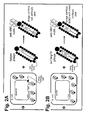
- the antibody sequence was first removed from the phagemid vector and placed into a generic plasmid that produced the antibody/pIII fusion protein in E. coli ( Figure 2B ). Then, by co-infecting the culture with M13 phage containing a normal wild-type packing signal, phage particles were produced containing the wild-type M13 genomic DNA, but not the antibody-containing DNA. Also shown in Figure 2B , a detection tag can also be incorporated into the phage used for co-infection, as described in greater detail elsewhere herein.
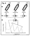
- m13mp18 phage were created, in which type differed in the length of DNA tag between common PCR primer sites ( Figure 3 ).
- the phage were constructed by starting with m13mp18 phage DNA (New England Biolabs, Inc.) and digesting the multiple cloning site with either EcoRI/HincII restriction enzymes (to remove a ⁇ 35-bp stretch of DNA) or EcoRI/HindIII restriction enzymes (to remove a ⁇ 50-bp stretch of DNA). Digested DNAs were then individually Klenow-treated and blunt-end ligated back to one another. Following electroporation into E. coli cultures, the 3 types of phage were obtained, distinguished based on the identity of the tag contained within.
- This set of experiments demonstrates a method for producing phage reagents that cannot be replicated by the end-user nor can the end-user determine the nucleotide sequence of the expressed antibody. Furthermore, the fact that the detection tag is introduced into the particle from the phage used for co-infection, obviates the need to clone tags into individual antibody constructs each time a different type of tag is desired in the final phage reagent product.
- the simultaneous binding and differentiation of each of the 3 types of anti-Rh(D) reagent to Rh(D)-positive cells but not Rh(D)-negative cells supports the concept of multiplexing phenotyping reactions, albeit in this example with 3 phage displaying the same specificity.
- the next sets of experiments will test the ability to multiplex phenotyping of cells for different specificities.
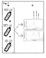
- Negative control phage (anti-dsg3), positive control phage (anti-Rh17), and anti-Rh(D) phage were then combined ( Figure 7 ) and incubated with Rh(D)-negative or Rh(D)-positive RBCs and processed for analysis. Melting point curves indicate binding of positive control phage to both cells, negative control phage to neither, and anti-Rh(D) phage to Rh(D)-positive RBCs only.
- This "quality control" aspect of the present invention was previously not possible with traditional methods of blood typing. Only with the present invention, and the multiplexing capabilities of the present invention, can both the positive and negative controls be included in a single assay.
- the methods of the present invention are similar to phage display methods well-known in the art, as described throughout the present specification.
- the methods of the present invention require the step of transforming an expression vector into a bacterial cell used to produce bacteriophage according to the present invention, wherein the expression vector comprises a nucleic acid sequence encoding the antibody or antibody fragment that will be displayed on the surface of the bacteriophage.
- the nucleic acid encoding the antibody or antibody fragment is cloned in-frame with a bacteriophage coat protein, such that an antibody/coat protein fusion protein is expressed in the bacterial cell. This fusion protein is subsequently incorporated into the progeny bacteriophage that are produced according to the present invention.
Landscapes
- Health & Medical Sciences (AREA)
- Life Sciences & Earth Sciences (AREA)
- Engineering & Computer Science (AREA)
- Immunology (AREA)
- Molecular Biology (AREA)
- Biomedical Technology (AREA)
- Hematology (AREA)
- Urology & Nephrology (AREA)
- Cell Biology (AREA)
- Chemical & Material Sciences (AREA)
- Microbiology (AREA)
- Food Science & Technology (AREA)
- Biotechnology (AREA)
- Medicinal Chemistry (AREA)
- Physics & Mathematics (AREA)
- Analytical Chemistry (AREA)
- Biochemistry (AREA)
- General Health & Medical Sciences (AREA)
- General Physics & Mathematics (AREA)
- Pathology (AREA)
- Proteomics, Peptides & Aminoacids (AREA)
- Mycology (AREA)
- Measuring Or Testing Involving Enzymes Or Micro-Organisms (AREA)
Claims (23)
- Procédé in vitro de détection de la présence d'un fragment portant un antigène sur une cellule, ledit procédé comprenant les étapes consistant à :a) prévoir un bactériophage, pour lequel ledit bactériophage :i) comprend une molécule marqueuse détectable ; etii) présente sur sa surface extérieure un anticorps connu pour se lier spécifiquement avec ledit fragment portant un antigène,- pour lequel le bactériophage est obtenu par un procédé comprenant les étapes consistant à insérer dans une cellule bactérienne une séquence d'acide nucléique codant l'anticorps à présenter sur la surface du bactériophage et à transfecter la même cellule bactérienne avec un phage d'assistance, pour lequel ladite séquence d'acide nucléique codant l'anticorps est dépourvue d'une séquence d'enrobage du bactériophage ;b) mettre en contact une cellule avec ledit bactériophage ;c) dénaturer le bactériophage spécifiquement lié à ladite cellule pour libérer ladite molécule marqueuse ; etd) détecter ladite molécule marqueuse, pour lequel cette détection de ladite molécule marqueuse détecte la présence dudit fragment portant un antigène sur ladite cellule.
- Procédé selon la revendication 1, pour lequel ladite molécule marqueuse est sélectionnée parmi le groupe constitué par un acide nucléique marqueur, une molécule fluorescente, un polypeptide, un lipide, un hydrate de carbone, un ligand, un récepteur, une enzyme, un substrat et une molécule inorganique.
- Procédé selon la revendication 2, consistant en outre à amplifier ledit acide nucléique marqueur avant l'étape (d).
- Procédé selon la revendication 1, ledit procédé consistant en outre à laver ladite cellule entre les étapes (b) et (c).
- Procédé selon la revendication 1, pour lequel ladite cellule est un globule rouge sanguin et ledit fragment portant un antigène est un antigène de globule rouge.
- Procédé selon la revendication 5, pour lequel ledit antigène de globule rouge est sélectionné parmi le groupe constitué par A, B, Rh(D), Rh(C), Rh(c), Rh(E), Rh(e), K, k, Jsa, Jsb, Kpa, Lea, Leb, Lua, Lub, Fya, Fyb, M, N, S, s, Doa, Dob, Jka et Jkb.
- Procédé selon la revendication 1, pour lequel ladite cellule est un globule blanc et pour lequel ledit fragment portant un antigène est sélectionné parmi le groupe constitué par un antigène lymphocyte, un antigène monocyte et un antigène granulocyte.
- Procédé selon la revendication 1, pour lequel ladite cellule est une plaquette et, en outre, pour lequel ledit fragment portant un antigène est un antigène de plaquette.
- Procédé selon la revendication 8, pour lequel ledit antigène de plaquette est sélectionné parmi le groupe constitué par HPA-1a, HPA-1b, HPA-2a, HPA-2b, HPA-3a, HPA-3b, HPA-4a, HPA-4b, HPA-5a, HPA-5b, HPA-6b, HPA-7b, HPA-8b, HPA-9b, HPA-10b, Gova et Govb.
- Procédé selon la revendication 1, pour lequel ledit acide nucléique comprend une séquence complémentaire à une sonde de balise moléculaire.
- Procédé selon la revendication 10, pour lequel ladite sonde de balise moléculaire comprend un fluorophore.
- Procédé selon la revendication 3, pour lequel ledit acide nucléique est amplifié en utilisant une réaction en chaine par polymérase (PCR).
- Procédé selon la revendication 3, pour lequel ledit acide nucléique est amplifié par une transcription utilisant une immuno-détection amplifiée par ARN T7 (IDAT).
- Procédé selon la revendication 2, pour lequel ladite molécule marqueuse est un acide nucléique marqueur.
- Procédé selon la revendication 14, pour lequel ledit acide nucléique marqueur est détecté en analysant la température de fusion de l'acide nucléique marqueur.
- Nécessaire de détection de la présence d'un fragment portant un antigène sur une cellule, ledit nécessaire comprenant un bactériophage, dans lequel ledit bactériophage :i) comprend une molécule marqueuse détectable ; etii) présente sur sa surface extérieure un anticorps connu pour se lier spécifiquement avec ledit fragment portant un antigène,- dans lequel le bactériophage est obtenu par un procédé comprenant les étapes consistant à insérer dans une cellule bactérienne une séquence d'acide nucléique codant l'anticorps à présenter sur la surface du bactériophage et à transfecter la même cellule bactérienne avec un phage d'assistance, pour lequel ladite séquence d'acide nucléique codant l'anticorps est dépourvue d'une séquence d'enrobage du bactériophage ;- ledit nécessaire comprenant en outre un applicateur et une documentation d'instructions pour son utilisation.
- Procédé in vitro de détection de la présence d'un fragment portant un antigène sur une cellule, ledit procédé comprenant les étapes consistant à :a) prévoir un bactériophage, pour lequel ledit bactériophage :i) comprend au moins deux molécules marqueuses, pour lequel lesdites molécules marqueuses sont chacune distinctement détectable ; etii) présente sur sa surface extérieure un anticorps connu pour se lier spécifiquement avec ledit fragment portant un antigène,- pour lequel le bactériophage est obtenu par un procédé comprenant les étapes consistant à insérer dans une cellule bactérienne une séquence d'acide nucléique codant l'anticorps à présenter sur la surface du bactériophage et à transfecter la même cellule bactérienne avec un phage d'assistance, pour lequel ladite séquence d'acide nucléique codant l'anticorps est dépourvue d'une séquence d'enrobage du bactériophage ;b) mettre en contact une cellule avec ledit bactériophage ;c) dénaturer le bactériophage spécifiquement lié à ladite cellule pour libérer lesdites molécules marqueuses ; etd) détecter au moins une desdites molécules marqueuses, pour lequel cette détection desdites molécules marqueuses détecte la présence dudit fragment portant un antigène sur ladite cellule.
- Procédé in vitro de détection de la présence d'au moins deux fragments différents portant un antigène sur une cellule, ledit procédé comprenant les étapes consistant à :a) prévoir un premier bactériophage, pour lequel ledit premier bactériophage :i) comprend une première molécule marqueuse détectable ; etii) présente sur sa surface extérieure un anticorps connu pour se lier spécifiquement avec un premier fragment portant un antigène,- pour lequel ledit premier bactériophage est obtenu par un procédé comprenant les étapes consistant à insérer dans une cellule bactérienne une première séquence d'acide nucléique codant l'anticorps à présenter sur la surface du premier bactériophage et à transfecter la même cellule bactérienne avec un phage d'assistance, pour lequel ladite première séquence d'acide nucléique codant l'anticorps est dépourvue d'une séquence d'enrobage du bactériophage ;b) prévoir un second bactériophage, pour lequel ledit second bactériophage :i) comprend une seconde molécule marqueuse détectable ; etii) présente sur sa surface extérieure un anticorps connu pour se lier spécifiquement avec un second fragment portant un antigène,- pour lequel ledit second bactériophage est obtenu par un procédé comprenant les étapes consistant à insérer dans une cellule bactérienne une seconde séquence d'acide nucléique codant l'anticorps à présenter sur la surface du second bactériophage et à transfecter la même cellule bactérienne avec un phage d'assistance, pour lequel ladite seconde séquence d'acide nucléique codant l'anticorps est dépourvue d'une séquence d'enrobage du bactériophage ;c) mettre en contact ladite cellule avec ledit premier bactériophage ;d) mettre en contact ladite cellule avec ledit second bactériophage ;e) détecter la liaison dudit premier bactériophage avec ledit premier fragment portant un antigène en détectant la présence de ladite première molécule marqueuse, pour lequel cette détection de ladite première molécule marqueuse détecte la présence dudit premier fragment portant un antigène sur ladite cellule ; etf) détecter la liaison dudit second bactériophage avec ledit second fragment portant un antigène en détectant la présence de ladite seconde molécule marqueuse, pour lequel cette détection de ladite seconde molécule marqueuse détecte la présence dudit second fragment portant un antigène sur ladite cellule.
- Procédé selon la revendication 18, pour lequel ladite première molécule marqueuse est un premier acide nucléique marqueur et, en outre, pour lequel ladite seconde molécule marqueuse est un second acide nucléique marqueur.
- Procédé selon la revendication 19, pour lequel lesdits premier et second acides nucléiques marqueurs sont détectés en analysant les températures de fusion desdits premier et second acides nucléiques marqueurs.
- Procédé de détection de la présence d'un anticorps anti globule rouge dans un sérum humain, ledit procédé comprenant les étapes consistant à :a) mettre en contact un globule rouge humain exprimant au moins un antigène de globule rouge humain sur la surface de ladite cellule avec ledit sérum ;b) laver ladite cellule pour retirer tout anticorps lié non-spécifiquement avec ladite cellule ;c) prévoir un bactériophage, pour lequel ledit bactériophage :i) comprend une molécule marqueuse détectable ; etii) présente sur sa surface extérieure un réactif anti-globuline humain,- pour lequel ledit réactif est obtenu par un procédé comprenant les étapes consistant à insérer dans une cellule bactérienne une séquence d'acide nucléique codant le réactif à présenter sur la surface du bactériophage et à transfecter la même cellule bactérienne avec un phage d'assistance, pour lequel ladite séquence d'acide nucléique codant le réactif est dépourvue d'une séquence d'enrobage du bactériophage ;d) mettre en contact ladite cellule avec ledit bactériophage ;e) laver ladite cellule pour retirer tout anticorps lié non-spécifiquement avec ladite cellule ;f) dénaturer le bactériophage spécifiquement lié à ladite cellule pour libérer ladite molécule marqueuse ; etg) détecter ladite molécule marqueuse, pour lequel cette détection de ladite molécule marqueuse détecte la présence dudit anticorps anti globule rouge dans ledit sérum.
- Procédé in vitro de détection de la présence d'un anticorps anti globule rouge chez un humain, ledit procédé comprenant les étapes consistant à :a) laver un globule rouge obtenu chez ledit humain pour retirer tout anticorps lié non-spécifiquement avec ladite cellule ;b) prévoir un bactériophage, pour lequel ledit bactériophage :i) comprend une molécule marqueuse détectable ; etii) présente sur sa surface extérieure un réactif anti-globuline humain,- pour lequel ledit réactif est obtenu par un procédé comprenant les étapes consistant à insérer dans une cellule bactérienne une séquence d'acide nucléique codant le réactif à présenter sur la surface du bactériophage et à transfecter la même cellule bactérienne avec un phage d'assistance, pour lequel ladite séquence d'acide nucléique codant le réactif est dépourvue d'une séquence d'enrobage du bactériophage ;c) mettre en contact ladite cellule avec ledit bactériophage ;d) dénaturer le bactériophage spécifiquement lié à ladite cellule pour libérer ladite molécule marqueuse ; etg) détecter ladite molécule marqueuse, pour lequel cette détection de ladite molécule marqueuse détecte la présence dudit anticorps anti globule rouge chez ledit humain.
- Procédé de détection de la présence d'un fragment portant un antigène dans une composition, ledit procédé comprenant les étapes consistant à :a) prévoir un bactériophage, pour lequel ledit bactériophage :i) comprend une molécule marqueuse détectable ; etii) présente sur sa surface extérieure un anticorps connu pour se lier spécifiquement avec ledit fragment portant un antigène,- pour lequel le bactériophage est obtenu par un procédé comprenant les étapes consistant à insérer dans une cellule bactérienne une séquence d'acide nucléique codant l'anticorps à présenter sur la surface du bactériophage et à transfecter la même cellule bactérienne avec un phage d'assistance, pour lequel ladite séquence d'acide nucléique codant l'anticorps est dépourvue d'une séquence d'enrobage du bactériophage ;b) mettre en contact ladite composition avec ledit bactériophage ; etc) détecter ladite molécule marqueuse, pour lequel cette détection de ladite molécule marqueuse détecte la présence dudit fragment portant un antigène sur ladite cellule.
Applications Claiming Priority (2)
| Application Number | Priority Date | Filing Date | Title |
|---|---|---|---|
| US74044005P | 2005-11-29 | 2005-11-29 | |
| PCT/US2006/044134 WO2007064462A1 (fr) | 2005-11-29 | 2006-11-14 | Reactifs diagnostiques de particules de phages |
Publications (3)
| Publication Number | Publication Date |
|---|---|
| EP1963494A1 EP1963494A1 (fr) | 2008-09-03 |
| EP1963494A4 EP1963494A4 (fr) | 2010-10-20 |
| EP1963494B1 true EP1963494B1 (fr) | 2014-10-08 |
Family
ID=38092561
Family Applications (1)
| Application Number | Title | Priority Date | Filing Date |
|---|---|---|---|
| EP06827795.3A Active EP1963494B1 (fr) | 2005-11-29 | 2006-11-14 | Reactifs diagnostiques de particules de phages |
Country Status (3)
| Country | Link |
|---|---|
| US (1) | US9658224B2 (fr) |
| EP (1) | EP1963494B1 (fr) |
| WO (1) | WO2007064462A1 (fr) |
Families Citing this family (10)
| Publication number | Priority date | Publication date | Assignee | Title |
|---|---|---|---|---|
| WO2004027028A2 (fr) * | 2002-09-18 | 2004-04-01 | The Trustees Of The University Of Pennsylvania | Preparations, procedes et kits pour la detection d'un antigene sur une cellule et dans un melange biologique |
| US20100099076A1 (en) * | 2008-10-16 | 2010-04-22 | Kent State University | Sensitive and rapid detection of viral particles in early viral infection by laser tweezers |
| WO2010046930A1 (fr) * | 2008-10-17 | 2010-04-29 | Genesis Labs Limited | Trousse de diagnostic fondee sur pcr et procede d'amplification et detection de pathogenes |
| PL218313B1 (pl) * | 2010-10-28 | 2014-11-28 | Inst Immunologii I Terapii Doświadczalnej Pan | Kompetytywna metoda otrzymywania preparatów bakteriofagowych |
| WO2013049121A2 (fr) * | 2011-09-26 | 2013-04-04 | Sample6 Technologies, Inc. | Phage recombinant et procédés associés |
| FR2991689B1 (fr) * | 2012-06-11 | 2018-04-20 | Diagast | Dispositif de diagnostic immuno-hematologique et utilisations |
| AU2014241191A1 (en) | 2013-03-27 | 2015-10-22 | Sample Technologies, Inc. | Recombinant phage and bacterial detection methods |
| US11326182B2 (en) | 2016-04-29 | 2022-05-10 | Voyager Therapeutics, Inc. | Compositions for the treatment of disease |
| WO2017189964A2 (fr) | 2016-04-29 | 2017-11-02 | Voyager Therapeutics, Inc. | Compositions pour le traitement de maladies |
| US11512356B2 (en) * | 2018-11-08 | 2022-11-29 | Tokitae Llc | Systems and methods for particle multiplexing in droplets |
Family Cites Families (9)
| Publication number | Priority date | Publication date | Assignee | Title |
|---|---|---|---|---|
| GB1303802A (fr) * | 1969-01-16 | 1973-01-24 | ||
| US5985543A (en) | 1996-10-11 | 1999-11-16 | The Trustees Of The University Of Pennsylvania | Compositions and methods for detection of antibody binding to cells |
| US5876925A (en) | 1996-10-11 | 1999-03-02 | The Trustees Of The University Of Pennsylvania | Magnetically activated cell sorting for production of proteins |
| US6255455B1 (en) | 1996-10-11 | 2001-07-03 | The Trustees Of The University Of Pennsylvania | Rh(D)-binding proteins and magnetically activated cell sorting method for production thereof |
| CA2368875A1 (fr) * | 1999-04-14 | 2000-10-19 | The Government Of The United States Of America | Detection de biomolecule d'expression de phage a sensibilite elevee |
| US20020025536A1 (en) * | 2000-06-26 | 2002-02-28 | Jeno Gyuris | Methods and reagents for isolating biologically active antibodies |
| DK1402025T3 (da) * | 2001-06-15 | 2006-06-06 | Crucell Holland Bv | Kimære fager |
| WO2004027028A2 (fr) * | 2002-09-18 | 2004-04-01 | The Trustees Of The University Of Pennsylvania | Preparations, procedes et kits pour la detection d'un antigene sur une cellule et dans un melange biologique |
| CA2585267A1 (fr) * | 2004-10-29 | 2007-02-08 | University Of Rochester | Vecteurs de bacteriophage modifie et utilisation de ceux-ci |
-
2006
- 2006-11-14 EP EP06827795.3A patent/EP1963494B1/fr active Active
- 2006-11-14 WO PCT/US2006/044134 patent/WO2007064462A1/fr not_active Ceased
- 2006-11-14 US US12/085,618 patent/US9658224B2/en active Active
Also Published As
| Publication number | Publication date |
|---|---|
| US9658224B2 (en) | 2017-05-23 |
| EP1963494A1 (fr) | 2008-09-03 |
| WO2007064462A1 (fr) | 2007-06-07 |
| HK1118863A1 (en) | 2009-02-20 |
| US20100015595A1 (en) | 2010-01-21 |
| EP1963494A4 (fr) | 2010-10-20 |
Similar Documents
| Publication | Publication Date | Title |
|---|---|---|
| CN110431233B (zh) | 用于单细胞的大量平行组合分析的系统和方法 | |
| JP2020043860A (ja) | ハイスループットなレセプター:リガンド同定の方法 | |
| US20040048311A1 (en) | Use of collections of binding sites for sample profiling and other applications | |
| US9658224B2 (en) | Phage particle diagnostic reagents | |
| US8617805B2 (en) | Compositions, methods and kits for detection of an antigen on a cell and in a biological mixture | |
| CN117545767A (zh) | 嵌合的免疫球蛋白 | |
| CN121652276A (zh) | 一种能够激活t细胞活性的抗cd3纳米抗体及其制备方法与应用 | |
| Song et al. | A cell-based multiplex immunoassay platform using fluorescent protein-barcoded reporter cell lines | |
| AU722978B2 (en) | Magnetically activated cell sorting for production of proteins | |
| Challa et al. | Function-first discovery of high affinity monoclonal antibodies using Nanovial-based plasma B cell screening | |
| JP7021949B2 (ja) | 抗体検出法及び抗体検出系 | |
| CN118388644B (zh) | 一种抗波形蛋白抗体及其应用 | |
| CN117683131B (zh) | 一种抗髓鞘少突胶质细胞糖蛋白(mog)抗体及其应用 | |
| CN101448934A (zh) | 用于粒细胞抗体检测的谱细胞 | |
| HK1118863B (en) | Phage particle diagnostic reagents | |
| Siegel | Phage display-based molecular methods in immunohematology. | |
| HK1077851B (en) | Compositions, methods and kits for detection of an antigen on a cell and in a biological mixture | |
| US20250163406A1 (en) | Methods for generating nucleic acid encoded protein libraries and uses thereof | |
| Avent | Recombinant technology in transfusion medicine | |
| Figini et al. | Isolation of human monoclonal antibodies using guided selection with mouse monoclonal antibodies | |
| Yang et al. | Use of CMFDA and CMTMR Fluorescent Dyes in FACS®-Based Antibody Screening | |
| CN119591714A (zh) | 一种抗人mog蛋白的人鼠嵌合单克隆抗体及其应用 |
Legal Events
| Date | Code | Title | Description |
|---|---|---|---|
| PUAI | Public reference made under article 153(3) epc to a published international application that has entered the european phase |
Free format text: ORIGINAL CODE: 0009012 |
|
| 17P | Request for examination filed |
Effective date: 20080529 |
|
| AK | Designated contracting states |
Kind code of ref document: A1 Designated state(s): AT BE BG CH CY CZ DE DK EE ES FI FR GB GR HU IE IS IT LI LT LU LV MC NL PL PT RO SE SI SK TR |
|
| AX | Request for extension of the european patent |
Extension state: AL BA HR MK RS |
|
| REG | Reference to a national code |
Ref country code: HK Ref legal event code: DE Ref document number: 1118863 Country of ref document: HK |
|
| A4 | Supplementary search report drawn up and despatched |
Effective date: 20100920 |
|
| RIC1 | Information provided on ipc code assigned before grant |
Ipc: G01N 33/53 20060101ALI20100914BHEP Ipc: C12P 21/06 20060101ALI20100914BHEP Ipc: C12N 7/00 20060101AFI20080606BHEP Ipc: G01N 33/80 20060101ALI20100914BHEP |
|
| 17Q | First examination report despatched |
Effective date: 20110715 |
|
| GRAP | Despatch of communication of intention to grant a patent |
Free format text: ORIGINAL CODE: EPIDOSNIGR1 |
|
| INTG | Intention to grant announced |
Effective date: 20140505 |
|
| GRAS | Grant fee paid |
Free format text: ORIGINAL CODE: EPIDOSNIGR3 |
|
| GRAA | (expected) grant |
Free format text: ORIGINAL CODE: 0009210 |
|
| AK | Designated contracting states |
Kind code of ref document: B1 Designated state(s): AT BE BG CH CY CZ DE DK EE ES FI FR GB GR HU IE IS IT LI LT LU LV MC NL PL PT RO SE SI SK TR |
|
| AX | Request for extension of the european patent |
Extension state: AL BA HR MK RS |
|
| REG | Reference to a national code |
Ref country code: GB Ref legal event code: FG4D |
|
| REG | Reference to a national code |
Ref country code: CH Ref legal event code: EP Ref country code: AT Ref legal event code: REF Ref document number: 690621 Country of ref document: AT Kind code of ref document: T Effective date: 20141015 |
|
| REG | Reference to a national code |
Ref country code: IE Ref legal event code: FG4D |
|
| REG | Reference to a national code |
Ref country code: DE Ref legal event code: R096 Ref document number: 602006043318 Country of ref document: DE Effective date: 20141120 |
|
| REG | Reference to a national code |
Ref country code: HK Ref legal event code: GR Ref document number: 1118863 Country of ref document: HK |
|
| REG | Reference to a national code |
Ref country code: NL Ref legal event code: VDEP Effective date: 20141008 |
|
| REG | Reference to a national code |
Ref country code: AT Ref legal event code: MK05 Ref document number: 690621 Country of ref document: AT Kind code of ref document: T Effective date: 20141008 |
|
| REG | Reference to a national code |
Ref country code: LT Ref legal event code: MG4D |
|
| PG25 | Lapsed in a contracting state [announced via postgrant information from national office to epo] |
Ref country code: NL Free format text: LAPSE BECAUSE OF FAILURE TO SUBMIT A TRANSLATION OF THE DESCRIPTION OR TO PAY THE FEE WITHIN THE PRESCRIBED TIME-LIMIT Effective date: 20141008 |
|
| PG25 | Lapsed in a contracting state [announced via postgrant information from national office to epo] |
Ref country code: LT Free format text: LAPSE BECAUSE OF FAILURE TO SUBMIT A TRANSLATION OF THE DESCRIPTION OR TO PAY THE FEE WITHIN THE PRESCRIBED TIME-LIMIT Effective date: 20141008 Ref country code: FI Free format text: LAPSE BECAUSE OF FAILURE TO SUBMIT A TRANSLATION OF THE DESCRIPTION OR TO PAY THE FEE WITHIN THE PRESCRIBED TIME-LIMIT Effective date: 20141008 Ref country code: PT Free format text: LAPSE BECAUSE OF FAILURE TO SUBMIT A TRANSLATION OF THE DESCRIPTION OR TO PAY THE FEE WITHIN THE PRESCRIBED TIME-LIMIT Effective date: 20150209 Ref country code: IS Free format text: LAPSE BECAUSE OF FAILURE TO SUBMIT A TRANSLATION OF THE DESCRIPTION OR TO PAY THE FEE WITHIN THE PRESCRIBED TIME-LIMIT Effective date: 20150208 Ref country code: ES Free format text: LAPSE BECAUSE OF FAILURE TO SUBMIT A TRANSLATION OF THE DESCRIPTION OR TO PAY THE FEE WITHIN THE PRESCRIBED TIME-LIMIT Effective date: 20141008 |
|
| PG25 | Lapsed in a contracting state [announced via postgrant information from national office to epo] |
Ref country code: AT Free format text: LAPSE BECAUSE OF FAILURE TO SUBMIT A TRANSLATION OF THE DESCRIPTION OR TO PAY THE FEE WITHIN THE PRESCRIBED TIME-LIMIT Effective date: 20141008 Ref country code: SE Free format text: LAPSE BECAUSE OF FAILURE TO SUBMIT A TRANSLATION OF THE DESCRIPTION OR TO PAY THE FEE WITHIN THE PRESCRIBED TIME-LIMIT Effective date: 20141008 Ref country code: GR Free format text: LAPSE BECAUSE OF FAILURE TO SUBMIT A TRANSLATION OF THE DESCRIPTION OR TO PAY THE FEE WITHIN THE PRESCRIBED TIME-LIMIT Effective date: 20150109 Ref country code: LV Free format text: LAPSE BECAUSE OF FAILURE TO SUBMIT A TRANSLATION OF THE DESCRIPTION OR TO PAY THE FEE WITHIN THE PRESCRIBED TIME-LIMIT Effective date: 20141008 Ref country code: CY Free format text: LAPSE BECAUSE OF FAILURE TO SUBMIT A TRANSLATION OF THE DESCRIPTION OR TO PAY THE FEE WITHIN THE PRESCRIBED TIME-LIMIT Effective date: 20141008 Ref country code: PL Free format text: LAPSE BECAUSE OF FAILURE TO SUBMIT A TRANSLATION OF THE DESCRIPTION OR TO PAY THE FEE WITHIN THE PRESCRIBED TIME-LIMIT Effective date: 20141008 |
|
| PG25 | Lapsed in a contracting state [announced via postgrant information from national office to epo] |
Ref country code: BE Free format text: LAPSE BECAUSE OF NON-PAYMENT OF DUE FEES Effective date: 20141130 |
|
| REG | Reference to a national code |
Ref country code: CH Ref legal event code: PL |
|
| REG | Reference to a national code |
Ref country code: DE Ref legal event code: R097 Ref document number: 602006043318 Country of ref document: DE |
|
| PG25 | Lapsed in a contracting state [announced via postgrant information from national office to epo] |
Ref country code: MC Free format text: LAPSE BECAUSE OF FAILURE TO SUBMIT A TRANSLATION OF THE DESCRIPTION OR TO PAY THE FEE WITHIN THE PRESCRIBED TIME-LIMIT Effective date: 20141008 Ref country code: EE Free format text: LAPSE BECAUSE OF FAILURE TO SUBMIT A TRANSLATION OF THE DESCRIPTION OR TO PAY THE FEE WITHIN THE PRESCRIBED TIME-LIMIT Effective date: 20141008 Ref country code: SK Free format text: LAPSE BECAUSE OF FAILURE TO SUBMIT A TRANSLATION OF THE DESCRIPTION OR TO PAY THE FEE WITHIN THE PRESCRIBED TIME-LIMIT Effective date: 20141008 Ref country code: RO Free format text: LAPSE BECAUSE OF FAILURE TO SUBMIT A TRANSLATION OF THE DESCRIPTION OR TO PAY THE FEE WITHIN THE PRESCRIBED TIME-LIMIT Effective date: 20141008 Ref country code: DK Free format text: LAPSE BECAUSE OF FAILURE TO SUBMIT A TRANSLATION OF THE DESCRIPTION OR TO PAY THE FEE WITHIN THE PRESCRIBED TIME-LIMIT Effective date: 20141008 Ref country code: CZ Free format text: LAPSE BECAUSE OF FAILURE TO SUBMIT A TRANSLATION OF THE DESCRIPTION OR TO PAY THE FEE WITHIN THE PRESCRIBED TIME-LIMIT Effective date: 20141008 Ref country code: CH Free format text: LAPSE BECAUSE OF NON-PAYMENT OF DUE FEES Effective date: 20141130 Ref country code: LI Free format text: LAPSE BECAUSE OF NON-PAYMENT OF DUE FEES Effective date: 20141130 |
|
| PLBE | No opposition filed within time limit |
Free format text: ORIGINAL CODE: 0009261 |
|
| STAA | Information on the status of an ep patent application or granted ep patent |
Free format text: STATUS: NO OPPOSITION FILED WITHIN TIME LIMIT |
|
| REG | Reference to a national code |
Ref country code: IE Ref legal event code: MM4A |
|
| PG25 | Lapsed in a contracting state [announced via postgrant information from national office to epo] |
Ref country code: IT Free format text: LAPSE BECAUSE OF FAILURE TO SUBMIT A TRANSLATION OF THE DESCRIPTION OR TO PAY THE FEE WITHIN THE PRESCRIBED TIME-LIMIT Effective date: 20141008 |
|
| 26N | No opposition filed |
Effective date: 20150709 |
|
| PG25 | Lapsed in a contracting state [announced via postgrant information from national office to epo] |
Ref country code: IE Free format text: LAPSE BECAUSE OF NON-PAYMENT OF DUE FEES Effective date: 20141114 |
|
| REG | Reference to a national code |
Ref country code: FR Ref legal event code: PLFP Year of fee payment: 10 |
|
| PG25 | Lapsed in a contracting state [announced via postgrant information from national office to epo] |
Ref country code: SI Free format text: LAPSE BECAUSE OF FAILURE TO SUBMIT A TRANSLATION OF THE DESCRIPTION OR TO PAY THE FEE WITHIN THE PRESCRIBED TIME-LIMIT Effective date: 20141008 |
|
| PG25 | Lapsed in a contracting state [announced via postgrant information from national office to epo] |
Ref country code: BG Free format text: LAPSE BECAUSE OF FAILURE TO SUBMIT A TRANSLATION OF THE DESCRIPTION OR TO PAY THE FEE WITHIN THE PRESCRIBED TIME-LIMIT Effective date: 20141008 |
|
| PG25 | Lapsed in a contracting state [announced via postgrant information from national office to epo] |
Ref country code: TR Free format text: LAPSE BECAUSE OF FAILURE TO SUBMIT A TRANSLATION OF THE DESCRIPTION OR TO PAY THE FEE WITHIN THE PRESCRIBED TIME-LIMIT Effective date: 20141008 Ref country code: HU Free format text: LAPSE BECAUSE OF FAILURE TO SUBMIT A TRANSLATION OF THE DESCRIPTION OR TO PAY THE FEE WITHIN THE PRESCRIBED TIME-LIMIT; INVALID AB INITIO Effective date: 20061114 Ref country code: LU Free format text: LAPSE BECAUSE OF NON-PAYMENT OF DUE FEES Effective date: 20141114 |
|
| REG | Reference to a national code |
Ref country code: FR Ref legal event code: PLFP Year of fee payment: 11 |
|
| REG | Reference to a national code |
Ref country code: FR Ref legal event code: PLFP Year of fee payment: 12 |
|
| REG | Reference to a national code |
Ref country code: FR Ref legal event code: PLFP Year of fee payment: 13 |
|
| P01 | Opt-out of the competence of the unified patent court (upc) registered |
Effective date: 20230526 |
|
| PGFP | Annual fee paid to national office [announced via postgrant information from national office to epo] |
Ref country code: DE Payment date: 20251028 Year of fee payment: 20 |
|
| PGFP | Annual fee paid to national office [announced via postgrant information from national office to epo] |
Ref country code: GB Payment date: 20251030 Year of fee payment: 20 |
|
| PGFP | Annual fee paid to national office [announced via postgrant information from national office to epo] |
Ref country code: FR Payment date: 20251117 Year of fee payment: 20 |