EP1990407A1 - Von einer gewebestammzelle abgeleitete feeder-zelle - Google Patents
Von einer gewebestammzelle abgeleitete feeder-zelle Download PDFInfo
- Publication number
- EP1990407A1 EP1990407A1 EP07706922A EP07706922A EP1990407A1 EP 1990407 A1 EP1990407 A1 EP 1990407A1 EP 07706922 A EP07706922 A EP 07706922A EP 07706922 A EP07706922 A EP 07706922A EP 1990407 A1 EP1990407 A1 EP 1990407A1
- Authority
- EP
- European Patent Office
- Prior art keywords
- cell
- cells
- feeder
- cultured
- epithelial
- Prior art date
- Legal status (The legal status is an assumption and is not a legal conclusion. Google has not performed a legal analysis and makes no representation as to the accuracy of the status listed.)
- Withdrawn
Links
- 210000004027 cell Anatomy 0.000 title claims abstract description 163
- 210000000130 stem cell Anatomy 0.000 title claims abstract description 80
- 238000000034 method Methods 0.000 claims abstract description 33
- 238000012258 culturing Methods 0.000 claims abstract description 19
- 210000004748 cultured cell Anatomy 0.000 claims abstract description 17
- 210000002919 epithelial cell Anatomy 0.000 claims description 36
- 210000001519 tissue Anatomy 0.000 claims description 35
- 210000002950 fibroblast Anatomy 0.000 claims description 17
- 241000124008 Mammalia Species 0.000 claims description 14
- 210000003683 corneal stroma Anatomy 0.000 claims description 13
- 210000001185 bone marrow Anatomy 0.000 claims description 11
- 230000012010 growth Effects 0.000 claims description 11
- 210000003491 skin Anatomy 0.000 claims description 9
- 238000002054 transplantation Methods 0.000 claims description 9
- 210000004927 skin cell Anatomy 0.000 claims description 6
- 210000002798 bone marrow cell Anatomy 0.000 claims description 4
- 230000002496 gastric effect Effects 0.000 claims description 4
- 210000002064 heart cell Anatomy 0.000 claims description 3
- 230000001939 inductive effect Effects 0.000 claims description 3
- 238000002360 preparation method Methods 0.000 abstract description 4
- 239000002609 medium Substances 0.000 description 13
- 210000000981 epithelium Anatomy 0.000 description 10
- WSFSSNUMVMOOMR-UHFFFAOYSA-N Formaldehyde Chemical compound O=C WSFSSNUMVMOOMR-UHFFFAOYSA-N 0.000 description 8
- 238000012744 immunostaining Methods 0.000 description 8
- 230000004069 differentiation Effects 0.000 description 7
- 239000003550 marker Substances 0.000 description 7
- 102100040443 Keratin, type I cytoskeletal 15 Human genes 0.000 description 6
- NWIBSHFKIJFRCO-WUDYKRTCSA-N Mytomycin Chemical compound C1N2C(C(C(C)=C(N)C3=O)=O)=C3[C@@H](COC(N)=O)[C@@]2(OC)[C@@H]2[C@H]1N2 NWIBSHFKIJFRCO-WUDYKRTCSA-N 0.000 description 6
- 238000003501 co-culture Methods 0.000 description 6
- 210000002901 mesenchymal stem cell Anatomy 0.000 description 6
- 108091003079 Bovine Serum Albumin Proteins 0.000 description 5
- 239000006144 Dulbecco’s modified Eagle's medium Substances 0.000 description 5
- 230000001332 colony forming effect Effects 0.000 description 5
- 210000004087 cornea Anatomy 0.000 description 5
- 239000012091 fetal bovine serum Substances 0.000 description 5
- 230000014509 gene expression Effects 0.000 description 5
- 102000001045 Connexin 43 Human genes 0.000 description 4
- 108010069241 Connexin 43 Proteins 0.000 description 4
- 229940079593 drug Drugs 0.000 description 4
- 239000003814 drug Substances 0.000 description 4
- 230000009786 epithelial differentiation Effects 0.000 description 4
- 210000005260 human cell Anatomy 0.000 description 4
- JYGXADMDTFJGBT-VWUMJDOOSA-N hydrocortisone Chemical compound O=C1CC[C@]2(C)[C@H]3[C@@H](O)C[C@](C)([C@@](CC4)(O)C(=O)CO)[C@@H]4[C@@H]3CCC2=C1 JYGXADMDTFJGBT-VWUMJDOOSA-N 0.000 description 4
- 230000006698 induction Effects 0.000 description 4
- NOESYZHRGYRDHS-UHFFFAOYSA-N insulin Chemical compound N1C(=O)C(NC(=O)C(CCC(N)=O)NC(=O)C(CCC(O)=O)NC(=O)C(C(C)C)NC(=O)C(NC(=O)CN)C(C)CC)CSSCC(C(NC(CO)C(=O)NC(CC(C)C)C(=O)NC(CC=2C=CC(O)=CC=2)C(=O)NC(CCC(N)=O)C(=O)NC(CC(C)C)C(=O)NC(CCC(O)=O)C(=O)NC(CC(N)=O)C(=O)NC(CC=2C=CC(O)=CC=2)C(=O)NC(CSSCC(NC(=O)C(C(C)C)NC(=O)C(CC(C)C)NC(=O)C(CC=2C=CC(O)=CC=2)NC(=O)C(CC(C)C)NC(=O)C(C)NC(=O)C(CCC(O)=O)NC(=O)C(C(C)C)NC(=O)C(CC(C)C)NC(=O)C(CC=2NC=NC=2)NC(=O)C(CO)NC(=O)CNC2=O)C(=O)NCC(=O)NC(CCC(O)=O)C(=O)NC(CCCNC(N)=N)C(=O)NCC(=O)NC(CC=3C=CC=CC=3)C(=O)NC(CC=3C=CC=CC=3)C(=O)NC(CC=3C=CC(O)=CC=3)C(=O)NC(C(C)O)C(=O)N3C(CCC3)C(=O)NC(CCCCN)C(=O)NC(C)C(O)=O)C(=O)NC(CC(N)=O)C(O)=O)=O)NC(=O)C(C(C)CC)NC(=O)C(CO)NC(=O)C(C(C)O)NC(=O)C1CSSCC2NC(=O)C(CC(C)C)NC(=O)C(NC(=O)C(CCC(N)=O)NC(=O)C(CC(N)=O)NC(=O)C(NC(=O)C(N)CC=1C=CC=CC=1)C(C)C)CC1=CN=CN1 NOESYZHRGYRDHS-UHFFFAOYSA-N 0.000 description 4
- 229960004857 mitomycin Drugs 0.000 description 4
- 238000010899 nucleation Methods 0.000 description 4
- 238000010186 staining Methods 0.000 description 4
- 102000004142 Trypsin Human genes 0.000 description 3
- 108090000631 Trypsin Proteins 0.000 description 3
- 210000001691 amnion Anatomy 0.000 description 3
- 238000004113 cell culture Methods 0.000 description 3
- 230000005757 colony formation Effects 0.000 description 3
- 210000001339 epidermal cell Anatomy 0.000 description 3
- 230000036541 health Effects 0.000 description 3
- 210000001232 limbus corneae Anatomy 0.000 description 3
- 238000011160 research Methods 0.000 description 3
- 239000012588 trypsin Substances 0.000 description 3
- ACNUVXZPCIABEX-UHFFFAOYSA-N 3',6'-diaminospiro[2-benzofuran-3,9'-xanthene]-1-one Chemical compound O1C(=O)C2=CC=CC=C2C21C1=CC=C(N)C=C1OC1=CC(N)=CC=C21 ACNUVXZPCIABEX-UHFFFAOYSA-N 0.000 description 2
- CSCPPACGZOOCGX-UHFFFAOYSA-N Acetone Chemical compound CC(C)=O CSCPPACGZOOCGX-UHFFFAOYSA-N 0.000 description 2
- IJGRMHOSHXDMSA-UHFFFAOYSA-N Atomic nitrogen Chemical compound N#N IJGRMHOSHXDMSA-UHFFFAOYSA-N 0.000 description 2
- 102000029816 Collagenase Human genes 0.000 description 2
- 108060005980 Collagenase Proteins 0.000 description 2
- KCXVZYZYPLLWCC-UHFFFAOYSA-N EDTA Chemical compound OC(=O)CN(CC(O)=O)CCN(CC(O)=O)CC(O)=O KCXVZYZYPLLWCC-UHFFFAOYSA-N 0.000 description 2
- 108090000790 Enzymes Proteins 0.000 description 2
- 102000004190 Enzymes Human genes 0.000 description 2
- 102000009024 Epidermal Growth Factor Human genes 0.000 description 2
- 102000009123 Fibrin Human genes 0.000 description 2
- 108010073385 Fibrin Proteins 0.000 description 2
- BWGVNKXGVNDBDI-UHFFFAOYSA-N Fibrin monomer Chemical compound CNC(=O)CNC(=O)CN BWGVNKXGVNDBDI-UHFFFAOYSA-N 0.000 description 2
- 102000004877 Insulin Human genes 0.000 description 2
- 108090001061 Insulin Proteins 0.000 description 2
- 102000012355 Integrin beta1 Human genes 0.000 description 2
- 108010022222 Integrin beta1 Proteins 0.000 description 2
- 102100023967 Keratin, type I cytoskeletal 12 Human genes 0.000 description 2
- 102100025759 Keratin, type II cytoskeletal 3 Human genes 0.000 description 2
- 108010065086 Keratin-12 Proteins 0.000 description 2
- 108010066330 Keratin-15 Proteins 0.000 description 2
- 108010070918 Keratin-3 Proteins 0.000 description 2
- 229930192392 Mitomycin Natural products 0.000 description 2
- 102000004338 Transferrin Human genes 0.000 description 2
- 108090000901 Transferrin Proteins 0.000 description 2
- 238000003556 assay Methods 0.000 description 2
- 238000005119 centrifugation Methods 0.000 description 2
- 229960002424 collagenase Drugs 0.000 description 2
- 230000000694 effects Effects 0.000 description 2
- 210000001671 embryonic stem cell Anatomy 0.000 description 2
- 229940088598 enzyme Drugs 0.000 description 2
- 210000003560 epithelium corneal Anatomy 0.000 description 2
- 238000002474 experimental method Methods 0.000 description 2
- 229950003499 fibrin Drugs 0.000 description 2
- 229960000890 hydrocortisone Drugs 0.000 description 2
- 208000015181 infectious disease Diseases 0.000 description 2
- 230000002401 inhibitory effect Effects 0.000 description 2
- 229940125396 insulin Drugs 0.000 description 2
- 239000000463 material Substances 0.000 description 2
- 239000012188 paraffin wax Substances 0.000 description 2
- 230000005855 radiation Effects 0.000 description 2
- 230000002829 reductive effect Effects 0.000 description 2
- 210000002966 serum Anatomy 0.000 description 2
- 239000012679 serum free medium Substances 0.000 description 2
- 239000000243 solution Substances 0.000 description 2
- UCSJYZPVAKXKNQ-HZYVHMACSA-N streptomycin Chemical compound CN[C@H]1[C@H](O)[C@@H](O)[C@H](CO)O[C@H]1O[C@@H]1[C@](C=O)(O)[C@H](C)O[C@H]1O[C@@H]1[C@@H](NC(N)=N)[C@H](O)[C@@H](NC(N)=N)[C@H](O)[C@H]1O UCSJYZPVAKXKNQ-HZYVHMACSA-N 0.000 description 2
- 238000001356 surgical procedure Methods 0.000 description 2
- 230000004083 survival effect Effects 0.000 description 2
- 239000012581 transferrin Substances 0.000 description 2
- 230000000007 visual effect Effects 0.000 description 2
- JWZZKOKVBUJMES-UHFFFAOYSA-N (+-)-Isoprenaline Chemical compound CC(C)NCC(O)C1=CC=C(O)C(O)=C1 JWZZKOKVBUJMES-UHFFFAOYSA-N 0.000 description 1
- FWBHETKCLVMNFS-UHFFFAOYSA-N 4',6-Diamino-2-phenylindol Chemical compound C1=CC(C(=N)N)=CC=C1C1=CC2=CC=C(C(N)=N)C=C2N1 FWBHETKCLVMNFS-UHFFFAOYSA-N 0.000 description 1
- 229930024421 Adenine Natural products 0.000 description 1
- GFFGJBXGBJISGV-UHFFFAOYSA-N Adenine Chemical compound NC1=NC=NC2=C1N=CN2 GFFGJBXGBJISGV-UHFFFAOYSA-N 0.000 description 1
- 108010039627 Aprotinin Proteins 0.000 description 1
- 102000009016 Cholera Toxin Human genes 0.000 description 1
- 108010049048 Cholera Toxin Proteins 0.000 description 1
- 208000035473 Communicable disease Diseases 0.000 description 1
- FBPFZTCFMRRESA-FSIIMWSLSA-N D-Glucitol Natural products OC[C@H](O)[C@H](O)[C@@H](O)[C@H](O)CO FBPFZTCFMRRESA-FSIIMWSLSA-N 0.000 description 1
- FBPFZTCFMRRESA-JGWLITMVSA-N D-glucitol Chemical compound OC[C@H](O)[C@@H](O)[C@H](O)[C@H](O)CO FBPFZTCFMRRESA-JGWLITMVSA-N 0.000 description 1
- IROWCYIEJAOFOW-UHFFFAOYSA-N DL-Isoprenaline hydrochloride Chemical compound Cl.CC(C)NCC(O)C1=CC=C(O)C(O)=C1 IROWCYIEJAOFOW-UHFFFAOYSA-N 0.000 description 1
- 101800003838 Epidermal growth factor Proteins 0.000 description 1
- 108010003272 Hyaluronate lyase Proteins 0.000 description 1
- 102000001974 Hyaluronidases Human genes 0.000 description 1
- 206010061218 Inflammation Diseases 0.000 description 1
- 108700020796 Oncogene Proteins 0.000 description 1
- 229930182555 Penicillin Natural products 0.000 description 1
- JGSARLDLIJGVTE-MBNYWOFBSA-N Penicillin G Chemical compound N([C@H]1[C@H]2SC([C@@H](N2C1=O)C(O)=O)(C)C)C(=O)CC1=CC=CC=C1 JGSARLDLIJGVTE-MBNYWOFBSA-N 0.000 description 1
- 101710098940 Pro-epidermal growth factor Proteins 0.000 description 1
- WDVSHHCDHLJJJR-UHFFFAOYSA-N Proflavine Chemical compound C1=CC(N)=CC2=NC3=CC(N)=CC=C3C=C21 WDVSHHCDHLJJJR-UHFFFAOYSA-N 0.000 description 1
- AUYYCJSJGJYCDS-LBPRGKRZSA-N Thyrolar Chemical compound IC1=CC(C[C@H](N)C(O)=O)=CC(I)=C1OC1=CC=C(O)C(I)=C1 AUYYCJSJGJYCDS-LBPRGKRZSA-N 0.000 description 1
- 208000027418 Wounds and injury Diseases 0.000 description 1
- 230000002159 abnormal effect Effects 0.000 description 1
- 229960000643 adenine Drugs 0.000 description 1
- 230000002411 adverse Effects 0.000 description 1
- 239000003513 alkali Substances 0.000 description 1
- 229960004405 aprotinin Drugs 0.000 description 1
- 210000000270 basal cell Anatomy 0.000 description 1
- 230000008901 benefit Effects 0.000 description 1
- 210000004556 brain Anatomy 0.000 description 1
- 210000004413 cardiac myocyte Anatomy 0.000 description 1
- 210000004240 ciliary body Anatomy 0.000 description 1
- 230000000112 colonic effect Effects 0.000 description 1
- 210000000795 conjunctiva Anatomy 0.000 description 1
- 230000006378 damage Effects 0.000 description 1
- 230000003247 decreasing effect Effects 0.000 description 1
- 210000002555 descemet membrane Anatomy 0.000 description 1
- 108010007093 dispase Proteins 0.000 description 1
- 238000012137 double-staining Methods 0.000 description 1
- 210000002257 embryonic structure Anatomy 0.000 description 1
- 210000003038 endothelium Anatomy 0.000 description 1
- 238000005516 engineering process Methods 0.000 description 1
- 229940116977 epidermal growth factor Drugs 0.000 description 1
- 210000004920 epithelial cell of skin Anatomy 0.000 description 1
- 230000008472 epithelial growth Effects 0.000 description 1
- 230000004438 eyesight Effects 0.000 description 1
- 235000013312 flour Nutrition 0.000 description 1
- 239000003102 growth factor Substances 0.000 description 1
- 238000007490 hematoxylin and eosin (H&E) staining Methods 0.000 description 1
- 238000007825 histological assay Methods 0.000 description 1
- 230000003054 hormonal effect Effects 0.000 description 1
- 229960002773 hyaluronidase Drugs 0.000 description 1
- 230000004054 inflammatory process Effects 0.000 description 1
- ZPNFWUPYTFPOJU-LPYSRVMUSA-N iniprol Chemical compound C([C@H]1C(=O)NCC(=O)NCC(=O)N[C@H]2CSSC[C@H]3C(=O)N[C@@H](CCCCN)C(=O)N[C@@H](C)C(=O)N[C@@H](CCCNC(N)=N)C(=O)N[C@H](C(N[C@H](C(=O)N[C@@H](CCCNC(N)=N)C(=O)N[C@@H](CC=4C=CC(O)=CC=4)C(=O)N[C@@H](CC=4C=CC=CC=4)C(=O)N[C@@H](CC=4C=CC(O)=CC=4)C(=O)N[C@@H](CC(N)=O)C(=O)N[C@@H](C)C(=O)N[C@@H](CCCCN)C(=O)N[C@@H](C)C(=O)NCC(=O)N[C@@H](CC(C)C)C(=O)N[C@@H](CSSC[C@H](NC(=O)[C@H](CC(O)=O)NC(=O)[C@H](CCC(O)=O)NC(=O)[C@H](C)NC(=O)[C@H](CO)NC(=O)[C@H](CCCCN)NC(=O)[C@H](CC=4C=CC=CC=4)NC(=O)[C@H](CC(N)=O)NC(=O)[C@H](CC(N)=O)NC(=O)[C@H](CCCNC(N)=N)NC(=O)[C@H](CCCCN)NC(=O)[C@H](C)NC(=O)[C@H](CCCNC(N)=N)NC2=O)C(=O)N[C@@H](CCSC)C(=O)N[C@@H](CCCNC(N)=N)C(=O)N[C@@H]([C@@H](C)O)C(=O)N[C@@H](CSSC[C@H](NC(=O)[C@H](CC=2C=CC=CC=2)NC(=O)[C@H](CC(O)=O)NC(=O)[C@H]2N(CCC2)C(=O)[C@@H](N)CCCNC(N)=N)C(=O)N[C@@H](CC(C)C)C(=O)N[C@@H](CCC(O)=O)C(=O)N2[C@@H](CCC2)C(=O)N2[C@@H](CCC2)C(=O)N[C@@H](CC=2C=CC(O)=CC=2)C(=O)N[C@@H]([C@@H](C)O)C(=O)NCC(=O)N2[C@@H](CCC2)C(=O)N3)C(=O)NCC(=O)NCC(=O)N[C@@H](C)C(O)=O)C(=O)N[C@@H](CCC(N)=O)C(=O)N[C@H](C(=O)N[C@@H](CC=2C=CC=CC=2)C(=O)N[C@H](C(=O)N1)C(C)C)[C@@H](C)O)[C@@H](C)CC)=O)[C@@H](C)CC)C1=CC=C(O)C=C1 ZPNFWUPYTFPOJU-LPYSRVMUSA-N 0.000 description 1
- 238000002347 injection Methods 0.000 description 1
- 239000007924 injection Substances 0.000 description 1
- 208000014674 injury Diseases 0.000 description 1
- 230000000968 intestinal effect Effects 0.000 description 1
- 230000009545 invasion Effects 0.000 description 1
- 210000000554 iris Anatomy 0.000 description 1
- 229940039009 isoproterenol Drugs 0.000 description 1
- 231100000636 lethal dose Toxicity 0.000 description 1
- 239000007788 liquid Substances 0.000 description 1
- 210000004185 liver Anatomy 0.000 description 1
- 238000005259 measurement Methods 0.000 description 1
- 210000002200 mouth mucosa Anatomy 0.000 description 1
- 210000003205 muscle Anatomy 0.000 description 1
- 210000005036 nerve Anatomy 0.000 description 1
- 229910052757 nitrogen Inorganic materials 0.000 description 1
- 238000012758 nuclear staining Methods 0.000 description 1
- 229940049954 penicillin Drugs 0.000 description 1
- 239000000047 product Substances 0.000 description 1
- 230000001737 promoting effect Effects 0.000 description 1
- 230000001681 protective effect Effects 0.000 description 1
- 230000005180 public health Effects 0.000 description 1
- 230000001172 regenerating effect Effects 0.000 description 1
- 230000000717 retained effect Effects 0.000 description 1
- PYWVYCXTNDRMGF-UHFFFAOYSA-N rhodamine B Chemical compound [Cl-].C=12C=CC(=[N+](CC)CC)C=C2OC2=CC(N(CC)CC)=CC=C2C=1C1=CC=CC=C1C(O)=O PYWVYCXTNDRMGF-UHFFFAOYSA-N 0.000 description 1
- 229940043267 rhodamine b Drugs 0.000 description 1
- 230000009528 severe injury Effects 0.000 description 1
- 210000000813 small intestine Anatomy 0.000 description 1
- 229960002920 sorbitol Drugs 0.000 description 1
- 210000000952 spleen Anatomy 0.000 description 1
- 238000013517 stratification Methods 0.000 description 1
- 229960005322 streptomycin Drugs 0.000 description 1
- 239000013589 supplement Substances 0.000 description 1
- 229940035722 triiodothyronine Drugs 0.000 description 1
- 229960001322 trypsin Drugs 0.000 description 1
- VBEQCZHXXJYVRD-GACYYNSASA-N uroanthelone Chemical compound C([C@@H](C(=O)N[C@H](C(=O)N[C@@H](CS)C(=O)N[C@@H](CC(N)=O)C(=O)N[C@@H](CS)C(=O)N[C@H](C(=O)N[C@@H]([C@@H](C)CC)C(=O)NCC(=O)N[C@@H](CC=1C=CC(O)=CC=1)C(=O)N[C@@H](CO)C(=O)NCC(=O)N[C@@H](CC(O)=O)C(=O)N[C@@H](CCCNC(N)=N)C(=O)N[C@@H](CS)C(=O)N[C@@H](CCC(N)=O)C(=O)N[C@@H]([C@@H](C)O)C(=O)N[C@@H](CCCNC(N)=N)C(=O)N[C@@H](CC(O)=O)C(=O)N[C@@H](CC(C)C)C(=O)N[C@@H](CCCNC(N)=N)C(=O)N[C@@H](CC=1C2=CC=CC=C2NC=1)C(=O)N[C@@H](CC=1C2=CC=CC=C2NC=1)C(=O)N[C@@H](CCC(O)=O)C(=O)N[C@@H](CC(C)C)C(=O)N[C@@H](CCCNC(N)=N)C(O)=O)C(C)C)[C@@H](C)O)NC(=O)[C@H](CO)NC(=O)[C@H](CC(O)=O)NC(=O)[C@H](CC(C)C)NC(=O)[C@H](CO)NC(=O)[C@H](CCC(O)=O)NC(=O)[C@@H](NC(=O)[C@H](CC=1NC=NC=1)NC(=O)[C@H](CCSC)NC(=O)[C@H](CS)NC(=O)[C@@H](NC(=O)CNC(=O)CNC(=O)[C@H](CC(N)=O)NC(=O)[C@H](CC(C)C)NC(=O)[C@H](CS)NC(=O)[C@H](CC=1C=CC(O)=CC=1)NC(=O)CNC(=O)[C@H](CC(O)=O)NC(=O)[C@H](CC=1C=CC(O)=CC=1)NC(=O)[C@H](CO)NC(=O)[C@H](CO)NC(=O)[C@H]1N(CCC1)C(=O)[C@H](CS)NC(=O)CNC(=O)[C@H]1N(CCC1)C(=O)[C@H](CC=1C=CC(O)=CC=1)NC(=O)[C@H](CO)NC(=O)[C@@H](N)CC(N)=O)C(C)C)[C@@H](C)CC)C1=CC=C(O)C=C1 VBEQCZHXXJYVRD-GACYYNSASA-N 0.000 description 1
- XLYOFNOQVPJJNP-UHFFFAOYSA-N water Substances O XLYOFNOQVPJJNP-UHFFFAOYSA-N 0.000 description 1
- 230000029663 wound healing Effects 0.000 description 1
- 238000002689 xenotransplantation Methods 0.000 description 1
Images



Classifications
-
- A—HUMAN NECESSITIES
- A61—MEDICAL OR VETERINARY SCIENCE; HYGIENE
- A61K—PREPARATIONS FOR MEDICAL, DENTAL OR TOILETRY PURPOSES
- A61K35/00—Medicinal preparations containing materials or reaction products thereof with undetermined constitution
- A61K35/12—Materials from mammals; Compositions comprising non-specified tissues or cells; Compositions comprising non-embryonic stem cells; Genetically modified cells
- A61K35/28—Bone marrow; Haematopoietic stem cells; Mesenchymal stem cells of any origin, e.g. adipose-derived stem cells
-
- A—HUMAN NECESSITIES
- A61—MEDICAL OR VETERINARY SCIENCE; HYGIENE
- A61K—PREPARATIONS FOR MEDICAL, DENTAL OR TOILETRY PURPOSES
- A61K35/00—Medicinal preparations containing materials or reaction products thereof with undetermined constitution
- A61K35/12—Materials from mammals; Compositions comprising non-specified tissues or cells; Compositions comprising non-embryonic stem cells; Genetically modified cells
-
- A—HUMAN NECESSITIES
- A61—MEDICAL OR VETERINARY SCIENCE; HYGIENE
- A61K—PREPARATIONS FOR MEDICAL, DENTAL OR TOILETRY PURPOSES
- A61K35/00—Medicinal preparations containing materials or reaction products thereof with undetermined constitution
- A61K35/12—Materials from mammals; Compositions comprising non-specified tissues or cells; Compositions comprising non-embryonic stem cells; Genetically modified cells
- A61K35/36—Skin; Hair; Nails; Sebaceous glands; Cerumen; Epidermis; Epithelial cells; Keratinocytes; Langerhans cells; Ectodermal cells
-
- A—HUMAN NECESSITIES
- A61—MEDICAL OR VETERINARY SCIENCE; HYGIENE
- A61K—PREPARATIONS FOR MEDICAL, DENTAL OR TOILETRY PURPOSES
- A61K35/00—Medicinal preparations containing materials or reaction products thereof with undetermined constitution
- A61K35/12—Materials from mammals; Compositions comprising non-specified tissues or cells; Compositions comprising non-embryonic stem cells; Genetically modified cells
- A61K35/44—Vessels; Vascular smooth muscle cells; Endothelial cells; Endothelial progenitor cells
-
- C—CHEMISTRY; METALLURGY
- C12—BIOCHEMISTRY; BEER; SPIRITS; WINE; VINEGAR; MICROBIOLOGY; ENZYMOLOGY; MUTATION OR GENETIC ENGINEERING
- C12N—MICROORGANISMS OR ENZYMES; COMPOSITIONS THEREOF; PROPAGATING, PRESERVING, OR MAINTAINING MICROORGANISMS; MUTATION OR GENETIC ENGINEERING; CULTURE MEDIA
- C12N5/00—Undifferentiated human, animal or plant cells, e.g. cell lines; Tissues; Cultivation or maintenance thereof; Culture media therefor
- C12N5/06—Animal cells or tissues; Human cells or tissues
- C12N5/0602—Vertebrate cells
- C12N5/0618—Cells of the nervous system
- C12N5/0621—Eye cells, e.g. cornea, iris pigmented cells
-
- C—CHEMISTRY; METALLURGY
- C12—BIOCHEMISTRY; BEER; SPIRITS; WINE; VINEGAR; MICROBIOLOGY; ENZYMOLOGY; MUTATION OR GENETIC ENGINEERING
- C12N—MICROORGANISMS OR ENZYMES; COMPOSITIONS THEREOF; PROPAGATING, PRESERVING, OR MAINTAINING MICROORGANISMS; MUTATION OR GENETIC ENGINEERING; CULTURE MEDIA
- C12N5/00—Undifferentiated human, animal or plant cells, e.g. cell lines; Tissues; Cultivation or maintenance thereof; Culture media therefor
- C12N5/06—Animal cells or tissues; Human cells or tissues
- C12N5/0602—Vertebrate cells
- C12N5/0618—Cells of the nervous system
- C12N5/0623—Stem cells
-
- C—CHEMISTRY; METALLURGY
- C12—BIOCHEMISTRY; BEER; SPIRITS; WINE; VINEGAR; MICROBIOLOGY; ENZYMOLOGY; MUTATION OR GENETIC ENGINEERING
- C12N—MICROORGANISMS OR ENZYMES; COMPOSITIONS THEREOF; PROPAGATING, PRESERVING, OR MAINTAINING MICROORGANISMS; MUTATION OR GENETIC ENGINEERING; CULTURE MEDIA
- C12N5/00—Undifferentiated human, animal or plant cells, e.g. cell lines; Tissues; Cultivation or maintenance thereof; Culture media therefor
- C12N5/06—Animal cells or tissues; Human cells or tissues
- C12N5/0602—Vertebrate cells
- C12N5/0652—Cells of skeletal and connective tissues; Mesenchyme
- C12N5/0656—Adult fibroblasts
-
- A—HUMAN NECESSITIES
- A61—MEDICAL OR VETERINARY SCIENCE; HYGIENE
- A61F—FILTERS IMPLANTABLE INTO BLOOD VESSELS; PROSTHESES; DEVICES PROVIDING PATENCY TO, OR PREVENTING COLLAPSING OF, TUBULAR STRUCTURES OF THE BODY, e.g. STENTS; ORTHOPAEDIC, NURSING OR CONTRACEPTIVE DEVICES; FOMENTATION; TREATMENT OR PROTECTION OF EYES OR EARS; BANDAGES, DRESSINGS OR ABSORBENT PADS; FIRST-AID KITS
- A61F2/00—Filters implantable into blood vessels; Prostheses, i.e. artificial substitutes or replacements for parts of the body; Appliances for connecting them with the body; Devices providing patency to, or preventing collapsing of, tubular structures of the body, e.g. stents
- A61F2/02—Prostheses implantable into the body
- A61F2/14—Eye parts, e.g. lenses or corneal implants; Artificial eyes
-
- C—CHEMISTRY; METALLURGY
- C12—BIOCHEMISTRY; BEER; SPIRITS; WINE; VINEGAR; MICROBIOLOGY; ENZYMOLOGY; MUTATION OR GENETIC ENGINEERING
- C12N—MICROORGANISMS OR ENZYMES; COMPOSITIONS THEREOF; PROPAGATING, PRESERVING, OR MAINTAINING MICROORGANISMS; MUTATION OR GENETIC ENGINEERING; CULTURE MEDIA
- C12N2500/00—Specific components of cell culture medium
- C12N2500/05—Inorganic components
- C12N2500/10—Metals; Metal chelators
- C12N2500/20—Transition metals
- C12N2500/24—Iron; Fe chelators; Transferrin
- C12N2500/25—Insulin-transferrin; Insulin-transferrin-selenium
-
- C—CHEMISTRY; METALLURGY
- C12—BIOCHEMISTRY; BEER; SPIRITS; WINE; VINEGAR; MICROBIOLOGY; ENZYMOLOGY; MUTATION OR GENETIC ENGINEERING
- C12N—MICROORGANISMS OR ENZYMES; COMPOSITIONS THEREOF; PROPAGATING, PRESERVING, OR MAINTAINING MICROORGANISMS; MUTATION OR GENETIC ENGINEERING; CULTURE MEDIA
- C12N2501/00—Active agents used in cell culture processes, e.g. differentation
- C12N2501/10—Growth factors
- C12N2501/11—Epidermal growth factor [EGF]
-
- C—CHEMISTRY; METALLURGY
- C12—BIOCHEMISTRY; BEER; SPIRITS; WINE; VINEGAR; MICROBIOLOGY; ENZYMOLOGY; MUTATION OR GENETIC ENGINEERING
- C12N—MICROORGANISMS OR ENZYMES; COMPOSITIONS THEREOF; PROPAGATING, PRESERVING, OR MAINTAINING MICROORGANISMS; MUTATION OR GENETIC ENGINEERING; CULTURE MEDIA
- C12N2501/00—Active agents used in cell culture processes, e.g. differentation
- C12N2501/30—Hormones
- C12N2501/38—Hormones with nuclear receptors
- C12N2501/39—Steroid hormones
-
- C—CHEMISTRY; METALLURGY
- C12—BIOCHEMISTRY; BEER; SPIRITS; WINE; VINEGAR; MICROBIOLOGY; ENZYMOLOGY; MUTATION OR GENETIC ENGINEERING
- C12N—MICROORGANISMS OR ENZYMES; COMPOSITIONS THEREOF; PROPAGATING, PRESERVING, OR MAINTAINING MICROORGANISMS; MUTATION OR GENETIC ENGINEERING; CULTURE MEDIA
- C12N2501/00—Active agents used in cell culture processes, e.g. differentation
- C12N2501/80—Neurotransmitters; Neurohormones
- C12N2501/81—Adrenaline
-
- C—CHEMISTRY; METALLURGY
- C12—BIOCHEMISTRY; BEER; SPIRITS; WINE; VINEGAR; MICROBIOLOGY; ENZYMOLOGY; MUTATION OR GENETIC ENGINEERING
- C12N—MICROORGANISMS OR ENZYMES; COMPOSITIONS THEREOF; PROPAGATING, PRESERVING, OR MAINTAINING MICROORGANISMS; MUTATION OR GENETIC ENGINEERING; CULTURE MEDIA
- C12N2502/00—Coculture with; Conditioned medium produced by
- C12N2502/03—Coculture with; Conditioned medium produced by non-embryonic pluripotent stem cells
-
- C—CHEMISTRY; METALLURGY
- C12—BIOCHEMISTRY; BEER; SPIRITS; WINE; VINEGAR; MICROBIOLOGY; ENZYMOLOGY; MUTATION OR GENETIC ENGINEERING
- C12N—MICROORGANISMS OR ENZYMES; COMPOSITIONS THEREOF; PROPAGATING, PRESERVING, OR MAINTAINING MICROORGANISMS; MUTATION OR GENETIC ENGINEERING; CULTURE MEDIA
- C12N2502/00—Coculture with; Conditioned medium produced by
- C12N2502/08—Coculture with; Conditioned medium produced by cells of the nervous system
-
- C—CHEMISTRY; METALLURGY
- C12—BIOCHEMISTRY; BEER; SPIRITS; WINE; VINEGAR; MICROBIOLOGY; ENZYMOLOGY; MUTATION OR GENETIC ENGINEERING
- C12N—MICROORGANISMS OR ENZYMES; COMPOSITIONS THEREOF; PROPAGATING, PRESERVING, OR MAINTAINING MICROORGANISMS; MUTATION OR GENETIC ENGINEERING; CULTURE MEDIA
- C12N2506/00—Differentiation of animal cells from one lineage to another; Differentiation of pluripotent cells
- C12N2506/08—Differentiation of animal cells from one lineage to another; Differentiation of pluripotent cells from cells of the nervous system
-
- C—CHEMISTRY; METALLURGY
- C12—BIOCHEMISTRY; BEER; SPIRITS; WINE; VINEGAR; MICROBIOLOGY; ENZYMOLOGY; MUTATION OR GENETIC ENGINEERING
- C12N—MICROORGANISMS OR ENZYMES; COMPOSITIONS THEREOF; PROPAGATING, PRESERVING, OR MAINTAINING MICROORGANISMS; MUTATION OR GENETIC ENGINEERING; CULTURE MEDIA
- C12N2509/00—Methods for the dissociation of cells, e.g. specific use of enzymes
-
- C—CHEMISTRY; METALLURGY
- C12—BIOCHEMISTRY; BEER; SPIRITS; WINE; VINEGAR; MICROBIOLOGY; ENZYMOLOGY; MUTATION OR GENETIC ENGINEERING
- C12N—MICROORGANISMS OR ENZYMES; COMPOSITIONS THEREOF; PROPAGATING, PRESERVING, OR MAINTAINING MICROORGANISMS; MUTATION OR GENETIC ENGINEERING; CULTURE MEDIA
- C12N2533/00—Supports or coatings for cell culture, characterised by material
- C12N2533/50—Proteins
- C12N2533/56—Fibrin; Thrombin
Definitions
- the present invention relates to a tissue stem cell-derived feeder cell and use thereof
- Stem cell exhaustion in the corneal limbus is a condition where stem cells in the corneal epithelium are dead because of alkali injury or the like, leading to severe damage in eye sight as a result of invasion of opaque conjunctive (Non-Patent Document 1).
- Non-Patent Document 2 There are two types of methods for culturing epithelial cells. One type does not use feeder cells and the other type uses feeder cells. The latter type (using feeder cells) is superior in epithelial cell maintaining capacity (Non-Patent Document 2).
- Feeder cells are fibroblasts whose growth has been terminated, for example, by treatment with drugs. Feeder cells have functions of promoting the growth of epithelial cells and inhibiting the growth of fibroblasts mixed in (Non-Patent Document 3). Epithelial sheets cultured with 3T3 cells as a feeder layer have been used in clinical scenes for more than 20 years.
- Non-Patent Document 4 Transplantation of epithelial sheets cultured with 3T3 cells is classified as xenotransplantation in the guidelines provided by Ministry of Health, Labour and Welfare.
- human cells may be used as a feeder layer.
- epidermal cells skin epithelial cells
- human fibroblasts as a feeder layer
- 3T3 which is an established cell strain (i.e. immortalized cells) from mouse
- normal human fibroblasts are known to get gradually aged through repeated subculture. This means that it is impossible to use cells of one lot permanently.
- the quality of feeder layers may vary because of subculture or difference in lot. It is predicted that such possibility will give adverse effect on the control of the quality of cultured epithelial sheets.
- Non-Patent Document 6 The group of the present inventors has also succeeded in isolating and culturing stem cells from mouse corneal stroma using this method.
- stem cells with the ability of self-replication i.e., without variation in quality
- feeder cells prepared from mouse corneal stroma stem cells it is expected that, theoretically, high quality cells of one lot can be used unlimitedly
- the present inventors have succeeded in culturing transplantable human epithelial sheets by using feeder cells prepared from mouse corneal stroma stem cells.
- the present invention has been achieved.
- the present invention relates to the following inventions.
- the advantage of the present invention resides in that homogeneous feeder cells can be supplied stably because stem cells are used as a cell source. Further, when a human cell is used as a feeder cell, it is possible to reduce the risk of xenogeneic infection.
- the present invention provides a feeder cell derived from a tissue stem and/or progenitor cell.
- the tissue stem and/or progenitor cell may be derived from a mammal (e.g., human or mouse).
- a mammal e.g., human or mouse
- the cell is derived from a mammal, especially, human.
- tissue stem and/or progenitor cell may be derived from either adults or embryos.
- the tissue stem and/or progenitor cell may be derived from any tissue, for example, bone marrow, muscle, nerve, skin, cornea, liver, spleen, small intestine, oral mucosa or fat.
- the cell is derived from cornea (especially, corneal stroma), skin or bone marrow (especially, bone marrow mesenchyme).
- the tissue stem and/or progenitor cell may be prepared as described below. Briefly, a tissue section of interest is collected and unnecessary portions are removed as much as possible. After an overnight treatment with disperse at 4°C, epithelium is removed. Then, the tissue is melted by collagenase treatment for 1 to 2 hours at 37°C. Cells are harvested by centrifugation and cultured in a serum-free medium for culturing tissue stem and/or progenitor cells. The cells are subcultured every 2 to 3 weeks.
- the feeder cell of the present invention may be prepared by treating a tissue stem and/or progenitor cell to decrease the growth ability thereof, optionally after inducing the tissue stem and/or progenitor cell into a fibroblast.
- tissue stem and/or progenitor cell into a fibroblast may be performed by the following procedures.
- a tissue stem and/or progenitor cell cultured in a serum-free medium is harvested by centrifugation and dispersed by treatment with an enzyme (Accumax TM , Innovative Cell Technologies, Inc.) at 37°C.
- the dispersed cell is cultured in a medium containing 10% serum to thereby induce differentiation into a fibroblast.
- This step of induction into a fibroblast is an optional step.
- tissue stem and progenitor cells those which inherently have the nature of fibroblast (such as bone marrow mesenchymal stem cell) do not need special differentiation induction and may be subjected to growth ability decreasing treatment directly.
- the fibroblast induced from a tissue stem and/or progenitor cell is treated with radiation, mitomycin or the like to decrease its growth ability.
- the fibroblast which has reached semi-confluence is cultured in mitomycin C-added medium at 37°C for 2 hours.
- the fibroblast may be given a lethal dose of radiation.
- the cell to be co-cultured with the feeder cell of the present invention is a human cell.
- the type of the cell to be co-cultured with the feeder cell of the present invention is not particularly limited.
- Examples of cells which may be co-cultured with the feeder cell of the present invention include, but are not limited to, corneal cells (e.g., corneal epithelial cell), skin cells (e.g., epidermal cell), cardiac cells (e.g., cardiomyocyte), gastrointestinal cells (e.g., gastric mucosal epithelial cell, small intestinal mucosal epithelial cell and colonic mucosal epithelial cell), bone marrow cells, embryonic stem cells and oral mucosal cells.
- corneal cells e.g., corneal epithelial cell
- skin cells e.g., epidermal cell
- cardiac cells e.g., cardiomyocyte
- gastrointestinal cells e.g., gastric mucosal epithelial cell, small intestinal mucosal epithelial cell and colonic mucosal epithelial cell
- stem and/or progenitor cells are present in the cell to be cultured, it is possible not only to maintain the survival of the cell but also to grow or differentiate the cell by co-culturing with the feeder cell of the present invention.
- the cell to be cultured is a tissue cell, the tissue may be regenerated in some occasions. The thus regenerated tissue may be transplanted.
- the resultant cultured cell may be grown and stratified.
- the resultant cultured cell may be grown but would not be stratified.
- Co-culture with the feeder cell of the present invention may be performed as described below.
- the feeder cell is seeded in culture dishes to give 70-90% confluence.
- the cell to be co-cultured is seeded.
- the cell may be cultured in contact with the feeder cell.
- the cell may be cultured without contact with the feeder cell by using culture inserts or the like.
- the cell When a cell such as corneal epithelial cell, skin epidermal cell or oral mucosal epithelial cell is to be stratified, the cell may be cultured until it reaches confluence on culture inserts in a serum-added medium. Subsequently, the medium may be reduced until the cell comes in contact with the liquid-air interface. Then, the cell may be cultured for about one week.
- a cultured cell for use in transplantation.
- a graft which is an epithelium cultured on an amniotic membrane is prepared in advance by the method as described above. Briefly, one layer of amniotic membrane is placed in a culture plate, and a cornea from a donor or a small amount of cells collected from a patient is cultured thereon. Transplantation of cultured epithelial sheets is advantageous compared to transplantation of amniotic membrane alone, because the former enables wound healing from an early stage and is effective in inhibiting inflammation. Further, it is suggested that undifferentiated cells are also contained in grown cells.
- corneal epithelium may be regenerated from a small amount of stem cells.
- abnormal epithelium covering the cornea is completely excised, followed by stanching.
- a cultured cell sheet is developed over the eye surface and fixed with a surgical suture.
- a protective contact lens is applied to the eye so that the cultured cell sheet does not fall off.
- the surgery is achieved.
- the present invention provides a cell culturing kit comprising the above-described feeder cell.
- the kit of the present invention may comprise, in addition to the feeder cell, a medium (e.g., DMEM/F12 or DMEM), growth factors (e.g., EGF, insulin, cholera toxin, hydrocortisone, transferrin, isoproterenol, triiodothyronine and adenine) and the like.
- a medium e.g., DMEM/F12 or DMEM
- growth factors e.g., EGF, insulin, cholera toxin, hydrocortisone, transferrin, isoproterenol, triiodothyronine and adenine
- Mouse corneal stroma progenitor cells treated with trypsin, collagenase and hyaluronidase were dispersed mechanically, suspended in DMEM/F12 supplemented with 10% fetal bovine serum (FBS), seeded in 75 cm 2 flasks so that 3x10 6 cells are present per flask and cultured at 37°C for 4 days to thereby induce fibroblasts according to the method described in Inventigative Ophthalmology & Visual Science, May 2005, Vol.46, No.5, pp.1653-1658 .
- the cells after induction were treated with 4 ⁇ g/ml mitomycin C (Sigma) at 37°C for 2 hours.
- the cells were dispersed by 0.05% trypsin EDTA (Invitrogen) treatment, diluted with a cell banker (Juzen Field) to give a density of 3x10 6 cells/ml and cryopreserved with a deep freezer.
- trypsin EDTA Invitrogen
- a cell banker Jozen Field
- FBS fetal bovine serum
- the limbus was separated from the remaining portion of sclerocorneal sections (from U.S. eye bank) used in corneal transplantation, followed by surgical removal of the iris, ciliary body, Descemet's membrane, endothelium and conjunctiva.
- the thus prepared sections were treated overnight in a serum-added medium supplemented with Dispase (Invitrogen, 10 mg/ml) and D-sorbitol (Wako, 9 mg/ml) at 4 °C.
- the epithelium was peeled off from the stroma and then epithelial cells were dispersed by treatment with 0.05% trypsin EDTA at 37°C for 30 minutes.
- the epithelial cells were seeded in fibrin gel (Bolheal TM ; KAKETSUKEN; 2 mg/cm 2 )-coated culture inserts (Corning; Transwell TM , cat no 3450) and co-cultured with the feeder cell prepared on the previous day for about two weeks.
- DMEM/F12 supplemented with FBS (10%), human recombinant epidermal growth factor (Invitrogen, 10 ng/ml), human recombinant insulin (Wako, 5 ⁇ g/ml), transferrin human cell culture tested (Sigma, 5 ⁇ g/ml), hydrocortisone water soluble (Sigma, 0.5 ⁇ g/ml), DL-isoproterenol hydrochloride (Sigma 0.25 ⁇ g/ml) and streptomycin/penicillin [i.e., Supplement hormonal epithelial medium (SHEM)] to which aprotinin (Wako, 666 KIU/ml) was further added was used and exchanged three times per week. When cells reached confluence, the medium was reduced to expose cells to air, followed by culturing for another 6 days with daily medium exchange.
- SHEM Supplement hormonal epithelial medium
- the thus cultured cells were fixed in formalin, followed by preparation of paraffin sections and HE staining.
- the cultured cells were embedded in 4% CMC solution (Sakura Finetek) and frozen with liquid nitrogen, followed by preparation of cryosections.
- the cryosections were fixed in acetone, blocked with PBS consisting of 10 % serum, stained with mouse monoclonal anti-keratin-3 antibody (AE5; Progen Biotechnik GmbH), anti-connexin 43 antibody (Chemicon International Inc.), anti-integrin ⁇ 1 antibody (Chemicon) and anti-p63 antibody (Oncogene Research Products) and reacted with Cy3-labeled anti-mouse antibody. Then, staining was detected with a fluorescence microscope.
- feeder cells were seeded in 100 mm dishes. On the next day, 1000 epithelial cells were seeded on the dishes. After a 2-week culture, cells were fixed with 10% formalin and stained with rhodamin B.
- Fig. 1A shows mouse stromal stem cells in culture.
- Fig. 1B shows feeder cells obtained from the cells shown in Fig. 1A by treatment with drugs.
- Fig. 1C shows practice of epithelial cell culture. Feeder cells are placed at the bottoms of culture dishes. Epithelial cells are cultured in culture inserts placed on the feeder cells.
- Fig. 1D shows epithelial cells in culture.
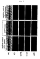
- the cultured epithelial sheet prepared using this feeder cell (shown in the central column in Fig. 2 ) is stratified as in the cultured epithelial sheet prepared using conventional 3T3 (shown in the left column in Fig. 2 ). Further, the former epithelial sheet and the latter epithelial sheet show almost similar gene expression patterns in differentiation markers and undifferentiation markers. That is, K3 positive cells were recognized in the outer most layer; Cx43 positive cells were recognized in allover the epithelium including basal cells; and the expression of integrin ⁇ 1 and p63 was recognized in allover the epithelium except for the outer most layer. These results suggest that the cultured epithelial sheets do not differ in quality between when a feeder cell derived from mouse corneal stroma stem cell is used and when 3T3 is used.
- Fig. 3 shows difference in colony forming efficiency when an extremely small number of epithelial cells were seeded. While no colony formation of epithelial cells was recognized without feeder cells, colony formation of epithelial cells was recognized when this feeder cell was used. These results suggest that this feeder cell is able to provide a microenvironment capable of supporting the survival and growth of epithelial cells.
- COPs Corneal stroma stem cells
- COPs Corneal stroma stem cells
- MSCs Human bone marrow mesenchymal stem cells
- human limbal epithelial cells were prepared from U.S. eye bank corneas by enzyme treatment, seeded on 100 mm culture dishes (1000 cells/dish) which contain COP or MASC feeder cell, and co-cultured for 2 weeks.
- SHEM the same medium as used in Example 1 was used. After culturing, the dishes were fixed with formalin and epithelial colonies were stained with rhodamin B. The percentage obtained by dividing the number of colonies by the number of cells seeded was taken as colony forming efficiency.
- each feeder cell was placed in 6-well plates and fibrin-coated cell culture inserts.
- human limbal epithelial cells were three-dimensionally cultured.
- Bolheal (KAKETSUKEN; 2 mg/cm 2 )-coated culture inserts (Coming; Transwell®, cat no 3450) were used.
- KAKETSUKEN 2 mg/cm 2
- Coming Coming; Transwell®, cat no 3450
- human epithelial cells reached confluence, a 1-week air lift culture was performed thereafter to thereby prepare cultured epithelial sheets.
- the resultant epithelial sheets were fixed in formalin, and paraffin sections were prepared therefrom, stained with HE and subjected to histological assay.
- the epithelial sheets were subjected to immunohistological assay in the form of fresh cryosections.
- immunohistological assay anti-keratin-3 antibody, anti-keratin-12 antibody, anti-keratin-15 antibody, anti-connexin 43 antibody, anti-integrin ⁇ 1 antibody or anti-p63 antibody was used as a primary antibody; Alexa flour-labeled antibody (Invitrogen) was used as a secondary antibody; and DAPI was used for nuclear staining. Then, the presence or absence of expression was examined with a fluorescence microscope (Axioplan 2; Carl Zeiss).
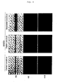
- cultured epithelial sheets could be prepared by co-culturing with corneal stroma stem cells. These sheets were equivalent to those sheets obtained by co-culturing with conventional mouse 3T3. Specifically, cultured epithelial sheets co-cultured with corneal stroma stem cells were stratified, contained cells expressing differentiation markers K3 and Cx43, and yet contained cells expressing undifferentiation markers Int b1 and p63. Further, as shown in Fig. 3 , cultured epithelial sheets co-cultured with corneal stroma stem cells were maintaining cells which retained such growth ability that enabled colony formation.
- epithelial cells co-cultured with no feeder cell were hardly stratified, showed little expression of differentiation markers, and did not maintain those cells with colony forming ability.
- epithelial cells co-cultured with a feeder cell derived from human mesenchymal stem cells could also form stratified epithelial sheets. Regardless of whether contacting with the feeder cell or not, these epithelial sheets contained differentiated cells expressing K3 and K12. Especially when humoral factors were continuously supplied (Duplex co-culture and Separated co-culture), expression of K15 was recognized as recognized in the corneal limbus where corneal epithelial stem cells are present.
- a feeder cell is provided.
- the feeder cell of the present invention it is possible to prepare transplantable cultured cells.
Landscapes
- Health & Medical Sciences (AREA)
- Life Sciences & Earth Sciences (AREA)
- Engineering & Computer Science (AREA)
- Biomedical Technology (AREA)
- Biotechnology (AREA)
- Cell Biology (AREA)
- Chemical & Material Sciences (AREA)
- Zoology (AREA)
- General Health & Medical Sciences (AREA)
- Organic Chemistry (AREA)
- Wood Science & Technology (AREA)
- Genetics & Genomics (AREA)
- Bioinformatics & Cheminformatics (AREA)
- Developmental Biology & Embryology (AREA)
- Immunology (AREA)
- Animal Behavior & Ethology (AREA)
- Medicinal Chemistry (AREA)
- Public Health (AREA)
- Veterinary Medicine (AREA)
- Virology (AREA)
- Epidemiology (AREA)
- Pharmacology & Pharmacy (AREA)
- Biochemistry (AREA)
- Microbiology (AREA)
- General Engineering & Computer Science (AREA)
- Neurology (AREA)
- Neurosurgery (AREA)
- Rheumatology (AREA)
- Hematology (AREA)
- Vascular Medicine (AREA)
- Dermatology (AREA)
- Ophthalmology & Optometry (AREA)
- Micro-Organisms Or Cultivation Processes Thereof (AREA)
- Materials For Medical Uses (AREA)
- Apparatus Associated With Microorganisms And Enzymes (AREA)
Priority Applications (1)
| Application Number | Priority Date | Filing Date | Title |
|---|---|---|---|
| EP09012264A EP2130911A1 (de) | 2006-02-08 | 2007-01-17 | Feeder-Zellen, aus Gewebestammzellen |
Applications Claiming Priority (2)
| Application Number | Priority Date | Filing Date | Title |
|---|---|---|---|
| JP2006030997 | 2006-02-08 | ||
| PCT/JP2007/050615 WO2007091409A1 (ja) | 2006-02-08 | 2007-01-17 | 組織幹細胞由来フィーダー細胞 |
Related Child Applications (1)
| Application Number | Title | Priority Date | Filing Date |
|---|---|---|---|
| EP09012264A Division EP2130911A1 (de) | 2006-02-08 | 2007-01-17 | Feeder-Zellen, aus Gewebestammzellen |
Publications (2)
| Publication Number | Publication Date |
|---|---|
| EP1990407A1 true EP1990407A1 (de) | 2008-11-12 |
| EP1990407A4 EP1990407A4 (de) | 2009-07-01 |
Family
ID=38345016
Family Applications (2)
| Application Number | Title | Priority Date | Filing Date |
|---|---|---|---|
| EP07706922A Withdrawn EP1990407A4 (de) | 2006-02-08 | 2007-01-17 | Von einer gewebestammzelle abgeleitete feeder-zelle |
| EP09012264A Withdrawn EP2130911A1 (de) | 2006-02-08 | 2007-01-17 | Feeder-Zellen, aus Gewebestammzellen |
Family Applications After (1)
| Application Number | Title | Priority Date | Filing Date |
|---|---|---|---|
| EP09012264A Withdrawn EP2130911A1 (de) | 2006-02-08 | 2007-01-17 | Feeder-Zellen, aus Gewebestammzellen |
Country Status (6)
| Country | Link |
|---|---|
| US (1) | US20090047738A1 (de) |
| EP (2) | EP1990407A4 (de) |
| JP (1) | JPWO2007091409A1 (de) |
| BR (1) | BRPI0708039A2 (de) |
| CA (1) | CA2637856A1 (de) |
| WO (1) | WO2007091409A1 (de) |
Families Citing this family (5)
| Publication number | Priority date | Publication date | Assignee | Title |
|---|---|---|---|---|
| JP2009225675A (ja) * | 2008-03-19 | 2009-10-08 | Tohoku Univ | 上皮系細胞シートの作製のための同種皮膚由来フィーダー細胞 |
| WO2018111908A1 (en) * | 2016-12-13 | 2018-06-21 | The Regents Of The University Of California | Xenobiotic-free culture system to expand human limbal stem cells |
| EP3680324A4 (de) * | 2017-09-08 | 2021-06-16 | Otsuka Pharmaceutical Factory, Inc. | Stammzellen von einem jungen schwein und verfahren zur herstellung dafür |
| KR102095346B1 (ko) * | 2018-11-16 | 2020-03-31 | 고려대학교 산학협력단 | 삼차원 공배양을 통한 신생혈관능이 증가된 이식용 지방줄기세포 시트 및 이의 제조방법 |
| TW202038976A (zh) * | 2018-11-30 | 2020-11-01 | 日商資生堂股份有限公司 | 用以治療或預防皮膚之色素沉著之組合物 |
Family Cites Families (2)
| Publication number | Priority date | Publication date | Assignee | Title |
|---|---|---|---|---|
| WO2005035739A1 (ja) * | 2003-10-14 | 2005-04-21 | Kohji Nishida | 再生治療システム |
| DE102004034997A1 (de) | 2004-07-16 | 2006-02-02 | Carl Zeiss Jena Gmbh | Lichtrastermikroskop mit bewegter Lochscheibe und Verwendung |
-
2007
- 2007-01-17 EP EP07706922A patent/EP1990407A4/de not_active Withdrawn
- 2007-01-17 WO PCT/JP2007/050615 patent/WO2007091409A1/ja not_active Ceased
- 2007-01-17 JP JP2007557769A patent/JPWO2007091409A1/ja active Pending
- 2007-01-17 US US12/162,905 patent/US20090047738A1/en not_active Abandoned
- 2007-01-17 BR BRPI0708039-5A patent/BRPI0708039A2/pt not_active IP Right Cessation
- 2007-01-17 CA CA002637856A patent/CA2637856A1/en not_active Abandoned
- 2007-01-17 EP EP09012264A patent/EP2130911A1/de not_active Withdrawn
Non-Patent Citations (6)
| Title |
|---|
| DATABASE MEDLINE [Online] US NATIONAL LIBRARY OF MEDICINE (NLM), BETHESDA, MD, US; December 1999 (1999-12), WANG Z ET AL: "[Preliminary experimental study on commitment differentiation of embryonic stem cells induced by corneal limbal stroma in vitro]" XP002524464 Database accession no. NLM12579666 & YAN KE XUE BAO = EYE SCIENCE / "YAN KE XUE BAO" BIAN JI BU DEC 1999, vol. 15, no. 4, December 1999 (1999-12), pages 195-198, ISSN: 1000-4432 * |
| KIM H -S ET AL: "Phenotypic characterization of human corneal epithelial cells expanded ex vivo from limbal explant and single cell cultures" EXPERIMENTAL EYE RESEARCH 200407 GB, vol. 79, no. 1, July 2004 (2004-07), pages 41-49, XP002524466 ISSN: 0014-4835 * |
| MIYASHITA H ET AL: "A novel NIH/3T3 duplex feeder system to engineer corneal epithelial sheets with enhanced cytokeratin 15-positive progenitor populations" TISSUE ENGINEERING - PART A. 20080701 US, vol. 14, no. 7, 1 July 2008 (2008-07-01), pages 1275-1282, XP002524467 ISSN: 1937-3341 * |
| See also references of WO2007091409A1 * |
| YOSHIDA SATORU ET AL: "Serum-free spheroid culture of mouse corneal keratocytes" IOVS, vol. 46, no. 5, May 2005 (2005-05), pages 1653-1658, XP002524465 ISSN: 0146-0404 * |
| ZHANG YI ET AL: "Human placenta-derived mesenchymal progenitor cells support culture expansion of long-term culture-initiating cells from cord blood CD34+ cells" EXPERIMENTAL HEMATOLOGY (NEW YORK), vol. 32, no. 7, July 2004 (2004-07), pages 657-664, XP002389863 ISSN: 0301-472X * |
Also Published As
| Publication number | Publication date |
|---|---|
| JPWO2007091409A1 (ja) | 2009-07-02 |
| US20090047738A1 (en) | 2009-02-19 |
| CA2637856A1 (en) | 2007-08-16 |
| WO2007091409A1 (ja) | 2007-08-16 |
| EP1990407A4 (de) | 2009-07-01 |
| EP2130911A1 (de) | 2009-12-09 |
| BRPI0708039A2 (pt) | 2011-05-17 |
Similar Documents
| Publication | Publication Date | Title |
|---|---|---|
| CN100554411C (zh) | 来自角膜缘的未分化干细胞组织系统 | |
| RU2331668C2 (ru) | Стромальные клетки, полученные из жировой ткани, для заживления дефектов роговицы и внутриглазных дефектов и их использование | |
| EP1980274B1 (de) | Hornhautendothelpräparation, die zellen das wachstum in vivo ermöglicht | |
| US20080039940A1 (en) | Biological Tissue Sheet, Method Of Forming The Same And Transplantation Method By Using The Sheet | |
| EP2975117B1 (de) | Verfahren zur herstellung einer menschlichen hornhautepithelschicht | |
| US20120149598A1 (en) | Feeder cells for target cell induction | |
| EP1675944A1 (de) | Menschliche hornhautendothelzellen und verfahren zur gewinnung und kultivierung von zellen zur hornhautzelltransplantation | |
| NZ570746A (en) | Conjunctival tissue system | |
| Lachaud et al. | Mesothelial cells: a cellular surrogate for tissue engineering of corneal endothelium | |
| EP1990407A1 (de) | Von einer gewebestammzelle abgeleitete feeder-zelle | |
| WO2007043255A1 (ja) | 培養角膜内皮シート及びその作製方法 | |
| US20070280993A1 (en) | Corneal Epithelial Sheet, Method Of constructing The Same, And Transplantation Method Using The Sheet | |
| JP2009225675A (ja) | 上皮系細胞シートの作製のための同種皮膚由来フィーダー細胞 | |
| JP7024974B2 (ja) | 培養上皮シートの製造方法 | |
| JP6703130B2 (ja) | 羊膜スライド支持体を利用した輪部幹細胞の培養方法 | |
| WO2009110647A1 (en) | Tissue sheet of cornea or conjunctiva using nasal mucosa epithelium | |
| Stewart | Identification of progenitor cell-rich sites of the conjunctiva |
Legal Events
| Date | Code | Title | Description |
|---|---|---|---|
| PUAI | Public reference made under article 153(3) epc to a published international application that has entered the european phase |
Free format text: ORIGINAL CODE: 0009012 |
|
| 17P | Request for examination filed |
Effective date: 20080829 |
|
| AK | Designated contracting states |
Kind code of ref document: A1 Designated state(s): AT BE BG CH CY CZ DE DK EE ES FI FR GB GR HU IE IS IT LI LT LU LV MC NL PL PT RO SE SI SK TR |
|
| A4 | Supplementary search report drawn up and despatched |
Effective date: 20090602 |
|
| 17Q | First examination report despatched |
Effective date: 20090805 |
|
| DAX | Request for extension of the european patent (deleted) | ||
| STAA | Information on the status of an ep patent application or granted ep patent |
Free format text: STATUS: THE APPLICATION IS DEEMED TO BE WITHDRAWN |
|
| 18D | Application deemed to be withdrawn |
Effective date: 20091216 |