EP1992277A1 - Dispositif optique et procédé d'acquisition d'images des structures oculaires - Google Patents
Dispositif optique et procédé d'acquisition d'images des structures oculaires Download PDFInfo
- Publication number
- EP1992277A1 EP1992277A1 EP07301039A EP07301039A EP1992277A1 EP 1992277 A1 EP1992277 A1 EP 1992277A1 EP 07301039 A EP07301039 A EP 07301039A EP 07301039 A EP07301039 A EP 07301039A EP 1992277 A1 EP1992277 A1 EP 1992277A1
- Authority
- EP
- European Patent Office
- Prior art keywords
- endoscope
- tip
- eye
- optical device
- images
- Prior art date
- Legal status (The legal status is an assumption and is not a legal conclusion. Google has not performed a legal analysis and makes no representation as to the accuracy of the status listed.)
- Ceased
Links
- 230000003287 optical effect Effects 0.000 title claims abstract description 29
- 238000000034 method Methods 0.000 title claims description 30
- 238000003384 imaging method Methods 0.000 claims abstract description 29
- 210000004087 cornea Anatomy 0.000 claims abstract description 17
- 239000013307 optical fiber Substances 0.000 claims abstract description 6
- 238000005286 illumination Methods 0.000 claims description 16
- 241000282326 Felis catus Species 0.000 claims description 4
- 241000282693 Cercopithecidae Species 0.000 claims description 3
- 230000004888 barrier function Effects 0.000 claims description 3
- 235000019687 Lamb Nutrition 0.000 claims description 2
- 239000004904 UV filter Substances 0.000 claims description 2
- 210000001508 eye Anatomy 0.000 description 40
- 210000004240 ciliary body Anatomy 0.000 description 7
- 230000002093 peripheral effect Effects 0.000 description 7
- 210000001525 retina Anatomy 0.000 description 7
- 230000004075 alteration Effects 0.000 description 6
- 210000000695 crystalline len Anatomy 0.000 description 6
- 210000005036 nerve Anatomy 0.000 description 5
- 210000001210 retinal vessel Anatomy 0.000 description 5
- 210000003462 vein Anatomy 0.000 description 5
- XKRFYHLGVUSROY-UHFFFAOYSA-N Argon Chemical compound [Ar] XKRFYHLGVUSROY-UHFFFAOYSA-N 0.000 description 4
- 241001465754 Metazoa Species 0.000 description 4
- 210000002565 arteriole Anatomy 0.000 description 4
- 208000010412 Glaucoma Diseases 0.000 description 3
- 230000011514 reflex Effects 0.000 description 3
- 210000003583 retinal pigment epithelium Anatomy 0.000 description 3
- 241000282472 Canis lupus familiaris Species 0.000 description 2
- 229910052786 argon Inorganic materials 0.000 description 2
- 210000001367 artery Anatomy 0.000 description 2
- 210000003161 choroid Anatomy 0.000 description 2
- 239000000835 fiber Substances 0.000 description 2
- 239000000049 pigment Substances 0.000 description 2
- 210000001519 tissue Anatomy 0.000 description 2
- 238000012800 visualization Methods 0.000 description 2
- 201000002862 Angle-Closure Glaucoma Diseases 0.000 description 1
- 208000003098 Ganglion Cysts Diseases 0.000 description 1
- 241000282412 Homo Species 0.000 description 1
- 241000283903 Ovis aries Species 0.000 description 1
- 201000007737 Retinal degeneration Diseases 0.000 description 1
- 206010038848 Retinal detachment Diseases 0.000 description 1
- 208000017442 Retinal disease Diseases 0.000 description 1
- 208000005400 Synovial Cyst Diseases 0.000 description 1
- 206010053648 Vascular occlusion Diseases 0.000 description 1
- 210000002159 anterior chamber Anatomy 0.000 description 1
- 238000013459 approach Methods 0.000 description 1
- 239000008280 blood Substances 0.000 description 1
- 210000004369 blood Anatomy 0.000 description 1
- 230000001684 chronic effect Effects 0.000 description 1
- 230000008878 coupling Effects 0.000 description 1
- 238000010168 coupling process Methods 0.000 description 1
- 238000005859 coupling reaction Methods 0.000 description 1
- 230000007423 decrease Effects 0.000 description 1
- 230000007547 defect Effects 0.000 description 1
- 230000007850 degeneration Effects 0.000 description 1
- 238000002059 diagnostic imaging Methods 0.000 description 1
- 238000002405 diagnostic procedure Methods 0.000 description 1
- 208000029436 dilated pupil Diseases 0.000 description 1
- 239000006185 dispersion Substances 0.000 description 1
- 230000005284 excitation Effects 0.000 description 1
- 230000007717 exclusion Effects 0.000 description 1
- 238000001914 filtration Methods 0.000 description 1
- GNBHRKFJIUUOQI-UHFFFAOYSA-N fluorescein Chemical compound O1C(=O)C2=CC=CC=C2C21C1=CC=C(O)C=C1OC1=CC(O)=CC=C21 GNBHRKFJIUUOQI-UHFFFAOYSA-N 0.000 description 1
- 238000001727 in vivo Methods 0.000 description 1
- 230000036512 infertility Effects 0.000 description 1
- 230000004410 intraocular pressure Effects 0.000 description 1
- 230000003902 lesion Effects 0.000 description 1
- 210000003041 ligament Anatomy 0.000 description 1
- 239000000463 material Substances 0.000 description 1
- 229920000609 methyl cellulose Polymers 0.000 description 1
- 239000001923 methylcellulose Substances 0.000 description 1
- 210000000056 organ Anatomy 0.000 description 1
- 230000008520 organization Effects 0.000 description 1
- 230000003071 parasitic effect Effects 0.000 description 1
- 230000019612 pigmentation Effects 0.000 description 1
- 230000008569 process Effects 0.000 description 1
- 210000001747 pupil Anatomy 0.000 description 1
- 238000011160 research Methods 0.000 description 1
- 230000004264 retinal detachment Effects 0.000 description 1
- 239000000523 sample Substances 0.000 description 1
- 238000012216 screening Methods 0.000 description 1
- 238000000926 separation method Methods 0.000 description 1
- 241000894007 species Species 0.000 description 1
- 208000021331 vascular occlusion disease Diseases 0.000 description 1
- 210000000264 venule Anatomy 0.000 description 1
- 229910052724 xenon Inorganic materials 0.000 description 1
- FHNFHKCVQCLJFQ-UHFFFAOYSA-N xenon atom Chemical compound [Xe] FHNFHKCVQCLJFQ-UHFFFAOYSA-N 0.000 description 1
Images
Classifications
-
- A—HUMAN NECESSITIES
- A61—MEDICAL OR VETERINARY SCIENCE; HYGIENE
- A61B—DIAGNOSIS; SURGERY; IDENTIFICATION
- A61B1/00—Instruments for performing medical examinations of the interior of cavities or tubes of the body by visual or photographical inspection, e.g. endoscopes; Illuminating arrangements therefor
- A61B1/06—Instruments for performing medical examinations of the interior of cavities or tubes of the body by visual or photographical inspection, e.g. endoscopes; Illuminating arrangements therefor with illuminating arrangements
- A61B1/07—Instruments for performing medical examinations of the interior of cavities or tubes of the body by visual or photographical inspection, e.g. endoscopes; Illuminating arrangements therefor with illuminating arrangements using light-conductive means, e.g. optical fibres
-
- A—HUMAN NECESSITIES
- A61—MEDICAL OR VETERINARY SCIENCE; HYGIENE
- A61B—DIAGNOSIS; SURGERY; IDENTIFICATION
- A61B1/00—Instruments for performing medical examinations of the interior of cavities or tubes of the body by visual or photographical inspection, e.g. endoscopes; Illuminating arrangements therefor
- A61B1/00163—Optical arrangements
- A61B1/00165—Optical arrangements with light-conductive means, e.g. fibre optics
- A61B1/00167—Details of optical fibre bundles, e.g. shape or fibre distribution
-
- A—HUMAN NECESSITIES
- A61—MEDICAL OR VETERINARY SCIENCE; HYGIENE
- A61B—DIAGNOSIS; SURGERY; IDENTIFICATION
- A61B1/00—Instruments for performing medical examinations of the interior of cavities or tubes of the body by visual or photographical inspection, e.g. endoscopes; Illuminating arrangements therefor
- A61B1/04—Instruments for performing medical examinations of the interior of cavities or tubes of the body by visual or photographical inspection, e.g. endoscopes; Illuminating arrangements therefor combined with photographic or television appliances
- A61B1/043—Instruments for performing medical examinations of the interior of cavities or tubes of the body by visual or photographical inspection, e.g. endoscopes; Illuminating arrangements therefor combined with photographic or television appliances for fluorescence imaging
-
- A—HUMAN NECESSITIES
- A61—MEDICAL OR VETERINARY SCIENCE; HYGIENE
- A61B—DIAGNOSIS; SURGERY; IDENTIFICATION
- A61B1/00—Instruments for performing medical examinations of the interior of cavities or tubes of the body by visual or photographical inspection, e.g. endoscopes; Illuminating arrangements therefor
- A61B1/06—Instruments for performing medical examinations of the interior of cavities or tubes of the body by visual or photographical inspection, e.g. endoscopes; Illuminating arrangements therefor with illuminating arrangements
- A61B1/0607—Instruments for performing medical examinations of the interior of cavities or tubes of the body by visual or photographical inspection, e.g. endoscopes; Illuminating arrangements therefor with illuminating arrangements for annular illumination
-
- A—HUMAN NECESSITIES
- A61—MEDICAL OR VETERINARY SCIENCE; HYGIENE
- A61B—DIAGNOSIS; SURGERY; IDENTIFICATION
- A61B3/00—Apparatus for testing the eyes; Instruments for examining the eyes
- A61B3/0008—Apparatus for testing the eyes; Instruments for examining the eyes provided with illuminating means
-
- A—HUMAN NECESSITIES
- A61—MEDICAL OR VETERINARY SCIENCE; HYGIENE
- A61B—DIAGNOSIS; SURGERY; IDENTIFICATION
- A61B3/00—Apparatus for testing the eyes; Instruments for examining the eyes
- A61B3/10—Objective types, i.e. instruments for examining the eyes independent of the patients' perceptions or reactions
- A61B3/12—Objective types, i.e. instruments for examining the eyes independent of the patients' perceptions or reactions for looking at the eye fundus, e.g. ophthalmoscopes
-
- A—HUMAN NECESSITIES
- A61—MEDICAL OR VETERINARY SCIENCE; HYGIENE
- A61B—DIAGNOSIS; SURGERY; IDENTIFICATION
- A61B1/00—Instruments for performing medical examinations of the interior of cavities or tubes of the body by visual or photographical inspection, e.g. endoscopes; Illuminating arrangements therefor
- A61B1/227—Instruments for performing medical examinations of the interior of cavities or tubes of the body by visual or photographical inspection, e.g. endoscopes; Illuminating arrangements therefor for ears, i.e. otoscopes
Definitions
- the present invention relates to medical imaging in the field of ophthalmology.
- it relates to means for acquiring images of eye structures of an animal or a human.
- Imaging eye structures has become an essential step for exploring eye structures in the diagnostic and follow up of retinal diseases or lesions in humans and animals.
- Imaging the fundus can for instance provide diagnostic of retinal detachment, retinal atrophy, and blood vascular occlusion whereas examination of the irido-corneal angle can explain increase intraocular pressure leading to glaucoma and ganglion cell degeneration.
- eye examinations of the cornea, anterior chamber, lens and posterior chamber and vitreous are generally first achieved with a slit lamp apparatus. Images of the fundus can then be obtained using indirect ophtahlmoscopy or confocal scanning laser ophthalmoscopes.
- the present invention provides a method and a corresponding device to increase the quality of images of the eye structures and, in particular, to improve the contrast of such images.
- the invention concerns an optical device for acquiring images of the eye structures, characterized in that it comprises: a light source; an endoscope connected to the light source, through an optical fibre, at one end for conducting and projecting an incident light beam through the tip, said tip being adapted to be applied nearby the cornea of an eye according to a given orientation; an imaging device connected to said endoscope through said imaging device being adapted to acquired imaged of the eye structures from a light beam coming from the structures of the eye.
- the device of the invention may present one or more of the following features:
- the invention concerns a method for acquiring images of the eye structures, characterized in that it comprises the following steps: an application, according to a given orientation, of the tip of an endoscope nearby the cornea of an eye, said endoscope being connected to a light source through an optical fibre at one end for conducting and projecting an incident light beam through the tip of the endoscope; an acquisition of images of the eye structures from light coming from the structures of the eye by means of an imaging device connected to said endoscope.
- the method of the invention may present one or more of the following features:
- the invention provides a low cost and a simply device to image the eye structures.
- Applicant has observed that an application of an illuminating endoscope nearby a cornea of an eye allows wide field fundus imaging.
- a fundus image can be obtained by placing an endoscope nearby the cornea, as long as the outer diameter does not exceed the diameter of the dilated pupil, that is, 3 to 4 mm in a mouse.
- an endoscope is used to observe the interior surfaces of an organ by inserting a tube into the body.
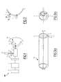
- Figure 1 shows an optical device used to implement a method for acquiring images of the structures of the eye.
- the optical device comprises an endoscope 1 with parallel illumination and observation channels, connected to an imaging device 5 such as a photographic camera (to acquire images) or a video camera (for screening procedures), through an adapter 3 containing a +5 lens to acquire images.
- an imaging device 5 such as a photographic camera (to acquire images) or a video camera (for screening procedures)
- an adapter 3 containing a +5 lens to acquire images.
- a reflex digital camera with a 6.1 million pixels Charged Couple Device (CCD) image sensor is preferably used for increasing the definition.
- CCD Charged Couple Device
- each pixel of a CCD codes corresponds to three color channels (RGB mode), that is, Red (peak approximately equal to 610 nm), Green (peak approximately equal to 550 nm) and Blue (peak approximately equal to 470nm), with a certain degree of overlapping between channels. (the resolution is better when necessary in black and white because 4 pixels in grey).
- the endoscope 1 is connected to a light source 2, preferably a cold light source 2 for instance, a xenon lamp, through a flexible optical fibre 9.
- a light source 2 preferably a cold light source 2 for instance, a xenon lamp
- the light source 2 can be an infrared source.
- the light source 2 is further suitable to provide an incident light beam with an adjustable intensity. Such adjustability permits the protection of the retina of the eye.
- the endoscope 1 is a rigid endoscope and advantageously an otoscope.
- the method of the invention performed with the optical device of the invention comprises an application of the tip 10 of the endoscope 1 nearby the cornea 7 of the eye 20.
- a gel 8 is applied on the cornea 7 of the eye 20; the gel 8 is for instance methylcellulose.
- the tip 10 of the endoscope 1 is then applied into the gel 8.
- a single use lens can be applied on the cornea or a transparent film can be inserted on the tip of the endoscope.
- the contact with the cornea 7 is not essential, but advantageously decreases parasitic reflections.
- the tip 10 of the endoscope 1 is applied according to a given orientation ⁇ 1 , ⁇ 2 , etc.
- the possibility to orient the tip 10 of the endoscope 1 under large angles with respect to the eye central optic permits to focus on the eye periphery.
- a lens can also be used to prepare interesting orientations by having designed positions for the tip of the endoscope.
- Figure 2 illustrates two configurations with different orientations of the tip 10 of the endoscope 1 applied into the gel 8.
- a classical endoscope comprises a first cylinder co-axial with a second cylinder having a diameter inferior to the diameter of the first cylinder. These two cylinders provide a circle-shaped illumination at the tip of such endoscope, and the light is emitted through the tip projecting a circle-shaped incident light beam on the eye.
- the second cylinder comprises means to acquire light beam from the area of interest.
- Figures 3a and 3b show different views of the endoscope preferably used in the invention.
- Figure 3a shows a perspective view of the tip 10 of the endoscope 1 and Figure 3b shows a side view of the tip 10 according to the axis CC' of the endoscope 1.
- the tip 10 of the endoscope 1 comprises a first cylinder 11 having a first diameter d1, and a second cylinder 12, which is non coaxial with the first cylinder 11, having a second diameter d 2 inferior to the first diameter d 1 .
- These two cylinders 11, 12 provide a crescent-shaped tip 10, and the light is emitted through the tip 10 projecting a lateral crescent-shaped incident light beam Bi.
- This illumination involves a minimization of axial reflections.
- the endoscope 1 has for instance, a 5cm length, 3mm outer diameter d1 with step index lenses that have an angle of view of 0°, a field of view of 80° in the air, and a crescent illuminating tip 10.
- the acquired images are obtained by measuring the light beam Br coming from the eye. They may be transferred to a computer 6 which processes them, in particular, for adjusting their contrast and/or their luminosity.
- the reflected light on the fundus is acquired.
- the fluorescent emitted by the dye is acquired.
- filters 4 can be placed between the light source 2 and the endoscope 1.
- These filters are used to control the light source 2 for UV exclusion and thus protecting eye tissues 20.
- each burst comprises four images.
- the acquired images can be used for a diagnostic method which may be implemented in an additional step after the method performed with the optical device for acquiring images of the eye structures in accordance with the present invention.
- the definition of the acquired images depends on the imaging device.
- the acquired images shown in this description have an estimated definition of the image acquisition system (e.g., 1.9 million pixels).
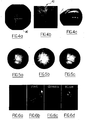
- FIG. 4a Examples of fundus images of a mouse are shown in figure 4a, 4b and 4c .
- Figure 4a shows a view of a posterior pole.
- the scale is given by the optical nerve 40, which is approximately 220 microns in diameter.
- Figure 4b shows a magnification of figure 4a .
- it is a magnification of the optical nerve 40 and vessels of Figure 4a .
- This figure accurately shows the delineation of the contour of the optical nerve 40 and the nerve fibre layer.
- Figure 4c shows a view of the peripheral retina and ciliary body which is acquired by changing the orientation of the endoscope, in particular by placing the endoscope perpendicularly to the limbus.
- This disposition allows imaging of the peripheral retina and the ciliary body.
- Figure 5a, 5b, 5c show an illustration of optical aberrations encountered in the images acquired with the method of the invention.
- Figure 5a shows a color fundus image
- figure 5b shows a monochromatic fundus image acquired through the red channel
- figure 5c shows a monochromatic fundus image acquired through the blue channel.
- Blue light images have indeed a larger field than red light images. This is consistent with the stronger dispersion of a blue incident light.
- Figures 6a, 6b, 6c and 6d show the reflectance of specific fundus structures.
- Figure 6a shows a color fundus image
- Figures 6b, 6c and 6d show monochromatic fundus images acquired through the red, green, and blue channel respectively.
- Red monochromatic imaging enhances the contrast of pigment alterations, as well as the axial reflex from vessels.
- Green monochromatic imaging enhances the contrast of veins, while arteries remain relatively isoreflective to the surrounding background.
- the blue channel image provides the best contrast for both arteries and veins; in particular, the best contrast for retinal vessels over the choroids 61 is obtained.
- the reflectance of the nerve fibre layer is more evidenced in blue or green monochromatic images.
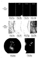
- Figures 7a, 7b, 7c, 7d, 7e, 7f, 7g and 7h show the variation in contrast of human retinal vessels with increased intensity of incident light.
- Figures 7a and 7e show color images
- Figures 7b and 7f show red monochromatic images
- Figures 7c and 7g show green monochromatic images
- Figures 7d and 7h show blue monochromatic images.
- fundus images of a pigmented human subject acquired with a conventional fundus camera (Topcon 50 IA from Topcon Corporation, Tokyo, Japan) at different flash intensities is magnified in order to visualize small retinal vessels (i.e. in the magnitude of 40-60 microns).
- fundus images in a pigmented subject are acquired with a normal flash intensity ( Figures 7a, 7b, 7c and 7d ) and with high intensity flash ( Figures 7e, 7f, 7g and 7h ).
- Figure 8a shows color image of the peripheral retina and the ciliary body 90 of an albino mouse.
- the overall organization of the ciliary body venous drainage into the vortex veins is discernable.
- Figure 8b shows a monochromatic high magnification image of Figure 8a .
- the venous drainage of individual ciliary body 91 is clearly visible.
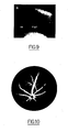
- Figure 9 show images of the irido-corneal angle in various species.
- the tip 10 of the endoscope 1 When the tip 10 of the endoscope 1 is oriented on the cornea a wide angle with the eye optic axis is obtained, the irido-corneal angle can be then imaged with great precision.
- Visualization of the irido-corneal angle is generally performed with the help of goniolens which enables to have a circular view of irido-corneal angle.
- the irido-corneal angle is advantageously observed with enlarged view of the pectinate ligament at the level of the trabeculum.
- Coupling an argon laser to the endoscope provides an easier way to achieve trabeculum retraction than with the current argon laser coupled to the slit lamp and using the goniolens.
- the method and its corresponding device offers a new approach to visualize the irido-corneal angle with great details and eventually to treat the trabeculum more easily thanks to the easiness to orient the tip of the endoscope.
- lasers could also be coupled to the endoscope 1 like a YAG laser.
- Figure 10 shows such a fluorescein angiogram obtained on a pigmented mouse after interposition of adequate excitation and barrier filters. The method can therefore be used to obtain fluorescent images of the different eye structures.
- the invention allows high resolution wide field digital fundus imaging in the mouse eye.
- the invention can be used with human eye or any other animals, such as cats, dogs, monkeys, lambs, etc.
- the invention significantly improves the quality of in vivo images of retina and ciliary body.
- resolution is affected by chromatic aberration
- resolution can be improved by monochromatic imaging.
- the endoscope probe can be handled in any position depending on the patient situation for the eye examination.
- optical device of the invention is suitable to obtain images of the fundus and images of the irido-corneal angle with only one device which is a very advantageous property.
- this is a low-cost and portable device which is very adapted not only for research laboratories and ophthalmology clinic but also to perform complete ophthalmologic exams in remote areas.
Landscapes
- Health & Medical Sciences (AREA)
- Life Sciences & Earth Sciences (AREA)
- Surgery (AREA)
- Physics & Mathematics (AREA)
- General Health & Medical Sciences (AREA)
- Animal Behavior & Ethology (AREA)
- Veterinary Medicine (AREA)
- Public Health (AREA)
- Biophysics (AREA)
- Engineering & Computer Science (AREA)
- Biomedical Technology (AREA)
- Heart & Thoracic Surgery (AREA)
- Medical Informatics (AREA)
- Molecular Biology (AREA)
- Optics & Photonics (AREA)
- Nuclear Medicine, Radiotherapy & Molecular Imaging (AREA)
- Radiology & Medical Imaging (AREA)
- Pathology (AREA)
- Ophthalmology & Optometry (AREA)
- Eye Examination Apparatus (AREA)
Priority Applications (2)
| Application Number | Priority Date | Filing Date | Title |
|---|---|---|---|
| EP07301039A EP1992277A1 (fr) | 2007-05-14 | 2007-05-14 | Dispositif optique et procédé d'acquisition d'images des structures oculaires |
| PCT/EP2008/055923 WO2008138953A1 (fr) | 2007-05-14 | 2008-05-14 | Dispositif optique et procédé d'acquisition d'images de structures de l'œil |
Applications Claiming Priority (1)
| Application Number | Priority Date | Filing Date | Title |
|---|---|---|---|
| EP07301039A EP1992277A1 (fr) | 2007-05-14 | 2007-05-14 | Dispositif optique et procédé d'acquisition d'images des structures oculaires |
Publications (1)
| Publication Number | Publication Date |
|---|---|
| EP1992277A1 true EP1992277A1 (fr) | 2008-11-19 |
Family
ID=38474217
Family Applications (1)
| Application Number | Title | Priority Date | Filing Date |
|---|---|---|---|
| EP07301039A Ceased EP1992277A1 (fr) | 2007-05-14 | 2007-05-14 | Dispositif optique et procédé d'acquisition d'images des structures oculaires |
Country Status (2)
| Country | Link |
|---|---|
| EP (1) | EP1992277A1 (fr) |
| WO (1) | WO2008138953A1 (fr) |
Cited By (5)
| Publication number | Priority date | Publication date | Assignee | Title |
|---|---|---|---|---|
| CN101853520A (zh) * | 2010-04-20 | 2010-10-06 | 珠海金联安警用技术研究发展中心有限公司 | 一种基于光学成像的视网膜三维建模方法及其装置 |
| CN101862177A (zh) * | 2010-04-20 | 2010-10-20 | 中山大学中山眼科中心 | 一种视网膜裂孔三维定位的方法及其装置 |
| CN111970957A (zh) * | 2018-04-18 | 2020-11-20 | 株式会社尼康 | 图像处理方法、程序及图像处理装置 |
| CN111989030A (zh) * | 2018-04-18 | 2020-11-24 | 株式会社尼康 | 图像处理方法、程序及图像处理装置 |
| CN112004457A (zh) * | 2018-04-18 | 2020-11-27 | 株式会社尼康 | 图像处理方法、程序、图像处理装置及眼科系统 |
Families Citing this family (3)
| Publication number | Priority date | Publication date | Assignee | Title |
|---|---|---|---|---|
| EP2164383A4 (fr) | 2007-06-15 | 2011-05-04 | Norbert Massie | Procédé et appareil d'imagerie d'un il d'un petit animal |
| DE112019001423T5 (de) * | 2018-03-20 | 2020-12-03 | Nikon Corporation | Bildverarbeitungsverfahren, Programm, ophthalmologische Vorrichtung und choroidales Blutgefäßbild-Erzeugungsverfahren |
| JP7696363B2 (ja) * | 2020-04-06 | 2025-06-20 | ボストン サイエンティフィック サイムド,インコーポレイテッド | 画像処理システム及びその使用方法 |
Citations (11)
| Publication number | Priority date | Publication date | Assignee | Title |
|---|---|---|---|---|
| US4259948A (en) * | 1978-11-13 | 1981-04-07 | Peter Urban | Endoscopic system |
| WO1982000580A1 (fr) * | 1980-08-27 | 1982-03-04 | P Urban | Systeme endoscopique |
| JPH01170437A (ja) * | 1987-12-25 | 1989-07-05 | M & M:Kk | 眼内内視鏡システム |
| US5213092A (en) * | 1991-10-31 | 1993-05-25 | Martin Uram | Aspirating endoscope |
| EP0554643A1 (fr) * | 1992-02-05 | 1993-08-11 | Istituto Nazionale Di Ottica | Dispositif à sensibilité élevée destiné à l'examen d'un objet par une source lumineuse à basse intensité |
| EP0589825A2 (fr) * | 1992-09-25 | 1994-03-30 | OPTIKO SCIENTIFIC Ltd. | Appareil d'ophtalmoscope pour l'examen et/ou traitement |
| US5701904A (en) * | 1996-01-11 | 1997-12-30 | Krug International | Telemedicine instrumentation pack |
| US20020101566A1 (en) * | 1998-01-30 | 2002-08-01 | Elsner Ann E. | Imaging apparatus and methods for near simultaneous observation of directly scattered light and multiply scattered light |
| US6485413B1 (en) * | 1991-04-29 | 2002-11-26 | The General Hospital Corporation | Methods and apparatus for forward-directed optical scanning instruments |
| US6501551B1 (en) * | 1991-04-29 | 2002-12-31 | Massachusetts Institute Of Technology | Fiber optic imaging endoscope interferometer with at least one faraday rotator |
| WO2003083434A2 (fr) * | 2002-04-02 | 2003-10-09 | Yeda Research And Development Co. Ltd. | Caracterisation d'objets en mouvement dans un fond stationnaire |
Family Cites Families (3)
| Publication number | Priority date | Publication date | Assignee | Title |
|---|---|---|---|---|
| US4607622A (en) * | 1985-04-11 | 1986-08-26 | Charles D. Fritch | Fiber optic ocular endoscope |
| IL121450A0 (en) * | 1997-08-01 | 1998-02-08 | Smollett Neil | Ophthalmic surgical equipment |
| US8403828B2 (en) * | 2003-07-21 | 2013-03-26 | Vanderbilt University | Ophthalmic orbital surgery apparatus and method and image-guide navigation system |
-
2007
- 2007-05-14 EP EP07301039A patent/EP1992277A1/fr not_active Ceased
-
2008
- 2008-05-14 WO PCT/EP2008/055923 patent/WO2008138953A1/fr not_active Ceased
Patent Citations (11)
| Publication number | Priority date | Publication date | Assignee | Title |
|---|---|---|---|---|
| US4259948A (en) * | 1978-11-13 | 1981-04-07 | Peter Urban | Endoscopic system |
| WO1982000580A1 (fr) * | 1980-08-27 | 1982-03-04 | P Urban | Systeme endoscopique |
| JPH01170437A (ja) * | 1987-12-25 | 1989-07-05 | M & M:Kk | 眼内内視鏡システム |
| US6485413B1 (en) * | 1991-04-29 | 2002-11-26 | The General Hospital Corporation | Methods and apparatus for forward-directed optical scanning instruments |
| US6501551B1 (en) * | 1991-04-29 | 2002-12-31 | Massachusetts Institute Of Technology | Fiber optic imaging endoscope interferometer with at least one faraday rotator |
| US5213092A (en) * | 1991-10-31 | 1993-05-25 | Martin Uram | Aspirating endoscope |
| EP0554643A1 (fr) * | 1992-02-05 | 1993-08-11 | Istituto Nazionale Di Ottica | Dispositif à sensibilité élevée destiné à l'examen d'un objet par une source lumineuse à basse intensité |
| EP0589825A2 (fr) * | 1992-09-25 | 1994-03-30 | OPTIKO SCIENTIFIC Ltd. | Appareil d'ophtalmoscope pour l'examen et/ou traitement |
| US5701904A (en) * | 1996-01-11 | 1997-12-30 | Krug International | Telemedicine instrumentation pack |
| US20020101566A1 (en) * | 1998-01-30 | 2002-08-01 | Elsner Ann E. | Imaging apparatus and methods for near simultaneous observation of directly scattered light and multiply scattered light |
| WO2003083434A2 (fr) * | 2002-04-02 | 2003-10-09 | Yeda Research And Development Co. Ltd. | Caracterisation d'objets en mouvement dans un fond stationnaire |
Cited By (9)
| Publication number | Priority date | Publication date | Assignee | Title |
|---|---|---|---|---|
| CN101853520A (zh) * | 2010-04-20 | 2010-10-06 | 珠海金联安警用技术研究发展中心有限公司 | 一种基于光学成像的视网膜三维建模方法及其装置 |
| CN101862177A (zh) * | 2010-04-20 | 2010-10-20 | 中山大学中山眼科中心 | 一种视网膜裂孔三维定位的方法及其装置 |
| CN101862177B (zh) * | 2010-04-20 | 2011-10-26 | 中山大学中山眼科中心 | 一种视网膜裂孔三维定位装置 |
| CN101853520B (zh) * | 2010-04-20 | 2012-04-25 | 珠海金联安警用技术研究发展中心有限公司 | 一种基于光学成像的视网膜三维建模装置 |
| CN111970957A (zh) * | 2018-04-18 | 2020-11-20 | 株式会社尼康 | 图像处理方法、程序及图像处理装置 |
| CN111989030A (zh) * | 2018-04-18 | 2020-11-24 | 株式会社尼康 | 图像处理方法、程序及图像处理装置 |
| CN112004457A (zh) * | 2018-04-18 | 2020-11-27 | 株式会社尼康 | 图像处理方法、程序、图像处理装置及眼科系统 |
| CN111970957B (zh) * | 2018-04-18 | 2024-02-27 | 株式会社尼康 | 图像处理方法、计算机可读的存储介质及图像处理装置 |
| CN111989030B (zh) * | 2018-04-18 | 2024-03-01 | 株式会社尼康 | 图像处理方法、程序及图像处理装置 |
Also Published As
| Publication number | Publication date |
|---|---|
| WO2008138953A1 (fr) | 2008-11-20 |
Similar Documents
| Publication | Publication Date | Title |
|---|---|---|
| Paques et al. | Panretinal, high-resolution color photography of the mouse fundus | |
| JP5275417B2 (ja) | 患者の眼の前方セグメントを照明して観察するための装置および方法 | |
| EP1992277A1 (fr) | Dispositif optique et procédé d'acquisition d'images des structures oculaires | |
| JP7293227B2 (ja) | 顕微鏡の短小鏡筒内での近赤外光イメージング及び可視光イメージングの結合 | |
| EP2632321B1 (fr) | Détection d'amyloïde dans la rétine | |
| AU2002363421B2 (en) | Device for digital retinal imaging | |
| US8488895B2 (en) | Laser scanning digital camera with pupil periphery illumination and potential for multiply scattered light imaging | |
| ES2601886T3 (es) | Aparato y método para obtener imágenes de un ojo | |
| JP7075178B2 (ja) | 広視野の網膜イメージャおよびその動作方法 | |
| EP2866640B1 (fr) | Système d'imagerie ophtalmologique composite de l'angle iridocornéen à vues multiples | |
| JP5170625B2 (ja) | 赤外光眼底撮影方法および装置 | |
| US9339179B2 (en) | Corneal confocal microscope | |
| WO2019146792A1 (fr) | Dispositif d'imagerie rapprochée | |
| US8882272B2 (en) | Method and apparatus for imaging the choroid | |
| CA2947630A1 (fr) | Visualisation de colorant vital dans des interventions chirurgicales ophtalmiques, dispositifs, systemes et procedes associes | |
| Parel et al. | The optics of the ophthalmoscope and related instruments | |
| EP3649923A1 (fr) | Caméra de fond d'oeil de polarisation pour supprimer efficacement la réflexion interne | |
| US10729319B2 (en) | Opthalmic device for fundus examination | |
| KR100796355B1 (ko) | 근적외선을 이용한 세극등 검사장치 | |
| ES2733978A1 (es) | Explorador oftalmológico | |
| US20250049317A1 (en) | Preserving polarization maintaining photons for enhanced contrast imaging of the retina | |
| JPH0680401U (ja) | 眼底内視鏡 | |
| US8888765B1 (en) | System and method for use of infrared binocular indirect ophthalmoscopes in imaging and photodynamic therapy | |
| US20170035288A1 (en) | Large patient head and chin rest | |
| CN110477853A (zh) | 一种图像引导和激光共焦扫描眼底相机 |
Legal Events
| Date | Code | Title | Description |
|---|---|---|---|
| PUAI | Public reference made under article 153(3) epc to a published international application that has entered the european phase |
Free format text: ORIGINAL CODE: 0009012 |
|
| AK | Designated contracting states |
Kind code of ref document: A1 Designated state(s): AT BE BG CH CY CZ DE DK EE ES FI FR GB GR HU IE IS IT LI LT LU LV MC MT NL PL PT RO SE SI SK TR |
|
| AX | Request for extension of the european patent |
Extension state: AL BA HR MK RS |
|
| AKX | Designation fees paid | ||
| STAA | Information on the status of an ep patent application or granted ep patent |
Free format text: STATUS: THE APPLICATION HAS BEEN REFUSED |
|
| REG | Reference to a national code |
Ref country code: DE Ref legal event code: 8566 |
|
| 18R | Application refused |
Effective date: 20090424 |