EP2019719B1 - Device for the regeneration and prevention of degeneration of the cartilaginous tissue and subchrondral bone and the proliferation of chondrocytes by means of a pulsed electromagnetic field - Google Patents
Device for the regeneration and prevention of degeneration of the cartilaginous tissue and subchrondral bone and the proliferation of chondrocytes by means of a pulsed electromagnetic field Download PDFInfo
- Publication number
- EP2019719B1 EP2019719B1 EP07704203A EP07704203A EP2019719B1 EP 2019719 B1 EP2019719 B1 EP 2019719B1 EP 07704203 A EP07704203 A EP 07704203A EP 07704203 A EP07704203 A EP 07704203A EP 2019719 B1 EP2019719 B1 EP 2019719B1
- Authority
- EP
- European Patent Office
- Prior art keywords
- electromagnetic field
- cartilage
- chondrocytes
- degeneration
- prevention
- Prior art date
- Legal status (The legal status is an assumption and is not a legal conclusion. Google has not performed a legal analysis and makes no representation as to the accuracy of the status listed.)
- Active
Links
Images




Classifications
-
- A—HUMAN NECESSITIES
- A61—MEDICAL OR VETERINARY SCIENCE; HYGIENE
- A61N—ELECTROTHERAPY; MAGNETOTHERAPY; RADIATION THERAPY; ULTRASOUND THERAPY
- A61N2/00—Magnetotherapy
- A61N2/02—Magnetotherapy using magnetic fields produced by coils, including single turn loops or electromagnets
-
- A—HUMAN NECESSITIES
- A61—MEDICAL OR VETERINARY SCIENCE; HYGIENE
- A61N—ELECTROTHERAPY; MAGNETOTHERAPY; RADIATION THERAPY; ULTRASOUND THERAPY
- A61N2/00—Magnetotherapy
- A61N2/004—Magnetotherapy specially adapted for a specific therapy
Definitions
- the present invention relates to a device for the regeneration and prevention of degeneration of the cartilaginous tissue and subchrondral bone and the proliferation of chondrocytes by means of a pulsed electromagnetic field.
- a solenoid is powered by a time-variable electrical signal (for example a current-variable signal) for the generation of a pulsed electromagnetic field which is addressed towards a portion of human body containing cartilaginous tissue/subchondral bone in which induced microcurrents form which contribute to the healing and/or improvement of inflammatory processes and/or lesions present in the portion of human body treated.
- a time-variable electrical signal for example a current-variable signal
- Patent document US-B1-6 858 000 discloses the preamble of claim 1.
- the object of the present invention is to provide a device capable of generating a pulsed electromagnetic field, the parameters of which are chosen and optimised on the basis of preclinical studies which evaluate the regeneration and prevention of degeneration of the cartilaginous tissue and subchondral bone; said preclinical studies also evaluate the proliferation of chondrocytes, in order for the device to be usable in a therapeutic type process which obtains tangible results that are clinically relevant and applicable to humans.
- the device can also be used in conjunction (before or after) with the administration of pharmacological agents (drugs, growth factors) aimed at stimulating cartilaginous repair, or also in conjunction with treatment of the subchondral bone by means of microfractures.
- pharmacological agents drug, growth factors
- the preceding object is achieved by the present invention since it relates to a device for the regeneration and prevention of degeneration of the cartilage and subchondral bone and the proliferation of chondrocytes by means of electromagnetic waves comprising: means for generating a periodic signal u(t); power amplification means suitable for applying said signal u(t) to at least one solenoid for the generation of a pulsed electromagnetic field M(t) addressed towards a portion of human/animal tissue containing cartilage, characterised in that it comprises setting means configured to generate an electromagnetic field having peak intensity between 0.5 and 2 milliTesla.
- the present disclosure also relates to a method for the treatment and/or prevention of pathologies affecting the cartilage and/or subchondral bone and for the proliferation of chondrocytes by means of electromagnetic waves comprising the following phases: generate a periodic signal u(t); apply said signal u(t) to at least one solenoid for the generation of a pulsed electromagnetic field M(t) addressed towards a portion of human/animal tissue containing cartilage, characterised in that said electromagnetic field has a peak intensity between 0.5 and 2 milliTesla.
- 1 indicates, as a whole, a device for the regeneration and prevention of degeneration of the cartilaginous tissue and subchondral bone and the proliferation of chondrocytes by means of a pulsed electromagnetic field.
- the device 1 comprises a signal generator device 3 (of known type) controlled by a microprocessor unit 4 and suitable for outputing a periodic type signal u(t).
- the microprocessor unit 4 is connected to a user interface 6 (for example keyboard/video) for selection of the waveform (square wave, saw tooth, linear ramp, etc.) of the signal u(t) and adjustment of the frequency and duty-cycle of said signal u(t).
- the user interface 6 also permits generation of the signal u(t) for a time interval Tmax that can be selected as required.
- the signal generator device 3 is connected at the output to the input of a variable gain power amplifier 8 which outputs a power signal U(t) transmitted to a pair of solenoids or to one single solenoid (of known type) 12, appropriately curved and modelled.
- the solenoid 12 can be one single solenoid.
- a portion of human/animal body containing a portion of cartilage/subchondral bone 14 to be treated is positioned between the pair of solenoids 12.
- a culture (not illustrated) of chondrocytes can also be positioned between the solenoids 12.
- the power amplifier 8 is controlled by the microprocessor unit 4 so that via the user interface 6 it is possible to adjust the amplitude of the signal U(t) transmitted to the solenoids and therefore the intensity of the electromagnetic field M(t) acting on the cartilage 14.
- each solenoid 12 consists of a support made of a sheet of flexible material on which a trace of conductive material (for example copper) is deposited, forming the coils of the solenoid.
- the solenoid could be made with a reduced number of coils (for example below 200) so that it can be modelled for good adhesion to the articular surface.
- the present invention it is possible, via the user interface and the microprocessor unit 4, to set the device 1 so that it generates a pulsed electromagnetic field with peak intensity between 0.5 and 2 milliTesla.
- the electromagnetic field can have a power of between 1 and 2 milliTesla.
- the electromagnetic field can have a power of around 1.5 milliTesla, as highlighted in figure 2 which shows on the X axis the value of the electromagnetic field and on the Y axis the synthesis of proteoglycans which indicate an anabolic effect on the cartilage and in particular on the cartilaginous matrix; the value of 1.5 m-Tesla permits maximisation of regeneration and prevention of degeneration of the cartilage and subchondral bone.
- proteoglycans As is known, a high synthesis of proteoglycans is an indicator of synthesis activity of the extracellular matrix of the articular cartilage.
- the frequency of the signal u(t) is set so as to present a frequency of between 2 Hz and 75Hz.
- the frequency can be between 37Hz and 75Hz, the frequency interval in which the greatest therapeutic effect is produced.
- the interval 37-75 Hz permits maximisation of the processes of regeneration and prevention of degeneration of the cartilage and subchondral bone as highlighted in figure 3 which shows on the X axis the frequency value of the signal u(t) and on the Y axis the synthesis of the proteoglycans.
- the studies of the applicant have highlighted that, subordinately to the intensity and frequency, the duration of the treatment also constitutes a factor for control of the processes of regeneration and prevention of degeneration of the cartilage and subchondral bone.
- the device 1 is set so that it generates an electromagnetic field for a variable time interval, if possible less than 9 hours.
- the setting interval of the electromagnetic field is between 4 and 9 hours as highlighted in figure 4 which shows on the X axis the application time (expressed in hours) and on the Y axis the synthesis of proteoglycans.
- the method was tested in vitro on bovine cartilage which has a high affinity with human cartilage.
- Explants of articular cartilage in the form of discs were performed from five different animals aged between 14 and 18 months.
- Each group of explants was divided at random into a first subgroup of explants with test function (therefore subject to the electromagnetic field) and a second group of explants with control function (therefore not subject to the electromagnetic field).
- the explants underwent pre-treatment by placing them for 48 hours in a culture of DMEM/F12 to which 10% of FBS (Fetal Bovine Serum) and antibiotics (penicillin 100 units/ml, streptomycin 0.1 mg/ml) (Life Technologies Paisley, U.K.) were added.
- FBS Fetal Bovine Serum
- antibiotics penicillin 100 units/ml, streptomycin 0.1 mg/ml
- each cartilage disc was placed between the solenoids 12 so that the plane of the solenoids was perpendicular to the discs and the direction of the electric field induced in the disc was perpendicular to the electromagnetic field.
- the device 1 was used adjusting the intensity of the electromagnetic field, the frequency of the signal u(t) and the application time as illustrated above.
- the intensity of the electromagnetic field produced was measured with a Hall effect sensor of a gaussmeter.
- Exposure to the pulsed electromagnetic field was performed with 10% FBS in the culture medium (0.5 ml/well). The evaluations were performed after 24 hours, independently of the exposure period.
- control cultures were arranged in the same incubator as the one containing the cultures subject to electromagnetic field.
- GAGs glycosaminoglycans
- the radioactive compound 5 ⁇ Ci/ml of Na 2 - 35 SO 4 (2.2 mCi/ml) (produced by the company Amersham Pharmacia Biotech, Buckinghamshire, England) was added at time 0 both to the explants subject to treatment by pulsed electromagnetic field and to the control explants not subject to pulsed electromagnetic field, thus performing radio-marking.
- the explants were washed and digested in a buffer containing 20 mM of phosphate (pH 6.8) and 4 mg/ml of papain (produced by the company Sigma-Aldrich S.r.l. Milan, Italy) and kept at 60°C for 12 hours.
- the content of the proteoglycans marked with compounds of radioactive sulphur 35 S belonging to new synthesis proteoglycans PGs was measured following precipitation of the radioactive sulphur compound 35 S-PGs by means of cetylpyridinium chloride (said compound is available from Sigma-Aldrich S.r.l. Milan, Italy) and filtering on fibreglass (Whatman GF/C).
- the filters were then dried and the radioactive sulphur compounds were quantified by scintillator count. The quantity of proteoglycans synthesised as a result of the cellular activity or activity of the chondrocytes was thus identified.
- Figures 5a, 5b illustrate radiographic images of an osteocartilaginous graft.
- figures 5a refer to an osteocartilaginous graft stimulated with device 1: in said figures the optimal integration of the transplant throughout the thickness as shown by the different sections can be observed.
- Figures 5b refer to an osteocartilaginous graft not stimulated with device 1: in said figures areas of re-absorption in the different sections can be observed.
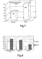
- the percentage of bone re-absorption areas (dark) in the transplanted cylinders of the stimulated animals is 31%, against 60% bone re-absorption areas in the transplanted cylinders of the control animals.
- the histogram of figure 7 illustrates the percentage of bone re-absorption areas present in the transplanted cylinders of the stimulated and control animals.
- the histological images 6a, 6b furthermore illustrate a section of the transplanted cartilage ( figure 6a ) treated with the device of figure 1 in which the viability of the transplanted cartilage, which has an adequate thickness and intense colouring of the cartilaginous matrix, is evident.
- figure 6a highlights the presence of hyaline tissue
- figure 6b non-treated cartilage
- treatment with the device 1 can effectively prevent cartilaginous degeneration in experimental animals (Dunkin Hartley), maintaining functionality. Animals with initial osteoarthrosis, aged 15 months, were treated for 6 months. Not only did treatment with the device 1 prevent degeneration of the cartilage, it also prevented osteosclerosis of the subchondral bone, indicating that the cartilage had maintained its mechanical characteristics. Indeed, when the cartilage loses the ability to absorb stress due to the load, the stress is transmitted directly to the subchondral bone tissue which reacts by increasing its density and thickness. The table shows that the histological evaluation (Mankin score) of the cartilage treated with the device 1 is clearly inferior (therefore better) than in the control animals. Control animals Animals treated with electromagnetic fields Mankin score 13.8 ⁇ 1.1 4.6 ⁇ 1.5***
- DEXA Dual Energy X-ray Absorptiometry
- Said chondrocytes can be used in different techniques, for example they can be used to perform Autologous Chondrocyte Implantation (ACI), a method introduced in the eighties by Petterson to promote healing of the cartilage.
- ACI Autologous Chondrocyte Implantation
- Said method provides for an initial collection of autologous chondrocytes by means of arthroscopy from patients affected by chondral lesions.
- the chondrocytes are then isolated by digestion of the cartilaginous matrix and cultivated in vitro, after dedifferentiation towards the chondroblastic phenotype.
- the cells thus obtained are then transplanted, in the form of suspension, into the patient's joint below a periostal flap sutured to the chondral cartilage during the operation.
- the chondrocytes can be used in the MACI (Matrix-Induced Autologous Chondrocyte Implantation) technique which involves initial arthroscopy and in vitro cultivation of autologous chondrocytes: the chondroblasts thus obtained are scattered three weeks after collection on a type I and III pig collagen scaffold.
- MACI Microx-Induced Autologous Chondrocyte Implantation
- This "membrane” can be implanted on the chondral lesion of the patient and affixed via the use of fibrin glue.
- the applicant has been able to demonstrate that on the one hand treatment with the device 1 (and with the parameters highlighted above) stimulates the proliferation of these cells which are transplanted. Stimulation of proliferation represents a fundamental element for colonisation of the cartilaginous lesion site to be treated.
- the device 1 is coupled with support means to make the device 1 portable by a person.
- These supporting means comprise ( figure 9 , 10 and 11 ) a supporting body 103 defined by at least one contoured wall 104 defining a cavity 105 for housing a portion of the human body.
- wall 104 is shaped to define a kneepad, which defines the elongated cavity 105 for housing a leg portion 107 of a patient (not shown in full) close to the knee 108.
- Cavity 105 may obviously house any portion of the human body, e.g. an arm, shoulder, etc..
- Wall 104 is preferably made of flexible synthetic material to adapt to the contour of the human body, and is obviously also made of anti-allergic, nontoxic material, such as neoprene.
- Supporting means also comprises an elastic connecting device 112 fitted to supporting body 103 to secure contoured wall 104 firmly to the portion of the human body.
- the elastic connecting device comprises two elastic straps 115, each having a portion fixed (e.g. stitched) to wall 104, and each having a fastening device, e.g. of VELCROTM, at the ends. Fastening devices other than those shown, however, may obviously be used.
- Supporting means also comprises a seat 120 for housing a the solenoid 12 of device 1 located adjacent to contoured wall 104 and therefore close to the portion of the human body.
- seat 120 is defined by a square pouch structure 124 made of fabric and connectable to an outer surface of contoured wall 4 by two reversible connecting devices 125, e.g. of VELCROTM, so that seat 120 is secured firmly in a predetermined position to contoured wall 104 when connecting devices 125 are connected firmly.
- two reversible connecting devices 125 e.g. of VELCROTM
- Solenoid 12 is conveniently made using a coiler (not shown), which forms a coil 126 ( Figure 11 ) comprising roughly 200 turns of copper wire with an average turn of 40 cm. Coil 126 is then wound with cotton tape to keep its shape. The two ends of coil 126 are connected to a bipolar power cable 127 of solenoid 122. Coil 126 is then covered with heat-sealed multilayer plastic material 128 comprising high-density sponge inside and a PVC sheet outside. Solenoid 122 is wound in air.
- Solenoid 12 is conveniently powered by the device 1, which may also be housed in a pouch 132 fixed to the outer surface of wall 104 by a releasable connecting device 133, e.g. of VELCROTM.
- more than one seat may be formed to house further solenoids.
- two separate seats may be formed for two solenoids located, in use, on opposite sides, one medial and one lateral, of the joint for treatment, which is an advantageous arrangement for treating the knee joint.
- a single solenoid is mainly indicated for treatment of the kneecap, and two solenoids for treatment of larger areas of the joint or for patients with larger than average joints. Two solenoids therefore ensure a uniform induced signal over the whole joint, even in patients with more extensive lesions.
Landscapes
- Health & Medical Sciences (AREA)
- Animal Behavior & Ethology (AREA)
- Biomedical Technology (AREA)
- Nuclear Medicine, Radiotherapy & Molecular Imaging (AREA)
- Radiology & Medical Imaging (AREA)
- Life Sciences & Earth Sciences (AREA)
- Engineering & Computer Science (AREA)
- General Health & Medical Sciences (AREA)
- Public Health (AREA)
- Veterinary Medicine (AREA)
- Prostheses (AREA)
- Magnetic Treatment Devices (AREA)
- Electrotherapy Devices (AREA)
Applications Claiming Priority (2)
| Application Number | Priority Date | Filing Date | Title |
|---|---|---|---|
| IT000344A ITTO20060344A1 (it) | 2006-05-12 | 2006-05-12 | Dispositivo per la rigenerazione e prevenzione della degenerazione del tessuto cartilagineo e dell'osso subcondrale e la proliferazione di condrociti mediante un campo elettromagnetico pulsante |
| PCT/EP2007/050849 WO2007131809A1 (en) | 2006-05-12 | 2007-01-29 | Device for the regeneration and prevention of degeneration of the cartilaginous tissue and subchrondral bone and the proliferation of chondrocytes by means of a pulsed electromagnetic field |
Publications (2)
| Publication Number | Publication Date |
|---|---|
| EP2019719A1 EP2019719A1 (en) | 2009-02-04 |
| EP2019719B1 true EP2019719B1 (en) | 2012-03-07 |
Family
ID=37904387
Family Applications (1)
| Application Number | Title | Priority Date | Filing Date |
|---|---|---|---|
| EP07704203A Active EP2019719B1 (en) | 2006-05-12 | 2007-01-29 | Device for the regeneration and prevention of degeneration of the cartilaginous tissue and subchrondral bone and the proliferation of chondrocytes by means of a pulsed electromagnetic field |
Country Status (10)
| Country | Link |
|---|---|
| US (1) | US20090299128A1 (it) |
| EP (1) | EP2019719B1 (it) |
| JP (1) | JP2009536841A (it) |
| CN (1) | CN101454046A (it) |
| AT (1) | ATE548077T1 (it) |
| AU (1) | AU2007251711B2 (it) |
| CA (1) | CA2652128A1 (it) |
| IT (1) | ITTO20060344A1 (it) |
| MX (1) | MX2008014449A (it) |
| WO (1) | WO2007131809A1 (it) |
Families Citing this family (8)
| Publication number | Priority date | Publication date | Assignee | Title |
|---|---|---|---|---|
| US7840272B2 (en) | 2005-06-03 | 2010-11-23 | Medrelief Inc. | Methods for modulating osteochondral development using bioelectrical stimulation |
| FI124387B (en) * | 2011-11-01 | 2014-08-15 | Oscare Medical Oy | Bone method and arrangement utilizing electromagnetic waves |
| CN105169561B (zh) * | 2015-09-01 | 2017-09-26 | 丁建民 | 一种癌症治疗装置 |
| US11517760B2 (en) | 2019-05-06 | 2022-12-06 | Kamran Ansari | Systems and methods of treating medical conditions using arrays of planar coils configured to generate pulsed electromagnetic fields and integrated into clothing |
| US11020603B2 (en) | 2019-05-06 | 2021-06-01 | Kamran Ansari | Systems and methods of modulating electrical impulses in an animal brain using arrays of planar coils configured to generate pulsed electromagnetic fields and integrated into clothing |
| US20250295931A1 (en) * | 2022-04-28 | 2025-09-25 | Ricoh Company, Ltd. | Cancer treatment system |
| JP2023163913A (ja) * | 2022-04-28 | 2023-11-10 | 株式会社リコー | がん治療システム |
| JP2023163906A (ja) * | 2022-04-28 | 2023-11-10 | 株式会社リコー | がん治療システム |
Family Cites Families (8)
| Publication number | Priority date | Publication date | Assignee | Title |
|---|---|---|---|---|
| JPS5843114B2 (ja) * | 1980-08-05 | 1983-09-24 | イレクトロ−バイオロジ−,インコ−ポレ−テツド | 電磁的治療装置 |
| US5067940A (en) * | 1988-03-23 | 1991-11-26 | Life Resonances, Inc. | Method and apparatus for controlling the growth of cartilage |
| US4993413A (en) * | 1988-09-22 | 1991-02-19 | The Research Foundation Of State University Of New York | Method and apparatus for inducing a current and voltage in living tissue |
| JPH078565A (ja) * | 1993-06-21 | 1995-01-13 | Teijin Ltd | 骨疾患治療用磁気刺激装置 |
| JPH1057510A (ja) * | 1996-08-21 | 1998-03-03 | Pitsupu Fujimoto Kk | 静磁場により腰痛に鎮痛効果を与える方法 |
| PT995463E (pt) * | 1998-10-21 | 2001-11-30 | A Kafka Prof Wolf | Dispositivo e sinal electrico ou electromagnetico para influenciar sequencias biologicas |
| RU2160130C1 (ru) * | 2000-07-10 | 2000-12-10 | Закрытое акционерное общество "ЭКОИНВЕНТ" | Способ нормализации биологических функций живых тканей и устройство для электромагнитного воздействия на живые ткани |
| US7160241B1 (en) * | 2000-10-06 | 2007-01-09 | Ewa Herbst | Treatment of living tissues using electromagnetic fields |
-
2006
- 2006-05-12 IT IT000344A patent/ITTO20060344A1/it unknown
-
2007
- 2007-01-29 EP EP07704203A patent/EP2019719B1/en active Active
- 2007-01-29 AU AU2007251711A patent/AU2007251711B2/en active Active
- 2007-01-29 CA CA002652128A patent/CA2652128A1/en not_active Abandoned
- 2007-01-29 US US12/300,233 patent/US20090299128A1/en not_active Abandoned
- 2007-01-29 JP JP2009508265A patent/JP2009536841A/ja active Pending
- 2007-01-29 MX MX2008014449A patent/MX2008014449A/es unknown
- 2007-01-29 WO PCT/EP2007/050849 patent/WO2007131809A1/en not_active Ceased
- 2007-01-29 AT AT07704203T patent/ATE548077T1/de active
- 2007-01-29 CN CNA2007800170176A patent/CN101454046A/zh active Pending
Also Published As
| Publication number | Publication date |
|---|---|
| ATE548077T1 (de) | 2012-03-15 |
| AU2007251711B2 (en) | 2011-08-18 |
| EP2019719A1 (en) | 2009-02-04 |
| US20090299128A1 (en) | 2009-12-03 |
| WO2007131809A1 (en) | 2007-11-22 |
| AU2007251711A1 (en) | 2007-11-22 |
| CN101454046A (zh) | 2009-06-10 |
| MX2008014449A (es) | 2009-03-13 |
| JP2009536841A (ja) | 2009-10-22 |
| ITTO20060344A1 (it) | 2007-11-13 |
| CA2652128A1 (en) | 2007-11-22 |
Similar Documents
| Publication | Publication Date | Title |
|---|---|---|
| EP2019719B1 (en) | Device for the regeneration and prevention of degeneration of the cartilaginous tissue and subchrondral bone and the proliferation of chondrocytes by means of a pulsed electromagnetic field | |
| US11826579B2 (en) | System and method for applying a low frequency magnetic field to biological tissues | |
| Aaron et al. | Treatment of nonunions with electric and electromagnetic fields | |
| CA2082170C (en) | Magnetic field therapy and apparatus | |
| Takano-Yamamoto et al. | Effect of a pulsing electromagnetic field on demineralized bone-matrix-induced bone formation in a bony defect in the premaxilla of rats | |
| JP2003518959A (ja) | Pemf生物物理学的刺激界発生器装置および方法 | |
| US20090163762A1 (en) | Device for the regeneration and prevention of degeneration of the cartilaginous tissue and subchrondral bone, the proliferation of chondrocytes and for blocking or reducing the fibroblastic evolution of chondrocytes and mesenchymal cells by means of a pulsed electromagnetic field | |
| CN113041502A (zh) | 一种基于静态磁场与动态磁场复合的骨科治疗装置 | |
| KR101630017B1 (ko) | 자성나노입자와 전자기장을 이용한 골 재생 촉진 방법 | |
| Ramli et al. | The effect of ultrasound on angiogenesis: an in vivo study using the chick chorioallantoic membrane | |
| Vicenti et al. | Biophysical stimulation of the knee with PEMFs: from bench to bedside | |
| Pickering et al. | Electromagnetic fields for bone healing | |
| Kubota et al. | Overview of effects of electrical stimulation on osteogenesis and alveolar bone | |
| Walker et al. | Electromagnetic field treatment of nerve crush injury in a rat model: effect of signal configuration on functional recovery | |
| PT1216076E (pt) | Um método e um aparelho para estimular/modular processos bioquímicos utilizando campos electromagnéticos pulsados | |
| NORTON | Implications of bioelectric growth control in orthodontics and dentistry | |
| AU2007236544B2 (en) | In vivo stimulation of cellular material | |
| Ding et al. | Effect of low-intensity pulsed ultrasound on bone formation during mandible distraction osteogenesis in a canine model—a preliminary study | |
| CN221384909U (zh) | 一种四肢磁疗舱 | |
| AU625756B2 (en) | Method and apparatus for controlling tissue growth with an applied fluctuating magnetic field | |
| CN102793975A (zh) | 一种促骨愈合的电磁场发生仪及应用 | |
| AU646597B2 (en) | Method and apparatus for controlling tissue growth with an applied fluctuating magnetic field | |
| Ghaib | Stimulation of rabbit condyle growth by using pulsed therapeutic ultrasound (A radiographical and histological experimental study) | |
| CN110448805A (zh) | 一种外加稳态磁场促骨折愈合的磁疗绷带 | |
| ELECTROMAGNETICFIELDSFORBONEHEALING | Electromagnetic Fields for Bone Healing |
Legal Events
| Date | Code | Title | Description |
|---|---|---|---|
| PUAI | Public reference made under article 153(3) epc to a published international application that has entered the european phase |
Free format text: ORIGINAL CODE: 0009012 |
|
| 17P | Request for examination filed |
Effective date: 20081129 |
|
| AK | Designated contracting states |
Kind code of ref document: A1 Designated state(s): AT BE BG CH CY CZ DE DK EE ES FI FR GB GR HU IE IS IT LI LT LU LV MC NL PL PT RO SE SI SK TR |
|
| AX | Request for extension of the european patent |
Extension state: AL BA HR MK RS |
|
| GRAP | Despatch of communication of intention to grant a patent |
Free format text: ORIGINAL CODE: EPIDOSNIGR1 |
|
| DAX | Request for extension of the european patent (deleted) | ||
| GRAS | Grant fee paid |
Free format text: ORIGINAL CODE: EPIDOSNIGR3 |
|
| GRAA | (expected) grant |
Free format text: ORIGINAL CODE: 0009210 |
|
| AK | Designated contracting states |
Kind code of ref document: B1 Designated state(s): AT BE BG CH CY CZ DE DK EE ES FI FR GB GR HU IE IS IT LI LT LU LV MC NL PL PT RO SE SI SK TR |
|
| REG | Reference to a national code |
Ref country code: GB Ref legal event code: FG4D |
|
| REG | Reference to a national code |
Ref country code: AT Ref legal event code: REF Ref document number: 548077 Country of ref document: AT Kind code of ref document: T Effective date: 20120315 Ref country code: CH Ref legal event code: EP |
|
| REG | Reference to a national code |
Ref country code: IE Ref legal event code: FG4D |
|
| REG | Reference to a national code |
Ref country code: DE Ref legal event code: R096 Ref document number: 602007021137 Country of ref document: DE Effective date: 20120503 |
|
| REG | Reference to a national code |
Ref country code: NL Ref legal event code: T3 |
|
| PG25 | Lapsed in a contracting state [announced via postgrant information from national office to epo] |
Ref country code: LT Free format text: LAPSE BECAUSE OF FAILURE TO SUBMIT A TRANSLATION OF THE DESCRIPTION OR TO PAY THE FEE WITHIN THE PRESCRIBED TIME-LIMIT Effective date: 20120307 |
|
| LTIE | Lt: invalidation of european patent or patent extension |
Effective date: 20120307 |
|
| PG25 | Lapsed in a contracting state [announced via postgrant information from national office to epo] |
Ref country code: GR Free format text: LAPSE BECAUSE OF FAILURE TO SUBMIT A TRANSLATION OF THE DESCRIPTION OR TO PAY THE FEE WITHIN THE PRESCRIBED TIME-LIMIT Effective date: 20120608 Ref country code: LV Free format text: LAPSE BECAUSE OF FAILURE TO SUBMIT A TRANSLATION OF THE DESCRIPTION OR TO PAY THE FEE WITHIN THE PRESCRIBED TIME-LIMIT Effective date: 20120307 Ref country code: FI Free format text: LAPSE BECAUSE OF FAILURE TO SUBMIT A TRANSLATION OF THE DESCRIPTION OR TO PAY THE FEE WITHIN THE PRESCRIBED TIME-LIMIT Effective date: 20120307 |
|
| REG | Reference to a national code |
Ref country code: AT Ref legal event code: MK05 Ref document number: 548077 Country of ref document: AT Kind code of ref document: T Effective date: 20120307 |
|
| PG25 | Lapsed in a contracting state [announced via postgrant information from national office to epo] |
Ref country code: CY Free format text: LAPSE BECAUSE OF FAILURE TO SUBMIT A TRANSLATION OF THE DESCRIPTION OR TO PAY THE FEE WITHIN THE PRESCRIBED TIME-LIMIT Effective date: 20120307 |
|
| PG25 | Lapsed in a contracting state [announced via postgrant information from national office to epo] |
Ref country code: EE Free format text: LAPSE BECAUSE OF FAILURE TO SUBMIT A TRANSLATION OF THE DESCRIPTION OR TO PAY THE FEE WITHIN THE PRESCRIBED TIME-LIMIT Effective date: 20120307 Ref country code: IS Free format text: LAPSE BECAUSE OF FAILURE TO SUBMIT A TRANSLATION OF THE DESCRIPTION OR TO PAY THE FEE WITHIN THE PRESCRIBED TIME-LIMIT Effective date: 20120707 Ref country code: PL Free format text: LAPSE BECAUSE OF FAILURE TO SUBMIT A TRANSLATION OF THE DESCRIPTION OR TO PAY THE FEE WITHIN THE PRESCRIBED TIME-LIMIT Effective date: 20120307 Ref country code: SI Free format text: LAPSE BECAUSE OF FAILURE TO SUBMIT A TRANSLATION OF THE DESCRIPTION OR TO PAY THE FEE WITHIN THE PRESCRIBED TIME-LIMIT Effective date: 20120307 Ref country code: BE Free format text: LAPSE BECAUSE OF FAILURE TO SUBMIT A TRANSLATION OF THE DESCRIPTION OR TO PAY THE FEE WITHIN THE PRESCRIBED TIME-LIMIT Effective date: 20120307 Ref country code: RO Free format text: LAPSE BECAUSE OF FAILURE TO SUBMIT A TRANSLATION OF THE DESCRIPTION OR TO PAY THE FEE WITHIN THE PRESCRIBED TIME-LIMIT Effective date: 20120307 Ref country code: SE Free format text: LAPSE BECAUSE OF FAILURE TO SUBMIT A TRANSLATION OF THE DESCRIPTION OR TO PAY THE FEE WITHIN THE PRESCRIBED TIME-LIMIT Effective date: 20120307 Ref country code: CZ Free format text: LAPSE BECAUSE OF FAILURE TO SUBMIT A TRANSLATION OF THE DESCRIPTION OR TO PAY THE FEE WITHIN THE PRESCRIBED TIME-LIMIT Effective date: 20120307 |
|
| PG25 | Lapsed in a contracting state [announced via postgrant information from national office to epo] |
Ref country code: PT Free format text: LAPSE BECAUSE OF FAILURE TO SUBMIT A TRANSLATION OF THE DESCRIPTION OR TO PAY THE FEE WITHIN THE PRESCRIBED TIME-LIMIT Effective date: 20120709 Ref country code: SK Free format text: LAPSE BECAUSE OF FAILURE TO SUBMIT A TRANSLATION OF THE DESCRIPTION OR TO PAY THE FEE WITHIN THE PRESCRIBED TIME-LIMIT Effective date: 20120307 |
|
| PLBE | No opposition filed within time limit |
Free format text: ORIGINAL CODE: 0009261 |
|
| STAA | Information on the status of an ep patent application or granted ep patent |
Free format text: STATUS: NO OPPOSITION FILED WITHIN TIME LIMIT |
|
| PG25 | Lapsed in a contracting state [announced via postgrant information from national office to epo] |
Ref country code: DK Free format text: LAPSE BECAUSE OF FAILURE TO SUBMIT A TRANSLATION OF THE DESCRIPTION OR TO PAY THE FEE WITHIN THE PRESCRIBED TIME-LIMIT Effective date: 20120307 Ref country code: AT Free format text: LAPSE BECAUSE OF FAILURE TO SUBMIT A TRANSLATION OF THE DESCRIPTION OR TO PAY THE FEE WITHIN THE PRESCRIBED TIME-LIMIT Effective date: 20120307 |
|
| 26N | No opposition filed |
Effective date: 20121210 |
|
| PG25 | Lapsed in a contracting state [announced via postgrant information from national office to epo] |
Ref country code: IT Free format text: LAPSE BECAUSE OF FAILURE TO SUBMIT A TRANSLATION OF THE DESCRIPTION OR TO PAY THE FEE WITHIN THE PRESCRIBED TIME-LIMIT Effective date: 20120307 |
|
| REG | Reference to a national code |
Ref country code: DE Ref legal event code: R097 Ref document number: 602007021137 Country of ref document: DE Effective date: 20121210 |
|
| PG25 | Lapsed in a contracting state [announced via postgrant information from national office to epo] |
Ref country code: ES Free format text: LAPSE BECAUSE OF FAILURE TO SUBMIT A TRANSLATION OF THE DESCRIPTION OR TO PAY THE FEE WITHIN THE PRESCRIBED TIME-LIMIT Effective date: 20120618 |
|
| PG25 | Lapsed in a contracting state [announced via postgrant information from national office to epo] |
Ref country code: BG Free format text: LAPSE BECAUSE OF FAILURE TO SUBMIT A TRANSLATION OF THE DESCRIPTION OR TO PAY THE FEE WITHIN THE PRESCRIBED TIME-LIMIT Effective date: 20120607 |
|
| PG25 | Lapsed in a contracting state [announced via postgrant information from national office to epo] |
Ref country code: MC Free format text: LAPSE BECAUSE OF NON-PAYMENT OF DUE FEES Effective date: 20130131 |
|
| REG | Reference to a national code |
Ref country code: CH Ref legal event code: PL |
|
| REG | Reference to a national code |
Ref country code: IE Ref legal event code: MM4A |
|
| PG25 | Lapsed in a contracting state [announced via postgrant information from national office to epo] |
Ref country code: LI Free format text: LAPSE BECAUSE OF NON-PAYMENT OF DUE FEES Effective date: 20130131 Ref country code: CH Free format text: LAPSE BECAUSE OF NON-PAYMENT OF DUE FEES Effective date: 20130131 |
|
| PG25 | Lapsed in a contracting state [announced via postgrant information from national office to epo] |
Ref country code: IE Free format text: LAPSE BECAUSE OF NON-PAYMENT OF DUE FEES Effective date: 20130129 |
|
| PG25 | Lapsed in a contracting state [announced via postgrant information from national office to epo] |
Ref country code: TR Free format text: LAPSE BECAUSE OF FAILURE TO SUBMIT A TRANSLATION OF THE DESCRIPTION OR TO PAY THE FEE WITHIN THE PRESCRIBED TIME-LIMIT Effective date: 20120307 |
|
| PG25 | Lapsed in a contracting state [announced via postgrant information from national office to epo] |
Ref country code: HU Free format text: LAPSE BECAUSE OF FAILURE TO SUBMIT A TRANSLATION OF THE DESCRIPTION OR TO PAY THE FEE WITHIN THE PRESCRIBED TIME-LIMIT; INVALID AB INITIO Effective date: 20070129 Ref country code: LU Free format text: LAPSE BECAUSE OF NON-PAYMENT OF DUE FEES Effective date: 20130129 |
|
| REG | Reference to a national code |
Ref country code: FR Ref legal event code: PLFP Year of fee payment: 10 |
|
| REG | Reference to a national code |
Ref country code: FR Ref legal event code: PLFP Year of fee payment: 11 |
|
| REG | Reference to a national code |
Ref country code: FR Ref legal event code: PLFP Year of fee payment: 12 |
|
| P01 | Opt-out of the competence of the unified patent court (upc) registered |
Effective date: 20230629 |
|
| PGFP | Annual fee paid to national office [announced via postgrant information from national office to epo] |
Ref country code: GB Payment date: 20251220 Year of fee payment: 20 |
|
| PGFP | Annual fee paid to national office [announced via postgrant information from national office to epo] |
Ref country code: FR Payment date: 20251217 Year of fee payment: 20 Ref country code: NL Payment date: 20251217 Year of fee payment: 20 |
|
| PGFP | Annual fee paid to national office [announced via postgrant information from national office to epo] |
Ref country code: DE Payment date: 20251217 Year of fee payment: 20 |