EP2044960B1 - Analogue phospholipidique pour le diagnostic in vivo de cancers - Google Patents
Analogue phospholipidique pour le diagnostic in vivo de cancers Download PDFInfo
- Publication number
- EP2044960B1 EP2044960B1 EP08010321.1A EP08010321A EP2044960B1 EP 2044960 B1 EP2044960 B1 EP 2044960B1 EP 08010321 A EP08010321 A EP 08010321A EP 2044960 B1 EP2044960 B1 EP 2044960B1
- Authority
- EP
- European Patent Office
- Prior art keywords
- tumor
- cancer
- tumors
- imaging
- mice
- Prior art date
- Legal status (The legal status is an assumption and is not a legal conclusion. Google has not performed a legal analysis and makes no representation as to the accuracy of the status listed.)
- Expired - Lifetime
Links
Images









Classifications
-
- A—HUMAN NECESSITIES
- A61—MEDICAL OR VETERINARY SCIENCE; HYGIENE
- A61K—PREPARATIONS FOR MEDICAL, DENTAL OR TOILETRY PURPOSES
- A61K51/00—Preparations containing radioactive substances for use in therapy or testing in vivo
- A61K51/02—Preparations containing radioactive substances for use in therapy or testing in vivo characterised by the carrier, i.e. characterised by the agent or material covalently linked or complexing the radioactive nucleus
- A61K51/04—Organic compounds
- A61K51/0489—Phosphates or phosphonates, e.g. bone-seeking phosphonates
-
- A—HUMAN NECESSITIES
- A61—MEDICAL OR VETERINARY SCIENCE; HYGIENE
- A61K—PREPARATIONS FOR MEDICAL, DENTAL OR TOILETRY PURPOSES
- A61K51/00—Preparations containing radioactive substances for use in therapy or testing in vivo
- A61K51/02—Preparations containing radioactive substances for use in therapy or testing in vivo characterised by the carrier, i.e. characterised by the agent or material covalently linked or complexing the radioactive nucleus
- A61K51/04—Organic compounds
-
- A—HUMAN NECESSITIES
- A61—MEDICAL OR VETERINARY SCIENCE; HYGIENE
- A61K—PREPARATIONS FOR MEDICAL, DENTAL OR TOILETRY PURPOSES
- A61K51/00—Preparations containing radioactive substances for use in therapy or testing in vivo
- A61K51/02—Preparations containing radioactive substances for use in therapy or testing in vivo characterised by the carrier, i.e. characterised by the agent or material covalently linked or complexing the radioactive nucleus
- A61K51/04—Organic compounds
- A61K51/0404—Lipids, e.g. triglycerides; Polycationic carriers
- A61K51/0408—Phospholipids
-
- A—HUMAN NECESSITIES
- A61—MEDICAL OR VETERINARY SCIENCE; HYGIENE
- A61P—SPECIFIC THERAPEUTIC ACTIVITY OF CHEMICAL COMPOUNDS OR MEDICINAL PREPARATIONS
- A61P35/00—Antineoplastic agents
-
- A—HUMAN NECESSITIES
- A61—MEDICAL OR VETERINARY SCIENCE; HYGIENE
- A61P—SPECIFIC THERAPEUTIC ACTIVITY OF CHEMICAL COMPOUNDS OR MEDICINAL PREPARATIONS
- A61P43/00—Drugs for specific purposes, not provided for in groups A61P1/00-A61P41/00
-
- C—CHEMISTRY; METALLURGY
- C12—BIOCHEMISTRY; BEER; SPIRITS; WINE; VINEGAR; MICROBIOLOGY; ENZYMOLOGY; MUTATION OR GENETIC ENGINEERING
- C12Q—MEASURING OR TESTING PROCESSES INVOLVING ENZYMES, NUCLEIC ACIDS OR MICROORGANISMS; COMPOSITIONS OR TEST PAPERS THEREFOR; PROCESSES OF PREPARING SUCH COMPOSITIONS; CONDITION-RESPONSIVE CONTROL IN MICROBIOLOGICAL OR ENZYMOLOGICAL PROCESSES
- C12Q1/00—Measuring or testing processes involving enzymes, nucleic acids or microorganisms; Compositions therefor; Processes of preparing such compositions
- C12Q1/34—Measuring or testing processes involving enzymes, nucleic acids or microorganisms; Compositions therefor; Processes of preparing such compositions involving hydrolase
-
- G—PHYSICS
- G01—MEASURING; TESTING
- G01N—INVESTIGATING OR ANALYSING MATERIALS BY DETERMINING THEIR CHEMICAL OR PHYSICAL PROPERTIES
- G01N33/00—Investigating or analysing materials by specific methods not covered by groups G01N1/00 - G01N31/00
- G01N33/48—Biological material, e.g. blood, urine; Haemocytometers
- G01N33/50—Chemical analysis of biological material, e.g. blood, urine; Testing involving biospecific ligand binding methods; Immunological testing
- G01N33/53—Immunoassay; Biospecific binding assay; Materials therefor
- G01N33/575—Immunoassay; Biospecific binding assay; Materials therefor for cancer
- G01N33/5758—Immunoassay; Biospecific binding assay; Materials therefor for cancer involving compounds serving as markers for tumours, cancers or neoplasias, e.g. cellular determinants, receptors, heat shock/stress proteins, A-protein, oligosaccharides or metabolites
- G01N33/5759—Immunoassay; Biospecific binding assay; Materials therefor for cancer involving compounds serving as markers for tumours, cancers or neoplasias, e.g. cellular determinants, receptors, heat shock/stress proteins, A-protein, oligosaccharides or metabolites involving compounds localised on the membrane of tumour or cancer cells
-
- A—HUMAN NECESSITIES
- A61—MEDICAL OR VETERINARY SCIENCE; HYGIENE
- A61K—PREPARATIONS FOR MEDICAL, DENTAL OR TOILETRY PURPOSES
- A61K49/00—Preparations for testing in vivo
- A61K49/001—Preparation for luminescence or biological staining
- A61K49/0013—Luminescence
- A61K49/0017—Fluorescence in vivo
-
- C—CHEMISTRY; METALLURGY
- C07—ORGANIC CHEMISTRY
- C07F—ACYCLIC, CARBOCYCLIC OR HETEROCYCLIC COMPOUNDS CONTAINING ELEMENTS OTHER THAN CARBON, HYDROGEN, HALOGEN, OXYGEN, NITROGEN, SULFUR, SELENIUM OR TELLURIUM
- C07F13/00—Compounds containing elements of Groups 7 or 17 of the Periodic Table
- C07F13/005—Compounds without a metal-carbon linkage
Definitions
- the invention generally relates to diagnostic imaging of tumors and specifically relates to diagnostic imaging of tumors using phospholipid analogs.
- CT computerized tomography
- MRI magnetic resonance imaging
- PET positron emission tomography
- SPECT single photon emission tomography
- the success of these nuclear imaging techniques depends in large part on the selective uptake and detection of appropriate radiopharmaceuticals. Selective uptake, in turn, depends upon the development of radiopharmaceuticals with a high degree of specificity for the target tissue.
- the tumor-localizing agents developed thus far for oncological applications have had only limited application.
- 67 Ga gallium citrate was originally identified for its ability to accumulate in tumor tissue.
- 67 Ga gallium citrate is taken up by a variety of other non-cancerous lesions as well, including inflammatory lesions, and unacceptable amounts of radioactivity can also accumulate in liver and spleen tissue.
- the rapid buildup of a radiopharmaceutical in these organs can seriously interfere with the imaging of nearby lesions and also negatively impacts the dosage that can safely be given to a patient.
- Mabs radiolabeled monoclonal antibodies
- these monoclonal antibodies are specific only to the particular tumor tissue for which they have been produced, and therefore will not localize generally in neoplastic tissue.
- the use of Mabs for diagnostic imaging has lead to additional problems, including varying deg rees of antigen expression, low tumor uptake, non-specific binding and adverse immunogenic reactions.
- radiopharmaceuticals which exhibit a rapid clearance from non-target tissues as well as an extended half-life in the plasma, while still retaining its specificity and avidity for neoplastic tissue.
- Such an agent should not only assist in the non-invasive imaging of primary tumors and metastases, but should also serve as a carrier for a cytotoxic agent for site-specific eradication of malignant tumor tissue, especially as it relates to most frequently diagnosed forms of cancers. It is further desirable that radiopharmaceuticals are selective for malignant tumors and not precancerous tissues including adenomas and hyperplasia.
- colorectal cancer is the fourth most common cancer, accounting for 60,000 deaths per year.
- Treatment depends primarily on the cancer stage, but may include surgery, radiation, chemotherapy, and/or radiofrequency or cryo ablation.
- CCA carcinoembryonic antigen
- a colorectal tumor marker a colorectal tumor marker
- repeat colonoscopies 5 fail to detect recurrent disease in over 50% of patients. 6 Therefore, there is a need for development of additional methods for detection of recurrent disease. Further, during treatment and diagnosis using CT scanning and RF ablation, functional information from CT scans is difficult to obtain.
- the tumor vascularity may be assessed to some degree, but there is no way of accurately determining if viable tumor cells remain within the RF lesion.
- the thermal lesions created by RF normally have a rim of inflammation surrounding them on post procedure CT scans for up to 6 months post-procedure. PET scanning has been used to follow post-ablation patients, but the rim of inflammation surrounding RF thermal lesions normally displays increased uptake, even in the absence of viable tumor. This decreases the sensitivity and specificity for early detection of recurrent tumor.
- agents like NM404 that are selective for and retained indefinitely by malignant tumor cells are preferable, unl ike FDG which is not selective for tumor cells and goes to infectious sites and hyperplasias (Barret's Esophagus).
- compounds like NM404 containing 124 I which has a 4 day physical half-life and can be shipped anywhere in the world, are preferable as compared to FDG which has a 110 minute half-life and therefore may only be have limited distribution within 200 miles of the production site.
- Further compounds like NM404 that undergo prolonged retention (not metabolized) are preferable since it is more likely that they may have significant therapeutic potential when mated with an appropriate radioisotope like 131 I.
- Response to radiation is a predictive factor for ultimate tumor control and survival for many cancers, including many gastrointestinal cancers, lung cancer, head and neck cancer, and gynecologic cancers. Most response characterization methods, while very predictive of response, are performed after completion of treatment. While some intra-treatment clinical assessments are useful in adjusting treatment, 14 in most cases there is no accurate method of predicting tumor response during actual treatment. Such a test, especially one applicable to a broad range of tumor sites and histologies, would obviously be very useful and desirable. Other treatment and diagnostic methods include molecular assays that have been proposed to predict response to therapy, and recent efforts include use of DNA microarrays to identify genetic changes that correlate with response or lack of response to treatment. These are investigational and none are in routine clinical use.
- Yet other methods of diagnosis and treatment include use of imaging modalities to predict response during XRT treatment.
- Intra-treatment PET scans using FDG are under active investigation, wherein the isotope uptake in the primary tumor midway through radiation therapy is compared to the pretreatment uptake.
- Several retrospective studies suggest patients with continued strong uptake during treatment have poorer tumor control outcomes than patients whose tumors are less FDG-avid during treatment. 15 However, more effect screening, diagnostic and treatment methods for various cancers are extremely desirable.
- malignant gliomas that are the most common type of primary brain tumors.
- MRI magnetic resonance imaging
- Most malignant gliomas have an infiltrative component, which is poorly differentiated from edematous brain tissue by present imaging techniques. It is often this component of the tumor that is most difficult to treat and responsible for local recurrence. Undoubtedly, better visualization of invasive glioma cells is desirable for significant therapeutic treatment.
- pancreatic cancer is a highly lethal disease with the poorest likelihood of survival among all of the major malignancies. It is the fifth leading cause of cancer death in the United States and of all the newly diagnosed cancers in the United States, 2% per year are due to pancreatic cancer. However, it is one of the most highly lethal diseases which accounts for 5% of all cancer deaths. Miller BA, et al. NIH Pub. No. 96-4104. Bethesda, MD. 1996. This is demonstrated by the fact that there are no five-year survivors in patients with unresectable disease. In addition, although surgical resection offers the only hope for cure, the five-year survival after resection is only 20%. Geer RJ, Brennan MF.
- PET scanning with 18-FDG has shown promise in imaging a variety of other primary cancers, it appears to have only limited ability to improve upon the imaging capability of CT scan for patients with pancreatic cancer, particularly in assessing for metastatic disease.
- Kasperk RK, Riesener KP et al., World J Surg 2001; 25:1134-1139 ; Sendler A, Avril N, et al., World J Surg 2000; 24:1121-1129 .
- Hepatocellular cancer is the most common solid organ malignancy worldwide, due to its common etiology of chronic liver damage from hepatitis or alcoholism. Incidence rates vary markedly, from 2.1 per 100,000 in North America to 80 per 100,000 in high-incidence regions of China. The risk of developing HCC in patients with cirrhosis is 1-6% per year. Although resection is the only curative option, only 10-30% of patients are candidates for surgery at the time of presentation, due to either poor hepatic reserve or the presence of unresectable or metastatic disease. Attesting to the aggressive nature of this disease, the five-year survival is only 15-35% after curative resection. Discusser G. Digestive Diseases (2001) 19:311-323 .
- Radiotherapy has a well-established role in the treatment of breast cancer due mainly to the sensitivity of many solid epithelial tumors, including infiltrating ductal carcinoma, to ionizing radiation.
- the most common indication for radiation in breast cancer is as adjuvant treatment following lumpectomy or mastectomy.
- rad iation therapy has been shown to dramatically decrease the incidence of local and regional recurrence by sterilizing microscopic deposits in these tissues.
- Chemotherapy is offered when the patient has metastatic disease or is deemed to have an increased risk for occult metastases. In this latter indication, that of adjuvant chemotherapy administration, studies confirm improved survival in patients receiving adjuvant chemotherapy or hormonal therapy. Radiation is also used in the palliative setting with good effect in reducing the pain and volume effects of metastases in solid organs and bone. Many patients relapse after definitive therapy for reasons that are multifactorial. Acquired resistance to radiation and chemotherapy undoubtedly contributes to recurrence after primary therapy. Additionally, the use of radiation is associated with specific toxicities which are generally late-occurring and d ose-limiting. Fibrosis, nerve damage, and soft tissue necrosis can be severe if excessive doses of radiation are used. Arm lymphedema is the most common and dreaded toxicity for breast cancer patients, and results most commonly from the combination of axillary dissection (done for diagnostic purposes) and adjuvant radiation to the axilla.
- Non-small cell lung cancer is the leading cause of cancer death in the United States today. Surgical resection in appropriately selected patients offers the best chance for long-term survival and may be curative. Accurate pre-operative assess ment of local, regional and distant metastatic spread is thus critical for optimal management. Evaluation of the mediastinal lymph node status is essential because nodal metastasis, which occu rs in nearly half of all patients with NSCLC, is probably the most frequent barrier to cure. Accurate staging may also spare patients the morbidity of unnecessary, non-curative surgical procedures.
- Imaging with FDG-PET scanning is quickly becoming the gold standard for imaging NSCLC, due to improved sensitivity rates, particularly when compared with CT imaging.
- this is an expensive imaging test which is not available in most community practices.
- Positron-emission tomography (PET) scanning with 18 F-FDG has generated considerable interest as an imaging technique.
- Preoperative Staging of Non-Small Cell Lung cancer with Positron-Emission Tomography New Eng J Med 343:254-261, 2000 .
- the present invention is directed to a compound for use in detecting and locating in vivo lung cancer, squamous cell carcinoma, melanoma, colorectal cancer, ovarian cancer, glioma, carcinosarcoma and pancreatic cancer in a human subject, which compound is 18-(p-iodophenyl)octadecyl phosphocholine labelled with 124 I.
- the present description generally provides background on methods and techniques for the detection and treatment of various cancers, in particular on a method for detecting and locating Lung cancer, Adrenal cancer, Melanoma, Colon cancer, Colorectal cancer, Ovarian cancer, Prostate cancer, Liver cancer, Subcutaneous cancer, Intestinal cancer, Hepatocellular carci noma, Retinoblastoma, Cervical cancer in subject that has or is suspected of having cancer.
- the background method comprises the steps of:
- the present description provides also background on a medicament for the treatment of cancer in a subject.
- the treatment comprises administering to the subject an effective amount of a molecule comprising a phospholipid ether analog, as described above.
- the cancer is selected from a group consisting of Lung cancer, Adrenal cancer, Melanoma, Colon cancer, Colorectal cancer, Ovarian cancer, Prostate cancer, Liver cancer, Subcutaneous cancer, Intestinal cancer, Hepatocellular carcinoma, Retinoblastoma, Cervical cancer, Glioma, Breast cancer, Pancreatic cancer, Carcinosarcoma and Prostrate cancer.
- the present description provides background information on a method of differentiating inflammation, adenoma and hyperplasia from neoplasia in a subject.
- the method comprises the steps of :
- a method of detecting neoplasia in a tissue sample having a phospholipase D comprises the step of:
- the PLD protein activity or the mRNA level may be quantified by contacting the tissue sample with a PLE analog, as described above.
- the present invention concerns only a compound for use in detecting and locating in vivo lung cancer, squamous cell carcinoma, melanoma, colorectal cancer, ovarian cancer, glioma, carcinosarcoma and pancreatic cancer in a human subject, which compound is 18-(p-iodophenyl)octadecyl phosphocholine labelled with 124 I.
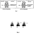
- Fig. 2 Scintigraphy of the anterior chest of Patie nt 03 acquired at 1, 2, and 6 days after IV administration of 1 mCi 131 I-NM324. Uptake is seen in the left lingular lung cancer (T) with increasing tumor-to-bac kground ratios over time.
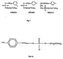
- Fig. 3 Structures of PLE analogs.
- FIG. 3A A NM404 analog.
- Fig. 4 Comparison of NM324 and NM404 in SCID mouse A549 lung tumor model following IV administration. Note that most of the NM324 activity is found in the gut and not in the tumor (implanted in the thigh) whereas NM404 identified one tumor in each thigh.
- Fig. 4A Scintigraphic NM404 images of Dunning R3327 metastatic prostate tumors in a Copenhagen rat with primary tumor site (leg) surgically removed. Two lymph node tumors were verified post mortem.
- FIG. 1 Digital Photo (A) of excised CT-26 tumor (T) and left and right lymph nodes (LN). Bioscan image (B) and fused photo/Bioscan image (C) showing correlation of radioactivity in tumor.
- Fig 7 MicroCT images of live mouse of Fig. 6 showing size and location of CT-26 tumor (arrows). 3D-surface rendered and planer slice images (A, B) as well as coronal (C) and axial (D) slices (40 ⁇ m thickness).
- Fig 8 Histologic section (H&E) of normal (left) and RF-ablated (right) CT-26 tumor. Ablated section has lost membrane integrity and appears pyknotic.
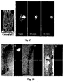
- Fig 9 Fused in vivo Bioscan/digital photo image of c-myc pancreatic tumor mouse 4 days post 125 I-NM404 injection (A). Ex vivo image of excised tumors (B) for comparison with digital photo (C). Color range same as in Fig 10 .
- FIG 10 Bioscan images of c-myc pancreatic tumor mouse 4-days post 125 I-NM404 administration.
- In vivo image (A) compared with digital photo of dissected mouse (B) showing presence of a large (2 cm) pa ncreatic tumor (T).
- Three tumors were excised and the remaining carcass scanned (C).
- the excised tumors were scanned (D) for comparison with digital photo (E).
- Color scale ranges from 0 (black) to 40 (white) cpm.
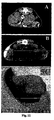
- FIG 11 MicroCT axial scans of pancreatic tumor-bearing mice. Two large tumors (T) are easily seen in the axial image in pan el A. Image of a different mouse in B depicts a pancreatic tumor (arrow) located adjacent to the spleen. In mice, the pancreas is a ubiquitous tissue. A digital photo of the excised spleen and attached tumor is shown in 11C for comparison.
- FIG 12 Bioscan image (4 days after IV injection of 125 I-NM404) of sham control rat brain (A) and same Bioscan image superimposed over the corresponding digital photograph of excised rat brain showing low background level of NM404 in normal brain tissue.
- FIG. 13 Digital photograph (A) and corresponding Bioscan image of excised C6-glioma bearing rat brain (B) 4 days after IV injection of 125 I-NM404. Position and size-matched fused Bioscan image and photograph (C) indicates intense localization of NM404 in tumor. The presence of tumor was histologically confirmed in H&E stained sample in D.
- FIG 15 Photograph (A) and Bioscan image (B) of excised CT-26 tumor-bearing mouse liver 7 days post NM404 injection. Liver tumor involvement was extensive. Tumor implant occurred 15 days prior to this scan. Bioscan image (C) and photograph (D) of excised dissected tumors (T) and normal uninvolved liver (L).
- NM404 Bioscan images of Min mouse with spontaneous right axillary mammary tumor (10 mm dia) at various times following IV administration of 125 I-NM404 (15 ⁇ Ci). Coronal microCT image (non-contrast-enhanced) is shown for anatomic comparison (left panel, T tumor).
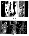
- Fig 18 Carmine stained photographs (A,C) and Bioscan images (B,D) of excised left and right abdominal mammary glands. Note 2mm tumor in panel A (T) which is easily detected in Bioscan Image (B) of the left gland. Lymph node (small arrow in A) shows no uptake of NM404. No tumors were visually detected in the right gland (C, D). Photograph (E) and Bioscan image (F) of colon indicates no uptake of NM404 in adenomatous polyps (arrows).
- Fig 19 MicroCT scans of Min mouse of Fig. 18 .
- Panel A is a low density surface rendering showing a large left axillary mammary tumor.
- Panel B is the high density surface rendering after blood pool CT contrast agent BP10 was administered to help locate tumor feeder vessels.
- Panel C is a composite coronal CT image and high density surface rendering showing absolu te feeder vessel localization. Orientation is from beneath in panel C, whereas Panels A and B are viewed from above.
- NM404 Bioscan images of Min mouse with spontaneous right axillary mammary adenocarcinoma (10 mm dia) at various times following IV administration of 125 I-NM404 (15 ⁇ Ci). Coronal microCT image (non-contrast-enhanced) is shown for anatomic comparison (left panel, T tumor).
- FIG 21 Bioscan image of excised mammary glands (A) and colon (E) from an FVBxB6Min mouse 8 days post NM404 administration. Corresponding digital photo of same excised tissues in B and D, resp ectively. Carmine stained enlarged photograph (C) shows the presence of hyperplasias (arrows) but no corresponding focal activity in the Bioscan Image (A). Tumor uptake on Bioscan image (A) corresponds to larger adenocarcinoma in B. Photograph (D) and Bioscan image (E) of excised colon indicates no uptake of NM404 in adenomatous polyps (arrows).
- FIG. 21 MicroCT scans of Min mouse shown in Fig. 21 .
- Panel A is a low density surface rendering showing a large left axial mammary tumor.
- Panel B is the high density surface rendering after blood pool CT contrast agent BP10 was administered to help locate tumor feeder vessels.
- Panel C is a composite coronal CT image and high density surface rendering showing absolute feeder vessel localization. Orientation is from beneath in panel C, whereas Panels A and B are viewed from above.
- FIG. 23 Comparison of 125 I-NM404 (A&B) and NM324 (C &D) uptake in excised SCID mouse lungs containing A549 lung CA micrornets ( ⁇ 1mm dia).
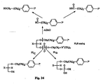
- Fig. 24 Enzymatic Metabolism of PLE's.
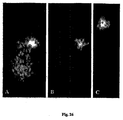
- FIG. 26 Bioscan images of prone FVBxB6 min mouse 1 ( A ) and 7 (B) days post 125 I-NM404 administration indicates presence of large axillary mammary tumor.
- Bioscan image of excised mammary gland (C) 10 days after injection shows incorporation of NM404 in large 10 mm adenocarcinoma and smaller adjacent 2 mm tumor that wasn't visible in the in vivo scan.
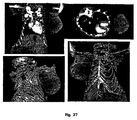
- FIG. 27 MicroCT images of same FVBxB6 min mouse, as shown in Fig. 26 , showing large axillary mammary tumor. Coronal and axial slices are shown in A and B, whereas 3D-surface (gold) and coronal slices are displayed simultaneously in posterior (C) and anterior (D) views.
- Fig. 28 Apparent SCC1 and 6 Tumor Reg ression after Injection of 125 I-NM404.
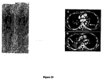
- Fig. 29 Patient 1 gamma cameral images (left panel) at 4 and 11 days following 131 I-NM404 injection showing intense and prolonged retention of the agent in both NSCLC tumors (arrows).
- Axial CT scans (right panel) showing location and size of focal 3 cm lesion in left lung (A) and large infiltrative mass in right lung (B) (arrows).
- Fig. 30 Patient 2 anterior and posterior wh ole body planar nuclear medicine images (left panel) following iv administration of 131 I-NM404. Axial (A) and coronal (B) CT scans (right panel) showing location of large 6 cm NSCLC (arrows).
- the term “isomer” includes optical isomers, structural isomers and analogs, conformational isomers. This description refers to the use of different optical isomers of an anti-tumor compound of Formula 3A. It will be appreciated by those skilled in the art that the anti-tumor compound useful in the present invention may contain at least one chiral center. Accordingly, the compound used in the the present invention and the compounds referred to as background may exist in, and be isolated in, optically-active or racemic forms. Some compounds may also exhibit polymorphism.
- the anti-tumor compounds may include pure (R)-isomers.
- the anti-tumor compounds may include pure (S)-isomers.
- the compounds may include a mixture of the (R) and the (S) isomers.
- the compounds may include a racemic mixture comprising both (R) and (S) isomers.
- optically-active forms for example, by resolution of the racemic form by recrystallization techniques, by synthesis from optically-active starting materials, by chiral synthesis, or by chromatographic separation using a chiral stationary phase).
- the background description includes the use of pharmaceutically acceptable salts of amino-substituted compounds with organic and inorganic acids, for example, citric acid and hydrochloric acid.
- Pharmaceutically acceptable salts can also be prepared from the phenolic compounds by treatment with inorganic bases, for example, sodium hydroxide.
- pharmaceutically acceptable salt refers to a compound formulated from a base background compound which achieves substantially the same pharmaceutical effect as the base compound.
- hydrate includes hemihydrate, monohydrate, dihydrate, trihydrate.
- metabolite means any substance produced from another substance by metabolism or a metabolic process.
- contacting means that the anti-tumor compound used in the background embodiments is introduced into a patient receiving treatment, and the compound is allowed to come in contact in vivo.
- the term “treating” includes preventative as well as disorder remittent treatment.
- the terms “reducing”, “suppressing” and “inhibiting” have their commonly understood meaning of lessening or decreasing.
- progression means increasing in scope or severity, advancing, growing or becoming worse.
- recurrence means the return of a disease after a remission.
- administering refers to bringing a patient, tissue, organ or cells in contact in vivo with an anti-tumor phospholipid ether compound.
- administration is accomplished in vivo , i.e. in cells or tissues of living organisms, for example, humans.
- the present description encompasses administering the described compounds to a patient or subject.
- a “patient” or “subject”, used equivalently herein, refers to a mammal, preferably a human, that either: (1) has a disorder remediable or treatable by administration of the anti-tumor substance using a reference phospholipid ether compound or (2) is susceptible to a disorder that is preventable by administering the anti-tumor compound using a reference phospholipid ether compound
- pharmaceutical composition means therapeutically effective amounts of the reference anti-tumor compound together with suitable diluents, preservatives, solubilizers, emulsifiers, and adjuvants, collectively “pharmaceutically-acceptable carriers.”
- effective amount and “therapeutically effective amount” refer to the quantity of active reference therapeutic agent sufficient to yield a desired therapeutic response without undue adverse side effects such as toxicity, irritation, or allergic response.
- the specific "effective amount” will, obviously, vary with such factors as the particular condition being treated, the physical condition of the patient, the type of animal being treated, the duration of the treatment, the nature of concurrent therapy (if any), and the specific formulations employed and the structure of the reference compounds described herein.
- an amount would be deemed therapeutically effective if it resulted in one or more of the following: (a) the prevention of disease (e.g., pancreatic cancer, breast cancer); and (b) the reversal or stabilization of such disease.
- the optimum effective amounts can be readily determined by one of ordinary skill in the art using routine experimentation.
- compositions are liquids or lyophilized or otherwise dried formulations and include diluents of various buffer content (e.g., Tris-HCl, acetate, phosphate), pH and ionic strength, additives such as albumin or gelatin to prevent absorption to surfaces, detergents (e.g., Tween (Polysorbate) 20, Tween 80, Pluronic F68, bile acid salts), solubilizing agents (e.g., glycerol, polyethylene glycerol), anti-oxidants (e.g., ascorbic acid, sodium metabisulfite), preservatives (e.g., Thimerosal, benzyl alcohol, parabens), bulking substances or tonicity modifiers (e.g., lactose, mannitol), covalent attachment of polymers such as polyethylene glycol to the protein, complexation with metal ions, or incorporation of the material into or onto particulate preparations of polymeric compounds such as polylactic acid, pol
- particulate compositions coated with polymers e.g., poloxamers or poloxamines.
- Other background embodiments of the compositions incorporate particulate forms protective coatings, protease inhibitors or permeation enhancers for various routes of administration, including topical, parenteral, pulmonary, nasal and oral.
- the background pharmaceutical composition is administered parenterally, paracancerally, transmucosally, tansdermally, intramuscularly, intravenously, intradermally, subcutaneously, intraperitonealy, intraventricularly, intracranially and intratumorally.
- pharmaceutically acceptable carriers are well known to those skilled in the art and include, but are not limited to, 0.01-0.1 M and preferably 0.05M phosphate buffer or 0.9% saline. Additionally, such pharmaceutically acceptable carriers may be aqueous or non-aqueous solutions, suspensions, and emulsions. Examples of non-aqueous solvents are propylene glycol, polyethylene glycol, vegetable oils such as olive oil, and injectable organic esters such as ethyl oleate. Aqueous carriers include water, alcoholic/aqueous solutions, emulsions or suspensions, including saline and buffered media.
- Parenteral vehicles include sodium chloride solution, Ringer's dextrose, dextrose and sodium chloride, lactated Ringer's and fixed oils.
- Intravenous vehicles include fluid and nutrient replenishers, electrolyte replenishers such as those based on Ringer's dextrose. Preservatives and other additives may also be present, such as, for example, antimicrobials, antioxidants, collating agents, inert gases.
- Controlled or sustained release compositions administerable according to the background description include formulation in lipophilic depots (e.g. fatty acids, waxes, oils). Also comprehended by the background description are particulate compositions coated with polymers (e.g. poloxamers or poloxamines).
- compositions administered according to the background description incorporate particulate forms, protective coatings, protease inhibitors or permeation enhancers for various routes of administration, including parenteral, pulmonary, nasal and oral.
- a pharmaceutical composition can be delivered in a controlled release system.
- the agent may be administered using intravenous infusion, an implantable osmotic pump, a transdermal patch, liposomes, or other modes of administration.
- a pump may be used (see Langer, supra; Sefton, CRC Crit. Ref. Biomed. Eng. 14:201 (1987 ); Buchwald et al., Surgery 88:507 (1980 ); Saudek et al., N. Engl. J. Med. 321:574 (1989 ).
- polymeric materials can be used.
- a controlled release system can be placed in proximity to the therapeutic target, for example liver, thus requiring only a fraction of the systemic dose (see, e.g., Goodson, in Medical Applications of Controlled Release, supra, vol. 2, pp. 115-138 (1984 ).
- Other controlled release systems are discussed in the review by Langer (Science 249:1527-1533 (1990 ).
- the pharmaceutical preparation described as background embodiments can comprise the reference anti-tumor compound alone, or can further include a pharmaceutically acceptable carrier, and can be in solid or liquid form such as tablets, powders, capsules, pellets, solutions, suspensions, elixirs, emulsions, gels, creams, or suppositories, including rectal and urethral suppositories.
- Pharmaceutically acceptable carriers include gums, starches, sugars, cellulosic materials, and mixtures thereof.
- the reference pharmaceutical preparation containing the reference anti-tumor compound can be administered to a patient by, for example, subcutaneous implantation of a pellet.
- a pellet provides for controlled release of reference anti-tumor compound over a period of time.
- the reference preparation can also be administered by intravenous, intra-arterial, or intramuscular injection of a liquid preparation oral administration of a liquid or solid preparation, or by topical application. Administration can also be accomplished by use of a rectal suppository or a urethral suppository.
- the reference pharmaceutical preparations administerable by the background information can be prepared by known dissolving, mixing, granulating, or tablet-forming processes.
- the reference anti-tumor compounds or their physiologically tolerated salts are mixed with additives customary for this purpose, such as vehicles, stabilizers, or inert diluents, and converted by customary methods into suitable forms for administration, such as tablets, coated tablets, hard or soft gelatin capsules, aqueous, alcoholic or oily solutions.
- suitable inert vehicles are conventional tablet bases such as lactose, sucrose, or cornstarch in combination with binders such as acacia, cornstarch, gelatin, with disintegrating agents such as cornstarch, potato starch, alginic acid, or with a lubricant such as stearic acid or magnesium stearate.
- binders such as acacia, cornstarch, gelatin
- disintegrating agents such as cornstarch, potato starch, alginic acid, or with a lubricant such as stearic acid or magnesium stearate.
- suitable oily vehicles or solvents are vegetable or animal oils such as sunflower oil or fish-liver oil. Preparations can be effected both as dry and as wet granules.
- parenteral administration subcutaneous, intravenous, intra-arterial, or intramuscular injection
- the reference anti-tumor compounds or their physiologically tolerated salts are converted into a solution, suspension, or expulsion, if desired with the substances customary and suitable for this purpose, for example, solubilizers or other auxiliaries.
- solubilizers or other auxiliaries examples are sterile liquids such as water and oils, with or without the addition of a surfactant and other pharmaceutically acceptable adjuvants.
- Illustrative oils are those of petroleum, animal, vegetable, or synthetic origin, for example, peanut oil, soybean oil, or mineral oil.
- water, saline, aqueous dextrose and related sugar solutions, and glycols such as propylene glycols or polyethylene glycol are preferred liquid carriers, particularly for injectable solutions.
- compositions which contain an active component are well understood in the art.
- Such reference compositions may be prepared as aerosols delivered to the nasopharynx or as injectables, either as liquid solutions or suspensions; however, solid forms suitable for solution in, or suspension in, liquid prior to injection can also be prepared.
- the preparation can also be emulsified.
- the active therapeutic ingredient is often mixed with excipients which are pharmaceutically acceptable and compatible with the active ingredient. Suitable excipients are, for example, water, saline, dextrose, glycerol, or ethanol or any combination thereof.
- the reference composition can contain minor amounts of auxiliary substances such as wetting or emulsifying agents, pH buffering agents which enhance the effectiveness of the active ingredient.
- An active component can be formulated into the reference composition as neutralized pharmaceutically acceptable salt forms.
- Pharmaceutically acceptable salts include the acid addition salts, which are formed with inorganic acids such as, for example, hydrochloric or phosphoric acids, or such organic acids as acetic, oxalic, tartaric, mandelic. Salts formed from the free carboxyl groups can also be derived from inorganic bases such as, for example, sodium, potassium, ammonium, calcium, or ferric hydroxides, and such organic bases as isopropylamine, trimethylamine, 2-ethylamino ethanol, histidine, procaine.
- the reference anti-tumor compounds or their physiologically tolerated salts are prepared and applied as solutions, suspensions, or emulsions in a physiologically acceptable diluent with or without a pharmaceutical carrier.
- the active compound in another method according to the background description, can be delivered in a vesicle, in particular a liposome (see Langer, Science 249:1527-1533 (1990 ); Treat et al., in Liposomes in the Therapy of Infectious Disease and Cancer, Lopez-Berestein and Fidler (eds.), Liss, N.Y., pp. 353-365 (1989 ); Lopez-Berestein ibid., pp. 317-327 ; see generally ibid).
- a liposome see Langer, Science 249:1527-1533 (1990 ); Treat et al., in Liposomes in the Therapy of Infectious Disease and Cancer, Lopez-Berestein and Fidler (eds.), Liss, N.Y., pp. 353-365 (1989 ); Lopez-Berestein ibid., pp. 317-327 ; see generally ibid).
- the salts of the reference anti-tumor compound may be pharmaceutically acceptable salts.
- Other salts may, however, be useful in the preparation of the compounds according to the background description or of their pharmaceutically acceptable salts.
- Suitable pharmaceutically acceptable salts of the compounds include acid addition salts which may, for example, be formed by mixing a solution of the compound according to the background description with a solution of a pharmaceutically acceptable acid such as hydrochloric acid, sulphuric acid, methanesulphonic acid, fumaric acid, maleic acid, succinic acid, acetic acid, benzoic acid, oxalic acid, citric acid, tartaric acid, carbonic acid or phosphoric acid.
- a pharmaceutically acceptable acid such as hydrochloric acid, sulphuric acid, methanesulphonic acid, fumaric acid, maleic acid, succinic acid, acetic acid, benzoic acid, oxalic acid, citric acid, tartaric acid, carbonic acid or phosphoric acid.
- NM404 is a promising new tumor-selective diagnostic imaging agent to monitor the treatment response of several tumor treatment modalities.
- Radioiodinated NM404 a second-generation phospholipid ether analog, had displayed remarkable tumor selectivity in 10/10 xenograft tumor models and more recently in another 14/14 spontaneous rodent tumor models. Due to a lack of metabolic phospholipase enzymes in the membranes of tumor cells, the prevailing hypothesis of this approach is that phospholipid ether analogs become trapped exclusively in tumor cell membranes because of their inability to become metabolized and eliminated. Thus, the differential clearance rates of phospholipid ethers from normal cells versus viable tumor cells form the basis of this concept.
- NM404 is sequestered and selectively retained by viable tumor cells and localizes in both primary and metastatic lesions regardless of anatomic location including those found in lymph nodes. Unlike FDG, this agent does not localize in infectious sites.
- Other advantages of NM404 over FDG include the following: NM404 is selective for and retained indefinitely by malignant tumor cells whereas FDG in not selective for tumor cells and goes to infectious sites and hyperplasias (Barret's Esophagus). Further, since 124 I has a 4 day physical half life it can be shipped anywhere in the world whereas FDG with its 110 min half-life, may have limited distribution within 200 miles of the production site.
- NM404 undergoes prolonged retention (not metabolized) and therefore affords a significant therapeutic potential when mated with an appropriate radioisotope like 131 I whereas FDG does not possess any therapeutic potential.
- NM404 can be labeled with a variety of iodine isotopes expanding it versatility (diagnosis and therapy as well as a tool for experimental animal studies) whereas FDG is limited to 18 F for PET scanning or potentially 19 F (stable) for magnetic resonance imaging albeit at very low sensitivity levels. Regardless of its tumor targeting ability, due to its rapid metabolism in tumor cells, it has not potential for therapy.
- NM404 affords the potential to not only accurately predict local tumor response to various treatment modalities, but also allows detection of distant metastatic lesions in cases of sub-therapeutic primary tumor treatment.
- the present invention is directed to a compound for use in detecting and locating in vivo lung cancer, squamous cell carcinoma, melanoma, colorectal cancer, ovarian cancer, glioma, carcinosarcoma and pancreatic cancer in a human subject, which compound is 18-(p-iodophenyl)octadecyl phosphocholine labelled with 124 I.
- the present description generally provides background information methods and techniques for the detection and treatment of various cancers, in particular on a method for detecting and locating Lung cancer, Adrenal cancer, Melanoma, Colon cancer, Colorectal cancer, Ovarian cancer, Prostate cancer, Liver cancer, Subcutaneous cancer, Intestinal cancer, Hepatocellular carcinoma, Retinoblastoma, Cervical cancer in subject that has or is suspected of having cancer.
- the background method comprises the steps of:
- the present description provides background information on the treatment of cancer in a subject.
- the treatment comprises administering to the subject an effective amount of a molecule comprising a phospholipid ether analog, as described above.
- the cancer is selected from a group consisting of Lung cancer, Adrenal cancer, Melanoma, Colon cancer, Colorectal cancer, Ovarian cancer, Prostate cancer, Liver cancer, Subcutaneous cancer, Intestinal cancer, Hepatocellular carcinoma, Retinoblastoma, Cervical cancer, Glioma, Breast cancer, Pancreatic cancer, carcinosarcoma and Prostrate cancer.
- the present invention provides background information on a method of differentiating inflammation, adenoma, hyperplasia from neoplasia in a subject.
- the method comprises the steps of :
- a method of detecting neoplasia in a tissue sample having a phospholipase D comprises the step of:
- the PLD protein activity or the mRNA level may be quantified by contacting the tissue sample with a PLE analog, as described above.
- NM404 a phospholipid ether has demonstrated marked specificity for neoplastic tissue but not in preneoplastic tissue in many experimental tumor models.
- the high tumor to background avidity and tumor selectivity of NM404 suggests it may be potentially superior to 18 F-FDG PET scanning for intra-treatment tumor imaging.
- the precise mechanism of tumor specificity of NM404 is under investigation, and currently is not as well described as the glucose utilization mechanism for 18 F-FDG uptake. It is not well established whether NM404 uptake in neoplastic tissue depends on the viability of that tissue, or if this uptake phenomenon is related to some membrane or matrix component that is independent of tissue viability.
- NM404 uptake and specificity are linked to tumor viability, it would follow that NM404 uptake in tumors recently sterilized by radiation would be non existent or poor, whereas tumors resistant to radiation would show continued uptake.
- Inventors demonstrated NM404 uptake and killing in both radio sensitive and radio resistant squamous cancer cells (SCC1 and 6) in nude mice.
- SCC1 and 6 radio sensitive and radio resistant squamous cancer cells
- One approach to the development of sensitive, more available imaging exams is to design carrier molecules which are capable of selectively delivering a radiopharmaceutical probe to the desired target tissue.
- the inventors approach has been to capitalize on unique biochemical or pharmacological properties of molecules displaying a high degree of tissue or tumor selectivity.
- the claimed invention is thus directed to imaging particular cancers with NH 404 labelled with 124I.
- Fig. 1 The inventors prevailing hypothesis ( Fig. 1 ) is that phospholipid ethers become trapped in viable tumor cell membranes because of their inability to become metabolized and eliminated. Extraction of tumors following administration of radioiodinated phospholipid ethers showed the presence of only the intact agent, whereas analysis of the urine and feces revealed only metabolites. Thus, it is the differential clearance rates of phospholipid ethers from normal cells versus tumor cells that form the basis of this concept. Preliminary results obtained in over 24 xenograft and spontaneous tumor models have universally shown NM404 to undergo selective uptake and prolonged retention in tumors. Because the agent is metabolized to some extent in the liver, the inventors avoided earlier compound evaluation in liver tumor models due to high liver background radioactivity levels.
- NM404 affords lower liver background levels than its predecessors
- the inventors expanded evaluation into liver tumors in light of the fact that imaging patients with HCC has been problematic. Many patients have underlying cirrhosis and therefore it is difficult to distinguish regenerating nodules from HCC on cross sectional imaging.
- preliminary studies evaluating PET scanning with FDG have shown only 20-50% sensitivity in detecting the disease. Verhoef C, Valkema R. et al., Liver (2002) 22:51-56 .
- PET-FDG is not useful in diagnostic screening in brain.
- FDG has not useful in evaluating disease in liver due to high natural uptake by hepatocytes.
- the inventors' synthetic approach was based on the copper-catalyzed cross-coupling reaction of Grignard reagents with alkyl tosylates or halides for the alkyl chain elongation (see the scheme below).
- the synthesis was started from p -iodobenzyl alcohol 7 which was converted into p-iodobenzyl bromide 2 by reaction with trimethylsilyl bromide.
- p -lodobenzyl bromide 2 was further coupled with Grignard reagent 3 in the presence of Li 2 CuCl 4 as a catalyst.
- 12-( p -lodophenyl)dodecanol 5 obtained after deprotection of the first coupling product 4 was converted into tosylate 6.
- tosylate 6 was coupled with Grignard reagent 7 containing 6 carbon atoms and this completed the chain elongation process.
- THP deprotection of 8 gave 18-( p -iodophenyl)octadecanol 9 which was converted into 10 (NM-404) by two-step procedure as shown in the scheme.
- an aluminum heating block apparatus was preheated to 145°C and a condenser was prepared using a 5 ml disposable syringe barrel fitted with a bent 1.5 inch 18 ga disposable needle and a rubber septum at the top.
- the HPLC system was initiated and the reservoir was filled with filtered degassed solvent (hexane/isopropanol/water (40:52:8).
- the system was equilibrated followed by a systematic check-up of the ancillary systems such as the pump, detectors, chart recorders and computer integrators.
- a 3-ml disposable syringe charcoal trap as prepared by using a glass wool plug in bottom, filling the syringe with 2.5mL with granulated charcoal, adding another glass wool plug and inserting a septum on top.
- a short tubing adaptor needle was placed on the syringe and an 18-ga needle was inserted through the septum on the top.
- the charcoal trap was connected to the top- of the condenser and vented to the atmosphere through a sodium thiosulfate trap.
- the septum was punctured with an 18-ga needle and the desired amount of aqueous sodium iodide-131 (in 0.1 N NaOH, typically 5 mCi in 15 ⁇ l) was added via a Hamilton microsyringe through the septum.
- the vial was again gently swirled or flicked to ensure mixing.
- the vial was assayed in a dose calibrator.
- the charcoal trap syringe was inserted into the reaction vial and the reaction vial was lowered into the heating block well (filled half way with sand).
- the reaction vial was heated at 145°C for 40 min during which most of the solvent distilled off and condensed in the condenser.
- a stream of air (4X25 ml) was slowly inserted through the reaction vial with a 25-ml syringe.
- the temperature of the reaction vile was increased to 155°C and heating was continued for an additional 30 minutes.
- the reaction vial was removed from the block heater and the condenser/trap assembly was disconnected and discarded and vial was allowed to cool to room temperature.
- Amberlite IRA 400-OH resin column was prepared by presoaking 1.0 g of resin in 5 ml of abs. ethanol for 30 minutes. Ethanol was decanted and the resin was rinsed with two additional 5 ml portions of ethanol. The wet resin was added into a 3 ml disposable syringe barrel with a glass wool plug at the bottom and fitted with an Acrodisc filter and a 1-way stopcock. The ethanolic solution of the crude radioiodinated product was gradually eluted through the resin column into a 5 ml vial.
- a septum was inserted and the solvent was blown off with a stream of nitrogen.
- a charcoal syringe was attached on the outlet of the vial prior to initiating nitrogen flow.
- 50 ⁇ l of ethanol was used to dilute and transfer contents to a 300 ⁇ l v-vial.
- the source vial was rinsed with a second 50 ⁇ l ethanol wash and transferred to the v-vial.
- Polysorbate 20 was added at a ratio of 0.1 ⁇ l/1.0 ⁇ g of NM-404 to the flask from a stock solution of 5% Polysorbate 20 in absolute ethanol.
- Polysorbate 20 is the pharmaceutical grade of Tween 20 that is now used in both human and animal studies with NM404.
- the solvent was removed by rotary evaporation for 10 min at ⁇ 30°C. The residue was dissolved with mixing in sufficient sterile water to yield a 2% Polysorbate 20 solution.
- the formulated product was passed through a sterile 0.2 ⁇ m Pall-Gelman Acrodisc filter (13 mm) into a dry, sterile, multidose vial (Hollister-Stier) vented with another sterile 0.2 ⁇ m filter. 100 ⁇ l of product solution was diverted into a vial for QC analysis.
- NM404 All unlabeled NM404 were taken from the original stock batch that recently underwent acute toxicology testing in order to minimize potential synthetic differences between studies. Radioiodination of NM404 was routinely achieved by an isotope exchange reaction in a melt of pivalic acid developed by the inventors 19 or by the new method described herein and prepared for injection according to standard methods described by the inventors. 22 This procedure was used effectively for preparing sterile material for the initial human trials with NM324, the predecessor of NM404 and has been used over 40 times to prepare reference 125 I- and reference 131 I-labeled NM404.
- the radiopharmaceutical was dissolved in absolute ethanol (50-500 ⁇ l) and Polysorbate 20 (0.1 ⁇ l/ ⁇ g of compound). The ethanol is removed under vacuum and the residue dissolved in sterile water to give a final solution containing no more than 2-3% Polysorbate 20. Sterilization was achieved by filtration through a sterile 0.2 ⁇ m filter unit. Final radiochemical purity must exceed 97% before using in animals. Quantification and calculation of final specific activity were achieved by HPLC analysis using known mass standards, and quantification of radioactivity ( 125 I) was accomplished by dilution and counting in a PE Wallac gamma-counter in order to avoid attenuation concerns.
- TD analysis Biodistribution studies were performed in female mice according to the standard procedure developed by the inventors. 27 Radioiodinated NM404 (5 ⁇ Ci in 100 ⁇ l) was administered via tail vein injection. At the predetermined time points animals (3/time point) were euthanized by exsanguination while under pentobarbital anesthesia. A total of 16 tissues including blood, plasma, adrenal glands, bladder, bone marrow, fat, heart, kidney, liver, lung, muscle, spleen, ovaries, skin, thyroid, and tumor were excised, rinsed, and dissected free of extraneous tissue. Large organs were minced and duplicate tissue samples will be weighed and placed in plastic tubes for isotope counting.
- Injection site and residual carcass radioactivity were also determined in a well counter. These standard procedures have been utilized for many years in the inventor's laboratory under appropriate animal care and radiation safety approval. Tissue distribution tables were generated by a computer program which produces decay-corrected tissue radioactivity concentration data on a percent injected dose/g, %kg dose, and percent injected dose/organ + SEM basis. At each time point, tumor to tissue ratios were calculated based on a percent injected dose per gram of tissue basis.
- a control TD study (3 mice/time point, 15 total mice) were performed on tumor bearing mice at 4, 7, 14, 21, and 28 days most NM404 injection in order to establish comparative TD tables for all of the therapeutic regimens.
- Animals (sodium pentobarbital anesthesia, 0.06 mg/g bw) underwent microCT scanning (lmtek MicroCAT I, 390 step acquisition/43Kvp/41 0 ⁇ A) using medium resolution acquisition parameters. Data sets were reconstructed 3-dimensionally and are visualized with AMIRA 3D-visualization software. The software allows for ROI density analysis and convenient on-screen measuring.
- Phospholipid ethers can easily be labeled with iodine radioisotopes using an isotope exchange method developed by the inventors. 19
- the iodophenyl phospholipid ether analogs are specifically designed so that the radioiodine affixed to each molecule is stable to facile in vivo deiodination.
- radiolabeled PLE compounds were synthesized and tested in vitro and in vivo. 20-22 Two of these, namely reference NM294 and reference NM324 [12-(3-iodophenyl)-dodecyl-phosphocholine], initially showed the most promise in animal tumor localization studies.
- reference NM324 and NM404 are similar in structure to miltefosine (hexadecylphosphocholine), an antitumor ether lipid studied most extensively in Europe.
- miltefosine hexadecylphosphocholine
- the antitumor properties of miltefosine and several other antitumor phospholipid ether analogs have been demonstrated in a wide range of tumor cell lines including prostate-, bladder-, and terato-carcinomas, murine and human leukemias, as well as lung, colon, ovarian, brain and breast cancers. 23
- these phospholipid ether analogs do not bind directly to DNA and are not mutagenic.
- O-Alkyl glycerol monooxygenase AGMO
- Phospholipases C (PLc) and D (PL D ) give rise to the glycerol or phosphatidic acid products, respectively.
- NM324 was not a substrate for this enzyme when compared to [ 3 H]-lyso-PAF (platelet activating factor), which was extensively metabolized.
- NM324 was analyzed as a substrate for PLc isolated from Bacillus cereus and was not hydrolyzed relative to 1-palmitoyl-2-[3H]-palmitoyl-L-3-phosphatidylcholine (DPPC), which underwent significant hydrolysis.
- DPPC 1-palmitoyl-2-[3H]-palmitoyl-L-3-phosphatidylcholine
- PLE analogs were subjected to a PL D assay.
- the PL D which was isolated from cabbage, is similar to mammalian PL D in that the cabbage form affords phosphatidylethanol-type products in addition to phosphatidic acid when the enzymatic reaction is performed in the presence of ethanol.
- Several of the PLE analogs subjected to these assay conditions did give rise to the phosphatidylethanol adduct, indicating possible interaction with PLn.
- NM404 precursors were also subjected to in vitro metabolism studies in various cell lines including Walker tumor cells, rat muscle (H9c2), and rat hepatocytes.
- the extent of metabolism was determined on the basis of radiolabeled products formed after incubation for various time periods and the results normalized to cell number or the amount of cellular protein.
- Subsequent lipid extraction of the incubation medium and cell suspension demonstrated little generation of PLE metabolites in the Walker tumor cells whereas a significant production of metabolites was seen in both the muscle cells and hepatocytes over the 48h time period studied.
- reference NM324 was more readily synthesized and was thus selected as the lead compound for initial clinical studies. Although images obtained in 5 human lung cancer patients detected tumors, images were complicated by high liver radioactivity ( Fig 2 ).
- Second Generation PLE Analogs In order to decrease liver uptake and prolong the plasma phase, 9 structural analogs of reference NM324 were synthesized and radiolabeled with 125 I for initial image analysis in Copenhagen rats bearing Dunning R3327 prostate tumors. Based upon this initial screen, reference NM347, NM404 [18-(4-iodophenyl)-octadecylphosphocholine] and NM412 ( Fig 3 ) were selected to undergo further imaging and biodistribution analysis in animal-tumor models.
- lymph node metastases were clearly delineated in a metastatic prostate tumor model following intravenous administration of either NM404 or reference NM412. Most importantly, the tracer was not retained by uninvolved lymph nodes. 24 ( Fig. 4A ). Although conducted in a prostate model, this finding is particularly relevant to breast cancer wherein lymph node involvement is such an important prognostic indicator.
- a preliminary pilot study conducted in SCID mice bearing human A549 NSCLC tumors were encouraging and demonstrated that NM404 overcomes the problem of high first pass clearance of reference NM324 by the liver.
- NM404 shows excellent tumor visualization, especially striking in the delayed images, with minimal liver and kidney uptake in comparison with reference NM324 ( Fig 4 ). Tissue biodistribution studies further confirmed the high levels of radioactivity residing in the tumors. Although imaging results were similar with NM404 and reference NM412, dosimetry data obtained in rats revealed lower kidney doses were found with NM404 relative to reference NM412, and thus NM404 was selected for further studies. Comparative biodistribution data for reference NM324 and NM404 in SCID mice with prostate and A549 lung cancer tumor models have revealed high tumor to normal tissue ratios and tumor uptake exceeding 25% of the injected dose with NM404.
- mice Animal imaging studies performed in mouse models aimed at determining the uptake characteristics in a wide variety of tumor models are summarized in Table 1.
- Preliminary results in B6 Apc Min /+ mice indicate that NM404 is not taken up by adenomatous polyps but is taken up and retained by mammary adenocarcinomas in this model, thus indicating a possible specificity for malignant tumor cells.
- These studies are aimed at determining the potential of NM404 to noninvasively characterize tumors.
- NM404 has displayed significant tumor uptake and retention in every adenocarcinoma model studied.
- CT26 Murine Tumor Model Inventors explored NM-404 as a predictor of tumor response in a murine (BALB/ c mice) model with subcutaneous CT26 cell inoculation into the flanks of the mice.
- the CT26 cell line is a poorly differentiated murine adenocarcinoma that was induced by rectal injection of N-nitroso-N-methylurethane in BALB/c mice.
- the cell line is simple to grow in vitro and results in a predictable growth pattern when injected in the vasculature (tail vein injection, metastatic model), or into the skin ( Fig 5 ) or liver. 25,26 Because the cell line is derived from a colorectal cancer, this murine model is highly clinically relevant for these studies.
- Fig 7 Each animal also underwent microCT scanning ( Fig 7 ) on day 7 prior to euthanasia and dissection of the tumor.
- Focal hot spots correlated visually with all tumors on ex vivo Bioscan images ( Fig 6 ). Although lymph nodes are visible, no radioactivity was associated with them indicating a lack of tumor cell infiltration.
- the main tumor in Figs 6 and 7 was histologically categorized as an adenocarcinoma. The inventors have scanned a wide variety of subcutaneous tumors via microCT and all are very easily detectable down to less than 300 microns in diameter.
- FIG 4 A comparison of reference NM324 and NM404 images in a SCID mouse-human NSCLC model are shown in Fig 4 . Note the relative lack of liver, kidney and gut activity with NM404, coupled with excellent tumor visualization. Although imaging results were similar with NM404 and reference NM412, recent dosimetry data obtained in rats revealed lower kidney doses were found with NM404 relative to reference NM412, and thus NM404 has been selected for further studies.
- the aim of that project is to evaluate the feasibility of labeling NM404 with iodine-124, a relatively new positron isotope with a 4-day physical half-life, and to evaluate its promise for PET imaging of tumors in small animal models.
- this approach would compliment the use of fluorine-18 FDG in that its uptake into tumor cells occurs via a different biochemical mechanism than glucose utilization.
- Unlabeled reference NM324 was administered to five normal, disease-free, humans in order to gain approval of the radiolabeled agent for human administration by the Radioactive Drug Research Committee (RDRC). These subjects had no evidence of toxicity, as manifested by symptoms, clinical examination, vital signs and sequential blood chemistries.
- RDRC Radioactive Drug Research Committee
- Patient 01 was a 55 year old woman with a right middle lobe lung mass eroding into the right ribs, histologically a mucin-producing adenocarcinoma of probable lung origin.
- Initial reference 131 I-NM324 scintigraphic images at 6 hours showed a focus of uptake in the right lateral mid-lung. For reasons unrelated to the scintigraphic study, the patient was unable to return to the hospital beyond 6 hours for further imaging session.
- Patient 02 was a 62 year old man with a large (9x7x7.5 cm), lobulated mediastinal mass extending from the aortopulmonary window and left hilum.
- Tissue type was a small cell undifferentiated (oat cell) carcinoma.
- 131 I-NM324 scintigraphic reference images revealed a focus of uptake in the left upper lung, which increased in intensity over time relative to the normal background activity.
- NM404 has displayed selective and prolonged tumor retention in 25/25 xenograft and spontaneous rodent models
- a physician sponsored IND recently initiated clinical evaluation of the agent in Stage 4 human non-small cell lung cancer patients in order to determine whether or not it would exhibit similar tumor uptake and retention properties in humans.
- two patients with advanced NSCLC were imaged after an injection of ⁇ 1 mCi of reference 131 I-NM404.
- Blood and urine samples were collected at predetermined times, and gamma imaging performed at several time points following administration.
- significant tumor uptake and retention of NM404 was demonstrated in the pri mary lung tumor, as seen in Figures 29 and 30 .
- Relative to the high liver uptake values seen previously with its first generation predecessor, reference NM324, liver an d abdominal activity are much lower with NM404, suggesting the feasibility of evaluating this agent in other abdominal cancers including pancreatic, colon, and prostate.
- NM404 does indeed localize in and undergo selective and prolonged tumor retention in human non-small cell lung cancer.
- Patient 1 55 year old male with bilateral 3cm left lobe and infiltrative right lobe NSCLC and a brain metastasis and a small right adrenal mass. He has participated in numerous standard and experimental treatment regimens. Images are included in Fig 29 .
- Patient 2 70 year old male recently diagnosed with 6 cm upper lobe non small cell mass, a 5 mm liver mass, an ilial bone met and a very small brain metastasis. He had recently completed low dose carboplatin/taxol chemotherapy and palliative radiotherapy to the ilial and brain metastases the week prior to initiating the NM404 trial. Images are shown in Fig. 30 .
- NM404 a second-generation PLE analog
- NM404 a second-generation PLE analog
- pancreatic acinar cells are linked to an elastase promoter, which is only expressed in the pancreas.
- ela-1-myc endogenous mice develop acinar and ductal neoplasia, which results in death between 2 and 7 months of age. By one month of age, the pancreas appears thickened and firm. Thus mice between the ages of 1-3 months serve as excellent models for the study of pancreatic cancer.
- Most human pancreatic neoplasms have a ductal morphology and Dr. Sandgren"s transgene targeting strategies are aimed at developing tumors that are specific for pancreatic ductal epithelium.
- the c-myc model produces tumors that are invasive adenocarcinomas, with mixed acinar/ductal phenotype.
- the biology of the k-ras model is markedly different.
- the k-ras tumors have been classified as "carcinoma in situ", meaning that they have features of neoplasia, but they do not invade and generally stay small ( ⁇ 2mm).
- Their cellular appearance is far more like the early human tumors so from a histological perspective they are a more relevant model of human disease. Further, they resemble the very early stages of the human disease.
- mice In order to determine if NM404 localizes in mouse pancreatic tumors, six c-myc endogenous mice were scanned on a Bioscan AR-2000 radioTLC scanner (modified in the inventors lab for mouse imaging) from 2-21 days after tail vein injection of reference 125 I-NM404 (15 ⁇ Ci/20g bw). On the last day, mice also underwent microCT scanning (42 kvp, 410 ⁇ A, 390 steps, MicroCAT-I, ImTek, Inc., Knoxville, TN).
- pancreatic tumors were excised and scanned ex vivo on the same scanner (equipped with high resolution 1 mm collimator and 2-D acquisition and analysis software) in order to avoid tissue attenuation associated with the low energy of reference iodine-125 ( Fig 9-10 )
- tissues were excised, weighed, and radioactivity quantitated in a gamma counter.
- NM404 displayed selective and prolonged retention in spontaneous pancreatic adenocarcinomas examined in this study, thus further extending the tumor selectivity of this agent.
- Rat C6 glioma cells were propagated in DMEM medium (Life Technologies, Gaithersburg, MD) supplemented with 10% heat-inactivated FBS (BioWhittaker, Walkersville, MD), 100 U/ml penicillin G, 100mg/ml streptomycin, and 0.01 M HEPES (Life Technologies, Gaithersburg, MD). Intracranial tumor implantation was performed as described previously (ref).
- NM404 [18-(4-iodophenyl)-octadecylphosphocholine] ( Fig 3A , 100mg) was radioiodinated with reference 125 I via isotope exchange with Na 125 I in a melt of pivalic acid. Weichert, et al. Int J Appl Rad Isotopes. 1986; 37:907-913. Following HPLC purification NM404 was dissolved in an aqueous 2% Polysorbate 20 solution prior to tail vein injection (5-20 ⁇ Ci/200g rat) into four tumor-bearing and three sham-operated rats.
- NM404 may also be useful for visualization of small invasive tumor foci.
- TGF ⁇ Endogenous Mouse HCC Model.
- the development of spontaneous hepatocellular cancer in endogenous mice over expressing the TGF ⁇ gene has been extensively evaluated and is an extremely promising animal model for study of this disease.
- TGF ⁇ is a mitogen for epithelial cells and binds to the EGF receptor; unregulated expression of TGF ⁇ results in tumor formation.
- MT1 zinc-inducible metallothionine 1
- DEN diethylnitrosamine
- CT26 Colon Adenocarcinoma Xenograft Model In addition to the spontaneous HCC model, NM404 was also evaluated in a xenograft colon adenocarcinoma tumor model whereby CT26 cells (5 ⁇ 10 5 cells/50 ⁇ l) were previously injected directly into the liver parenchyma of female BALB/c mice for creation of focal liver tumors.
- NM404 ( Fig 3A , 100 ⁇ g) was radioiodinated with 125 I via isotope exchange in a melt of pivalic acid.
- HPLC purification it was dissolved in an aqueous 2% Polysorbate 20 solution prior to tail vein injection (15 ⁇ Ci/20g mouse) into 3 TGF ⁇ endogenous mice or alternatively into 3 CT26-tumor bearing mice.
- mice were anesthetized and scanned for up to 21 days post injection on a modified Bioscan AR2000 radio-TLC scanner (1 mm increments at 2 min acquisition/ lane and 1 mm high-resolution collimator) and also in an ImTek microCT scanner (390steps) for anatomic correlation.
- MicroCT images were displayed using Amira software.
- tumor-bearing livers were initially excised and scanned ex vivo. Tumors were then excised, weighed, scanned ex vivo, and radioactivity quantitated. Lesion samples were submitted for histologic classification.
- NM404 displayed selective and prolonged retention by both spontaneous and xenograft murine liver tumor models evaluated in this study.
- Apc Min /+Mouse Model This model is comprised of mice carrying the Min allele of Apc ( Apc Min /+ mice). This model offers specific advantages over xenograft models in that female Apc min /+ mice are predisposed to develop mammary hyperplasias and carcinomas and intestinal adenomas. On the C57BL/6J genetic background, about 5% of untreated females will develop a mammary tumor by 100 days of age. Moser AR, Dove, et al. Proc Natl Acad Sci USA (1993) 90:8977-81 .
- the incidence and multiplicity of the mammary lesions can be increased by a single dose of ethylnitrosourea (ENU), a direct acting alkylating agent.
- ENU ethylnitrosourea
- Treatment with ENU results in 90% of B6 Apc Min / + females developing an average of 3 mammary squamous cell carcinomas (SCC), but few hyperplasic lesions within 60 days after treatment.
- Apc Min / + mice carry a single base pair change in the Apc (adenomatous polyposis coli) gene.
- the APC / Apc gene encodes a large protein with several potential functional domains. Groden, J., Thliveris, A., Samowitz, W., Carlson, M., Gelbert, L., Albertsen, H., Joslyn, G., Stevens, J., Spirio, L., Robertson, M. and et al. Identification and characterization of the familial adenomatous polyposis coli gene.
- ⁇ -catenin has multiple roles in the cell, including stabilization of E-cadherin and regulation of transcription through the LEF and TCF family of transcription factors.
- Axin a negative regulator of the Wnt signaling pathway, forms a complex with GSK-3beta and beta-catenin and promotes GSK-3beta-dependent phosphorylation of beta-catenin.
- Axin a negative regulator of the wnt signaling pathway, directly interacts with adenomatous polyposis coli and regulates the stabilization of beta-catenin.
- APC adenomatous polyposis coli
- ⁇ -catenin would accumulate in the cytoplasm and nucleus, possibly affected both transcription of target genes and cell-cell interaction through E-cadherin.
- APC mutations are frequent in several tumor types in humans including intestinal tumors and other epithelial tumors. Loss of heterozygosity at the APC locus or increased levels of ⁇ -catenin have been found in more than 25% of breast cancers. Furuuchi, K., Tada, M., Yamada, H., Kataoka, A., Furuuchi, N., Hamada, J., Takahashi, M., Todo, S., and Moriuchi, T.
- Somatic mutations of the APC gene in primary breast cancers Somatic mutations of the APC gene in primary breast cancers. Somatic mutations of the APC gene in primary breast cancers, American Journalof Pathology. (2000) 156:: 1997-2005 ; Jonsson, M., Borg, A., Nilbert, M., and Andersson, T. Involvement of adenomatous polyposis coli (APC)/beta-catenin signaling in human breast cancer. Involvement of adenomatous polyposis coli (APC)/beta-catenin signaling in human breast cancer, European Journal of Cancer. (2000) 36: 242-248 . Thus, the types of lesions that appear in these mice will be molecularly and histologically similar to breast cancers in humans.
- FVBxB6 Apc Min / + female mice develop an average of 0.2 mammary tumors per mouse, but 4 hyperplasias per mouse within 120 days of treatment.
- BALB/xB6 Apc Min / + develop an average of 1.8 mammary tumors and 0.6 hyperplasias per mouse.
- FVBxB6 and BALBxB6 Apc min / + mice develop both mammary SCC and adenocarcinomas (AC).
- the hyperplastic lesions in the FVBxB6 Apc Min /+ mice can be classified as either alveolar hyperplasias or squamous nodules.
- Alveolar hyperplasias are precursors to the adenocarcinomas and the squamous nodules are precursor lesions to the SCC.
- mice that develop multiple types of hyperplasias and carcinomas may be generated, often within the same animal.
- the alveolar hyperplasias resemble atypical lobules (type A) commonly found in samples from human breasts.
- Edinburgh, R. D. and Wellings, S. R. The comparative pathology of human and mouse mammary glands.
- the comparative pathology of human and mouse mammary glands Journalof Mammary Gland Biology& Neoplasia. (1999) 4: 105-22 .
- These atypical lobules are more common in cancerous breasts or in the contralateral breast in women with breast cancer. While SCC is not a frequent type of breast tumor, the AC resembles a common type of human breast tumor.
- Polyoma virus infection of mice leads to the development of numerous tumor types including mammary tumors.
- Endogenous mice expressing the polyoma middle T antigen (PyVT) under the control of the mouse mammary tumor virus LTR (MMTV) develop multifocal mammary dysplasias and tumors rapidly.
- Induction of mammary tumors by expression of polyomavirus middle T oncogene a endogenous mouse model for metastatic disease.
- Induction of mammary tumors by expression of polyomavirus middle T oncogene a transgenic mouse model for metastatic disease, Molecular & Cellular Biology, (1992) 12: 954-61 .
- mice provide a rapid model for metastatic mammary cancer.
- genetic background affects the time course of tumor development and metastatic spread. Lifsted, T., Le Voyer, T., Williams, M., Muller, W., Klein-Szanto, AA., Buetow, K. H., and Hunter, K. W. Identification of inbred mouse strains harboring genetic modifiers of mammary tumor age of onset and metastatic progression. Identification of inbred mouse strains harboring genetic modifiers of mammary tumor age of onset and metastatic progression, lnt J of Cancer. (1998) 77: 640-4 .
- NM404 ( Fig 3A , 100 ⁇ g) was radioiodinated with reference 125 I via isotope exchange in a melt of pivalic acid. Following HPLC purification it was dissolved in an aqueous 2% tween-20 solution prior to tail vein injection (15 ⁇ Ci/20g mouse) into 6 female Apc Min / + mice. Mice were anesthetized and scanned for up to 50 days post-injection on a modified Bioscan AR2000 radio-TLC scanner (1 mm increments at 2 min acquisition/lane and 1 mm high-resolution collimator) and also in an ImTek microCT scanner (390steps) for anatomic comparison. MicroCT images were displayed using Amira software.
- NM404 has displayed striking tumor avidity in animal and human xenograft tumor models examined to date. Moreover, while it displayed selective and prolonged retention by mammary tumors in this spontaneous tumor model it did not localize in associated intestinal adenomatous polyps.
- Apc Min /+Mouse Model This model is comprised of mice carrying the Min allele of Apc ( Apc Min / + mice). This model offers specific advantages over xenograft models in that female Apc Min / + mice are predisposed to develop mammary hyperplasias and carcinomas and intestinal adenomas. On the C57BL/6J genetic background, about 5% of untreated females will develop a mammary tumor by 100 days of age. Moser AR, Dove, et al. Proc Natl Acad Sci USA (1993) 90:8977-81 .
- the incidence and multiplicity of the mammary lesions can be increased by a single dose of ethylnitrosourea (ENU), a direct acting alkylating agent.
- ENU ethylnitrosourea
- Treatment with ENU results in 90% of B6 Apc Min / + females developing an average of 3 mammary squamous cell carcinomas (SCC), but few hyperplasic lesions within 60 days after treatment.
- FVBxB6 Apc Min / + female mice develop an average of 0.2 mammary tumors per mouse, but 4 hyperplasias per mouse within 120 days of treatment.
- BALB/xB6 Apc Min / + develop an average of 1.8 mammary tumors and 0.6 hyperplasias per mouse.
- FVBxB6 and BALBxB6 Apc Min / + mice develop both mammary SCC and adenocarcinomas (AC).
- NM404 ( Fig 3A , 100 ⁇ g) was radioiodinated with 125 I via isotope exchange in a melt of pivalic acid. Following HPLC purification it was dissolved in an aqueous 2% tween-20 solution prior to tail vein injection (15 ⁇ Ci/20g mouse) into 6 female Apc Min / + mice. Mice were anesthetized and scanned for up to 30 days post injection on a modified Bioscan AR2000 radio-TLC scanner (1 mm increments at 2 min acquisition/lane and 1 mm high-resolution collimator) and also in an ImTek microCT scanner (390steps) for anatomic comparison. MicroCT images were displayed using Amira software.
- NM404 did not localize, however, in either focal alveolar hyperplasias or in intestinal adenomatous polyps found frequently in these mice ( Fig 21 ). MicroCT images confirmed the presence and precise location of all mammary tumors ( Fig 22 ). NM404 apparently is metabolized and eliminated from normal cells but becomes metabolically trapped in tumor cell membranes.
- NM404 has displayed striking tumor avidity in 20/20 animal and human xenograft tumor models examined to date. Moreover, while it displayed selective and prolonged retention by mammary adeno- and squamous cell carcinomas in this spontaneous tumor model, it did not localize in associated focal alveolar hyperplasias or intestinal adenomatous polyps and thus appears to be selective for malignant tumor cells.
- Certain phospholipid ether analogs such as NM404, are selectively retained within many types of tumor cells for a prolonged time.
- the inventors sought to evaluate the mechanism of selective retention of NM404 in tumor cells using both an enzymatic assay to evaluate the activity of phospholipase D (PLD) protein and quantitative PCR.
- PLD phospholipase D
- hepa-1 hepatoma
- CT26 colonal adenocarcinoma
- TS/A breast adenocarcinoma
- Single cell suspensions of murine tumor cell lines including hepa-1 (hepatoma), CT26 (colorectal adenocarcinoma), and TS/A were analyzed with two assays:(1) Amplex ® Red assay, using a commercially available kit (Molecular Probes) that evaluates PLD protein activity using a fluorescence microplate reader, and (2) quantitative PCR to determine the level of PLD mRNA.
- Tumor cell lines were compared to normal liver tissue, which exhibits higher levels of uptake and elimination of NM404 and thus likely has lower PLD levels than other normal tissues.
- B6 and BRB6 F1 Min / + mice were very similar with respect to the mammary gland phenotype, but quite different in susceptibility to intestinal tumors.
- the B6 and BR strains can be considered sensitive backgrounds for Min -induced mammary tumorigenesis as the mice developed a large number of tumors within a short time after ENU treatment.
- the BR strain carries dominant resistance alleles at modifier loci affecting intestinal tumor development, which may prove relevant to the proposed therapy study.
- a comparison of these strains is presented in Table 2.
- mice % with mammary tumor 0
- Average # of mammary lesions/mouse Average # of intestinal tumors/mouse Average survival in days after ENU (range)
- B6 45 93 3.3 ⁇ 2.0 17(38) 0.6 ⁇ 0.9 34 ⁇ 10
- BRxB6 18 94 4.9 ⁇ 2.6 7(39) 0.5 ⁇ 0.7 14 ⁇ 4 91 (58-118)
- FVBxB6 18 17 0.2 ⁇ 0.5 18(100) 4.1 ⁇ 2.4 12 ⁇ 6 a 127 (93-178) a Information on 16 mice as the intestines of 2 mice were lost in processing.
- mice were generated by crossing females of each strain with B6 Min / + males.
- Female mice were treated with ENU when between 30-45 days of age and sacrificed when moribund, Only the results from the Minl+ mice are shown.
- Mammary tumors are defined as those tumors identified at necropsy, while mammary lesions are the small focal lesions noted in the whole mounts of the 1 st , 4 th and 5 th mammary glands.
- Intestinal tumors were counted in three 4cm sections from the small intestine (duodenum, jejunum, and ileum), and the entire colon. All Min / + mice developed intestinal tumors. Values are the means ⁇ SD
- Fig 27C The main tumor in Fig 27C was histologically categorized as an adenocarcinoma. There were four mammary tumors in both mice and all were easily detectable in ex vivo Bioscan images of the excised mammary glands.
- Radiotherapeutic Potential of NM404 NM404 During the course of recent mouse tumor uptake and retention studies with "imaging" doses (15-20 ⁇ Ci/20g mouse) of 125 I-labeled NM404, several apparent therapeutic responses have been observed (unpublished results). In an Apc Min / + mouse mammary tumor model it has generally been noted that tumor growth remains static following a single intravenous injection of NM404. Some of these animals also lost all hair above larger mammary tumors at around 8 days after injection. Moreover, these mice also get intestinal tumors and usually suffer from intestinal bleeding resulting in severe anemia, which renders their feet white. Dr. Moser noted that the feet of these mice had reverted to a pink color around 5 days after a single injection of NM404.
- iodine-125 Due to its 60-day physical half-life and low energy 28 KeV photon emission, iodine-125 is suitable for imaging experiments in mice and rats. lodine-125 also affords therapeutic characteristics as well and is currently used in permanent prostate brachytherapy implants.
- 2 nude mice were each inoculated with subcutaneous squamous cell 1 and 6 tumor cell implants on opposing flanks. SCC 1 and 6 cells were used because one is radiosensitive relative to the other. After 14 days when the average tumor size (4 total) was approaching 0.5 cm in diameter, one of the mice received 20 ⁇ Ci of 125 I-labeled NM404 and the other one receive unlabeled NM404 in an equal mass dose.
- the mouse that had received only the unlabeled compound had to be euthanized 20 days after injection due to both tumors reaching the termination size limit as defined in our animal use protocol.
- Both tumors in the 125 I-NM404 mouse regressed dramatically and unexpectedly over the course of several weeks ( Fig 28 ). In fact, the tumors of this mouse never did reach terminal size and the mouse was actually euthanized after 90 days in order to collect histology sections. At this time, the center of the tumor had become necrotic while the peripheral rim appeared somewhat viable. Histologic examination confirmed a necrotic center and viable rim.
- Iodine-131 has been used with great efficacy in the treatment of thyroid cancer. Very safe doses of 131 I can control subclinical deposits of well-differentiated thyroid cancer, which concentrates iodine very avidly as does the normal thyroid. This active uptake process helps limit the dose to normal tissues. lodine-131 has both beta and several gamma emissions, but the predomi nant tissue dose arises from the beta emissions. The inventors have selected 131 I-labeled NM404 based upon the clinical success with thyroid cancer coupled with results obtained with Bexxar (an iodine-131-labeled antibody-based agent) in low-grade lymphoma patients.
- Bexxar an iodine-131-labeled antibody-based agent
- the predominant beta emissions and mostly low energy gamma emissions optimize dose homogeneity within the tumor nodule itself. Also, the shorter half-life (8-days) provides more clinically relevant dose-intensity compared to the 60-day half-life of 125 I. These factors will permit the inventors to make the best assessment of the anti-tumor efficacy of this agent.
- a potential disadvantage of 131 I is that there is a higher energy gamma emission as well which could actually expose adjacent surrounding tissues to more radiation than would occur with 125 I.
- the tumors in the endogenous model proposed herein are peripherally located in the mammary glands and thus should not represent an immediate threat to the overall well-being of the animal.
- organ toxicity is also one of the study endpoints, the reaction of the surrounding tissue and key organ systems (marrow, liver, kidneys, bowel, brain, etc) is assessed. Tissue distribution data and actual dosimetry of radiolabeled NM404 will determine its optimal therapeutic potential. It is possible that different isotopes will complement each other in the therapeutic setting.
- DIAPEUTIC is a trademark of Cellectar, LLC
Landscapes
- Health & Medical Sciences (AREA)
- Life Sciences & Earth Sciences (AREA)
- Chemical & Material Sciences (AREA)
- General Health & Medical Sciences (AREA)
- Engineering & Computer Science (AREA)
- Proteomics, Peptides & Aminoacids (AREA)
- Molecular Biology (AREA)
- Physics & Mathematics (AREA)
- Medicinal Chemistry (AREA)
- Organic Chemistry (AREA)
- Immunology (AREA)
- Animal Behavior & Ethology (AREA)
- Public Health (AREA)
- Veterinary Medicine (AREA)
- Pharmacology & Pharmacy (AREA)
- Biophysics (AREA)
- Epidemiology (AREA)
- Optics & Photonics (AREA)
- Biochemistry (AREA)
- Wood Science & Technology (AREA)
- Zoology (AREA)
- Analytical Chemistry (AREA)
- Biotechnology (AREA)
- Microbiology (AREA)
- Urology & Nephrology (AREA)
- Hematology (AREA)
- Biomedical Technology (AREA)
- Bioinformatics & Cheminformatics (AREA)
- Cell Biology (AREA)
- General Engineering & Computer Science (AREA)
- Genetics & Genomics (AREA)
- Food Science & Technology (AREA)
- Chemical Kinetics & Catalysis (AREA)
- General Chemical & Material Sciences (AREA)
- Nuclear Medicine, Radiotherapy & Molecular Imaging (AREA)
- General Physics & Mathematics (AREA)
- Pathology (AREA)
- Medicines Containing Antibodies Or Antigens For Use As Internal Diagnostic Agents (AREA)
- Medicines That Contain Protein Lipid Enzymes And Other Medicines (AREA)
- Pharmaceuticals Containing Other Organic And Inorganic Compounds (AREA)
Claims (4)
- Composé destiné à être utilisé pour détecter et localiser in vivo le cancer du poumon, un carcinome à cellules squameuses, un mélanome, le cancer colorectal, le cancer de l'ovaire, un gliome, un carcinosarcome et le cancer du pancréas chez un sujet humain, le composé étant la 18-(p-iodophényl)octadécyl phosphocholine marquée à l'I124.
- Composé destiné à être utilisé selon la revendication 1, où le composé est administré au sujet pour cette utilisation par une injection intraveineuse d'une préparation liquide.
- Utilisation d'un composé pour la fabrication d'un médicament destiné à détecter et à localiser in vivo le cancer du poumon, un carcinome à cellules squameuses, un mélanome, le cancer colorectal, le cancer de l'ovaire, un gliome, un carcinosarcome et le cancer du pancréas chez un sujet humain, où le composé est la 18-(p-iodophényl)octadécyl phosphocholine marquée à l'I124.
- Utilisation selon la revendication 3, où le médicament est sous la forme d'une préparation liquide et est administré au sujet par une injection intraveineuse.
Applications Claiming Priority (2)
| Application Number | Priority Date | Filing Date | Title |
|---|---|---|---|
| US52116604P | 2004-03-02 | 2004-03-02 | |
| EP05729873A EP1729824B1 (fr) | 2004-03-02 | 2005-03-02 | Analogues phospholipidiques pour le traitement de cancers |
Related Parent Applications (1)
| Application Number | Title | Priority Date | Filing Date |
|---|---|---|---|
| EP05729873A Division EP1729824B1 (fr) | 2004-03-02 | 2005-03-02 | Analogues phospholipidiques pour le traitement de cancers |
Publications (3)
| Publication Number | Publication Date |
|---|---|
| EP2044960A2 EP2044960A2 (fr) | 2009-04-08 |
| EP2044960A3 EP2044960A3 (fr) | 2009-11-04 |
| EP2044960B1 true EP2044960B1 (fr) | 2013-11-20 |
Family
ID=34919310
Family Applications (3)
| Application Number | Title | Priority Date | Filing Date |
|---|---|---|---|
| EP08020805.1A Expired - Lifetime EP2044961B1 (fr) | 2004-03-02 | 2005-03-02 | Analogues phospholipidiques pour le traitement des cancers du pancréas, des ovaires et du colon, des mélanomes, gliomes et carcinosarcomes |
| EP05729873A Expired - Lifetime EP1729824B1 (fr) | 2004-03-02 | 2005-03-02 | Analogues phospholipidiques pour le traitement de cancers |
| EP08010321.1A Expired - Lifetime EP2044960B1 (fr) | 2004-03-02 | 2005-03-02 | Analogue phospholipidique pour le diagnostic in vivo de cancers |
Family Applications Before (2)
| Application Number | Title | Priority Date | Filing Date |
|---|---|---|---|
| EP08020805.1A Expired - Lifetime EP2044961B1 (fr) | 2004-03-02 | 2005-03-02 | Analogues phospholipidiques pour le traitement des cancers du pancréas, des ovaires et du colon, des mélanomes, gliomes et carcinosarcomes |
| EP05729873A Expired - Lifetime EP1729824B1 (fr) | 2004-03-02 | 2005-03-02 | Analogues phospholipidiques pour le traitement de cancers |
Country Status (16)
| Country | Link |
|---|---|
| US (3) | US8535641B2 (fr) |
| EP (3) | EP2044961B1 (fr) |
| JP (7) | JP2007528374A (fr) |
| KR (1) | KR20070015518A (fr) |
| CN (1) | CN1929869B (fr) |
| AT (1) | ATE437657T1 (fr) |
| AU (1) | AU2005219412A1 (fr) |
| BR (1) | BRPI0508425A (fr) |
| CA (1) | CA2557698A1 (fr) |
| DE (1) | DE602005015687D1 (fr) |
| DK (1) | DK1729824T3 (fr) |
| ES (3) | ES2430065T3 (fr) |
| IL (1) | IL177645A0 (fr) |
| MX (1) | MXPA06009681A (fr) |
| WO (1) | WO2005084716A2 (fr) |
| ZA (1) | ZA200607042B (fr) |
Families Citing this family (37)
| Publication number | Priority date | Publication date | Assignee | Title |
|---|---|---|---|---|
| US8540968B2 (en) * | 2004-03-02 | 2013-09-24 | Cellectar, Inc. | Phospholipid ether analogs as agents for detecting and locating cancer, and methods thereof |
| ATE437657T1 (de) * | 2004-03-02 | 2009-08-15 | Cellectar Inc | Phospholipid-analoga für die behandlung von krebs |
| EP1773408A2 (fr) * | 2004-07-08 | 2007-04-18 | Cellectar, Llc | Colonoscopie virtuelle comprenant l'utilisation d'analogues d'ethers phospholipidiques radiomarques |
| MX2007007497A (es) * | 2004-12-20 | 2008-01-11 | Cellectar Llc | Analogos de fosfolipido eter para la deteccion y tratamiento del cancer. |
| US20080075660A1 (en) * | 2006-08-15 | 2008-03-27 | Cellectar, Llc | Near infrared-fluorescence using phospholipid ether analog dyes in endoscopic applications |
| US7893286B2 (en) | 2007-06-01 | 2011-02-22 | Cellectar, Inc. | Method for the synthesis of phospholipid ethers |
| US8022235B2 (en) | 2007-06-01 | 2011-09-20 | Cellectar, Inc. | Compositions of phospholipid ether boronic acids and esters and methods for their synthesis and use |
| WO2010011844A1 (fr) * | 2008-07-25 | 2010-01-28 | Tufts Medical Center | Système et procédé de planification du traitement clinique de distributions de doses de curiethérapie complexe de type monte-carlo |
| US8871181B2 (en) | 2009-05-11 | 2014-10-28 | Cellectar, Inc. | Fluorescent phospholipid ether compounds, compositions, and methods of use |
| US7811548B1 (en) | 2009-05-11 | 2010-10-12 | Cellectar, Inc. | Fluorescent phospholipid ether compounds and compositions |
| ES2549459T3 (es) | 2009-05-11 | 2015-10-28 | Cellectar, Inc. | Compuestos de éter de fosfolípido fluorescentes, composiciones y procedimientos de uso |
| JP6092624B2 (ja) * | 2009-06-12 | 2017-03-08 | セレクター,インコーポレイティド | 癌の治療並びに癌幹細胞の造影および検出のためのエーテルおよびアルキルリン脂質化合物 |
| US10007961B2 (en) * | 2009-09-09 | 2018-06-26 | Wisconsin Alumni Research Foundation | Treatment planning system for radiopharmaceuticals |
| WO2011031919A2 (fr) * | 2009-09-11 | 2011-03-17 | Cellectar, Inc. | Composés phospholipidiques non radioactifs, compositions et procédés d'utilisation |
| EP4282419A1 (fr) | 2012-12-20 | 2023-11-29 | Purdue Research Foundation | Cellules t exprimant un récepteur d'antigène chimérique et produits thérapeutiques anticancéreux |
| CA2968145C (fr) | 2014-11-17 | 2023-06-27 | Cellectar Biosciences, Inc. | Analogues d'ether phospholipidique utilises en tant que vehicules de medicament ciblant le cancer |
| CA3004458C (fr) | 2015-11-06 | 2021-03-30 | Wisconsin Alumni Research Foundation | Agents a duree de vie longue a base de gadolinium capables de cibler des tumeurs, destines a l'imagerie et a la therapie |
| EP3439675A4 (fr) | 2016-04-08 | 2019-12-18 | Purdue Research Foundation | Méthodes et compositions pour thérapie par lymphocytes t car |
| WO2017218702A1 (fr) * | 2016-06-14 | 2017-12-21 | Cellectar Biosciences, Inc. | Analogues d'éther phospholipidique pour l'identification et l'isolement de cellules tumorales circulantes |
| US11147823B2 (en) | 2016-07-13 | 2021-10-19 | Wisconsin Alumni Research Foundation | Phospholipid ether analogs for imaging and targeted treatment of pediatric solid tumors |
| US11633506B2 (en) * | 2016-07-18 | 2023-04-25 | Wisconsin Alumni Research Foundation | Using targeted radiotherapy (TRT) to drive anti-tumor immune response to immunotherapies |
| EP3484513B1 (fr) | 2016-07-18 | 2023-06-07 | Wisconsin Alumni Research Foundation | Agents radiohalogenés pour thérapie combinée contre le cancer |
| IL264365B2 (en) * | 2016-07-25 | 2026-03-01 | Wisconsin Alumni Res Found | Chelates for targeted radiotherapy for in situ immune-modulated cancer vaccination |
| IL264363B2 (en) * | 2016-07-25 | 2024-05-01 | Wisconsin Alumni Res Found | Radioactive phospholipid metal chelates for cancer imaging and therapy |
| US11649288B2 (en) | 2017-02-07 | 2023-05-16 | Seattle Children's Hospital | Phospholipid ether (PLE) CAR T cell tumor targeting (CTCT) agents |
| WO2018160622A1 (fr) | 2017-02-28 | 2018-09-07 | Endocyte, Inc. | Compositions et méthodes de thérapie par lymphocytes t car |
| AU2018366219B2 (en) * | 2017-11-10 | 2025-09-11 | Wisconsin Alumni Research Foundation | Using targeted radiotherapy (TRT) to drive anti-tumor immune response to immunotherapies |
| US20190184040A1 (en) * | 2017-12-15 | 2019-06-20 | Wisconsin Alumni Research Foundation | Targeted Macrocyclic Agents for Dual Modality PET and MRI Imaging of Cancer |
| US11311576B2 (en) | 2018-01-22 | 2022-04-26 | Seattle Children's Hospital | Methods of use for CAR T cells |
| US12312416B2 (en) | 2018-02-06 | 2025-05-27 | Seattle Children's Hospital | Fluorescein-specific cars exhibiting optimal t cell function against FL-PLE labelled tumors |
| AU2019225174B2 (en) | 2018-02-23 | 2025-11-20 | Endocyte, Inc. | Sequencing method for CAR T cell therapy |
| CA3095515A1 (fr) | 2018-04-10 | 2019-10-17 | Cellectar Biosciences, Inc. | Conjugues phospholipide-flavagline et procedes d'utilisation de ceux-ci pour une therapie anticancereuse ciblee |
| BR112022006739B1 (pt) | 2019-10-10 | 2024-01-23 | Cellectar Biosciences, Inc | Conjugados fosfolipídio-flavagina e métodos de uso do mesmo para terapia direcionada a câncer |
| US20240131204A1 (en) * | 2019-10-10 | 2024-04-25 | Cellectar Biosciences, Inc. | Fractionated Dosing of a Phospholipid Ether Analog for the Treatment of Cancer |
| CN115210252A (zh) | 2020-02-04 | 2022-10-18 | 西雅图儿童医院(Dba西雅图儿童研究所) | 抗二硝基苯酚的嵌合抗原受体 |
| EP3885452A1 (fr) * | 2020-03-23 | 2021-09-29 | Koninklijke Philips N.V. | Prédiction de la réponse à la radiothérapie pour un sujet atteint d'un cancer de la prostate sur la base des gènes de chimiokine |
| CA3212963A1 (fr) | 2021-04-23 | 2022-10-27 | Reinier HERNANDEZ | Ligands ciblant le psma ayant des proprietes optimales d'imagerie et de therapie |
Family Cites Families (39)
| Publication number | Priority date | Publication date | Assignee | Title |
|---|---|---|---|---|
| US5851526A (en) * | 1985-04-19 | 1998-12-22 | Ludwig Institute For Cancer Research | Methods of treating colon cancer utilizing tumor-specific antibodies |
| US4873075A (en) * | 1985-09-10 | 1989-10-10 | The University Of Michigan | Polyiodinated triglyceride analogs as radiologic agents |
| US4957729A (en) * | 1985-09-10 | 1990-09-18 | The University Of Michigan | Polyiodinated triglyceride analogs as radiologic agents |
| US5093042A (en) * | 1985-09-10 | 1992-03-03 | The University Of Michigan | Polyiodinated triglyceride analogs as radiologic agents |
| US4925649A (en) | 1987-06-12 | 1990-05-15 | The University Of Michigan | Radioiodinated diacylglycerol analogues and methods of use |
| US5347030A (en) | 1987-10-23 | 1994-09-13 | The Board Of Regents Of The University Of Michigan | Radioiodinated phospholipid ether analogues and methods of using same |
| US4965391A (en) | 1987-10-23 | 1990-10-23 | The University Of Michigan | Radioiodinated phospholipid ether analogues |
| US5087721A (en) | 1987-10-23 | 1992-02-11 | The University Of Michigan | Radioiodinated phosphate esters |
| US5041859A (en) * | 1988-10-04 | 1991-08-20 | Nikon Corporation | Automatic focusing detection device for cameras |
| US5274113A (en) | 1991-11-01 | 1993-12-28 | Molecular Probes, Inc. | Long wavelength chemically reactive dipyrrometheneboron difluoride dyes and conjugates |
| US5369097A (en) | 1991-04-25 | 1994-11-29 | The University Of British Columbia | Phosphonates as anti-cancer agents |
| US5783170A (en) * | 1991-11-27 | 1998-07-21 | Diatide, Inc. | Peptide-metal chelate conjugates |
| US5626654A (en) | 1995-12-05 | 1997-05-06 | Xerox Corporation | Ink compositions containing liposomes |
| CA2276284C (fr) | 1996-12-04 | 2006-10-10 | The Regents Of The University Of Michigan | Analogues radio-iodes d'ethers phospholipidiques et leurs procedes d'utilisation |
| US6255519B1 (en) * | 1996-12-04 | 2001-07-03 | Regents Of The University Of Michigan | Radioiodinated phospholipid ether analogs and methods of using the same |
| US6503478B2 (en) | 1999-01-13 | 2003-01-07 | Lightouch Medical, Inc. | Chemically specific imaging of tissue |
| AU5799200A (en) * | 1999-06-24 | 2001-01-31 | Academic Hospital At The University Of Amsterdam | Lpl variant therapeutics |
| ATE355374T1 (de) * | 2000-11-27 | 2006-03-15 | Praecis Pharm Inc | Therapeutische agentien und methoden ihrer verwendung zur behandlung von amyloidogenen erkrankungen |
| EP1404319B1 (fr) * | 2001-06-06 | 2006-03-22 | Brookhaven Science Associates | Nouvelles metalloporphyrines et leurs utilisations comme radiosensibilisateurs en radiotherapie |
| US7220539B1 (en) | 2002-06-12 | 2007-05-22 | The Salk Institute For Biological Studies | Protein kinase B/Akt modulators and methods for the use thereof |
| RU2005100777A (ru) * | 2002-06-14 | 2005-08-27 | Иммуномедикс, Инк. (Us) | Моноклональное антитело рам4 и его применение для диагностики и лечения рака поджелудочной железы |
| US20070037825A1 (en) * | 2002-07-19 | 2007-02-15 | Janina Baranowska-Kortylewicz | Enhancing the effect of radioimmunotherapy in the treatment of tumors |
| CA2539107A1 (fr) | 2003-09-19 | 2005-03-31 | Arcturus Bioscience, Inc. | Prevision du resultat d'un traitement contre le cancer du sein |
| US7829742B2 (en) | 2003-12-22 | 2010-11-09 | Johns Hopkins University | Boronic acid aryl analogs |
| US7632644B2 (en) | 2004-03-02 | 2009-12-15 | Cellectar, Inc. | Imaging and selective retention of phospholipid ether analogs |
| ATE437657T1 (de) * | 2004-03-02 | 2009-08-15 | Cellectar Inc | Phospholipid-analoga für die behandlung von krebs |
| US8540968B2 (en) | 2004-03-02 | 2013-09-24 | Cellectar, Inc. | Phospholipid ether analogs as agents for detecting and locating cancer, and methods thereof |
| WO2005097158A1 (fr) | 2004-03-29 | 2005-10-20 | The Arizona Board Of Regents On Behalf Of The University Of Arizona | Glycopeptides amphipathiques |
| EP1773408A2 (fr) | 2004-07-08 | 2007-04-18 | Cellectar, Llc | Colonoscopie virtuelle comprenant l'utilisation d'analogues d'ethers phospholipidiques radiomarques |
| US20060115426A1 (en) | 2004-08-11 | 2006-06-01 | Weichert Jamey P | Methods of detecting breast cancer, brain cancer, and pancreatic cancer |
| US7041859B1 (en) | 2004-09-09 | 2006-05-09 | University Of Tennessee Research Foundation | Method for halogenating or radiohalogenating a chemical compound |
| MX2007007497A (es) * | 2004-12-20 | 2008-01-11 | Cellectar Llc | Analogos de fosfolipido eter para la deteccion y tratamiento del cancer. |
| US20080075660A1 (en) | 2006-08-15 | 2008-03-27 | Cellectar, Llc | Near infrared-fluorescence using phospholipid ether analog dyes in endoscopic applications |
| US8022235B2 (en) * | 2007-06-01 | 2011-09-20 | Cellectar, Inc. | Compositions of phospholipid ether boronic acids and esters and methods for their synthesis and use |
| US7893286B2 (en) | 2007-06-01 | 2011-02-22 | Cellectar, Inc. | Method for the synthesis of phospholipid ethers |
| GB0819280D0 (en) | 2008-10-21 | 2008-11-26 | Gen Electric | Imgaing and radiotherapy methods |
| JP6092624B2 (ja) | 2009-06-12 | 2017-03-08 | セレクター,インコーポレイティド | 癌の治療並びに癌幹細胞の造影および検出のためのエーテルおよびアルキルリン脂質化合物 |
| WO2011031919A2 (fr) | 2009-09-11 | 2011-03-17 | Cellectar, Inc. | Composés phospholipidiques non radioactifs, compositions et procédés d'utilisation |
| WO2011031911A1 (fr) | 2009-09-11 | 2011-03-17 | Cellectar, Inc. | Composés alkylphospholipides deutérés, compositions, et méthodes d'utilisation |
-
2005
- 2005-03-02 AT AT05729873T patent/ATE437657T1/de not_active IP Right Cessation
- 2005-03-02 DK DK05729873T patent/DK1729824T3/da active
- 2005-03-02 ES ES08020805T patent/ES2430065T3/es not_active Expired - Lifetime
- 2005-03-02 EP EP08020805.1A patent/EP2044961B1/fr not_active Expired - Lifetime
- 2005-03-02 MX MXPA06009681A patent/MXPA06009681A/es active IP Right Grant
- 2005-03-02 BR BRPI0508425-3A patent/BRPI0508425A/pt not_active Application Discontinuation
- 2005-03-02 CN CN2005800070569A patent/CN1929869B/zh not_active Expired - Lifetime
- 2005-03-02 KR KR1020067018488A patent/KR20070015518A/ko not_active Ceased
- 2005-03-02 JP JP2007501917A patent/JP2007528374A/ja not_active Withdrawn
- 2005-03-02 US US10/906,687 patent/US8535641B2/en active Active
- 2005-03-02 WO PCT/US2005/006681 patent/WO2005084716A2/fr not_active Ceased
- 2005-03-02 EP EP05729873A patent/EP1729824B1/fr not_active Expired - Lifetime
- 2005-03-02 AU AU2005219412A patent/AU2005219412A1/en not_active Abandoned
- 2005-03-02 DE DE602005015687T patent/DE602005015687D1/de not_active Expired - Lifetime
- 2005-03-02 CA CA002557698A patent/CA2557698A1/fr not_active Abandoned
- 2005-03-02 EP EP08010321.1A patent/EP2044960B1/fr not_active Expired - Lifetime
- 2005-03-02 ES ES05729873T patent/ES2330951T3/es not_active Expired - Lifetime
- 2005-03-02 ES ES08010321.1T patent/ES2440257T3/es not_active Expired - Lifetime
-
2006
- 2006-08-22 IL IL177645A patent/IL177645A0/en unknown
- 2006-08-23 ZA ZA200607042A patent/ZA200607042B/en unknown
-
2012
- 2012-01-13 JP JP2012005213A patent/JP2012097113A/ja not_active Withdrawn
- 2012-10-24 JP JP2012234608A patent/JP2013040201A/ja active Pending
-
2013
- 2013-08-12 US US13/964,315 patent/US8877159B2/en not_active Expired - Lifetime
-
2014
- 2014-07-18 JP JP2014147869A patent/JP6228080B2/ja not_active Expired - Lifetime
- 2014-09-30 US US14/501,373 patent/US9550002B2/en not_active Expired - Lifetime
-
2015
- 2015-01-05 JP JP2015000135A patent/JP6342818B2/ja not_active Expired - Lifetime
-
2016
- 2016-07-08 JP JP2016135920A patent/JP6453818B2/ja not_active Expired - Lifetime
-
2017
- 2017-05-31 JP JP2017108141A patent/JP2017173339A/ja active Pending
Also Published As
Similar Documents
| Publication | Publication Date | Title |
|---|---|---|
| EP2044960B1 (fr) | Analogue phospholipidique pour le diagnostic in vivo de cancers | |
| US7700075B2 (en) | Virtual colonoscopy with radiolabeled phospholipid ether analogs | |
| US20150030538A1 (en) | Phospholipid ether analogs as agents for detecting and locating cancer, and methods thereof | |
| HK1130186B (en) | Phospholipid analogue for the in vivo diagnosis of cancers | |
| HK1109333A (en) | Virtual colonoscopy with radiolabeled phospholipid ether analogs |
Legal Events
| Date | Code | Title | Description |
|---|---|---|---|
| PUAI | Public reference made under article 153(3) epc to a published international application that has entered the european phase |
Free format text: ORIGINAL CODE: 0009012 |
|
| 17P | Request for examination filed |
Effective date: 20080703 |
|
| AC | Divisional application: reference to earlier application |
Ref document number: 1729824 Country of ref document: EP Kind code of ref document: P |
|
| AK | Designated contracting states |
Kind code of ref document: A2 Designated state(s): AT BE BG CH CY CZ DE DK EE ES FI FR GB GR HU IE IS IT LI LT LU MC NL PL PT RO SE SI SK TR |
|
| RAP1 | Party data changed (applicant data changed or rights of an application transferred) |
Owner name: CELLECTAR, INC. |
|
| RIC1 | Information provided on ipc code assigned before grant |
Ipc: G01N 33/574 20060101ALI20090707BHEP Ipc: A61K 51/04 20060101AFI20090227BHEP Ipc: A61P 35/00 20060101ALI20090707BHEP Ipc: A61K 101/02 20060101ALN20090227BHEP |
|
| PUAL | Search report despatched |
Free format text: ORIGINAL CODE: 0009013 |
|
| AK | Designated contracting states |
Kind code of ref document: A3 Designated state(s): AT BE BG CH CY CZ DE DK EE ES FI FR GB GR HU IE IS IT LI LT LU MC NL PL PT RO SE SI SK TR |
|
| REG | Reference to a national code |
Ref country code: HK Ref legal event code: DE Ref document number: 1130186 Country of ref document: HK |
|
| AKX | Designation fees paid |
Designated state(s): AT BE BG CH CY CZ DE DK EE ES FI FR GB GR HU IE IS IT LI LT LU MC NL PL PT RO SE SI SK TR |
|
| 17Q | First examination report despatched |
Effective date: 20100804 |
|
| GRAP | Despatch of communication of intention to grant a patent |
Free format text: ORIGINAL CODE: EPIDOSNIGR1 |
|
| RIC1 | Information provided on ipc code assigned before grant |
Ipc: A61K 101/02 20060101ALN20130121BHEP Ipc: A61K 51/04 20060101AFI20130121BHEP |
|
| GRAP | Despatch of communication of intention to grant a patent |
Free format text: ORIGINAL CODE: EPIDOSNIGR1 |
|
| RIC1 | Information provided on ipc code assigned before grant |
Ipc: A61K 101/02 20060101ALN20130725BHEP Ipc: A61K 51/04 20060101AFI20130725BHEP |
|
| INTG | Intention to grant announced |
Effective date: 20130805 |
|
| GRAS | Grant fee paid |
Free format text: ORIGINAL CODE: EPIDOSNIGR3 |
|
| GRAA | (expected) grant |
Free format text: ORIGINAL CODE: 0009210 |
|
| AC | Divisional application: reference to earlier application |
Ref document number: 1729824 Country of ref document: EP Kind code of ref document: P |
|
| AK | Designated contracting states |
Kind code of ref document: B1 Designated state(s): AT BE BG CH CY CZ DE DK EE ES FI FR GB GR HU IE IS IT LI LT LU MC NL PL PT RO SE SI SK TR |
|
| REG | Reference to a national code |
Ref country code: GB Ref legal event code: FG4D |
|
| REG | Reference to a national code |
Ref country code: CH Ref legal event code: EP |
|
| REG | Reference to a national code |
Ref country code: AT Ref legal event code: REF Ref document number: 641190 Country of ref document: AT Kind code of ref document: T Effective date: 20131215 |
|
| REG | Reference to a national code |
Ref country code: IE Ref legal event code: FG4D |
|
| REG | Reference to a national code |
Ref country code: DE Ref legal event code: R096 Ref document number: 602005041964 Country of ref document: DE Effective date: 20140116 |
|
| REG | Reference to a national code |
Ref country code: ES Ref legal event code: FG2A Ref document number: 2440257 Country of ref document: ES Kind code of ref document: T3 Effective date: 20140128 |
|
| REG | Reference to a national code |
Ref country code: NL Ref legal event code: VDEP Effective date: 20131120 |
|
| REG | Reference to a national code |
Ref country code: AT Ref legal event code: MK05 Ref document number: 641190 Country of ref document: AT Kind code of ref document: T Effective date: 20131120 |
|
| REG | Reference to a national code |
Ref country code: LT Ref legal event code: MG4D |
|
| PG25 | Lapsed in a contracting state [announced via postgrant information from national office to epo] |
Ref country code: IS Free format text: LAPSE BECAUSE OF FAILURE TO SUBMIT A TRANSLATION OF THE DESCRIPTION OR TO PAY THE FEE WITHIN THE PRESCRIBED TIME-LIMIT Effective date: 20140320 Ref country code: NL Free format text: LAPSE BECAUSE OF FAILURE TO SUBMIT A TRANSLATION OF THE DESCRIPTION OR TO PAY THE FEE WITHIN THE PRESCRIBED TIME-LIMIT Effective date: 20131120 Ref country code: LT Free format text: LAPSE BECAUSE OF FAILURE TO SUBMIT A TRANSLATION OF THE DESCRIPTION OR TO PAY THE FEE WITHIN THE PRESCRIBED TIME-LIMIT Effective date: 20131120 Ref country code: FI Free format text: LAPSE BECAUSE OF FAILURE TO SUBMIT A TRANSLATION OF THE DESCRIPTION OR TO PAY THE FEE WITHIN THE PRESCRIBED TIME-LIMIT Effective date: 20131120 Ref country code: SE Free format text: LAPSE BECAUSE OF FAILURE TO SUBMIT A TRANSLATION OF THE DESCRIPTION OR TO PAY THE FEE WITHIN THE PRESCRIBED TIME-LIMIT Effective date: 20131120 |
|
| PG25 | Lapsed in a contracting state [announced via postgrant information from national office to epo] |
Ref country code: BE Free format text: LAPSE BECAUSE OF FAILURE TO SUBMIT A TRANSLATION OF THE DESCRIPTION OR TO PAY THE FEE WITHIN THE PRESCRIBED TIME-LIMIT Effective date: 20131120 Ref country code: AT Free format text: LAPSE BECAUSE OF FAILURE TO SUBMIT A TRANSLATION OF THE DESCRIPTION OR TO PAY THE FEE WITHIN THE PRESCRIBED TIME-LIMIT Effective date: 20131120 |
|
| PG25 | Lapsed in a contracting state [announced via postgrant information from national office to epo] |
Ref country code: PT Free format text: LAPSE BECAUSE OF FAILURE TO SUBMIT A TRANSLATION OF THE DESCRIPTION OR TO PAY THE FEE WITHIN THE PRESCRIBED TIME-LIMIT Effective date: 20140320 |
|
| PG25 | Lapsed in a contracting state [announced via postgrant information from national office to epo] |
Ref country code: EE Free format text: LAPSE BECAUSE OF FAILURE TO SUBMIT A TRANSLATION OF THE DESCRIPTION OR TO PAY THE FEE WITHIN THE PRESCRIBED TIME-LIMIT Effective date: 20131120 |
|
| REG | Reference to a national code |
Ref country code: HK Ref legal event code: GR Ref document number: 1130186 Country of ref document: HK |
|
| REG | Reference to a national code |
Ref country code: DE Ref legal event code: R097 Ref document number: 602005041964 Country of ref document: DE |
|
| PG25 | Lapsed in a contracting state [announced via postgrant information from national office to epo] |
Ref country code: CZ Free format text: LAPSE BECAUSE OF FAILURE TO SUBMIT A TRANSLATION OF THE DESCRIPTION OR TO PAY THE FEE WITHIN THE PRESCRIBED TIME-LIMIT Effective date: 20131120 Ref country code: SK Free format text: LAPSE BECAUSE OF FAILURE TO SUBMIT A TRANSLATION OF THE DESCRIPTION OR TO PAY THE FEE WITHIN THE PRESCRIBED TIME-LIMIT Effective date: 20131120 Ref country code: RO Free format text: LAPSE BECAUSE OF FAILURE TO SUBMIT A TRANSLATION OF THE DESCRIPTION OR TO PAY THE FEE WITHIN THE PRESCRIBED TIME-LIMIT Effective date: 20131120 Ref country code: PL Free format text: LAPSE BECAUSE OF FAILURE TO SUBMIT A TRANSLATION OF THE DESCRIPTION OR TO PAY THE FEE WITHIN THE PRESCRIBED TIME-LIMIT Effective date: 20131120 |
|
| PLBE | No opposition filed within time limit |
Free format text: ORIGINAL CODE: 0009261 |
|
| STAA | Information on the status of an ep patent application or granted ep patent |
Free format text: STATUS: NO OPPOSITION FILED WITHIN TIME LIMIT |
|
| PG25 | Lapsed in a contracting state [announced via postgrant information from national office to epo] |
Ref country code: DK Free format text: LAPSE BECAUSE OF FAILURE TO SUBMIT A TRANSLATION OF THE DESCRIPTION OR TO PAY THE FEE WITHIN THE PRESCRIBED TIME-LIMIT Effective date: 20131120 |
|
| 26N | No opposition filed |
Effective date: 20140821 |
|
| PG25 | Lapsed in a contracting state [announced via postgrant information from national office to epo] |
Ref country code: LU Free format text: LAPSE BECAUSE OF FAILURE TO SUBMIT A TRANSLATION OF THE DESCRIPTION OR TO PAY THE FEE WITHIN THE PRESCRIBED TIME-LIMIT Effective date: 20140302 |
|
| REG | Reference to a national code |
Ref country code: CH Ref legal event code: PL |
|
| REG | Reference to a national code |
Ref country code: DE Ref legal event code: R097 Ref document number: 602005041964 Country of ref document: DE Effective date: 20140821 |
|
| REG | Reference to a national code |
Ref country code: IE Ref legal event code: MM4A |
|
| PG25 | Lapsed in a contracting state [announced via postgrant information from national office to epo] |
Ref country code: IE Free format text: LAPSE BECAUSE OF NON-PAYMENT OF DUE FEES Effective date: 20140302 Ref country code: LI Free format text: LAPSE BECAUSE OF NON-PAYMENT OF DUE FEES Effective date: 20140331 Ref country code: CH Free format text: LAPSE BECAUSE OF NON-PAYMENT OF DUE FEES Effective date: 20140331 |
|
| PG25 | Lapsed in a contracting state [announced via postgrant information from national office to epo] |
Ref country code: SI Free format text: LAPSE BECAUSE OF FAILURE TO SUBMIT A TRANSLATION OF THE DESCRIPTION OR TO PAY THE FEE WITHIN THE PRESCRIBED TIME-LIMIT Effective date: 20131120 |
|
| REG | Reference to a national code |
Ref country code: FR Ref legal event code: PLFP Year of fee payment: 12 |
|
| PG25 | Lapsed in a contracting state [announced via postgrant information from national office to epo] |
Ref country code: MC Free format text: LAPSE BECAUSE OF FAILURE TO SUBMIT A TRANSLATION OF THE DESCRIPTION OR TO PAY THE FEE WITHIN THE PRESCRIBED TIME-LIMIT Effective date: 20131120 Ref country code: BG Free format text: LAPSE BECAUSE OF FAILURE TO SUBMIT A TRANSLATION OF THE DESCRIPTION OR TO PAY THE FEE WITHIN THE PRESCRIBED TIME-LIMIT Effective date: 20131120 |
|
| PG25 | Lapsed in a contracting state [announced via postgrant information from national office to epo] |
Ref country code: CY Free format text: LAPSE BECAUSE OF FAILURE TO SUBMIT A TRANSLATION OF THE DESCRIPTION OR TO PAY THE FEE WITHIN THE PRESCRIBED TIME-LIMIT Effective date: 20131120 Ref country code: GR Free format text: LAPSE BECAUSE OF FAILURE TO SUBMIT A TRANSLATION OF THE DESCRIPTION OR TO PAY THE FEE WITHIN THE PRESCRIBED TIME-LIMIT Effective date: 20140221 |
|
| PG25 | Lapsed in a contracting state [announced via postgrant information from national office to epo] |
Ref country code: HU Free format text: LAPSE BECAUSE OF FAILURE TO SUBMIT A TRANSLATION OF THE DESCRIPTION OR TO PAY THE FEE WITHIN THE PRESCRIBED TIME-LIMIT; INVALID AB INITIO Effective date: 20050302 Ref country code: TR Free format text: LAPSE BECAUSE OF FAILURE TO SUBMIT A TRANSLATION OF THE DESCRIPTION OR TO PAY THE FEE WITHIN THE PRESCRIBED TIME-LIMIT Effective date: 20131120 |
|
| REG | Reference to a national code |
Ref country code: FR Ref legal event code: PLFP Year of fee payment: 13 |
|
| REG | Reference to a national code |
Ref country code: FR Ref legal event code: PLFP Year of fee payment: 14 |
|
| P01 | Opt-out of the competence of the unified patent court (upc) registered |
Effective date: 20230526 |
|
| PGFP | Annual fee paid to national office [announced via postgrant information from national office to epo] |
Ref country code: DE Payment date: 20240327 Year of fee payment: 20 Ref country code: GB Payment date: 20240327 Year of fee payment: 20 |
|
| PGFP | Annual fee paid to national office [announced via postgrant information from national office to epo] |
Ref country code: IT Payment date: 20240321 Year of fee payment: 20 Ref country code: FR Payment date: 20240325 Year of fee payment: 20 |
|
| PGFP | Annual fee paid to national office [announced via postgrant information from national office to epo] |
Ref country code: ES Payment date: 20240401 Year of fee payment: 20 |
|
| REG | Reference to a national code |
Ref country code: DE Ref legal event code: R071 Ref document number: 602005041964 Country of ref document: DE |
|
| REG | Reference to a national code |
Ref country code: GB Ref legal event code: PE20 Expiry date: 20250301 |
|
| PG25 | Lapsed in a contracting state [announced via postgrant information from national office to epo] |
Ref country code: ES Free format text: LAPSE BECAUSE OF EXPIRATION OF PROTECTION Effective date: 20250303 |
|
| PG25 | Lapsed in a contracting state [announced via postgrant information from national office to epo] |
Ref country code: GB Free format text: LAPSE BECAUSE OF EXPIRATION OF PROTECTION Effective date: 20250301 |