EP2110441A1 - Verfahren und Sonden zur Diagnose von Hämochromatose - Google Patents
Verfahren und Sonden zur Diagnose von Hämochromatose Download PDFInfo
- Publication number
- EP2110441A1 EP2110441A1 EP09165173A EP09165173A EP2110441A1 EP 2110441 A1 EP2110441 A1 EP 2110441A1 EP 09165173 A EP09165173 A EP 09165173A EP 09165173 A EP09165173 A EP 09165173A EP 2110441 A1 EP2110441 A1 EP 2110441A1
- Authority
- EP
- European Patent Office
- Prior art keywords
- hfe
- mutation
- mutations
- tfr2
- hemochromatosis
- Prior art date
- Legal status (The legal status is an assumption and is not a legal conclusion. Google has not performed a legal analysis and makes no representation as to the accuracy of the status listed.)
- Withdrawn
Links
- 208000018565 Hemochromatosis Diseases 0.000 title claims abstract description 49
- 239000000523 sample Substances 0.000 title claims abstract description 33
- 238000000034 method Methods 0.000 title claims abstract description 23
- 238000003745 diagnosis Methods 0.000 title description 14
- 230000002068 genetic effect Effects 0.000 title description 9
- 230000035772 mutation Effects 0.000 claims abstract description 161
- 239000012472 biological sample Substances 0.000 claims abstract description 27
- 101150065637 Hfe gene Proteins 0.000 claims description 104
- 102000048988 Hemochromatosis Human genes 0.000 claims description 91
- 108700022944 Hemochromatosis Proteins 0.000 claims description 91
- 238000009396 hybridization Methods 0.000 claims description 16
- 238000012360 testing method Methods 0.000 claims description 11
- 102200071038 rs1800562 Human genes 0.000 claims description 3
- 238000012300 Sequence Analysis Methods 0.000 claims description 2
- 102200071191 rs1799945 Human genes 0.000 claims description 2
- 102100026143 Transferrin receptor protein 2 Human genes 0.000 abstract description 80
- 101150050472 Tfr2 gene Proteins 0.000 abstract description 76
- 239000002773 nucleotide Substances 0.000 abstract description 45
- 125000003729 nucleotide group Chemical group 0.000 abstract description 45
- 239000002299 complementary DNA Substances 0.000 abstract description 36
- 108020004707 nucleic acids Proteins 0.000 abstract description 15
- 102000039446 nucleic acids Human genes 0.000 abstract description 15
- 150000007523 nucleic acids Chemical class 0.000 abstract description 15
- 238000006467 substitution reaction Methods 0.000 abstract description 15
- OPTASPLRGRRNAP-UHFFFAOYSA-N cytosine Chemical compound NC=1C=CNC(=O)N=1 OPTASPLRGRRNAP-UHFFFAOYSA-N 0.000 abstract description 12
- 238000003780 insertion Methods 0.000 abstract description 11
- 230000037431 insertion Effects 0.000 abstract description 11
- 230000037433 frameshift Effects 0.000 abstract description 8
- 229940104302 cytosine Drugs 0.000 abstract description 6
- 231100000221 frame shift mutation induction Toxicity 0.000 abstract description 6
- 125000003275 alpha amino acid group Chemical group 0.000 abstract 1
- XEEYBQQBJWHFJM-UHFFFAOYSA-N Iron Chemical compound [Fe] XEEYBQQBJWHFJM-UHFFFAOYSA-N 0.000 description 70
- 208000019425 cirrhosis of liver Diseases 0.000 description 48
- 235000001014 amino acid Nutrition 0.000 description 37
- 150000001413 amino acids Chemical class 0.000 description 37
- 229910052742 iron Inorganic materials 0.000 description 35
- 208000036487 Arthropathies Diseases 0.000 description 28
- 208000012659 Joint disease Diseases 0.000 description 28
- 238000003752 polymerase chain reaction Methods 0.000 description 24
- 108020004705 Codon Proteins 0.000 description 23
- 102200134460 rs80338879 Human genes 0.000 description 22
- 108090000623 proteins and genes Proteins 0.000 description 18
- 108020004414 DNA Proteins 0.000 description 17
- 238000006243 chemical reaction Methods 0.000 description 17
- 238000012163 sequencing technique Methods 0.000 description 14
- 101000835086 Homo sapiens Transferrin receptor protein 2 Proteins 0.000 description 13
- 108700028369 Alleles Proteins 0.000 description 11
- 102000004338 Transferrin Human genes 0.000 description 11
- 108090000901 Transferrin Proteins 0.000 description 11
- 208000037265 diseases, disorders, signs and symptoms Diseases 0.000 description 11
- 201000000354 hemochromatosis type 3 Diseases 0.000 description 11
- 102000004169 proteins and genes Human genes 0.000 description 11
- 210000002966 serum Anatomy 0.000 description 11
- 239000012581 transferrin Substances 0.000 description 11
- 102000008857 Ferritin Human genes 0.000 description 10
- 108050000784 Ferritin Proteins 0.000 description 10
- 238000008416 Ferritin Methods 0.000 description 10
- 206010065973 Iron Overload Diseases 0.000 description 10
- 238000004458 analytical method Methods 0.000 description 10
- 210000004369 blood Anatomy 0.000 description 10
- 239000008280 blood Substances 0.000 description 10
- 210000000349 chromosome Anatomy 0.000 description 10
- 235000018102 proteins Nutrition 0.000 description 10
- 206010016654 Fibrosis Diseases 0.000 description 9
- 206010058359 Hypogonadism Diseases 0.000 description 8
- 201000010099 disease Diseases 0.000 description 8
- 230000012953 feeding on blood of other organism Effects 0.000 description 8
- 102220087967 rs146519482 Human genes 0.000 description 8
- 108091032973 (ribonucleotides)n+m Proteins 0.000 description 7
- 101001053395 Arabidopsis thaliana Acid beta-fructofuranosidase 4, vacuolar Proteins 0.000 description 7
- 108700024394 Exon Proteins 0.000 description 7
- WHUUTDBJXJRKMK-UHFFFAOYSA-N Glutamic acid Natural products OC(=O)C(N)CCC(O)=O WHUUTDBJXJRKMK-UHFFFAOYSA-N 0.000 description 7
- OUYCCCASQSFEME-QMMMGPOBSA-N L-tyrosine Chemical compound OC(=O)[C@@H](N)CC1=CC=C(O)C=C1 OUYCCCASQSFEME-QMMMGPOBSA-N 0.000 description 7
- 108091034117 Oligonucleotide Proteins 0.000 description 7
- 230000003321 amplification Effects 0.000 description 7
- 230000007882 cirrhosis Effects 0.000 description 7
- ZDXPYRJPNDTMRX-UHFFFAOYSA-N glutamine Natural products OC(=O)C(N)CCC(N)=O ZDXPYRJPNDTMRX-UHFFFAOYSA-N 0.000 description 7
- 230000002440 hepatic effect Effects 0.000 description 7
- 229930182817 methionine Natural products 0.000 description 7
- 238000003199 nucleic acid amplification method Methods 0.000 description 7
- OUYCCCASQSFEME-UHFFFAOYSA-N tyrosine Natural products OC(=O)C(N)CC1=CC=C(O)C=C1 OUYCCCASQSFEME-UHFFFAOYSA-N 0.000 description 7
- 201000000361 Hemochromatosis type 2 Diseases 0.000 description 6
- 108020004485 Nonsense Codon Proteins 0.000 description 6
- 206010003246 arthritis Diseases 0.000 description 6
- 239000012634 fragment Substances 0.000 description 6
- 235000013922 glutamic acid Nutrition 0.000 description 6
- 239000004220 glutamic acid Substances 0.000 description 6
- 208000035203 thalassemia minor Diseases 0.000 description 6
- 108010033576 Transferrin Receptors Proteins 0.000 description 5
- 102100026144 Transferrin receptor protein 1 Human genes 0.000 description 5
- 238000003556 assay Methods 0.000 description 5
- 235000018417 cysteine Nutrition 0.000 description 5
- XUJNEKJLAYXESH-UHFFFAOYSA-N cysteine Natural products SCC(N)C(O)=O XUJNEKJLAYXESH-UHFFFAOYSA-N 0.000 description 5
- 238000001514 detection method Methods 0.000 description 5
- 206010012601 diabetes mellitus Diseases 0.000 description 5
- 102000054766 genetic haplotypes Human genes 0.000 description 5
- HNDVDQJCIGZPNO-UHFFFAOYSA-N histidine Natural products OC(=O)C(N)CC1=CN=CN1 HNDVDQJCIGZPNO-UHFFFAOYSA-N 0.000 description 5
- 239000012528 membrane Substances 0.000 description 5
- 102200071192 rs111033557 Human genes 0.000 description 5
- 102200071042 rs111033563 Human genes 0.000 description 5
- 102200071189 rs28934595 Human genes 0.000 description 5
- 102200071193 rs28934889 Human genes 0.000 description 5
- 238000005204 segregation Methods 0.000 description 5
- 238000010186 staining Methods 0.000 description 5
- 108091026890 Coding region Proteins 0.000 description 4
- 108010014303 DNA-directed DNA polymerase Proteins 0.000 description 4
- 102000016928 DNA-directed DNA polymerase Human genes 0.000 description 4
- 206010022971 Iron Deficiencies Diseases 0.000 description 4
- FFEARJCKVFRZRR-BYPYZUCNSA-N L-methionine Chemical compound CSCC[C@H](N)C(O)=O FFEARJCKVFRZRR-BYPYZUCNSA-N 0.000 description 4
- KDXKERNSBIXSRK-UHFFFAOYSA-N Lysine Natural products NCCCCC(N)C(O)=O KDXKERNSBIXSRK-UHFFFAOYSA-N 0.000 description 4
- 239000004472 Lysine Substances 0.000 description 4
- 208000007502 anemia Diseases 0.000 description 4
- 239000000969 carrier Substances 0.000 description 4
- 210000004185 liver Anatomy 0.000 description 4
- 210000005259 peripheral blood Anatomy 0.000 description 4
- 239000011886 peripheral blood Substances 0.000 description 4
- 108091008146 restriction endonucleases Proteins 0.000 description 4
- 230000002441 reversible effect Effects 0.000 description 4
- 101001053401 Arabidopsis thaliana Acid beta-fructofuranosidase 3, vacuolar Proteins 0.000 description 3
- 238000001712 DNA sequencing Methods 0.000 description 3
- 108010075704 HLA-A Antigens Proteins 0.000 description 3
- 102000011786 HLA-A Antigens Human genes 0.000 description 3
- CKLJMWTZIZZHCS-REOHCLBHSA-N L-aspartic acid Chemical compound OC(=O)[C@@H](N)CC(O)=O CKLJMWTZIZZHCS-REOHCLBHSA-N 0.000 description 3
- 108091092878 Microsatellite Proteins 0.000 description 3
- 108700011325 Modifier Genes Proteins 0.000 description 3
- HEMHJVSKTPXQMS-UHFFFAOYSA-M Sodium hydroxide Chemical compound [OH-].[Na+] HEMHJVSKTPXQMS-UHFFFAOYSA-M 0.000 description 3
- 230000007488 abnormal function Effects 0.000 description 3
- 235000003704 aspartic acid Nutrition 0.000 description 3
- OQFSQFPPLPISGP-UHFFFAOYSA-N beta-carboxyaspartic acid Natural products OC(=O)C(N)C(C(O)=O)C(O)=O OQFSQFPPLPISGP-UHFFFAOYSA-N 0.000 description 3
- 210000004027 cell Anatomy 0.000 description 3
- 239000003153 chemical reaction reagent Substances 0.000 description 3
- 208000035475 disorder Diseases 0.000 description 3
- 230000000694 effects Effects 0.000 description 3
- 238000000605 extraction Methods 0.000 description 3
- 230000010438 iron metabolism Effects 0.000 description 3
- 125000001360 methionine group Chemical group N[C@@H](CCSC)C(=O)* 0.000 description 3
- 238000007479 molecular analysis Methods 0.000 description 3
- 230000037434 nonsense mutation Effects 0.000 description 3
- 238000007894 restriction fragment length polymorphism technique Methods 0.000 description 3
- 238000003757 reverse transcription PCR Methods 0.000 description 3
- YBJHBAHKTGYVGT-ZKWXMUAHSA-N (+)-Biotin Chemical group N1C(=O)N[C@@H]2[C@H](CCCCC(=O)O)SC[C@@H]21 YBJHBAHKTGYVGT-ZKWXMUAHSA-N 0.000 description 2
- 238000007400 DNA extraction Methods 0.000 description 2
- 238000007399 DNA isolation Methods 0.000 description 2
- KCXVZYZYPLLWCC-UHFFFAOYSA-N EDTA Chemical compound OC(=O)CN(CC(O)=O)CCN(CC(O)=O)CC(O)=O KCXVZYZYPLLWCC-UHFFFAOYSA-N 0.000 description 2
- 101100233116 Escherichia coli insC gene Proteins 0.000 description 2
- 108010013476 HLA-A24 Antigen Proteins 0.000 description 2
- 241000282414 Homo sapiens Species 0.000 description 2
- 241000699670 Mus sp. Species 0.000 description 2
- 239000000020 Nitrocellulose Substances 0.000 description 2
- ONIBWKKTOPOVIA-UHFFFAOYSA-N Proline Natural products OC(=O)C1CCCN1 ONIBWKKTOPOVIA-UHFFFAOYSA-N 0.000 description 2
- MTCFGRXMJLQNBG-UHFFFAOYSA-N Serine Natural products OCC(N)C(O)=O MTCFGRXMJLQNBG-UHFFFAOYSA-N 0.000 description 2
- FAPWRFPIFSIZLT-UHFFFAOYSA-M Sodium chloride Chemical compound [Na+].[Cl-] FAPWRFPIFSIZLT-UHFFFAOYSA-M 0.000 description 2
- 108091081024 Start codon Proteins 0.000 description 2
- 208000002903 Thalassemia Diseases 0.000 description 2
- KZSNJWFQEVHDMF-UHFFFAOYSA-N Valine Natural products CC(C)C(N)C(O)=O KZSNJWFQEVHDMF-UHFFFAOYSA-N 0.000 description 2
- JLCPHMBAVCMARE-UHFFFAOYSA-N [3-[[3-[[3-[[3-[[3-[[3-[[3-[[3-[[3-[[3-[[3-[[5-(2-amino-6-oxo-1H-purin-9-yl)-3-[[3-[[3-[[3-[[3-[[3-[[5-(2-amino-6-oxo-1H-purin-9-yl)-3-[[5-(2-amino-6-oxo-1H-purin-9-yl)-3-hydroxyoxolan-2-yl]methoxy-hydroxyphosphoryl]oxyoxolan-2-yl]methoxy-hydroxyphosphoryl]oxy-5-(5-methyl-2,4-dioxopyrimidin-1-yl)oxolan-2-yl]methoxy-hydroxyphosphoryl]oxy-5-(6-aminopurin-9-yl)oxolan-2-yl]methoxy-hydroxyphosphoryl]oxy-5-(6-aminopurin-9-yl)oxolan-2-yl]methoxy-hydroxyphosphoryl]oxy-5-(6-aminopurin-9-yl)oxolan-2-yl]methoxy-hydroxyphosphoryl]oxy-5-(6-aminopurin-9-yl)oxolan-2-yl]methoxy-hydroxyphosphoryl]oxyoxolan-2-yl]methoxy-hydroxyphosphoryl]oxy-5-(5-methyl-2,4-dioxopyrimidin-1-yl)oxolan-2-yl]methoxy-hydroxyphosphoryl]oxy-5-(4-amino-2-oxopyrimidin-1-yl)oxolan-2-yl]methoxy-hydroxyphosphoryl]oxy-5-(5-methyl-2,4-dioxopyrimidin-1-yl)oxolan-2-yl]methoxy-hydroxyphosphoryl]oxy-5-(5-methyl-2,4-dioxopyrimidin-1-yl)oxolan-2-yl]methoxy-hydroxyphosphoryl]oxy-5-(6-aminopurin-9-yl)oxolan-2-yl]methoxy-hydroxyphosphoryl]oxy-5-(6-aminopurin-9-yl)oxolan-2-yl]methoxy-hydroxyphosphoryl]oxy-5-(4-amino-2-oxopyrimidin-1-yl)oxolan-2-yl]methoxy-hydroxyphosphoryl]oxy-5-(4-amino-2-oxopyrimidin-1-yl)oxolan-2-yl]methoxy-hydroxyphosphoryl]oxy-5-(4-amino-2-oxopyrimidin-1-yl)oxolan-2-yl]methoxy-hydroxyphosphoryl]oxy-5-(6-aminopurin-9-yl)oxolan-2-yl]methoxy-hydroxyphosphoryl]oxy-5-(4-amino-2-oxopyrimidin-1-yl)oxolan-2-yl]methyl [5-(6-aminopurin-9-yl)-2-(hydroxymethyl)oxolan-3-yl] hydrogen phosphate Polymers Cc1cn(C2CC(OP(O)(=O)OCC3OC(CC3OP(O)(=O)OCC3OC(CC3O)n3cnc4c3nc(N)[nH]c4=O)n3cnc4c3nc(N)[nH]c4=O)C(COP(O)(=O)OC3CC(OC3COP(O)(=O)OC3CC(OC3COP(O)(=O)OC3CC(OC3COP(O)(=O)OC3CC(OC3COP(O)(=O)OC3CC(OC3COP(O)(=O)OC3CC(OC3COP(O)(=O)OC3CC(OC3COP(O)(=O)OC3CC(OC3COP(O)(=O)OC3CC(OC3COP(O)(=O)OC3CC(OC3COP(O)(=O)OC3CC(OC3COP(O)(=O)OC3CC(OC3COP(O)(=O)OC3CC(OC3COP(O)(=O)OC3CC(OC3COP(O)(=O)OC3CC(OC3COP(O)(=O)OC3CC(OC3COP(O)(=O)OC3CC(OC3CO)n3cnc4c(N)ncnc34)n3ccc(N)nc3=O)n3cnc4c(N)ncnc34)n3ccc(N)nc3=O)n3ccc(N)nc3=O)n3ccc(N)nc3=O)n3cnc4c(N)ncnc34)n3cnc4c(N)ncnc34)n3cc(C)c(=O)[nH]c3=O)n3cc(C)c(=O)[nH]c3=O)n3ccc(N)nc3=O)n3cc(C)c(=O)[nH]c3=O)n3cnc4c3nc(N)[nH]c4=O)n3cnc4c(N)ncnc34)n3cnc4c(N)ncnc34)n3cnc4c(N)ncnc34)n3cnc4c(N)ncnc34)O2)c(=O)[nH]c1=O JLCPHMBAVCMARE-UHFFFAOYSA-N 0.000 description 2
- 238000000376 autoradiography Methods 0.000 description 2
- 108010058966 bacteriophage T7 induced DNA polymerase Proteins 0.000 description 2
- 208000005980 beta thalassemia Diseases 0.000 description 2
- 230000033228 biological regulation Effects 0.000 description 2
- 239000000872 buffer Substances 0.000 description 2
- 230000015556 catabolic process Effects 0.000 description 2
- 230000001413 cellular effect Effects 0.000 description 2
- 230000001684 chronic effect Effects 0.000 description 2
- 230000006378 damage Effects 0.000 description 2
- 230000002950 deficient Effects 0.000 description 2
- 238000006731 degradation reaction Methods 0.000 description 2
- 235000005911 diet Nutrition 0.000 description 2
- 230000000378 dietary effect Effects 0.000 description 2
- 230000007613 environmental effect Effects 0.000 description 2
- 238000001976 enzyme digestion Methods 0.000 description 2
- 230000004761 fibrosis Effects 0.000 description 2
- 230000006870 function Effects 0.000 description 2
- 239000000499 gel Substances 0.000 description 2
- 238000003205 genotyping method Methods 0.000 description 2
- 125000000291 glutamic acid group Chemical class N[C@@H](CCC(O)=O)C(=O)* 0.000 description 2
- 208000019622 heart disease Diseases 0.000 description 2
- 230000006303 immediate early viral mRNA transcription Effects 0.000 description 2
- 238000011534 incubation Methods 0.000 description 2
- 238000011835 investigation Methods 0.000 description 2
- 230000002427 irreversible effect Effects 0.000 description 2
- 230000007774 longterm Effects 0.000 description 2
- 238000007403 mPCR Methods 0.000 description 2
- 239000003550 marker Substances 0.000 description 2
- 210000004914 menses Anatomy 0.000 description 2
- 229920001220 nitrocellulos Polymers 0.000 description 2
- 210000000056 organ Anatomy 0.000 description 2
- 230000036961 partial effect Effects 0.000 description 2
- 238000000746 purification Methods 0.000 description 2
- 238000012216 screening Methods 0.000 description 2
- ATHGHQPFGPMSJY-UHFFFAOYSA-N spermidine Chemical compound NCCCCNCCCN ATHGHQPFGPMSJY-UHFFFAOYSA-N 0.000 description 2
- 238000010561 standard procedure Methods 0.000 description 2
- 208000024891 symptom Diseases 0.000 description 2
- 239000004474 valine Substances 0.000 description 2
- 125000002987 valine group Chemical class [H]N([H])C([H])(C(*)=O)C([H])(C([H])([H])[H])C([H])([H])[H] 0.000 description 2
- QRXMUCSWCMTJGU-UHFFFAOYSA-N 5-bromo-4-chloro-3-indolyl phosphate Chemical compound C1=C(Br)C(Cl)=C2C(OP(O)(=O)O)=CNC2=C1 QRXMUCSWCMTJGU-UHFFFAOYSA-N 0.000 description 1
- 208000020446 Cardiac disease Diseases 0.000 description 1
- 208000031229 Cardiomyopathies Diseases 0.000 description 1
- 206010008909 Chronic Hepatitis Diseases 0.000 description 1
- 201000003883 Cystic fibrosis Diseases 0.000 description 1
- 230000004544 DNA amplification Effects 0.000 description 1
- 108010008286 DNA nucleotidylexotransferase Proteins 0.000 description 1
- 102100033215 DNA nucleotidylexotransferase Human genes 0.000 description 1
- 102000004190 Enzymes Human genes 0.000 description 1
- 108090000790 Enzymes Proteins 0.000 description 1
- LFQSCWFLJHTTHZ-UHFFFAOYSA-N Ethanol Chemical compound CCO LFQSCWFLJHTTHZ-UHFFFAOYSA-N 0.000 description 1
- NYHBQMYGNKIUIF-UUOKFMHZSA-N Guanosine Chemical compound C1=NC=2C(=O)NC(N)=NC=2N1[C@@H]1O[C@H](CO)[C@@H](O)[C@H]1O NYHBQMYGNKIUIF-UUOKFMHZSA-N 0.000 description 1
- 108010017480 Hemosiderin Proteins 0.000 description 1
- 241000711549 Hepacivirus C Species 0.000 description 1
- 206010019799 Hepatitis viral Diseases 0.000 description 1
- 208000033981 Hereditary haemochromatosis Diseases 0.000 description 1
- 241000282412 Homo Species 0.000 description 1
- 208000026350 Inborn Genetic disease Diseases 0.000 description 1
- QIVBCDIJIAJPQS-VIFPVBQESA-N L-tryptophane Chemical compound C1=CC=C2C(C[C@H](N)C(O)=O)=CNC2=C1 QIVBCDIJIAJPQS-VIFPVBQESA-N 0.000 description 1
- 206010025476 Malabsorption Diseases 0.000 description 1
- 208000004155 Malabsorption Syndromes Diseases 0.000 description 1
- 208000024556 Mendelian disease Diseases 0.000 description 1
- 241001602730 Monza Species 0.000 description 1
- 241000699666 Mus <mouse, genus> Species 0.000 description 1
- 101500006448 Mycobacterium bovis (strain ATCC BAA-935 / AF2122/97) Endonuclease PI-MboI Proteins 0.000 description 1
- 206010028980 Neoplasm Diseases 0.000 description 1
- 108020005187 Oligonucleotide Probes Proteins 0.000 description 1
- 108700026244 Open Reading Frames Proteins 0.000 description 1
- 102000004160 Phosphoric Monoester Hydrolases Human genes 0.000 description 1
- 108090000608 Phosphoric Monoester Hydrolases Proteins 0.000 description 1
- 101710137500 T7 RNA polymerase Proteins 0.000 description 1
- 239000007983 Tris buffer Substances 0.000 description 1
- QIVBCDIJIAJPQS-UHFFFAOYSA-N Tryptophan Natural products C1=CC=C2C(CC(N)C(O)=O)=CNC2=C1 QIVBCDIJIAJPQS-UHFFFAOYSA-N 0.000 description 1
- 239000006035 Tryptophane Substances 0.000 description 1
- 230000002159 abnormal effect Effects 0.000 description 1
- 230000005856 abnormality Effects 0.000 description 1
- 239000011543 agarose gel Substances 0.000 description 1
- 238000000246 agarose gel electrophoresis Methods 0.000 description 1
- 210000004381 amniotic fluid Anatomy 0.000 description 1
- 238000000137 annealing Methods 0.000 description 1
- 239000000074 antisense oligonucleotide Substances 0.000 description 1
- 238000012230 antisense oligonucleotides Methods 0.000 description 1
- 238000013459 approach Methods 0.000 description 1
- 238000003321 atomic absorption spectrophotometry Methods 0.000 description 1
- 208000025341 autosomal recessive disease Diseases 0.000 description 1
- 229960002685 biotin Drugs 0.000 description 1
- 235000020958 biotin Nutrition 0.000 description 1
- 239000011616 biotin Substances 0.000 description 1
- 230000000740 bleeding effect Effects 0.000 description 1
- 102220106904 c.Exon Human genes 0.000 description 1
- 201000011510 cancer Diseases 0.000 description 1
- 239000003795 chemical substances by application Substances 0.000 description 1
- 210000005220 cytoplasmic tail Anatomy 0.000 description 1
- NHVNXKFIZYSCEB-XLPZGREQSA-N dTTP Chemical compound O=C1NC(=O)C(C)=CN1[C@@H]1O[C@H](COP(O)(=O)OP(O)(=O)OP(O)(O)=O)[C@@H](O)C1 NHVNXKFIZYSCEB-XLPZGREQSA-N 0.000 description 1
- 230000034994 death Effects 0.000 description 1
- 230000007547 defect Effects 0.000 description 1
- 238000012217 deletion Methods 0.000 description 1
- 230000037430 deletion Effects 0.000 description 1
- 238000004925 denaturation Methods 0.000 description 1
- 230000036425 denaturation Effects 0.000 description 1
- 238000003935 denaturing gradient gel electrophoresis Methods 0.000 description 1
- 230000001419 dependent effect Effects 0.000 description 1
- 238000013399 early diagnosis Methods 0.000 description 1
- 238000001962 electrophoresis Methods 0.000 description 1
- 238000011846 endoscopic investigation Methods 0.000 description 1
- 230000002255 enzymatic effect Effects 0.000 description 1
- 238000009585 enzyme analysis Methods 0.000 description 1
- 230000000925 erythroid effect Effects 0.000 description 1
- 238000002474 experimental method Methods 0.000 description 1
- 230000003485 founder effect Effects 0.000 description 1
- 230000002496 gastric effect Effects 0.000 description 1
- 238000012252 genetic analysis Methods 0.000 description 1
- 210000002216 heart Anatomy 0.000 description 1
- 230000002489 hematologic effect Effects 0.000 description 1
- 230000023597 hemostasis Effects 0.000 description 1
- 208000006454 hepatitis Diseases 0.000 description 1
- 230000003100 immobilizing effect Effects 0.000 description 1
- 238000009399 inbreeding Methods 0.000 description 1
- 239000012678 infectious agent Substances 0.000 description 1
- 230000003834 intracellular effect Effects 0.000 description 1
- 210000000265 leukocyte Anatomy 0.000 description 1
- 238000012317 liver biopsy Methods 0.000 description 1
- 230000003908 liver function Effects 0.000 description 1
- 238000007449 liver function test Methods 0.000 description 1
- 238000011068 loading method Methods 0.000 description 1
- 230000005923 long-lasting effect Effects 0.000 description 1
- 238000013507 mapping Methods 0.000 description 1
- 230000013011 mating Effects 0.000 description 1
- 230000002175 menstrual effect Effects 0.000 description 1
- 239000000203 mixture Substances 0.000 description 1
- 239000003607 modifier Substances 0.000 description 1
- 210000000214 mouth Anatomy 0.000 description 1
- 230000007935 neutral effect Effects 0.000 description 1
- 239000002751 oligonucleotide probe Substances 0.000 description 1
- 230000008816 organ damage Effects 0.000 description 1
- 210000000496 pancreas Anatomy 0.000 description 1
- 230000008506 pathogenesis Effects 0.000 description 1
- 230000002093 peripheral effect Effects 0.000 description 1
- 230000002688 persistence Effects 0.000 description 1
- 150000008300 phosphoramidites Chemical class 0.000 description 1
- 229920003023 plastic Polymers 0.000 description 1
- 229920002401 polyacrylamide Polymers 0.000 description 1
- 230000001124 posttranscriptional effect Effects 0.000 description 1
- 230000002028 premature Effects 0.000 description 1
- 238000002360 preparation method Methods 0.000 description 1
- 238000002203 pretreatment Methods 0.000 description 1
- 230000000750 progressive effect Effects 0.000 description 1
- 230000001681 protective effect Effects 0.000 description 1
- 230000005855 radiation Effects 0.000 description 1
- 230000006798 recombination Effects 0.000 description 1
- 238000005215 recombination Methods 0.000 description 1
- 239000002342 ribonucleoside Substances 0.000 description 1
- 238000011309 routine diagnosis Methods 0.000 description 1
- 102200071196 rs1800730 Human genes 0.000 description 1
- 102200071182 rs28934597 Human genes 0.000 description 1
- 238000007423 screening assay Methods 0.000 description 1
- 239000011780 sodium chloride Substances 0.000 description 1
- 239000012064 sodium phosphate buffer Substances 0.000 description 1
- 239000000243 solution Substances 0.000 description 1
- 229940063673 spermidine Drugs 0.000 description 1
- 230000037436 splice-site mutation Effects 0.000 description 1
- 230000007863 steatosis Effects 0.000 description 1
- 231100000240 steatosis hepatitis Toxicity 0.000 description 1
- 239000000758 substrate Substances 0.000 description 1
- 230000009469 supplementation Effects 0.000 description 1
- 230000001225 therapeutic effect Effects 0.000 description 1
- 210000001519 tissue Anatomy 0.000 description 1
- 238000013518 transcription Methods 0.000 description 1
- 230000035897 transcription Effects 0.000 description 1
- 238000013519 translation Methods 0.000 description 1
- 102000035160 transmembrane proteins Human genes 0.000 description 1
- 108091005703 transmembrane proteins Proteins 0.000 description 1
- LENZDBCJOHFCAS-UHFFFAOYSA-N tris Chemical compound OCC(N)(CO)CO LENZDBCJOHFCAS-UHFFFAOYSA-N 0.000 description 1
- 229960004799 tryptophan Drugs 0.000 description 1
- 238000000870 ultraviolet spectroscopy Methods 0.000 description 1
- 201000001862 viral hepatitis Diseases 0.000 description 1
Images


Classifications
-
- C—CHEMISTRY; METALLURGY
- C12—BIOCHEMISTRY; BEER; SPIRITS; WINE; VINEGAR; MICROBIOLOGY; ENZYMOLOGY; MUTATION OR GENETIC ENGINEERING
- C12Q—MEASURING OR TESTING PROCESSES INVOLVING ENZYMES, NUCLEIC ACIDS OR MICROORGANISMS; COMPOSITIONS OR TEST PAPERS THEREFOR; PROCESSES OF PREPARING SUCH COMPOSITIONS; CONDITION-RESPONSIVE CONTROL IN MICROBIOLOGICAL OR ENZYMOLOGICAL PROCESSES
- C12Q1/00—Measuring or testing processes involving enzymes, nucleic acids or microorganisms; Compositions therefor; Processes of preparing such compositions
- C12Q1/68—Measuring or testing processes involving enzymes, nucleic acids or microorganisms; Compositions therefor; Processes of preparing such compositions involving nucleic acids
- C12Q1/6876—Nucleic acid products used in the analysis of nucleic acids, e.g. primers or probes
- C12Q1/6883—Nucleic acid products used in the analysis of nucleic acids, e.g. primers or probes for diseases caused by alterations of genetic material
-
- C—CHEMISTRY; METALLURGY
- C12—BIOCHEMISTRY; BEER; SPIRITS; WINE; VINEGAR; MICROBIOLOGY; ENZYMOLOGY; MUTATION OR GENETIC ENGINEERING
- C12Q—MEASURING OR TESTING PROCESSES INVOLVING ENZYMES, NUCLEIC ACIDS OR MICROORGANISMS; COMPOSITIONS OR TEST PAPERS THEREFOR; PROCESSES OF PREPARING SUCH COMPOSITIONS; CONDITION-RESPONSIVE CONTROL IN MICROBIOLOGICAL OR ENZYMOLOGICAL PROCESSES
- C12Q2600/00—Oligonucleotides characterized by their use
- C12Q2600/156—Polymorphic or mutational markers
-
- C—CHEMISTRY; METALLURGY
- C12—BIOCHEMISTRY; BEER; SPIRITS; WINE; VINEGAR; MICROBIOLOGY; ENZYMOLOGY; MUTATION OR GENETIC ENGINEERING
- C12Q—MEASURING OR TESTING PROCESSES INVOLVING ENZYMES, NUCLEIC ACIDS OR MICROORGANISMS; COMPOSITIONS OR TEST PAPERS THEREFOR; PROCESSES OF PREPARING SUCH COMPOSITIONS; CONDITION-RESPONSIVE CONTROL IN MICROBIOLOGICAL OR ENZYMOLOGICAL PROCESSES
- C12Q2600/00—Oligonucleotides characterized by their use
- C12Q2600/16—Primer sets for multiplex assays
-
- C—CHEMISTRY; METALLURGY
- C12—BIOCHEMISTRY; BEER; SPIRITS; WINE; VINEGAR; MICROBIOLOGY; ENZYMOLOGY; MUTATION OR GENETIC ENGINEERING
- C12Q—MEASURING OR TESTING PROCESSES INVOLVING ENZYMES, NUCLEIC ACIDS OR MICROORGANISMS; COMPOSITIONS OR TEST PAPERS THEREFOR; PROCESSES OF PREPARING SUCH COMPOSITIONS; CONDITION-RESPONSIVE CONTROL IN MICROBIOLOGICAL OR ENZYMOLOGICAL PROCESSES
- C12Q2600/00—Oligonucleotides characterized by their use
- C12Q2600/172—Haplotypes
Definitions
- the invention relates to a method for the genetic diagnosis of hemochromatosis as well as to probes for the genetic diagnosis of hemochromatosis.
- Hereditary Hemochromatosis (HH) /11/ is a common autosomal recessive disease characterized by a disorder of the iron metabolism in the human body leading to progressive iron overload.
- the excess iron is deposited in various organs, primarily liver, heart and pancreas, causing irreversible damages and diseases as liver cirrhosis, diabetes, arthritis, cardiomyopathies and premature death.
- organ damage can be completely prevented and life expectancy of patients is normal.
- clinical symptoms of hemochromatosis often appear not till the age of 40, 50 or older, when irreversible damages of organs already may exist, a presymptomatic diagnosis is favourized.
- HFE histone deficiency virus
- HH /1,7,9/ HH /1,7,9/.
- This gene encodes a protein that interacts with transferrin receptor (TFRC) and negatively affects cellular iron uptake from transferrin /36/.
- TFRC transferrin receptor
- the HFE gene was identified and two mutations have been originally described, C282Y and H63D /1/. These two mutations are presently used for the genetic diagnosis of hemochromatosis.
- H63D is a C ⁇ G mutation in codon 63, converting the amino acid 63 from histidine into aspartic acid. Because of its high frequency in the general population, its role in the diagnosis of the disease remains uncertain.
- C282Y is a G ⁇ A mutation at nucleotide 845 of the HFE cDNA sequence that converts the amino acid 282 from cysteine into tyrosine. So most HH patients are homozygous for C282Y /1,13,14,15/. Among HH probands, compound heterozygotes C282Y/H63D account for about 5-7% /1,16/, H63D homozygotes are very rare and both have generally a mild form of hemochromatosis /17/.
- HFE mutations Three novel HFE mutations have been recently discovered in the heterozygous state in association with C282Y in single cases with classical HH: a splice site mutation (IVS3 + 1G ⁇ T) causing obligate skipping of exon 3 /22/, and two missense mutations (I105T and G93R) located in ⁇ 1 domain that may affect the binding of the HFE protein to the transferrin receptor /23/.
- the S65C missense mutation was found to be significantly enriched in HH chromosomes that were neither C282Y or H63D, suggesting that the S65C could be another variant contributing to a mild form of HH in association with C282Y or H63D /9/.
- Other missense mutations have been recently described but their role in the pathogenesis of HH is still unclear /7/.
- the problem of the present invention is to find further possibilities to diagnose genetic causes for hemochromatosis.
- the problem is solved by examining a biological sample for the presence of a G ⁇ A mutation at nucleotide 506 and/or a G ⁇ T mutation at nucleotide 502 of the HFE cDNA sequence.
- Such mutations have been found during examinations of five unrelated Italian patients heterozygous for C282Y with the classical hemochromatosis phenotype.
- the mutations (nt 506 G ⁇ A or W169X and nt 502 G ⁇ T or E168X) were identified in exon 3 of the C282Y negative chromosome.
- the G ⁇ A mutation at nucleotide 506 causes the conversion of the corresponding amino acid 169 from tryptophane into a stop codon (W169X), as well as the G ⁇ T mutation at nucleotide 502 causes the conversion of the corresponding amino acid 168 from glutamic acid into a stop codon (E168X).
- the problem is also solved by examining a biological sample for the presence of a G ⁇ C mutation at nucleotide 502 of the HFE cDNA sequence.
- This exon 3 mutation (nt 502 G ⁇ C or E168Q) had been identified in a 79-year old Caucasian female from South-Africa. She was also heterozygous for the H63D mutation and her serum ferritin was moderately elevated at the time of testing (242 ng/ml; ref. values: 12-119 ng/ml). She presented without noteworthy clinical symptoms, but had a family history of both heart disease and different types of cancer.
- the E168Q mutation was initially identified by single-strand conformation polymorphism (SSCP) analysis /7/ and subsequently confirmed by DNA sequencing.
- SSCP single-strand conformation polymorphism
- the G ⁇ C mutation at nucleotide 502 causes the conversion of the corresponding amino acid 168 from glutamic acid into glutamine (E168Q).
- the problem is further solved by examining a biological sample for the presence of a C ⁇ G mutation at nucleotide 750 of the TFR2 cDNA sequence.
- This mutation in exon 6 (nt 750 C ⁇ G or Y250X) had been identified in six affected patients from two unrelated families of Sicilian origin. All of them were homozygous for Y250X.
- the C ⁇ G mutation at position 750 of the cDNA sequence replaces a tyrosine with a stop codon at amino acid 250 (Y250X). The position of this mutation lies in 7q22. And this locus will subsequently be termed HFE3 (HFE: locus 6p; HFE2: locus 1q - juvenile hemochromatosis /26/).
- TFR2 In iron metabolism is poorly understood. TFR2 lacks iron responsive elements and does not show the iron-dependent post-transcriptional regulation present in other key genes of iron metabolism. Two transcripts have been identified /37/. The ⁇ -transcript product is a transmembrane protein that is primarily expressed in liver /37/. The TFR2 ⁇ -transcript is the result of alternative splicing; its protein product may be an intracellular protein and, though widley distributed, is expressed at low levels. The Y250X mutation is located in a region shared by both transcripts. A phenotype of iron overload associated with the absence of a functional gene suggests that TFR2 is more likely involved in iron regulation (as is HFE) rather than in iron uptake.
- HFE iron regulation
- TFR2 is involved in iron export, because the examined patients attained iron depletion by phlebotomies, implying that excess iron can be mobilized from the liver.
- TFR2 is highly expressed in the erythroid K562 cell line /37/, none of the patients showed erythocyte abnormalities. Rather, they tolerated long-term phlebotomies without developing anaemia.
- T ⁇ A mutation at nucleotide 515 of the TFR2 cDNA sequence This mutation in exon 4 (nt 515 T ⁇ A or M172K) had been found in a single patient with non-HFE hemochromatosis originating from central Italy (Lazio region).
- the T ⁇ A mutation at position 515 of the cDNA sequence results in a lysine for methionine substitution at amino acid 172 (M172K).
- the problem is further solved by examining a biological sample for the presence of a frameshift mutation by insertion of a cytosine in a polyC tract at nucleotides 84 to 88 of the TFR2 cDNA sequence.
- This mutation in exon 2 (84-88 insC or E60X) had been identified in six affected patients of a large family from Campania in the area South of Naples.
- the cytosine insertion in the polyC tract at positions 84 to 88 of the TFR2 cDNA sequence results in a frameshift followed by a premature stop codon at amino acid 60 (E60X).
- the biological sample is additionally examined for the presence of a of a G ⁇ A mutation at nucleotide 845 and/or a C ⁇ G mutation at nucleotide 187 of the HFE cDNA sequence, so that a diagnosis of hemochromatosis may take place even if only one of these known mutations is found.
- the problem is further solved by examining a biological sample for the presence of nucleic acids coding for HFE products with a substitution of valine with methionine at amino acid 53 or 59 (V53M, V59M) and/or of histidine with aspartic acid at amino acid 63 (H63D) and/or of serine with cysteine at amino acid 65 (S65C) and/or of glutamine with histidine at amino acid 127 (Q127H) and/or of glutamic acid with glutamine or a stop codon at amino acid 168 (E168Q, E168X) and/or of thryptophan with a stop codon at amino acid 169 (W169X) and/or of cysteine with tyrosine at amino acid 282 (C282Y) and/or of glutamine with proline at amino acid 283 (Q283P) or examining the biological sample for the presence of a HFE gene product with a substitution of valine with methionine at amino acid 53 or 59 (V53
- the problem is further solved by examining a biological sample for the presence of nucleic acids coding for TFR2 products with a substitution of glutamic acid with a stop codon at amino acid 60 (E60X) and/or of methionine with a lysine at amino acid 172 (M172K) and/or of tyrosine with a stop codon at amino acid 250 (Y20X) or examining the biological sample for the presence of a TFR2 gene product with a substitution of glutamic acid with a stop codon at amino acid 60 (E60X) and/or of methionine with a lysine at amino acid 172 (M172K) and/or of tyrosine with a stop codon at amino acid 250 (Y20X).
- the point mutations known from the state of art or according to the present invention and described above and the frameshift mutation of the present invention as described above cause certain amino acid substitutions and have been found with patients affected by hemochromatosis.
- the amino acid substitutions described above may be created also by mutations different from the said known mutations or mutations according to the present invention and will never the less lead to an affection or a carrier status. Examining a biological sample for the presence of one or more of the above amino acid substitutions, will therefore give information whether the patient the biological sample originates from is affected by or is a carrier of hemochromatosis.
- biopsic tissue smear from the oral cavity, amniotic fluid or the like may be used and the biological sample is subjected to known pretreatments according to the selected known examination procedure.
- the examination may be accomplished in a known manner by sequence analysis of the nucleic acids derived from the biological sample.
- nucleic acids of the biological sample are brought into contact with at least one probe capable of hybridizing with a region of said nucleic acids corresponding to a region of the HFE cDNA sequence containing nucleotide 506 if a G ⁇ A mutation exists at position 506 of the HFE cDNA sequence and/or containing nucleotide 502 if a G ⁇ T mutation exists at position 502 of the HFE cDNA sequence and/or containing nucleotide 502 if a G ⁇ C mutation exists at position 502 of the HFE gene and/or capable of hybridizing with a region of said nucleic acids corresponding to a region of the TFR2 cDNA sequence containing nucleotide 750 if a C ⁇ G mutation exists at position 750 of the TFR2 cDNA sequence and/or containing nucleotide 515 if a T ⁇ A mutation exists at position 515 of the TFR2 cDNA sequence and/or containing nucleotides 84 to 88 if a
- nucleic acids of the biological sample are also contacted with at least one further probe capable of hybridising with a region of said nucleic acids corresponding to a region of the HFE cDNA sequence containing nucleotide 845 if a G ⁇ A mutation exists at nucleotide 845 and/or nucleotide 187 if a C ⁇ G mutation exists at nucleotide 187 of the HFE cDNA sequence and an examination is accomplished whether respective hybridization products have been created.
- the nucleic acids of the sample are preferably brought also into contact with at least one further probe capable of hybridising with a region of said nucleic acids corresponding to a region of the HFE or TFR2 cDNA sequence containing at least one of said nucleotides if no mutation exists and an examination is accomplished whether respective hybridization products have been created.
- RNA is examined this may be accomplished for example by "NASBA” or RNA is transcribed into cDNA by reverse transcripton, which is amplified thereafter by PCR and which is charcterized successively. All these known analysis methods may be used for the method for diagnosis of the present invention.
- the probe for the diagnosis of hemochromatosis by means of mutations in the HFE gene according to the invention is capable of hybridising with nucleic acids of a biological sample in a region corresponding to a region of the HFE cDNA sequence containing nucleotide 506 if a G ⁇ A mutation exists at position 506 of the HFE cDNA sequence and/or containing nucleotide 502 if a G ⁇ T mutation exists at position 502 of the HFE cDNA sequence and/or containing nucleotide 502 if a G ⁇ C mutation exists at position 502 of the HFE cDNA sequence.
- the probe for the diagnosis of hemochromatosis by means of mutations in the TFR2 gene according to the invention is capable of hybridising with nucleic acids of a biological sample in a region corresponding to a region of the TFR2 cDNA sequence containing nucleotide 750 if a C ⁇ G mutation exists at position 750 of the TFR2 cDNA sequence and/or containing nucleotide 515 if a T ⁇ A mutation exists at position 515 of the TFR2 cDNA sequence and/or containing nucleotides 84 to 88 if a frameshift mutation by insertion of a cytosine in a polyC tract at nucleotides 84 to 88 of the TFR2 cDNA sequence exists.
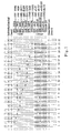
- Fig. 1 shows some possible staining patterns obtainable with DNAs carrying different mutant and wild-type HFE or TFR2 alleles using reverse hybridization as explained in example 2.
- Fig. 2a, 2b , 2c and 2d show the pedigrees of the four families (filled symbols affected, open symbols unaffected).
- Fig.3a, 3b and 3c show sequencing chromatographs of sequences of the TFR2 gene of exon 2, spanning the C insertion which originates the E60X mutation, of exon 4 spanning the M172K mutation at position 515 and of exon 6 spanning the Y250X mutation at position 750.
- Fig.4 shows a schematic representation of TFR2 structure in the 5'region of the gene.
- DNA was isolated from anticoagulated blood using standard extraction methods /10/ or commercially available reagents (GenXtract DNA extraction system, ViennaLab). HFE gene exon 3 sequences were amplified in a PCR reaction using primers 5'-GGA TTG GAG AGC AGC AGA AC-3' and 5'-AAA GTC CAA CCA GGG ATT CC-3'. Each primer was used at a final concentration of 0.4 ⁇ M in 1xPCR buffer containing 1.5mM Mg2+, 200 ⁇ M dNTPs (Epicentre, Madison, WI) and 0.02 U/ ⁇ l AmpliTaq DNA polymerase (PE Biosystems, Foster City, CA). A thermocycling program of 30 cycles (94°C for 15s, 58°C for 30s, and 72°C for 30s) was performed on the GeneAmp PCR System 2400 (PE Biosystems).
- PCR products were purified with "Centricon-100" columns according to the manufacturers recommendations. Amplified and purified DNA was quantitated by UV-spectrophotometry and subsequently sequenced strictly following the protocol described in the "BigDye Terminator Cycle Sequencing Ready Reaction Kit - with AmpliTaq DNA Polymerase, FS" (PE Applied Biosystems) for the ABI Prism instrumentation (PE Applied Biosystems).
- the test is based on multiplex DNA amplification and ready-to-use membrane teststrips, which contain oligonucleotide probes for each wild-type and mutated allele immobilized as an array of parallel lines.
- the test is rapid, easy to perform and accessible to automation on commercially available equipment, and by adding new probes the teststrip can easily be adapted to cover an increasing number of mutations.
- DNA was isolated from anticoagulated blood using standard extraction methods /10/ or commercially available reagents (GenXtract DNA extraction system, ViennaLab). HFE gene exon 2, 3 and 4 sequences and TFR2 gene exon 2, 4 and 6 sequences were amplified in a single, multiplex PCR reaction using 5'-biotinylated primers (/1,31,32/; table 3). Each primer was used at a final concentration of 0.4 ⁇ M in 1x PCR buffer including 1.5 mM Mg2+, 200 ⁇ M dNTPs (Epicentre, Madison WI), and 0.02 U/ ⁇ l AmpliTaq DNA polymerase (PE Biosystems, Foster City, CA). A thermocycling program of 30 cycles (94°C for 15 s, 58°C for 30 s and 72°C for 30 s) was performed on the GeneAmp PCR System 2400 (PE Biosystems).
- Probes (16-24 mers) specific for the wild-type or mutant allele of each mutation were selected from the HFE and TFR2 databank sequences (GenBank accession numbers: HFE-Z92910, TFR2-AF067864) and synthesized using standard phosphoramidite chemistry.
- a 3'-poly(dT) tail (300-500 residues) was enzymatically added by incubating oligonucleotides (200 pmol) in the presence of 60 U terminal deoxynucleotidyl transferase (Amersham Pharmacia, Buckinghamshire, UK) and 150 nmol dTTP (Epicentre) for 8 h at 37°C. The reaction was terminated by adding EDTA to 10 mM, and tailing efficiency was monitored by agarose gel electrophoresis.
- Poly(dT)-tailed probes were applied onto nitrocellulose membrane sheets (Whatman, Maidstone, UK) as individual parallel lines of 1 mm width using a custom-made slot-blot apparatus, dried and fixed by overnight baking at 60°C.
- a 5'-biotinylated control oligonucleotide was included to allow performance control of the detection reagents.
- the membrane sheet was finally sliced into 3 mm ready-to-use teststrips.
- the entire hybridization and detection procedure was carried out in individual lanes of a thin-walled plastic incubation tray (Bio-Rad, Hercules, CA). Equal volumes (10 ⁇ l) of PCR product and denaturing solution (400 mM NaOH, 10 mM EDTA) were mixed and incubated at room temperature for 5 min. The alkaline reaction was neutralized with sodium phosphate buffer, pH 7, a teststrip was added and hybridization was carried out for 30 min. at 45°C in a shaking waterbath. After three stringent washes at 45°C, bound PCR fragments were detected using a streptavidin-alkaline phosphatase conjugate and color substrates (NBT/BCIP). Upon positive reaction, a purple staining was visible after 15 min. incubation at room temperature.
- NBT/BCIP streptavidin-alkaline phosphatase conjugate and color substrates
- a multiplex PCR system was established to obtain biotinylated DNA fragments spanning exons 2, 3 and 4 of the HFE gene and exons 2, 4 and 6 of the TFR2 gene in a single amplification reaction.
- HFE V53M, V59M, H63D, H63H, S65C, Q127H, E168Q, E168X, W169X, C282Y, Q283P
- TFR2 E60X, M172K and Y250X
- Table 1 Line Probe specificity Mutation designation Reference 1 (control) 2 nt 157 G ⁇ A exon 2 HFE V53M V Amsterdam et al. /7/ 3 nt 175 G ⁇ A exon 2 HFE V59M V Amsterdam et al. /7/ 4 nt 187 C ⁇ G exon 2 HFE H63D Feder et al.
- a variety of candidate probes between 16 to 24 nucleotides in length encoding either the wild-type or mutant allele of each mutation were chosen from the GenBank sequences for HFE and TFR2. After enzymatic poly(dT)-tailing the oligonucleotides were applied to a nitrocellulose membrane as an array of parallel lines and fixed by baking. Finally, the membrane sheet was sliced into individual teststrips. Amplification products from different DNA samples covering all 14 mutations were hybridized to these preliminary teststrips under identical and accurately controlled stringency conditions. Bound biotinylated sequences were detected using a streptavidin-enzyme conjugate and color reaction.
- Fig. 1 shows some possible staining patterns obtainable with DNAs carrying different mutant and wild-type HFE or TFR2 alleles. The following samples were used:
- the staining pattern of a mutant and its corresponding wild-type probe allows the discrimination of three possible genotypes: the mutation is absent if only the wild-type probe stains positive, both signals are visible in heterozygotes, and only the mutant probe is positive in homozygous mutant samples.
- the same principle applies to the majority of compound heterozygotes for two mutations ( Fig. 1 , lanes 8-12, 14-16).
- the HFE gene contains at least two regions, one in exon 2 (nt 187-193) and the other in exon 3 (nt 502-506), where several mutations have been found in close proximity. As the hybridization probes span more than one mutation in this area, no wild-type signal is observed if two mutations from the same cluster (e.g.
- H63D and S65C occur on different chromosomes in the same sample ( Fig. 1 , lane 8). The occurance of two neighbouring mutations on a single chromosome would again lead to a different pattern, but has not been described to date.
- the reverse-hybridization assay presented here has the potential of becoming a valuable tool for routine diagnosis of multiple HFE and/or TFR2 mutations.
- the procedure is simple, reliable, and can be accomplished within a few hours from blood drawing to final results.
- testing can be automated on existing, commercially available equipment, such as the profiBlot II T (TECAN AG, Hombrechtikon, Switzerland).
- the number of mutations covered by the present assay can easily be extended using the same approach to select additional hybridization probes.
- HIC Hepatic iron concentration
- HII hepatic iron index
- genomic DNA was extracted from peripheral blood leukocytes. C282Y and H63D mutations were detected using standard polymerase chain reaction (PCR) and restriction enzyme digestion with Rsa I and Bcl I, respectively /18/. Microsatellites D6S265, D6S105 and D6S1281 were analysed as described /26/. HLA-A antigens were defined by the microlymphotoxicity test. Haplotypes were constructed manually on the basis of intrafamilial segregation of the marker alleles.
- the entire coding sequence and exon-intron boundaries of the HFE gene were amplified by PCR according to Carella et al /18/.
- the nucleotide sequence of both DNA strands was independently determined in all the probands and in relatives with the C282Y negative chromosome identical to the proband by using the dideoxy chain-termination reaction with Sequenase version 2.0 (USB Amersham, Cleveland,Ohio) and 35 S- ⁇ -dATP, according to the protocol recommended by the manufacturer. Reactions were separated on a 6% denaturing polyacrylamide gel and visualised by autoradiography. The primers employed for sequencing were the same used for PCR reactions.
- RFLP restriction length polymorphism analysis
- TS transferrin saturation
- SF serum ferritin
- HIC hepatic iron concentration
- HII hepatic iron index
- IR iron removed
- C hepatic cirrhosis
- H hypogonadism
- A arthropathy
- D diabetes.
- Table 2 reports iron and clinical data of the 5 probands.
- Three patients (1, 2 and 3) originated from the same subalpine valley in the north west (Ossola) and two (4 and 5) from an area around the city of Monza (Brianza).
- Sequence analyses of the HFE gene in probands showed two novel nonsense mutations in exon 3 in the heterozygous state. The first one, a G ⁇ T mutation, at the nucleotide 502 of the HFE cDNA sequence (codon 168) (GAG ⁇ TAG, Glu ⁇ Stop) was found in probands from the Ossola valley.
- the second, a G ⁇ A mutation at nucleotide 506 of HFE cDNA sequence (codon 169) (TGG ⁇ TAG, Trp ⁇ Stop). was detected in probands from Brianza region.
- the new variants were named HFE-Ossola and HFE-Brianza from the origin of the patients.
- HFE-Ossola was associated with haplotype D6S265-3, HLA-A24, D6S105-5 and D6S1281-6 in two families, whereas in the third family, the haplotype changed at D6S265 and HLA-A loci suggesting that a recombination event occurred between HLA-A and D6S105.
- HFE-Brianza was associated with haplotype D6S265-3, HLA-A24, D6S105-6 and D6S1281-6.
- the two mutations determine a stop codon at nucleotide 502 and 506 of the open reading frame (nt 502 GAG ⁇ TAG and nt 506 TGG ⁇ TAG) respectively, leading to proteins that lack the ⁇ 3 domain, the transmembrane domain and the cytoplasmic tail.
- These mutations probably produce, differently from C282Y, a complete disruption of the function of HFE similar to that induced by the partial deletion of exon 4 in HFE-deficient mice /27,28/.
- these mutated alleles may behave as a null allele. It is expected that the combination of these mutations in the compound heterozygous state with C282Y leads to severe phenotype expression.
- proband 3 had a mild phenotype probably due to the fact he was a blood donor for the last 10 years, whereas proband 1 and his affected brother had a relatively mild phenotype and no other obvious protective factors such as blood loss or malabsorption.
- phenotype variability exists in these patients and could be related to both environmental and genetic factors.
- the existence of modifying genes located around the D6S105 region which influence phenotypic expression of C282Y homozygous HH patients has been recently proposed /25,29/, but further studies are needed to clarify genotype-phenotype correlations in HH patients.
- HFE-Ossola and HFE-Brianza mutations did not have elevated serum iron indices indicating that neither mutation was able to produce iron overload in the heterozygous state.
- the three compound heterozygotes for either HFE-Ossola or HFE-Brianza and H63D did not show evidence of iron overload. Since they were females and/or young persons, it is possible that, as occur for C282Y, the combination of the two nonsense mutations with H63D or other mild HFE mutations may produce a HH phenotype with low penetrance /17/.
- C282Y/HFE-Ossola compound heterozygotes account for 25% of HH probands in the Ossola region (Vs 8.3% of C282Y/H63D), and C282Y/HFE-Brianza and C282Y/H63D compound heterozygotes each account for 8.4% of HH probands in Brianza region.
- DNA was prepared from peripheral blood buffy coats or patient's lymphoblastoid cell lines by standard phenolclorophorm extraction /38/.
- C282Y and H63D mutations in HFE were studied on genomic DNA using PCR-based tests and restriction enzyme digestion with RsaI and MboI (New England Biolabs, Berkeley, MA) respectively /18/.
- Linkage to the HFE3 locus was established in family 3 analyzing microsatellite D7S651, D7S2498, D7S662, D7S477, D7S1588 allele segregation and two TFR2 intragenic repeats (R1 and R2). Pairwise linkage analysis was performed using the MLINK program from the LINKAGE computer package, as previously described /39/.
- PCR was performed in a Thermal Cycler, using 10 pMol of each primer, with an protocol of 32 cycles (denaturation: 94°C 30", annealing: 56°C 45", extension: 72°C 45") and 1 U of AmpliTaq DNApolymerase (Perkin Elmer).
- PCR primers for TFR2 exon amplification were obtained from databases and are reported in Table 3. Table 3 . Sequence of TFR2 primers used in the PCR reactions.
- MaeI enzyme was used to detect Y250X mutant on the amplification product of exon 6. Restriction enzyme analysis of the PCR products was performed according to the manufacturer recommendations.
- RNA-SSCP was performed according to previously described protocols /38, 40/. After PCR reaction, transcription was carried out with 10U of T7RNA polymerase in a final volume of 10 ⁇ l containing 10mM DTT, 40mM Tris pH 7.5, 6mM MgCl, 2mM spermidine, 10mM NaCl, 5 nmol of each ribonucleoside, 10U of RNAse and 0.2 ⁇ l of S35 UTP. After electrophoresis, gels were dried and subjected to autoradiography. Bands showing an electrophoretically altered mobility were directly sequenced.
- TFR2 The gene TFR2 was recently isolated and mapped to 7q22 by radiation hybrids /37/. TFR2 shows 66% homology to the transferrin receptor (encoded by TFRC) in its extracellular domain, binds transferrin and is presumed to mediate cellular iron uptake /37/. Using available sequence information, two intragenic polymorphic repeats, (R1 and R2) were identified and homozygosity for these markers was detected in all affected individuals. TFR2 was mapped within the homozygosity region. The TFR2 coding sequence and exon-intron boundaries for mutations were scanned and a C ⁇ G transversion in exon 6 at position 750 .
- Fig.3c shows sequencing chromatographs of the forward sequence of exon 6 spanning the C750G (Y250X) mutation. Subject V-11 (+/+) and VII-1 (+/+) are shown compared to a normal control. The mutation is indicated by an arrow. The Y250X substitution creates a MaeI site. The amplified TFR2 exon 6 was digested with MaeI to analyse segregation of the mutation in family 1. All affected members of family 1 were homozygous for Y250X, whereas obligate carriers were heterozygous. Patient II-3 of family 2 (in which no consanguinity was reported) was homozygous for Y250X. The Y250X mutation was not found in 100 normal chromosomes or in 12 patients, who did not have mutations in HFE.
- FIG. 3a shows sequencing chromatographs of the forward sequence of exon 2 spanning the C insertion which originates the E60X mutation.
- Subjects VI-6(+/+) and VI-2(+/-) are shown compared to a normal control (-/-). The position of the insertion is indicated by the arrow.
- the C insertion was investigated by sequencing DNA of all family members available.
- Subjects VI-2, VI-3, VI-4 and VI-5 had the mutation at the homozygous state (Table 4) and subjects VI-4, VI-1, VI-2, VI-6, VII-3 and VII-4 had the mutation at the heterozygous state (Table 5). The same mutation was not found in 50 normal DNA samples analyzed by RNA-SSCP.
- Fig.3b shows sequencing chromatographs of the forward sequence of exon 4 spanning the T515A (M172K) mutation. Subjects II-2(+/+) and I-2(+/-) are shown compared to a normal control (-/-). The mutation is indicated by the arrow.
- This nucleotide change results in a lysine for methionine substitution at position 172 of the protein (M172K).
- T515A segregation within the family was studied by direct sequencing. I-2, III-1 and III-2 were heterozygous for the mutation and II-1 had the normal genotype. The same mutation was not found by RNA-SSCP amoung 50 normal controls.
- RT-PCR was performed on total RNA obtained from peripheral blood buffy coats and lymphoblastoid cell lines (LCL) of patients with different TFR2 substitutions and in a normal control.
- LCL lymphoblastoid cell lines
- attempts at amplifying the ⁇ -transcripts by RT-PCR using RNA from buffy coats or LCL were unsuccessful. Fragments corresponding to the ⁇ -transcript were obtained in the normal control and in all patients, except in Y250X homozygotes (not shown).
- VI-4 and V-2 were undiagnosed and their identification as E60X homozygotes was obtained through family studies (Table 4). VI-4, a premenopausal woman, had remarkably low transferrin saturation and serum ferritin. The finding of iron deficiency without anemia was unexpected in an homozygous mutant subject. Low dietary iron intake and long lasting blood losses through menses in the absence of iron supplementation were identified as the causes of iron deficiency. No history of other blood losses, nor evidence of a hemostasis defect were recorded, but the patient refused a thorough gastrointestinal endoscopic investigation. V-2 had altered iron parameters, abnormal liver function tests and severe arthritis, but never had phlebotomies. His older brother (V-1) was not available for the study, but was reported to be under a regular phlebotomy treatment.
- HFE3 carriers were parents or children of the patients and/or siblings with a documented TFR2 mutation. Clinical data and iron parameters of 15 HFE3 heterozygotes are shown in Table 5. All carriers had normal transferrin saturation and serum ferritin, except V-9 of familiy 1. This subject, who showed increased transferrin saturation and serum ferritin, was affected by HCV chronic hepatitis and underwent occasional phlebotomies. In all the other cases the condition of HFE3 heterozygosity, even in combination with H63D at the heterozygous state (1 case) or with ⁇ -thalassemia trait (3 cases), was not associated with iron overload.
- TFR2 as the HFE3 gene.
- Scanning for mutations the coding sequence of TFR2 a C insertion (84-88insC), which causes a premature stop codon (E60X) in exon 2 was found at the homozygous state in five affected subjects and in one non-expressing, iron deficient female, from an inbred family.
- the presence of HFE3 homozygotes in two consecutive generations in family 3 witnesses the high degree of inbreeding.
- the homozygote-heterozygote mating (between V-2 and V-4) explains the absence of wild type TFR2 in the last but one generation, and the seemingly dominant inheritance in this branch of the family.
- HFE C282Y mutations H63D Y250X TFR2 mutations E60X M172K Family 1 V-2 M 68 36 97 166 -/- -/- +/- V-9 n M 70 74 310 159 -/- -/- +/- VI-1 F 59 27 79 142 +/- * F 17 38 47 155 +/- * ⁇ M 13 25 58 132 +/- * M 7 34 22 145 +/- Family 3 V-4 F 78 ND 32 ND -/- -/- +/- VI-1 F 50 33 19 ND -/- -/- +/- VI-2 F 46 15 30 128 -/- -/- +/- VI-6 M 34 26 183 156 -/- -/- +/- VII-3 M 27 49 84 147 -/-/-
- ⁇ -transcript lacks exon 1-3 and has additional nucleotides at the 5'end (dotted box) as compared to the ⁇ -transcript. The position of the mutations identified and their possible effect on the proteins are shown.
- ⁇ V indicates the variant with the aminoacid change M172K.
- E60X occurs in exon 2 and disrupts the predicted ⁇ -TFR2 transcript, but at variance with Y250X, does not interfere with the ⁇ -transcript.
- T515A causes a missense in the ⁇ -protein (M172K).
- M172K The methionine at position 172 is conserved in the mouse and the substitution of a basic for a neutral aminoacid might change the ⁇ -protein properties.
- the same nucleotide substitution affects also the putative initiation codon of the ⁇ -variant, preventing its translation.
- TFR2 as the HFE3 gene is of relevance to the issue of modifier genes in hemochromatosis.
- modifier genes that may modulate (either ameliorate or worsen) the phenotype has been demonstrated in mice and hypothesized in humans.
- TFR2 is the first obvious modifier to be investigated in C282Y homozygotes.
Landscapes
- Chemical & Material Sciences (AREA)
- Life Sciences & Earth Sciences (AREA)
- Proteomics, Peptides & Aminoacids (AREA)
- Health & Medical Sciences (AREA)
- Organic Chemistry (AREA)
- Wood Science & Technology (AREA)
- Analytical Chemistry (AREA)
- Zoology (AREA)
- Genetics & Genomics (AREA)
- Engineering & Computer Science (AREA)
- Pathology (AREA)
- Immunology (AREA)
- Microbiology (AREA)
- Molecular Biology (AREA)
- Biotechnology (AREA)
- Biophysics (AREA)
- Physics & Mathematics (AREA)
- Biochemistry (AREA)
- Bioinformatics & Cheminformatics (AREA)
- General Engineering & Computer Science (AREA)
- General Health & Medical Sciences (AREA)
- Measuring Or Testing Involving Enzymes Or Micro-Organisms (AREA)
Applications Claiming Priority (3)
| Application Number | Priority Date | Filing Date | Title |
|---|---|---|---|
| AT7662000 | 2000-05-02 | ||
| AT0079900A AT412282B (de) | 2000-05-02 | 2000-05-08 | Verfahren und sonde zur diagnose von hämochromatose |
| EP01933906A EP1278891B1 (de) | 2000-05-02 | 2001-04-30 | Verfahren und sonden zur diagnose von hämochromatose |
Related Parent Applications (1)
| Application Number | Title | Priority Date | Filing Date |
|---|---|---|---|
| EP01933906A Division EP1278891B1 (de) | 2000-05-02 | 2001-04-30 | Verfahren und sonden zur diagnose von hämochromatose |
Publications (1)
| Publication Number | Publication Date |
|---|---|
| EP2110441A1 true EP2110441A1 (de) | 2009-10-21 |
Family
ID=25608405
Family Applications (2)
| Application Number | Title | Priority Date | Filing Date |
|---|---|---|---|
| EP01933906A Expired - Lifetime EP1278891B1 (de) | 2000-05-02 | 2001-04-30 | Verfahren und sonden zur diagnose von hämochromatose |
| EP09165173A Withdrawn EP2110441A1 (de) | 2000-05-02 | 2001-04-30 | Verfahren und Sonden zur Diagnose von Hämochromatose |
Family Applications Before (1)
| Application Number | Title | Priority Date | Filing Date |
|---|---|---|---|
| EP01933906A Expired - Lifetime EP1278891B1 (de) | 2000-05-02 | 2001-04-30 | Verfahren und sonden zur diagnose von hämochromatose |
Country Status (9)
| Country | Link |
|---|---|
| US (1) | US20040086862A1 (de) |
| EP (2) | EP1278891B1 (de) |
| AT (3) | AT412282B (de) |
| AU (2) | AU6026301A (de) |
| CA (1) | CA2408263A1 (de) |
| DE (2) | DE20180363U1 (de) |
| ES (1) | ES2333518T3 (de) |
| NZ (1) | NZ522521A (de) |
| WO (1) | WO2001083812A2 (de) |
Families Citing this family (2)
| Publication number | Priority date | Publication date | Assignee | Title |
|---|---|---|---|---|
| GB2459099A (en) * | 2008-04-08 | 2009-10-14 | Ethicon Inc | Genetic markers of wound development |
| CN107760780A (zh) * | 2017-12-05 | 2018-03-06 | 天津脉络医学检验有限公司 | 一种检测儿童铁吸收基因多态性的扩增引物及应用 |
Citations (1)
| Publication number | Priority date | Publication date | Assignee | Title |
|---|---|---|---|---|
| WO1998014466A1 (en) * | 1996-10-01 | 1998-04-09 | Progentior, Inc. | Polymorphisms and new genes in the region of the human hemochromatosis gene |
Family Cites Families (2)
| Publication number | Priority date | Publication date | Assignee | Title |
|---|---|---|---|---|
| US6025130A (en) * | 1996-04-04 | 2000-02-15 | Mercator Genetics, Inc. | Hereditary hemochromatosis gene |
| US20030092019A1 (en) * | 2001-01-09 | 2003-05-15 | Millennium Pharmaceuticals, Inc. | Methods and compositions for diagnosing and treating neuropsychiatric disorders such as schizophrenia |
-
2000
- 2000-05-08 AT AT0079900A patent/AT412282B/de not_active IP Right Cessation
-
2001
- 2001-04-30 EP EP01933906A patent/EP1278891B1/de not_active Expired - Lifetime
- 2001-04-30 AU AU6026301A patent/AU6026301A/xx active Pending
- 2001-04-30 DE DE20180363U patent/DE20180363U1/de not_active Expired - Lifetime
- 2001-04-30 CA CA002408263A patent/CA2408263A1/en not_active Abandoned
- 2001-04-30 DE DE60139929T patent/DE60139929D1/de not_active Expired - Lifetime
- 2001-04-30 AT AT01933906T patent/ATE443157T1/de active
- 2001-04-30 WO PCT/EP2001/004835 patent/WO2001083812A2/en not_active Ceased
- 2001-04-30 NZ NZ522521A patent/NZ522521A/en unknown
- 2001-04-30 AT AT0904101U patent/AT6806U1/de not_active IP Right Cessation
- 2001-04-30 EP EP09165173A patent/EP2110441A1/de not_active Withdrawn
- 2001-04-30 ES ES01933906T patent/ES2333518T3/es not_active Expired - Lifetime
- 2001-04-30 US US10/275,453 patent/US20040086862A1/en not_active Abandoned
- 2001-04-30 AU AU2001260263A patent/AU2001260263B2/en not_active Ceased
Patent Citations (1)
| Publication number | Priority date | Publication date | Assignee | Title |
|---|---|---|---|---|
| WO1998014466A1 (en) * | 1996-10-01 | 1998-04-09 | Progentior, Inc. | Polymorphisms and new genes in the region of the human hemochromatosis gene |
Non-Patent Citations (49)
| Title |
|---|
| BACON B.R. ET AL.: "Molecular medicine and hemochromatosis: at the crossroads", GASTROENTEROLOGY, vol. 116, 1999, pages 193 - 207 |
| BARTON J C ET AL: "TWO NOVEL MISSENSE MUTATIONS OF THE HFE GENE (I05T AND G93R) AND IDENTIFICATION OF THE S65C MUTATION IN ALABAMA HEMOCHROMATOSIS PROBANDS", BLOOD CELLS, MOLECULES AND DISEASES, LAJOLLA, US, vol. 25, no. 9, 1999, pages 147 - 155, XP000929556, ISSN: 1079-9796 * |
| BARTON J.C. ET AL.: "Two novel missense mutations of the HFE gene (I105T and G93R) and identification of the S65C mutation", ALABAMA HEMOCHROMATOSIS PROBANDS |
| BEUTLER E. ET AL.: "Mutation analysis in hereditary hemochromatosis", BLOOD CELLS MOL DIS, vol. 22, 1996, pages 187 - 94 |
| BEUTLER E.: "Genetic iron beyond haemocromatosis: clinical effects of HLA-H mutations", LANCET, vol. 349, 1997, pages 296 - 7 |
| BEUTLER E.: "The significance of 187G (H63D) mutation in hemochromatosis", AM J HUM GENET, vol. 61, 1997, pages 762 - 4 |
| BLOOD CELLS MOL DIS, vol. 25, 1999, pages 146 - 54 |
| BOLLHALDER M. ET AL.: "LightCycler PCR assay for simultaneous detection of the H63D and S65C mutations in the HFE hemochromatosis gene based on opposite melting temperature", CLIN. CHEM., vol. 45, 1999, pages 2275 - 2278 |
| CAMASCHELLA C. ET AL.: "Inherited HFE-unrelated hemochromatosis in Italian families", HEPATOLOGY, vol. 29, 1999, pages 1563 - 4 |
| CAMASCHELLA C. ET AL.: "Juvenile and adult hemochromatosis are distinct genetic disorders", EUR J HUM GENET, vol. 5, 1997, pages 371 - 5 |
| CAMASCHELLA C.; PIPERNO A.: "Hereditary Hemochromatosis: recent advances in molecular genetics and clinical management", HAEMATOLOGICA, vol. 82, 1997, pages 77 - 84 |
| CARELLA M. ET AL.: "Mutation analysis of HLA-H gene in Italian hemochromatosis patients", AM J HUM GENET, vol. 60, 1997, pages 828 - 32 |
| CHEHAB F.F.; WALL J.: "Detection of multiple cystic fibrosis mutations by reverse dot blot hybridization: a technology for carrier screening", HUM. GENET., vol. 89, 1992, pages 163 - 168 |
| CHRISTIANSEN L. ET AL.: "Denaturing gradient gel electrophoresis analysis of the hemochromatosis (HFE) gene: impact of HFE gene mutations on the manifestation of porphyria cutanea tarda", CLIN. CHEM., vol. 45, 1999, pages 2025 - 2026 |
| DE VILLIERS J. ET AL: "Spectrum of mutations in the HFE gene implicated in heamochromatosis and porphyria", HUMAN MOLECULAR GENETICS, vol. 8, no. 8, August 1999 (1999-08-01), pages 1517 - 22, XP002175621 * |
| DOUABIN V ET AL: "POLYMORPHISMS IN THE HFE GENE", HUMAN HEREDITY, KARGER, BASEL, CH, vol. 49, no. 1, January 1999 (1999-01-01), pages 21 - 26, XP000983474, ISSN: 0001-5652 * |
| DOUABIN V. ET AL.: "Polymorphisms in the HFE gene", HUM. HERED., vol. 49, 1999, pages 21 - 26 |
| FEDER J.N. ET AL.: "A novel MHC class I-like gene is mutated in patients with hereditary haemochromatosis", NAT GENET, vol. 13, 1996, pages 399 - 408 |
| JAZWINSKA, E.C. ET AL.: "Haemochromatosis and HLA-H", NATURE GENET., vol. 14, 1997, pages 249 - 251 |
| JEFFREY G.P. ET AL.: "Polymorphism in intron 4 of HFE may cause overestimation of C282Y homozygote prevalence in haemochromatosis", NATURE GENET., vol. 22, 1999, pages 325 - 326 |
| JOUANOLLE A.M. ET AL.: "Haemochromatosis and HLA-H.", NAT GENET, vol. 14, 1996, pages 251 - 2 |
| KAWABATA H.; YANG R.; HIRAMA T. ET AL.: "Molecular cloning of Transferrin receptor 2", J.BIOL CHEM., vol. 274, 1999, pages 20826 - 20832 |
| LEBRON J.A.; BENNETT M.J.; VAUGHN D.E. ET AL.: "Crystal structure of the hemochromatosis protein HFE and characterization of its interaction with transferrin receptor", CELL, vol. 93, 1998, pages 111 - 123 |
| LEVY J.E. ET AL.: "The C282Y mutation causing hereditary hemochromatosis does not produce a null allele", BLOOD, vol. 94, 1999, pages 9 - 11 |
| MAGGIO A. ET AL.: "Rapid and simultaneous typing of hemoglobin S, hemoglobin C, and seven Mediterranean (-thalassemia mutations by covalent reverse dot-blot analysis: application to prenatal diagnosis in Si- cily", BLOOD, vol. 81, 1993, pages 239 - 242 |
| MANGASSER-STEPHAN K. ET AL.: "Rapid genotyping of haemochromatosis gene mutations on the LightCycler with fluorescent hybridization probes", CLIN. CHEM., vol. 45, 1999, pages 1875 - 1878 |
| MILLER S.A.; DYKES D.D.; POLESKY H.F.: "A simple salting out procedure for extracting DNA from human nucleated cells", NUCLEIC ACIDS RES., vol. 16, 1988, pages 1215 |
| MURA C ET AL: "HFE mutations analysis in 711 hemochromatosis probands: evidence for S65C implication in mild form of hemochromatosis", BLOOD, W.B.SAUNDERS COMPAGNY, ORLANDO, FL, US, vol. 93, no. 8, 15 April 1999 (1999-04-15), pages 2502 - 2505, XP002143645, ISSN: 0006-4971 * |
| MURA C.; RAGUENES 0.; FEREC C.: "HFE mutations analysis in 711 hemochromatosis probands: evidence for S65C implication in mild form of hemochromatosis", BLOOD, vol. 93, 1999, pages 2502 - 5 |
| OBERKANINS C. ET AL.: "Genotyping of the hereditary haemochromatosis mutation (HLA-H:C282Y) in microtiter plates", AM. J. HUM. GENET., vol. 61, 1997, pages A225 |
| O'DONNELL-MALONEY M J; SMITH C L; CANTOR C R: "The development of microfabricated arrays for DNA sequencing and analysis", TRENDS IN BIOTECHNOLOGY, ELSEVIER PUBLICATIONS, CAMBRIDGE, GB, vol. 14, no. 10, 1 October 1996 (1996-10-01), pages 401 - 407, XP004035731, ISSN: 0167-7799 * |
| PIETRANGELO A. ET AL.: "Hereditary Hemochromatosis in adults without pathogenic mutations in the hemochromatosis gene", NEW ENGL J MED, vol. 341, 1999, pages 725 - 32 |
| PIETRANGELO A.; CAMASCHELLA C.: "Molecular genetics and control of iron metabolism in Hemochromatosis", HAEMATOLOGICA, vol. 83, 1998, pages 456 - 61 |
| PIPERNO A. ET AL.: "Heterogeneity of hemochromatosis in Italy", GASTROENTEROLOGY, vol. 114, 1998, pages 996 - 1002 |
| PIPERNO A. ET AL.: "The ancestral hemochromatosis haplotype is associated with a severe phenotype expression in italian patients", HEPATOLOGY, vol. 24, 1996, pages 43 - 6 |
| PRATIWI R. ET AL.: "Linkage disequilibrium analysis in Australian haemochromatosis patients indicates bipartite association with clinical expression", JOURNAL OF HEPATOLOGY, vol. 31, 1999, pages 39 - 46 |
| ROETTO A.; TOTARO A.; CAZZOLA M. ET AL.: "The Juvenile Hemochromatosis Locus Maps To Chromosome lq", AM J HUM GENET., vol. 64, 1999, pages 1388 - 1393 |
| RUBINI M ET AL: "A novel mutation of HFE explains the phenotype of hereditary hemochromatosis in a C282Y carrier", EUROPEAN JOURNAL OF HUMAN GENETICS, vol. 8, no. Suppl 1, 27 February 2000 (2000-02-27), European Human Genetics Conference 2000 Amsterdam May 27-Feb. 30, pages 158, XP001016153 * |
| RUBINI, M. ET AL., HUM.GENET., vol. 8, no. 1, 2000, pages 158 |
| SAIKI R.K. ET AL.: "Genetic analysis of amplified DNA with immobilized sequence-specific oligonucleotide probes", PROC. NATL. ACAD. SCI. USA, vol. 86, 1989, pages 6230 - 6234 |
| SAMBROOK J.; FRISCH E.; MANIATIS T.: "Molecular cloning: a laboratory manual", vol. 1-3, 1989, COLD SPRING HARBOR LABORATORY PRESS |
| SANCHEZ ET AL: "PREVALENCE OF THE Cys282Tyr AND His63Asp HFE GENE MUTATIONS IN SPANISH PATIENTS WITH HEREDITARY HEMOCHROMATOSIS", JOURNAL OF HEPATOLOGY, XX, XX, vol. 29, no. 5, November 1998 (1998-11-01), pages 725 - 728, XP002113587 * |
| SARKAR G.; YOON H.S.; SOMMER S.: "Screening for mutations by RNA single strand conformation polymorphisms (rSSCP): comparison with DNA-SSCP", NUCLEIC ACIDS RES., vol. 20, 1992, pages 871 - 878 |
| SCHEUER P.J.; WILLIAMS R.; MUIR A.R.: "Hepatic pathology in relatives of patients with hemochromatosis", J. PATHOL BACTERIOL, vol. 84, 1962, pages 53 - 64 |
| STEFFENSEN R.; VARMING K.; JERSILD C.: "Determination of gene frequencies for two common haemochromatosis mutations in the Danish population by a novel polymerase chain reaction with sequence-specific primers", TISSUE ANTIGENS, vol. 52, 1998, pages 230 - 235 |
| STUYVER L. ET AL.: "Typing of hepatitis C virus isolates and characterization of new subtypes using a line probe assay", J. GEN. VIROL., 1993 |
| VILLIERS J.N. ET AL.: "Spectrum of mutations in the HFE gene implicated in haemochromatosis and porphyria", HUM MOL GENET, vol. 8, 1999, pages 1517 - 22 |
| WALLACE D.F.; DOOLEY J.S; WALKER A.P.: "A novel mutation of HFE explains the classical phenotype of genetic hemochromatosis in a C282Y heterozygote", GASTROENTEROLOGY, vol. 116, 1999, pages 1409 - 12 |
| ZHOU X.Y. ET AL.: "HFE gene Knockout produces mouse model of hereditary hemochromatosis", PROC NATL ACAD SCI USA, vol. 95, 1998, pages 2492 - 7 |
Also Published As
| Publication number | Publication date |
|---|---|
| ATA7992000A (de) | 2004-05-15 |
| DE20180363U1 (de) | 2003-01-09 |
| ES2333518T3 (es) | 2010-02-23 |
| AT412282B (de) | 2004-12-27 |
| AU6026301A (en) | 2001-11-12 |
| ATE443157T1 (de) | 2009-10-15 |
| US20040086862A1 (en) | 2004-05-06 |
| AT6806U1 (de) | 2004-04-26 |
| WO2001083812A3 (en) | 2002-04-11 |
| EP1278891B1 (de) | 2009-09-16 |
| EP1278891A2 (de) | 2003-01-29 |
| AU2001260263B2 (en) | 2005-07-21 |
| CA2408263A1 (en) | 2001-11-08 |
| WO2001083812A2 (en) | 2001-11-08 |
| DE60139929D1 (de) | 2009-10-29 |
| NZ522521A (en) | 2004-06-25 |
Similar Documents
| Publication | Publication Date | Title |
|---|---|---|
| US5750400A (en) | Coding sequences of the human BRCA1 gene | |
| EP0256630A1 (de) | Spezifische Sonde zur Identifikation von Mutationen in den humanen Phenylalaningenen | |
| AU779477B2 (en) | Alterations in the long QT syndrome genes KVLQT1 and SCN5A and methods for detecting same | |
| JPH11504809A (ja) | 精神分裂症の診断方法 | |
| Oberkanins et al. | A reverse-hybridization assay for the rapid and simultaneous detection of nine HFE gene mutations | |
| Kenney et al. | Characterization of the p67phox gene: genomic organization and restriction fragment length polymorphism analysis for prenatal diagnosis in chronic granulomatous disease | |
| EP1130123A2 (de) | Diagnosemethode | |
| JP2003513620A (ja) | Rhd陰性遺伝子座の分子構造 | |
| Frank et al. | Identification of a founder mutation in the protoporphyrinogen oxidase gene in variegate porphyria patients from Chile | |
| Cama et al. | Multiplex PCR analysis and genotype–phenotype correlations of frequent APC mutations | |
| EP2110441A1 (de) | Verfahren und Sonden zur Diagnose von Hämochromatose | |
| Shimokawa et al. | Analysis of the glucokinase gene promoter in Japanese subjects with noninsulin-dependent diabetes mellitus | |
| AU2001260263A1 (en) | Method and probes for the genetic diagnosis of hemochromatosis | |
| Beutler et al. | A common intron 3 mutation (IVS3− 48c→ g) leads to misdiagnosis of the c. 845G→ A (C282Y) HFE gene mutation | |
| TW201311908A (zh) | 診斷犬之青光眼的方法及套組 | |
| RU2722564C1 (ru) | Молекулярно-генетические маркеры цветовых вариаций американской норки и способ выявления особей, являющихся носителем аллелей, обуславливающих формирование желаемой цветовой вариации | |
| US7833710B2 (en) | Polynucleotide associated with breast cancer comprising single nucleotide polymorphism, microarray and diagnostic kit comprising the same and method for diagnosing breast cancer using the same | |
| EP1247861A1 (de) | Verfahren zur untersuchung der gemeinen psoriasis | |
| US7674583B2 (en) | Alterations in the copy number of the SULT1A1 gene | |
| JP7602893B2 (ja) | イヌ白内障の検査方法、イヌ白内障の検査をするための試薬、及びイヌ白内障の検査キット | |
| JP4444576B2 (ja) | 2型糖尿病関連遺伝子 | |
| EP1100962A1 (de) | Genetische polymorphismen des humanen neurokinin-1-receptor-gens und deren verwendung zur diagnose und behandlung von krankheiten | |
| JP3682688B2 (ja) | 骨粗鬆症薬剤感受性予測方法およびそのための試薬キット | |
| US7449291B1 (en) | Methods for identifying subjects susceptible to Charcot-Marie-Tooth neuropathy type 1C | |
| EP1100961A1 (de) | Genetische polymorphismen des humanen neurokinin-2-rezeptor-gens und deren verwendung zur diagnose und behandlung von krankheiten |
Legal Events
| Date | Code | Title | Description |
|---|---|---|---|
| PUAI | Public reference made under article 153(3) epc to a published international application that has entered the european phase |
Free format text: ORIGINAL CODE: 0009012 |
|
| 17P | Request for examination filed |
Effective date: 20090710 |
|
| AC | Divisional application: reference to earlier application |
Ref document number: 1278891 Country of ref document: EP Kind code of ref document: P |
|
| AK | Designated contracting states |
Kind code of ref document: A1 Designated state(s): AT BE CH CY DE DK ES FI FR GB GR IE IT LI LU MC NL PT SE TR |
|
| AX | Request for extension of the european patent |
Extension state: AL LT LV MK RO SI |
|
| RIN1 | Information on inventor provided before grant (corrected) |
Inventor name: KURY, FRIEDRICH Inventor name: OBERKANINS, CHRISTIAN Inventor name: DE VILLIERS, NICO Inventor name: CAMASCHELLA, CLARA Inventor name: GASPARINI, PAOLO Inventor name: PIPERNO, ALBERTO |
|
| RIN1 | Information on inventor provided before grant (corrected) |
Inventor name: KURY, FRIEDRICH Inventor name: OBERKANINS, CHRISTIAN Inventor name: DE VILLIERS, NICO Inventor name: CAMASCHELLA, CLARA Inventor name: GASPARINI, PAOLO Inventor name: PIPERNO, ALBERTO |
|
| RIN1 | Information on inventor provided before grant (corrected) |
Inventor name: KURY, FRIEDRICH Inventor name: OBERKANINS, CHRISTIAN Inventor name: DE VILLIERS, NICO Inventor name: CAMASCHELLA, CLARA Inventor name: GASPARINI, PAOLO Inventor name: PIPERNO, ALBERTO |
|
| RIN1 | Information on inventor provided before grant (corrected) |
Inventor name: KURY, FRIEDRICH Inventor name: OBERKANINS, CHRISTIAN Inventor name: DE VILLIERS, NICO Inventor name: CAMASCHELLA, CLARA Inventor name: GASPARINI, PAOLO Inventor name: PIPERNO, ALBERTO |
|
| RIN1 | Information on inventor provided before grant (corrected) |
Inventor name: KURY, FRIEDRICH Inventor name: OBERKANINS, CHRISTIAN Inventor name: DE VILLIERS, NICO Inventor name: CAMASCHELLA, CLARA Inventor name: GASPARINI, PAOLO Inventor name: PIPERNO, ALBERTO |
|
| 17Q | First examination report despatched |
Effective date: 20101105 |
|
| GRAP | Despatch of communication of intention to grant a patent |
Free format text: ORIGINAL CODE: EPIDOSNIGR1 |
|
| INTG | Intention to grant announced |
Effective date: 20140108 |
|
| STAA | Information on the status of an ep patent application or granted ep patent |
Free format text: STATUS: THE APPLICATION IS DEEMED TO BE WITHDRAWN |
|
| 18D | Application deemed to be withdrawn |
Effective date: 20140520 |