EP2112228A1 - Method for detecting pathogenic microorganism and antimicrobial agent, method for evaluating effect of antimicrobial agent, and antimicrobial agent - Google Patents
Method for detecting pathogenic microorganism and antimicrobial agent, method for evaluating effect of antimicrobial agent, and antimicrobial agent Download PDFInfo
- Publication number
- EP2112228A1 EP2112228A1 EP09163421A EP09163421A EP2112228A1 EP 2112228 A1 EP2112228 A1 EP 2112228A1 EP 09163421 A EP09163421 A EP 09163421A EP 09163421 A EP09163421 A EP 09163421A EP 2112228 A1 EP2112228 A1 EP 2112228A1
- Authority
- EP
- European Patent Office
- Prior art keywords
- antimicrobial agent
- skin
- fungus
- agent
- pathogenic microorganism
- Prior art date
- Legal status (The legal status is an assumption and is not a legal conclusion. Google has not performed a legal analysis and makes no representation as to the accuracy of the status listed.)
- Withdrawn
Links
Images


Classifications
-
- C—CHEMISTRY; METALLURGY
- C07—ORGANIC CHEMISTRY
- C07D—HETEROCYCLIC COMPOUNDS
- C07D231/00—Heterocyclic compounds containing 1,2-diazole or hydrogenated 1,2-diazole rings
- C07D231/02—Heterocyclic compounds containing 1,2-diazole or hydrogenated 1,2-diazole rings not condensed with other rings
- C07D231/10—Heterocyclic compounds containing 1,2-diazole or hydrogenated 1,2-diazole rings not condensed with other rings having two or three double bonds between ring members or between ring members and non-ring members
- C07D231/12—Heterocyclic compounds containing 1,2-diazole or hydrogenated 1,2-diazole rings not condensed with other rings having two or three double bonds between ring members or between ring members and non-ring members with only hydrogen atoms, hydrocarbon or substituted hydrocarbon radicals, directly attached to ring carbon atoms
-
- A—HUMAN NECESSITIES
- A61—MEDICAL OR VETERINARY SCIENCE; HYGIENE
- A61K—PREPARATIONS FOR MEDICAL, DENTAL OR TOILETRY PURPOSES
- A61K31/00—Medicinal preparations containing organic active ingredients
- A61K31/33—Heterocyclic compounds
- A61K31/395—Heterocyclic compounds having nitrogen as a ring hetero atom, e.g. guanethidine or rifamycins
- A61K31/41—Heterocyclic compounds having nitrogen as a ring hetero atom, e.g. guanethidine or rifamycins having five-membered rings with two or more ring hetero atoms, at least one of which being nitrogen, e.g. tetrazole
- A61K31/4196—1,2,4-Triazoles
-
- A—HUMAN NECESSITIES
- A61—MEDICAL OR VETERINARY SCIENCE; HYGIENE
- A61K—PREPARATIONS FOR MEDICAL, DENTAL OR TOILETRY PURPOSES
- A61K31/00—Medicinal preparations containing organic active ingredients
- A61K31/33—Heterocyclic compounds
- A61K31/395—Heterocyclic compounds having nitrogen as a ring hetero atom, e.g. guanethidine or rifamycins
- A61K31/435—Heterocyclic compounds having nitrogen as a ring hetero atom, e.g. guanethidine or rifamycins having six-membered rings with one nitrogen as the only ring hetero atom
- A61K31/44—Non condensed pyridines; Hydrogenated derivatives thereof
- A61K31/445—Non condensed piperidines, e.g. piperocaine
- A61K31/4523—Non condensed piperidines, e.g. piperocaine containing further heterocyclic ring systems
- A61K31/454—Non condensed piperidines, e.g. piperocaine containing further heterocyclic ring systems containing a five-membered ring with nitrogen as a ring hetero atom, e.g. pimozide, domperidone
-
- A—HUMAN NECESSITIES
- A61—MEDICAL OR VETERINARY SCIENCE; HYGIENE
- A61P—SPECIFIC THERAPEUTIC ACTIVITY OF CHEMICAL COMPOUNDS OR MEDICINAL PREPARATIONS
- A61P31/00—Antiinfectives, i.e. antibiotics, antiseptics, chemotherapeutics
- A61P31/10—Antimycotics
-
- C—CHEMISTRY; METALLURGY
- C07—ORGANIC CHEMISTRY
- C07D—HETEROCYCLIC COMPOUNDS
- C07D233/00—Heterocyclic compounds containing 1,3-diazole or hydrogenated 1,3-diazole rings, not condensed with other rings
- C07D233/54—Heterocyclic compounds containing 1,3-diazole or hydrogenated 1,3-diazole rings, not condensed with other rings having two double bonds between ring members or between ring members and non-ring members
- C07D233/56—Heterocyclic compounds containing 1,3-diazole or hydrogenated 1,3-diazole rings, not condensed with other rings having two double bonds between ring members or between ring members and non-ring members with only hydrogen atoms or radicals containing only hydrogen and carbon atoms, attached to ring carbon atoms
-
- C—CHEMISTRY; METALLURGY
- C07—ORGANIC CHEMISTRY
- C07D—HETEROCYCLIC COMPOUNDS
- C07D249/00—Heterocyclic compounds containing five-membered rings having three nitrogen atoms as the only ring hetero atoms
- C07D249/02—Heterocyclic compounds containing five-membered rings having three nitrogen atoms as the only ring hetero atoms not condensed with other rings
- C07D249/08—1,2,4-Triazoles; Hydrogenated 1,2,4-triazoles
-
- C—CHEMISTRY; METALLURGY
- C07—ORGANIC CHEMISTRY
- C07D—HETEROCYCLIC COMPOUNDS
- C07D401/00—Heterocyclic compounds containing two or more hetero rings, having nitrogen atoms as the only ring hetero atoms, at least one ring being a six-membered ring with only one nitrogen atom
- C07D401/02—Heterocyclic compounds containing two or more hetero rings, having nitrogen atoms as the only ring hetero atoms, at least one ring being a six-membered ring with only one nitrogen atom containing two hetero rings
- C07D401/12—Heterocyclic compounds containing two or more hetero rings, having nitrogen atoms as the only ring hetero atoms, at least one ring being a six-membered ring with only one nitrogen atom containing two hetero rings linked by a chain containing hetero atoms as chain links
-
- C—CHEMISTRY; METALLURGY
- C12—BIOCHEMISTRY; BEER; SPIRITS; WINE; VINEGAR; MICROBIOLOGY; ENZYMOLOGY; MUTATION OR GENETIC ENGINEERING
- C12Q—MEASURING OR TESTING PROCESSES INVOLVING ENZYMES, NUCLEIC ACIDS OR MICROORGANISMS; COMPOSITIONS OR TEST PAPERS THEREFOR; PROCESSES OF PREPARING SUCH COMPOSITIONS; CONDITION-RESPONSIVE CONTROL IN MICROBIOLOGICAL OR ENZYMOLOGICAL PROCESSES
- C12Q1/00—Measuring or testing processes involving enzymes, nucleic acids or microorganisms; Compositions therefor; Processes of preparing such compositions
- C12Q1/02—Measuring or testing processes involving enzymes, nucleic acids or microorganisms; Compositions therefor; Processes of preparing such compositions involving viable microorganisms
- C12Q1/04—Determining presence or kind of microorganism; Use of selective media for testing antibiotics or bacteriocides; Compositions containing a chemical indicator therefor
-
- C—CHEMISTRY; METALLURGY
- C12—BIOCHEMISTRY; BEER; SPIRITS; WINE; VINEGAR; MICROBIOLOGY; ENZYMOLOGY; MUTATION OR GENETIC ENGINEERING
- C12Q—MEASURING OR TESTING PROCESSES INVOLVING ENZYMES, NUCLEIC ACIDS OR MICROORGANISMS; COMPOSITIONS OR TEST PAPERS THEREFOR; PROCESSES OF PREPARING SUCH COMPOSITIONS; CONDITION-RESPONSIVE CONTROL IN MICROBIOLOGICAL OR ENZYMOLOGICAL PROCESSES
- C12Q1/00—Measuring or testing processes involving enzymes, nucleic acids or microorganisms; Compositions therefor; Processes of preparing such compositions
- C12Q1/02—Measuring or testing processes involving enzymes, nucleic acids or microorganisms; Compositions therefor; Processes of preparing such compositions involving viable microorganisms
- C12Q1/18—Testing for antimicrobial activity of a material
-
- G—PHYSICS
- G01—MEASURING; TESTING
- G01N—INVESTIGATING OR ANALYSING MATERIALS BY DETERMINING THEIR CHEMICAL OR PHYSICAL PROPERTIES
- G01N33/00—Investigating or analysing materials by specific methods not covered by groups G01N1/00 - G01N31/00
- G01N33/48—Biological material, e.g. blood, urine; Haemocytometers
- G01N33/50—Chemical analysis of biological material, e.g. blood, urine; Testing involving biospecific ligand binding methods; Immunological testing
- G01N33/53—Immunoassay; Biospecific binding assay; Materials therefor
- G01N33/569—Immunoassay; Biospecific binding assay; Materials therefor for microorganisms, e.g. protozoa, bacteria, viruses
- G01N33/56911—Bacteria
-
- G—PHYSICS
- G01—MEASURING; TESTING
- G01N—INVESTIGATING OR ANALYSING MATERIALS BY DETERMINING THEIR CHEMICAL OR PHYSICAL PROPERTIES
- G01N33/00—Investigating or analysing materials by specific methods not covered by groups G01N1/00 - G01N31/00
- G01N33/48—Biological material, e.g. blood, urine; Haemocytometers
- G01N33/50—Chemical analysis of biological material, e.g. blood, urine; Testing involving biospecific ligand binding methods; Immunological testing
- G01N33/53—Immunoassay; Biospecific binding assay; Materials therefor
- G01N33/569—Immunoassay; Biospecific binding assay; Materials therefor for microorganisms, e.g. protozoa, bacteria, viruses
- G01N33/56961—Plant cells or fungi
-
- G—PHYSICS
- G01—MEASURING; TESTING
- G01N—INVESTIGATING OR ANALYSING MATERIALS BY DETERMINING THEIR CHEMICAL OR PHYSICAL PROPERTIES
- G01N2500/00—Screening for compounds of potential therapeutic value
Definitions
- the present invention relates to a method for detecting pathogenic microorganism, method for evaluating an effect of an antimicrobial agent on pathogenic microorganism and a method for detecting an antimicrobial agent.
- the present invention also relates to an antimicrobial agent and a therapeutic agent for onychomycosis, which are obtained according to the above-mentioned method for evaluating the drug effect.
- a method for evaluating a drug effect with an animal model is needed in order to explore a novel antimicrobial agent (also hereinafter referred to "drug"). Further, a method enabling a drug effect to be evaluated with accuracy is needed because of grate importance in view of predicting a clinical therapeutic efficiency thereof.
- the evaluation of the effect of such antifungal agent carried out by applying the antifungal agent to the infected animal, by excising the skin after the certain period of time to cut into plural small pieces, by cultivating the skin pieces on the medium, and by counting the number of pieces wherein no growth of fungus is seen or the number of animals or feet wherein no growth of fungus is seen in all skin pieces, as an indicator ( Antimicrobial Agents and Chemotherapy, 36: 2523-2525, 1992 , 39: 2353-2355, 1995 ).
- the conventional method for evaluating the drug effect is referred to as "the conventional method".
- the conventional method has the problem that the drug effect can not be accurately evaluated, because the apparent therapeutic effect need to be evaluated after removing the drug remaining in the skin.
- a kind of mycosis is the superficial dermatosis which is caused by dermatophyte parasitizing the keratin such as skin (stratum corneum), the nail and the hair.
- tinea unguium formed in the nail is known as the intractable disease among dermatomycoses based on dermatophytoses, and is accompanied by symptom such as opacity, tylosis, destruction and deformation of nail plate.
- the oral preparation (such as griseofulvin or terbinafine) is used for the treatment of such tinea unguium.
- the present invention has been accomplished based on findings that it is desirable that an effect of antimicrobial agent such as particularly antifungal agent is evaluated after removing a drug remaining in the infected site after treatment of an animal or a biosample such as skin with the pathogenic microorganism.
- An object of the present invention is to provide a novel method for evaluating the effect of the antimicrobial agent and the antimicrobial agent obtained according to the method for evaluating the drug effect.
- the present invention provides the method for detecting the viable pathogenic microorganism in the above-mentioned infected site of the animal or the biosample with the pathogenic microorganism after removing the antimicrobial agent which has been administered to the animal or the biosample, and the method for evaluating the effect of antimicrobial agent which can accurately evaluate the effect of the antimicrobial agent without the influence of the antimicrobial agent remaining in the infected site of the animal or the biosample with a pathogenic microorganism.
- the present invention provides the antimicrobial agent obtained according to the above-mentioned the method for evaluating the drug effect, and the detecting method of the antimicrobial agent which comprises detecting the existing antimicrobial agent in the infected site of the animal or the biosample with the pathogenic microorganism, to which the antimicrobial agent is administered.
- a detection of a pathogenic microorganism and an evaluation of an effect of an antimicrobial agent can be carried out by infecting an animal or a biosample with the pathogenic microorganism, administering the antimicrobial agent comprising a compound having an antimicrobial effect or a composition thereof before or after the infection, then removing the antimicrobial agent, and thereafter detecting the viable pathogenic microorganism in the infected site with the pathogenic microorganism.
- a detection of an existing antimicrobial agent can be carried out by infecting an animal or a biosample with a pathogenic microorganism, administering the antimicrobial agent comprising a compound having an antimicrobial effect or a composition thereof before or after the infection, then excising the infected site with the pathogenic microorganism, placing and cultivating it on a medium containing the pathogenic microorganism, and thereafter observing a growth inhibition of the pathogenic microorganism around the infected site with the pathogenic microorganism.
- an object of the present invention is to provide the evaluation method of a drug which enables the effect of an antifungal agent to accurately evaluate in a guinea pig model of tinea unguium.
- Another object of present invention is to provide a therapeutic agent for onychomycosis which exhibits the effect on tinea unguium by topical applicartion and which is capable of curing tinea unguium shorter period than that of the marketed oral preparation due to good permeability, good retention capacity and conservation of high activity in nail plate as well as the potent antifungal activity thereof based on the present invention.
- Another object of the present invention is to provide the effective therapeutic agent for onychomycosis exhibiting no side effect even if therapeutically effective amounts of it are administered sufficiently.
- the present invention provides a therapeutic agent for onychomycosis containing a compound having a formula (1):
- R 1 and R 2 are the same or different and are hydrogen atom, C 1-6 alkyl group, a non-substituted aryl group, an aryl group substituted with 1 to 3 substituents selected from a halogen atom, trifluoromethyl group, nitro group and C 1-6 alkyl group, C 2-8 alkenyl group, C 2-6 alkinyl group, or C 7-12 aralkyl group,
- Presence includes the mean of "remaining”.
- an animal employed in the present invention there includes mammal such as mice, rat, guinea pig or rabbit.
- mammal such as mice, rat, guinea pig or rabbit.
- a biosample there includes a skin of back or planta, a nail or the like, which is taken from such animal.
- a method for infecting such animal or biosample with a pathogenic microorganism includes an inoculation percutaneouslly, orally, intravenously, transbronchially, transnasally or intraperitoneally.
- a method for inoculating it on the skin a method for inoculating on the exposed demis, the closed patch method, intracutaneous injection or the like.
- a method for inoculating on nail a method in which a skin of the animals' foot is infected by the above-mentioned infecting method to the skin, and thereafter the infection is moved into the nail by leaving it for several months.
- skin means a tissue including the three layers being epidermis, demis and subcutaneous tissue, accompanied by pilus (hair), nail, glandulae sebaceae, glandulae sudoriferae and glandulae mammaria as appendages.
- the epidermis is separated five layers being stratum corneum, stratum lucidum, granulosum epidermidis, stratum spinosum, and stratum basale from surface in order.
- stratum corneum, the stratum lucidum and the stratum granulosum epidermidis is referred to as a stratum corneum in a broad sense.
- keratin sbustance means a part of the above-mentioned stratum corneum.
- nail includes nail plate, nail bed, nail matrix, further side nail wall, posterial nail wall, eponychium and hyponychium which make up a tissue around thereof.
- pathogenic microorganism means a microorganism which causes human and animal disease in one way or another.
- An example of the pathogenic microorganism is bacteria including aerobic Gram-negative bacillus and coccus such as Pseudomonas and Neisseriaceae species; facultative anaerobic Gram-negative bacillus such as Eschrichia , Salmonella and Enterobacter species; Gram-positive coccus such as Staphylococcus and Streptococcus species.
- microorganism examples include fungi including Hyphomycetes such as Trichophyton, Microsporum and Epidermophyton species; Blastomycetes such as Candida and Malassezia ; Ascomycetes such as Aspergillus species; Zygomycetes such as Mucor species; and variants thereof.
- Hyphomycetes such as Trichophyton, Microsporum and Epidermophyton species
- Blastomycetes such as Candida and Malassezia
- Ascomycetes such as Aspergillus species
- Zygomycetes such as Mucor species
- variants thereof examples include resistant strain which naturally obtains drug resistance; auxotrophic mutation strain which comes to have nutritious dependency; artificial mutation strain which is artificially mutated by treatment with mutagenic agent; and the like.
- Mycosis means a disease which is caused by invading and proliferating in the tissue of human or animal. Usually, mycosis is broadly divided into superficial mycosis and deep mycosis. A seat of the disease lie in the skin or visible mucosa in case of the former, in viscus, central nervous system, subcutaneous tissue, muscle, born or articulation in case of the latter.
- superficial mycosis is dermatophytosis which is caused by infecting with dermatophyte such as Trichophyton , Microsporum and Epidermophyton species, including three disease, tinea, tinea favosa and tinea imbricata. Tinea may be conventionally employed a synonymous with dermatophytosis.
- dermatophyte belonging to Trichophyton species is referred usually to as trichophytosis.
- an antimicrobial agent means a compound having an antimicrobial effect or a composition containing the compound.
- the composition includes a preparation form being artificial composition and a natural composition such as a natural product.
- a method for administration of the antimicrobial agent in the present invention depends on the kind thereof and includes topical application, subcutaneous administration, oral administration, intravenous administration or the like.
- the method for evaluating the drug effect and the method for detecting the antimicrobial agent according to the present invention is carried out, either an infection with microorganism or an administration of the antimicrobial agent may be carried out first.
- the present evaluation method a therapeutic effect of the antimicrobial agent can be evaluated in case where the antimicrobial agent is administered after the infection with microorganism, meanwhile, a effect of the antimicrobial agent protecting from the infection and the retention capacity thereof can be evaluated in case where the infection with microorganism is carried out after the administration of the antimicrobial agent.
- the evaluation can be carried out with varying the period until infection with microorganism from the administration of the antimicrobial agent.
- dialysis or ultra filtration for removing the antimicrobial agent in view point of the usefulness, but not limited thereto as long as a microorganism to be a detecting target or a microorganism used in the present evaluation method and the like is not affected by it.
- a marketed dialysis membrane made of cellulose is convenient.
- a membrane made of the other material can be used without problem, as long as the microorganism to be the detecting target or the microorganism used in the present evaluation method and the like can not be passed, and the antimicrobial agent can be passed through it. Since sizes of most fungi and bacteria are at least 0.2 ⁇ m, it is preferable to use the membrane having less than 0.2 ⁇ m of the pore size, particularly it is suitable to use dialysis membrane having fractional molecular weight of 1,000 to 50,000.
- the antimicrobial agent In removing the antimicrobial agent according to the present invention, even though the infected site with the microorganism is the nail, organ or the like as well as the skin, the antimicrobial agent can be efficiently removed. Usually, since there is the case where it takes longer time dialysis to remove the antimicrobial agent from nail than skin, the following treatment with digestive enzyme may be carried out before removing it in order to enhance the removal effect.
- Dialysis conditions depend on variety, dose concentration, dose term and the drug holidays (the term until evaluation from last day of treatment) of an antimicrobial agent. Therefore, it is preferable to previously investigate the dialysis conditions enabling the antimicrobial agent to be removed from the treated skin about individual cases_using the following detecting method of the existing antimicrobial agent in the infected site with a microorganism in the present invention (hereinafter referred to "the present method for detecting an agent”) to adjust the conditions appropriately.
- the present method for detecting an agent is carried out by placing and cultivating the infected site with a microorganism which is subjected to the removing method of the antimicrobial agent (e.g. an skin piece) or a suspension obtained according to the following extraction procedure of the microorganism from the above skin piece on an agar medium containing the microorganism, and observing a growth inhibition of the microorganism found around it. When there is the remaining antimicrobial agent, the growth inhibition of the microorganism is observed.
- a microorganism which is subjected to the removing method of the antimicrobial agent (e.g. an skin piece) or a suspension obtained according to the following extraction procedure of the microorganism from the above skin piece on an agar medium containing the microorganism, and observing a growth inhibition of the microorganism found around it.
- the remaining antimicrobial agent the growth inhibition of the microorganism is observed.
- the present evaluation method can be carried out by locating and cultivating, on a medium, the skin piece in which a removal of an antimicrobial agent has been determined using the above-mentioned present method for detecting the agent after carrying out the appropriate removal of the antimicrobial agent and observing whether there is a growth of microorganism or not, or by smearing and cultivating a suspension obtained according to the extraction procedure of the microorganism from the skin piece on an agar medium and observing whether there is the growth of microorganism or not or counting colonies emerging on those medium.
- a treatment with trypsin can be carried out in order to extract a microorganism efficiently from a biosample such as a skin or a nail.
- Other digestive enzyme than trypsin such as pronase or keratinase, or a keratin resolvent such as urea also can be used without limitation to trypsin as long as they have an extraction effect. It is necessary to adjust concentrations of the digestive enzyme such as trypsin and keratin resolvent in a treating solution, and reaction time to no affect range to a microorganism.
- the treatment with digestive enzyme such as trypsin may be carried out either before or after dialysis. When the treatment with trypsin is carried out before dialysis, it is necessary to remove the digestive enzyme adequately so that the microorganism is not affected on dialysis.
- any medium used for a cultivation of a microorganism in the present invention any medium can be used as long as it can be conventionally used for the cultivation and a separation of the microorganism.
- the medium in case of fungi, example of the medium is Sabouraud medium, modified Sabouraud medium, Czapek agar medium, Potato dextrose agar medium or the like.
- example of the medium in case of bacteria, example of the medium is Mueller Hinton medium, modified Mueller Hinton medium, Heart Infusion agar medium, Brain Heart Infusion agar medium, normal agar medium or the like.
- a reacting temperature is 10 to 40°C, preferably 20 to 40°C.
- a microorganism may be cultivated with standing during a sufficient time when the microorganism can be growth, for example, 1 to 20 days in case of fungi, 1 to 5 days in bacteria.
- the present evaluation method be utilizable as a evaluation method of a drug effect in exo vivo which comprises infecting a skin, a nail excised from an animal body with a microorganism, thereafter administering an antimicrobial agent as a test compound, then removing the antimicrobial agent and detecting and determining quantity of the microorganism in the sample.
- the present evaluation method also can be applied to an evaluation of an antimicrobial agent such as a therapeutic agent for deep mycosis or an antibacterial agent as well as an evaluation of an effect of a therapeutic agent for superficial mycosis. That is to say, it is possible to evaluate an effect of a therapeutic agent for deep mycosis or an antibacterial agent by means of administering an antimicrobial agent to an animal infected with a microorganism such as a fungus or a bacterium by inoculating percutaneouslly, orally, intravenously, transbronchially, transnasally, intraperitoneally, then obtaining biosample such as skin, kidney, lung or brain, and detecting the viable microorganism in the biosample in which removed the remaining antimicrobial agent has been removed.
- an antimicrobial agent such as a therapeutic agent for deep mycosis or an antibacterial agent
- a microorganism such as a fungus or a bacterium
- the present evaluation method enables a quantitative comparison of antimicrobial effects by means of determining the number of viable microorganisms in the treated biosample.
- a significant deference test is carried out about the number of microorganisms in the infected site with the microorganism for the treated group with drug and for the reference infected group using a statistical method such as Kruskal-Wallis Test, and thereby a quantitative comparison between the groups can be done by using a multiple test such as Tukey method.
- the present invention is useful as either a method for evaluating a drug effect or a method for detecting the antimicotics in keratin substance or nail, after administering the antifungus to the patient infected with fungus.
- an effect of an antifungal agent can be evaluated by administering it to the patient whose skin or nail is infected with fungus, obtaining the keratin substance or nail, then removing the above-mentioned antifungal agent, and thereafter detecting the viable fungus in the keratin substance or nail.
- a detection of an antifungal agent can be carried out by administering it to the patient whose skin or nail is infected with fungus, then obtaining the keratin substance or nail, cultivating it on agar medium containing fungus, and thereafter detecting the existing antifungal agent in the keratin substance or nail through a growth inhibition of fungus observed around the keratin substance or nail.
- Such evaluation of an antifungal agent administered to a patient with fungus and detection of the antifungal agent from the keratin substance or nail can be carried out in the same manner as in the above-mentioned evaluation method of a drug effect and detecting method of the antimicrobial agent administered to an animal or a biosample.
- the present invention provides various useful antimicrobial agents according to the present evaluation method.
- an antimicrobial agent comprising a compound having an eradication effect for microorganism in vivo or a composition for therapy of the superficial mycosis, deep mycosis or bacterial infection containing the compound; an antimicrobial agent having the true effect selected by means of showing a statistically significant effect; furthermore, an antimicrobial agent having an excellent eradication effect for microorganism in vivo , which is selected by means of appearing the pure antimicrobial activity thereof; or an antimicrobial agent of the complete cure type without relapse.
- a concrete example is a therapeutic agent for onychomycosis comprising a compound having the group represented by the above-mentioned formula (I).
- a therapeutic agent for onychomycosis comprising the compound represented by the formula (II): wherein, Ar is a non-substituted phenyl group or a phenyl group substituted with 1 to 3 substituents selected from a halogen atom and trifluoromethyl group,
- the substituted phenyl group is a phenyl group having 1 to 3 substituents selected from a halogen atom and trifluoromethyl, and includes, for instance, 2,4-difluorophenyl, 2,4-dichlorophenyl, 4-fluorophenyl, 4-chlorophenyl, 2-chlorophenyl, 4-trifluoromethylphenyl, 2-chloro-4-fluorophenyl, 4-bromophenyl or the like.
- C 1-6 alkyl group includes, for example, a straight chain, branched chain or cyclic alkyl group having 1 to 6 carbon atoms such as methyl, ethyl, n-propyl, isopropyl, n-butyl, isobutyl, sec-butyl, tert-butyl, n-pentyl, isopentyl, neopentyl, tert-pentyl and cyclohexyl.
- the non-substituted aryl group includes, for example, phenyl, naphthyl, biphenyl or the like.
- the substituted aryl group includes, for example, 2,4-difluorophenyl, 2,4-dichlorophenyl, 4-fluorophenyl, 4-chlorophenyl, 2-chlorophenyl, 4-trifluoromethylphenyl, 2-chloro-4-fluorophenyl, 4-bromophenyl, 4-tert-butylphenyl, 4-nitrophenyl or the like.
- C 2-8 alkenyl group includes, for example, vinyl, 1-propenyl, styryl or the like.
- C 2-6 alkynyl group includes, for example, ethynyl or the like.
- C 7-12 aralkyl group includes, for example, benzyl, naphthylmethyl, 4-nitrobenzyl or the like.
- the most preferable compound among the above-mentioned antimicrobial agent includes the compound which shows the therapeutic efficiency like the following KP-103.
- KP-103 means an antifungal indicated by (2R,3R)-2-(2,4-difluorophenyl)-3-(4-methylenepiperidine-l-yl)-1-(1H-1,2,4-triazole-1-yl)butane-2-ol.
- the compound can be prepared by allowing (2R,3S)-2-(2,4-difluorophenyl)-3-methyl-2-[(1H-1,2,4-triazole-1-yl)methyl]oxirane to react with 4-methylenepiperidine based on Example 1 in WO94/26734 .
- the antimicrobial agent obtained in such manner can be used as a drug composition, the drug composition in order to sterilize a microorganism.
- the drug composition in order to sterilize a microorganism.
- Onychomycosis means a kind of the above-mentioned superficial mycosis, in the other word a disease which is caused by invading and proliferating in the nail of human or an animal.
- Trichophyton rubrum and Trichophyton mentagrophytes mainly cause onychomycosis in human.
- Microsporum , Epidermophyton , Candida , Aspergillus or Fusarium causes it.
- tinea unguium caused by Trichophyton species
- Onychocandidasis caused by Candida species
- onychomycosis caused by the other fungus.
- a therapeutic agent for onychomycosis being a kind of antimicrobial agent in the present invention is given as topical preparation
- liquid preparation cream, ointoment or manicure preparation as dosage form.
- it can be prepared using oil vehicle, emulsion vehicle or the like.
- the preferable amount of active ingredient is in 0.1 to 10 % by weight.
- a dose amount may be appropriately aligned depending on the width of affected area and condition of disease.
- an oral preparation it is used as powder, tablet, granule, capsule or syrup.
- it is used in form of injection such as subcutaneous injection, intramuscular injection or intravenous injection.
- the dosage amount of a therapeutic agent for onychomycosis depends on age, weight and individual conditions of a patient, it is about 10 mg to about 10 g per day, preferably about 50 mg to about 5 g as amount of the active ingredient.
- the agent was given in the above-mentioned daily dose at once or several divided portions.
- Millipore Filter made by Millipore Corporation, HA, diameter 47 mm, 0.45 ⁇ m
- Brain-Heart-infusion agar medium available from Nissui Pharmaceutical Co., LTD.
- 10 6 cells of microcondium of Trichophyton mentagrophytes KD-04 strain were applied thereon.
- the cultivation was carried out at 30°C under 17 % of CO 2 for 7days.
- just amount of physiological saline containing 0.05 % of Tween 80 was dropped on the filter and arthroconidia were collected using a platinum loop.
- the number of arthroconidia in the filtrate was calculated by hemocytometer to adjust in concentration of 1 ⁇ 10 8 arthroconidia / ml to obtain a fungal inocula.
- a guinea pig model of interdigital type of tinea pedis was prepared according to the method of Arika et al (Antimicrobial Agents and Chemotherapy, 36: 2523-2525, 1992 ). Concretely, in two hind foots of male Hartley strain guinea pigs of 7 weeks age, the interdigital skin was lightly abraded with sandpaper.
- a paper disc (AAdisc available from Whatmen International Ltd cut in 8 ⁇ 4 mm) moisten with the above-mentioned solution of the inoculated organism was applied onto the region between the interdigital toes of the hind feet and fixed using Self-adhering-Foam Pad (Restone 1560M; available from 3M) and adhesive stretch bandage (ELASTPORE; available from Nichiban Co., Ltd). The paper disc and the bandage were removed seven days after of the infection.
- a marketed 1 % lanoconazole solution (commercial name: Astat (trade name) solution) and a solution in which KP-103 was solved in a concentration of 1 % in polyethylene grycole #400 : ethanol (75 : 25 v/v) mixture were used as test substance.
- Each solution in amount of 0.1 ml was applied to the plantar skin once a day from 10 days after the infection for 10 days.
- the conventional method was described as follows.
- the KP-103-treated group and the lanoconazole-treated group 10 guinea pigs (hereinafter referred to "animal") were employed, respectively.
- Animals of each group were sacrificed two days after and 30 days after the last treatment. Their two hind feet were excised and wiped with the cotton sweb containing alcohol sufficiently.
- a skin of whole sole was excised and cut into 15 skin pieces in total including 12 skin pieces from plantar parts and 3 skin pieces from an interdigital part.
- Each skin pieces were placed on 20 ml of Sabouraud dextrose agar medium (available from Difco laboratories) containing 50 ⁇ g of chloramphenicol (available from Wako Pure Chemical Industries, Ltd.), 100 ⁇ g of gentamicin (available from Schering-Plough Corporation), 50 ⁇ g of 5-fluorocytosine (available from Wako Pure Chemical Industries, Ltd.) and 1 mg of cycloheximide (available from nacalai tesque, Inc.) per ml.
- An antibiotic substance added to the medium was set to a condition which enable bacteria not to grow and which enable fungi to grow without problem.
- a model was prepared according to Comparative Example 1.
- Lanoconazole being a test compound was used for a therapeutic experience as 1 % solution with the same vehicle as KP-103.
- the KP-103-treated group and the lanoconazole-treated group 20 animals were employed, respectively.
- the two hind feet were excised from each animal five days after the last treatment in the same manner as in Comparative Example 1.
- a total of 20 light feet were used for an evaluation by the conventional method, and a total of 20 right feet were used in the present evaluation method.
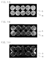
- FIG. 1 is an electronic date of the photograph of the skin after the cultivation in the above-mentioned condition. (a) indicates the infected control group without the drug application, (b) the KP-103-treated group and (c) the lanoconazole-treated group.
- Example 1 20 right feet were excised from each animal five days after the last treatment, and sufficiently wiped with the cotton sweb containing alcohol. The planta was cut off from each foot. The skin mincced by a scissors was put into dialysis membrane (fractional molecular weight: 12,000-14,000, made of cellulose, available from VISKASE SALES Corporation) together with 4 ml of distilled water. Dialysis was carried out under 3 L of distilled water at 4°C for 2 days. The dialysis water was changed twice a day 4 times in total. The content was transfer into a glass homogenizer.
- dialysis membrane fractional molecular weight: 12,000-14,000, made of cellulose, available from VISKASE SALES Corporation
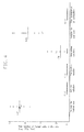
- Figure 2 is an electronic date of the photograph of the skin after the cultivation in the above-mentioned condition.
- (a) indicates the infected control group without the drug application,
- One plate was explained as a representative of ten plates corresponding to each animal in the infected control group (a).
- E indicates the skin suspension prepared from planta of the animal and M the above-mentioned medium.
- E and M described in both the KP-103-treated group (b) and the lanoconazole-treated group (c) are also the same.
- the white zone shows the growth of fungus
- the black zone around the well shows the inhibition of the growth of fungus.
- a result in the present evaluation method correlates with a result obtained by evaluation in the conventional method described in Comparative Example 1 in 30 days after the last treatment.
- a fungal solution was prepared in the same manner as in the pretreatment of Comparative Example 1 except for changing Trichophyton mentagrophytes KD-04 strain to Trichophyton mentagrophytes SM-110 strain.
- a guinea pig model of tinea unguium and tinea pedis was prepared in the same manner as in the above-mentioned preparation of the guinea pig model in interdigital tinea pedis except for changing male Hartley strain guinea pigs of 7 weeks age to male Hartley strain guinea pigs of 5 weeks age and except that the paper disc and the bandage was removed 21 days after the infection changing from seven days after the infection. The invasion of dermatophytes in plantar skin and nail plate was observed 60 days after the infection.
- solutions were prepared by dissolving raw powders of KP-103, amorolfine and terbinafine in a concentration of 1 % thereof to mixture solution of polyethylene grycole #400 : ethanol (75 : 25 v/v), respectivly.
- Capsule of terbinafine was prepared by crushing the marketed tablet, suspending in the concentration of 100 mg/ml into Miglyol 812 (available from Mitsuba trade Co., Ltd)with glass homogenizer uniformity, and injecting the resulting suspension into each capsule in the concentration of 40 mg/kg depending on body weight measured on administration day.
- a solution of KP-103, amorolfine or terbinafine in the amount of 0.1 ml was applied a plantar skin and nail of one foot once a day for 30 consecutive days.
- terbinafine capsule one capsule (40 mg/kg) was administered orally.
- the obtaining precipitate was washed three times by centrifuging with phosphate buffered saline in order to remove trypsin.
- the precipitate was suspended with 4 ml of distilled water and put into dialysis membrane (fractional molecular weight: 12,000-14,000, made of cellulose, available from VISKASE SALES Corporation). Dialysis was carried out into 3 L of distilled water at 4°C for 14 days. Dialysis water was replaced twice a day 28 times in total.
- 1 ml of phosphate buffered saline was added to the precipitate obtained by removing the supernatant to prepare a suspension. This suspension was defined as stock solution and was diluted by tenfold.
- an effect of an antimicrobial agent can be evaluated accurately, since a remaining drug can be removed by dialyzing the infected site with a microorganism of animal or biosample such as the treated skin using a dialysis membrane.
- the present evaluation method enables the antimicrobial effects to compare quantitatively, since the number of viable fungi in the infected site of an animal or a bioample such as a skin can be determined precisely.
- the therapeutic effect based on the present evaluation method reflect a result as to relapse in the conventional method and therefore an effect to prevent relapse can be estimated by evaluating at earlier time after the treatment according to the present evaluation method. Therefore, in the present evaluation method, a true effect of an antimicrobial agent can be evaluated and it is possible to select an antimicrobial agent having an excellent sterilization effect against fungi in vivo or an antimicrobial agent of complete cure type which does not bring about relapse. As mentioned above, the present evaluation method is very useful as a method for evaluating the antimicrobial agent.
- onychomycosis it is the first time that it is possible to evaluate a therapeutic effect against onychomycosis on a model of tinea unguium by the present evaluation method.
- KP-103 exhibits the excellent therapeutic effect against onychomycosis with a simple application on which the effect is not exhibited using the conventional topical antifungal agent. Therefore, KP-103 is a beneficial agent for treating onychomycosis, industrially.
Landscapes
- Chemical & Material Sciences (AREA)
- Health & Medical Sciences (AREA)
- Life Sciences & Earth Sciences (AREA)
- Organic Chemistry (AREA)
- Engineering & Computer Science (AREA)
- Immunology (AREA)
- General Health & Medical Sciences (AREA)
- Molecular Biology (AREA)
- Microbiology (AREA)
- Medicinal Chemistry (AREA)
- Biotechnology (AREA)
- Hematology (AREA)
- Proteomics, Peptides & Aminoacids (AREA)
- Biomedical Technology (AREA)
- Urology & Nephrology (AREA)
- Biochemistry (AREA)
- Zoology (AREA)
- Physics & Mathematics (AREA)
- Analytical Chemistry (AREA)
- Wood Science & Technology (AREA)
- Cell Biology (AREA)
- Veterinary Medicine (AREA)
- Public Health (AREA)
- Animal Behavior & Ethology (AREA)
- Pharmacology & Pharmacy (AREA)
- Virology (AREA)
- Epidemiology (AREA)
- Toxicology (AREA)
- Biophysics (AREA)
- Bioinformatics & Cheminformatics (AREA)
- General Engineering & Computer Science (AREA)
- Genetics & Genomics (AREA)
- Tropical Medicine & Parasitology (AREA)
- Pathology (AREA)
- General Physics & Mathematics (AREA)
- Food Science & Technology (AREA)
- Botany (AREA)
- Mycology (AREA)
- Communicable Diseases (AREA)
- Oncology (AREA)
Applications Claiming Priority (2)
| Application Number | Priority Date | Filing Date | Title |
|---|---|---|---|
| JP21436999 | 1999-07-28 | ||
| EP00944390A EP1205559B1 (en) | 1999-07-28 | 2000-07-11 | Method for detecting pathogenic microorganisms and antimicrobial agents, method for evaluating the drug effect of antimicrobial agents, and antimicrobial agents |
Related Parent Applications (1)
| Application Number | Title | Priority Date | Filing Date |
|---|---|---|---|
| EP00944390A Division EP1205559B1 (en) | 1999-07-28 | 2000-07-11 | Method for detecting pathogenic microorganisms and antimicrobial agents, method for evaluating the drug effect of antimicrobial agents, and antimicrobial agents |
Publications (1)
| Publication Number | Publication Date |
|---|---|
| EP2112228A1 true EP2112228A1 (en) | 2009-10-28 |
Family
ID=16654658
Family Applications (2)
| Application Number | Title | Priority Date | Filing Date |
|---|---|---|---|
| EP00944390A Expired - Lifetime EP1205559B1 (en) | 1999-07-28 | 2000-07-11 | Method for detecting pathogenic microorganisms and antimicrobial agents, method for evaluating the drug effect of antimicrobial agents, and antimicrobial agents |
| EP09163421A Withdrawn EP2112228A1 (en) | 1999-07-28 | 2000-07-11 | Method for detecting pathogenic microorganism and antimicrobial agent, method for evaluating effect of antimicrobial agent, and antimicrobial agent |
Family Applications Before (1)
| Application Number | Title | Priority Date | Filing Date |
|---|---|---|---|
| EP00944390A Expired - Lifetime EP1205559B1 (en) | 1999-07-28 | 2000-07-11 | Method for detecting pathogenic microorganisms and antimicrobial agents, method for evaluating the drug effect of antimicrobial agents, and antimicrobial agents |
Country Status (12)
| Country | Link |
|---|---|
| US (2) | US7214506B2 (da) |
| EP (2) | EP1205559B1 (da) |
| JP (2) | JP4414623B2 (da) |
| AT (1) | ATE434667T1 (da) |
| AU (1) | AU5852400A (da) |
| CA (1) | CA2391274C (da) |
| CY (1) | CY1109281T1 (da) |
| DE (1) | DE60042453D1 (da) |
| DK (1) | DK1205559T3 (da) |
| ES (1) | ES2328785T3 (da) |
| PT (1) | PT1205559E (da) |
| WO (1) | WO2001007643A1 (da) |
Families Citing this family (24)
| Publication number | Priority date | Publication date | Assignee | Title |
|---|---|---|---|---|
| US20060013862A1 (en) * | 2004-07-19 | 2006-01-19 | Held Jerry M | Onychomycosis: a new process for cure |
| JP4824493B2 (ja) * | 2005-08-31 | 2011-11-30 | 株式会社ポーラファルマ | 抗真菌剤の評価法 |
| JP5372523B2 (ja) * | 2006-12-28 | 2013-12-18 | 科研製薬株式会社 | 真菌症治療用ゲル組成物 |
| JP5250562B2 (ja) * | 2007-12-25 | 2013-07-31 | 株式会社ポーラファルマ | 真菌を感染させた動物の被感染爪 |
| US20090175810A1 (en) * | 2008-01-03 | 2009-07-09 | Gareth Winckle | Compositions and methods for treating diseases of the nail |
| US20100291182A1 (en) * | 2009-01-21 | 2010-11-18 | Arsenal Medical, Inc. | Drug-Loaded Fibers |
| US10420862B2 (en) | 2009-08-24 | 2019-09-24 | Aresenal AAA, LLC. | In-situ forming foams for treatment of aneurysms |
| US20110202016A1 (en) * | 2009-08-24 | 2011-08-18 | Arsenal Medical, Inc. | Systems and methods relating to polymer foams |
| US12544491B2 (en) | 2009-08-24 | 2026-02-10 | Arsenal Medical, Inc. | In situ forming hemostatic foam implants |
| US9173817B2 (en) | 2009-08-24 | 2015-11-03 | Arsenal Medical, Inc. | In situ forming hemostatic foam implants |
| US9044580B2 (en) | 2009-08-24 | 2015-06-02 | Arsenal Medical, Inc. | In-situ forming foams with outer layer |
| EP3284831B1 (en) * | 2010-04-16 | 2019-07-24 | Momentum Bioscience Limited | Methods for measuring enzyme activity useful in determining cell viability in non-purified samples |
| US8039494B1 (en) | 2010-07-08 | 2011-10-18 | Dow Pharmaceutical Sciences, Inc. | Compositions and methods for treating diseases of the nail |
| US8968626B2 (en) | 2011-01-31 | 2015-03-03 | Arsenal Medical, Inc. | Electrospinning process for manufacture of multi-layered structures |
| US9034240B2 (en) | 2011-01-31 | 2015-05-19 | Arsenal Medical, Inc. | Electrospinning process for fiber manufacture |
| US9194058B2 (en) | 2011-01-31 | 2015-11-24 | Arsenal Medical, Inc. | Electrospinning process for manufacture of multi-layered structures |
| US8993831B2 (en) | 2011-11-01 | 2015-03-31 | Arsenal Medical, Inc. | Foam and delivery system for treatment of postpartum hemorrhage |
| KR102320051B1 (ko) | 2013-10-03 | 2021-10-29 | 다우 파마슈티컬 사이언시즈, 인코포레이티드 | 안정화된 에피나코나졸 조성물 |
| CA3052643A1 (en) | 2013-11-22 | 2015-05-28 | Bausch Health Ireland Limited | Anti-infective methods, compositions, and devices |
| EP3247399A4 (en) * | 2015-01-20 | 2018-09-05 | Wavelength Enterprises Ltd | Crystalline forms of efinaconazole |
| KR102060546B1 (ko) | 2019-06-07 | 2019-12-30 | 주식회사 바이오빌리프 | 에피나코나졸을 함유하는 용액 형태의 국소 투여용 약학 조성물 |
| JP7440073B2 (ja) * | 2020-07-30 | 2024-02-28 | 国立大学法人広島大学 | 生存細菌の検出方法及びそのためのキット |
| KR102301743B1 (ko) * | 2020-12-29 | 2021-09-13 | 대봉엘에스 주식회사 | 에피나코나졸 경구용 조성물 |
| KR102863832B1 (ko) | 2021-05-04 | 2025-09-25 | 주식회사 제뉴원사이언스 | 에피나코나졸을 함유하는 액상 형태의 약학적 조성물 |
Citations (4)
| Publication number | Priority date | Publication date | Assignee | Title |
|---|---|---|---|---|
| EP0402989A2 (en) * | 1989-06-09 | 1990-12-19 | Janssen Pharmaceutica N.V. | Antifungal 4-[4-[4-[4-[[2-(2,4-difluorophenyl)-2-(1H-azolylmethyl)-1,3-dioxolan-4-yl]methoxy]phenyl]-1-piperazinyl]phenyl]triazolones and imidazolones |
| WO1994026734A1 (fr) | 1993-05-10 | 1994-11-24 | Kaken Pharmaceutical Co., Ltd. | Derive d'azolylamine |
| JPH08103291A (ja) * | 1994-10-04 | 1996-04-23 | Pola Chem Ind Inc | 抗真菌剤の評価法 |
| WO1999039680A1 (en) * | 1998-02-09 | 1999-08-12 | Macrochem Corporation | Antifungal nail lacquer and method using same |
Family Cites Families (2)
| Publication number | Priority date | Publication date | Assignee | Title |
|---|---|---|---|---|
| EP0725818A1 (en) * | 1993-10-25 | 1996-08-14 | Ribogene, Inc. | Methods for screening for antimycotics |
| JP3129662B2 (ja) * | 1996-07-16 | 2001-01-31 | グンゼ株式会社 | 皮膚刺激判定法 |
-
2000
- 2000-07-11 JP JP2001512909A patent/JP4414623B2/ja not_active Expired - Lifetime
- 2000-07-11 ES ES00944390T patent/ES2328785T3/es not_active Expired - Lifetime
- 2000-07-11 DK DK00944390T patent/DK1205559T3/da active
- 2000-07-11 EP EP00944390A patent/EP1205559B1/en not_active Expired - Lifetime
- 2000-07-11 AT AT00944390T patent/ATE434667T1/de active
- 2000-07-11 PT PT00944390T patent/PT1205559E/pt unknown
- 2000-07-11 WO PCT/JP2000/004617 patent/WO2001007643A1/ja not_active Ceased
- 2000-07-11 DE DE60042453T patent/DE60042453D1/de not_active Expired - Lifetime
- 2000-07-11 EP EP09163421A patent/EP2112228A1/en not_active Withdrawn
- 2000-07-11 CA CA2391274A patent/CA2391274C/en not_active Expired - Lifetime
- 2000-07-11 AU AU58524/00A patent/AU5852400A/en not_active Abandoned
-
2003
- 2003-10-14 US US10/685,266 patent/US7214506B2/en not_active Expired - Lifetime
-
2007
- 2007-03-27 US US11/728,766 patent/US20080003636A1/en not_active Abandoned
-
2009
- 2009-08-10 CY CY20091100847T patent/CY1109281T1/el unknown
- 2009-10-07 JP JP2009233792A patent/JP2010004893A/ja active Pending
Patent Citations (4)
| Publication number | Priority date | Publication date | Assignee | Title |
|---|---|---|---|---|
| EP0402989A2 (en) * | 1989-06-09 | 1990-12-19 | Janssen Pharmaceutica N.V. | Antifungal 4-[4-[4-[4-[[2-(2,4-difluorophenyl)-2-(1H-azolylmethyl)-1,3-dioxolan-4-yl]methoxy]phenyl]-1-piperazinyl]phenyl]triazolones and imidazolones |
| WO1994026734A1 (fr) | 1993-05-10 | 1994-11-24 | Kaken Pharmaceutical Co., Ltd. | Derive d'azolylamine |
| JPH08103291A (ja) * | 1994-10-04 | 1996-04-23 | Pola Chem Ind Inc | 抗真菌剤の評価法 |
| WO1999039680A1 (en) * | 1998-02-09 | 1999-08-12 | Macrochem Corporation | Antifungal nail lacquer and method using same |
Non-Patent Citations (5)
| Title |
|---|
| 36TH INTERSCIENCE CONFERENCE ON ANTIMICROBIAL AGENTS AND CHEMOTHERAPY, 1996 |
| ANTIMICROBIAL AGENTS AND CHEMOTHERAPY, vol. 36, 1992, pages 2523 - 2525 |
| ARIKA ET AL., ANTIMICROBIAL AGENTS AND CHEMOTHERAPY, vol. 36, 1992, pages 2523 - 2525 |
| KITAZAKI T ET AL: "OPTICALLY ACTIVE ANTIFUNGAL AZOLES. VI.1) SYNTHESIS AND ANTIFUNGAL ACTIVITY OF N-U(1R,2R)-2-(2,4-DIFLUOROPHENYL)-2-HYDROXY-1-METHYL-3-(1 H-1,2,4-TRIAZOL-1-YL)PROPYL-N'-(4-SUBSTITUTED PHENYL)-3(2H,4H)- 1,2,4-TRIAZOLONES AND 5(1H,4H)-TETRAZOLONES", CHEMICAL AND PHARMACEUTICAL BULLETIN, PHARMACEUTICAL SOCIETY OF JAPAN. TOKYO, JP, vol. 44, no. 2, 1996, pages 314 - 327, XP002067032, ISSN: 0009-2363 * |
| MARKUS NIEWERTH; HANS C. KORTING, MANAGEMENT OF ONYCHOMYCOSES, DRUGS, vol. 58, 1999, pages 283 - 296 |
Also Published As
| Publication number | Publication date |
|---|---|
| CA2391274C (en) | 2010-12-14 |
| CY1109281T1 (el) | 2014-07-02 |
| WO2001007643A1 (en) | 2001-02-01 |
| ATE434667T1 (de) | 2009-07-15 |
| US20070082375A1 (en) | 2007-04-12 |
| ES2328785T3 (es) | 2009-11-18 |
| EP1205559B1 (en) | 2009-06-24 |
| EP1205559A1 (en) | 2002-05-15 |
| US7214506B2 (en) | 2007-05-08 |
| CA2391274A1 (en) | 2001-02-01 |
| AU5852400A (en) | 2001-02-13 |
| US20080003636A1 (en) | 2008-01-03 |
| JP4414623B2 (ja) | 2010-02-10 |
| DE60042453D1 (de) | 2009-08-06 |
| EP1205559A4 (en) | 2004-11-17 |
| DK1205559T3 (da) | 2009-09-28 |
| PT1205559E (pt) | 2009-08-04 |
| JP2010004893A (ja) | 2010-01-14 |
Similar Documents
| Publication | Publication Date | Title |
|---|---|---|
| US20080003636A1 (en) | Method for detecting pathogenic microorganism and antimicrobial agent, method for evaluating effect of antimicrobial agent, and antimicrobial agent | |
| JPWO2001007643A1 (ja) | 病原微生物および抗微生物剤の検出法、抗微生物剤の薬効評価法ならびに抗微生物剤 | |
| Welsh et al. | Onychomycosis | |
| Levin et al. | Candida parapsilosis fungemia associated with implantable and semi-implantable central venous catheters and the hands of healthcare workers | |
| Casals | Tablet sensitivity testing on pathogenic fungi. | |
| Shimamura et al. | Animal model of dermatophytosis | |
| JPS63500664A (ja) | 亜鉛グリセロラ−ト錯体及び医薬的適用のための添加 | |
| Ringel | In vitro and in vivo studies of ambruticin (W7783): new class of antifungal antibiotics | |
| Arika et al. | Effects of butenafine hydrochloride, a new benzylamine derivative, on experimental tinea pedis in guinea pigs | |
| Youssef et al. | Ecological effects of antibiotic production by dermatophyte fungi | |
| Haller et al. | Experimental in vitro and in vivo comparison of modern antimycotics | |
| Nakamura et al. | A comparative study between two antifungal agents, Luliconazole and Efinaconazole, of their preventive effects in a Trichophyton-infected guinea pig onychomycosis model | |
| Pfaller et al. | Fungicidal activity of cilofungin (LY121019) alone and in combination with anticapsin or other antifungal agents | |
| Yousif et al. | Leucocyte chemotaxis to mycetoma agents—the effect of the antifungal drugs griseofulvin and ketoconazole | |
| Andrews et al. | Repurposing sunscreen as an antibiotic: zinc-activated avobenzone inhibits methicillin-resistant Staphylococcus aureus | |
| Tatsumi et al. | In Vivo Fungicidal Effect of KP‐103 in a Guinea Pig Model of Interdigital Tinea Pedis Determined by Using a New Method for Removing the Antimycotic Carryover Effect | |
| Hussien et al. | Neutrophil hypochlorous acid production is impaired in multiple organ failure patients with candidaemia; reversal with antifungal agents | |
| EP1306445B1 (en) | Method of evaluating antifungal agent | |
| Popa et al. | Microsporum Canis strains sensitivity to antifungal drugs. | |
| JP3120965B2 (ja) | 水虫外用薬 | |
| Majima et al. | A novel mycological analysis valuable for evaluating therapeutic efficacy of antimycotics against experimental dermatophytosis in guinea pigs | |
| Macura | Fungal Resistance to Antimycotic Drugs. | |
| Gürer et al. | Effect of fluconazole on human polymorphonuclear leucocyte functions ex vivo against Candida albicans | |
| Clayton | Dermatophyte infections | |
| JP3163378B2 (ja) | 抗真菌剤及び抗細菌剤 |
Legal Events
| Date | Code | Title | Description |
|---|---|---|---|
| PUAI | Public reference made under article 153(3) epc to a published international application that has entered the european phase |
Free format text: ORIGINAL CODE: 0009012 |
|
| AC | Divisional application: reference to earlier application |
Ref document number: 1205559 Country of ref document: EP Kind code of ref document: P |
|
| AK | Designated contracting states |
Kind code of ref document: A1 Designated state(s): AT BE CH CY DE DK ES FI FR GB GR IE IT LI LU MC NL PT SE |
|
| RIN1 | Information on inventor provided before grant (corrected) |
Inventor name: NAKAMURA, KOSHO Inventor name: ARIKA, TADAHI Inventor name: YOKOO, MAMORU Inventor name: TATSUMI, YOSHIYUKI |
|
| 17P | Request for examination filed |
Effective date: 20091223 |
|
| 17Q | First examination report despatched |
Effective date: 20100122 |
|
| STAA | Information on the status of an ep patent application or granted ep patent |
Free format text: STATUS: THE APPLICATION IS DEEMED TO BE WITHDRAWN |
|
| 18D | Application deemed to be withdrawn |
Effective date: 20100602 |