EP2117582B1 - Periostine induisant une proliferation de cardiomyocytes et favorisant une regeneration cardiaque - Google Patents
Periostine induisant une proliferation de cardiomyocytes et favorisant une regeneration cardiaque Download PDFInfo
- Publication number
- EP2117582B1 EP2117582B1 EP08713899.6A EP08713899A EP2117582B1 EP 2117582 B1 EP2117582 B1 EP 2117582B1 EP 08713899 A EP08713899 A EP 08713899A EP 2117582 B1 EP2117582 B1 EP 2117582B1
- Authority
- EP
- European Patent Office
- Prior art keywords
- periostin
- cardiomyocytes
- composition
- cardiomyocyte
- proliferation
- Prior art date
- Legal status (The legal status is an assumption and is not a legal conclusion. Google has not performed a legal analysis and makes no representation as to the accuracy of the status listed.)
- Not-in-force
Links
- 210000004413 cardiac myocyte Anatomy 0.000 title claims description 427
- 101710199268 Periostin Proteins 0.000 title claims description 417
- 102100037765 Periostin Human genes 0.000 title claims description 415
- 230000035755 proliferation Effects 0.000 title claims description 101
- 230000000747 cardiac effect Effects 0.000 title description 21
- 230000008929 regeneration Effects 0.000 title description 17
- 238000011069 regeneration method Methods 0.000 title description 17
- 102000006495 integrins Human genes 0.000 claims description 76
- 108010044426 integrins Proteins 0.000 claims description 76
- 239000000203 mixture Substances 0.000 claims description 69
- 239000012634 fragment Substances 0.000 claims description 47
- 210000001519 tissue Anatomy 0.000 claims description 26
- 238000000034 method Methods 0.000 claims description 23
- 101150008545 FAS1 gene Proteins 0.000 claims description 16
- 150000001875 compounds Chemical class 0.000 claims description 15
- 230000006378 damage Effects 0.000 claims description 12
- 208000027418 Wounds and injury Diseases 0.000 claims description 11
- 208000014674 injury Diseases 0.000 claims description 11
- 210000005003 heart tissue Anatomy 0.000 claims description 8
- 238000006467 substitution reaction Methods 0.000 claims description 8
- 108010022222 Integrin beta1 Proteins 0.000 claims description 7
- 101710172176 Fasciclin-1 Proteins 0.000 claims description 6
- 239000001963 growth medium Substances 0.000 claims description 6
- 238000004519 manufacturing process Methods 0.000 claims description 6
- 230000004936 stimulating effect Effects 0.000 claims description 6
- 108010020950 Integrin beta3 Proteins 0.000 claims description 5
- 238000013270 controlled release Methods 0.000 claims description 4
- 239000003085 diluting agent Substances 0.000 claims description 4
- 239000003937 drug carrier Substances 0.000 claims description 4
- 108010021518 integrin beta5 Proteins 0.000 claims description 4
- 239000008194 pharmaceutical composition Substances 0.000 claims description 4
- 238000012258 culturing Methods 0.000 claims description 3
- 239000003814 drug Substances 0.000 claims description 2
- 239000002609 medium Substances 0.000 claims description 2
- 238000007911 parenteral administration Methods 0.000 claims description 2
- 208000010125 myocardial infarction Diseases 0.000 description 94
- 230000022131 cell cycle Effects 0.000 description 87
- 210000002216 heart Anatomy 0.000 description 82
- 230000006820 DNA synthesis Effects 0.000 description 78
- 241000700159 Rattus Species 0.000 description 74
- 230000002861 ventricular Effects 0.000 description 57
- 230000021953 cytokinesis Effects 0.000 description 46
- 210000004027 cell Anatomy 0.000 description 37
- WOVKYSAHUYNSMH-RRKCRQDMSA-N 5-bromodeoxyuridine Chemical compound C1[C@H](O)[C@@H](CO)O[C@H]1N1C(=O)NC(=O)C(Br)=C1 WOVKYSAHUYNSMH-RRKCRQDMSA-N 0.000 description 35
- WOVKYSAHUYNSMH-UHFFFAOYSA-N BROMODEOXYURIDINE Natural products C1C(O)C(CO)OC1N1C(=O)NC(=O)C(Br)=C1 WOVKYSAHUYNSMH-UHFFFAOYSA-N 0.000 description 34
- 206010061216 Infarction Diseases 0.000 description 33
- 230000007574 infarction Effects 0.000 description 32
- 210000004940 nucleus Anatomy 0.000 description 32
- 230000001965 increasing effect Effects 0.000 description 30
- 230000037361 pathway Effects 0.000 description 28
- 230000000694 effects Effects 0.000 description 25
- 230000002107 myocardial effect Effects 0.000 description 25
- 206010020880 Hypertrophy Diseases 0.000 description 23
- 230000000638 stimulation Effects 0.000 description 23
- 102000019149 MAP kinase activity proteins Human genes 0.000 description 22
- 108040008097 MAP kinase activity proteins Proteins 0.000 description 22
- 230000014509 gene expression Effects 0.000 description 22
- 108090000623 proteins and genes Proteins 0.000 description 22
- 108020004414 DNA Proteins 0.000 description 20
- 230000006870 function Effects 0.000 description 20
- 239000013615 primer Substances 0.000 description 18
- 230000002062 proliferating effect Effects 0.000 description 18
- 108091007960 PI3Ks Proteins 0.000 description 17
- 210000001054 cardiac fibroblast Anatomy 0.000 description 17
- 102000010834 Extracellular Matrix Proteins Human genes 0.000 description 16
- 108010037362 Extracellular Matrix Proteins Proteins 0.000 description 16
- 102000038030 PI3Ks Human genes 0.000 description 16
- 210000002744 extracellular matrix Anatomy 0.000 description 16
- 238000010820 immunofluorescence microscopy Methods 0.000 description 16
- 238000001727 in vivo Methods 0.000 description 16
- 102000004169 proteins and genes Human genes 0.000 description 16
- 238000011002 quantification Methods 0.000 description 16
- 108010067306 Fibronectins Proteins 0.000 description 15
- 102000016359 Fibronectins Human genes 0.000 description 15
- 206010016654 Fibrosis Diseases 0.000 description 15
- 230000004913 activation Effects 0.000 description 15
- 239000000872 buffer Substances 0.000 description 15
- 230000004761 fibrosis Effects 0.000 description 15
- 238000000338 in vitro Methods 0.000 description 15
- 102000003993 Phosphatidylinositol 3-kinases Human genes 0.000 description 14
- 108090000430 Phosphatidylinositol 3-kinases Proteins 0.000 description 14
- 210000004165 myocardium Anatomy 0.000 description 14
- 210000000130 stem cell Anatomy 0.000 description 14
- 230000004217 heart function Effects 0.000 description 13
- 239000012190 activator Substances 0.000 description 12
- 230000000903 blocking effect Effects 0.000 description 12
- 239000007924 injection Substances 0.000 description 11
- 238000002347 injection Methods 0.000 description 11
- 241000282414 Homo sapiens Species 0.000 description 10
- 230000001351 cycling effect Effects 0.000 description 10
- 230000001605 fetal effect Effects 0.000 description 10
- 108090000765 processed proteins & peptides Proteins 0.000 description 10
- 230000004083 survival effect Effects 0.000 description 10
- 101001095308 Homo sapiens Periostin Proteins 0.000 description 9
- 102000016971 Proto-Oncogene Proteins c-kit Human genes 0.000 description 9
- 108010014608 Proto-Oncogene Proteins c-kit Proteins 0.000 description 9
- 230000006907 apoptotic process Effects 0.000 description 9
- 102000048431 human POSTN Human genes 0.000 description 9
- 230000001976 improved effect Effects 0.000 description 9
- 229920001184 polypeptide Polymers 0.000 description 9
- 102000004196 processed proteins & peptides Human genes 0.000 description 9
- 238000011282 treatment Methods 0.000 description 9
- 101800001288 Atrial natriuretic factor Proteins 0.000 description 8
- 101800001890 Atrial natriuretic peptide Proteins 0.000 description 8
- 102400001282 Atrial natriuretic peptide Human genes 0.000 description 8
- 206010019280 Heart failures Diseases 0.000 description 8
- 208000033774 Ventricular Remodeling Diseases 0.000 description 8
- 230000033115 angiogenesis Effects 0.000 description 8
- NSQLIUXCMFBZME-MPVJKSABSA-N carperitide Chemical compound C([C@H]1C(=O)NCC(=O)NCC(=O)N[C@@H](CCCNC(N)=N)C(=O)N[C@@H](CCSC)C(=O)N[C@@H](CC(O)=O)C(=O)N[C@@H](CCCNC(N)=N)C(=O)N[C@H](C(NCC(=O)N[C@@H](C)C(=O)N[C@@H](CCC(N)=O)C(=O)N[C@@H](CO)C(=O)NCC(=O)N[C@@H](CC(C)C)C(=O)NCC(=O)N[C@@H](CSSC[C@@H](C(=O)N1)NC(=O)[C@H](CO)NC(=O)[C@H](CO)NC(=O)[C@H](CCCNC(N)=N)NC(=O)[C@H](CCCNC(N)=N)NC(=O)[C@H](CC(C)C)NC(=O)[C@@H](N)CO)C(=O)N[C@@H](CC(N)=O)C(=O)N[C@@H](CO)C(=O)N[C@@H](CC=1C=CC=CC=1)C(=O)N[C@@H](CCCNC(N)=N)C(=O)N[C@@H](CC=1C=CC(O)=CC=1)C(O)=O)=O)[C@@H](C)CC)C1=CC=CC=C1 NSQLIUXCMFBZME-MPVJKSABSA-N 0.000 description 8
- 210000002253 embryonic cardiomyocyte Anatomy 0.000 description 8
- 230000005764 inhibitory process Effects 0.000 description 8
- 102000002574 p38 Mitogen-Activated Protein Kinases Human genes 0.000 description 8
- 108010068338 p38 Mitogen-Activated Protein Kinases Proteins 0.000 description 8
- 230000004044 response Effects 0.000 description 8
- 230000019491 signal transduction Effects 0.000 description 8
- 102000013394 Troponin I Human genes 0.000 description 7
- 108010065729 Troponin I Proteins 0.000 description 7
- 125000003275 alpha amino acid group Chemical group 0.000 description 7
- 230000007423 decrease Effects 0.000 description 7
- 230000001419 dependent effect Effects 0.000 description 7
- 239000000463 material Substances 0.000 description 7
- 238000007634 remodeling Methods 0.000 description 7
- 238000010186 staining Methods 0.000 description 7
- 108010047303 von Willebrand Factor Proteins 0.000 description 7
- 102100036537 von Willebrand factor Human genes 0.000 description 7
- 229960001134 von willebrand factor Drugs 0.000 description 7
- 102000007469 Actins Human genes 0.000 description 6
- 108010085238 Actins Proteins 0.000 description 6
- 102000004228 Aurora kinase B Human genes 0.000 description 6
- 108090000749 Aurora kinase B Proteins 0.000 description 6
- PEDCQBHIVMGVHV-UHFFFAOYSA-N Glycerine Chemical compound OCC(O)CO PEDCQBHIVMGVHV-UHFFFAOYSA-N 0.000 description 6
- 108010011536 PTEN Phosphohydrolase Proteins 0.000 description 6
- 102000014160 PTEN Phosphohydrolase Human genes 0.000 description 6
- 108050002653 Retinoblastoma protein Proteins 0.000 description 6
- 102000005937 Tropomyosin Human genes 0.000 description 6
- 108010030743 Tropomyosin Proteins 0.000 description 6
- 102000004987 Troponin T Human genes 0.000 description 6
- 108090001108 Troponin T Proteins 0.000 description 6
- 238000004458 analytical method Methods 0.000 description 6
- 239000003795 chemical substances by application Substances 0.000 description 6
- 239000005090 green fluorescent protein Substances 0.000 description 6
- 230000007246 mechanism Effects 0.000 description 6
- 230000008439 repair process Effects 0.000 description 6
- 231100000241 scar Toxicity 0.000 description 6
- 230000002459 sustained effect Effects 0.000 description 6
- 238000013518 transcription Methods 0.000 description 6
- 230000035897 transcription Effects 0.000 description 6
- 238000010361 transduction Methods 0.000 description 6
- 230000026683 transduction Effects 0.000 description 6
- 238000002054 transplantation Methods 0.000 description 6
- 238000001262 western blot Methods 0.000 description 6
- 102000002554 Cyclin A Human genes 0.000 description 5
- 108010068192 Cyclin A Proteins 0.000 description 5
- 102100022337 Integrin alpha-V Human genes 0.000 description 5
- 108010040765 Integrin alphaV Proteins 0.000 description 5
- 102100024193 Mitogen-activated protein kinase 1 Human genes 0.000 description 5
- FAPWRFPIFSIZLT-UHFFFAOYSA-M Sodium chloride Chemical compound [Na+].[Cl-] FAPWRFPIFSIZLT-UHFFFAOYSA-M 0.000 description 5
- 238000007792 addition Methods 0.000 description 5
- 150000001413 amino acids Chemical class 0.000 description 5
- 230000034994 death Effects 0.000 description 5
- 231100000517 death Toxicity 0.000 description 5
- 230000003247 decreasing effect Effects 0.000 description 5
- 238000001514 detection method Methods 0.000 description 5
- 238000011161 development Methods 0.000 description 5
- 230000018109 developmental process Effects 0.000 description 5
- 239000006185 dispersion Substances 0.000 description 5
- 230000009368 gene silencing by RNA Effects 0.000 description 5
- 230000001939 inductive effect Effects 0.000 description 5
- 230000003993 interaction Effects 0.000 description 5
- 239000003550 marker Substances 0.000 description 5
- 230000011278 mitosis Effects 0.000 description 5
- SONNWYBIRXJNDC-VIFPVBQESA-N phenylephrine Chemical compound CNC[C@H](O)C1=CC=CC(O)=C1 SONNWYBIRXJNDC-VIFPVBQESA-N 0.000 description 5
- 229960001802 phenylephrine Drugs 0.000 description 5
- 239000013641 positive control Substances 0.000 description 5
- 230000002829 reductive effect Effects 0.000 description 5
- 238000004904 shortening Methods 0.000 description 5
- 230000001629 suppression Effects 0.000 description 5
- 238000001356 surgical procedure Methods 0.000 description 5
- 230000008685 targeting Effects 0.000 description 5
- 230000001225 therapeutic effect Effects 0.000 description 5
- 238000012800 visualization Methods 0.000 description 5
- QDLHCMPXEPAAMD-QAIWCSMKSA-N wortmannin Chemical compound C1([C@]2(C)C3=C(C4=O)OC=C3C(=O)O[C@@H]2COC)=C4[C@@H]2CCC(=O)[C@@]2(C)C[C@H]1OC(C)=O QDLHCMPXEPAAMD-QAIWCSMKSA-N 0.000 description 5
- QDLHCMPXEPAAMD-UHFFFAOYSA-N wortmannin Natural products COCC1OC(=O)C2=COC(C3=O)=C2C1(C)C1=C3C2CCC(=O)C2(C)CC1OC(C)=O QDLHCMPXEPAAMD-UHFFFAOYSA-N 0.000 description 5
- 230000007730 Akt signaling Effects 0.000 description 4
- 229940123587 Cell cycle inhibitor Drugs 0.000 description 4
- AOJJSUZBOXZQNB-TZSSRYMLSA-N Doxorubicin Chemical compound O([C@H]1C[C@@](O)(CC=2C(O)=C3C(=O)C=4C=CC=C(C=4C(=O)C3=C(O)C=21)OC)C(=O)CO)[C@H]1C[C@H](N)[C@H](O)[C@H](C)O1 AOJJSUZBOXZQNB-TZSSRYMLSA-N 0.000 description 4
- 108010007457 Extracellular Signal-Regulated MAP Kinases Proteins 0.000 description 4
- 241001465754 Metazoa Species 0.000 description 4
- 230000018199 S phase Effects 0.000 description 4
- 108700019146 Transgenes Proteins 0.000 description 4
- 125000000539 amino acid group Chemical group 0.000 description 4
- 238000013459 approach Methods 0.000 description 4
- 230000004071 biological effect Effects 0.000 description 4
- 230000032823 cell division Effects 0.000 description 4
- 230000001413 cellular effect Effects 0.000 description 4
- 210000000349 chromosome Anatomy 0.000 description 4
- 239000002299 complementary DNA Substances 0.000 description 4
- 238000002592 echocardiography Methods 0.000 description 4
- 230000003176 fibrotic effect Effects 0.000 description 4
- 230000004927 fusion Effects 0.000 description 4
- 238000001476 gene delivery Methods 0.000 description 4
- 230000001969 hypertrophic effect Effects 0.000 description 4
- 230000006872 improvement Effects 0.000 description 4
- 239000004615 ingredient Substances 0.000 description 4
- 230000002401 inhibitory effect Effects 0.000 description 4
- 238000002372 labelling Methods 0.000 description 4
- 230000007774 longterm Effects 0.000 description 4
- 230000002018 overexpression Effects 0.000 description 4
- 239000002935 phosphatidylinositol 3 kinase inhibitor Substances 0.000 description 4
- 230000026731 phosphorylation Effects 0.000 description 4
- 238000006366 phosphorylation reaction Methods 0.000 description 4
- 108091033319 polynucleotide Proteins 0.000 description 4
- 102000040430 polynucleotide Human genes 0.000 description 4
- 239000002157 polynucleotide Substances 0.000 description 4
- 238000002360 preparation method Methods 0.000 description 4
- 102000005962 receptors Human genes 0.000 description 4
- 108020003175 receptors Proteins 0.000 description 4
- 230000001172 regenerating effect Effects 0.000 description 4
- 230000036573 scar formation Effects 0.000 description 4
- 239000003826 tablet Substances 0.000 description 4
- 238000012360 testing method Methods 0.000 description 4
- -1 troches Substances 0.000 description 4
- FWBHETKCLVMNFS-UHFFFAOYSA-N 4',6-Diamino-2-phenylindol Chemical compound C1=CC(C(=N)N)=CC=C1C1=CC2=CC=C(C(N)=N)C=C2N1 FWBHETKCLVMNFS-UHFFFAOYSA-N 0.000 description 3
- 206010007572 Cardiac hypertrophy Diseases 0.000 description 3
- 208000006029 Cardiomegaly Diseases 0.000 description 3
- 102000008186 Collagen Human genes 0.000 description 3
- 108010035532 Collagen Proteins 0.000 description 3
- 108010060273 Cyclin A2 Proteins 0.000 description 3
- 108010058544 Cyclin D2 Proteins 0.000 description 3
- 102100025191 Cyclin-A2 Human genes 0.000 description 3
- 239000012824 ERK inhibitor Substances 0.000 description 3
- 102100024185 G1/S-specific cyclin-D2 Human genes 0.000 description 3
- 241000282412 Homo Species 0.000 description 3
- 102000012355 Integrin beta1 Human genes 0.000 description 3
- 102000043136 MAP kinase family Human genes 0.000 description 3
- 108091054455 MAP kinase family Proteins 0.000 description 3
- DNIAPMSPPWPWGF-UHFFFAOYSA-N Propylene glycol Chemical compound CC(O)CO DNIAPMSPPWPWGF-UHFFFAOYSA-N 0.000 description 3
- 108060008682 Tumor Necrosis Factor Proteins 0.000 description 3
- 102000000852 Tumor Necrosis Factor-alpha Human genes 0.000 description 3
- 230000009471 action Effects 0.000 description 3
- 238000000540 analysis of variance Methods 0.000 description 3
- 230000001640 apoptogenic effect Effects 0.000 description 3
- 238000003556 assay Methods 0.000 description 3
- 230000037396 body weight Effects 0.000 description 3
- 239000002775 capsule Substances 0.000 description 3
- 239000003153 chemical reaction reagent Substances 0.000 description 3
- 238000003776 cleavage reaction Methods 0.000 description 3
- 238000000576 coating method Methods 0.000 description 3
- 229920001436 collagen Polymers 0.000 description 3
- 230000000875 corresponding effect Effects 0.000 description 3
- 238000012217 deletion Methods 0.000 description 3
- 230000037430 deletion Effects 0.000 description 3
- 230000004069 differentiation Effects 0.000 description 3
- 239000002612 dispersion medium Substances 0.000 description 3
- 210000002950 fibroblast Anatomy 0.000 description 3
- 208000019622 heart disease Diseases 0.000 description 3
- 230000002779 inactivation Effects 0.000 description 3
- 208000015181 infectious disease Diseases 0.000 description 3
- 230000000302 ischemic effect Effects 0.000 description 3
- 210000005240 left ventricle Anatomy 0.000 description 3
- 239000003446 ligand Substances 0.000 description 3
- 239000002502 liposome Substances 0.000 description 3
- 230000031864 metaphase Effects 0.000 description 3
- 244000005700 microbiome Species 0.000 description 3
- 230000027291 mitotic cell cycle Effects 0.000 description 3
- 230000010016 myocardial function Effects 0.000 description 3
- 208000037891 myocardial injury Diseases 0.000 description 3
- 208000031225 myocardial ischemia Diseases 0.000 description 3
- 239000002773 nucleotide Substances 0.000 description 3
- 125000003729 nucleotide group Chemical group 0.000 description 3
- 230000003287 optical effect Effects 0.000 description 3
- 210000000056 organ Anatomy 0.000 description 3
- 230000007017 scission Effects 0.000 description 3
- 210000002460 smooth muscle Anatomy 0.000 description 3
- 239000011780 sodium chloride Substances 0.000 description 3
- 239000000243 solution Substances 0.000 description 3
- 239000002904 solvent Substances 0.000 description 3
- 239000000126 substance Substances 0.000 description 3
- 238000012546 transfer Methods 0.000 description 3
- 241000701161 unidentified adenovirus Species 0.000 description 3
- XLYOFNOQVPJJNP-UHFFFAOYSA-N water Substances O XLYOFNOQVPJJNP-UHFFFAOYSA-N 0.000 description 3
- MZOFCQQQCNRIBI-VMXHOPILSA-N (3s)-4-[[(2s)-1-[[(2s)-1-[[(1s)-1-carboxy-2-hydroxyethyl]amino]-4-methyl-1-oxopentan-2-yl]amino]-5-(diaminomethylideneamino)-1-oxopentan-2-yl]amino]-3-[[2-[[(2s)-2,6-diaminohexanoyl]amino]acetyl]amino]-4-oxobutanoic acid Chemical compound OC[C@@H](C(O)=O)NC(=O)[C@H](CC(C)C)NC(=O)[C@H](CCCN=C(N)N)NC(=O)[C@H](CC(O)=O)NC(=O)CNC(=O)[C@@H](N)CCCCN MZOFCQQQCNRIBI-VMXHOPILSA-N 0.000 description 2
- 108091032973 (ribonucleotides)n+m Proteins 0.000 description 2
- PRDFBSVERLRRMY-UHFFFAOYSA-N 2'-(4-ethoxyphenyl)-5-(4-methylpiperazin-1-yl)-2,5'-bibenzimidazole Chemical compound C1=CC(OCC)=CC=C1C1=NC2=CC=C(C=3NC4=CC(=CC=C4N=3)N3CCN(C)CC3)C=C2N1 PRDFBSVERLRRMY-UHFFFAOYSA-N 0.000 description 2
- IYMAXBFPHPZYIK-BQBZGAKWSA-N Arg-Gly-Asp Chemical compound NC(N)=NCCC[C@H](N)C(=O)NCC(=O)N[C@@H](CC(O)=O)C(O)=O IYMAXBFPHPZYIK-BQBZGAKWSA-N 0.000 description 2
- 241000894006 Bacteria Species 0.000 description 2
- 208000031229 Cardiomyopathies Diseases 0.000 description 2
- 208000024172 Cardiovascular disease Diseases 0.000 description 2
- 102000002585 Contractile Proteins Human genes 0.000 description 2
- 108010068426 Contractile Proteins Proteins 0.000 description 2
- 241000252212 Danio rerio Species 0.000 description 2
- 102100031480 Dual specificity mitogen-activated protein kinase kinase 1 Human genes 0.000 description 2
- 101710146526 Dual specificity mitogen-activated protein kinase kinase 1 Proteins 0.000 description 2
- LFQSCWFLJHTTHZ-UHFFFAOYSA-N Ethanol Chemical compound CCO LFQSCWFLJHTTHZ-UHFFFAOYSA-N 0.000 description 2
- 230000020172 G2/M transition checkpoint Effects 0.000 description 2
- 108010010803 Gelatin Proteins 0.000 description 2
- 102100033636 Histone H3.2 Human genes 0.000 description 2
- 108010033040 Histones Proteins 0.000 description 2
- 206010021143 Hypoxia Diseases 0.000 description 2
- ROHFNLRQFUQHCH-YFKPBYRVSA-N L-leucine Chemical compound CC(C)C[C@H](N)C(O)=O ROHFNLRQFUQHCH-YFKPBYRVSA-N 0.000 description 2
- 241000713666 Lentivirus Species 0.000 description 2
- ROHFNLRQFUQHCH-UHFFFAOYSA-N Leucine Natural products CC(C)CC(N)C(O)=O ROHFNLRQFUQHCH-UHFFFAOYSA-N 0.000 description 2
- 229940124647 MEK inhibitor Drugs 0.000 description 2
- 108700011259 MicroRNAs Proteins 0.000 description 2
- 102100031790 Myelin expression factor 2 Human genes 0.000 description 2
- 101710107751 Myelin expression factor 2 Proteins 0.000 description 2
- 108091034117 Oligonucleotide Proteins 0.000 description 2
- 208000006735 Periostitis Diseases 0.000 description 2
- 101000890996 Rattus norvegicus Fibronectin Proteins 0.000 description 2
- 208000006011 Stroke Diseases 0.000 description 2
- 238000010521 absorption reaction Methods 0.000 description 2
- 230000000996 additive effect Effects 0.000 description 2
- 101150045355 akt1 gene Proteins 0.000 description 2
- 239000003242 anti bacterial agent Substances 0.000 description 2
- 230000000844 anti-bacterial effect Effects 0.000 description 2
- 239000003429 antifungal agent Substances 0.000 description 2
- 229940121375 antifungal agent Drugs 0.000 description 2
- 210000002565 arteriole Anatomy 0.000 description 2
- 230000002238 attenuated effect Effects 0.000 description 2
- 230000033228 biological regulation Effects 0.000 description 2
- 230000015572 biosynthetic process Effects 0.000 description 2
- 210000001185 bone marrow Anatomy 0.000 description 2
- 239000011575 calcium Substances 0.000 description 2
- 230000001269 cardiogenic effect Effects 0.000 description 2
- 230000003293 cardioprotective effect Effects 0.000 description 2
- 230000015556 catabolic process Effects 0.000 description 2
- 230000030833 cell death Effects 0.000 description 2
- 210000000170 cell membrane Anatomy 0.000 description 2
- 230000004663 cell proliferation Effects 0.000 description 2
- 230000005754 cellular signaling Effects 0.000 description 2
- 230000001086 cytosolic effect Effects 0.000 description 2
- 230000007547 defect Effects 0.000 description 2
- 230000008021 deposition Effects 0.000 description 2
- 238000009792 diffusion process Methods 0.000 description 2
- 238000010494 dissociation reaction Methods 0.000 description 2
- 230000005593 dissociations Effects 0.000 description 2
- 229960004679 doxorubicin Drugs 0.000 description 2
- 238000005516 engineering process Methods 0.000 description 2
- 238000002474 experimental method Methods 0.000 description 2
- 239000013604 expression vector Substances 0.000 description 2
- 239000008273 gelatin Substances 0.000 description 2
- 229920000159 gelatin Polymers 0.000 description 2
- 235000019322 gelatine Nutrition 0.000 description 2
- 235000011852 gelatine desserts Nutrition 0.000 description 2
- 238000001415 gene therapy Methods 0.000 description 2
- 230000002068 genetic effect Effects 0.000 description 2
- 230000012010 growth Effects 0.000 description 2
- 239000003102 growth factor Substances 0.000 description 2
- 210000003709 heart valve Anatomy 0.000 description 2
- 230000000004 hemodynamic effect Effects 0.000 description 2
- 230000007954 hypoxia Effects 0.000 description 2
- 238000010166 immunofluorescence Methods 0.000 description 2
- 238000011065 in-situ storage Methods 0.000 description 2
- 230000006698 induction Effects 0.000 description 2
- 239000003112 inhibitor Substances 0.000 description 2
- 239000007928 intraperitoneal injection Substances 0.000 description 2
- 238000002955 isolation Methods 0.000 description 2
- 239000007951 isotonicity adjuster Substances 0.000 description 2
- 238000011813 knockout mouse model Methods 0.000 description 2
- 239000007788 liquid Substances 0.000 description 2
- 238000012423 maintenance Methods 0.000 description 2
- 230000031852 maintenance of location in cell Effects 0.000 description 2
- 238000005259 measurement Methods 0.000 description 2
- 239000012528 membrane Substances 0.000 description 2
- 239000002679 microRNA Substances 0.000 description 2
- 230000003278 mimic effect Effects 0.000 description 2
- 230000004048 modification Effects 0.000 description 2
- 238000012986 modification Methods 0.000 description 2
- 230000001114 myogenic effect Effects 0.000 description 2
- 239000002245 particle Substances 0.000 description 2
- 210000003460 periosteum Anatomy 0.000 description 2
- 230000002093 peripheral effect Effects 0.000 description 2
- 239000000546 pharmaceutical excipient Substances 0.000 description 2
- 239000006187 pill Substances 0.000 description 2
- 230000001681 protective effect Effects 0.000 description 2
- 108091008598 receptor tyrosine kinases Proteins 0.000 description 2
- 102000027426 receptor tyrosine kinases Human genes 0.000 description 2
- 230000007115 recruitment Effects 0.000 description 2
- 230000009719 regenerative response Effects 0.000 description 2
- 238000011160 research Methods 0.000 description 2
- 238000003757 reverse transcription PCR Methods 0.000 description 2
- 238000000926 separation method Methods 0.000 description 2
- 150000003384 small molecules Chemical class 0.000 description 2
- 238000013222 sprague-dawley male rat Methods 0.000 description 2
- 238000003860 storage Methods 0.000 description 2
- 235000000346 sugar Nutrition 0.000 description 2
- 239000006228 supernatant Substances 0.000 description 2
- 238000003786 synthesis reaction Methods 0.000 description 2
- 230000017423 tissue regeneration Effects 0.000 description 2
- 239000003981 vehicle Substances 0.000 description 2
- 230000035899 viability Effects 0.000 description 2
- VIBDVOOELVZGDU-UHFFFAOYSA-N 4-(1h-indol-2-yl)benzene-1,3-dicarboximidamide Chemical compound NC(=N)C1=CC(C(=N)N)=CC=C1C1=CC2=CC=CC=C2N1 VIBDVOOELVZGDU-UHFFFAOYSA-N 0.000 description 1
- WRDABNWSWOHGMS-UHFFFAOYSA-N AEBSF hydrochloride Chemical compound Cl.NCCC1=CC=C(S(F)(=O)=O)C=C1 WRDABNWSWOHGMS-UHFFFAOYSA-N 0.000 description 1
- 102000005446 Anaphase-Promoting Complex-Cyclosome Human genes 0.000 description 1
- 108010031677 Anaphase-Promoting Complex-Cyclosome Proteins 0.000 description 1
- 206010002383 Angina Pectoris Diseases 0.000 description 1
- 208000010392 Bone Fractures Diseases 0.000 description 1
- OYPRJOBELJOOCE-UHFFFAOYSA-N Calcium Chemical compound [Ca] OYPRJOBELJOOCE-UHFFFAOYSA-N 0.000 description 1
- 241000283707 Capra Species 0.000 description 1
- 108010022452 Collagen Type I Proteins 0.000 description 1
- 102000012422 Collagen Type I Human genes 0.000 description 1
- 206010009944 Colon cancer Diseases 0.000 description 1
- 108020004635 Complementary DNA Proteins 0.000 description 1
- 206010056370 Congestive cardiomyopathy Diseases 0.000 description 1
- 241000514744 Cyclina Species 0.000 description 1
- 102000004127 Cytokines Human genes 0.000 description 1
- 108090000695 Cytokines Proteins 0.000 description 1
- FBPFZTCFMRRESA-FSIIMWSLSA-N D-Glucitol Natural products OC[C@H](O)[C@H](O)[C@@H](O)[C@H](O)CO FBPFZTCFMRRESA-FSIIMWSLSA-N 0.000 description 1
- FBPFZTCFMRRESA-KVTDHHQDSA-N D-Mannitol Chemical compound OC[C@@H](O)[C@@H](O)[C@H](O)[C@H](O)CO FBPFZTCFMRRESA-KVTDHHQDSA-N 0.000 description 1
- FBPFZTCFMRRESA-JGWLITMVSA-N D-glucitol Chemical compound OC[C@H](O)[C@@H](O)[C@H](O)[C@H](O)CO FBPFZTCFMRRESA-JGWLITMVSA-N 0.000 description 1
- 101710177611 DNA polymerase II large subunit Proteins 0.000 description 1
- 101710184669 DNA polymerase II small subunit Proteins 0.000 description 1
- 239000003155 DNA primer Substances 0.000 description 1
- 101000802895 Dendroaspis angusticeps Fasciculin-1 Proteins 0.000 description 1
- 101000876610 Dictyostelium discoideum Extracellular signal-regulated kinase 2 Proteins 0.000 description 1
- 201000010046 Dilated cardiomyopathy Diseases 0.000 description 1
- KCXVZYZYPLLWCC-UHFFFAOYSA-N EDTA Chemical compound OC(=O)CN(CC(O)=O)CCN(CC(O)=O)CC(O)=O KCXVZYZYPLLWCC-UHFFFAOYSA-N 0.000 description 1
- 241000588724 Escherichia coli Species 0.000 description 1
- 102000007665 Extracellular Signal-Regulated MAP Kinases Human genes 0.000 description 1
- 101710165567 Extracellular signal-regulated kinase 1 Proteins 0.000 description 1
- 101710165576 Extracellular signal-regulated kinase 2 Proteins 0.000 description 1
- 101150089023 FASLG gene Proteins 0.000 description 1
- 108091008794 FGF receptors Proteins 0.000 description 1
- 102000003971 Fibroblast Growth Factor 1 Human genes 0.000 description 1
- 108090000386 Fibroblast Growth Factor 1 Proteins 0.000 description 1
- 102000044168 Fibroblast Growth Factor Receptor Human genes 0.000 description 1
- 241000233866 Fungi Species 0.000 description 1
- 102000003688 G-Protein-Coupled Receptors Human genes 0.000 description 1
- 108090000045 G-Protein-Coupled Receptors Proteins 0.000 description 1
- 230000008051 G1/S transition checkpoint Effects 0.000 description 1
- 102000018715 GATA4 Transcription Factor Human genes 0.000 description 1
- 108010052320 GATA4 Transcription Factor Proteins 0.000 description 1
- 241000287828 Gallus gallus Species 0.000 description 1
- 206010071602 Genetic polymorphism Diseases 0.000 description 1
- 102100031181 Glyceraldehyde-3-phosphate dehydrogenase Human genes 0.000 description 1
- 108010043121 Green Fluorescent Proteins Proteins 0.000 description 1
- 102000004144 Green Fluorescent Proteins Human genes 0.000 description 1
- 208000013875 Heart injury Diseases 0.000 description 1
- 101001052493 Homo sapiens Mitogen-activated protein kinase 1 Proteins 0.000 description 1
- 101001087045 Homo sapiens Phosphatidylinositol 3,4,5-trisphosphate 3-phosphatase and dual-specificity protein phosphatase PTEN Proteins 0.000 description 1
- 102100032832 Integrin alpha-7 Human genes 0.000 description 1
- 108010047852 Integrin alphaVbeta3 Proteins 0.000 description 1
- 102000008607 Integrin beta3 Human genes 0.000 description 1
- 101710128836 Large T antigen Proteins 0.000 description 1
- GDBQQVLCIARPGH-UHFFFAOYSA-N Leupeptin Natural products CC(C)CC(NC(C)=O)C(=O)NC(CC(C)C)C(=O)NC(C=O)CCCN=C(N)N GDBQQVLCIARPGH-UHFFFAOYSA-N 0.000 description 1
- 102100024192 Mitogen-activated protein kinase 3 Human genes 0.000 description 1
- 241001529936 Murinae Species 0.000 description 1
- 241000699666 Mus <mouse, genus> Species 0.000 description 1
- 101100013973 Mus musculus Gata4 gene Proteins 0.000 description 1
- 241000699670 Mus sp. Species 0.000 description 1
- 208000029549 Muscle injury Diseases 0.000 description 1
- 241000238367 Mya arenaria Species 0.000 description 1
- 229910020700 Na3VO4 Inorganic materials 0.000 description 1
- 206010028980 Neoplasm Diseases 0.000 description 1
- 102000014413 Neuregulin Human genes 0.000 description 1
- 108050003475 Neuregulin Proteins 0.000 description 1
- 239000000020 Nitrocellulose Substances 0.000 description 1
- 206010033128 Ovarian cancer Diseases 0.000 description 1
- 229940116355 PI3 kinase inhibitor Drugs 0.000 description 1
- 102000004160 Phosphoric Monoester Hydrolases Human genes 0.000 description 1
- 108090000608 Phosphoric Monoester Hydrolases Proteins 0.000 description 1
- 108091000080 Phosphotransferase Proteins 0.000 description 1
- 239000004698 Polyethylene Substances 0.000 description 1
- 102000001253 Protein Kinase Human genes 0.000 description 1
- 108010076504 Protein Sorting Signals Proteins 0.000 description 1
- 241000700157 Rattus norvegicus Species 0.000 description 1
- 101000935035 Rattus norvegicus Integrin beta-1 Proteins 0.000 description 1
- 229920001800 Shellac Polymers 0.000 description 1
- VMHLLURERBWHNL-UHFFFAOYSA-M Sodium acetate Chemical compound [Na+].CC([O-])=O VMHLLURERBWHNL-UHFFFAOYSA-M 0.000 description 1
- 238000000692 Student's t-test Methods 0.000 description 1
- 238000012288 TUNEL assay Methods 0.000 description 1
- 208000007536 Thrombosis Diseases 0.000 description 1
- IQFYYKKMVGJFEH-XLPZGREQSA-N Thymidine Chemical class O=C1NC(=O)C(C)=CN1[C@@H]1O[C@H](CO)[C@@H](O)C1 IQFYYKKMVGJFEH-XLPZGREQSA-N 0.000 description 1
- 108091023040 Transcription factor Proteins 0.000 description 1
- 102000040945 Transcription factor Human genes 0.000 description 1
- 206010060872 Transplant failure Diseases 0.000 description 1
- 239000007983 Tris buffer Substances 0.000 description 1
- 239000013504 Triton X-100 Substances 0.000 description 1
- 229920004890 Triton X-100 Polymers 0.000 description 1
- 208000024248 Vascular System injury Diseases 0.000 description 1
- 208000012339 Vascular injury Diseases 0.000 description 1
- 108010051583 Ventricular Myosins Proteins 0.000 description 1
- 206010047281 Ventricular arrhythmia Diseases 0.000 description 1
- 241000251539 Vertebrata <Metazoa> Species 0.000 description 1
- 239000003070 absorption delaying agent Substances 0.000 description 1
- 239000002253 acid Substances 0.000 description 1
- 150000007513 acids Chemical class 0.000 description 1
- 230000003213 activating effect Effects 0.000 description 1
- 239000013543 active substance Substances 0.000 description 1
- 230000009692 acute damage Effects 0.000 description 1
- 230000002730 additional effect Effects 0.000 description 1
- 230000002411 adverse Effects 0.000 description 1
- 239000000443 aerosol Substances 0.000 description 1
- 238000013019 agitation Methods 0.000 description 1
- 239000000556 agonist Substances 0.000 description 1
- 230000008877 allosteric signaling Effects 0.000 description 1
- 230000002491 angiogenic effect Effects 0.000 description 1
- 230000002424 anti-apoptotic effect Effects 0.000 description 1
- 239000012062 aqueous buffer Substances 0.000 description 1
- 239000007864 aqueous solution Substances 0.000 description 1
- 108010072041 arginyl-glycyl-aspartic acid Proteins 0.000 description 1
- 206010003119 arrhythmia Diseases 0.000 description 1
- 230000006793 arrhythmia Effects 0.000 description 1
- 230000003190 augmentative effect Effects 0.000 description 1
- DHCLVCXQIBBOPH-UHFFFAOYSA-N beta-glycerol phosphate Natural products OCC(CO)OP(O)(O)=O DHCLVCXQIBBOPH-UHFFFAOYSA-N 0.000 description 1
- GHRQXJHBXKYCLZ-UHFFFAOYSA-L beta-glycerolphosphate Chemical compound [Na+].[Na+].CC(CO)OOP([O-])([O-])=O GHRQXJHBXKYCLZ-UHFFFAOYSA-L 0.000 description 1
- 230000002457 bidirectional effect Effects 0.000 description 1
- 230000001588 bifunctional effect Effects 0.000 description 1
- 239000011230 binding agent Substances 0.000 description 1
- 239000008280 blood Substances 0.000 description 1
- 210000004369 blood Anatomy 0.000 description 1
- 239000006189 buccal tablet Substances 0.000 description 1
- 210000004899 c-terminal region Anatomy 0.000 description 1
- 238000010804 cDNA synthesis Methods 0.000 description 1
- 229910052791 calcium Inorganic materials 0.000 description 1
- 201000011510 cancer Diseases 0.000 description 1
- 210000001736 capillary Anatomy 0.000 description 1
- 230000003683 cardiac damage Effects 0.000 description 1
- 230000009787 cardiac fibrosis Effects 0.000 description 1
- 230000021164 cell adhesion Effects 0.000 description 1
- 230000000453 cell autonomous effect Effects 0.000 description 1
- 238000004113 cell culture Methods 0.000 description 1
- 230000011712 cell development Effects 0.000 description 1
- 239000013592 cell lysate Substances 0.000 description 1
- 230000003833 cell viability Effects 0.000 description 1
- 230000007248 cellular mechanism Effects 0.000 description 1
- 239000013000 chemical inhibitor Substances 0.000 description 1
- 238000006243 chemical reaction Methods 0.000 description 1
- 230000001684 chronic effect Effects 0.000 description 1
- 239000011248 coating agent Substances 0.000 description 1
- 229940096422 collagen type i Drugs 0.000 description 1
- 210000001072 colon Anatomy 0.000 description 1
- 230000001447 compensatory effect Effects 0.000 description 1
- 238000009833 condensation Methods 0.000 description 1
- 230000005494 condensation Effects 0.000 description 1
- 208000029078 coronary artery disease Diseases 0.000 description 1
- 210000004351 coronary vessel Anatomy 0.000 description 1
- 230000002596 correlated effect Effects 0.000 description 1
- 238000011461 current therapy Methods 0.000 description 1
- 108010045325 cyclic arginine-glycine-aspartic acid peptide Proteins 0.000 description 1
- 125000004122 cyclic group Chemical group 0.000 description 1
- 230000001934 delay Effects 0.000 description 1
- 230000010454 developmental mechanism Effects 0.000 description 1
- 235000005911 diet Nutrition 0.000 description 1
- 230000037213 diet Effects 0.000 description 1
- UGMCXQCYOVCMTB-UHFFFAOYSA-K dihydroxy(stearato)aluminium Chemical compound CCCCCCCCCCCCCCCCCC(=O)O[Al](O)O UGMCXQCYOVCMTB-UHFFFAOYSA-K 0.000 description 1
- 230000010339 dilation Effects 0.000 description 1
- 229960003722 doxycycline Drugs 0.000 description 1
- XQTWDDCIUJNLTR-CVHRZJFOSA-N doxycycline monohydrate Chemical compound O.O=C1C2=C(O)C=CC=C2[C@H](C)[C@@H]2C1=C(O)[C@]1(O)C(=O)C(C(N)=O)=C(O)[C@@H](N(C)C)[C@@H]1[C@H]2O XQTWDDCIUJNLTR-CVHRZJFOSA-N 0.000 description 1
- 238000009509 drug development Methods 0.000 description 1
- 238000001493 electron microscopy Methods 0.000 description 1
- 239000000839 emulsion Substances 0.000 description 1
- 210000002472 endoplasmic reticulum Anatomy 0.000 description 1
- 210000002889 endothelial cell Anatomy 0.000 description 1
- DEFVIWRASFVYLL-UHFFFAOYSA-N ethylene glycol bis(2-aminoethyl)tetraacetic acid Chemical compound OC(=O)CN(CC(O)=O)CCOCCOCCN(CC(O)=O)CC(O)=O DEFVIWRASFVYLL-UHFFFAOYSA-N 0.000 description 1
- 101150078221 fas2 gene Proteins 0.000 description 1
- 239000012530 fluid Substances 0.000 description 1
- 235000003599 food sweetener Nutrition 0.000 description 1
- 238000010230 functional analysis Methods 0.000 description 1
- 239000000499 gel Substances 0.000 description 1
- 239000007903 gelatin capsule Substances 0.000 description 1
- 238000012239 gene modification Methods 0.000 description 1
- 230000005017 genetic modification Effects 0.000 description 1
- 235000013617 genetically modified food Nutrition 0.000 description 1
- 108020004445 glyceraldehyde-3-phosphate dehydrogenase Proteins 0.000 description 1
- 230000035876 healing Effects 0.000 description 1
- 230000005802 health problem Effects 0.000 description 1
- 230000009067 heart development Effects 0.000 description 1
- 208000024348 heart neoplasm Diseases 0.000 description 1
- 102000045726 human PTEN Human genes 0.000 description 1
- 210000004408 hybridoma Anatomy 0.000 description 1
- 238000003384 imaging method Methods 0.000 description 1
- 238000003119 immunoblot Methods 0.000 description 1
- 230000001771 impaired effect Effects 0.000 description 1
- 238000002513 implantation Methods 0.000 description 1
- 239000012535 impurity Substances 0.000 description 1
- 239000003701 inert diluent Substances 0.000 description 1
- 238000001802 infusion Methods 0.000 description 1
- 239000007972 injectable composition Substances 0.000 description 1
- 108010024084 integrin alpha7 Proteins 0.000 description 1
- 239000007925 intracardiac injection Substances 0.000 description 1
- 230000003834 intracellular effect Effects 0.000 description 1
- 238000007912 intraperitoneal administration Methods 0.000 description 1
- 238000001990 intravenous administration Methods 0.000 description 1
- 208000028867 ischemia Diseases 0.000 description 1
- 101150066555 lacZ gene Proteins 0.000 description 1
- GDBQQVLCIARPGH-ULQDDVLXSA-N leupeptin Chemical compound CC(C)C[C@H](NC(C)=O)C(=O)N[C@@H](CC(C)C)C(=O)N[C@H](C=O)CCCN=C(N)N GDBQQVLCIARPGH-ULQDDVLXSA-N 0.000 description 1
- 108010052968 leupeptin Proteins 0.000 description 1
- 239000000314 lubricant Substances 0.000 description 1
- 239000006166 lysate Substances 0.000 description 1
- 238000013507 mapping Methods 0.000 description 1
- 239000011159 matrix material Substances 0.000 description 1
- QSHDDOUJBYECFT-UHFFFAOYSA-N mercury Chemical compound [Hg] QSHDDOUJBYECFT-UHFFFAOYSA-N 0.000 description 1
- 229910052753 mercury Inorganic materials 0.000 description 1
- 238000000386 microscopy Methods 0.000 description 1
- 239000003226 mitogen Substances 0.000 description 1
- 230000000394 mitotic effect Effects 0.000 description 1
- 230000003562 morphometric effect Effects 0.000 description 1
- 238000013425 morphometry Methods 0.000 description 1
- 230000004899 motility Effects 0.000 description 1
- 238000011201 multiple comparisons test Methods 0.000 description 1
- 230000009756 muscle regeneration Effects 0.000 description 1
- 238000002703 mutagenesis Methods 0.000 description 1
- 231100000350 mutagenesis Toxicity 0.000 description 1
- 239000013642 negative control Substances 0.000 description 1
- 230000007935 neutral effect Effects 0.000 description 1
- 229920001220 nitrocellulos Polymers 0.000 description 1
- 231100000252 nontoxic Toxicity 0.000 description 1
- 230000003000 nontoxic effect Effects 0.000 description 1
- 230000004942 nuclear accumulation Effects 0.000 description 1
- 239000003921 oil Substances 0.000 description 1
- 235000019198 oils Nutrition 0.000 description 1
- 230000003204 osmotic effect Effects 0.000 description 1
- 210000000963 osteoblast Anatomy 0.000 description 1
- 210000002379 periodontal ligament Anatomy 0.000 description 1
- 230000000865 phosphorylative effect Effects 0.000 description 1
- 102000020233 phosphotransferase Human genes 0.000 description 1
- 239000013612 plasmid Substances 0.000 description 1
- 229920002401 polyacrylamide Polymers 0.000 description 1
- 229920001223 polyethylene glycol Polymers 0.000 description 1
- 229920005862 polyol Polymers 0.000 description 1
- 150000003077 polyols Chemical class 0.000 description 1
- 239000000843 powder Substances 0.000 description 1
- 230000036316 preload Effects 0.000 description 1
- 239000003755 preservative agent Substances 0.000 description 1
- 230000002335 preservative effect Effects 0.000 description 1
- 230000002265 prevention Effects 0.000 description 1
- 230000000861 pro-apoptotic effect Effects 0.000 description 1
- 239000000047 product Substances 0.000 description 1
- 230000002035 prolonged effect Effects 0.000 description 1
- 230000001737 promoting effect Effects 0.000 description 1
- 108060006633 protein kinase Proteins 0.000 description 1
- 238000001243 protein synthesis Methods 0.000 description 1
- 230000005180 public health Effects 0.000 description 1
- 238000011552 rat model Methods 0.000 description 1
- 238000011084 recovery Methods 0.000 description 1
- 230000009467 reduction Effects 0.000 description 1
- 230000022983 regulation of cell cycle Effects 0.000 description 1
- 230000010410 reperfusion Effects 0.000 description 1
- 238000009256 replacement therapy Methods 0.000 description 1
- 238000002864 sequence alignment Methods 0.000 description 1
- ZLGIYFNHBLSMPS-ATJNOEHPSA-N shellac Chemical compound OCCCCCC(O)C(O)CCCCCCCC(O)=O.C1C23[C@H](C(O)=O)CCC2[C@](C)(CO)[C@@H]1C(C(O)=O)=C[C@@H]3O ZLGIYFNHBLSMPS-ATJNOEHPSA-N 0.000 description 1
- 239000004208 shellac Substances 0.000 description 1
- 229940113147 shellac Drugs 0.000 description 1
- 235000013874 shellac Nutrition 0.000 description 1
- 230000011664 signaling Effects 0.000 description 1
- 210000002027 skeletal muscle Anatomy 0.000 description 1
- 239000001632 sodium acetate Substances 0.000 description 1
- 235000017281 sodium acetate Nutrition 0.000 description 1
- FQENQNTWSFEDLI-UHFFFAOYSA-J sodium diphosphate Chemical compound [Na+].[Na+].[Na+].[Na+].[O-]P([O-])(=O)OP([O-])([O-])=O FQENQNTWSFEDLI-UHFFFAOYSA-J 0.000 description 1
- 238000002415 sodium dodecyl sulfate polyacrylamide gel electrophoresis Methods 0.000 description 1
- 229940048086 sodium pyrophosphate Drugs 0.000 description 1
- 239000000600 sorbitol Substances 0.000 description 1
- 241000894007 species Species 0.000 description 1
- 238000007619 statistical method Methods 0.000 description 1
- 238000007920 subcutaneous administration Methods 0.000 description 1
- 150000005846 sugar alcohols Polymers 0.000 description 1
- 150000008163 sugars Chemical class 0.000 description 1
- 239000004094 surface-active agent Substances 0.000 description 1
- 239000000725 suspension Substances 0.000 description 1
- 239000003765 sweetening agent Substances 0.000 description 1
- 239000006188 syrup Substances 0.000 description 1
- 235000020357 syrup Nutrition 0.000 description 1
- 238000012353 t test Methods 0.000 description 1
- 235000019818 tetrasodium diphosphate Nutrition 0.000 description 1
- 239000001577 tetrasodium phosphonato phosphate Substances 0.000 description 1
- 210000002303 tibia Anatomy 0.000 description 1
- 230000000699 topical effect Effects 0.000 description 1
- 230000002103 transcriptional effect Effects 0.000 description 1
- 230000002463 transducing effect Effects 0.000 description 1
- 230000009261 transgenic effect Effects 0.000 description 1
- 230000007704 transition Effects 0.000 description 1
- 230000014616 translation Effects 0.000 description 1
- 102000035160 transmembrane proteins Human genes 0.000 description 1
- 108091005703 transmembrane proteins Proteins 0.000 description 1
- LENZDBCJOHFCAS-UHFFFAOYSA-N tris Chemical compound OCC(N)(CO)CO LENZDBCJOHFCAS-UHFFFAOYSA-N 0.000 description 1
- IHIXIJGXTJIKRB-UHFFFAOYSA-N trisodium vanadate Chemical compound [Na+].[Na+].[Na+].[O-][V]([O-])([O-])=O IHIXIJGXTJIKRB-UHFFFAOYSA-N 0.000 description 1
- 230000005747 tumor angiogenesis Effects 0.000 description 1
- 230000002792 vascular Effects 0.000 description 1
- 235000015112 vegetable and seed oil Nutrition 0.000 description 1
- 239000008158 vegetable oil Substances 0.000 description 1
- 238000010865 video microscopy Methods 0.000 description 1
- 235000012431 wafers Nutrition 0.000 description 1
Images






Classifications
-
- C—CHEMISTRY; METALLURGY
- C12—BIOCHEMISTRY; BEER; SPIRITS; WINE; VINEGAR; MICROBIOLOGY; ENZYMOLOGY; MUTATION OR GENETIC ENGINEERING
- C12N—MICROORGANISMS OR ENZYMES; COMPOSITIONS THEREOF; PROPAGATING, PRESERVING, OR MAINTAINING MICROORGANISMS; MUTATION OR GENETIC ENGINEERING; CULTURE MEDIA
- C12N5/00—Undifferentiated human, animal or plant cells, e.g. cell lines; Tissues; Cultivation or maintenance thereof; Culture media therefor
- C12N5/06—Animal cells or tissues; Human cells or tissues
- C12N5/0602—Vertebrate cells
- C12N5/0652—Cells of skeletal and connective tissues; Mesenchyme
- C12N5/0657—Cardiomyocytes; Heart cells
-
- A—HUMAN NECESSITIES
- A61—MEDICAL OR VETERINARY SCIENCE; HYGIENE
- A61K—PREPARATIONS FOR MEDICAL, DENTAL OR TOILETRY PURPOSES
- A61K38/00—Medicinal preparations containing peptides
- A61K38/16—Peptides having more than 20 amino acids; Gastrins; Somatostatins; Melanotropins; Derivatives thereof
- A61K38/17—Peptides having more than 20 amino acids; Gastrins; Somatostatins; Melanotropins; Derivatives thereof from animals; from humans
- A61K38/1703—Peptides having more than 20 amino acids; Gastrins; Somatostatins; Melanotropins; Derivatives thereof from animals; from humans from vertebrates
- A61K38/1709—Peptides having more than 20 amino acids; Gastrins; Somatostatins; Melanotropins; Derivatives thereof from animals; from humans from vertebrates from mammals
-
- A—HUMAN NECESSITIES
- A61—MEDICAL OR VETERINARY SCIENCE; HYGIENE
- A61P—SPECIFIC THERAPEUTIC ACTIVITY OF CHEMICAL COMPOUNDS OR MEDICINAL PREPARATIONS
- A61P9/00—Drugs for disorders of the cardiovascular system
-
- A—HUMAN NECESSITIES
- A61—MEDICAL OR VETERINARY SCIENCE; HYGIENE
- A61P—SPECIFIC THERAPEUTIC ACTIVITY OF CHEMICAL COMPOUNDS OR MEDICINAL PREPARATIONS
- A61P9/00—Drugs for disorders of the cardiovascular system
- A61P9/10—Drugs for disorders of the cardiovascular system for treating ischaemic or atherosclerotic diseases, e.g. antianginal drugs, coronary vasodilators, drugs for myocardial infarction, retinopathy, cerebrovascula insufficiency, renal arteriosclerosis
-
- C—CHEMISTRY; METALLURGY
- C12—BIOCHEMISTRY; BEER; SPIRITS; WINE; VINEGAR; MICROBIOLOGY; ENZYMOLOGY; MUTATION OR GENETIC ENGINEERING
- C12N—MICROORGANISMS OR ENZYMES; COMPOSITIONS THEREOF; PROPAGATING, PRESERVING, OR MAINTAINING MICROORGANISMS; MUTATION OR GENETIC ENGINEERING; CULTURE MEDIA
- C12N2501/00—Active agents used in cell culture processes, e.g. differentation
- C12N2501/50—Cell markers; Cell surface determinants
- C12N2501/585—Integrins
-
- C—CHEMISTRY; METALLURGY
- C12—BIOCHEMISTRY; BEER; SPIRITS; WINE; VINEGAR; MICROBIOLOGY; ENZYMOLOGY; MUTATION OR GENETIC ENGINEERING
- C12N—MICROORGANISMS OR ENZYMES; COMPOSITIONS THEREOF; PROPAGATING, PRESERVING, OR MAINTAINING MICROORGANISMS; MUTATION OR GENETIC ENGINEERING; CULTURE MEDIA
- C12N2501/00—Active agents used in cell culture processes, e.g. differentation
- C12N2501/998—Proteins not provided for elsewhere
Definitions
- Cardiovascular diseases are a leading cause of death resulting in almost 40% of deaths annually in the United States. Inadequate human myocardial regeneration poses a significant public health problem. It is estimated that 13 million Americans have coronary artery disease, and more than half a million experience a myocardial infarction every year. Human cardiac tissue responds to injury, e.g. myocardial infarction, with scar formation. Because the human heart is incapable of adequate muscle regeneration, survivors of a myocardial infarction typically develop heart failure, arrhythmias, thrombosis, and other complications.
- Heart disease results in the loss of cardiomyocytes. It has been a significant challenge to develop effective treatments for cardiac repair because adult mammalian cardiomyocytes are highly differentiated cells and have been believed to be unable to proliferate. Mammalian cardiomyocytes withdraw from the cell cycle soon after birth and have lowered levels of cyclin A ( Yoshizumi, M.,et. al.. (1995). J Clin Invest 95, 2275-2280 ). The fact that primary cardiac tumors occur rarely supports the notion that adult cardiomyocytes are highly restricted in their ability to divide. Because of its lack of proliferative potential, the primary response of the mammalian heart to injury is scar formation, which prevents cardiac repair. Thus the loss of cardiomyocytes after damage caused by events such as myocardial infarction generally results in compensatory responses that are inadequate to restore function. Unreplaced loss of cardiomyocytes leads to heart failure, a significant health problem worldwide.
- the invention provides the use of a compound comprising a periostin composition in the manufacture of a medicament for stimulating proliferation of post-mitotic cardiomyocytes after injury wherein the periostin composition comprises periostin, fragments or variants thereof and wherein the periostin composition has the ability to stimulate proliferation of post-mitotic cardiomyocytes.
- the invention provides a method for producing de-differentiated cardiomyocytes, the method comprising the steps of:
- the invention provides a pharmaceutical composition
- a pharmaceutical composition comprising an effective amount of a periostin composition and a pharmaceutically acceptable carrier, diluent or medium for use to stimulate proliferation of post-mitotic cardiomyocytes to treat damaged heart tissue, wherein the periostin composition comprises periostin, fragments or variants thereof and wherein the periostin composition has the ability to stimulate proliferation of post-mitotic cardiomyocytes.
- compositions and methods for increasing proliferation, increasing cell cycle activity, and/or proliferation of postmitotic mammalian differentiated cardiomyocytes are disclosed. They can be used to slow, reduce, or prevent the onset of cardiac damage caused by, for example, myocardial ischemia, hypoxia, stroke, or myocardial infarction in vivo.
- the methods and compositions disclosed herein can be used to enhance proliferation of differentiated cardiomyocytes in vitro and/or ex vivo, which can then be used in tissue grafting.
- the invention is based, in part, on the discovery that periostin, a component of the extracellular matrix, and fragments thereof promote cardiomyocyte proliferation and myocardial regeneration.
- the adult mammalian heart responds to injury with scar formation, not with proliferation, the cellular basis for regeneration.
- the insufficient regeneration of mammalian hearts is explained by the contractile apparatus impinging on cardiomyocyte division.
- the invention demonstrates that extracellular periostin can induce cell cycle re-entry of differentiated mammalian cardiomyocytes.
- Periostin stimulates mononuclear cardiomyocytes, present in the adult mammalian heart, to undergo the full mitotic cell cycle.
- Periostin activates ⁇ V ⁇ 1/3/5 integrins located in the cardiomyocyte cell membrane.
- Periostin-induced cardiomyocyte proliferation results from activation the ERK1/2 and Akt signaling pathways. After myocardial infarction, recombinant periostin induces cardiomyocyte cell cycle re-entry, improves cardiac remodeling and function, reduces fibrosis and infarct size, and increases angiogenesis. These results demonstrate that periostin and the pathway it regulates is a new target for innovative strategies to treat heart failure.
- Agents that activate the signal transduction cascade consisting of extracellular periostin, integrins, and the ERK1/2 and the PI3-kinase/Akt pathways.
- agents include periostin, truncated biologically active constructs of periostin, functional analogs of periostin and pharmaceutically acceptable derivatives of periostin or its functional analogs, and combinations thereof.
- the post-mitotic cells can be cardiomyocytes, and preferably mammalian cardiomyocytes.
- proliferation comprises at least one of cell cycle reentry, increased cardiomyocyte DNA and periostin, synthesis and cytokinesis.
- periostin, fragments or variants thereof is delivered locally.
- a method of inducing division of post mitotic cells comprising administering an integrin activator or a pharmaceutically acceptable derivative thereof to a subject in an amount effective to stimulate de-differentiation of post-mitotic cells.
- the integrin activator activates at least one of the integrin subunits selected from the group comprising ⁇ V, ⁇ 1, ⁇ 3, and ⁇ 5 integrin subunits.
- the integrin activator can be periostin or fragments thereof.
- activation of integrin causes ERK 1/2 and/or Akt phosphorylation.
- the integrin activator or a pharmaceutically acceptable derivative thereof can be delivered locally to the target cells or target area. Local and/or targeted delivery can be administered using a slow controlled release delivery system, such as, for example, a biodegradable matrix.
- the invention can be used with a long-term, short-term and/or controlled release delivery systems.
- a method of repairing heart tissue comprising identifying a subject in need of heart tissue repair, and administering to the subject an effective amount of ⁇ V ⁇ 1/3/5 integrin activator, such that proliferation of cardiomyocytes increases.
- the integrin activator can be periostin or fragments thereof.
- the subject in need of heart tissue repair has undergone myocardial ischemia, hypoxia, stroke, or myocardial infarction.
- the invention provides a method for producing de-differentiated of cardiomyocytes comprising selecting differentiated cells from a tissue that includes said cells; resuspending said concentrated cells in a growth medium containing an effective amount of integrin activator; and culturing said resuspended cells in the growth medium for a time and under conditions to effect de-differentiation of at least a portion of said selected cells in culture, wherein at least a portion of said selected terminally differentiated cells in culture undergo at least one round of cardiomyocyte division.
- the integrin activator can be periostin or fragments thereof.
- portion refers to an amino acid sequence of the periostin genes that has fewer amino acids than the entire sequence of the periostin genes.
- a periostin fragment can comprise one, two, three or four of the fasciclin 1 (fas1) domains.
- the periostin fragment comprises the four fasciclin 1 (fas1) domains.
- the periostin fragment that comprise a fas1 domain can include additional amino acids to facilitate the binding of the protein fragment.
- a periostin fragment comprising fas1 can include 10%, 20%, 30%, 40%, or 50%, etc. of the amino acids comprising fas2.
- Variant is a protein that differs from a reference protein (i.e. a periostin protein or fragment thereof consistent with embodiments of the present invention), but retains essential properties (i.e., biological activity).
- a typical variant of a polynucleotide differs in nucleotide sequence from another, reference polynucleotide. Changes in the nucleotide sequence of the variant may or may not alter the amino acid sequence of a polypeptide encoded by the reference polynucleotide. Nucleotide changes may result in amino acid substitutions, additions, deletions, fusions and truncations in the polypeptide encoded by the reference sequence, as discussed below. Generally, differences are limited so that the sequences of the reference polypeptide and the variant are closely similar overall and, in many regions, identical.
- a variant and reference protein may differ in amino acid sequence by one or more substitutions, additions, and deletions in any combination.
- a substituted or inserted amino acid residue may or may not be one encoded by the genetic code.
- a variant of a protein may be naturally occurring such as an allelic variant, or it may be a variant that is not known to occur naturally.
- Non-naturally occurring variants of polynucleotides and polypeptides may be made by mutagenesis techniques or by direct synthesis. For instance, a conservative amino acid substitution may be made with respect to the amino acid sequence encoding the polypeptide.
- Variant proteins encompassed by the present application are biologically active, that is they continue to possess the desired biological activity of the native protein, as described herein.
- the term "variant” includes any polypeptide having an amino acid residue sequence substantially identical to a sequence specifically shown herein in which one or more residues have been conservatively substituted with a functionally similar residue, and which displays the ability to mimic the biological activity of periostin, such as for example, activating integrins, phosphorylating ERK1/2 and Akt, and/or increasing proliferation of cardiomyocytes.
- Biological activity refers to the ability of the protein to increase DNA synthesis in cardiomyocytes, as can be tested by methods known to one skilled in the art, such as, but not limited to, BrdU uptake assay.
- Variants may result from, for example, genetic polymorphism or from human manipulation.
- Biologically active variants of a periostin protein of the invention will have at least about 80%, 85%, 90%, 91%, 92%, 93%, 94%, 95%, 96%, 97%, 98%, 99% or more sequence identity to the amino acid sequence for the human periostin protein as determined by sequence alignment programs and parameters described elsewhere herein.
- a biologically active variant of a protein consistent with an embodiment of the invention may differ from that protein by as few as 1-15 amino acid residues, as few as 1-10, such as 6-10, as few as 5, as few as 4, 3, 2, or even 1 amino acid residue.
- cardiomyocyte proliferation the cellular basis of regeneration, can be stimulated by periostin and biologically active fragments thereof.
- Periostin activates integrins, transmembrane proteins involved in cell adhesion and signaling, and thus stimulates specific signal transduction cascades in cardiomyocytes.
- Periostin is absent from normal myocardium but present after myocardial injury; its function in the heart, however, is unknown.
- periostin and biologically active fragments thereof are cardioprotective. Manipulating periostin function after myocardial injury can enhance the regenerative capacity of mammalian hearts. The studies shown in the Examples provide the first detailed functional analysis of periostin in the recovery of the myocardium after injury, and provides new therapeutic approaches to heart failure.
- cardiomyocytes carry the pump function of the human heart. Loss of cardiomyocytes, such as after myocardial infarction, typically results in heart failure. Heart transplantation is currently the only biological myocardial replacement therapy. A small proportion of cardiomyocytes in adult hearts proliferates. Cardiomyocyte proliferation increases to 0.004% in the region bordering a myocardial infarction. This proliferative rate is not sufficient for myocardial regeneration.
- Stem and progenitor cells can contribute to maintenance of the cardiomyocyte number in the adult mammalian heart. Although the stem cell population can maintain the balance between cardiomyocyte death and renewal, it is insufficient to mount a significant regenerative response after injury. Transplantation of bone marrow stem cells has variable effects on cardiac function in humans. Furthermore, regenerated myocardium derived from transplanted cells has been difficult to detect in vivo. Proliferating endogenous cardiomyocytes provide an alternative approach to regenerate myocardium.
- cardiomyocytes In contrast to adult cardiomyocytes, fetal cardiomyocytes do proliferate. After birth, cardiomyocytes binucleate, down-regulate cell cycle activators (e.g. cyclin A), up-regulate cell cycle inhibitors (e.g. retinoblastoma protein, Rb), and withdraw from the cell cycle. While modifications of intrinsic cell cycle regulators increase cell cycle activity of differentiated cardiomyocytes, extrinsic factors inducing cardiomyocyte proliferation are unknown.
- cell cycle activators e.g. cyclin A
- up-regulate cell cycle inhibitors e.g. retinoblastoma protein, Rb
- Periostin is a secreted protein of 834 amino acids (osteoblast specific factor (Homo sapiens) Accession No. NP_006466 (Seq ID. No. 1)).
- periostin increases cell cycle activity and proliferation of differentiated cardiomyocytes.
- Local administration of periostin, and fragments thereof, in adult rat hearts induce cardiomyocyte DNA synthesis and cytokinesis in vivo.
- periostin Long-term administration of periostin after experimental myocardial infarction improves cardiac function, reduces infarct size, and increases the number of cardiomyocytes.
- periostin can be applied to regenerate mammalian myocardium by stimulating proliferation of endogenous cardiomyocytes. cardiomyocytes.
- periostin and biologically active fragments (such as periostin fragment polypeptide comprising fas1) delivered through the cardiac extracellular matrix, increases cardiomyocyte proliferation.
- Periostin induces cell cycle re-entry of differentiated mononucleated cardiomyocytes.
- Periostin stimulates integrins in cardiomyocytes leading to proliferation through both PI3-kinase/Akt and ERK1/2 dependent pathways.
- periostin induces cardiomyocyte cell cycle re-entry, reduces infarct size and fibrosis, and improves cardiac function.
- Periostin, and biologically active variants and fragments thereof can be applied to enhance the regenerative capacity of adult mammalian hearts.
- Periostin is a secreted protein of 834 amino acids named after its expression in the periosteum and the periodontal ligament.
- the nascent periostin peptide contains a signal peptide and four fasciclin 1 (fas1) domains (Scheme 1).
- the fas1 domains are interact with integrins.
- Periostin is expressed during myocardial development and reexpressed in adult life after myocardial injury and after vascular injury in rats, and after skeletal muscle injury and bone fracture in mice.
- periostin is a component of the response of different tissues to injury; however, the precise function remains unclear.
- the phenotype of the periostin knock-out mouse indicates that periostin has important functions in adult life.
- Periostin activates integrins in colon and ovarian carcinoma cells. Integrins connect cells to the extracellular matrix and function as receptors for extracellular signals, e.g. mechanical stress. Integrins are heterodimeric transmembranous receptors consisting of one ⁇ and one ⁇ subunit. To date, 18 ⁇ and 8 ⁇ subunits have been identified giving rise to 24 distinct subunit combinations ( Hynes RO. Integrins: bidirectional, allosteric signaling machines. Cell 110:673-87. 2002 ). The integrin ⁇ heterodimers form an extracellular domain, which binds components of the extracellular matrix.
- integrins With their intracellular domain integrins activate signaling pathways including the phosphoinositide 3-kinase/Akt (PI3-kinase/Akt) pathway and the extracellular signal-regulated kinase 1 and 2 (ERK1/2) pathway. Through the interaction with multiple signaling pathways, integrins control cell survival, proliferation, motility, and differentiation. The inventors show that periostin activates integrins and protein kinases in cardiomyocytes.
- PI3-kinase/Akt phosphoinositide 3-kinase/Akt
- ERK1/2 extracellular signal-regulated kinase 1 and 2
- integrins Through the interaction with integrins, components of the extracellular matrix control important cardiomyocyte functions, such as proliferation and survival. Fetal cardiomyocytes proliferate only when attached to the cell culture dish through ⁇ 1-integrins. In vitro fetal mouse cardiomyocytes proliferate in response to an epicardial-derived extracellular factor, a response which can be disrupted at the level of the MEK1/ERK and PI3-kinase/Akt pathways.
- cardiac contractile proteins tropomyosin and troponin I
- GATA-4 cardiac contractile proteins
- MEF-2 myogenic markers
- Transgenic overexpression of the cell cycle activators SV40 large T antigen, cyclin D2, and cyclin A2 increase cardiomyocyte cell cycle activity in vivo. Because these genetic modifications were present during development, inhibition ofterminal differentiation of cardiomyocytes could not be excluded. Recombinant periostin was applied in adult animals, thus ruling out a developmental mechanism for adult cardiomyocyte proliferation.
- periostin-induced cardiomyocyte proliferation Activation of integrins is required for periostin-dependent cardiomyocyte proliferation.
- Previous studies have shown that proliferation of fetal cardiomyocytes requires attachment to ⁇ 1 integrins. Although the Examples suggest an important role for ⁇ 3 integrins, periostin can activate other integrin subunit combinations.
- Two other components of the extracellular matrix, fibronectin and collagen, did not stimulate proliferation of postnatal cardiomyocytes. This underscores the high level of specificity of periostinstimulated cardiomyocyte proliferation.
- receptor tyrosine kinases such as FGF receptors, stimulate cardiomyocyte proliferation in vitro but induce cardiac hypertrophy in vivo.
- periostin-stimulated cardiomyocyte proliferation requires phosphorylation of ERK1/2 and Akt. Sustained activation of either pathway is sufficient to induce cardiac hypertrophy, but does not induce cardiomyocyte proliferation.
- the requirement of ERK1/2 activation for periostin-stimulated cardiomyocyte proliferation is consistent with a requirement of ERK2 for transit through the G2/M checkpoint of the cell cycle.
- the present data together with a recent report of increased cardiomyocyte cell cycling after overexpression of nuclear-targeted Akt suggest a previously uncharacterized function of Akt in cardiomyocyte proliferation.
- Cardiomyocyte hypertrophy is the major mechanism to increase myocardial mass of the adult mammalian heart.
- Different extracellular factors including ligands for G protein coupled receptors and receptor tyrosine kinases, induce cardiomyocyte hypertrophy.
- Extracellular factors inducing proliferation of differentiated cardiomyocytes in vivo are unknown.
- the Examples show that the signal transduction cascade consisting of extracellular periostin, integrins, and the ERK1/2 and the PI3-kinase/Akt pathways induces proliferation of postnatal cardiomyocytes.
- periostin a component of the extracellular matrix, and fragments thereof can induce myocardial regeneration by proliferating endogenous cardiomyocytes.
- Five lines of evidence support this conclusion.
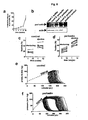
- periostin induces DNA synthesis followed by cytokinesis in fully differentiated rod-shaped adult cardiomyocytes.
- periostin stimulates cardiomyocyte proliferation in vitro.
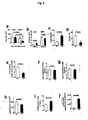
- periostin inactivates the cell cycle inhibitor retinoblastoma protein and increases levels of the cell cycle activator cyclin A in cardiomyocytes.
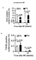
- injection of recombinant periostin increases cardiomyocyte DNA synthesis and cytokinesis in the adult rat heart in vivo.
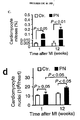
- periostin-induced cardiomyocyte proliferation improves cardiac function and reduces scar size after experimental myocardial infarction.
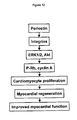
- Figure 12 depicts a schematic flow chart of periostin-induced cardiomyocyte regeneration.
- periostin increases cardiomyocyte cell cycle activity
- periostin attenuates fibrosis and hypertrophy and increases angiogenesis.
- Periostin improves the maladaptive myocardial remodeling following myocardial infarction.
- periostin activates ⁇ V ⁇ 1/3/5 integrins, which is consistent with the activation of ⁇ V ⁇ 1/3/5 integrins by periostin in other systems.
- attachment to ⁇ 1 integrins is required for proliferation of fetal cardiomyocytes.
- the fas1 domains of periostin are sufficient to induce proliferation, suggesting that the integrin interaction domain lies within this region. This is in accord with the mapping of the integrin interaction sites to the fas1 domains of the structurally related protein ⁇ igH3.
- ⁇ 1 and ⁇ 3 integrins are upregulated in peri-infarct cardiomyocytes, endothelial cells, and fibroblasts.
- the cardiac-specific knock-outs of ⁇ 1 or ⁇ 3 integrins show increased fibrosis, consistent with the finding that periostin, an agonist of ⁇ 1 and ⁇ 3 integrins, markedly decreases fibrosis after myocardial infarction and with increased extracellular matrix deposition in the heart valves of periostin knock-outs.
- ⁇ V ⁇ 1/3/5 integrins are also involved in angiogenesis and periostin promotes tumor angiogenesis.
- periostin-treated hearts have a higher capillary density in the infarct and border zone.
- PI3-kinase/Akt pathway Periostin, like FGF, requires the PI3-kinase/Akt pathway to induce cardiomyocyte cell cycle re-entry, although activation of this pathway is not sufficient to induce proliferation.
- Extracellular periostin in the absence of increased hemodynamic load, does not induce cardiomyocyte hypertrophy, but induces cell cycle re-entry and division. This is in contrast to two previous studies of cardiac overexpression of periostin or periostin-like factor under control of the CMV promoter.
- periostin-treated hearts determined by catheterization decreases the hypertrophic drive after myocardial infarction, consistent with smaller cardiomyocyte cross-sectional area.
- Sustained cardiomyocyte replacement attenuated the increased wall stress after myocardial infarction, resulting in improved ventricular remodeling.
- Increased cardiomyocyte DNA synthesis in the border zone of a myocardial infarction demonstrates that some differentiated cardiomyocytes reenter the cell cycle.
- the Examples show that differentiated mononucleated cardiomyocytes have proliferative potential.
- periostin-induced cycling of differentiated cardiomyocytes is associated with improved myocardial function and remodeling and with infarct regression.
- periostin requires at least one integrin subunit selected from the group consisting of ⁇ v , an ⁇ 1 , ⁇ 3 and ⁇ 5 integrin subunit. Integrins are upregulated in the infarct border zone, but have not been implicated in cell-cycle reentry of differentiated cardiomyocytes.
- Cardiac-specific knockout of ⁇ 1 or ⁇ 3 integrin results in increased fibrosis; this is consistent with our finding that periostin, a ligand of ⁇ 1 and ⁇ 3 integrins, markedly decreases fibrosis after myocardial infarction, and is also consistent with the increased extracellular matrix deposition in the heart valves of periostin knockout mice.
- periostin-treated hearts determined by catheterization decreased the hypertrophic drive after myocardial infarction, consistent with the smaller cardiomyocyte cross-sectional area observed.
- Sustained cardiomyocyte replacement attenuated the increase in wall stress after myocardial infarction, resulting in improved ventricular remodeling.
- periostin like FGF, requires the DI3K/Akt pathway to induce cardiomyocytes cell-cycle reentry. Because of the myocardial protective effect of Akt, it is plausible to propose that periostin has an anti-apoptotic effect. However, our data show that periostin has no protective effect, suggesting that the pro-apoptotic signals dominate over possible protection through periostin-induced PI3K activation. After myocardial infarction, the epicardial delivery strategy may not effectively reach all cardiomyocytes in the ischemic area to induce functionally and structurally relevant myocardial protection.
- myocardial function can be achieved with circulating or resident stem cells, which requires their isolation, in vitro propagation, and transplantation. Although our results do not exclude the possibility that cardiogenic stem cells contribute to periostin-induced myocardial repair, we have shown that a subpopulation of differentiated cardiomyocytes has proliferative potential. Stimulating proliferation of endogenous cardiomyocytes with periostin, periostin derivatives, or mimetics provide an innovative approach to induce myocardial repair.
- the disclosure is also applicable to tissue engineering where cells can be induced to proliferate by treatment with periostin, variants or fragments thereof (or such compositions together with growth factors) ex vivo. Following such treatment, the resulting tissue can be used for implantation or transplantation.
- perostin or biologically active variants or fragments thereof, can be used as reagents in ex vivo applications.
- perostin fragments are introduced into tissue or cells that are transplanted into a subject for therapeutic effect.
- the cells and/or tissue can be derived from an organism or subject that later receives the explant, or can be derived from another organism or subject prior to transplantation.
- the perostin compositions can be used to modulate the signaling pathway in the cells (i.e., cardiomyocytes), such that the cells or tissue obtain a desired phenotype or are able to perform a function (i.e., cardiomyocyte proliferation) when transplanted in vivo.
- Certain target cells from a patient may be extracted.
- ex vivo applications include use in organ/tissue transplant, tissue grafting, or treatment of heart disease. Such ex vivo applications can also used to treat conditions associated with coronary and peripheral bypass graft failure, for example, such methods can be used in conjunction with peripheral vascular bypass graft surgery and coronary artery bypass graft surgery.
- compositions and methods described herein have utility in research and drug development, as well as in surgery, tissue engineering, and organ transplantation.
- Periostin, variants or fragments thereof may be delivered locally, both continuously and transiently, and systemically.
- the disclosure could be used to modify or reduce scar tissue around the heart, speed up healing, and enhance cardiac tissue generation.
- the methods and compositions described herein provide the ability to successfully generate new tissue, augment organ function, and preserve the viability of impaired tissues, such as ischemic tissues.
- the present disclosure can enhance the viability of tissue.
- Heart failure in humans begins with reduced myocardial contractility, which leads to reduced cardiac output.
- the methods and composition described herein can be used to augment heart function. For example, they can be used to enhance growth of cardiomyocytes in an area of the heart that has been damaged or has become ischaemic.
- Heart diseases include, but are not limited to angina pectoris, myocardial infarction, and chronic ischemic heart disease.
- Periostin, variants or fragments thereof, or a combination of one or more variants or fragments thereof can be administered as compositions by various known methods, such as by injection (direct needle injection at the delivery site, subcutaneous, intravenous, etc.), oral administration, inhalation, transdermal application, catheter infusion, biolistic injectors, particle accelerators, Gelfoam, other commercially available depot materials, osmotic pumps, oral or suppositorial sond pharmaceutical formulations, decanting or topical applications during surgery, or aerosol delivery.
- the composition can be coated with a material to protect the compound from the action of acids and other natural conditions which can inactivate the compound.
- the composition can further include both the periostin compound and another agent, such as, but not limited to, a growth factor.
- the composition can be coated with, or co-administer the composition with, a material to prevent its inactivation.
- the composition can be administered to a subject in an appropriate diluent or in an appropriate carrier such as liposomes.
- Pharmaceutically acceptable diluents include saline and aqueous buffer solutions.
- Liposomes include water-in-oil-in-water CGF emulsions as well as conventional liposomes ( Strejan et al., J. Neuroimmunol. 7:27 (1984 )).
- composition containing at least one periostin protein, variants or fragments thereof can also be administered parenterally or intraperitoneally.
- Dispersions can also be prepared in glycerol, liquid polyethylene glycols, and mixtures thereof and in oils. Under ordinary conditions of storage and use, these preparations can contain a preservative to prevent the growth of microorganisms.
- compositions suitable for injectable use include sterile aqueous solutions (where water soluble) or dispersions and sterile powders for the extemporaneous preparation of sterile injectable solutions or dispersion.
- the composition must be sterile and must be fluid to the extent that easy syringability exists. It must be stable under the conditions of manufacture and storage and must be preserved against the contaminating action of microorganisms such as bacteria and fungi.
- the carrier can be a solvent or dispersion medium containing, for example, water, ethanol, polyol (for example, glycerol, propylene glycol, and liquid polyetheylene gloycol, and the like), suitable mixtures thereof, and vegetable oils.
- the proper fluidity can be maintained, for example, by the use of a coating such as licithin, by the maintenance of the required particle size in the case of dispersion and by the use of surfactants.
- Prevention of the action of microorganisms can be achieved by various antibacterial and antifungal agents. In many cases, it will be preferable to include isotonic agents, for example, sugars, polyalcohols such as manitol, sorbitol, sodium chloride in the composition.
- Prolonged absorption of the injectable compositions can be brought about by including in the composition an agent which delays absorption, for example, aluminum monostearate and gelatin.
- Sterile injectable solutions can be prepared by incorporating the composition containing the periostin molecule, variants or fragments thereof in the required amount in an appropriate solvent with one or a combination of ingredients enumerated above, as required.
- dispersions are prepared by incorporating the composition into a sterile vehicle which contains a basic dispersion medium and the required other ingredients from those enumerated above.
- the composition containing the periostin composition can be orally administered, for example, with an inert diluent or an assimilable edible carrier.
- the composition and other ingredients can also be enclosed in a hard or soft shell gelatin capsule, compressed into tablets, or incorporated directly into the subject's diet.
- the composition can be incorporated with excipients and used in the form of ingestible tablets, buccal tablets, troches, capsules, elixirs, suspensions, syrups, wafers, and the like.
- the percentage of the compositions and preparations can, of course, be varied.
- the amount of active compound in such therapeutically useful compositions is such that a suitable dosage will be obtained.
- the tablets, troches, pills, capsules and the like can also contain a binder, an excipient, a lubricant, or a sweetening agent.
- Various other materials can be present as coatings or to otherwise modify the physical form of the dosage unit.
- tablets, pills, or capsules can be coated with shellac, sugar or both.
- any material used in preparing any dosage unit form should be pharmaceutically pure and substantially non-toxic in the amounts employed.
- pharmaceutically acceptable carrier includes any solvents, dispersion media, coatings, antibacterial and antifungal agents, isotonic and absorption delaying agents, and the like. The use of such media and agents for pharmaceutically active substances is well known in the art. Except insofar as any conventional media or agent is incompatible with the active compound, use thereof in compositions of the invention is contemplated.
- Dosage unit form refers to physically discrete units suited as unitary dosages for the subjects to be treated. Each dosage contains a predetermined quantity of active compound calculated to produce the desired therapeutic effect in association with the required pharmaceutical carrier.
- the specification for the novel dosage unit forms of the invention is dependent on the unique characteristics of the composition containing periostin, variants or fragments thereof, and the particular therapeutic effect to be achieved. Dosages are determined by reference to the usual dose and manner of administration of the ingredients.
- periostin stimulates mononuclear cardiomyocytes, present in the adult mammalian heart, to undergo the full mitotic cell cycle.
- Periostin activates ⁇ V ⁇ 1/3/5 integrins located in the cardiomyocyte cell membrane.
- Periostin-induced cardiomyocyte proliferation requires activation of the ERK1/2 and Akt signaling pathways.
- recombinant periostin induces cardiomyocyte cell cycle re-entry, improves cardiac remodeling and function, reduces fibrosis and infarct size, and increases angiogenesis.
- Periostin and the pathway it regulates provides a new target for innovative strategies to treat heart failure.
- periostin Recombinant periostin, purified from HEK293 cells expressing the full-length human cDNA, was provided by Drs. Lan-Bo Chen and Meiru Dai (DFCI, Boston, MA) ( Tai, I.T., Dai, M. & Chen, L.B. Carcinogenesis 26, 908-15 (2005 )).
- the periostin fasl-only polypeptide was purified from E. coli and provided by BioVendor (Chandler, NC). Purity of >90% was established by SDS-PAGE and Coomassie staining.
- the periostin, derivative consisting of 4 fas1 domains and purified from bacteria was provided by BioVendor (Chandler, NC).
- FGF1 was provided by R&D Systems (Minneapolis, MN)
- the integrin ⁇ V (RMV-7) and ⁇ 1 (HM ⁇ 1-1) antibodies were provided by BioLegend (San Diego, CA).
- the integrin ⁇ V ⁇ 3 antibody (LM609) was kindly provided by Bing Luo (Center for Blood Research, Harvard Medical School) and the integrin ⁇ 7 antibody (6A11) was provided by MBL (Woburn, MA).
- the integrin ⁇ 3 (F11) and ⁇ 5 (MAB 1961Z), the aurora B kinase antibody, all chemical inhibitors, and type 1 rat collagen were provided by BD Biosciences (Franklin Lakes, NJ).
- Rat fibronectin was from Biomedical Technologies (Stoughton, MA).
- the tropomyosin (CH1) and troponin T (CT3) antibodies were provided by the Developmental Studies Hybridoma Bank (Iowa City, IA).
- Antibodies against human periostin, 5-Bromo-2'-deoxyuridine (BrdU), phosphorylated histone H3, and Ki67 were provided by AbCam (Cambridge, MA).
- the Cyclin A, troponin I, vWF, and c-kit antibodies were provided by Santa Cruz Biotechnology (Santa Cruz, CA).
- the phospho-Rb, phospho-ERK1/2, and phospho-Akt antibodies were provided by Cell Signaling Technology (Danvers, MA). Fluorophore-conjugated secondary antibodies raised in goat were provided by Invitrogen.
- Primary neonatal rat ventricular cardiomyocytes were isolated using the Neomyts kit (Cellutron, Highland Park, NJ), seeded at a density of 10 5 cells/cm 2 onto gelatin-coated cover slips, stimulated for 3 days, and labeled with BrdU for the terminal 2 days.
- Primary adult ventricular cardiomyocytes were isolated from male (300 gm) Wistar rats (Charles River Laboratories) and maintained as described ( Engel, F.B. et al. p38 MAP kinase inhibition enables proliferation of adult mammalian cardiomyocytes. Genes Dev 19, 1175-87 (2005 )).
- Adult cardiomyocytes were stimulated daily for 9 days and labeled with 30 ⁇ M BrdU for the terminal 3 days.
- Cardiomyocytes were stimulated with periostin (500 ng/mL) and FGF (100 ng/mL). Inhibiting antibodies were added at a concentration of 10 ⁇ g/mL, cRGD at 10 ⁇ M, U0126 at 10 ⁇ M, PD98059 at 1 ⁇ M, LY294002 at 10 ⁇ M, and Wortmannin at 0.1 ⁇ M 30 min prior to stimulation.
- the vehicle used to dissolve factors or chemicals was added to unstimulated samples, which were then processed together with the test samples.
- DNA-synthesis was analyzed by visualization of BrdU uptake and cytokinesis was determined by detection of the cleavage furrow and the midbody with an antibody against aurora B kinase as described ( Engel, F.B. et al. p38 MAP kinase inhibition enables proliferation of adult mammalian cardiomyocytes. Genes Dev 19, 1175-87 (2005 )). Signals were visualized with secondary antibodies conjugated to Alexa 488, Alexa 546, Alexa 594, and Alexa 647 (Invitrogen). Nuclei were visualized with 4',6'-diamidino-2-phenylindole (DAPI, Invitrogen). For image acquisition, the value for y was set at 1 and the lookup table settings were linear.
- Cardiomyocyle proliferation, survival, and apoptosis are important roles in cardiovascular disease.
- cardiomyocyte proliferation primary neonatal rat ventricular cardiomyocytes were quantified by counting with a hemocytometer after 3 days and 6 days of stimulation.
- cardiomyocyte survival primary neonatal rat ventricular cardiomyocytes were pre-treated with 500 ng/mL periostin or buffer for 30 min and then exposed to 0.5 ⁇ M doxycycline or 100 ng/mL TNF ⁇ for 24 hr and quantified by counting.
- Corresponding samples were stained with an antibody directed against tropomyosin as a cardiomyocyte marker to control for the percentage of differentiated cardiomyocytes.
- Apoptotic cardiomyocyte nuclei were determined after 24 hr with the In situ Cell Death Detection Kit (Roche, Indianapolis, IN) in combination with staining for troponin I.
- RNA was prepared using Trizol reagent (Invitrogen) from neonatal rat ventricular cardiomyocytes after 72 hr of stimulation.
- Complementary DNA was synthesized from 2 ⁇ g RNA with the SuperScriptIII kit (Invitrogen) using oligo-dT primers.
- One ⁇ L cDNA was used as template for 20 ⁇ L PCR-reactions (95 °C for 2 min, 30 cycles of 95 °C for 2 min, 55°C for 2 min, 72 °C for 5 min, followed by 72 °C for 10 min).
- Oligonucleotide primers used for detection of ANF were F: 5'-ATACAGTGCGGTGTCCAACA-3' (Seq ID No.
- R 5'-CTTTTCCAGGGAGGAGGAAG-3' (Seq ID No. 9); SERCA2 were F: 5'-ACTGGTGATGGTGTGAACGA-3' (Seq ID No. 10), R: 5'-TACGGGGACTCAAAGATTGC-3' (Seq ID No. 11), and for GAPDH: 5'-CTCATGACCACAGTCCATGC-3' (Seq ID No. 12), R: 5'-ATGTAGGCCATGAGGTCCAC-3' (Seq ID No. 13).
- rat ventricular cardiomyocytes Primary adult rat ventricular cardiomyocytes were incubated with 0.2 ⁇ g/mL Hoechst 33342 (Invitrogen) for 10 min. Nuclei were visualized with a mercury arc lamp and 4 random 20x microscopic fields were counted.
- Cardiomyocytes Primary adult rat ventricular cardiomyocytes were grown on gridded cover slips (Electron Microscopy Services, Ft. Washington, PA) and imaged in phase contrast at 10x magnification. Cardiomyocytes were transduced with an adenovirus leading to nuclear expression of histone2B-GFP ( Shi, Q. & King, R.W. Nature 437, 1038-42 (2005 )) under control of the chicken troponin T promoter ( Ma, H., Sumbilla, C.M., Farrance, I.K., Klein, M.G. & Inesi, G. Am JPhysiol Cell Physiol 286, C556-64 (2004 )).
- histone2B-GFP Shi, Q. & King, R.W. Nature 437, 1038-42 (2005 )
- the chicken troponin T promoter Ma, H., Sumbilla, C.M., Farrance, I.K., Klein, M.G. & Inesi, G. Am J
- Type 5 recombinant adenoviruses were generated as described ( He, T.C. et al. A simplified system for generating recombinant adenoviruses. Proc Natl Acad Sci USA 95, 2509-14 (1998 )).
- the -268 bp chick troponin T promoter segment was used to control expression of the histone2B-GFP fusion construct (BD Biosciences).
- the CMV promoter was used for expression of the human PTEN cDNA.
- Adult rat ventricular cardiomyocytes were infected at multiplicity of infection of 20.
- the diffusion range of recombinant periostin in myocardium was determined with a polyclonal antibody directed against human periostin followed by immunofluorescence microscopy. Presence of periostin in the myocardium was determined by Western blot using a polyclonal antibody directed against human periostin (Abcam).
- the infarct border zone was defined as myocardial tissue within 0.5 mm of the fibrous scar tissue. Fibrosis was determined on 10x images of Masson's Trichrome stainings and quantified using the Metamorph software package (Molecular Devices Corp.). Cardiomyocyte cross-sectional area was determined after staining with Masson's Trichrome at 40x magnification and quantified using the Metamorph software package. BrdU-positive cardiac fibroblast nuclei were determined at 5 cross-sections per heart at the level of the myocardial infarction. Cardiomyocyte nuclei were counted on troponin T and DAPI-stained sections in 32 - 60 random sample volumes of 84,500 ⁇ m 3 per heart. BrdU-positive cardiomyocyte nuclei were quantified on 16-20 sections per heart. Capillaries and stem cells were detected with antibodies against von Willebrand factor
- Oligomers directed against the coding sequences of ⁇ V and ⁇ 1 integrin subunits were designed and subcloned into the mammalian expression vector pcDNA 6.2-GW/EmEGFP-miR (Block-IT Pol II miR RNAi Expression Vector Kit, Invitrogen, Table 2). Table 2. Oligonucleotide sequences used for suppression of integrin subunits by RNAi. Gene name and ID Construct Oligonucleotide sequence Rat integrin ⁇ V XM_001068715 #1 #2 #3 Rat integrin ⁇ 1 NM_017022 #1 #2 #3
- Periostin stimulates cell cycle reentry and cell division of differentiated adult ventricular cardiomyocytes in vitro.
- periostin stimulates cell cycle reentry and cell division of differentiated adult ventricular cardiomyocytes in vitro.
- Periostin was identified in a screen of candidate compounds for induction of DNA synthesis in primary neonatal cardiomyocytes.
- Primary adult rat ventricular cardiomyocytes uniformly displayed rod-shaped phenotype, characteristic of full differentiation (data not shown).
- DNA synthesis was determined by uptake of the thymidine analog BrdU.
- Cardiomyocytes were identified by expression of the cardiac contractile protein tropomyosin and by expression of the cardiac transcription factor GATA-4 (data not shown).
- Cytokinesis was determined by visualization of the aurora B kinase, located at the contractile ring at the site of cytoplasmic separation and required for cytokinesis.
- Rod-shaped cardiomyocytes underwent cytokinesis after stimulation with periostin or FGF ( Fig. 1b ).
- Nonstimulated cardiomyocytes did not progress into cytokinesis.
- aurora B kinase after dissociation from the midbody, characteristic for completion of cytokinesis ( Fig. 1b ).
- Periostin stimulated cytokinesis in 0.5 ⁇ 0.2% and FGF in 0.3 ⁇ 0.1% of all differentiated cardiomyocytes ( Fig. 1b ).
- periostin stimulates DNA synthesis and cytokinesis in fully differentiated adult cardiomyocytes.
- periostin stimulates proliferation of neonatal cardiomyocytes.
- the presence of periostin in the developing myocardium prompted us to investigate if it induces proliferation of primary neonatal rat ventricular cardiomyocytes.
- Recombinant human periostin at a concentration of 500 ng/mL induced DNA synthesis in 35 ⁇ 5% of cardiomyocytes, a 5-fold increase over non-stimulated cardiomyocytes ( Fig. 1a ).
- Periostin induced a 12-fold increase of cardiomyocyte mitoses and 8-fold increase of cytokineses ( Fig. 1a ).
- Co-stimulation with FGF a factor known to induce cardiomyocyte DNA synthesis, had an additive effect ( Fig.
- Periostin increased the population of proliferating cardiomyocytes, determined by expression of the marker Ki-67, by 4-fold, similar to FGF ( Fig. 1c ).
- Periostin induced a 3.6-fold increase of cardiomyocyte proliferation after 3 days and a 4.2-fold increase after 6 days ( Fig. 1d ).
- Inactivation of the cell cycle inhibitor retinoblastoma protein (Rb) by phosphorylation is necessary for release of the G1/S checkpoint of the cell cycle ( Pasumarthi, K.B. & Field, L.J. Cardiomyocyte cell cycle regulation.
- Periostin increased the number of cardiomyocytes with detectable levels of phosphorylated Rb by 12-fold ( Fig. 1e ).
- periostin induced cardiomyocyte DNA synthesis with half-maximal stimulation at 155 ⁇ 25 nM, indicating a highly specific interaction of periostin with its receptor ( Fig. 2a ). Because periostin activates integrins in other systems, we determined if blocking integrins would inhibit periostin-stimulated cardiomyocyte cell cycle activity.
- Cyclic Arg-Gly-Asp which blocks the activation site of a broad range of integrins, inhibited periostin-stimulated DNA synthesis, but not FGF-stimulated DNA synthesis ( Fig. 2b ).
- Blocking antibodies against ⁇ V, ⁇ 1, ⁇ 3, and ⁇ 5 integrins inhibited periostin-stimulated DNA synthesis ( Fig. 2b ) and proliferation ( Fig. 2c ).
- Blocking ⁇ 7 integrins had no effect. Fibronectin and collagen, known to activate integrins, did not induce DNA synthesis ( Fig. 2d ).
- periostin a derivative comprised of 4 fas1 domains.
- the periostin fas 1 domain-only polypeptide is sufficient to stimulate cardiomyocyte cell cycle reentry.
- V. ERK1/2 and Akt are required for periostin-induced cardiomyocyte proliferation.
- Integrins activate the ERK1/2 and PI3-kinase/Akt pathways of the mitogen activated protein kinase (MAPK) cascade ( Miranti, C.K. & Brugge, J.S. Sensing the environment: a historical perspective on integrin signal transduction. Nat Cell Biol 4, E83-90 (2002 )).
- Periostin activated ERK1/2 and Akt in neonatal cardiomyocytes ( Fig. 2f ). Because the ERK1/2 and PI3-kinase/Akt pathways control cardiomyocyte size and hypertrophy, we determined if periostin induces the hypertrophic gene program.
- periostin did not induce transcription of atrial natriuretic factor (ANF), a marker for cardiomyocyte hypertrophy ( Sadoshima, J. & Izumo, S. Annu Rev Physiol 59, 551-71 (1997 )) ( Fig. 2g ).
- ANF atrial natriuretic factor
- ERK1/2 activation by MEK1/2 was blocked with the compounds U0126 (10 ⁇ M) and PD98059 (1 ⁇ M) and Akt activation by PI3-kinase was blocked with LY294002 (10 ⁇ M) and Wortmannin (0.1 ⁇ M).
- Periostin-stimulated DNA synthesis was inhibited only by blocking Akt activation ( Fig. 2h ).
- cardiomyocyte cytokinesis ( Fig. 2i ) and proliferation ( Fig. 2j ) were inhibited by both, ERK1/2 and Akt inhibition.
- ERK1/2 is not required for periostin-induced cardiomyocyte cell cycle re-entry, but periostin must activate both pathways to complete the cell cycle and induce proliferation, suggesting specific functions for distinct cell cycle events. Because periostin augments cancer cell survival, we tested if periostin enhances cardiomyocyte survival. Baseline and induced cardiomyocyte apoptosis ( Fig. 2k ) and survival ( Fig. 2l ) were not affected by periostin.
- periostin and fragments thereof induce cell cycle reentry of differentiated cardiomyocytes.
- Primary adult rat ventricular cardiomyocytes uniformly displayed a rod-shaped phenotype, characteristic of terminal differentiation ( Fig. 3a, b ).
- DNA synthesis was determined. Tracking the fate of individual rod-shaped cardiomyocytes revealed that they undergo DNA synthesis while expressing the cardiac contractile protein tropomyosin.
- Periostin induced a 14-fold increase of cardiomyocyte DNA synthesis to 1.1 ⁇ 0.2%, similar to FGF, known to promote cell cycle re-entry of differentiated cardiomyocytes ( Fig. 3a ).
- cardiomyocytes In cardiomyocytes, cytokinesis is not obligatory.
- We detected cardiomyocyte cytokineses by visualizing aurora B kinase, located at the contractile ring at the site of cytoplasmic separation and required for cytokinesis.
- Nonstimulated cardiomyocytes did not progress into cytokinesis.
- aurora B kinase After stimulation with periostin, we detected aurora B kinase after dissociation from the midbody, characteristic for completion of cytokinesis ( Fig. 3b ).
- Periostin stimulated cytokinesis in 0.5 ⁇ 0.2% of cardiomyocytes, similar to FGF ( Fig. 3b ).
- extracellular periostin re-activates the cell cycle and induces division in terminally differentiated cardiomyocytes.
- cardiomyocyte DNA synthesis and binucleation during murine development Am J Physiol 271, H2183-9 (1996 )
- a possible origin of cardiomyocyte cytokinesis is breakdown of binucleated cardiomyocytes into mononucleated cardiomyocytes, without prior DNA synthesis.
- Periostin-induced cardiomyocyte DNA synthesis began after 3 days, whereas cytokinesis was not detected before 6 days ( Fig. 3c ).
- cardiomyocytes in cytokinesis should be BrdU-positive.
- a third-generation lentivirus Rubinson, D.A. et al. Nat. Genet. 33, 401-406 (2003 )
- we genetically labeled autologous cardiac fibroblasts to exclude the possibility that their fusion with differentiated cardiomyocytes induced cell-cycle reentry. Cardiac fibroblasts form cell-cell contacts with differentiated cardiomyocytes, but do not fuse to induce cell cycle re-entry in vitro.
- Isolated adult rat ventricular cardiomyocytes were co-cultured for 9 days with autologous cardiac fibroblasts genetically labeled by infection with a third generation lentivirus directing expression of GFP under control of the CMV promoter. Cell-cell contact between cardiomyocytes and cardiac fibroblasts without fusion was observed. In conclusion, periostin induces the full mitotic cell cycle in differentiated cardiomyocytes.
- the PI3-kinase/Akt pathway controls the size of differentiated cardiomyocytes. To determine if this pathway is required for cell cycle re-entry, we transduced the PI3-kinase inhibitor PTEN ( Crackower, M.A. et al. Regulation of myocardial contractility and cell size by distinct PI3K-PTEN signaling pathways. Cell 110, 737-49 (2002 )). Expression of PTEN abolished periostin-stimulated cardiomyocyte DNA synthesis and cytokinesis ( Fig. 3d &e), indicating that periostin releases the proliferative potential of differentiated cardiomyocytes in a PI3-kinase/Akt dependent mechanism.
- Cardiomyocyte cycling requires integrins and PI3K Periostin-induced cardiomyocyte cell-cycle reentry was concentration dependent ( Fig. 2A ) and did not correlate with the abundance of cardiac fibroblasts ( Fig. 5a-c ), suggesting that periostin has a direct effect on cardiomyocytes. As shown in Fig. 5a-c , periostin-induced cardiomyocyte cell cycle re-entry does not correlate with abundance of cardiac fibroblasts. Assays were performed in isolated adult rat ventricular cardiomyocytes. Isolated adult rat ventricular cardiomyocytes were seeded with different amounts of autologous cardiac fibroblasts. Periostin-induced cardiomyocyte DNA synthesis was determined by immunofluorescence microscopy.
- Fig. 5a Cardiomyocyte cell cycle re-entry in the absence and presence of periostin and different percentages of autologous cardiac fibroblasts ( Fig. 5b ). Isolated adult rat ventricular cardiomyocytes were seeded at different densities and periostin-induced cardiomyocyte cell cycle re-entry was determined by immunofluorescence microscopy ( Fig. 5c ).
- Integrins can function as periostin receptors in cardiomyocytes. We therefore determined whether blocking specific integrin subunits would inhibit periostin-stimulated cardiomyocyte cell-cycle activity. Antibodies specific to ⁇ v , ⁇ 1 , ⁇ 3 and ⁇ 5 integrins inhibited periostinstimulated DNA synthesis in differentiated cardiomyocytes, whereas blocking ⁇ 7 integrin had no effect ( Fig. 5d ). Reducing the expression of integrin ⁇ v or ⁇ 1 by RNA-mediated interference (RNAi) inhibited periostin-induced cell-cycle reentry in neonatal cardiomyocytes ( Fig. 2b, 2c , 6 ). Periostin requires integrin ⁇ v and a ⁇ 1 , ⁇ 3 or ⁇ 5 subunit to induce cardiomyocyte cellcycle reentry.
- RNAi RNA-mediated interference
- Periostin-stimulated proliferation of neonatal cardiomyocytes requires integrins.
- Periostin-induced DNA synthesis ( Fig. 2b ) and proliferation ( Fig. 2c ) of neonatal ventricular cardiomyocytes is blocked by cRGD (10 ⁇ M) and integrin blocking antibodies (10 ⁇ g/mL).
- Suppression of periostin-induced cardiomyocyte BrdU uptake with microRNA (miR) sequences targeting integrin ⁇ v and ⁇ 1 was analyzed (not shown).
- Fig. 6 shows quantification of periostin-induced BrdU-uptake after transduction with different targeting constructs. Dotted line indicates periostin-induced cardiomyocyte DNA synthesis in the presence of miR-negative control. Suppression of integrin ⁇ v and ⁇ 1 expression by miR constructs was visualized (not shown). Statistical significance tested with ANOVA.
- FGF-induced cardiomyocyte cell-cycle reentry is enhanced by concurrently inhibiting p38 mitogen-activated protein (MAP) kinase.
- MAP mitogen-activated protein
- Integrins activate the extracellular signal-regulated kinases 1 and 2 (ERK1/2) and PI3K pathways of the MAP kinase cascade24.
- ERK1/2 extracellular signal-regulated kinases 1 and 2
- PI3K pathways of the MAP kinase cascade24.
- compounds blocking the ERK1/2 pathway did not inhibit periostin-induced cell-cycle reentry.
- blocking the ERK1/2 pathway with the compounds PD98059 (1 mM) and U0126 (10 mM) did not reduce periostin-induced cell-cycle reentry, whereas under the same conditions they inhibited neuregulin-induced cell-cycle reentry ( Fig. 7B ).
- Akt protein kinase Akt is an important downstream target of PI3K in cardiomyocytes25. Inhibition of Akt signaling with two different compounds, SH-6 (10 mM) or an Akt1/2 inhibitor (1 mM), reduced periostin-induced cardiomyocyte cell-cycle reentry ( Fig. 7C ).
- adenoviral transduction to express regulators of the PI3K pathway.
- Activation of the PI3K cascade was functionally disrupted by transducing the bifunctional phosphatase PTEN26.
- Expression of PTEN abolished periostin-stimulated DNA synthesis in cardiomyocytes ( Fig. 7D ), indicating that periostin releases the proliferative potential of differentiated cardiomyocytes in a PI3K-dependent mechanism.
- Transduction of a constitutively active form of PI3K increased cardiomyocyte DNA synthesis in the absence of periostin ( Fig. 7D ), and adding periostin did not further augment cardiomyocyte cell-cycle reentry ( Fig. 7D ).
- Cardiomyocyte cell-cycle reentry induced by constitutively active PI3K was associated with abnormally shaped nuclei (data not shown). Whereas constitutively active Akt induces cardiomyocyte hypertrophy, nuclear targeted Akt is associated with cardiomyocyte proliferation. Accordingly, we transduced nuclear targeted Akt, which had no effect on cardiomyocyte DNA synthesis in the absence of periostin. In the presence of periostin, nuclear targeted Akt doubled the proportion of DNA-synthesizing cardiomyocytes ( Fig. 7D ). Taken together, these results suggest that PI3K signaling is required for periostin-induced reentry of differentiated cardiomyocytes into the cell cycle and is sufficient for cell-cycle reentry in the absence of periostin.
- the PI3K pathway is associated with cardiomyocyte hypertrophy and apoptosis.
- periostin induces hypertrophy of differentiated cardiomyocytes.
- Transcription of ⁇ -myosin heavy chain ( ⁇ -MHC), smooth muscle actin (SMA) and atrial natriuretic factor (ANF) markers of the fetal gene program induced in hypertrophy, was not increased by periostin in adult ventricular cardiomyocytes (RT-PCR data not shown).
- RT-PCR data positive control samples treated with phenylephrine showed increased transcription of ⁇ -MHC, SMA and ANF.
- SERCA2 sarco-/endoplasmic reticulum Ca 2+ -ATPase
- Induction of the fetal gene program is accompanied by increased protein synthesis leading to cardiomyocyte hypertrophy.
- the positive control phenylephrine (10 mM) induced [ 3 H]leucine uptake
- periostin did not induce uptake at any of the time points tested ( Fig. 7E ).
- periostin did not reduce doxorubicin or TNF- ⁇ -induced cardiomyocyte death ( Fig. 2L ).
- periostin stimulates cardiomyocyte cell cycle re-entry in vivo
- periostin was introduced into the myocardial extracellular matrix by injecting the left ventricular free wall. The presence of injected periostin was confirmed by immunofluorescence microscopy (immunofluorescence microscopy images not shown). Recombinant periostin did not induce fibrosis ( Fig. 4a ) or hypertrophy ( Fig. 4b ). Three intraperitoneal injections of BrdU with a half life of 2 hr were administered to detect a proportion of cycling cardiomyocytes. After 7 days, cycling cardiomyocytes were present near the injected areas (immunofluorescence microscopy images not shown).
- BrdU-positive cardiomyocytes had a differentiated phenotype as shown by striations, expression of contractile protein (troponin T), and expression of the myogenic transcription factor, MEF-2 (data not shown). Reconstruction of optical sections enabled us to assign BrU-positive nuclei unequivocally to cardiomyocytes.
- Periostin induced DNA synthesis in 0.7 ⁇ 0.1% and cytokinesis in 0.1 ⁇ 0.03% of cardiomyocyte nuclei in the injected area ( Fig. 4c ).
- periostin stimulates cardiomyocytes to divide in vivo.
- Differentiated cardiomyocytes were detected during and after cytokinesis.
- Reconstruction of optical sections provided unequivocal assignment of cleavage furrow and midbody to dividing cardiomyocytes.
- the proportion of cardiomyocytes visualized during cytokinesis in periostin-injected hearts was 0.008 ⁇ 0.002%.
- periostin induces DNA synthesis and division of differentiated cardiomyocytes in vivo.
- fibronectin was injected into control hearts. Fibronectin did not increase cardiomyocyte DNA synthesis or cytokinesis ( Fig. 4c & 4d ). These results indicate that periostin induces DNA synthesis and division of differentiated cardiomyocytes in vivo.
- Periostin and fragments thereof improve cardiac function and reduces fibrosis and hypertrophy after myocardial infarction.
- Sustained cardiomyocyte cell cycle activity improve function and decrease infarct size after myocardial infarction.
- Periostin's ability to associate with Gelfoam a biodegradable extracellular matrix preparation is demonstrated in this Example.
- the invention can be used with a long-term and/or controlled release delivery system. Periostin bound to Gelfoam and was gradually released ( Fig. 8a ).
- Fig. 8a Periostin bound to Gelfoam and was gradually released.
- we administered epicardial Gelfoam patches loaded with periostin or with buffer and determined the presence of recombinant periostin in the left ventricle after 12 weeks.
- the distance from the epicardium to the farthest diffusion range of periostin was 461 ⁇ 62 ⁇ m.
- periostin Treatment with periostin improved myocardial function as indicated by a steeper slope of the end-systolic pressure-volume relationship (E es ), higher preload-recruitable stroke work (PRSW), higher maximum rate of ventricular pressure rise (dP/dt max ), and a higher maximum ventricular elastance (E max , Fig. 8e &f and Table 3).
- E es end-systolic pressure-volume relationship
- PRSW preload-recruitable stroke work
- dP/dt max higher maximum rate of ventricular pressure rise
- E max a higher maximum ventricular elastance
- periostin-treated and control hearts had the same infarct size 1 week after myocardial infarction.
- periostin-treated hearts had a smaller scar volume ( Fig. 9a ).
- the infarct size was 50% smaller compared with control hearts ( Fig. 9a ).
- the left ventricular free wall was thicker and the septum was thinner in periostin-treated animals ( Fig. 9b ).
- ventricular remodeling was analyzed by measuring the thickness of the left ventricular free wall and the interventricular septum 12 weeks after myocardial infarction.
- the left ventricular free wall was thicker and the septum was thinner in periostin-treated hearts ( Fig. 9b ), consistent with improved remodeling.
- cardiac hypertrophy a component of ventricular remodeling.
- heart weight was similar to that in buffer-treated rats ( Fig. 9f & 9g ).
- periostin-treated rats had a smaller crosssectional cardiomyocyte area than did control rats, indicating less cellular hypertrophy ( Fig. 9h ).
- cardiomyocyte nuclei To determine whether the decreased infarct size of periostin-treated hearts was related to an increased number of cardiomyocytes, we quantified cardiomyocyte nuclei. Control rats had 12.6 million left ventricular cardiomyocyte nuclei, whereas periostin-treated rats had 18.8 million left ventricular cardiomyocyte nuclei ( Fig. 9i ). To determine directly if periostin induces cardiomyocyte cell cycle re-entry after myocardial infarction, we quantified DNA synthesis. Differentiated cardiomyocytes, identified by expression of the cardiac contractile protein troponin I and characteristic shape, underwent DNA synthesis (not shown).
- apoptotic cardiomyocytes The proportion of apoptotic cardiomyocytes was similar in control and periostin-treated hearts at both 1 and 12 weeks ( Fig. 10b ). In periostin-treated hearts, however, the proportion of cycling cardiomyocytes was fivefold higher than the proportion of apoptotic cardiomyocytes at 1 week after myocardial infarction and 100-fold higher at 12 weeks ( Fig. 10c ), suggesting that proliferation of differentiated cardiomyocytes can be the cellular mechanism underlying the decrease in infarct size in periostin-treated hearts.
- We investigated this possibility by visualizing H3P in condensed metaphase chromosomes in differentiated cardiomyocytes during mitosis (not shown). Treatment with periostin induced mitosis in 0.1 ⁇ 0.01% of cardiomyocyte nuclei at 1 week and in 0.2 ⁇ 0.5% at 12 weeks ( Fig. 10c ).
- This rate of cardiomyocyte mitosis is calculated to result in the generation of 5.5 x 10 6 cardiomyocyte nuclei over a period of 12 weeks.
- control and periostin-treated hearts had the same number of nuclei ( Fig. 10d ).
- periostin-treated hearts had 6.2 x 10 6 more cardiomyocyte nuclei than did control hearts ( Fig. 10d ), consistent with the results based on proliferative indices.
- Cell-cycle reentry and division of differentiated cardiomyocytes can therefore account for the periostin-induced functional and structural improvements.
- periostin stimulates angiogenesis after myocardial infarction
- the capillary ( Fig. 10e ) and arteriolar (Fig. 10f ) densities did not differ between control and periostin-treated hearts.
- periostin-treated hearts had a 4-fold increase in capillary and a 1.5-fold increase in arteriolar density as compared with control hearts.
- Improved cardiac function is associated with recruitment of c-kit-positive stem cells.
- the number of c-kit-positive cells in the infarct and border zone did not differ between periostin-treated and control hearts at 1 and at 12 weeks after myocardial infarction ( Fig. 10g ).
- Periostin stimulates angiogenesis.
- periostin stimulates angiogenesis after myocardial infarction
- Periostin-treated hearts had a 4-fold increased capillary density compared with control animals ( Fig. 11A ).
- the number of stem cells, identified with the marker c-kit Fazel, S. et al. Cardioprotective c-kit+ cells are from the bone marrow and regulate the myocardial balance of angiogenic cytokines. J Clin Invest 116, 1865-77 (2006 )
- periostin induces angiogenesis in the infarct area without increasing recruitment of stem cells.
Landscapes
- Health & Medical Sciences (AREA)
- Life Sciences & Earth Sciences (AREA)
- Engineering & Computer Science (AREA)
- Bioinformatics & Cheminformatics (AREA)
- Chemical & Material Sciences (AREA)
- Zoology (AREA)
- Biomedical Technology (AREA)
- Organic Chemistry (AREA)
- General Health & Medical Sciences (AREA)
- Biotechnology (AREA)
- Genetics & Genomics (AREA)
- Wood Science & Technology (AREA)
- Veterinary Medicine (AREA)
- Medicinal Chemistry (AREA)
- Cardiology (AREA)
- Public Health (AREA)
- Animal Behavior & Ethology (AREA)
- Pharmacology & Pharmacy (AREA)
- Proteomics, Peptides & Aminoacids (AREA)
- Biochemistry (AREA)
- Immunology (AREA)
- Marine Sciences & Fisheries (AREA)
- Rheumatology (AREA)
- Microbiology (AREA)
- Epidemiology (AREA)
- Cell Biology (AREA)
- General Engineering & Computer Science (AREA)
- Gastroenterology & Hepatology (AREA)
- Heart & Thoracic Surgery (AREA)
- Chemical Kinetics & Catalysis (AREA)
- General Chemical & Material Sciences (AREA)
- Nuclear Medicine, Radiotherapy & Molecular Imaging (AREA)
- Urology & Nephrology (AREA)
- Vascular Medicine (AREA)
- Medicines That Contain Protein Lipid Enzymes And Other Medicines (AREA)
- Medicines Containing Material From Animals Or Micro-Organisms (AREA)
Claims (16)
- Utilisation d'un composé comprenant une composition de périostine, dans la fabrication d'un médicament pour stimuler la prolifération de cardiomyocytes post-mitotiques suite à une lésion, où la composition de périostine comprend de la périostine, un fragment ou une variante de celle-ci et où la composition de périostine a l'aptitude de stimuler la prolifération de cardiomyocytes post-mitotiques, où la séquence variante diffère seulement en des substitutions conservatrices et a l'aptitude de stimuler la prolifération de cardiomyocytes post-mitotiques.
- Utilisation selon la revendication 1, où la composition de périostine active au moins l'une des sous-unités d'intégrines sélectionnées parmi le groupe comprenant les sous-unités d'intégrines αV, β1, β3 et β5.
- Utilisation selon la revendication 1, où la composition de périostine comprend au moins un fragment de la composition de périostine de la SEQ ID NO:1 1 ou une séquence qui est au moins identique à 80% à celle de la SEQ ID NO:1 1 et où le fragment ou la séquence a l'aptitude de stimuler la prolifération de cardiomyocytes post-mitotiques.
- Utilisation selon la revendication 1, où la composition de périostine comprend au moins un domaine de fascicline 1 (fas1) ou une séquence variante qui diffère seulement en des substitutions conservatrices et a l'aptitude de stimuler la prolifération de cardiomyocytes post-mitotiques.
- Procédé de production de cardiomyocytes dédifférenciés, le procédé comprenant les étapes consistant à :sélectionner des cardiomyocytes différenciés dans un tissu obtenu préalablement d'un sujet, lequel comprend lesdits cardiomyocytes;remettre lesdits cardiomyocytes concentrés en suspension dans un milieu de croissance contenant une quantité efficace de la composition de périostine; etcultiver lesdits cardiomyocytes remis en suspension dans le milieu de croissance pendant un temps et dans des conditions pour induire la prolifération d'au moins une portion desdits cardiomyocytes sélectionnés en culture, où au moins une portion desdits cardiomyocytes différenciés sélectionnés en culture subit au moins un tour de division de cardiomyocytes.
- Procédé selon la revendication 5, dans lequel la composition de périostine active au moins l'une des sous-unités d'intégrines sélectionnées parmi le groupe comprenant les sous-unités d'intégrines αV, β1, β3 et β5.
- Procédé selon la revendication 5, dans lequel la composition de périostine comprend au moins un fragment de la composition de périostine de la SEQ ID NO:1 1 ou une séquence qui est au moins identique à 80% à celle de la SEQ ID NO:1 1 et où le fragment ou la séquence a l'aptitude de stimuler la prolifération de cardiomyocytes post-mitotiques.
- Procédé selon la revendication 5, dans lequel la composition de périostine comprend au moins un domaine de fascicline 1 (fas1) ou une séquence variante qui diffère seulement en des substitutions conservatrices et a l'aptitude de stimuler la prolifération de cardiomyocytes post-mitotiques.
- Composition pharmaceutique comprenant une quantité efficace d'une composition de périostine et un véhicule, un diluant ou un milieu pharmaceutiquement acceptable, à utiliser pour stimuler la prolifération de cardiomyocytes post-mitotiques pour traiter un tissu cardiaque endommagé, où la composition de périostine comprend de la périostine, un fragment ou une variante de celle-ci et où la composition de périostine a l'aptitude de stimuler la prolifération de cardiomyocytes post-mitotiques, où la séquence variante diffère seulement en des substitutions conservatrices et a l'aptitude de stimuler la prolifération de cardiomyocytes post-mitotiques.
- Composition à utiliser selon la revendication 9, où la composition de périostine active au moins l'une des sous-unités d'intégrines sélectionnées parmi le groupe comprenant les sous-unités d'intégrines αV, β1, β3 et β5.
- Composition à utiliser selon la revendication 9, où la quantité efficace induit la prolifération des cardiomyocytes.
- Composition à utiliser selon la revendication 9, où la composition de périostine comprend au moins un fragment de la composition de périostine de la SEQ ID NO:1 1 ou une séquence qui est au moins identique à 80% à celle de la SEQ ID NO:1, où le fragment ou la séquence a l'aptitude de stimuler la prolifération de cardiomyocytes post-mitotiques.
- Composition à utiliser selon la revendication 9, où la composition de périostine comprend au moins un domaine de fascicline 1 (fas1) ou une séquence variante qui diffère seulement en des substitutions conservatrices et a l'aptitude de stimuler la prolifération de cardiomyocytes post-mitotiques.
- Composition à utiliser selon la revendication 9, où la composition de périostine est formulée pour administration parentérale.
- Composition à utiliser selon la revendication 9, où la composition de périostine est formulée pour administration avec un système d'administration à libération lente contrôlée.
- Composition à utiliser selon la revendication 9, où la composition de périostine est incorporée dans un échafaudage biologique biodégradable.
Applications Claiming Priority (2)
| Application Number | Priority Date | Filing Date | Title |
|---|---|---|---|
| US88193807P | 2007-01-22 | 2007-01-22 | |
| PCT/US2008/051659 WO2008091867A2 (fr) | 2007-01-22 | 2008-01-22 | Périostine induisant une prolifération de cardiomyocytes et favorisant une régénération cardiaque |
Publications (2)
| Publication Number | Publication Date |
|---|---|
| EP2117582A2 EP2117582A2 (fr) | 2009-11-18 |
| EP2117582B1 true EP2117582B1 (fr) | 2013-06-26 |
Family
ID=39645121
Family Applications (1)
| Application Number | Title | Priority Date | Filing Date |
|---|---|---|---|
| EP08713899.6A Not-in-force EP2117582B1 (fr) | 2007-01-22 | 2008-01-22 | Periostine induisant une proliferation de cardiomyocytes et favorisant une regeneration cardiaque |
Country Status (3)
| Country | Link |
|---|---|
| US (1) | US8936806B2 (fr) |
| EP (1) | EP2117582B1 (fr) |
| WO (1) | WO2008091867A2 (fr) |
Families Citing this family (11)
| Publication number | Priority date | Publication date | Assignee | Title |
|---|---|---|---|---|
| US8361960B2 (en) * | 2008-09-08 | 2013-01-29 | Ottawa Hospital Research Institute | Periostin-induced pancreatic regeneration |
| WO2011031299A1 (fr) * | 2009-08-28 | 2011-03-17 | Mount Sinai School Of Medicine Of New York University | Injections intrapéricardiques |
| RU2012143607A (ru) * | 2010-03-12 | 2014-04-20 | Дайити Санкио Компани, Лимитед | Способ пролиферации кардиомиоцитов с применением микро-рнк |
| US20120071407A1 (en) * | 2010-06-18 | 2012-03-22 | Douglas Hamilton | Method of treating wounds |
| KR101584146B1 (ko) * | 2014-01-02 | 2016-01-11 | 부산대학교 산학협력단 | 페리오스틴 단백질 또는 이의 단편을 유효성분으로 포함하는 허혈성 질환의 예방 또는 치료용 조성물 |
| US10017574B2 (en) * | 2015-05-07 | 2018-07-10 | Yeda Research And Development Co. Ltd. | Methods, kits and devices for promoting cardiac regeneration |
| WO2017151717A1 (fr) * | 2016-03-01 | 2017-09-08 | French Brent A | Compositions et procédés pour l'expression génique faisant intervenir un virus adéno-associé dans des cellules de type myofibroblastes |
| JPWO2018070397A1 (ja) * | 2016-10-12 | 2019-08-29 | 株式会社シノテスト | ペリオスチンの吸着防止剤及び吸着防止方法 |
| WO2019077810A1 (fr) * | 2017-10-17 | 2019-04-25 | 国立大学法人富山大学 | Promoteur de sécrétion de périostine et de pkm2 |
| EP3597211A1 (fr) | 2018-07-19 | 2020-01-22 | Chemotherapeutisches Forschungsinstitut Georg-Speyer-Haus | Composés de périostine pour le traitement de complications hématologiques |
| WO2020037076A1 (fr) | 2018-08-14 | 2020-02-20 | Emory University | Induction de la prolifération de cardiomyocytes et utilisations thérapeutiques associées |
Family Cites Families (3)
| Publication number | Priority date | Publication date | Assignee | Title |
|---|---|---|---|---|
| US7115554B1 (en) | 1993-05-06 | 2006-10-03 | Acorda Therapeutics, Inc. | Methods of increasing myotube formation or survival or muscle cell mitogenesis differentiation or survival using neuregulin GGF III |
| AUPP785098A0 (en) | 1998-12-21 | 1999-01-21 | Victor Chang Cardiac Research Institute, The | Treatment of heart disease |
| EP1649008A2 (fr) * | 2003-07-16 | 2006-04-26 | Boston Scientific Limited | Structures d'echafaudage alignees pour regeneration myocardique accrue |
-
2008
- 2008-01-22 US US12/524,097 patent/US8936806B2/en not_active Expired - Fee Related
- 2008-01-22 EP EP08713899.6A patent/EP2117582B1/fr not_active Not-in-force
- 2008-01-22 WO PCT/US2008/051659 patent/WO2008091867A2/fr not_active Ceased
Also Published As
| Publication number | Publication date |
|---|---|
| US8936806B2 (en) | 2015-01-20 |
| US20100166827A1 (en) | 2010-07-01 |
| WO2008091867A2 (fr) | 2008-07-31 |
| WO2008091867A3 (fr) | 2010-08-05 |
| EP2117582A2 (fr) | 2009-11-18 |
Similar Documents
| Publication | Publication Date | Title |
|---|---|---|
| US8936806B2 (en) | Periostin induces proliferation of cardiomyocytes and promotes cardiac regeneration | |
| Gao et al. | Therapeutic delivery of microRNA-125a-5p oligonucleotides improves recovery from myocardial ischemia/reperfusion injury in mice and swine | |
| Durrani et al. | Skeletal myoblasts for cardiac repair | |
| US11913021B2 (en) | Methods and compositions for promoting survival and proliferation of endothelial cells and stimulating angiogenesis | |
| Zhang et al. | Endothelial ERG alleviates cardiac fibrosis via blocking endothelin-1-dependent paracrine mechanism | |
| Kühn et al. | Periostin induces proliferation of differentiated cardiomyocytes and promotes cardiac repair | |
| Hahn et al. | Pre-treatment of mesenchymal stem cells with a combination of growth factors enhances gap junction formation, cytoprotective effect on cardiomyocytes, and therapeutic efficacy for myocardial infarction | |
| Testa et al. | Vascular endothelial growth factors in cardiovascular medicine | |
| EP1061944B1 (fr) | Antagonistes therapeutiques du recepteur de chemokine | |
| Brilla et al. | Renin-angiotensin system and myocardial collagen matrix: modulation of cardiac fibroblast function by angiotensin II type 1 receptor antagonism | |
| Lutz et al. | Local injection of stem cell factor (SCF) improves myocardial homing of systemically delivered c-kit+ bone marrow-derived stem cells | |
| Melchior-Becker et al. | Deficiency of biglycan causes cardiac fibroblasts to differentiate into a myofibroblast phenotype | |
| Castillero et al. | Activin type II receptor ligand signaling inhibition after experimental ischemic heart failure attenuates cardiac remodeling and prevents fibrosis | |
| Iso et al. | Priming with ligands secreted by human stromal progenitor cells promotes grafts of cardiac stem/progenitor cells after myocardial infarction | |
| Wang et al. | Combining neuropeptide Y and mesenchymal stem cells reverses remodeling after myocardial infarction | |
| US20120121557A1 (en) | Neuregulin induced proliferation of cardiomyocytes | |
| Smith et al. | Stem cells in the heart: what's the buzz all about?—Part 1: preclinical considerations | |
| Li et al. | Young bone marrow Sca-1 cells rejuvenate the aged heart by promoting epithelial-to-mesenchymal transition | |
| JP2009507494A (ja) | キメラのhcnチャネル | |
| AU2016246070B2 (en) | Epicardial-derived paracrine factors for repairing cardiac tissue | |
| Jurado Acosta et al. | Phosphorylation of GATA4 at serine 105 is required for left ventricular remodelling process in angiotensin II‐induced hypertension in rats | |
| EP3512528A1 (fr) | Modulation de l'expression de tjp1 pour réguler la régénération de cellules cardiaques | |
| Hamoudane et al. | Caveolin-1 and polymerase I and transcript release factor: New players in insulin-like growth factor-I receptor signaling | |
| WO2002017899A2 (fr) | Procede servant a reguler l'angiogenese | |
| US8592387B2 (en) | Methods for treating or preventing heart damage with integrin-linked kinase (ILK) compositions |
Legal Events
| Date | Code | Title | Description |
|---|---|---|---|
| PUAI | Public reference made under article 153(3) epc to a published international application that has entered the european phase |
Free format text: ORIGINAL CODE: 0009012 |
|
| 17P | Request for examination filed |
Effective date: 20090814 |
|
| AK | Designated contracting states |
Kind code of ref document: A2 Designated state(s): AT BE BG CH CY CZ DE DK EE ES FI FR GB GR HR HU IE IS IT LI LT LU LV MC MT NL NO PL PT RO SE SI SK TR |
|
| DAX | Request for extension of the european patent (deleted) | ||
| R17D | Deferred search report published (corrected) |
Effective date: 20100805 |
|
| 17Q | First examination report despatched |
Effective date: 20101018 |
|
| RIC1 | Information provided on ipc code assigned before grant |
Ipc: A61P 9/10 20060101ALI20121128BHEP Ipc: C12N 5/077 20100101ALI20121128BHEP Ipc: A61K 38/18 20060101AFI20121128BHEP |
|
| GRAP | Despatch of communication of intention to grant a patent |
Free format text: ORIGINAL CODE: EPIDOSNIGR1 |
|
| GRAS | Grant fee paid |
Free format text: ORIGINAL CODE: EPIDOSNIGR3 |
|
| GRAA | (expected) grant |
Free format text: ORIGINAL CODE: 0009210 |
|
| AK | Designated contracting states |
Kind code of ref document: B1 Designated state(s): AT BE BG CH CY CZ DE DK EE ES FI FR GB GR HR HU IE IS IT LI LT LU LV MC MT NL NO PL PT RO SE SI SK TR |
|
| REG | Reference to a national code |
Ref country code: GB Ref legal event code: FG4D |
|
| REG | Reference to a national code |
Ref country code: CH Ref legal event code: EP |
|
| REG | Reference to a national code |
Ref country code: AT Ref legal event code: REF Ref document number: 618392 Country of ref document: AT Kind code of ref document: T Effective date: 20130715 |
|
| REG | Reference to a national code |
Ref country code: IE Ref legal event code: FG4D |
|
| REG | Reference to a national code |
Ref country code: DE Ref legal event code: R096 Ref document number: 602008025568 Country of ref document: DE Effective date: 20130822 |
|
| PG25 | Lapsed in a contracting state [announced via postgrant information from national office to epo] |
Ref country code: SI Free format text: LAPSE BECAUSE OF FAILURE TO SUBMIT A TRANSLATION OF THE DESCRIPTION OR TO PAY THE FEE WITHIN THE PRESCRIBED TIME-LIMIT Effective date: 20130626 Ref country code: NO Free format text: LAPSE BECAUSE OF FAILURE TO SUBMIT A TRANSLATION OF THE DESCRIPTION OR TO PAY THE FEE WITHIN THE PRESCRIBED TIME-LIMIT Effective date: 20130926 Ref country code: SE Free format text: LAPSE BECAUSE OF FAILURE TO SUBMIT A TRANSLATION OF THE DESCRIPTION OR TO PAY THE FEE WITHIN THE PRESCRIBED TIME-LIMIT Effective date: 20130626 Ref country code: FI Free format text: LAPSE BECAUSE OF FAILURE TO SUBMIT A TRANSLATION OF THE DESCRIPTION OR TO PAY THE FEE WITHIN THE PRESCRIBED TIME-LIMIT Effective date: 20130626 Ref country code: LT Free format text: LAPSE BECAUSE OF FAILURE TO SUBMIT A TRANSLATION OF THE DESCRIPTION OR TO PAY THE FEE WITHIN THE PRESCRIBED TIME-LIMIT Effective date: 20130626 Ref country code: GR Free format text: LAPSE BECAUSE OF FAILURE TO SUBMIT A TRANSLATION OF THE DESCRIPTION OR TO PAY THE FEE WITHIN THE PRESCRIBED TIME-LIMIT Effective date: 20130927 |
|
| REG | Reference to a national code |
Ref country code: AT Ref legal event code: MK05 Ref document number: 618392 Country of ref document: AT Kind code of ref document: T Effective date: 20130626 |
|
| REG | Reference to a national code |
Ref country code: LT Ref legal event code: MG4D |
|
| PG25 | Lapsed in a contracting state [announced via postgrant information from national office to epo] |
Ref country code: HR Free format text: LAPSE BECAUSE OF FAILURE TO SUBMIT A TRANSLATION OF THE DESCRIPTION OR TO PAY THE FEE WITHIN THE PRESCRIBED TIME-LIMIT Effective date: 20130626 Ref country code: BG Free format text: LAPSE BECAUSE OF FAILURE TO SUBMIT A TRANSLATION OF THE DESCRIPTION OR TO PAY THE FEE WITHIN THE PRESCRIBED TIME-LIMIT Effective date: 20130926 |
|
| REG | Reference to a national code |
Ref country code: NL Ref legal event code: VDEP Effective date: 20130626 |
|
| PG25 | Lapsed in a contracting state [announced via postgrant information from national office to epo] |
Ref country code: LV Free format text: LAPSE BECAUSE OF FAILURE TO SUBMIT A TRANSLATION OF THE DESCRIPTION OR TO PAY THE FEE WITHIN THE PRESCRIBED TIME-LIMIT Effective date: 20130626 |
|
| PG25 | Lapsed in a contracting state [announced via postgrant information from national office to epo] |
Ref country code: CZ Free format text: LAPSE BECAUSE OF FAILURE TO SUBMIT A TRANSLATION OF THE DESCRIPTION OR TO PAY THE FEE WITHIN THE PRESCRIBED TIME-LIMIT Effective date: 20130626 Ref country code: BE Free format text: LAPSE BECAUSE OF FAILURE TO SUBMIT A TRANSLATION OF THE DESCRIPTION OR TO PAY THE FEE WITHIN THE PRESCRIBED TIME-LIMIT Effective date: 20130626 Ref country code: CY Free format text: LAPSE BECAUSE OF FAILURE TO SUBMIT A TRANSLATION OF THE DESCRIPTION OR TO PAY THE FEE WITHIN THE PRESCRIBED TIME-LIMIT Effective date: 20130814 Ref country code: EE Free format text: LAPSE BECAUSE OF FAILURE TO SUBMIT A TRANSLATION OF THE DESCRIPTION OR TO PAY THE FEE WITHIN THE PRESCRIBED TIME-LIMIT Effective date: 20130626 Ref country code: SK Free format text: LAPSE BECAUSE OF FAILURE TO SUBMIT A TRANSLATION OF THE DESCRIPTION OR TO PAY THE FEE WITHIN THE PRESCRIBED TIME-LIMIT Effective date: 20130626 Ref country code: IS Free format text: LAPSE BECAUSE OF FAILURE TO SUBMIT A TRANSLATION OF THE DESCRIPTION OR TO PAY THE FEE WITHIN THE PRESCRIBED TIME-LIMIT Effective date: 20131026 Ref country code: PT Free format text: LAPSE BECAUSE OF FAILURE TO SUBMIT A TRANSLATION OF THE DESCRIPTION OR TO PAY THE FEE WITHIN THE PRESCRIBED TIME-LIMIT Effective date: 20131028 Ref country code: AT Free format text: LAPSE BECAUSE OF FAILURE TO SUBMIT A TRANSLATION OF THE DESCRIPTION OR TO PAY THE FEE WITHIN THE PRESCRIBED TIME-LIMIT Effective date: 20130626 |
|
| PG25 | Lapsed in a contracting state [announced via postgrant information from national office to epo] |
Ref country code: ES Free format text: LAPSE BECAUSE OF FAILURE TO SUBMIT A TRANSLATION OF THE DESCRIPTION OR TO PAY THE FEE WITHIN THE PRESCRIBED TIME-LIMIT Effective date: 20131007 Ref country code: NL Free format text: LAPSE BECAUSE OF FAILURE TO SUBMIT A TRANSLATION OF THE DESCRIPTION OR TO PAY THE FEE WITHIN THE PRESCRIBED TIME-LIMIT Effective date: 20130626 Ref country code: PL Free format text: LAPSE BECAUSE OF FAILURE TO SUBMIT A TRANSLATION OF THE DESCRIPTION OR TO PAY THE FEE WITHIN THE PRESCRIBED TIME-LIMIT Effective date: 20130626 Ref country code: RO Free format text: LAPSE BECAUSE OF FAILURE TO SUBMIT A TRANSLATION OF THE DESCRIPTION OR TO PAY THE FEE WITHIN THE PRESCRIBED TIME-LIMIT Effective date: 20130626 |
|
| PG25 | Lapsed in a contracting state [announced via postgrant information from national office to epo] |
Ref country code: CY Free format text: LAPSE BECAUSE OF FAILURE TO SUBMIT A TRANSLATION OF THE DESCRIPTION OR TO PAY THE FEE WITHIN THE PRESCRIBED TIME-LIMIT Effective date: 20130626 |
|
| PG25 | Lapsed in a contracting state [announced via postgrant information from national office to epo] |
Ref country code: DK Free format text: LAPSE BECAUSE OF FAILURE TO SUBMIT A TRANSLATION OF THE DESCRIPTION OR TO PAY THE FEE WITHIN THE PRESCRIBED TIME-LIMIT Effective date: 20130626 |
|
| PLBE | No opposition filed within time limit |
Free format text: ORIGINAL CODE: 0009261 |
|
| STAA | Information on the status of an ep patent application or granted ep patent |
Free format text: STATUS: NO OPPOSITION FILED WITHIN TIME LIMIT |
|
| PG25 | Lapsed in a contracting state [announced via postgrant information from national office to epo] |
Ref country code: IT Free format text: LAPSE BECAUSE OF FAILURE TO SUBMIT A TRANSLATION OF THE DESCRIPTION OR TO PAY THE FEE WITHIN THE PRESCRIBED TIME-LIMIT Effective date: 20130626 |
|
| 26N | No opposition filed |
Effective date: 20140327 |
|
| REG | Reference to a national code |
Ref country code: DE Ref legal event code: R097 Ref document number: 602008025568 Country of ref document: DE Effective date: 20140327 |
|
| PG25 | Lapsed in a contracting state [announced via postgrant information from national office to epo] |
Ref country code: LU Free format text: LAPSE BECAUSE OF FAILURE TO SUBMIT A TRANSLATION OF THE DESCRIPTION OR TO PAY THE FEE WITHIN THE PRESCRIBED TIME-LIMIT Effective date: 20140122 Ref country code: MC Free format text: LAPSE BECAUSE OF FAILURE TO SUBMIT A TRANSLATION OF THE DESCRIPTION OR TO PAY THE FEE WITHIN THE PRESCRIBED TIME-LIMIT Effective date: 20130626 |
|
| REG | Reference to a national code |
Ref country code: CH Ref legal event code: PL |
|
| PG25 | Lapsed in a contracting state [announced via postgrant information from national office to epo] |
Ref country code: LI Free format text: LAPSE BECAUSE OF NON-PAYMENT OF DUE FEES Effective date: 20140131 Ref country code: CH Free format text: LAPSE BECAUSE OF NON-PAYMENT OF DUE FEES Effective date: 20140131 |
|
| REG | Reference to a national code |
Ref country code: IE Ref legal event code: MM4A |
|
| PG25 | Lapsed in a contracting state [announced via postgrant information from national office to epo] |
Ref country code: IE Free format text: LAPSE BECAUSE OF NON-PAYMENT OF DUE FEES Effective date: 20140122 |
|
| REG | Reference to a national code |
Ref country code: FR Ref legal event code: PLFP Year of fee payment: 9 |
|
| PG25 | Lapsed in a contracting state [announced via postgrant information from national office to epo] |
Ref country code: MT Free format text: LAPSE BECAUSE OF FAILURE TO SUBMIT A TRANSLATION OF THE DESCRIPTION OR TO PAY THE FEE WITHIN THE PRESCRIBED TIME-LIMIT Effective date: 20130626 |
|
| PG25 | Lapsed in a contracting state [announced via postgrant information from national office to epo] |
Ref country code: HU Free format text: LAPSE BECAUSE OF FAILURE TO SUBMIT A TRANSLATION OF THE DESCRIPTION OR TO PAY THE FEE WITHIN THE PRESCRIBED TIME-LIMIT; INVALID AB INITIO Effective date: 20080122 Ref country code: TR Free format text: LAPSE BECAUSE OF FAILURE TO SUBMIT A TRANSLATION OF THE DESCRIPTION OR TO PAY THE FEE WITHIN THE PRESCRIBED TIME-LIMIT Effective date: 20130626 |
|
| REG | Reference to a national code |
Ref country code: FR Ref legal event code: PLFP Year of fee payment: 10 |
|
| REG | Reference to a national code |
Ref country code: FR Ref legal event code: PLFP Year of fee payment: 11 |
|
| PGFP | Annual fee paid to national office [announced via postgrant information from national office to epo] |
Ref country code: GB Payment date: 20220127 Year of fee payment: 15 Ref country code: DE Payment date: 20220127 Year of fee payment: 15 |
|
| PGFP | Annual fee paid to national office [announced via postgrant information from national office to epo] |
Ref country code: FR Payment date: 20220125 Year of fee payment: 15 |
|
| P01 | Opt-out of the competence of the unified patent court (upc) registered |
Effective date: 20230523 |
|
| REG | Reference to a national code |
Ref country code: DE Ref legal event code: R119 Ref document number: 602008025568 Country of ref document: DE |
|
| GBPC | Gb: european patent ceased through non-payment of renewal fee |
Effective date: 20230122 |
|
| PG25 | Lapsed in a contracting state [announced via postgrant information from national office to epo] |
Ref country code: GB Free format text: LAPSE BECAUSE OF NON-PAYMENT OF DUE FEES Effective date: 20230122 Ref country code: DE Free format text: LAPSE BECAUSE OF NON-PAYMENT OF DUE FEES Effective date: 20230801 |
|
| PG25 | Lapsed in a contracting state [announced via postgrant information from national office to epo] |
Ref country code: FR Free format text: LAPSE BECAUSE OF NON-PAYMENT OF DUE FEES Effective date: 20230131 |