EP2123227A1 - Procédure chirurgicale endoscopique et appareil chirurgical - Google Patents
Procédure chirurgicale endoscopique et appareil chirurgical Download PDFInfo
- Publication number
- EP2123227A1 EP2123227A1 EP09006724A EP09006724A EP2123227A1 EP 2123227 A1 EP2123227 A1 EP 2123227A1 EP 09006724 A EP09006724 A EP 09006724A EP 09006724 A EP09006724 A EP 09006724A EP 2123227 A1 EP2123227 A1 EP 2123227A1
- Authority
- EP
- European Patent Office
- Prior art keywords
- spacial structure
- spacial
- body cavity
- treatment
- surgical apparatus
- Prior art date
- Legal status (The legal status is an assumption and is not a legal conclusion. Google has not performed a legal analysis and makes no representation as to the accuracy of the status listed.)
- Granted
Links
Images




Classifications
-
- A—HUMAN NECESSITIES
- A61—MEDICAL OR VETERINARY SCIENCE; HYGIENE
- A61B—DIAGNOSIS; SURGERY; IDENTIFICATION
- A61B17/00—Surgical instruments, devices or methods
- A61B17/02—Surgical instruments, devices or methods for holding wounds open, e.g. retractors; Tractors
- A61B17/0218—Surgical instruments, devices or methods for holding wounds open, e.g. retractors; Tractors for minimally invasive surgery
-
- A—HUMAN NECESSITIES
- A61—MEDICAL OR VETERINARY SCIENCE; HYGIENE
- A61B—DIAGNOSIS; SURGERY; IDENTIFICATION
- A61B17/00—Surgical instruments, devices or methods
- A61B17/00234—Surgical instruments, devices or methods for minimally invasive surgery
-
- A—HUMAN NECESSITIES
- A61—MEDICAL OR VETERINARY SCIENCE; HYGIENE
- A61B—DIAGNOSIS; SURGERY; IDENTIFICATION
- A61B17/00—Surgical instruments, devices or methods
- A61B17/00234—Surgical instruments, devices or methods for minimally invasive surgery
- A61B2017/00238—Type of minimally invasive operation
- A61B2017/00278—Transorgan operations, e.g. transgastric
-
- A—HUMAN NECESSITIES
- A61—MEDICAL OR VETERINARY SCIENCE; HYGIENE
- A61B—DIAGNOSIS; SURGERY; IDENTIFICATION
- A61B17/00—Surgical instruments, devices or methods
- A61B17/28—Surgical forceps
- A61B17/29—Forceps for use in minimally invasive surgery
- A61B2017/2901—Details of shaft
- A61B2017/2906—Multiple forceps
Definitions
- the present invention relates to an endoscopic surgical procedure and surgical apparatus.
- endoscopic surgeries such as cholecystectomy have been used, in which the inside of a body cavity is observed by an endoscope, internal organs are operated by treatment instruments or the like, and operations are performed.
- endoscopic surgeries it is important to displace internal organs which are not related to the observation or the operation and assure a field of view and operative field by enlarging a narrow cavity.
- the operations performed are namely; moving a body tissue or an internal organ located above observation or operation target with grasping forceps; slightly moving the internal organ or the like, which is not under observation or the operation target, during operations, with grasping forceps or the like; moving the internal organ or the like, which is not under observation or the operation target, with another grasping forceps or the like.
- an instrument having an axis body, a hollow tube provided in a revolvable manner around the axis body, and a plurality of elastic striatums, one end of which being fixed to the axis body and the other end of which being fixed to the hollow tube, is proposed in Japanese Unexamined Patent Application, First Publication No. H08-336538 .
- the apparatus proposed in the publication is able to select from a stored state in which the elastic striatum is constricted and an operation state in which the elastic striatum is enlarged by allowing the axis body and the hollow tube to rotate relatively. By selecting the operation state in which the elastic striatum is enlarged, it is possible to support the body tissue or the internal organ.
- a first aspect of the present invention is a surgical apparatus which includes: a spacial structure which is deformable to be shrunken or enlarged, when it is disposed in a body cavity in an enlarged state, it is possible to support tissues inside of the body cavity and to be temporarily placed in a body; a spacial structure introduction mechanism which is able to insert the spacial structure in a shrunken state into the body cavity; an endoscope or a treatment instrument, a distal end of which is inserted from an opening provided in the spacial structure disposed in the body cavity, the distal end is oriented to the pathological lesion located on the inner side of the tissues inside of the body cavity which is supported by the spacial structure.
- FIG 1 is a perspective view showing the overall picture of a surgical apparatus in accordance with the first embodiment of the present invention.
- a surgical apparatus 1 includes: a treatment endoscope 10; a spacial structure 20 capable of supporting tissues inside of a body cavity when disposed in an arbitral position inside of the body cavity; a spacial structure introduction mechanism for inserting the spacial structure 20 in a shrunken state into the body cavity (refer FIG. 2 ).
- the spacial structure 20 is inserted into the body cavity by using a channel 11a of an insertion portion 11 of the treatment endoscope 10. That is, the treatment endoscope 10 has a function as the spacial structure introduction mechanism for inserting the spacial structure 20 into the body cavity.
- the treatment endoscope 10 includes: the channel 11a; the insertion portion 11 which is inserted into the body cavity from a distal end thereof; an operation portion 12 for curving a distal end of the insertion portion 11; and a treatment instrument guide 14 for guiding right and left treatment instruments 17A and 17B into the insertion portion 11, via a pipe 13 and the operation portion 12, toward a front side of the distal end of the insertion portion 11.
- the treatment instrument guide 14 has right and left arms 15A and 15B which protrudes from the distal end of the insertion portion 11.
- the right and left arms 15A and 15B are independently curved right, left, up, and down by operating the first operation portions 16A and 16B on the proximal side of the treatment instrument guide 14.
- the right and left arms 15A and 15B are selected from a straight state suitable for insertion into the body cavity or a curved state suitable for the treatment.
- FIG 1 shows each of the right and left arms 15A and 15B in the curved state.
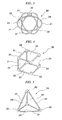
- FIGS. 3 to 5 show specific structural examples of the spacial structure 20.
- the spacial structure 20 assures adequate space in the body cavity during procedures. For example, in the case of extracting the cholecyst, the spacial structure 20 supports the liver R located above the cholecyst, exposes the cholecyst inside of the body cavity, and assures space necessary for the treatment around the cholecyst.
- Specific structures for the spacial structure 20 includes, as shown in FIGS. 3 to 5 , a plurality of rod members 21 made of elastic material, and connection portions 22 for connecting end portions of the rod members 21, for example.
- the rod members 21 and the connection portions 22 are flexible so that, when the entire spacial structure 20 is folded, the diameter of the spacial structure 20 is able to be inserted into the channel 11a of the insertion portion 11.
- Preferable materials for the rod members 21 and the connection portion 22 include shape-memory metal, a soft plastic material which is harmless to the human body, or the like.
- the entire shape of the spacial structure 20 includes a spherical shape as shown in FIG 3 , a rectangular hexahedron or a rectangular parallelepipe as shown in FIG 4 , or a three-sided pyramid as shown in FIG 5 .
- the spacial structure 20 is the rectangular parallelepipe or the three-sided pyramid, since at least a flat portion 23 is formed on the outer surface thereof, it is advantageous in that it can be stably placed on a flat place in the body cavity.
- these spacial structures 20 are formed by connecting the end portions of the rod members 21, which are assembled so as to interpose spaces therebetween, opening portions 24 are formed between the rod members 21. Also, when the spacial structure 20 is placed in the body cavity in the enlarged state so as to support the tissues inside of the body cavity such as internal organs in the body cavity, by using the opening portions 24, it is possible to orient the distal end of the endoscope or the treatment instrument to pathological lesion such as cholecyst A located on the inner side of the tissues inside of the body cavity supported by the spacial structure 20. (refer FIG 2 )
- spacial structure 20 connects end portions of the rod members 21 by the connection portions 22, the structure of the spacial structure 20 is not limited to this, but the end portions of the rod members 21 can be directly glued with adhesives or brazing alloys.
- the insertion portion 11 of the treatment endoscope 10 is inserted into a abdominal cavity by penetrating, for example, the umbilicus of the patient (step S1).
- necessary treatment instruments such as grasping forceps may be provided to the treatment instrument 10 in advance, or the treatment instruments 17A and 17B may be set to the treatment endoscope 10 when the distal end of the insertion portion 10 is inserted to a required position in the body cavity.
- the abdominal portion of the patient is made to swell by sending air into the abdominal cavity.
- the spacial structure 20 folded in advance is inserted from a forceps plug 11b of the insertion portion 11.
- a pushing instrument such as a wire is inserted into the forceps plug 11b, and the spacial structure 20 is sent into the abdominal cavity by pushing the spacial structure 20 through the channel 11a (step S2).
- the spacial structure 20 sent into the abdominal cavity from the channel 11a enlarges by its own elasticity (step S3).
- the enlarged spacial structure 20 is moved to a predetermined position in the abdominal cavity by using the treatment instruments 17A and 17B such as forceps or the like protruded from the distal end of the arms 15A and 15B.
- the treatment instruments 17A and 17B such as forceps or the like protruded from the distal end of the arms 15A and 15B.
- the liver R is lifted and supported to be separated from the cholecyst A (step S4). Accordingly, the cholectst A is exposed inside of the abdominal cavity and a space that is necessary for the observation or the treatment is assured around the cholecyst A by the space of the spacial structure 20.
- step S5 the distal ends of an observation portion or the treatment instruments 17A and 17B located at the distal end of the insertion portion 11 are oriented to the cholecyst A which is the pathological lesion by using the opening portion 24 of the spacial structure 20.
- step S6 damaged condition of the cholecyst A is confirmed by the observation portion of the treatment endoscope 10 (step S6), an adequate treatment such as extraction or the like is operated in accordance with the damaged condition of the cholecyst A.
- the spacial structure 20 is recovered by using the channel 11a of the insertion portion 11. That is, a recovery instrument for the grasping forceps or the like is inserted into the channel 11 from the forceps plug 11b for example, the spacial structure 20 is locked by protruding a distal end of the recovery instrument from the distal end of the channel 11a. Then, the spacial structure 20 is retracted into the channel 11a by the recovery instrument. The spacial structure 20 shrinks while being folded by its own elasticity and is retracted into the channel 11a. Then, the spacial structure 20 is retracted to the outside of the forceps plug 11b by passing through the channel 11.
- the treatment endoscope 1 is recovered with the treatment instruments 17A and 17B (step S8).
- the body tissues or the internal organs such as the liver R is supported by the spacial structure 20 enlarged in the body, due to the space inside of the spacial structure 20, it is possible to assure a space that is necessary for the observation or the treatment around the pathological lesion such as cholecyst A or the like located below the supported body tissues. By using the space, required treatments can be performed on the pathological lesion such as cholecyst A or the like.
- the spacial structure 20 can enlarge itself by its own elasticity without any operations from the outside of the body and support body tissues while maintaining the enlarged state. In this manner, since operations from the outside for enlarging the spacial structure 20 or maintaining the enlarged state are not necessary, it is possible to reduce the burden of operators, for example.
- FIG 7 is a partially cross sectional view showing an endoscopic surgical procedure using a surgical apparatus in accordance with a second embodiment of the present invention.
- the same reference numbers shall be given to identical portions and descriptions of overlapping portions with the first embodiment shall be omitted. This applies to a third embodiment and a fourth embodiment described later as well.
- the second embodiment is different from the first embodiment in that a spacial structure using a different structure is used and the spacial structure is inserted into the abdominal cavity by penetrating a wall portion of the anus or the large intestine not using the umbilicus as the insertion portion of the treatment endoscope.
- the spacial structure 40 of the embodiment has a plurality of pipes 41 and wire 42 inserted through the pipes 41.
- the spacial structure forms three-sided pyramids in the enlarged usage state.
- the spacial structure 40 becomes substantially a line shape as shown in FIG. 10 , when folded and shrunken.
- a relationship between the pipe 41 and the wire 42 shall be explained with reference to a usage state shown in FIG. 12 as an example.
- Two wires are used.
- the first wire 42a of the two wires is passed through the channel 11a of the insertion portion 11 of the treatment endoscope, inserted into a first pipe 41a, a second pipe 41b, and a third pipe 41c, and returned to the channel 11a again.
- the other second wire 42b is passed through the channel 11 a of the insertion portion 11 of the treatment endoscope, inserted into the fourth pipe 41d, a fifth pipe 41e, and the second pipe 41b, and also a sixth pipe 41 f, and returned to the channel 11a.
- the pipe 41 is made of a hard material of relatively high rigidity such as stainless steel and rigid plastic for example.
- the wire 42 is made of a material with relatively high rigidity so that when pushed in the longitudinal direction, the suppress strength can be transmitted to the distal end thereof.
- the distal end of the insertion portion 11 of the treatment endoscope 10 is inserted from a natural opening of the patient such as the anus to the large intestine, and is further inserted to the vicinity of the liver R in the abdominal cavity by penetrating the wall portion of the large intestine.
- the spacial structure 40 folded in advance is inserted from a forceps plug of the insertion portion 11, the spacial structure 40 is extruded from the distal end of the channel 11a by inserting and operating the distal ends of the wires 42a and 42b.
- the first pipe 41 a, the fifth pipe 41e, and the second pipe 41 b are extruded from the distal end of the channel 11a.
- one side of the first wire 42a inserted through the first pipe 41 a is no longer pushed, and the other side of the first wire 42a and both ends of the second wire 42b are pushed forward.
- the third pipe 41c and the sixth pipe 41f are extruded from the distal end of the channel 11a.
- both ends of the first wire 42a are no longer pushed, and both ends of the second wire 42b are pushed.
- the fourth pipe 41 d is extruded from the distal end of the channel 11a.
- the three-sided pyramid is formed by these extruded six pipes 41. That is, it is possible to enlarge the spacial structure 40 in the abdominal cavity.
- the spacial structure 40 By moving the enlarged spacial structure 40 to the distal end of the treatment endoscope, the spacial structure 40 is moved to a predetermined position in the abdominal cavity.
- the liver R is supported by the spacial structure 40 so as to be apart from the cholecyst A. Accordingly, the cholecyst A is exposed in the abdominal cavity, and a space necessary for the observation or treatment is assured around the cholecyst A by the space inside of the spacial structure 40.
- distal ends of the observation portion and the treatment instruments 17A and 17B located at the distal end of the insertion portion 11 are oriented to the cholecyst A which is the pathological lesion by using the opening portion 44 of the spacial structure 40, confirms the damaged condition of the cholecyst A, and performs necessary treatments thereafter.
- the spacial structure 40 is recovered by using the channel 11a of the insertion portion 11.
- the recovering procedure of the spacial structure 40 is opposite to the enlarging procedure of the spacial structure 40.
- the fourth pipe 41d is stored in the channel 11a.
- the fourth pipe 42d is extracted to the further proximal side of the channel 11a, and the third pipe 41 c and the sixth pipe 41 f are stored in the channel 11 a.
- the treatment endoscope 1 is recovered with the spacial structure 40 and the treatment instruments 17A and 17B stored in the channel 11a.
- the spacial structure 40 composed of pipe and wire is directly inserted into the channel 11a of the insertion portion 11.
- the embodiment is not limited to this but the spacial structure 40 may be first stored in the sheath, and the then spacial structure may be inserted into the channel with the sheath.
- FIG 13 is a partially cross sectional view showing an endoscopic surgical procedure using a surgical apparatus in accordance with a third embodiment of the present invention.
- the third embodiment is different from the second embodiment in that a spacial structure 45 using a different structure is used.
- a spacial structure 45 of the present embodiment has a plurality of pipes 46, a wire 47 inserted through the pipes 46, and a sheath 48 capable of storing the wire and the pipe.
- the spacial structure 45 forms a triangle with pipes by connecting three pipes 46 through one wire 47.
- the spacial structure 45 becomes substantially a line shape as shown in FIG 15 , when folded and shrunken.
- a relationship between the pipe 46 and the wire 47 shall be explained with reference to a usage state in FIG 13 .
- One side of the wire 47 protrudes from the distal end of the sheath 48, inserted into a first pipe 46a, a second pipe 46b, and a third pipe 46c, and is returned to the sheath 48 again. Both ends of the wire 47 protrude outward from a proximal end of the sheath 48, therefore, it is possible to push and extract only the wire 47.
- the sheath 48 is inserted into the channel 11a of the insertion portion 11 of the treatment endoscope.
- the pipe 46 is made of a hard material of relatively high rigidity such as stainless steel and rigid plastic for example.
- the wire 47 is made of a material with relatively high rigidity so that when pushed in the longitudinal direction, the force can be transmitted to the distal end thereof.
- the spacial structure 45 folded in advance as shown in FIG. 15 , is extruded from the distal end of the channel 11a of the insertion portion 11.
- the distal end of the sheath 48 is disposed in substantially the same position as the distal end of the insertion portion 11 of the treatment endoscope, with this state, a middle portion of the wire 47 is pushed forward. Then, as shown in FIG 14 , the first pipe 46a, the second pipe 46b, and the third pipe 46c are extruded from the distal end of the sheath 48, and those pipes form a triangle. Next, after abutting the position of the second pipe 46b to an adequate position of the body tissue, the distal end portion of the sheath 48 is extruded from the distal end of the channel 11a. Then, as shown in FIG.
- the plane of the triangle is substantially along the axis of the sheath 48 and is bent away from the axis of the sheath 48 at an apex of the triangle being pushed forward. Therefore, it is possible to lift and support the liver R so as to be separated from the cholecyst by the spacial structure 45 in which the pipes form a triangle in a bent state. Accordingly, the cholecyst is exposed inside of the abdominal cavity, and a space necessary for the observation or the treatment is assured around the cholecyst by the space inside of the spacial structure 45.
- distal ends of the observation portion and the treatment instrument located at the distal end of the insertion portion 11 are oriented to the cholecyst which is the pathological lesion by using the opening portion 49 of the spacial structure 45, confirms the damaged condition of the cholecyst, and performs necessary treatments thereafter.
- the spacial structure 45 is recovered by using the channel 11a of the insertion portion 11.
- the recovering procedure of the spacial structure 45 is opposite to the enlarging procedure of the spacial structure 45.
- the sheath 48 is stored in the channel 11a.
- the first pipe 46a, the second pipe 46b, and the third pipe 46c are respectively stored in the sheath 48, and then the sheath 48 is stored in the channel 11a.
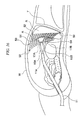
- FIG 16 is a partially cross sectional view showing an endoscopic surgical procedure using a surgical apparatus in accordance with a fourth embodiment of the present invention.
- the fourth embodiment is different from the first embodiment in that a spacial structure 50 using a different structure is used, and the spacial structure is inserted into the abdominal cavity by penetrating a wall portion of the anus or the large intestine not using the umbilicus as the insertion portion of the treatment endoscope.
- the spacial structure 50 of the embodiment has a mesh member 51, a fixture 52 for fixing the mesh member 51 in an enlarged state to the tissues inside of the body cavity.
- the mesh member 51 is made of silicon system materials, for example, which are flexible and harmless to the human body.
- a quadrangular grid is used for a basic shape, but it is not limited to this.
- the basic shape may be a triangle or hexagon.
- the fixture 52 is attached to a predetermined position of the mesh member 51 in advance.
- a member used for a clip which is able to grasp a portion of a biological tissue by a biasing force of a spring or the like, is used for the fixture 52 for example.
- the fixture 52 may be a wedge shape which is attached to a predetermined position of the mesh member 51 in advance, and be inserted and fixed to a biological tissue.
- the distal end of the insertion portion 11 of the treatment endoscope 10 is inserted from a natural opening of the patient such as the anus to the large intestine, and is further inserted to the vicinity of the liver R in the abdominal cavity by penetrating the wall portion of the large intestine.
- the spacial structure 50 folded in advance is inserted from a forceps plug of the insertion portion 11, and the pushing instrument such as the wire is inserted, and the spacial structure 50 is sent into the abdominal cavity by pushing the spacial structure 50 through the channel 11a.
- the spacial structure 50 extruded into the abdominal cavity from the channel 11a is slightly enlarged by its own elasticity.
- the plurality of fixtures 52 is fixed to the biological tissues such as the abdominal wall W or the diaphragm T, and the spacial structure 50 is set to the enlarged state.
- the mesh member 51 of the enlarged spacial structure 50 the biological tissue, which is not the treatment target such as the liver R, is directly supported.
- using a grasping member such as the clip attached to the mesh member 51 is also available other than directly supporting the biological tissue or the internal organs by the mesh member 51.
- a portion of the mesh member 51 is dissected by a treatment instrument for dissection. Accordingly, it is possible to expose the cholecyst A, which is the treatment target, closer to the treatment endoscope than the mesh member 51. Also, it is possible to assure a space necessary for the observation or the treatment around the cholecyst A by the space inside of the spacial structure 50.
- the distal ends of the observation portion and the treatment instruments 17A and 17B located at the distal end of the insertion portion 11 are oriented to the cholecyst A, which is the pathological lesion, by using an opening portion 54 of the spacial structure 50, confirms the damaged condition of the cholecyst A, and performs necessary treatments thereafter.
- the spacial structure 50 is recovered by using the channel 11 a of the insertion portion 11.
- a recovery instrument for the grasping forceps for example, is inserted into the channel 11a from the forceps plug 11b, the distal end of the recovery instrument is locked to the spacial structure 50, and the spacial structure 50 is retracted into the channel 11a.
- the treatment instrument 1 is recovered with the spacial structure 50 and the treatment instruments 17A and 17B stored in the channel.
- the body tissue is supported by the spacial structure 50 enlarged in the body, it is possible to assure a space that is necessary for the observation or the treatment around the pathological lesion such as the cholecyst A or the like located below the supported body tissues by the space 54 inside of the spacial structure 50.
- the space By using the space, required treatments can be operated to the pathological lesion such as cholecyst A or the like.
- the spacial structure 50 is composed mainly of the mesh member 51 which is freely deformable, it is possible to support in arbitral shape the internal organs or the like, which are not the treatment target, it is easier to assure the space for the treatment as a result.
- the spacial structure 50 is configured mainly by the mesh member 51 which is the mesh member flexible in itself, it is possible to dispose the spacial structure 50 in an arbitrary location in the body cavity. Also, since the size of the spacial structure 50 is large, it is advantageous in that it is easy to confirm that the spacial structure 50 is left behind in the body or not.
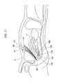
- FIG 17 shows an alternative example of the fourth embodiment.
- the spacial structure 50 composed of the mesh member 51 and the fixture 52, the bladder U is supported and the appendico AP located below the bladder U is treated.
- the insertion portion 11 of the treatment endoscope 1 is inserted into the body cavity from the mouth, for example, passes the esophagus and stomach, penetrates the stomach wall and reaches the inside of the abdominal cavity. Since the spacial structure 50 is made mainly of mesh member and is flexible, it is possible to introduce the spacial structure into the body cavity passing through the natural orifices, for example, by mouth, where only a relatively small curvature radius is available.
- the spacial structure is inserted into the body cavity via the channel 11 a of the insertion portion 11 of the treatment endoscope, it is not limited to this.
- a special instrument made of a pipe member or the like for inserting the spacial structure into the body cavity is prepared, and the spacial structure may be introduced into the body cavity by using the special instrument.
- FIGS. 1 and 2 examples in the case of using the grasping forceps and the dissection knife are shown, but the embodiment is not limited to those.
- Other treatment instruments such as a rotating grip, a high-frequency snare, a balloon, or the like may be used in accordance with treatment relative to the pathological lesion.
- the treatment endoscope 1, in which two treatment instruments can be introduced into the insertion portion 11, is used, but it is not limited to this.
- a treatment endoscope, in which only one treatment instrument can be introduced, may be used, or the endoscope may only have an observation function, and the treatment instrument may be introduced into the body cavity by using another pipe line.
- the above embodiments are described by treating the cholecyst A or the appendico AP for example, but it is not limited to this.
- the present invention can also be applied to cases in which the treatment is operated by using the spacial structure in the digestive canal.
- the spacial structure may support the tissue inside of the body cavity, which is not the treatment target, by introducing air therein.
Landscapes
- Health & Medical Sciences (AREA)
- Life Sciences & Earth Sciences (AREA)
- Surgery (AREA)
- Molecular Biology (AREA)
- Engineering & Computer Science (AREA)
- Biomedical Technology (AREA)
- Heart & Thoracic Surgery (AREA)
- Medical Informatics (AREA)
- Nuclear Medicine, Radiotherapy & Molecular Imaging (AREA)
- Animal Behavior & Ethology (AREA)
- General Health & Medical Sciences (AREA)
- Public Health (AREA)
- Veterinary Medicine (AREA)
- Surgical Instruments (AREA)
- Media Introduction/Drainage Providing Device (AREA)
- Endoscopes (AREA)
Applications Claiming Priority (1)
| Application Number | Priority Date | Filing Date | Title |
|---|---|---|---|
| US12/122,802 US8764630B2 (en) | 2008-05-19 | 2008-05-19 | Endoscopic surgical procedure and surgical apparatus |
Publications (2)
| Publication Number | Publication Date |
|---|---|
| EP2123227A1 true EP2123227A1 (fr) | 2009-11-25 |
| EP2123227B1 EP2123227B1 (fr) | 2019-04-10 |
Family
ID=40834456
Family Applications (1)
| Application Number | Title | Priority Date | Filing Date |
|---|---|---|---|
| EP09006724.0A Not-in-force EP2123227B1 (fr) | 2008-05-19 | 2009-05-19 | Appareil chirurgical endoscopique |
Country Status (3)
| Country | Link |
|---|---|
| US (1) | US8764630B2 (fr) |
| EP (1) | EP2123227B1 (fr) |
| JP (1) | JP5173930B2 (fr) |
Cited By (2)
| Publication number | Priority date | Publication date | Assignee | Title |
|---|---|---|---|---|
| GB2479179A (en) * | 2010-03-31 | 2011-10-05 | Neosurgical Ltd | Laparoscopic surgical retraction device |
| WO2014195422A1 (fr) * | 2013-06-05 | 2014-12-11 | Ladjali Mustapha | Dispositif de traitement d'un tissu corporel et nécessaire de traitement associé |
Families Citing this family (30)
| Publication number | Priority date | Publication date | Assignee | Title |
|---|---|---|---|---|
| US8100822B2 (en) | 2004-03-16 | 2012-01-24 | Macroplata Systems, Llc | Anoscope for treating hemorrhoids without the trauma of cutting or the use of an endoscope |
| US20100069947A1 (en) | 2006-11-27 | 2010-03-18 | Surgical Structure Ltd. | Device especially useful for hernia repair surgeries and methods thereof |
| CA2925611C (fr) * | 2007-10-17 | 2018-05-29 | Davol Inc. | Moyens de fixation entre une maille et des moyens de deploiement d'une maille, utiles notamment pour les chirurgies de reparation des hernies et procedes associes |
| ES2576079T3 (es) | 2008-05-07 | 2016-07-05 | Davol Inc. | Método y aparato para reparar una hernia |
| US20100069710A1 (en) * | 2008-09-02 | 2010-03-18 | Ken Yamatani | treatment method |
| US8870759B2 (en) * | 2009-12-04 | 2014-10-28 | Covidien Lp | Suspension system for minimally invasive surgery |
| US12376737B1 (en) | 2009-12-16 | 2025-08-05 | Boston Scientific Scimed, Inc. | Tissue retractor for minimally invasive surgery |
| US10595711B2 (en) | 2009-12-16 | 2020-03-24 | Boston Scientific Scimed, Inc. | System for a minimally-invasive, operative gastrointestinal treatment |
| US10531869B2 (en) | 2009-12-16 | 2020-01-14 | Boston Scientific Scimed, Inc. | Tissue retractor for minimally invasive surgery |
| US10966701B2 (en) | 2009-12-16 | 2021-04-06 | Boston Scientific Scimed, Inc. | Tissue retractor for minimally invasive surgery |
| US11344285B2 (en) | 2009-12-16 | 2022-05-31 | Boston Scientific Scimed, Inc. | Multi-lumen-catheter retractor system for a minimally-invasive, operative gastrointestinal treatment |
| US9565998B2 (en) | 2009-12-16 | 2017-02-14 | Boston Scientific Scimed, Inc. | Multi-lumen-catheter retractor system for a minimally-invasive, operative gastrointestinal treatment |
| US10758116B2 (en) | 2009-12-16 | 2020-09-01 | Boston Scientific Scimed, Inc. | System for a minimally-invasive, operative gastrointestinal treatment |
| USRE48850E1 (en) | 2009-12-16 | 2021-12-14 | Boston Scientific Scimed, Inc. | Multi-lumen-catheter retractor system for a minimally-invasive, operative gastrointestinal treatment |
| US9186131B2 (en) | 2009-12-16 | 2015-11-17 | Macroplata, Inc. | Multi-lumen-catheter retractor system for a minimally-invasive, operative gastrointestinal treatment |
| ES2874194T3 (es) | 2009-12-16 | 2021-11-04 | Boston Scient Scimed Inc | Disposiciones para realizar una estructura anatómica endoluminal |
| US8932211B2 (en) | 2012-06-22 | 2015-01-13 | Macroplata, Inc. | Floating, multi-lumen-catheter retractor system for a minimally-invasive, operative gastrointestinal treatment |
| WO2012047414A1 (fr) | 2010-10-05 | 2012-04-12 | C.R. Bard, Inc. | Prothèse de réparation de tissu mou et dispositif extensible |
| JP5989003B2 (ja) * | 2011-01-04 | 2016-09-07 | ザ・ジョンズ・ホプキンス・ユニバーシティ | 最小侵襲腹腔鏡手術用リトラクタ |
| US8758235B2 (en) | 2011-07-13 | 2014-06-24 | Cook Medical Technologies Llc | Foldable surgical retractor |
| EP2830416A4 (fr) | 2012-03-29 | 2015-12-16 | Lapspace Medical Ltd | Écarteur de tissus |
| CN104470414B (zh) * | 2012-06-22 | 2018-07-03 | 波士顿科学希梅德公司 | 用于最低限度侵入胃肠道手术治疗的多腔导管式牵开器系统 |
| US10758218B2 (en) * | 2015-07-02 | 2020-09-01 | Atlantic Health System, Inc. | Lighted polyhedral retractor |
| US10123791B2 (en) * | 2015-07-02 | 2018-11-13 | Atlantic Health System, Inc. | Lighted polyhedral retractor |
| KR101739225B1 (ko) | 2016-04-11 | 2017-05-24 | 계명대학교 산학협력단 | 복강경 수술용 파우치 |
| EP3562374A1 (fr) | 2016-12-30 | 2019-11-06 | Boston Scientific Scimed, Inc. | Système pour un traitement mini-invasif à l'intérieur d'une lumière corporelle |
| CN110913773B (zh) | 2017-03-18 | 2023-03-28 | 波士顿科学国际有限公司 | 用于体腔内的微创治疗的系统 |
| JP7302202B2 (ja) * | 2019-02-28 | 2023-07-04 | 日本ゼオン株式会社 | 内視鏡用スペーサ部材 |
| US11832789B2 (en) | 2019-12-13 | 2023-12-05 | Boston Scientific Scimed, Inc. | Devices, systems, and methods for minimally invasive surgery in a body lumen |
| US20240341742A1 (en) * | 2021-08-10 | 2024-10-17 | Atricure, Inc. | Expandable spacemaker and related methods |
Citations (8)
| Publication number | Priority date | Publication date | Assignee | Title |
|---|---|---|---|---|
| US5242456A (en) * | 1991-11-21 | 1993-09-07 | Kensey Nash Corporation | Apparatus and methods for clamping tissue and reflecting the same |
| FR2726993A1 (fr) * | 1994-11-18 | 1996-05-24 | Sgro Jean Claude | Dispositif gonflable a usage unique pour ecarter les tissus anatomiques notamment en chirurgie coelioscopique et son materiel d'application |
| JPH08336538A (ja) | 1995-06-12 | 1996-12-24 | Olympus Optical Co Ltd | 生体操作器具 |
| US5743851A (en) * | 1991-05-29 | 1998-04-28 | Origin Medsystems, Inc. | Retraction apparatus and methods for endoscopic surgery |
| US20030225432A1 (en) * | 2002-05-31 | 2003-12-04 | Baptiste Reginald C. | Soft tissue retraction device for an endoscopic instrument |
| WO2005104959A1 (fr) * | 2004-04-29 | 2005-11-10 | Umc Utrecht Holding B.V. | Dispositif de dilatation chirurgical |
| US20060069408A1 (en) * | 2004-09-29 | 2006-03-30 | Terumo Kabushiki Kaisha | Device for treating a patent foramen ovale |
| WO2007083305A2 (fr) * | 2006-01-17 | 2007-07-26 | Ez Surgical Ltd. | Dispositif d’ouverture d’acces pour procedures chirurgicales |
Family Cites Families (20)
| Publication number | Priority date | Publication date | Assignee | Title |
|---|---|---|---|---|
| JPS60168801A (ja) | 1984-02-09 | 1985-09-02 | 冨田 織江 | 道路の継目部の連続舗装化工法 |
| JPS60168801U (ja) * | 1984-04-19 | 1985-11-09 | 富士写真光機株式会社 | 内視鏡の先端部固定保持装置 |
| US6120437A (en) * | 1988-07-22 | 2000-09-19 | Inbae Yoon | Methods for creating spaces at obstructed sites endoscopically and methods therefor |
| US5275610A (en) * | 1991-05-13 | 1994-01-04 | Cook Incorporated | Surgical retractors and method of use |
| US5501653A (en) * | 1991-05-29 | 1996-03-26 | Origin Medsystems, Inc. | Abdominal wall lifting retractor with hinged cross-member |
| US5865728A (en) * | 1991-05-29 | 1999-02-02 | Origin Medsystems, Inc. | Method of using an endoscopic inflatable lifting apparatus to create an anatomic working space |
| US5361752A (en) | 1991-05-29 | 1994-11-08 | Origin Medsystems, Inc. | Retraction apparatus and methods for endoscopic surgery |
| NL9201118A (nl) * | 1992-06-24 | 1994-01-17 | Leuven K U Res & Dev | Gereedschapset voor laparoscopische vaginale hysterectomie. |
| JPH0631705A (ja) | 1992-07-15 | 1994-02-08 | Matsushita Electric Works Ltd | なら材スライス単板の製造方法 |
| US5681349A (en) * | 1992-08-13 | 1997-10-28 | Brigham & Women's Hospital | Expanding polygonal surgical compressor |
| JPH0631705U (ja) * | 1992-10-09 | 1994-04-26 | 住友ベークライト株式会社 | 医療用バルーン |
| JPH07116262A (ja) | 1993-10-22 | 1995-05-09 | Olympus Optical Co Ltd | 医療用膨張体 |
| JP4002649B2 (ja) | 1996-12-17 | 2007-11-07 | オリンパス株式会社 | 外科手術用腔確保具 |
| JP2000037388A (ja) | 1998-05-20 | 2000-02-08 | Osamu Yoshida | 臓器収納バッグと臓器収納バッグ挿入具 |
| US6231561B1 (en) * | 1999-09-20 | 2001-05-15 | Appriva Medical, Inc. | Method and apparatus for closing a body lumen |
| CA2405710C (fr) * | 2000-04-25 | 2010-04-20 | Impres Medical, Inc. | Procede et appareil pour creer des adherences intra-uterines |
| US7860555B2 (en) * | 2005-02-02 | 2010-12-28 | Voyage Medical, Inc. | Tissue visualization and manipulation system |
| US20070060939A1 (en) * | 2005-09-02 | 2007-03-15 | Zimmer Spine, Inc. | Expandable and retractable cannula |
| WO2007106079A2 (fr) | 2006-03-10 | 2007-09-20 | The Board Of Trustees Of The Leland Stanford Junior University | Acces par voie percutanee et visualisation de la colonne vertebrale |
| US9259233B2 (en) * | 2007-04-06 | 2016-02-16 | Hologic, Inc. | Method and device for distending a gynecological cavity |
-
2008
- 2008-05-19 US US12/122,802 patent/US8764630B2/en active Active
-
2009
- 2009-05-15 JP JP2009118920A patent/JP5173930B2/ja not_active Expired - Fee Related
- 2009-05-19 EP EP09006724.0A patent/EP2123227B1/fr not_active Not-in-force
Patent Citations (8)
| Publication number | Priority date | Publication date | Assignee | Title |
|---|---|---|---|---|
| US5743851A (en) * | 1991-05-29 | 1998-04-28 | Origin Medsystems, Inc. | Retraction apparatus and methods for endoscopic surgery |
| US5242456A (en) * | 1991-11-21 | 1993-09-07 | Kensey Nash Corporation | Apparatus and methods for clamping tissue and reflecting the same |
| FR2726993A1 (fr) * | 1994-11-18 | 1996-05-24 | Sgro Jean Claude | Dispositif gonflable a usage unique pour ecarter les tissus anatomiques notamment en chirurgie coelioscopique et son materiel d'application |
| JPH08336538A (ja) | 1995-06-12 | 1996-12-24 | Olympus Optical Co Ltd | 生体操作器具 |
| US20030225432A1 (en) * | 2002-05-31 | 2003-12-04 | Baptiste Reginald C. | Soft tissue retraction device for an endoscopic instrument |
| WO2005104959A1 (fr) * | 2004-04-29 | 2005-11-10 | Umc Utrecht Holding B.V. | Dispositif de dilatation chirurgical |
| US20060069408A1 (en) * | 2004-09-29 | 2006-03-30 | Terumo Kabushiki Kaisha | Device for treating a patent foramen ovale |
| WO2007083305A2 (fr) * | 2006-01-17 | 2007-07-26 | Ez Surgical Ltd. | Dispositif d’ouverture d’acces pour procedures chirurgicales |
Cited By (5)
| Publication number | Priority date | Publication date | Assignee | Title |
|---|---|---|---|---|
| GB2479179A (en) * | 2010-03-31 | 2011-10-05 | Neosurgical Ltd | Laparoscopic surgical retraction device |
| US9492072B2 (en) | 2010-03-31 | 2016-11-15 | Neosurgical Limited | Laparoscopic surgical device |
| WO2014195422A1 (fr) * | 2013-06-05 | 2014-12-11 | Ladjali Mustapha | Dispositif de traitement d'un tissu corporel et nécessaire de traitement associé |
| FR3006582A1 (fr) * | 2013-06-05 | 2014-12-12 | Mustapha Ladjali | Dispositif de traitement d'un tissu corporel et necessaire de traitement associe |
| US10398554B2 (en) | 2013-06-05 | 2019-09-03 | Mustapha LADJALI | Device for treatment of body tissue, and associated treatment kit |
Also Published As
| Publication number | Publication date |
|---|---|
| JP2009279406A (ja) | 2009-12-03 |
| US8764630B2 (en) | 2014-07-01 |
| EP2123227B1 (fr) | 2019-04-10 |
| US20090287046A1 (en) | 2009-11-19 |
| JP5173930B2 (ja) | 2013-04-03 |
Similar Documents
| Publication | Publication Date | Title |
|---|---|---|
| US8764630B2 (en) | Endoscopic surgical procedure and surgical apparatus | |
| US7396329B2 (en) | Endoscopic retractor instrument and associated method | |
| JP7048861B2 (ja) | 体管腔内低侵襲性治療用システム | |
| US9427223B2 (en) | Frame device | |
| US8636651B2 (en) | Medical immobilization device and related methods of use | |
| US20180092636A1 (en) | Method and apparatus for straightening and flattening the side wall of a body lumen or body cavity so as to provide three dimensional exposure of a lesion or abnormality within the body lumen or body cavity, and/or for stabilizing an instrument relative to the same | |
| JP2018122126A5 (fr) | ||
| US20130211415A1 (en) | Steerable tissue manipulation medical devices and related methods of use | |
| KR102718748B1 (ko) | 조직 퇴축을 위한 장치 및 방법 | |
| EP2679169A1 (fr) | Écarteur à trois dimensions | |
| EP2481336A1 (fr) | Endoscope | |
| JP2007313314A (ja) | 内視鏡的粘膜切除術用の管腔スタビライザ | |
| AU2021336228B2 (en) | Grappling systems and methods for lumen apposition or wound defects | |
| JPH08317928A (ja) | 医療用処置具 | |
| CN108882931B (zh) | 缝合装置 | |
| JP2012080902A (ja) | 軟性内視鏡用リトラクター | |
| JP5912473B2 (ja) | マイクロスネークリトラクター | |
| JP2017205162A (ja) | 組織回収器具 |
Legal Events
| Date | Code | Title | Description |
|---|---|---|---|
| PUAI | Public reference made under article 153(3) epc to a published international application that has entered the european phase |
Free format text: ORIGINAL CODE: 0009012 |
|
| AK | Designated contracting states |
Kind code of ref document: A1 Designated state(s): AT BE BG CH CY CZ DE DK EE ES FI FR GB GR HR HU IE IS IT LI LT LU LV MC MK MT NL NO PL PT RO SE SI SK TR |
|
| 17P | Request for examination filed |
Effective date: 20091126 |
|
| RAP1 | Party data changed (applicant data changed or rights of an application transferred) |
Owner name: OLYMPUS MEDICAL SYSTEMS CORP. |
|
| 17Q | First examination report despatched |
Effective date: 20100118 |
|
| RAP1 | Party data changed (applicant data changed or rights of an application transferred) |
Owner name: OLYMPUS CORPORATION |
|
| RAP1 | Party data changed (applicant data changed or rights of an application transferred) |
Owner name: OLYMPUS CORPORATION |
|
| RAP1 | Party data changed (applicant data changed or rights of an application transferred) |
Owner name: OLYMPUS CORPORATION |
|
| STAA | Information on the status of an ep patent application or granted ep patent |
Free format text: STATUS: EXAMINATION IS IN PROGRESS |
|
| GRAP | Despatch of communication of intention to grant a patent |
Free format text: ORIGINAL CODE: EPIDOSNIGR1 |
|
| STAA | Information on the status of an ep patent application or granted ep patent |
Free format text: STATUS: GRANT OF PATENT IS INTENDED |
|
| INTG | Intention to grant announced |
Effective date: 20181009 |
|
| GRAS | Grant fee paid |
Free format text: ORIGINAL CODE: EPIDOSNIGR3 |
|
| GRAJ | Information related to disapproval of communication of intention to grant by the applicant or resumption of examination proceedings by the epo deleted |
Free format text: ORIGINAL CODE: EPIDOSDIGR1 |
|
| GRAL | Information related to payment of fee for publishing/printing deleted |
Free format text: ORIGINAL CODE: EPIDOSDIGR3 |
|
| STAA | Information on the status of an ep patent application or granted ep patent |
Free format text: STATUS: EXAMINATION IS IN PROGRESS |
|
| GRAP | Despatch of communication of intention to grant a patent |
Free format text: ORIGINAL CODE: EPIDOSNIGR1 |
|
| STAA | Information on the status of an ep patent application or granted ep patent |
Free format text: STATUS: GRANT OF PATENT IS INTENDED |
|
| GRAA | (expected) grant |
Free format text: ORIGINAL CODE: 0009210 |
|
| STAA | Information on the status of an ep patent application or granted ep patent |
Free format text: STATUS: THE PATENT HAS BEEN GRANTED |
|
| INTG | Intention to grant announced |
Effective date: 20190222 |
|
| AK | Designated contracting states |
Kind code of ref document: B1 Designated state(s): AT BE BG CH CY CZ DE DK EE ES FI FR GB GR HR HU IE IS IT LI LT LU LV MC MK MT NL NO PL PT RO SE SI SK TR |
|
| REG | Reference to a national code |
Ref country code: GB Ref legal event code: FG4D |
|
| REG | Reference to a national code |
Ref country code: CH Ref legal event code: EP Ref country code: AT Ref legal event code: REF Ref document number: 1117604 Country of ref document: AT Kind code of ref document: T Effective date: 20190415 |
|
| REG | Reference to a national code |
Ref country code: IE Ref legal event code: FG4D |
|
| REG | Reference to a national code |
Ref country code: DE Ref legal event code: R096 Ref document number: 602009057788 Country of ref document: DE |
|
| REG | Reference to a national code |
Ref country code: NL Ref legal event code: MP Effective date: 20190410 |
|
| REG | Reference to a national code |
Ref country code: LT Ref legal event code: MG4D |
|
| REG | Reference to a national code |
Ref country code: AT Ref legal event code: MK05 Ref document number: 1117604 Country of ref document: AT Kind code of ref document: T Effective date: 20190410 |
|
| PG25 | Lapsed in a contracting state [announced via postgrant information from national office to epo] |
Ref country code: NL Free format text: LAPSE BECAUSE OF FAILURE TO SUBMIT A TRANSLATION OF THE DESCRIPTION OR TO PAY THE FEE WITHIN THE PRESCRIBED TIME-LIMIT Effective date: 20190410 |
|
| PG25 | Lapsed in a contracting state [announced via postgrant information from national office to epo] |
Ref country code: ES Free format text: LAPSE BECAUSE OF FAILURE TO SUBMIT A TRANSLATION OF THE DESCRIPTION OR TO PAY THE FEE WITHIN THE PRESCRIBED TIME-LIMIT Effective date: 20190410 Ref country code: SE Free format text: LAPSE BECAUSE OF FAILURE TO SUBMIT A TRANSLATION OF THE DESCRIPTION OR TO PAY THE FEE WITHIN THE PRESCRIBED TIME-LIMIT Effective date: 20190410 Ref country code: HR Free format text: LAPSE BECAUSE OF FAILURE TO SUBMIT A TRANSLATION OF THE DESCRIPTION OR TO PAY THE FEE WITHIN THE PRESCRIBED TIME-LIMIT Effective date: 20190410 Ref country code: PT Free format text: LAPSE BECAUSE OF FAILURE TO SUBMIT A TRANSLATION OF THE DESCRIPTION OR TO PAY THE FEE WITHIN THE PRESCRIBED TIME-LIMIT Effective date: 20190910 Ref country code: NO Free format text: LAPSE BECAUSE OF FAILURE TO SUBMIT A TRANSLATION OF THE DESCRIPTION OR TO PAY THE FEE WITHIN THE PRESCRIBED TIME-LIMIT Effective date: 20190710 Ref country code: LT Free format text: LAPSE BECAUSE OF FAILURE TO SUBMIT A TRANSLATION OF THE DESCRIPTION OR TO PAY THE FEE WITHIN THE PRESCRIBED TIME-LIMIT Effective date: 20190410 Ref country code: FI Free format text: LAPSE BECAUSE OF FAILURE TO SUBMIT A TRANSLATION OF THE DESCRIPTION OR TO PAY THE FEE WITHIN THE PRESCRIBED TIME-LIMIT Effective date: 20190410 |
|
| PG25 | Lapsed in a contracting state [announced via postgrant information from national office to epo] |
Ref country code: PL Free format text: LAPSE BECAUSE OF FAILURE TO SUBMIT A TRANSLATION OF THE DESCRIPTION OR TO PAY THE FEE WITHIN THE PRESCRIBED TIME-LIMIT Effective date: 20190410 Ref country code: GR Free format text: LAPSE BECAUSE OF FAILURE TO SUBMIT A TRANSLATION OF THE DESCRIPTION OR TO PAY THE FEE WITHIN THE PRESCRIBED TIME-LIMIT Effective date: 20190711 Ref country code: BG Free format text: LAPSE BECAUSE OF FAILURE TO SUBMIT A TRANSLATION OF THE DESCRIPTION OR TO PAY THE FEE WITHIN THE PRESCRIBED TIME-LIMIT Effective date: 20190710 Ref country code: LV Free format text: LAPSE BECAUSE OF FAILURE TO SUBMIT A TRANSLATION OF THE DESCRIPTION OR TO PAY THE FEE WITHIN THE PRESCRIBED TIME-LIMIT Effective date: 20190410 |
|
| REG | Reference to a national code |
Ref country code: CH Ref legal event code: PL |
|
| PG25 | Lapsed in a contracting state [announced via postgrant information from national office to epo] |
Ref country code: IS Free format text: LAPSE BECAUSE OF FAILURE TO SUBMIT A TRANSLATION OF THE DESCRIPTION OR TO PAY THE FEE WITHIN THE PRESCRIBED TIME-LIMIT Effective date: 20190810 Ref country code: AT Free format text: LAPSE BECAUSE OF FAILURE TO SUBMIT A TRANSLATION OF THE DESCRIPTION OR TO PAY THE FEE WITHIN THE PRESCRIBED TIME-LIMIT Effective date: 20190410 |
|
| REG | Reference to a national code |
Ref country code: DE Ref legal event code: R097 Ref document number: 602009057788 Country of ref document: DE |
|
| PG25 | Lapsed in a contracting state [announced via postgrant information from national office to epo] |
Ref country code: DK Free format text: LAPSE BECAUSE OF FAILURE TO SUBMIT A TRANSLATION OF THE DESCRIPTION OR TO PAY THE FEE WITHIN THE PRESCRIBED TIME-LIMIT Effective date: 20190410 Ref country code: MC Free format text: LAPSE BECAUSE OF FAILURE TO SUBMIT A TRANSLATION OF THE DESCRIPTION OR TO PAY THE FEE WITHIN THE PRESCRIBED TIME-LIMIT Effective date: 20190410 Ref country code: CH Free format text: LAPSE BECAUSE OF NON-PAYMENT OF DUE FEES Effective date: 20190531 Ref country code: LI Free format text: LAPSE BECAUSE OF NON-PAYMENT OF DUE FEES Effective date: 20190531 Ref country code: SK Free format text: LAPSE BECAUSE OF FAILURE TO SUBMIT A TRANSLATION OF THE DESCRIPTION OR TO PAY THE FEE WITHIN THE PRESCRIBED TIME-LIMIT Effective date: 20190410 Ref country code: EE Free format text: LAPSE BECAUSE OF FAILURE TO SUBMIT A TRANSLATION OF THE DESCRIPTION OR TO PAY THE FEE WITHIN THE PRESCRIBED TIME-LIMIT Effective date: 20190410 Ref country code: CZ Free format text: LAPSE BECAUSE OF FAILURE TO SUBMIT A TRANSLATION OF THE DESCRIPTION OR TO PAY THE FEE WITHIN THE PRESCRIBED TIME-LIMIT Effective date: 20190410 Ref country code: RO Free format text: LAPSE BECAUSE OF FAILURE TO SUBMIT A TRANSLATION OF THE DESCRIPTION OR TO PAY THE FEE WITHIN THE PRESCRIBED TIME-LIMIT Effective date: 20190410 |
|
| REG | Reference to a national code |
Ref country code: BE Ref legal event code: MM Effective date: 20190531 |
|
| PLBE | No opposition filed within time limit |
Free format text: ORIGINAL CODE: 0009261 |
|
| STAA | Information on the status of an ep patent application or granted ep patent |
Free format text: STATUS: NO OPPOSITION FILED WITHIN TIME LIMIT |
|
| PG25 | Lapsed in a contracting state [announced via postgrant information from national office to epo] |
Ref country code: LU Free format text: LAPSE BECAUSE OF NON-PAYMENT OF DUE FEES Effective date: 20190519 Ref country code: IT Free format text: LAPSE BECAUSE OF FAILURE TO SUBMIT A TRANSLATION OF THE DESCRIPTION OR TO PAY THE FEE WITHIN THE PRESCRIBED TIME-LIMIT Effective date: 20190410 |
|
| 26N | No opposition filed |
Effective date: 20200113 |
|
| GBPC | Gb: european patent ceased through non-payment of renewal fee |
Effective date: 20190710 |
|
| PG25 | Lapsed in a contracting state [announced via postgrant information from national office to epo] |
Ref country code: TR Free format text: LAPSE BECAUSE OF FAILURE TO SUBMIT A TRANSLATION OF THE DESCRIPTION OR TO PAY THE FEE WITHIN THE PRESCRIBED TIME-LIMIT Effective date: 20190410 |
|
| PG25 | Lapsed in a contracting state [announced via postgrant information from national office to epo] |
Ref country code: GB Free format text: LAPSE BECAUSE OF NON-PAYMENT OF DUE FEES Effective date: 20190710 Ref country code: IE Free format text: LAPSE BECAUSE OF NON-PAYMENT OF DUE FEES Effective date: 20190519 |
|
| PG25 | Lapsed in a contracting state [announced via postgrant information from national office to epo] |
Ref country code: SI Free format text: LAPSE BECAUSE OF FAILURE TO SUBMIT A TRANSLATION OF THE DESCRIPTION OR TO PAY THE FEE WITHIN THE PRESCRIBED TIME-LIMIT Effective date: 20190410 Ref country code: BE Free format text: LAPSE BECAUSE OF NON-PAYMENT OF DUE FEES Effective date: 20190531 |
|
| PG25 | Lapsed in a contracting state [announced via postgrant information from national office to epo] |
Ref country code: FR Free format text: LAPSE BECAUSE OF NON-PAYMENT OF DUE FEES Effective date: 20190610 |
|
| PG25 | Lapsed in a contracting state [announced via postgrant information from national office to epo] |
Ref country code: CY Free format text: LAPSE BECAUSE OF FAILURE TO SUBMIT A TRANSLATION OF THE DESCRIPTION OR TO PAY THE FEE WITHIN THE PRESCRIBED TIME-LIMIT Effective date: 20190410 |
|
| PG25 | Lapsed in a contracting state [announced via postgrant information from national office to epo] |
Ref country code: MT Free format text: LAPSE BECAUSE OF FAILURE TO SUBMIT A TRANSLATION OF THE DESCRIPTION OR TO PAY THE FEE WITHIN THE PRESCRIBED TIME-LIMIT Effective date: 20190410 Ref country code: HU Free format text: LAPSE BECAUSE OF FAILURE TO SUBMIT A TRANSLATION OF THE DESCRIPTION OR TO PAY THE FEE WITHIN THE PRESCRIBED TIME-LIMIT; INVALID AB INITIO Effective date: 20090519 |
|
| PG25 | Lapsed in a contracting state [announced via postgrant information from national office to epo] |
Ref country code: MK Free format text: LAPSE BECAUSE OF FAILURE TO SUBMIT A TRANSLATION OF THE DESCRIPTION OR TO PAY THE FEE WITHIN THE PRESCRIBED TIME-LIMIT Effective date: 20190410 |
|
| PGFP | Annual fee paid to national office [announced via postgrant information from national office to epo] |
Ref country code: DE Payment date: 20220519 Year of fee payment: 14 |
|
| REG | Reference to a national code |
Ref country code: DE Ref legal event code: R119 Ref document number: 602009057788 Country of ref document: DE |
|
| PG25 | Lapsed in a contracting state [announced via postgrant information from national office to epo] |
Ref country code: DE Free format text: LAPSE BECAUSE OF NON-PAYMENT OF DUE FEES Effective date: 20231201 |