EP2156193B1 - Optisches messverfahren zur ermittlung des ph-werts eines mediums unter anwendung von ageladine a als fluoreszierendem ph-wert-indikator - Google Patents
Optisches messverfahren zur ermittlung des ph-werts eines mediums unter anwendung von ageladine a als fluoreszierendem ph-wert-indikator Download PDFInfo
- Publication number
- EP2156193B1 EP2156193B1 EP08734458A EP08734458A EP2156193B1 EP 2156193 B1 EP2156193 B1 EP 2156193B1 EP 08734458 A EP08734458 A EP 08734458A EP 08734458 A EP08734458 A EP 08734458A EP 2156193 B1 EP2156193 B1 EP 2156193B1
- Authority
- EP
- European Patent Office
- Prior art keywords
- ageladine
- indicator
- fluorescence
- medium
- value
- Prior art date
- Legal status (The legal status is an assumption and is not a legal conclusion. Google has not performed a legal analysis and makes no representation as to the accuracy of the status listed.)
- Not-in-force
Links
- KMWCUXRHUPOEFD-UHFFFAOYSA-N N=C1Nc(cnc(-c([nH]2)cc(Br)c2Br)c2)c2N1 Chemical compound N=C1Nc(cnc(-c([nH]2)cc(Br)c2Br)c2)c2N1 KMWCUXRHUPOEFD-UHFFFAOYSA-N 0.000 description 1
- ADKKFRSRRMDUBP-AATRIKPKSA-N NC(N1)N=C(C=N2)C1=C/C2=C(/C=C1Br)\N=C1Br Chemical compound NC(N1)N=C(C=N2)C1=C/C2=C(/C=C1Br)\N=C1Br ADKKFRSRRMDUBP-AATRIKPKSA-N 0.000 description 1
Images


Classifications
-
- G—PHYSICS
- G01—MEASURING; TESTING
- G01N—INVESTIGATING OR ANALYSING MATERIALS BY DETERMINING THEIR CHEMICAL OR PHYSICAL PROPERTIES
- G01N33/00—Investigating or analysing materials by specific methods not covered by groups G01N1/00 - G01N31/00
- G01N33/48—Biological material, e.g. blood, urine; Haemocytometers
- G01N33/50—Chemical analysis of biological material, e.g. blood, urine; Testing involving biospecific ligand binding methods; Immunological testing
- G01N33/84—Chemical analysis of biological material, e.g. blood, urine; Testing involving biospecific ligand binding methods; Immunological testing involving inorganic compounds or pH
Definitions
- Hydrogen concentration or pH is an extremely important parameter in biological and chemical / technical systems. Many chemical and biological reactions require accurate regulation of the pH for proper operation. For example, there is a complex, natural method of controlling the pH in human blood, which normally has a pH of about 7.4. Changes of even a few tenths of a pH unit can already have or cause serious disease conditions to the ground. Although a variety of techniques have been developed to measure pH, they are generally based on either electrochemical or optical principles. For example, a standard laboratory pH meter includes a standard electrode of known potential, a special glass electrode that changes the potential depending on the concentration of hydrogen ions in the medium in which it is immersed, and a potentiometer that shows the potential between the two Measures electrodes from which a numerical pH is determined. However, methods of this kind are not very good for Measurements in intact biological systems are suitable because a measuring electrode has to be inserted.
- pH indicators which are dyes whose detectable optical properties, such as absorbance or fluorescence, also change as the pH changes.
- pH indicators indicate the current pH of the solution by their hue and color intensity.
- the greatest sensitivity of indicators to small changes in pH is when the equilibrium constant (pK a ) between the acidic and basic forms of the indicator is close to the value of the medium to be tested, usually a solution.
- optical measurements of pH are considered inferior to electrochemical techniques, mainly because factors other than hydrogen ion concentration, such as temperature, ionic strength and protein concentration, can affect the dyes and interfere with pH measurements. Nevertheless, optical techniques have significant advantages in terms of cost and size, and especially when it is desired to measure the pH in living cells and tissues where no probe can be inserted. Measurements of pH-dependent emission intensity in single excitation wavelength cells suffer from inaccuracies related to dye concentration, photobleaching of the dye, measuring cell thickness or path length.
- One solution to the problem of dye concentration is to determine the ratio of fluorescence intensity at a fixed wavelength with excitation at a pH-sensitive wavelength to the fluorescence intensity at the same wavelength with excitation at a relatively pH-insensitive wavelength. This method is commonly used to estimate the pH inside cells with fluorescent derivatives, and is useful for suspensions of cells and in vivo homogeneous fluids such as media in a research fluorometer or microscope.
- An optical biosensor is out of the DE 39 23 921 A1 known, in which a chemical reaction catalyzing enzyme is bound to a pH indicator.
- the indicator may be a fluorescent dye, in particular based on coumarin. By measurable change in the fluorescence intensity of the dye, a change in the pH during the chemical reaction can be detected.
- a culture medium for the detection and quantification of microorganisms is known which comprises a pH indicator chemically bound to a high molecular weight hydrophilic substance.
- a water-soluble pH indicator is provided with ballast, which also supports the hydrated growth of microorganisms as a nutrient gel.
- the DE 101 52 994 A1 discloses an optical method for the simultaneous determination of pH and dissolved oxygen.
- two fluorescent pH indicators for example based on ruthenium, are used in a common matrix in the medium.
- a pH indicator based on fluorescent xanthine dyes is known.
- two emission maxima are measured in the single-wavelength excitation with particular selectivity for the independent excitation of acid and base form at either a single or two wavelengths and the resulting pH-dependent absorbance or fluorescence excitation spectra.
- DE 196 81 363 C2 of which the present invention as the closest prior Technique, a pH indicator based on fluorescent carbazine dyes and derivatives is known. Compared with the xanthine dyes, the carbazine dyes show higher fluorescence, greater stability, lower temperature sensitivity, and higher Stokes shifts.
- the carbazine dyes are better immobilized on a solid support.
- a method is disclosed for indicating the pH of a solution as a medium in which the pH indicator is placed in the solution and contacted with light of a selected wavelength to excite the carbazine dye for fluorescence, the intensity of the fluorescence is measured at two different wavelengths, the ratio of the fluorescence intensities at the two selected wavelengths is calculated and the ratio is correlated with a predetermined relationship for such ratios with the pH.
- fluorescence dyes already used to measure the pH are, for example, the dyes BCECF and DNP-160.
- Further molecular probes from the pH indicator family can be found, for example, in the following table (sorted by decreasing equilibrium constant pK a , tables taken from http://probes.invitrogen.com/handbook/tables/0361.html, state 27.05 .2007). Clearly visible is the respective small extent of the detectable pH range.
- the bioactive marine natural product ageladine A (chemical formula C 10 H 7 N 5 Br 2 ) is a pyrrole-imidazole alkaloid, which can be isolated, for example, from sponges of the genus Agelas (see PUBLICATION I by M. Fujita et al., "Ageladine A: An Antiangiogenic Matrix Metalloproteinase Inhibitor from Marine Sponge Agelas nakamurai", J. Am. Chem. Soc. 2003, 125, 15700-15701 and Supporting Information SI 1-15). Ageladine A can now also be completely synthesized (see PUBLICATION II by M.
- Meketa et al . "Total Synthesis of Ageladine A, to Angiogenesis Inhibitor from the Marine Sponge Agelas nakamurai" Org. Lett. 2006, 8, 7, 1443-1446 , PUBLICATION III S. S. Shengule et al .: Concise Total Synthesis of the Marine Natural Product Ageladine A, Org. Lett. 2006, 8, 18, 4083-4084 and PUBLICATION IV by M. Meketa et al .: "A New Total Synthesis of the Zinc Matrix Metalloproteinase Inhibitor Ageladine A Featuring a Biogenetically Patterned 6 ⁇ -2 Azatriene Electrocyclization", Org. Lett.
- Ageladine A is thus available to the public in unlimited quantities. Ageladine A shows a disturbing intrinsic fluorescence in scientific studies of Ageladine A with measurements of the cellular effects of marine natural products (see PUBLISHING V by U. Bickmeyer et al .: “Disturbance of Voltage Induced Cellular Calcium Entry by the Marine Pyrrole Imidazole Alkaloids", doi: 10.1016 / j.toxicon.2007.04.015 ). Ageladine A has a pronounced fluorescence in the green area after UV excitation (see PUBLICATION I ).
- the DE 10 2004 002 885 B4 discloses a series of new bioactive compounds from the class of pyrrole alkaloids.
- Sea sponges are a rich source of pyrrole alkaloids, a group of natural products that are characterized by their structural diversity and interesting biological activities.
- pyrrole-imidazole alkaloids obtained from various Caribbean sponges of the genus Agelas, with a selective antihistaminic action and Agelongin (structural formula 22) from Agelas longissima with an antiserotonergic effect on Fundus rempliparationen of rat stomachs. All said bioactive compounds are used exclusively for medical purposes, in particular for combating neurodegenerative diseases.
- the object of the present invention is to provide an improved generic measuring method in which an alternative fluorescent pH-meter to the known fluorescent pH indicators is used.
- Value indicator is used, which shows a reduced sensitivity to potentially disturbing factors and a large pH measuring range.
- a significantly improved measurement performance in biological systems most of which operate in the range of pH5 to pH9, should be ensured
- the optical measuring method according to the invention is characterized in that an alternative fluorescent pH indicator based on Ageladine A is used in addition to the known fluorescent pH indicators.
- Ageladine A is a known marine natural product in the form of a pyrrole-imidazole alkaloid, which in the prior art exclusively used for medical purposes.
- Ageladine A can be obtained from marine sponges as well as synthetically produced, as stated above. The described total syntheses thus make it readily possible to provide larger amounts of Ageladine A without having to resort to marine sponges.
- Ageladine A For the use of Ageladine A in the context of the present invention, it has been found that the self-fluorescence occurring by excitation amplification by means of irradiation with light in the UV range between 315 nm and 400 nm wavelength can be used for measurement purposes. Ageladine A then shows a pronounced fluorescence in the green range between 490 nm and 575 nm wavelength.
- Ageladine A as a fluorescent pH indicator
- pH changes of the medium to be examined between pH4 and pH9 can be reliably visualized by a variable fluorescence intensity in such a way that the emitted fluorescence intensity increases with decreasing pH value.
- the pH range displayed by Ageladine A is unusually large for a fluorescent pH indicator and the stains are very stable over time.
- the fluorescent properties of molecules are due to their chemical structure.
- the following general points should be fulfilled by the chemical molecular structure: aromatic or delocalized ⁇ -electron system, chromophore, rigid and planar molecular structure. All these points are fulfilled by the molecular structure of Ageladine A.
- the structure has an aromatic system (pyrrole, pyridine) and the ⁇ -electron system of guanidine.
- the basic guanidine structure makes protonation in acidic media very likely at this point.
- the resulting positive charge can be delocalized over the entire molecule, which is a prerequisite for a chromophoric system.
- the formulated boundary structure has a rigid or planar molecular structure.
- Ageladine A shows a particularly high sensitivity of the emitted fluorescence intensity in the physiologically relevant range between pH6 and pH8.
- Ageladine A can be used as a pH indicator particularly well for physiological pH measurements.
- taken physiological samples (in vitro) but also living cells, tissues and whole organisms (in vivo) can be incubated with Ageladine A as a fluorescent pH indicator and stained by the fluorescence in the green area in this way.
- the fluorescence in the green area increases significantly with decreasing pH. The intense stains turn out to be stable for hours to days.
- Ageladine A as the fluorescent pH indicator
- pH measurements of solutions and pH measurements within living cells, tissues and whole organisms as a medium can be made. Fluorescence staining of cells and tissues by incubation of these media with Ageladine A is possible.
- an in vivo or in vitro incubation of cells, tissues or whole organisms may be carried out as a medium in a solution containing the fluorescent pH indicator.
- acid tissue parts digestive organs
- Ageladine A can therefore be used as an extremely intensive cell dye, which simultaneously displays pH value changes reproducibly as a pH indicator.
- Qualitative pH readings and quantitative pH measurements within living systems can be greatly simplified by the invention.
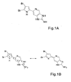
- the structural formula of Ageladine A is the FIGURE 1A refer to.
- the FIG. 1B shows the protonation of the guanidine group in Ageladine A and the stabilization of the positive charge over the entire structure.
- the right-hand structure contains a rigid and planar arrangement, which among other things is responsible for the fluorescence.
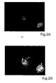
- FIGURES 2A . 1B show in 400x magnification images of Ageladine A incubated and thus stained live cells (PC-12 cells, rat, deposited at DSZM). The excitation was carried out with UV light with a wavelength of 380 nm.
- the FIG. 3 Figure 12 shows the fluorescence ratio value (measured at a fluorescence emission at 510 nm) of the excitation wavelengths 340 nm / 380 nm at various pH values for addition of 20 ⁇ M Ageladine A into a physiological buffer.
- the measurable large pH value range can clearly be recognized by a good assignability of the measured ratio values to the individual pH values.
- the FIG. 4 shows the excitation spectrum (emitted fluorescence intensity above the excitation wavelength in nm) of Ageladine A at different pH values of the solution.
- the fluorescence intensity was measured at 510 nm.
- the high pH sensitivity of Ageladine A can be seen in the physiologically relevant range around pH7.
- the FIG. 5 shows the measurement protocol of a measurement of the intracellular pH of PC12 cells using Ageladine A when changing the external pH of the solution. Plotted is the fluorescence ratio value (340 nm / 380 nm) over time (time in s). The different pH values of the buffer solution are marked as parameters on the graph. It can be clearly seen that the fluorescence emission follows the extracellular increase in the pH of the solution in a wide range (pH 6.5 to pH 3). However, the change in intracellular pH is only a few tenths of pH7, giving a very high pH reading of the pH indicator.
- the FIG. 6A shows in 100x magnification a microscopic transparency image of a living organism, here a shrimp ( Palaemonetes argentinus ) from above.
- the FIGURE 6B shows the fluorescence image of this shrimp after it has been incubated with Ageladine A and irradiated with UV light. Due to the intensity of the fluorescence emitted, it can be seen that acidic structures (oral digestive area) were stained in the shrimp.
- the FIG. 7 shows the excitation spectrum (emitted fluorescence intensity above excitation wavelength) of Ageladine A in the shrimp in the mouth digestion region measured at 510 nm fluorescence emission.
- the green curve (lowermost, even curve) indicates fluorescence intensity values within the buffer solution outside the shrimp
- the red and blue curves (middle and upper curved curve) show fluorescence intensity values within the shrimp and thus easily detectable and detectable acidic components of the shrimp.
Landscapes
- Health & Medical Sciences (AREA)
- Life Sciences & Earth Sciences (AREA)
- Engineering & Computer Science (AREA)
- Chemical & Material Sciences (AREA)
- Immunology (AREA)
- Urology & Nephrology (AREA)
- Biomedical Technology (AREA)
- Molecular Biology (AREA)
- Hematology (AREA)
- Biotechnology (AREA)
- Analytical Chemistry (AREA)
- Cell Biology (AREA)
- Inorganic Chemistry (AREA)
- Food Science & Technology (AREA)
- Medicinal Chemistry (AREA)
- Physics & Mathematics (AREA)
- Microbiology (AREA)
- Biochemistry (AREA)
- General Health & Medical Sciences (AREA)
- General Physics & Mathematics (AREA)
- Pathology (AREA)
- Investigating Or Analysing Materials By The Use Of Chemical Reactions (AREA)
- Investigating, Analyzing Materials By Fluorescence Or Luminescence (AREA)
- Measuring Or Testing Involving Enzymes Or Micro-Organisms (AREA)
Description
- Die Erfindung bezieht sich auf ein optisches Messverfahren zur Ermittlung des pH-Werts eines Mediums durch
- Zugabe eines fluoreszierenden pH-Wert-Indikators in das Medium,
- Fluoreszenzanregung des pH-Wert-Indikators durch Bestrahlung des pH-Wert-Indikators mit Licht zumindest einer ausgewählten Wellenlänge und
- Detektion der emittierten Fluoreszenzintensitäten des pH-Wert-Indikators als Maß für den pH-Wert des Mediums.
- Die Wasserstoffkonzentration oder der pH-Wert ist ein extrem wichtiger Parameter in biologischen und chemisch/technischen Systemen. Viele chemische und biologische Reaktionen erfordern eine genaue Regelung des pH-Werts für einen korrekten Ablauf. Beispielsweise findet ein komplexes natürliches Verfahren zur Regelung des pH-Werts in menschlichem Blut statt, das normalerweise einen pH-Wert von ungefähr 7,4 hat. Veränderungen von selbst einigen Zehnteln einer pH-Werteinheit können bereits ernste Krankheitszustände zum Grund haben oder verursachen. Obwohl eine Vielfalt von Techniken entwickelt worden ist, um den pH-Wert zu messen, basieren diese allgemein auf entweder elektrochemischen oder optischen Prinzipien. Ein Standard-Labormessgerät für den pH-Wert umfasst beispielsweise eine Standardelektrode mit bekanntem Potenzial, eine spezielle Glaselektrode, die das Potenzial in Abhängigkeit von der Konzentration der Wasserstoffionen im Medium ändert, in das sie eintaucht, und ein Potenziometer, welches das Potenzial zwischen den beiden Elektroden misst, aus dem ein numerischer pH-Wert ermittelt wird. Verfahren dieser Art sind jedoch nicht sehr gut für Messungen in intakten biologischen Systemen geeignet, da eine Messelektrode eingeführt werden muss.
- Im Rahmen von optischen Messungen werden pH-Wert-Indikatoren verwendet, bei denen es sich um Farbstoffe handelt, deren detektierbare optische Eigenschaften, wie Extinktion (Absorption) oder Fluoreszenz, sich mit Veränderung des pH-Werts ebenfalls ändern. Somit zeigen pH-Wert-Indikatoren durch ihren Farbton und ihre Farbintensität den aktuellen pH-Wert der Lösung an. Die größte Empfindlichkeit von Indikatoren auf kleine Änderungen des pH-Werts liegt vor, wenn die Gleichgewichtskonstante (pKa) zwischen den sauren und basischen Formen des Indikators nahe dem Wert des zu untersuchenden Mediums, in der Regel eine Lösung, liegt.
- Als breite Verallgemeinerung werden optische Messungen des pH-Werts als den elektrochemischen Techniken unterlegen angesehen, hauptsächlich weil von der Wasserstoffionenkonzentration verschiedenen Faktoren wie Temperatur, Ionenstärke und Proteinkonzentration die Farbstoffe beeinflussen und die pH-Wertmessungen stören können. Dennoch haben optische Techniken erhebliche Vorteile, wenn Kosten und Größe eine Rolle spielen und besonders, wenn in lebenden Zellen und Geweben der pH-Wert gemessen werden soll, in die keine Messsonde eingeführt werden kann. Messungen von pH-abhängiger Emissionsintensität in einzelnen Zellen mit einer einzigen Anregungswellenlänge leiden unter Ungenauigkeiten, die mit der Farbstoffkonzentration, Photobleichung des Farbstoffs, Messzellendicke oder Weglänge zusammenhängen. Eine Lösung des Problems der Farbstoffkonzentration besteht in der Bestimmung des Verhältnisses der Fluoreszenzintensität bei einer festen Wellenlänge mit Anregung bei einer pH-empfindlichen Wellenlänge zu der Fluoreszenzintensität bei der gleichen Wellenlänge mit Anregung bei einer relativ pH-unempfindlichen Wellenlänge. Dieses Verfahren wird üblicherweise verwendet, um den pH-Wert im Inneren von Zellen mit Fluoreszenzderivaten zu schätzen, und ist praktisch geeignet für Suspensionen von Zellen und in homogenen Flüssigkeiten wie Medien in einem Forschungsfluorometer oder Mikroskop.
- Ein optischer Biosensor ist aus der
DE 39 23 921 A1 bekannt, bei dem ein chemische Reaktionen katalysierendes Enzym an einen pH-Wert-Indikator gebunden ist. Bei dem Indikator kann es sich um einen Fluoreszenzfarbstoff, insbesondere auf der Basis von Cumarin, handeln. Durch messbare Änderung der Fluoreszenzintensität des Farbstoffs kann eine Änderung des pH-Wertes während der chemischen Reaktion festgestellt werden. Aus derDE 600 13 613 T2 ist ein Kulturmedium für den Nachweis und die zahlenmäßige Bestimmung von Mikroorganismen bekannt, das einen pH-Wert-Indikator umfasst, der chemisch an eine hydrophile Substanz mit hohem Molekulargewicht gebunden ist. Somit wird ein wasserlöslicher pH-Wert-Indikator mit Ballast zur Verfügung gestellt, der gleichzeitig als Nährstoffgel das hydratisierte Wachstum der Mikroorganismen unterstützt. Ein Wachstumsnachweis wird durch Nachweis der Fluoreszenzveränderung des pH-Wert-Indikators, der Carboxyphenolrot umfassen kann, im Kulturmedium erbracht. DieDE 101 52 994 A1 offenbart ein optisches Verfahren zur gleichzeitigen Bestimmung von pH-Wert und Gelöstsauerstoff. Dazu werden zwei fluoreszierende pH-Wert-Indikatoren, beispielsweise auf der Basis von Ruthenium, in einer gemeinsamen Matrix im Medium eingesetzt. - Aus der
US 4.945 171 A ist ein pH-Wert-Indikator auf der Basis von fluoreszierenden Xanthinfarbstoffen bekannt. Hierbei werden zwei Emissionsmaxima bei der Anregung mit nur einer Wellenlänge mit besonderer Selektivität für die unabhängige Anregung von Säure- und Baseform bei entweder einer einzigen oder zwei Wellenlängen und die daraus resultierenden pH-Wert-abhängigen Absorptions- oder Fluoreszenzanregungsspektren gemessen. Aus derDE 196 81 363 C2 , von der die vorliegende Erfindung als nächstliegendem Stand der Technik ausgeht, ist ein pH-Wert-Indikator auf der Basis von fluoreszierenden Carbazinfarbstoffen und Derivaten bekannt. Gegenüber den Xanthinfarbstoffen zeigen die Carbazinfarbstoffe eine höhere Fluoreszenz, eine größeren Stabilität, eine geringere Temperaturempfindlichkeit und höhere Stokes-Verschiebungen. Außerdem sind die Carbazinfarbstoffe besser auf einem festen Träger zu immobilisieren. Es wird ein Verfahren zum Anzeigen des pH-Werts einer Lösung als Medium offenbart, bei dem der pH-Wert-Indikator in die Lösung gegeben und mit Licht einer ausgewählten Wellenlänge in Kontakt gebracht wird, um den Carbazinfarbstoff zur Fluoreszenz anzuregen, die Intensität der Fluoreszenz bei zwei verschiedenen Wellenlängen gemessen wird, das Verhältnis der Fluoreszenzintensitäten bei den zwei gewählten Wellenlängen berechnet wird und das Verhältnis mit einer vorgegebenen Beziehung für solche Verhältnisse mit dem pH-Wert korreliert wird. - Weitere Fluoreszenz-Farbstoffe, die bereits zu Messung des pH-Werts eingesetzt werden, sind beispielsweise die Farbstoffe BCECF und DNP-160. Weitere molekulare Sonden aus der pH-Wert-Indikator-Familie sind beispielsweise der nachfolgenden Tabelle zu entnehmen (sortiert nach abnehmender Gleichgewichtskonstante pKa, Tabellen entnommen aus http://probes.invitrogen.com/handbook/tables/0361.html, Stand 27.05.2007). Deutlich zu erkennen ist der jeweils geringe Umfang des detektierbaren pH-Wert-Bereichs.
VORGÄNGER- FLUOROPHOR PH-BEREICH TYPISCHE MESSUNG SNARF Indikatoren 6,0-8,0 Emissionsrate 580/640 nm HPTS (Pyranin) 7,0-8,0 Anregungsrate 450/405 nm BCECF 6,5-7,5 Anregungsrate 490/440 nm Fluoreszeine und Carboxyfluoreszeine 6,0-7,2 Anregungsrate 490/450 nm LysoSensor grün DND-189 4,5-6,0 einzelne Emission 520 nm Oregon grüne Farbstoffe 4,2-5,7 Anregungsrate 510/450 nm oder 490/440 nm LysoSensor gelb/blau DND-160 3,5-6,0 Emissionsrate 450/510 nm - Bei dem bioaktiven marinen Naturstoff Ageladine A (chemische Formel C10H7N5Br2) handelt es sich um ein Pyrrol-Imidazol Alkaloid, das beispielsweise aus Schwämmen der Gattung Agelas isoliert werden kann (vergleiche VERÖFFENTLICHUNG I von M. Fujita et al.: "Ageladine A: An Antiangiogenetic Matrixmetalloproteinase Inhibitor from Marine Sponge Agelas nakamurai", J. Am. Chem. Soc. 2003, 125, 15700-15701 und Supporting Information S.I. 1-15). Ageladine A kann mittlerweile auch vollständig synthetisiert werden (vergleiche VERÖFFENTLICHUNG II von M. Meketa et al.: "Total Synthesis of Ageladine A, an Angiogenesis Inhibitor from the Marine Sponge Agelas nakamurai" Org. Lett. 2006, 8, 7, 1443-1446, VERÖFFENTLICHUNG III VON S. Shengule et al.: "Concise Total Synthesis of the Marine Natural Product Ageladine A", Org. Lett. 2006, 8, 18, 4083-4084 und VERÖFFENTLICHUNG IV von M. Meketa et al.: "A New Total Synthesis of the Zinc Matrixmetalloproteinase Inhibitor Ageladine A Featuring a Biogenetically Patterned 6π-2-Azatriene Electrocyclization", Org. Lett. 2007, 9, 5, 853-855). Ageladine A steht somit der Öffentlichkeit in unbegrenztem Umfang zur Verfügung. Bei wissenschaftlichen Untersuchungen von Ageladine A mit Messungen zur zellulären Wirkung von marinen Naturstoffen zeigt Ageladine A eine störende Eigenfluoreszenz (vergleiche VERÖFFENTLICHUNG V von U. Bickmeyer et al.: "Disturbance of Voltage Induced Cellular Calcium Entry by the Marine Pyrrole-Imidazole Alkaloids", doi:10.1016/j.toxicon.2007.04.015). Ageladine A hat nach UV-Anregung eine ausgeprägte Fluoreszenz im Grünbereich (siehe VERÖFFENTLICHUNG I).
- Die
DE 10 2004 002 885 B4 offenbart eine Reihe neuer bioaktiver Verbindungen aus der Klasse der Pyrrolalkaloide. Dabei sind Meeresschwämme eine reichhaltige Quelle an Pyrrolalkaloiden, einer Gruppe von Naturstoffen, die sich durch ihre strukturelle Vielfalt und interessante biologische Aktivitäten auszeichnet. Genannt werden auch Pyrrol-Imidazol Alkaloide, gewonnen aus verschiedenen karibischen Schwammarten der Gattung Agelas, mit einer selektiven antihistaminischen Wirkung und Agelongin (Strukturformel 22) aus Agelas longissima mit einer antiserotonergen Wirkung an Funduspräparationen von Rattenmägen. Alle genannten bioaktiven Verbindungen werden ausschließlich für medizinische Zwecke eingesetzt, insbesondere zur Bekämpfung von neurodegenerativen Erkrankungen. - In Anbetracht der vorangehenden Ausführungen zu bekannten optischen Messverfahren zur Ermittlung des pH-Werts eines Mediums ist die AUFGABE für die vorliegende Erfindung darin zu sehen, ein verbessertes gattungsgemäßes Messverfahren anzugeben, bei dem ein zu den bekannten fluoreszierenden pH-Wert-Indikatoren alternativer fluoreszierender pH-Wert-Indikator verwendet wird, der eine verminderte Empfindlichkeit gegenüber potenziell störenden Faktoren und einen großen pH-Wert-Messbereich zeigt. Insbesondere soll eine wesentlich verbesserte Messleistung in biologischen Systemen, von denen die meisten im Bereich von pH5 bis pH9 arbeiten, gewährleistet sein
- Die LÖSUNG für diese Aufgabe ist dem Verfahrensanspruch zu entnehmen. Vorteilhafte Weiterbildungen des Verfahrens sind in den Unteransprüchen aufgezeigt und werden im Folgenden im Zusammenhang mit der Erfindung näher erläutert.
- Das erfindungsgemäße optische Messverfahren ist dadurch gekennzeichnet, dass ein zu den bekannten fluoreszierenden pH-Wert-Indikatoren alternativer fluoreszierender pH-Wert-Indikator auf Basis von Ageladine A verwendet wird. Bei Ageladine A handelt es sich um einen bekannten marinen Naturstoff in Form eines Pyrrol-Imidazol Alkaloids, der im Stand der Technik ausschließlich zu medizinischen Zwecken eingesetzt wird. Ageladine A kann sowohl aus marinen Schwämmen gewonnen als auch synthetisch hergestellt werden, wie oben bereits ausgeführt. Durch die beschriebenen Totalsynthesen ist es somit ohne weiteres möglich, größere Mengen von Ageladine A bereitzustellen, ohne dabei auf marine Schwämme zurückgreifen zu müssen. Zur Nutzung von Ageladine A im Rahmen der vorliegenden Erfindung hat sich herausgestellt, dass die auftretende Eigenfluoreszenz durch Anregungsverstärkung mittels Bestrahlung mit Licht im UV-Bereich zwischen 315 nm und 400 nm Wellenlänge für Messzwecke einsetzbar ist. Ageladine A zeigt dann eine ausgeprägte Fluoreszenz im Grünbereich zwischen 490 nm und 575 nm Wellenlänge. Bei der Verwendung von Ageladine A als fluoreszierendem pH-Wert-Indikator können pH-Wert-Änderungen des zu untersuchenden Mediums zwischen pH4 und pH9 durch eine veränderliche Fluoreszenzintensität in der Weise zuverlässig optisch angezeigt werden, dass mit sinkendem pH-Wert die emittierte Fluoreszenzintensität ansteigt. Der von Ageladine A anzeigbare pH-Wert-Bereich ist ungewöhnlich groß für einen fluoreszierenden pH-Wert-Indikator und die Färbungen erweisen sich als zeitlich sehr stabil.
- Die fluoreszierenden Eigenschaften von Molekülen sind auf ihre chemische Struktur zurückzuführen. Dabei sollten folgende, allgemeine Punkte durch die chemische Molekülstruktur erfüllt sein: aromatisches bzw. delokalisiertes π-Elektronensystem, Chromophor, starrer und planarer Molekülaufbau. Alle diese Punkte werden von der Molekülstruktur von Ageladine A erfüllt. Die Struktur weist ein aromatisches System (Pyrrol, Pyridin) sowie das π-Elektronensystem des Guanidins auf. Durch die basische Guanidinstruktur ist eine Protonierung in sauren Medien an dieser Stelle sehr wahrscheinlich. Die dabei auftretende positive Ladung kann dabei über das gesamte Molekül delokalisiert werden, was Voraussetzung für ein chromophores System ist. Die formulierte Grenzstruktur weist eine starre bzw. planare Molekülstruktur auf. Damit können die fluoreszierenden Eigenschaften und auch pH-Wertabhängige Änderungen in der Fluoreszenz erklärt werden (vergleiche
Figuren 1A, 1B des speziellen Beschreibungsteil). - Überraschend hat sich weiterhin herausgestellt, dass Ageladine A eine besonders hohe Empfindlichkeit der emittierten Fluoreszenzintensität im physiologisch relevanten Bereich zwischen pH6 und pH8 aufzeigt. Somit kann Ageladine A als pH-Wert-Indikator besonders gut für physiologische pH-Wert-Messungen eingesetzt werden. Vorteilhafterweise können entnommene physiologische Proben (in vitro) aber auch lebende Zellen, Gewebe und ganze Organismen (in vivo) mit Ageladine A als fluoreszierendem pH-Wert-Indikator inkubiert und durch die Fluoreszenz im Grünbereich auf diese Art gefärbt werden. Dies ermöglicht Messungen des pH-Werts in vivo und in vitro in einem weiten Bereich und damit die Erkennung saurer Gewebe in Organismen durch seine Fluoreszenz-Eigenschaften. Die Fluoreszenz im Grünbereich nimmt mit sinkendem pH-Wert deutlich zu. Die intensiven Färbungen erweisen sich als stabil über Stunden bis zu Tagen.
- Mit dem Verfahren nach der Erfindung unter der Anwendung von Ageladine A als fluoreszierendem pH-Wert-Indikator können somit pH-Wert-Messungen von Lösungen und pH-Wert-Messungen innerhalb lebender Zellen, Geweben und ganzen Organismen als Medium durchgeführt werden. Eine Fluoreszenz-Färbung von Zellen und Geweben durch Inkubation dieser Medien mit Ageladine A ist möglich. Vorteilhafterweise kann bei der Erfindung eine in vivo-oder in vitro-Inkubation von Zellen, Geweben oder kompletten Organismen als Medium in einer den fluoreszierenden pH-Wert-Indikator enthaltenden Lösung erfolgen. Durch eine derartige einfache, schnelle und biologische Färbung können beispielsweise saure Gewebsanteile (Verdauungsorgane) in lebenden Organismen markiert und unter dem Fluoreszenz-Mikroskop leicht erkannt werden. Ageladine A kann deshalb als äußerst intensiver Zellfarbstoff angewendet werden, der gleichzeitig als pH-Wert-Indikator pH-Wert-Änderungen reproduzierbar anzeigt. Qualitative pH-Wert-Anzeigen und quantitative pH-Wert-Messungen innerhalb lebender Systeme können durch die Erfindung stark vereinfacht werden.
- Zusammenfassend zeigt das Verfahren nach der Erfindung mit einer Verwendung von Ageladine A als pH-Wert-Indikator folgende besondere Vorteile:
- Leichte Handhabbarkeit. Durch Vollsynthese steht der marine Naturstoff Ageladine A in großen Mengen zur Verfügung, ohne den Artenschutz zu gefährden.
- Große Empfindlichkeit von Ageladine A im physiologisch besonders interessanten Bereich zwischen pH6 und pH8.
- Die Fluoreszenz ist trotz UV-Bestrahlung. über Stunden stabil. Die Färbungen halten sich wenigstens über mehrere Tage.
- Kurzfristige Inkubation von Zellen und Geweben mit Ageladine A führt zu deutlicher und starker Fluoreszenz.
- Sogar intakte, lebende Organismen lassen sich mit Ageladine A färben, da es auf Grund seiner lipophilen Struktur (Bromierung) sehr schnell und weit in die Gewebe eindringt.
- Saure Bestandteile von Organismen lassen sich sofort nach Inkubation auf einen Blick im Fluoreszenz-Mikroskop erkennen, da die Fluoreszenz mit sinkendem pH-Wert stark zunimmt.
- Weiter oben wurde bereits ausgeführt, dass sich verschiedene Störfaktoren ggfs. negativ auf die pH-Wert-Messungen auswirken können, wenn nur bei einer einzigen Anregungswellenlänge gemessen wird. Vorteilhaft kann deshalb auch das Verfahren nach der Erfindung unter Anwendung von Ageladine A als fluoreszierendem pH-Wert-Indikator modifiziert werden durch
- eine Fluoreszenzanregung bei zwei ausgewählten Wellenlängen
- Detektion der emittierten Fluoreszenzintensitäten
- Berechnung des Verhältnisses der Fluoreszenzintensitäten und
- Korrelation des Verhältnisses mit einer vorgegebenen Beziehung für solche Verhältnisse mit dem pH-Wert der Lösung.
- Das optische Messverfahren nach der Erfindung unter Anwendung von Ageladine A als fluoreszierendem pH-Wert-Indikator wird nachfolgend anhand der schematischen Figuren noch zum weiteren Verständnis der Erfindung näher erläutert. Dabei zeigt:
- FIGUR 1A
- die Strukturformel von Ageladine A,
- FIGUR 1B
- die Protonierung und Stabilisierung von Ageladine A,
- FIGUR 2A,2B
- Fluoreszenz von mit Ageladine A gefärbten Zellen,
- FIGUR 3
- das Fluoreszenzintensitätsdiagramm von Ageladine A in Abhängigkeit de pH-Werts,
- FIGUR 4
- das Anregungsspektrum von Ageladine A in Abhängigkeit des pH-Werts,
- FIGUR 5
- Messungen des intrazellulären pH-Werts während der Änderung des extrazellulären pH-Werts,
- FIGUR 6A
- einen Organismus (Garnele),
- FIGUR 6B
- eine Lebendfärbung der Garnele mit Ageladine A und
- FIGUR 7
- das Anregungsspektrum von Ageladine A in der Garnele.
- Die Strukturformel von Ageladine A ist der
FIGUR 1A zu entnehmen. DieFIGUR 1B zeigt die Protonierung der Guanidingruppe in Ageladine A und die Stabilisierung der positiven Ladung über die gesamte Struktur. Die rechte Struktur beinhaltet eine starre und planare Anordnung, die unter anderem für die Fluoreszenz verantwortlich ist. - Die
FIGUREN 2A ,1B zeigen in 400facher Vergrößerung Aufnahmen von mit Ageladine A inkubierten und dadurch gefärbten lebenden Zellen (PC-12-Zellen, Ratte, hinterlegt bei DSZM). Die Anregung erfolgte mit UV-Licht mit einer Wellenlänge von 380 nm. - Die
FIGUR 3 zeigt den Fluoreszenz-Ratio-Wert (gemessen bei einer Fluoreszenzemission bei 510 nm) der Anregungswellenlängen 340 nm / 380 nm bei verschiedenen pH-Werten für eine Zugabe von 20 µM Ageladine A in einen physiologischen Puffer. Deutlich ist der messbare große pH-Wert-Bereich mit einer guten Zuordenbarkeit der gemessenen Ratio-Werte zu den einzelnen pH-Werten zu erkennen. - Die
FIGUR 4 zeigt das Anregungsspektrum (emittierte Fluoreszenzintensität über der Anregungswellenlänge in nm) von Ageladine A bei verschiedenen pH-Werten der Lösung. Die Fluoreszenzintensität wurde bei 510 nm gemessen. Zu erkennen ist die große pH-Wert-Empfindlichkeit von Ageladine A im physiologisch relevanten Bereich um pH7 herum. - Die
FIGUR 5 zeigt das Messprotokoll einer Messung des intrazellulären pH-Werts von PC12 Zellen mittels Ageladine A bei Änderung des äußeren pH-Werts der Lösung. Aufgetragen ist der Fluoreszenz-Ratio-Wert (340 nm/380 nm) über der Zeit (time in s). Die verschiedenen pH-Werte der Pufferlösung sind als Parameter am Graphen markiert. Deutlich ist zu erkennen, dass die Fluoreszenzemission dem extrazellulären Ansteigen des pH-Wertes der Lösung in einem weiten Bereich (pH6,5 bis pH3) folgt. Die Änderung des intrazellulären pH-Werts beträgt jedoch nur einige Zehntel um pH7 herum, wodurch eine sehr hohe Messauflösung des pH-Wert-Indikators gegeben ist. - Die
FIGUR 6A zeigt in 100facher Vergrößerung ein mikroskopisches Durchlichtbild eines lebenden Organismus, hier einer Garnele (Palaemonetes argentinus) von oben. DieFIGUR 6B zeigt das Fluoreszenzbild dieser Garnele, nachdem diese mit Ageladine A inkubiert und mit UV-Licht bestrahlt wurde. Aufgrund der Intensität der emittierten Fluoreszenz ist zu erkennen, dass saure Strukturen (Mund-Verdauungsbereich) in der Garnele gefärbt wurden. - Die
FIGUR 7 zeigt das Anregungsspektrum (emittierte Fluoreszenzintensität über Anregungswellenlänge) von Ageladine A in der Garnele im Mund-Verdauungsbereich, gemessen bei 510nm Fluoreszenzemission. Die grüne Kurve (unterste, gerade Kurve) zeigt Fluoreszenz-Intensitätswerte innerhalb der Pufferlösung außerhalb der Garnele an, die rote und die blaue Kurve (mittlere und obere gekrümmte Kurve) zeigen Fluoreszenz-Intensitätswerte innerhalb der Garnele und damit in einfacher Weise leicht erkenn- und detektierbare saure Bestandteile der Garnele an.
Claims (4)
- Optisches Messverfahren zur Ermittlung des pH-Werts eines Mediums durch• Zugabe eines fluoreszierenden pH-Wert-Indikators in das Medium,• Fluoreszenzanregung des pH-Wert-Indikators durch Bestrahlung des pH-Wert-Indikators mit Licht zumindest einer ausgewählten Wellenlänge und• Detektion der emittierten Fluoreszenzintensität des pH-Wert-Indikators als Maß für den pH-Wert des Mediums,GEKENNZEICHNET DURCH
die Verwendung eines fluoreszierenden pH-Wert-Indikators auf Basis von natürlich gewonnenem oder synthetisiertem Ageladine A mit einer Fluoreszenz im Grünbereich zwischen 490 nm und 575 nm Wellenlänge bei einer Fluoreszenzanregung durch Bestrahlung mit Licht im UV-Bereich zwischen 315 und 400 nm Wellenlänge und einer hohen Empfindlichkeit der emittierten Fluoreszenzintensität im physiologisch besonders relevanten Bereich zwischen pH6 und pH8, wobei pH-Wert-Änderungen des zu untersuchenden Mediums zwischen pH4 und pH9 durch eine veränderliche Fluoreszenzintensität in der Weise optisch angezeigt werden, dass mit sinkendem pH-Wert die emittierte Fluoreszenzintensität ansteigt. - Optisches Messverfahren nach Anspruch 1,
GEKENNZEICHNET DURCH
eine in vivo- oder in vitro-Inkubation von Zellen, Geweben oder kompletten Organismen als Medium in einer den fluoreszierenden pH-Wert-Indikator enthaltenden Lösung. - Optisches Messverfahren nach Anspruch 2,
GEKENNZEICHNET DURCH
eine Betrachtung des inkubierten Mediums in einem Fluoreszenz-Mikroskop, - Optisches Messverfahren nach einem der Ansprüche 1 bis 3, GEKENNZEICHNET DURCH• eine Fluoreszenzanregung bei zwei ausgewählten Wellenlängen• Detektion der emittierten Fluoreszenzintensitäten• Berechnung des Verhältnisses der Fluoreszenzintensitäten und• Korrelation des Verhältnisses mit einer vorgegebenen Beziehung für solche Verhältnisse mit dem pH-Wert der Lösung.
Applications Claiming Priority (3)
| Application Number | Priority Date | Filing Date | Title |
|---|---|---|---|
| DE102007024985 | 2007-05-28 | ||
| DE102007034886A DE102007034886B4 (de) | 2007-05-28 | 2007-07-15 | Optisches Messverfahren zur Ermittlung des pH-Wertes eines Mediums unter Anwendung von Ageladine A als fluoreszierendem pH-Wert-Indikator |
| PCT/DE2008/000570 WO2008145080A1 (de) | 2007-05-28 | 2008-04-01 | Optisches messverfahren zur ermittlung des ph-werts eines mediums unter anwendung von ageladine a als fluoreszierendem ph-wert-indikator |
Publications (2)
| Publication Number | Publication Date |
|---|---|
| EP2156193A1 EP2156193A1 (de) | 2010-02-24 |
| EP2156193B1 true EP2156193B1 (de) | 2010-09-01 |
Family
ID=39942238
Family Applications (1)
| Application Number | Title | Priority Date | Filing Date |
|---|---|---|---|
| EP08734458A Not-in-force EP2156193B1 (de) | 2007-05-28 | 2008-04-01 | Optisches messverfahren zur ermittlung des ph-werts eines mediums unter anwendung von ageladine a als fluoreszierendem ph-wert-indikator |
Country Status (6)
| Country | Link |
|---|---|
| US (1) | US8198098B2 (de) |
| EP (1) | EP2156193B1 (de) |
| AT (1) | ATE479901T1 (de) |
| AU (1) | AU2008255407C1 (de) |
| DE (2) | DE102007034886B4 (de) |
| WO (1) | WO2008145080A1 (de) |
Families Citing this family (7)
| Publication number | Priority date | Publication date | Assignee | Title |
|---|---|---|---|---|
| DE102009024937A1 (de) | 2009-06-08 | 2010-12-09 | Braatz, Udo, Dr. | Verfahren und Anordnung zur Bestimmung von Flüssigkeitsparametern |
| DE102011101207B3 (de) * | 2011-05-11 | 2012-05-10 | Sartorius Stedim Biotech Gmbh | Fluoreszenzfarbstoff für pH-Sensor |
| DE102012210055B4 (de) | 2012-06-14 | 2017-03-02 | Alfred-Wegener-Institut Helmholtz-Zentrum für Polar- und Meeresforschung | Nachweis der Viabilität biologischer Proben |
| DE102012021933B4 (de) * | 2012-11-09 | 2015-12-31 | Airbus Defence and Space GmbH | Optischer pH-Wert-Sensor |
| DE102019124795A1 (de) * | 2019-09-16 | 2021-03-18 | Abberior GmbH | Optischer pH-Sensor |
| KR20220161286A (ko) * | 2020-04-03 | 2022-12-06 | 다이애그널 바이오 에이비 | 샘플의 pH 측정 방법 |
| CN119915887B (zh) * | 2024-12-13 | 2025-12-05 | 齐鲁工业大学(山东省科学院) | 一种基于铜铟硫/卡巴肼体系的pH型电化学发光传感器及制备方法 |
Family Cites Families (8)
| Publication number | Priority date | Publication date | Assignee | Title |
|---|---|---|---|---|
| DE2560064C3 (de) * | 1975-02-28 | 1983-12-01 | Max Planck Gesellschaft zur Förderung der Wissenschaften e.V., 3400 Göttingen | Verfahren zur optischen Messung von Blutgasen |
| US4945171A (en) | 1987-08-10 | 1990-07-31 | Molecular Probes, Inc. | Xanthene dyes having a fused (C) benzo ring |
| DE3923921A1 (de) | 1989-07-19 | 1991-01-24 | Biotechnolog Forschung Gmbh | Optischer biosensor |
| US5567624A (en) | 1995-04-27 | 1996-10-22 | Utah Medical Products, Inc. | Carbazine dyes and derivatives for pH measurement |
| US6391626B1 (en) | 1999-08-23 | 2002-05-21 | 3M Innovative Properties Company | Culture medium and device using ballasted pH indicators for detection and enumeration of microorganisms |
| DE10152994A1 (de) | 2001-10-26 | 2003-08-07 | Chromeon Gmbh | Methode zur gleichzeitigen optischen Bestimmung von pH-Wert und Gelöstsauerstoff |
| CA2489054A1 (en) | 2002-06-10 | 2003-12-18 | Riken | Chromoprotein |
| DE102004002885B4 (de) | 2004-01-20 | 2007-03-15 | Johannes-Gutenberg-Universität Mainz | Neue medizinisch einsetzbare Pyrrolalkaloide: Verfahren zu ihrer Isolierung, Synthese, sie enthaltende Arzneimittel und deren Verwendung |
-
2007
- 2007-07-15 DE DE102007034886A patent/DE102007034886B4/de not_active Expired - Fee Related
-
2008
- 2008-04-01 EP EP08734458A patent/EP2156193B1/de not_active Not-in-force
- 2008-04-01 AU AU2008255407A patent/AU2008255407C1/en not_active Ceased
- 2008-04-01 DE DE502008001271T patent/DE502008001271D1/de active Active
- 2008-04-01 US US12/601,908 patent/US8198098B2/en not_active Expired - Fee Related
- 2008-04-01 WO PCT/DE2008/000570 patent/WO2008145080A1/de not_active Ceased
- 2008-04-01 AT AT08734458T patent/ATE479901T1/de active
Also Published As
| Publication number | Publication date |
|---|---|
| AU2008255407C1 (en) | 2011-02-17 |
| EP2156193A1 (de) | 2010-02-24 |
| WO2008145080A1 (de) | 2008-12-04 |
| AU2008255407B2 (en) | 2010-09-16 |
| AU2008255407A1 (en) | 2008-12-04 |
| DE102007034886B4 (de) | 2009-06-18 |
| DE502008001271D1 (de) | 2010-10-14 |
| DE102007034886A1 (de) | 2008-12-11 |
| US8198098B2 (en) | 2012-06-12 |
| ATE479901T1 (de) | 2010-09-15 |
| US20100178664A1 (en) | 2010-07-15 |
Similar Documents
| Publication | Publication Date | Title |
|---|---|---|
| EP2156193B1 (de) | Optisches messverfahren zur ermittlung des ph-werts eines mediums unter anwendung von ageladine a als fluoreszierendem ph-wert-indikator | |
| DE69334079T2 (de) | Fluoreszenzfarbstoff, der Pyryliumsalze oder ähnliche Salze enthält, Nachweisverfahren von Nukleinsäuren durch seine Verwendung | |
| EP1144586B1 (de) | Verfahren, gefäss und vorrichtung zur überwachung der stoffwechselaktivität von zellkulturen in flüssigen medien | |
| DE2953524C2 (de) | ||
| EP0215772B1 (de) | Verfahren und Vorrichtung zur Tumordiagnose mittels Sera | |
| DE3109252C2 (de) | Verfahren und Zusammensetzung zur Bestimmung einzelner Leukozyten durch metachromatische Farbstoffdifferentialabsorption | |
| DE69022819T2 (de) | Verbindungen und Reagenzien zur Bestimmung von Retikulozyten. | |
| Kim et al. | A ratiometric two-photon probe for Ca2+ in live tissues and its application to spinal cord injury model | |
| CN112745287B (zh) | 一种荧光探针hm及其制备方法和应用 | |
| DE69223851T2 (de) | Nitro- oder Nitroso-substituierte polyhalogenierte Phenolsulfonphtaleine | |
| DE3104078C2 (de) | Verfahren zur Bestimmung des pH-Wertes im Innern einer Zelle; 1,4-Dibutyryloxy-2,3-dicyanobenzol; 1,4-Di(-tert-butyloxycarbonyl-l-alanyloxy)-2-3-dicyanobenzol | |
| DE102007003873B4 (de) | Verfahren zur Fluoreszenzfärbung und zur Untersuchung von Gewebe | |
| EP1999457A1 (de) | Verfahren zur bestimmung von molekülen oder molekülteilen in biologischen proben | |
| CN119161342B (zh) | 一种高信噪比检测生物体内、体外线粒体粘度变化荧光探针的开发及其应用 | |
| EP3017290B1 (de) | Diffusionskammer zur ermittlung unterschiedlicher parameter einer wässrigen substanz | |
| WO2003036293A1 (de) | Methode zur gleichzeitigen optischen bestimmung von ph-wert und gelöstsauerstoff | |
| EP2122364B1 (de) | Verfahren zur identifizierung der agonistischen aktivität einer zielverbindung auf einen kaliumkanal | |
| DE102007011913A1 (de) | Fluoreszenz-basiertes Assay zum Erkennen von Verbindungen zum Modulieren des Natrium-Calcium-Austauschers (NCX) im "Vorwärtsmodus" | |
| CN117486913A (zh) | 一种对粘度和过氧化氢双响应的近红外探针及其制备和应用 | |
| Nett et al. | Simultaneous measurements of intracellular pH in the leech giant glial cell using 2', 7'-bis-(2-carboxyethyl)-5, 6-carboxyfluorescein and ion-sensitive microelectrodes | |
| DE3813503A1 (de) | Traegergebundenes mehrkomponentiges nachweissystem zur kolorimetrischen bestimmung esterolytisch oder/und proteolytisch aktiver inhaltsstoffe von koerperfluessigkeiten | |
| DE102012210055B4 (de) | Nachweis der Viabilität biologischer Proben | |
| DE102019124795A1 (de) | Optischer pH-Sensor | |
| Inglefield et al. | [26] Using confocal microscopy and the fluorescent indicator, 6-methoxy-N-ethylquinolinium iodide, to measure changes in intracellular chloride | |
| DE10202482B4 (de) | Molekularer Sensorkomplex, Messsondenanordnung und Verfahren zur pharmakologischen Wirkstoff- und/oder Wirkorttestung |
Legal Events
| Date | Code | Title | Description |
|---|---|---|---|
| PUAI | Public reference made under article 153(3) epc to a published international application that has entered the european phase |
Free format text: ORIGINAL CODE: 0009012 |
|
| 17P | Request for examination filed |
Effective date: 20091207 |
|
| AK | Designated contracting states |
Kind code of ref document: A1 Designated state(s): AT BE BG CH CY CZ DE DK EE ES FI FR GB GR HR HU IE IS IT LI LT LU LV MC MT NL NO PL PT RO SE SI SK TR |
|
| AX | Request for extension of the european patent |
Extension state: AL BA MK RS |
|
| RIN1 | Information on inventor provided before grant (corrected) |
Inventor name: BICKMEYER, ULF-GEORG Inventor name: KLINGS, KARL-WALTER Inventor name: KOECK, MATTHIAS Inventor name: GRUBE, ACHIM |
|
| GRAP | Despatch of communication of intention to grant a patent |
Free format text: ORIGINAL CODE: EPIDOSNIGR1 |
|
| GRAS | Grant fee paid |
Free format text: ORIGINAL CODE: EPIDOSNIGR3 |
|
| GRAA | (expected) grant |
Free format text: ORIGINAL CODE: 0009210 |
|
| AK | Designated contracting states |
Kind code of ref document: B1 Designated state(s): AT BE BG CH CY CZ DE DK EE ES FI FR GB GR HR HU IE IS IT LI LT LU LV MC MT NL NO PL PT RO SE SI SK TR |
|
| REG | Reference to a national code |
Ref country code: GB Ref legal event code: FG4D Free format text: NOT ENGLISH |
|
| REG | Reference to a national code |
Ref country code: CH Ref legal event code: EP |
|
| REG | Reference to a national code |
Ref country code: IE Ref legal event code: FG4D Free format text: LANGUAGE OF EP DOCUMENT: GERMAN |
|
| REF | Corresponds to: |
Ref document number: 502008001271 Country of ref document: DE Date of ref document: 20101014 Kind code of ref document: P |
|
| REG | Reference to a national code |
Ref country code: NL Ref legal event code: T3 |
|
| PG25 | Lapsed in a contracting state [announced via postgrant information from national office to epo] |
Ref country code: LT Free format text: LAPSE BECAUSE OF FAILURE TO SUBMIT A TRANSLATION OF THE DESCRIPTION OR TO PAY THE FEE WITHIN THE PRESCRIBED TIME-LIMIT Effective date: 20100901 Ref country code: FI Free format text: LAPSE BECAUSE OF FAILURE TO SUBMIT A TRANSLATION OF THE DESCRIPTION OR TO PAY THE FEE WITHIN THE PRESCRIBED TIME-LIMIT Effective date: 20100901 Ref country code: NO Free format text: LAPSE BECAUSE OF FAILURE TO SUBMIT A TRANSLATION OF THE DESCRIPTION OR TO PAY THE FEE WITHIN THE PRESCRIBED TIME-LIMIT Effective date: 20101201 |
|
| LTIE | Lt: invalidation of european patent or patent extension |
Effective date: 20100901 |
|
| PG25 | Lapsed in a contracting state [announced via postgrant information from national office to epo] |
Ref country code: CY Free format text: LAPSE BECAUSE OF FAILURE TO SUBMIT A TRANSLATION OF THE DESCRIPTION OR TO PAY THE FEE WITHIN THE PRESCRIBED TIME-LIMIT Effective date: 20100901 Ref country code: HR Free format text: LAPSE BECAUSE OF FAILURE TO SUBMIT A TRANSLATION OF THE DESCRIPTION OR TO PAY THE FEE WITHIN THE PRESCRIBED TIME-LIMIT Effective date: 20100901 Ref country code: PL Free format text: LAPSE BECAUSE OF FAILURE TO SUBMIT A TRANSLATION OF THE DESCRIPTION OR TO PAY THE FEE WITHIN THE PRESCRIBED TIME-LIMIT Effective date: 20100901 Ref country code: SI Free format text: LAPSE BECAUSE OF FAILURE TO SUBMIT A TRANSLATION OF THE DESCRIPTION OR TO PAY THE FEE WITHIN THE PRESCRIBED TIME-LIMIT Effective date: 20100901 |
|
| REG | Reference to a national code |
Ref country code: IE Ref legal event code: FD4D |
|
| PG25 | Lapsed in a contracting state [announced via postgrant information from national office to epo] |
Ref country code: LV Free format text: LAPSE BECAUSE OF FAILURE TO SUBMIT A TRANSLATION OF THE DESCRIPTION OR TO PAY THE FEE WITHIN THE PRESCRIBED TIME-LIMIT Effective date: 20100901 Ref country code: GR Free format text: LAPSE BECAUSE OF FAILURE TO SUBMIT A TRANSLATION OF THE DESCRIPTION OR TO PAY THE FEE WITHIN THE PRESCRIBED TIME-LIMIT Effective date: 20101202 Ref country code: SE Free format text: LAPSE BECAUSE OF FAILURE TO SUBMIT A TRANSLATION OF THE DESCRIPTION OR TO PAY THE FEE WITHIN THE PRESCRIBED TIME-LIMIT Effective date: 20100901 |
|
| PG25 | Lapsed in a contracting state [announced via postgrant information from national office to epo] |
Ref country code: IE Free format text: LAPSE BECAUSE OF FAILURE TO SUBMIT A TRANSLATION OF THE DESCRIPTION OR TO PAY THE FEE WITHIN THE PRESCRIBED TIME-LIMIT Effective date: 20100901 |
|
| PG25 | Lapsed in a contracting state [announced via postgrant information from national office to epo] |
Ref country code: SK Free format text: LAPSE BECAUSE OF FAILURE TO SUBMIT A TRANSLATION OF THE DESCRIPTION OR TO PAY THE FEE WITHIN THE PRESCRIBED TIME-LIMIT Effective date: 20100901 Ref country code: CZ Free format text: LAPSE BECAUSE OF FAILURE TO SUBMIT A TRANSLATION OF THE DESCRIPTION OR TO PAY THE FEE WITHIN THE PRESCRIBED TIME-LIMIT Effective date: 20100901 Ref country code: IS Free format text: LAPSE BECAUSE OF FAILURE TO SUBMIT A TRANSLATION OF THE DESCRIPTION OR TO PAY THE FEE WITHIN THE PRESCRIBED TIME-LIMIT Effective date: 20110101 Ref country code: PT Free format text: LAPSE BECAUSE OF FAILURE TO SUBMIT A TRANSLATION OF THE DESCRIPTION OR TO PAY THE FEE WITHIN THE PRESCRIBED TIME-LIMIT Effective date: 20110103 Ref country code: EE Free format text: LAPSE BECAUSE OF FAILURE TO SUBMIT A TRANSLATION OF THE DESCRIPTION OR TO PAY THE FEE WITHIN THE PRESCRIBED TIME-LIMIT Effective date: 20100901 Ref country code: RO Free format text: LAPSE BECAUSE OF FAILURE TO SUBMIT A TRANSLATION OF THE DESCRIPTION OR TO PAY THE FEE WITHIN THE PRESCRIBED TIME-LIMIT Effective date: 20100901 |
|
| PG25 | Lapsed in a contracting state [announced via postgrant information from national office to epo] |
Ref country code: ES Free format text: LAPSE BECAUSE OF FAILURE TO SUBMIT A TRANSLATION OF THE DESCRIPTION OR TO PAY THE FEE WITHIN THE PRESCRIBED TIME-LIMIT Effective date: 20101212 |
|
| PLBE | No opposition filed within time limit |
Free format text: ORIGINAL CODE: 0009261 |
|
| STAA | Information on the status of an ep patent application or granted ep patent |
Free format text: STATUS: NO OPPOSITION FILED WITHIN TIME LIMIT |
|
| 26N | No opposition filed |
Effective date: 20110606 |
|
| PG25 | Lapsed in a contracting state [announced via postgrant information from national office to epo] |
Ref country code: DK Free format text: LAPSE BECAUSE OF FAILURE TO SUBMIT A TRANSLATION OF THE DESCRIPTION OR TO PAY THE FEE WITHIN THE PRESCRIBED TIME-LIMIT Effective date: 20100901 |
|
| REG | Reference to a national code |
Ref country code: DE Ref legal event code: R097 Ref document number: 502008001271 Country of ref document: DE Effective date: 20110606 |
|
| BERE | Be: lapsed |
Owner name: STIFTUNG ALFRED-WEGENER-INSTITUT FUR POLAR- UND M Effective date: 20110430 |
|
| PG25 | Lapsed in a contracting state [announced via postgrant information from national office to epo] |
Ref country code: MC Free format text: LAPSE BECAUSE OF NON-PAYMENT OF DUE FEES Effective date: 20110430 |
|
| PG25 | Lapsed in a contracting state [announced via postgrant information from national office to epo] |
Ref country code: MT Free format text: LAPSE BECAUSE OF FAILURE TO SUBMIT A TRANSLATION OF THE DESCRIPTION OR TO PAY THE FEE WITHIN THE PRESCRIBED TIME-LIMIT Effective date: 20100901 |
|
| PG25 | Lapsed in a contracting state [announced via postgrant information from national office to epo] |
Ref country code: BE Free format text: LAPSE BECAUSE OF NON-PAYMENT OF DUE FEES Effective date: 20110430 |
|
| PG25 | Lapsed in a contracting state [announced via postgrant information from national office to epo] |
Ref country code: LU Free format text: LAPSE BECAUSE OF NON-PAYMENT OF DUE FEES Effective date: 20110401 |
|
| PG25 | Lapsed in a contracting state [announced via postgrant information from national office to epo] |
Ref country code: TR Free format text: LAPSE BECAUSE OF FAILURE TO SUBMIT A TRANSLATION OF THE DESCRIPTION OR TO PAY THE FEE WITHIN THE PRESCRIBED TIME-LIMIT Effective date: 20100901 Ref country code: BG Free format text: LAPSE BECAUSE OF FAILURE TO SUBMIT A TRANSLATION OF THE DESCRIPTION OR TO PAY THE FEE WITHIN THE PRESCRIBED TIME-LIMIT Effective date: 20101201 |
|
| PG25 | Lapsed in a contracting state [announced via postgrant information from national office to epo] |
Ref country code: HU Free format text: LAPSE BECAUSE OF FAILURE TO SUBMIT A TRANSLATION OF THE DESCRIPTION OR TO PAY THE FEE WITHIN THE PRESCRIBED TIME-LIMIT Effective date: 20100901 |
|
| REG | Reference to a national code |
Ref country code: DE Ref legal event code: R081 Ref document number: 502008001271 Country of ref document: DE Owner name: ALFRED-WEGENER-INSTITUT HELMHOLTZ-ZENTRUM FUER, DE Free format text: FORMER OWNER: STIFTUNG ALFRED-WEGENER-INSTITUT FUER POLAR- UND MEERESFORSCHUNG, 27570 BREMERHAVEN, DE Effective date: 20140103 |
|
| REG | Reference to a national code |
Ref country code: AT Ref legal event code: MM01 Ref document number: 479901 Country of ref document: AT Kind code of ref document: T Effective date: 20130401 |
|
| PG25 | Lapsed in a contracting state [announced via postgrant information from national office to epo] |
Ref country code: AT Free format text: LAPSE BECAUSE OF NON-PAYMENT OF DUE FEES Effective date: 20130401 |
|
| REG | Reference to a national code |
Ref country code: FR Ref legal event code: PLFP Year of fee payment: 8 |
|
| REG | Reference to a national code |
Ref country code: CH Ref legal event code: PFA Owner name: ALFRED-WEGENER-INSTITUT HELMHOLTZ-ZENTRUM FUER, DE Free format text: FORMER OWNER: STIFTUNG ALFRED-WEGENER-INSTITUT FUER POLAR- UND MEERESFORSCHUNG, DE |
|
| REG | Reference to a national code |
Ref country code: FR Ref legal event code: CD Owner name: ALFRED-WEGENER-INSTITUT, HELMHOLTZ-ZENTRUM FUE, DE Effective date: 20151119 |
|
| REG | Reference to a national code |
Ref country code: FR Ref legal event code: PLFP Year of fee payment: 9 |
|
| REG | Reference to a national code |
Ref country code: NL Ref legal event code: HC Owner name: ALFRED-WEGENER-INSTITUT HELMHOLTZ-ZENTRUM FUER PO Free format text: DETAILS ASSIGNMENT: VERANDERING VAN EIGENAAR(S), VERANDERING VAN NAAM VAN DE EIGENAAR(S); FORMER OWNER NAME: STIFTUNG ALFRED-WEGENER-INSTITUT FUER POLAR- UND MEERESFORSCHUNG Effective date: 20160127 |
|
| REG | Reference to a national code |
Ref country code: FR Ref legal event code: PLFP Year of fee payment: 10 |
|
| PGFP | Annual fee paid to national office [announced via postgrant information from national office to epo] |
Ref country code: CH Payment date: 20170313 Year of fee payment: 10 Ref country code: FR Payment date: 20170309 Year of fee payment: 10 |
|
| PGFP | Annual fee paid to national office [announced via postgrant information from national office to epo] |
Ref country code: NL Payment date: 20170216 Year of fee payment: 10 Ref country code: GB Payment date: 20170313 Year of fee payment: 10 |
|
| PGFP | Annual fee paid to national office [announced via postgrant information from national office to epo] |
Ref country code: IT Payment date: 20170315 Year of fee payment: 10 |
|
| PGFP | Annual fee paid to national office [announced via postgrant information from national office to epo] |
Ref country code: DE Payment date: 20170309 Year of fee payment: 10 |
|
| REG | Reference to a national code |
Ref country code: DE Ref legal event code: R119 Ref document number: 502008001271 Country of ref document: DE |
|
| REG | Reference to a national code |
Ref country code: CH Ref legal event code: PL |
|
| REG | Reference to a national code |
Ref country code: NL Ref legal event code: MM Effective date: 20180501 |
|
| GBPC | Gb: european patent ceased through non-payment of renewal fee |
Effective date: 20180401 |
|
| PG25 | Lapsed in a contracting state [announced via postgrant information from national office to epo] |
Ref country code: DE Free format text: LAPSE BECAUSE OF NON-PAYMENT OF DUE FEES Effective date: 20181101 Ref country code: NL Free format text: LAPSE BECAUSE OF NON-PAYMENT OF DUE FEES Effective date: 20180501 |
|
| PG25 | Lapsed in a contracting state [announced via postgrant information from national office to epo] |
Ref country code: CH Free format text: LAPSE BECAUSE OF NON-PAYMENT OF DUE FEES Effective date: 20180430 Ref country code: GB Free format text: LAPSE BECAUSE OF NON-PAYMENT OF DUE FEES Effective date: 20180401 Ref country code: LI Free format text: LAPSE BECAUSE OF NON-PAYMENT OF DUE FEES Effective date: 20180430 |
|
| PG25 | Lapsed in a contracting state [announced via postgrant information from national office to epo] |
Ref country code: IT Free format text: LAPSE BECAUSE OF NON-PAYMENT OF DUE FEES Effective date: 20180401 Ref country code: FR Free format text: LAPSE BECAUSE OF NON-PAYMENT OF DUE FEES Effective date: 20180430 |