EP2199776B1 - Zellenbildanalysevorrichtung, Zellenbildanalyseverfahren und Programm - Google Patents
Zellenbildanalysevorrichtung, Zellenbildanalyseverfahren und Programm Download PDFInfo
- Publication number
- EP2199776B1 EP2199776B1 EP09015814.8A EP09015814A EP2199776B1 EP 2199776 B1 EP2199776 B1 EP 2199776B1 EP 09015814 A EP09015814 A EP 09015814A EP 2199776 B1 EP2199776 B1 EP 2199776B1
- Authority
- EP
- European Patent Office
- Prior art keywords
- cell nucleus
- region
- fragmented
- threshold value
- cell
- Prior art date
- Legal status (The legal status is an assumption and is not a legal conclusion. Google has not performed a legal analysis and makes no representation as to the accuracy of the status listed.)
- Not-in-force
Links
Images


Classifications
-
- G—PHYSICS
- G01—MEASURING; TESTING
- G01N—INVESTIGATING OR ANALYSING MATERIALS BY DETERMINING THEIR CHEMICAL OR PHYSICAL PROPERTIES
- G01N15/00—Investigating characteristics of particles; Investigating permeability, pore-volume or surface-area of porous materials
- G01N15/10—Investigating individual particles
- G01N15/14—Optical investigation techniques, e.g. flow cytometry
- G01N15/1429—Signal processing
- G01N15/1433—Signal processing using image recognition
-
- G—PHYSICS
- G06—COMPUTING OR CALCULATING; COUNTING
- G06T—IMAGE DATA PROCESSING OR GENERATION, IN GENERAL
- G06T7/00—Image analysis
- G06T7/10—Segmentation; Edge detection
- G06T7/11—Region-based segmentation
-
- G—PHYSICS
- G06—COMPUTING OR CALCULATING; COUNTING
- G06T—IMAGE DATA PROCESSING OR GENERATION, IN GENERAL
- G06T7/00—Image analysis
- G06T7/10—Segmentation; Edge detection
- G06T7/136—Segmentation; Edge detection involving thresholding
-
- G—PHYSICS
- G06—COMPUTING OR CALCULATING; COUNTING
- G06V—IMAGE OR VIDEO RECOGNITION OR UNDERSTANDING
- G06V20/00—Scenes; Scene-specific elements
- G06V20/60—Type of objects
- G06V20/69—Microscopic objects, e.g. biological cells or cellular parts
- G06V20/695—Preprocessing, e.g. image segmentation
-
- G—PHYSICS
- G06—COMPUTING OR CALCULATING; COUNTING
- G06T—IMAGE DATA PROCESSING OR GENERATION, IN GENERAL
- G06T2207/00—Indexing scheme for image analysis or image enhancement
- G06T2207/10—Image acquisition modality
- G06T2207/10056—Microscopic image
-
- G—PHYSICS
- G06—COMPUTING OR CALCULATING; COUNTING
- G06T—IMAGE DATA PROCESSING OR GENERATION, IN GENERAL
- G06T2207/00—Indexing scheme for image analysis or image enhancement
- G06T2207/30—Subject of image; Context of image processing
- G06T2207/30004—Biomedical image processing
- G06T2207/30024—Cell structures in vitro; Tissue sections in vitro
Definitions
- the present invention relates to a cell image analysis apparatus, a cell image analysis method, and a program for analyzing a cell image in which a cell nucleus is stained with a fluorescent substance.
- a technique which captures an image of a cell stained with a fluorescent substance by a microscope and analyzes the captured cell image (see Japanese Patent No. 3576491 ).
- a cell is stained with a fluorescent substance, only a cell nucleus is stained, and other portions (cytoplasm) of the cell are hardly stained. For this reason, in the captured cell image, only the cell nucleus has high luminance.
- a portion with high luminance in the cell image is recognized as the cell nucleus.
- Apoptosis causes DNA (Deoxyribonucleic acid) fragmentation, and DNA is cut in short units. For this reason, a plurality of fragmented cell nuclei are generated inside the cell nucleus. In the known technique, while one cell nucleus can be detected, fragmented cell nuclei cannot be detected.
- SHANNON HENERY ET AL "Quantitative image based apoptotic index measurement using multispectral imaging flow cytometry: a comparison with standard photometric methods", APOPTOSIS; AN INTERNATIONAL JOURNAL ON PROGRAMMED CELL DEATH, KLUWER ACADEMIC PUBLISHERS, BO, vol. 13, no. 8, 10 June 2008 (2008-06-10), pages 1054-1063 , discloses a technique for quantitative image based apoptotic index measurement using multispectral imaging flow cytometry.
- WO 2004/046994 A1 discloses a method of histological assessment of nuclear pleomorphism to identify potential cell nuclei.
- the method uses principal component analysis to derive monochromatic image data, followed by Otsu thresholding to produce a binary image.
- the method removes image regions at sub-image boundaries, unsuitably small image regions and holes in relatively large image regions. It then reassembles the resulting sub-images into a single image. Perimeters and areas of image regions which are potential cell nuclei are determined and used in calculating nuclear shape factors.
- WO 2006/047502 A2 discloses a method and an apparatus for the automated analysis of images of living cells acquired by time-lapse microscopy.
- the methods and the apparatus can be used for the segmentation, classification and tracking of individual cells in a cell population, and for the extraction of biologically significant features from the cell images.
- the image analysis methods can characterize a cell as mitotic or interphase and/or can classify a cell into one of the following mitotic phases: prophase, metaphase, arrested metaphase, and anaphase with high accuracy.
- WO 2004/099773 A1 concerns an automated process for determining the presence and/or size and/or shape and/or location of target objects inside or outside cells in a sample or portion thereof, the cells normally comprising cytoplasm, the sample or portion thereof being treated to highlight the presence of cytoplasm and to highlight the presence of the target objects, one or more images of the sample or portion thereof showing the resulting highlighting having been collected, each of the one or more images comprising image data, there being image data for a plurality of locations within each of the one or more images, one or more of the cells in one or more of the images possibly appearing to be joined together in cellular clumps and one or more of the target objects in one or more of the images possibly appearing to be joined together in target object clumps, the process comprising the steps of: (a) automatically determining the outlines of the cells in the sample or portion thereof from the image data using means that can resolve cellular clumps into individual cells with an error rate no greater than 20%; (b) automatically determining the outlines of the target objects in the
- the invention uses the following means so as to solve the above-described problem and to solve the relevant object.
- a cell image analysis apparatus includes the features of claim 1.
- the fragmented cell nucleus region extraction unit includes a candidate region extraction unit extracting, from the cell nucleus region, a region having a luminance value equal to or larger than the fragmented cell nucleus threshold value as a candidate region, and a region determination unit performing boundary detection processing based on the gradient of the luminance value of each pixel to detect a boundary in the candidate region, and extracting a region surrounded by the detected boundary as the fragmented cell nucleus region.
- the fragmented cell nucleus region extraction unit may further include a peak detection unit detecting a pixel having a peak luminance value from the candidate region.
- the region determination unit may detect a boundary which includes a pixel having a peak and on which the average value of the gradient of the luminance value of each pixel has a maximum value.
- the threshold value storage unit may further store a fragmented cell nucleus area threshold value, and the region determination unit may extract a region surrounded by a boundary having an inner area equal to or smaller than the fragmented cell nucleus area threshold value from among the detected boundaries as the fragmented cell nucleus region.
- the fragmented cell nucleus region extraction unit may extract, from a region other than the cell nucleus region, a region having a luminance value equal to or larger than the fragmented cell nucleus threshold value as the fragmented cell nucleus region.
- the cell image analysis apparatus may further include a fragmented cell nucleus region selection unit determining, for each fragmented cell nucleus region extracted by the fragmented cell nucleus region extraction unit, whether one of conditions, a statistical value of the luminance values of pixels in the relevant region, a comparison result of the statistical value of the luminance values of the pixels in the relevant region and a statistical value of the luminance values of pixels around the relevant region, the size of the relevant region, a comparison result of the size of the relevant region and the size of a cell nucleus region including the relevant region, and the shape of the relevant region, or a plurality of conditions are satisfied or not and selecting only a fragmented cell nucleus region satisfying the conditions.
- a cell image analysis method according to the invention includes the steps of claim 8.
- the cell image analysis method according to the invention may be specified as a cell image analysis method which is executed by a cell image analysis apparatus having the threshold value storage unit.
- a program according to the invention may be specified as a computer program which causes a computer including the threshold value storage unit to execute the cell image analysis method.
- two threshold values regarding a luminance value are used, so a cell nucleus region and a fragmented cell nucleus region can be extracted from a cell image.
- FIG. 1 is a schematic block diagram showing the functional configuration of a cell image analysis apparatus 1a which is a first embodiment of a cell image analysis apparatus 1.
- the cell image analysis apparatus 1a includes an image input unit 101 inputting image data, a threshold value storage unit 102 recording threshold values, a cell nucleus region extraction unit 103 detecting a cell nucleus region, a fragmented cell nucleus region extraction unit 104a detecting a fragmented cell nucleus region, and an analysis result output unit 107 outputting an analysis result.
- an information processing apparatus such as a personal computer or a workstation, may be used, or an exclusive-use apparatus which is incorporated into a microscope may be used.
- the image input unit 101 inputs digital data of a cell image of a cell stained with a fluorescent substance captured by a microscope to the cell image analysis apparatus 1a.
- the threshold value storage unit 102 stores a cell nucleus threshold value, a fragmented cell nucleus threshold value, and a cell nucleus area threshold value, which are used for processing in the cell nucleus region extraction unit 103 and the fragmented cell nucleus region extraction unit 104a, in advance.
- the cell nucleus region extraction unit 103 extracts a cell nucleus region (a region where a cell nucleus is present) from the input cell image on the basis of the cell nucleus threshold value and the cell nucleus area threshold value, and acquires information representing the position and range of the cell nucleus region.
- the term "area” may mean not only an accurate numerical value of the area of the region, but also the number of pixels in the region.
- the fragmented cell nucleus region extraction unit 104a extracts a fragmented cell nucleus region (a region where a fragmented cell nucleus is present) from the cell nucleus region of the input cell image on the basis of the fragmented cell nucleus threshold value, and acquires information representing the position and range of the fragmented cell nucleus region.
- fragmented cell nucleus indicates the nucleus of an apoptosis cell with DNA fragmented by apoptosis.
- the fragmented cell nucleus region extraction unit 104a includes a candidate region extraction unit 105a extracting a region as a candidate of a fragmented cell nucleus region, and a region determination unit 106a determining a fragmented cell nucleus region.
- the candidate region extraction unit 105a extracts, from inside of the cell nucleus region, a region having a luminance value (a value of luminance) equal to or larger than the fragmented cell nucleus threshold value as a candidate region.
- the region determination unit 106a performs boundary detection processing based on the gradient of the luminance value of each pixel in the candidate region to detect a boundary between the cell nucleus region and the fragmented cell nucleus region, and extracts a region surrounded by the detected boundary as the fragmented cell nucleus region.
- luminance is a value indicating a gray-scale level of a gray-scale image, and is a value indicating brightness of each pixel in an image.
- the analysis result output unit 107 generates an analysis result on the basis of the information representing the positions and ranges of the cell nucleus region and the fragmented cell nucleus region, and outputs the analysis result.
- the analysis result may be displayed on an image output device in the form of characters or figures such as graphs, or may be printed by a printer.
- the analysis result output from the analysis result output unit 107 includes, for example, the following matters.
- FIG. 2 is a schematic view showing the outline of the cell nucleus threshold value and the fragmented cell nucleus threshold value stored in the threshold value storage unit 102.
- the graph of FIG. 2 shows changes in the luminance value of a cell image obtained by an experiment in advance in the x-axis direction when the value on the y axis is fixed to a predetermined value.
- the value of the x coordinate as the boundary between regions A1 to A3 is set by a designer or an experimenter.
- the region A1 corresponds to neither the cell nucleus region nor the fragmented cell nucleus region.
- the cell nucleus is stained by using a fluorescent substance which selectively stains the cell nucleus.
- a region where no cell nucleus and fragmented cell nucleus exist includes pixels having a small luminance value.
- the region A2 is a cell nucleus region where no fragmented cell nucleus is present.
- the region A3 is a region where a fragmented cell nucleus is present In the cell image, the higher cell nucleus density the region has, the larger the luminance value is, so a region where a fragmented cell nucleus is present has a luminance value larger than that of a cell nucleus region where no fragmented cell nucleus is present.
- the cell nucleus threshold value and the fragmented cell nucleus threshold value are set on the basis of such an analysis result. That is, the luminance value of each pixel at the boundary between the region A1 and the region A2 is set as the cell nucleus threshold value, and the luminance value of each pixel at the boundary between the region A2 and the region A3 is set as the fragmented cell nucleus threshold value. At this time, the fragmented cell nucleus threshold value is set larger than the cell nucleus threshold value.
- the cell nucleus threshold value and the fragmented cell nucleus threshold value are determined to optimum values in accordance with the experiment environment by carrying out a plurality of experiments in advance and compiling statistics of the luminance values of a cell nucleus region and a region where a fragmented cell nucleus is present.
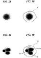
- FIGS. 3A and 3B are diagrams showing an example of an image of a cell nucleus in which apoptosis is not present.
- display is white as the luminance value is small, and display is black as the luminance value is large.
- FIG. 3A is a diagram showing an example of an image of a cell nucleus in which apoptosis is not present.
- FIG. 3B is a diagram showing an example of a cell nucleus region extracted by the cell nucleus region extraction unit 103.
- the region surrounded by a broken line indicated by symbol X is the cell nucleus region extracted by the cell nucleus region extraction unit 103.
- FIG. 4A is a diagram showing an example of an image of a cell nucleus in which apoptosis is present. As shown in FIG. 4A , in an image of a cell nucleus in which apoptosis is present, a plurality of fragmented cell nuclei are present inside the cell nucleus.
- FIG. 4B is a diagram showing an example of the cell nucleus region extracted by the cell nucleus region extraction unit 103 and the fragmented cell nucleus region extracted by the fragmented cell nucleus region extraction unit 104a.
- a region surrounded by a broken line indicated by symbol X is the cell nucleus region extracted by the cell nucleus region extraction unit 103.
- the region surrounded by a solid line indicated by symbol Y is the fragmented cell nucleus region extracted by the fragmented cell nucleus region extraction unit 104a.
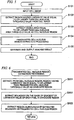
- FIG. 5 is a flowchart showing an operation example of the cell image analysis apparatus 1a.
- the operation example of the cell image analysis apparatus 1a will be described with reference to FIG. 5 .
- Step S101 The image input unit 101 inputs a digital image of a cell image to the cell image analysis apparatus 1a.
- Step S102 The cell nucleus region extraction unit 103 reads the cell nucleus threshold value stored in the threshold value storage unit 102, and extracts all regions having a luminance value equal to or larger than the cell nucleus threshold value from the cell image.
- Step S103 The cell nucleus region extraction unit 103 reads the cell nucleus area threshold value stored in the threshold value storage unit 102, and extracts all regions having an area equal to or larger than the cell nucleus area threshold value from among the regions extracted in Step S102 as a cell nucleus region.
- Step S104 The fragmented cell nucleus region extraction unit 104a executes fragmented cell nucleus region extraction processing.
- FIG. 6 is a flowchart showing an operation example of the fragmented cell nucleus region extraction unit 104a during the fragmented cell nucleus region extraction processing.
- the operation example of the fragmented cell nucleus region extraction unit 104a will be described with reference to FIG. 6 .
- the candidate region extraction unit 105 reads the fragmented cell nucleus threshold value stored in the threshold value storage unit 102, and extracts all regions having a luminance value equal to or larger than the fragmented cell nucleus threshold value from inside of each cell nucleus region extracted in Step S103 as a candidate region.
- Step S202 The region determination unit 106a performs boundary detection processing based on the gradient of the luminance value of each pixel in each candidate region to detect all boundaries.
- the boundary detection processing is implemented by applying boundary extraction processing (boundary detection processing) in the known image processing technique.
- the region determination unit 106a calculates a differential value regarding the luminance value of each pixel in the candidate region, and successively connects pixels, in which the differential value is locally maximized, to extract a boundary.
- Step S203 The region determination unit 106a extracts each region surrounded by each extracted boundary as the fragmented cell nucleus region, and ends the fragmented cell nucleus region extraction processing.
- Step S105 If the fragmented cell nucleus region extraction processing ends, the analysis result output unit 107 generates an analysis result on the basis of the detection result of the fragmented cell nucleus region extraction unit 104a and outputs the analysis result. Thus, the processing shown in FIG. 5 ends.
- FIG. 7 is a diagram showing an example of the analysis result of the cell image analysis apparatus 1a.
- the cell nucleus region extraction unit 103 extracts all the captured cell nucleus regions (regions inside three broken lines indicated by symbol X in FIG. 7 ).
- the fragmented cell nucleus region extraction unit 104a extracts all the captured fragmented cell nucleus regions (regions inside eight solid lines indicated by symbol Y in FIG. 7 ).
- the cell nucleus threshold value and the fragmented cell nucleus threshold value larger than the cell nucleus threshold value are used. That is, two threshold values regarding the luminance value are used, so the cell nucleus region and the fragmented cell nucleus region can be extracted.
- the fragmented cell nucleus region extraction unit 104a performs processing using the fragmented cell nucleus threshold value and the boundary extraction processing based on the gradient of the luminance value of each pixel to extract a boundary. That is, a region surrounded by the extracted boundary is extracted as the fragmented cell nucleus region, such that a fragmented cell nucleus region can be more accurately extracted.
- the fragmented cell nucleus region extraction unit 104a may extract all the regions as a candidate extracted in Step S201 as the fragmented cell nucleus region, without executing Steps S202 and S203. In other words, the fragmented cell nucleus region extraction unit 104a may extract, from the cell nucleus region, a region having a luminance value equal to or larger than the fragmented cell nucleus threshold value as the fragmented cell nucleus region.
- FIG. 8 is a schematic block diagram showing the functional configuration of a cell image analysis apparatus 1b which is a second embodiment of the cell image analysis apparatus.
- the same functional parts as those in the cell image analysis apparatus 1a of the first embodiment are represented by the same reference numerals as those in FIG. 1 , and description thereof will not be repeated.
- the cell image analysis apparatus 1b is different from the cell image analysis apparatus 1a in that a candidate region extraction unit 105b, instead of the candidate region extraction unit 105a, is provided. Other parts are the same as those in the cell image analysis apparatus 1a.
- the candidate region extraction unit 105b extracts, not only from the cell nucleus region but also from regions other than the cell nucleus region of the input cell image, all regions having a luminance value equal to or larger than the fragmented cell nucleus threshold value as a candidate region.
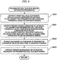
- FIG. 9 is a flowchart showing fragmented cell nucleus region extraction processing which is executed by a fragmented cell nucleus region extraction unit 104b of the second embodiment.
- the fragmented cell nucleus region extraction processing in the second embodiment will be described with reference to FIG. 9 .
- FIG. 9 the same steps as those in FIG. 6 are represented by the same reference numerals as those in FIG. 6 , and description thereof will not be repeated.
- Step S301 is executed.
- Step S301 For a region which is determined in Step S102 that the luminance value is equal to or larger than the cell nucleus threshold value and also determined in Step S103 that the area is smaller than the cell nucleus area threshold value, the candidate region extraction unit 105b extracts a region having a luminance value equal to or larger than the fragmented cell nucleus threshold value as an additional candidate region.
- Step S301 the region determination unit 106a performs Steps S202 and S203, and the fragmented cell nucleus region extraction processing ends.
- FIG. 10A is a diagram showing an example of an image in a state where the nuclear membrane of the cell nucleus is broken due to presence of apoptosis and stage advancement.
- FIG. 10A if the nuclear membrane is broken due to presence of apoptosis and stage advancement, a plurality of fragmented cell nuclei which are densely spaced inside the nuclear membrane as shown in FIG. 4A are scattered as shown in FIG 10A.
- FIG 10B is a diagram showing an example of a fragmented cell nucleus region extracted by the fragmented cell nucleus region extraction unit 104b.
- FIG. 10A is a diagram showing an example of an image in a state where the nuclear membrane of the cell nucleus is broken due to presence of apoptosis and stage advancement.
- FIG. 10B is a diagram showing an example of a fragmented cell nucleus region extracted by the fragmented cell nucleus region extraction unit 104b.
- each region surrounded by a solid line indicated by symbol Y is a fragmented cell nucleus region extracted by the fragmented cell nucleus region extraction unit 104b.
- the fragmented cell nucleus region extraction unit 104b extracts a fragmented cell nucleus region from a region other than the cell nucleus region, so a fragmented cell nucleus region in a state where the nuclear membrane is broken due to stage advancement as shown in FIG. 10A can be extracted.
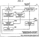
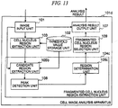
- FIG 11 is a schematic block diagram showing the functional configuration of a cell image analysis apparatus 1c which is a third embodiment of the cell image analysis apparatus 1.
- the same functional parts as those in the cell image analysis apparatus 1b of the second embodiment are represented by the same reference numerals as those in FIG. 8 , and description thereof will not be repeated.
- the cell image analysis apparatus 1c is different from the cell image analysis apparatus 1b in that the threshold value storage unit 102 further stores a fragmented cell nucleus area threshold value, a region determination unit 106c, instead of the region determination unit 106a, is provided, and a peak detection unit 108 is further provided. Other parts are the same as those in cell image analysis apparatus 1b.
- the peak detection unit 108 detects all pixels (hereinafter, referred to as peak pixel) having a peak (maximum) luminance value in each candidate region extracted by the candidate region extraction unit 105b.
- the region determination unit 106c extracts the boundary of the fragmented cell nucleus region on the basis of the detection result of the peak detection unit 108. The details of processing in the region determination unit 106c will be described below.
- FIG. 12 is a flowchart showing fragmented cell nucleus region extraction processing which is executed by a fragmented cell nucleus region extraction unit 104c of the third embodiment
- the fragmented cell nucleus extraction processing in the third embodiment will be described with reference to FIG. 12 .
- FIG. 12 the same steps as those in FIG. 9 are represented by the same reference numerals as those in FIG. 9 , and description thereof will not be repeated.
- Step S301 Steps S401 to S403 are executed sequentially.
- Step S401 The peak detection unit 108 detects a peak pixel in each candidate region extracted by the candidate region extraction unit 105b.
- the peak pixel includes not only a pixel having the maximum luminance value in the candidate region, but also all pixels having a luminance value larger than peripheral pixels and the gradient of the luminance value is zero or close to zero (a value equal to or smaller than a predetermined threshold value).
- Step S402 The region determination unit 106c extracts a boundary on the basis of the detection result of the peak detection unit 108.
- the region determination unit 106c searches and extracts a boundary satisfying the following conditions.
- the region determination unit 106c first sets nine pixels adjacent to the peak pixel as an initial boundary. Next, the area inside the boundary is calculated and it is determined whether or not the calculated area is smaller than the fragmented cell nucleus area threshold value. When the inner area is smaller than the fragmented cell nucleus area threshold value, the luminance value of each pixel on the boundary and the luminance value of each pixel on an outer periphery are compared with each other for each pixel.
- the gradient of the luminance value is calculated for each pixel on the current boundary, the outer boundary is set as a new boundary, and processing including comparison with the fragmented cell nucleus area threshold value is repeated. After this processing is repeated, when the area inside the boundary exceeds the fragmented cell nucleus area threshold value, or when the luminance value of each pixel on the outer periphery is larger than the luminance value of each pixel on the boundary, a boundary on which the average value of the gradient of the luminance value of each pixel has a maximum value is selected by using the gradient of the luminance value of each pixel calculated in the interim.
- the method of searching for a boundary satisfying the three conditions may be implemented by other methods using the known technique.
- Step S403 After Step S402, the region determination unit 106c extracts a region surrounded by each boundary extracted in Step S402 as the fragmented cell nucleus region.
- the peak detection unit 108 detects the peak pixel from the candidate region, and the region determination unit 106c extracts a region including the peak pixel as the fragmented cell nucleus region.
- the region determination unit 106c extracts a region including the peak pixel as the fragmented cell nucleus region.
- the fragmented cell nucleus region around the center of the region has a maximum luminance value. For this reason, as described above, a region including the peak pixel is extracted as the fragmented cell nucleus region, so extraction accuracy of the fragmented cell nucleus region can be improved.
- FIG. 13 is a schematic block diagram showing the functional configuration of a cell image analysis apparatus 1d which is a fourth embodiment of the cell image analysis apparatus.
- the same functional parts as those in the cell image analysis apparatus 1c of the third embodiment are represented by the same reference numerals as those in FIG. 11 , and description thereof will not be repeated.
- the cell image analysis apparatus 1d is different from the cell image analysis apparatus 1c in that a fragmented cell nucleus region selection unit 109 is further provided. Other parts are the same as those in the cell image analysis apparatus 1c.
- the fragmented cell nucleus region selection unit 109 selects only a fragmented cell nucleus region satisfying predetermined conditions from among one or more fragmented cell nucleus regions extracted by the fragmented cell nucleus region extraction unit 104c and outputs the selected fragmented cell nucleus region to the analysis result output unit 107.
- the predetermined conditions include, for example, the following conditions.
- the fragmented cell nucleus region selection unit 109 selects a fragmented cell nucleus region, for example, under the following conditions.
- the fragmented cell nucleus region selection unit 109 may be designed to perform determination one of the conditions, or may be designed to perform determination a plurality of conditions and to select a region satisfying all the conditions. Alternatively, the fragmented cell nucleus region selection unit 109 may be designed to perform determination a plurality of conditions and to select a region satisfying one of the conditions.
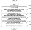
- FIG. 14 is a flowchart showing processing which is executed by the cell image analysis apparatus 1d of the fourth embodiment.
- the processing in the cell image analysis apparatus 1d of the fourth embodiment will be described with reference to FIG. 14 .
- the same steps as those in FIG. 5 are represented by the same reference numerals as those in FIG. 5 , and description thereof will not be repeated.
- Step S104 After the fragmented cell nucleus region extraction processing of Step S104, Steps S501 and S105 are executed.
- Step S501 The fragmented cell nucleus region selection unit 109 determines whether or not each fragmented cell nucleus region extracted in the fragmented cell nucleus region extraction processing satisfies predetermined conditions. Then, the fragmented cell nucleus region selection unit 109 selects only a fragmented cell nucleus region satisfying the conditions and outputs the selected fragmented cell nucleus region to the analysis result output unit 107.
- Step S105 After Step S105, the analysis result output unit 107 generates an analysis result only on the basis of the fragmented cell nucleus region selected by the fragmented cell nucleus region selection unit 109, and outputs the generated analysis result
- the fragmented cell nucleus region selection unit 109 selects only a fragmented cell nucleus region satisfying predetermined conditions, and outputs the selected fragmented cell nucleus region to the analysis result output unit 107. For this reason, extraction accuracy of the fragmented cell nucleus region can be improved.
- the functions of the cell image analysis apparatuses 1a to 1d in the foregoing embodiments may be implemented by a computer.
- a program for implementing each function may be recorded in a computer-readable recording medium, and a computer system may read and execute the program recorded on the recording medium.
- the term "computer system” includes an OS or hardware such as peripheral devices.
- the term "computer-readable recording medium” indicates a storage device, for example, a portable medium, such as a flexible disk, a magnetooptical disk, a ROM, or a CD-ROM, or a hard disk built in a computer system.
- computer-readable recording medium may be a medium for dynamically holding a program for a short time, for example, a communication line when a program is transmitted through a network, such as the Internet, or a communication line, such as a telephone line.
- computer-readable recording medium may be a medium for holding a program for a predetermined time, for example, a volatile memory in a computer system serving as a server or a client.
- the program may implement part of the above-described functions.
- the above-described functions may be implemented in combination with programs recorded in a computer system.
Landscapes
- Engineering & Computer Science (AREA)
- Physics & Mathematics (AREA)
- General Physics & Mathematics (AREA)
- Theoretical Computer Science (AREA)
- Computer Vision & Pattern Recognition (AREA)
- Health & Medical Sciences (AREA)
- Life Sciences & Earth Sciences (AREA)
- Chemical & Material Sciences (AREA)
- General Health & Medical Sciences (AREA)
- Multimedia (AREA)
- Molecular Biology (AREA)
- Signal Processing (AREA)
- Biomedical Technology (AREA)
- Dispersion Chemistry (AREA)
- Analytical Chemistry (AREA)
- Biochemistry (AREA)
- Immunology (AREA)
- Pathology (AREA)
- Investigating Or Analysing Biological Materials (AREA)
- Apparatus Associated With Microorganisms And Enzymes (AREA)
- Investigating, Analyzing Materials By Fluorescence Or Luminescence (AREA)
- Image Processing (AREA)
Claims (8)
- Zellbildanalysevorrichtung (1a), die umfasst:eine Schwellenwertspeichereinheit (102), die so konfiguriert ist, dass sie einen Zellkern-Schwellenwert, einen Fragmentierter-Zellkern-Schwellenwert und einen Zellkernflächen-Schwellenwert vorab speichert;eine Bildeingabeeinheit (101), die so konfiguriert ist, dass sie ein Zellbild eingibt, das aus einer mit einer fluoreszierenden Substanz gefärbten Zelle aufgenommen wird;eine Zellkernregionextraktionseinheit (103), die so konfiguriert ist, dass sie eine Region mit einer Fläche, die größer oder gleich dem Zellkernflächen-Schwellenwert ist, aus Regionen mit einem Luminanzwert, der größer oder gleich dem Zellkern-Schwellenwert ist, als Zellkernregion aus dem eingegebenen Zellbild extrahiert und Information, die Position und Bereich der Zellkernregion repräsentiert, erfasst;eine Einheit (104a) zum Extrahieren einer Region fragmentierten Zellkerns, die so konfiguriert ist, dass sie eine Region mit einem Luminanzwert, der größer oder gleich dem Fragmentierter-Zellkern-Schwellenwert ist, als Region fragmentierten Zellkerns aus der Zellkernregion extrahiert und Information, die Position und Bereich der Region fragmentierten Zellkerns repräsentiert, erfasst; wobei:die Einheit (104a) zum Extrahieren einer Region fragmentierten Zellkerns umfasst:eine Kandidatenregionextraktionseinheit (105a), die so konfiguriert ist, dass sie eine Region mit einem Luminanzwert, der größer gleich dem Fragmentierter-Zellkern-Schwellenwert ist, als Kandidatenregion aus der Zellkernregion extrahiert, undeine Regionbestimmungseinheit (106a), die so konfiguriert ist, dass sie eine Grenzenerkennungsverarbeitung auf Basis des Gradienten des Luminanzwerts jedes Pixels durchführt, um eine Grenze in der Kandidatenregion zu erkennen, und eine Region, die von der erkannten Grenze umgeben ist, als Region fragmentierten Zellkerns extrahiert; undeine Analyseergebnisausgabeeinheit (107), die so konfiguriert ist, dass sie ein Analyseergebnis auf Basis von Information, die die Positionen und Bereiche der Zellkernregion und der Region fragmentierten Zellkerns repräsentiert, die jeweils aus der Zellkernregionextraktionseinheit (103) und der Einheit (104a) zum Extrahieren einer Region fragmentierten Zellkerns extrahiert werden, wobei die Analyseergebnisausgabeeinheit (107) so konfiguriert ist, dass sie zumindest die Anzahl von Regionen fragmentierten Zellkerns ausgibt.
- Zellbildanalysevorrichtung (1a) nach Anspruch 1,
wobei die Einheit (104a) zum Extrahieren einer Region fragmentierten Zellkerns ferner umfasst:eine Spitzenerkennungseinheit (108), die so konfiguriert ist, dass sie ein Pixel mit einem Spitzenluminanzwert in der Kandidatenregion erkennt,wobei die Regionbestimmungseinheit (106a) so konfiguriert ist, dass sie eine Grenze erkennt, die ein Pixel mit einer Spitze umfasst und auf der der Durchschnittswert des Gradienten des Luminanzwerts jedes Pixels einen Maximalwert aufweist. - Zellbildanalysevorrichtung (1a) nach Anspruch 1,
wobei die Schwellenwertspeichereinheit (102) ferner so konfiguriert ist, dass sie einen Fragmentierter-Zellkern-Flächen-Schwellenwert speichert, und
die Regionbestimmungseinheit (106a) so konfiguriert ist, dass sie eine Region, die von einer Grenze mit einer Innenfläche kleiner oder gleich dem Fragmentierter-Zellkern-Flächen-Schwellenwert umgeben ist, als Region fragmentierten Zellkerns aus den erkannten Grenzen extrahiert. - Zellbildanalysevorrichtung (1a) nach Anspruch 2,
wobei die Schwellenwertspeichereinheit (102) ferner so konfiguriert ist, dass sie einen Fragmentierter-Zellkern-Flächen-Schwellenwert speichert, und
die Regionbestimmungseinheit (106a) so konfiguriert ist, dass sie eine Region, die von einer Grenze mit einer Innenfläche kleiner gleich dem Fragmentierter-Zellkern-Flächen-Schwellenwert umgeben ist, als Region fragmentierten Zellkerns aus den erkannten Grenzen extrahiert. - Zellbildanalysevorrichtung (1a) nach Anspruch 1,
wobei die Einheit (104a) zum Extrahieren einer Region fragmentierten Zellkerns so konfiguriert ist, dass sie eine Region mit einem Luminanzwert, der größer oder gleich dem Fragmentierter-Zellkern-Schwellenwert ist, als Region fragmentierten Zellkerns aus einer anderen Region als der Zellkernregion extrahiert. - Zellbildanalysevorrichtung (1a) nach Anspruch 1, die ferner umfasst:
eine Einheit (109) zum Auswählen einer Region fragmentierten Zellkerns, die so konfiguriert ist, dass sie für jede Region fragmentierten Zellkerns, die von der Einheit (104a) zum Extrahieren einer Region fragmentierten Zellkerns extrahiert wird, bestimmt, ob eine der folgenden Bedingungen oder eine Mehrzahl der Bedingungen erfüllt ist, und nur eine Region fragmentierten Zellkerns auswählt, die die Bedingungen erfüllt:einen statistischen Wert der Luminanzwerte von Pixeln in der relevanten Region;ein Vergleichsergebnis des statistischen Werts der Luminanzwerte der Pixel in der relevanten Region und eines statistischen Werts der Luminanzwerte von Pixeln um die relevante Region herum;eine Größe der relevanten Region;ein Vergleichsergebnis der Größe der relevanten Region und einer Größe einer Zellkernregion, die die relevante Region umfasst; undeine Form der relevanten Region. - Programm, das einen Computer, der eine Schwellenwertspeichereinheit umfasst, in der ein Zellkern-Schwellenwert, ein Fragmentierter-Zellkern-Schwellenwert und ein Zellkernflächen-Schwellenwert vorab gespeichert werden, dazu veranlasst, die Schritte durchzuführen:Eingeben eines Zellbilds, das aus einer mit einer fluoreszierenden Substanz gefärbten Zelle aufgenommen wird;Extrahieren einer Region mit einer Fläche, die größer oder gleich dem Zellkernflächen-Schwellenwert ist, aus Regionen mit einem Luminanzwert, der größer oder gleich dem Zellkern-Schwellenwert ist, als Zellkernregion aus dem eingegebenen Zellbild und Erfassen von Information, die die Position und den Bereich der Zellkernregion repräsentiert;Extrahieren einer Region mit einem Luminanzwert, der größer oder gleich dem Fragmentierter-Zellkern-Schwellenwert ist, als Region fragmentierten Zellkerns aus der Zellkernregion und Erfassen von Information, die die Position und den Bereich der Region fragmentierten Zellkerns repräsentiert, das umfasst:Extrahieren einer Region mit einem Luminanzwert, der größer oder gleich dem Fragmentierter-Zellkern-Schwellenwert ist, als Kandidatenregion, aus der Zellkernregion, undDurchführen einer Grenzenerkennungsverarbeitung auf Basis des Gradienten des Luminanzwerts jedes Pixels, um eine Grenze in der Kandidatenregion zu erkennen, undExtrahieren einer Region, die von der erkannten Grenze umgeben ist, als Region fragmentierten Zellkerns; undAusgeben eines Analyseergebnisses auf Basis von Information, die die Positionen und Bereiche der Zellkernregion und der Region fragmentierten Zellkerns repäsentiert, die jeweils im Zellkernregionextraktionsschritt und im Schritt des Extrahierens einer Region fragmentierten Zellkerns extrahiert werden, wobei der Schritt des Ausgebens eines Analyseergebnisses zumindest die Anzahl von Regionen fragmentierten Zellkerns ausgibt.
- Zellbildanalyseverfahren, das die Schritte umfasst:Eingeben eines Zellbilds, das aus einer mit einer fluoreszierenden Substanz gefärbten Zelle aufgenommen wird;Extrahieren einer Region mit einer Fläche, die größer oder gleich einem Zellkernflächen-Schwellenwert ist, aus Regionen mit einem Luminanzwert, der größer oder gleich einem Zellkern-Schwellenwert ist, als Zellkernregion aus dem eingegebenen Zellbild und Erfassen von Information, die die Position und den Bereich der Zellkernregion repräsentiert;Extrahieren einer Region mit einem Luminanzwert, der größer oder gleich einem Fragmentierter-Zellkern-Schwellenwert ist, als Region fragmentierten Zellkerns aus der Zellkernregion und Erfassen von Information, die die Position und den Bereich der Region fragmentierten Zellkerns repräsentiert, das umfasst:Extrahieren einer Region mit einem Luminanzwert, der größer oder gleich dem Fragmentierter-Zellkern-Schwellenwert ist, als Kandidatenregion, aus der Zellkernregion,Durchführen einer Grenzenerkennungsverarbeitung auf Basis des Gradienten des Luminanzwerts jedes Pixels, um eine Grenze in der Kandidatenregion zu erkennen, undExtrahieren einer Region, die von der erkannten Grenze umgeben ist, als Region fragmentierten Zellkerns; undAusgeben eines Analyseergebnisses auf Basis von Information, die die Positionen und Bereiche der Zellkernregion und der Region fragmentierten Zellkerns repräsentiert, die jeweils im Zellkernregionextraktionsschritt und im Schritt des Extrahierens einer Region fragmentierten Zellkerns extrahiert werden, wobei der Schritt des Ausgebens eines Analyseergebnisses zumindest die Anzahl von Regionen fragmentierten Zellkerns ausgibt.
Applications Claiming Priority (1)
| Application Number | Priority Date | Filing Date | Title |
|---|---|---|---|
| JP2008326108A JP5058962B2 (ja) | 2008-12-22 | 2008-12-22 | 細胞画像解析装置、細胞画像解析方法、及びプログラム |
Publications (3)
| Publication Number | Publication Date |
|---|---|
| EP2199776A2 EP2199776A2 (de) | 2010-06-23 |
| EP2199776A3 EP2199776A3 (de) | 2013-08-07 |
| EP2199776B1 true EP2199776B1 (de) | 2019-04-17 |
Family
ID=42045368
Family Applications (1)
| Application Number | Title | Priority Date | Filing Date |
|---|---|---|---|
| EP09015814.8A Not-in-force EP2199776B1 (de) | 2008-12-22 | 2009-12-21 | Zellenbildanalysevorrichtung, Zellenbildanalyseverfahren und Programm |
Country Status (3)
| Country | Link |
|---|---|
| US (1) | US9176043B2 (de) |
| EP (1) | EP2199776B1 (de) |
| JP (1) | JP5058962B2 (de) |
Families Citing this family (11)
| Publication number | Priority date | Publication date | Assignee | Title |
|---|---|---|---|---|
| JP5745919B2 (ja) * | 2011-04-28 | 2015-07-08 | 浜松ホトニクス株式会社 | 細胞解析方法、細胞解析装置、および細胞解析プログラム |
| JP2015103217A (ja) * | 2013-11-28 | 2015-06-04 | ソニー株式会社 | 画像処理装置および画像処理方法 |
| EP3124956B1 (de) * | 2014-03-27 | 2018-12-26 | Konica Minolta, Inc. | Bildverarbeitungsvorrichtung und bildverarbeitungsprogramm |
| CN105067520B (zh) * | 2015-07-28 | 2018-10-23 | 爱威科技股份有限公司 | 一种镜检识别方法及装置 |
| TWI586954B (zh) * | 2015-12-09 | 2017-06-11 | 財團法人金屬工業研究發展中心 | 檢測細胞受人類乳突病毒(hpv)感染的裝置及其檢測方法 |
| ES3024557T3 (en) | 2016-06-10 | 2025-06-04 | Univ California | Image-based cell sorting systems and methods |
| TWI595372B (zh) * | 2016-08-31 | 2017-08-11 | 義守大學 | 用以檢測蛋白特徵物之顯微影像檢測系統以及顯微影像檢測方法 |
| WO2021134508A1 (zh) * | 2019-12-31 | 2021-07-08 | 深圳迈瑞生物医疗电子股份有限公司 | 一种细胞图像分析装置和样本分析方法 |
| CN113935983B (zh) * | 2021-10-28 | 2024-08-02 | 北京中科与点科技中心(有限合伙) | 一种核周溶酶体分布全自动定量分析方法 |
| CN114912493B (zh) * | 2022-05-27 | 2022-11-29 | 深圳见康智能科技有限公司 | 基于机器学习的流式免疫细胞智能分析系统 |
| CN115620284B (zh) * | 2022-12-19 | 2023-04-18 | 广东工业大学 | 基于卷积注意力机制的细胞凋亡计数方法、系统及平台 |
Citations (1)
| Publication number | Priority date | Publication date | Assignee | Title |
|---|---|---|---|---|
| WO2004099773A1 (en) * | 2003-04-30 | 2004-11-18 | Pfizer Products Inc. | Automated in vitro cellular imaging assays for micronuclei and other target objects |
Family Cites Families (17)
| Publication number | Priority date | Publication date | Assignee | Title |
|---|---|---|---|---|
| ATE239907T1 (de) * | 1999-02-26 | 2003-05-15 | Cellomics Inc | Ein system für zellbasierte reihenuntersuchungen |
| JP2001211896A (ja) * | 1999-11-26 | 2001-08-07 | Olympus Optical Co Ltd | 画像解析方法、装置、及び記録媒体 |
| JP4854832B2 (ja) * | 2000-03-27 | 2012-01-18 | オリンパス株式会社 | 細胞の画像解析方法,装置、及び記録媒体 |
| JP2002142800A (ja) * | 2000-11-13 | 2002-05-21 | Olympus Optical Co Ltd | 細胞の形態解析方法および記憶媒体 |
| JP4583670B2 (ja) * | 2001-07-04 | 2010-11-17 | パナソニック株式会社 | 画像歪み補正装置及び方法 |
| JP4749637B2 (ja) * | 2001-09-28 | 2011-08-17 | オリンパス株式会社 | 画像解析方法、装置、及び記録媒体 |
| JP3946590B2 (ja) * | 2002-07-16 | 2007-07-18 | 富士通株式会社 | 画像処理方法、画像処理プログラムおよび画像処理装置 |
| GB0227160D0 (en) | 2002-11-21 | 2002-12-24 | Qinetiq Ltd | Histological assessment of pleomorphism |
| US20070031818A1 (en) * | 2004-07-15 | 2007-02-08 | Cytokinetics, Inc., A Delaware Corporation | Assay for distinguishing live and dead cells |
| WO2006047502A2 (en) * | 2004-10-25 | 2006-05-04 | Brigham And Women's Hospital | Automated segmentation, classification and tracking of cell nuclei in time-lapse microscopy |
| WO2006104201A1 (ja) * | 2005-03-29 | 2006-10-05 | Olympus Corporation | 細胞画像解析方法、細胞画像解析プログラム、細胞画像解析装置、スクリーニング方法およびスクリーニング装置 |
| JP2006285310A (ja) * | 2005-03-31 | 2006-10-19 | Kanazawa Univ | 森林の樹冠の評価方法及びその樹冠評価プログラム |
| JP4744187B2 (ja) | 2005-05-10 | 2011-08-10 | オリンパス株式会社 | 細胞観察装置 |
| US20070124085A1 (en) * | 2005-11-30 | 2007-05-31 | Geert Kalusche | Method of processing a biological image |
| TWI406664B (zh) * | 2006-03-30 | 2013-09-01 | Univ Kyoto | 硫氧還蛋白(thioredoxin)產生促進劑 |
| JP5347272B2 (ja) * | 2008-01-18 | 2013-11-20 | 日本電気株式会社 | スポット定量装置、スポット定量方法及びプログラム |
| JP5365011B2 (ja) * | 2008-01-29 | 2013-12-11 | 日本電気株式会社 | 病理診断支援装置、病理診断支援方法、およびプログラム |
-
2008
- 2008-12-22 JP JP2008326108A patent/JP5058962B2/ja not_active Expired - Fee Related
-
2009
- 2009-12-21 EP EP09015814.8A patent/EP2199776B1/de not_active Not-in-force
- 2009-12-21 US US12/643,241 patent/US9176043B2/en not_active Expired - Fee Related
Patent Citations (1)
| Publication number | Priority date | Publication date | Assignee | Title |
|---|---|---|---|---|
| WO2004099773A1 (en) * | 2003-04-30 | 2004-11-18 | Pfizer Products Inc. | Automated in vitro cellular imaging assays for micronuclei and other target objects |
Also Published As
| Publication number | Publication date |
|---|---|
| US20100172569A1 (en) | 2010-07-08 |
| JP5058962B2 (ja) | 2012-10-24 |
| EP2199776A2 (de) | 2010-06-23 |
| JP2010145366A (ja) | 2010-07-01 |
| US9176043B2 (en) | 2015-11-03 |
| EP2199776A3 (de) | 2013-08-07 |
Similar Documents
| Publication | Publication Date | Title |
|---|---|---|
| EP2199776B1 (de) | Zellenbildanalysevorrichtung, Zellenbildanalyseverfahren und Programm | |
| US11430131B2 (en) | Foreground segmentation and nucleus ranking for scoring dual ISH images | |
| CN112703531B (zh) | 生成组织图像的注释数据 | |
| EP3251088B1 (de) | Systeme und verfahren für interessensbereichserkennung mit folienminiaturbildern | |
| JP5413408B2 (ja) | 画像処理装置、プログラム及び画像処理システム | |
| EP2803015B1 (de) | Zweistufige kategorisierung von objekten in bildern | |
| CA2200457C (en) | Biological analysis system self-calibration apparatus | |
| EP3657154A1 (de) | Verfahren und system zur bestimmung der fluoreszenzintensität eines fluoreszenzbildes | |
| Chang et al. | Nuclear segmentation in H&E sections via multi-reference graph cut (MRGC) | |
| JP4037869B2 (ja) | 画像解析支援方法、画像解析支援プログラムおよび画像解析支援装置 | |
| EP2889619A1 (de) | Bildverarbeitungsvorrichtung, programm, bildverarbeitungsverfahren, computerlesbares medium und bildverarbeitungssystem | |
| JP2014041084A (ja) | 画像処理装置、プログラム及び画像処理システム | |
| WO2014087689A1 (ja) | 画像処理装置、画像処理システム及びプログラム | |
| JP6733983B2 (ja) | 画像解析装置 | |
| Hu et al. | Automated interpretation of subcellular patterns from immunofluorescence microscopy | |
| Lee et al. | Image analysis using machine learning for automated detection of hemoglobin H inclusions in blood smears-a method for morphologic detection of rare cells | |
| CN113763370B (zh) | 数字病理图像的处理方法、装置、电子设备及存储介质 | |
| CN110622168A (zh) | 医学图像检测 | |
| CN113537253A (zh) | 一种红外图像目标检测方法、装置、计算设备及存储介质 | |
| Rijpkema et al. | Reducing manual labour in forensic microtrace recognition with deep learning | |
| JP5861678B2 (ja) | 画像処理装置、プログラム及び画像処理システム |
Legal Events
| Date | Code | Title | Description |
|---|---|---|---|
| PUAI | Public reference made under article 153(3) epc to a published international application that has entered the european phase |
Free format text: ORIGINAL CODE: 0009012 |
|
| 17P | Request for examination filed |
Effective date: 20091221 |
|
| AK | Designated contracting states |
Kind code of ref document: A2 Designated state(s): AT BE BG CH CY CZ DE DK EE ES FI FR GB GR HR HU IE IS IT LI LT LU LV MC MK MT NL NO PL PT RO SE SI SK SM TR |
|
| AX | Request for extension of the european patent |
Extension state: AL BA RS |
|
| PUAL | Search report despatched |
Free format text: ORIGINAL CODE: 0009013 |
|
| AK | Designated contracting states |
Kind code of ref document: A3 Designated state(s): AT BE BG CH CY CZ DE DK EE ES FI FR GB GR HR HU IE IS IT LI LT LU LV MC MK MT NL NO PL PT RO SE SI SK SM TR |
|
| AX | Request for extension of the european patent |
Extension state: AL BA RS |
|
| RIC1 | Information provided on ipc code assigned before grant |
Ipc: G06T 7/00 20060101ALN20130628BHEP Ipc: G01N 15/14 20060101AFI20130628BHEP Ipc: G06M 11/04 20060101ALN20130628BHEP Ipc: G06K 9/46 20060101ALN20130628BHEP |
|
| RBV | Designated contracting states (corrected) |
Designated state(s): AT BE BG CH CY CZ DE DK EE ES FI FR GB GR HR HU IE IS IT LI LT LU LV MC MK MT NL NO PL PT RO SE SI SK SM TR |
|
| RAP1 | Party data changed (applicant data changed or rights of an application transferred) |
Owner name: OLYMPUS CORPORATION |
|
| RAP1 | Party data changed (applicant data changed or rights of an application transferred) |
Owner name: OLYMPUS CORPORATION |
|
| RIN1 | Information on inventor provided before grant (corrected) |
Inventor name: KOBAYASHI, TAMIYO Inventor name: TAKAGI, KOSUKE |
|
| STAA | Information on the status of an ep patent application or granted ep patent |
Free format text: STATUS: EXAMINATION IS IN PROGRESS |
|
| 17Q | First examination report despatched |
Effective date: 20180905 |
|
| RIC1 | Information provided on ipc code assigned before grant |
Ipc: G06M 11/04 20060101ALN20181001BHEP Ipc: G06K 9/46 20060101ALN20181001BHEP Ipc: G06T 7/00 20060101ALN20181001BHEP Ipc: G01N 15/14 20060101AFI20181001BHEP |
|
| GRAP | Despatch of communication of intention to grant a patent |
Free format text: ORIGINAL CODE: EPIDOSNIGR1 |
|
| STAA | Information on the status of an ep patent application or granted ep patent |
Free format text: STATUS: GRANT OF PATENT IS INTENDED |
|
| RIC1 | Information provided on ipc code assigned before grant |
Ipc: G06K 9/46 20060101ALN20181031BHEP Ipc: G06T 7/00 20060101ALN20181031BHEP Ipc: G06M 11/04 20060101ALN20181031BHEP Ipc: G01N 15/14 20060101AFI20181031BHEP |
|
| INTG | Intention to grant announced |
Effective date: 20181116 |
|
| GRAS | Grant fee paid |
Free format text: ORIGINAL CODE: EPIDOSNIGR3 |
|
| GRAA | (expected) grant |
Free format text: ORIGINAL CODE: 0009210 |
|
| STAA | Information on the status of an ep patent application or granted ep patent |
Free format text: STATUS: THE PATENT HAS BEEN GRANTED |
|
| AK | Designated contracting states |
Kind code of ref document: B1 Designated state(s): AT BE BG CH CY CZ DE DK EE ES FI FR GB GR HR HU IE IS IT LI LT LU LV MC MK MT NL NO PL PT RO SE SI SK SM TR |
|
| REG | Reference to a national code |
Ref country code: GB Ref legal event code: FG4D |
|
| REG | Reference to a national code |
Ref country code: CH Ref legal event code: EP |
|
| REG | Reference to a national code |
Ref country code: DE Ref legal event code: R096 Ref document number: 602009057904 Country of ref document: DE |
|
| REG | Reference to a national code |
Ref country code: AT Ref legal event code: REF Ref document number: 1122103 Country of ref document: AT Kind code of ref document: T Effective date: 20190515 Ref country code: IE Ref legal event code: FG4D |
|
| REG | Reference to a national code |
Ref country code: NL Ref legal event code: MP Effective date: 20190417 |
|
| REG | Reference to a national code |
Ref country code: LT Ref legal event code: MG4D |
|
| PG25 | Lapsed in a contracting state [announced via postgrant information from national office to epo] |
Ref country code: NL Free format text: LAPSE BECAUSE OF FAILURE TO SUBMIT A TRANSLATION OF THE DESCRIPTION OR TO PAY THE FEE WITHIN THE PRESCRIBED TIME-LIMIT Effective date: 20190417 |
|
| PG25 | Lapsed in a contracting state [announced via postgrant information from national office to epo] |
Ref country code: PT Free format text: LAPSE BECAUSE OF FAILURE TO SUBMIT A TRANSLATION OF THE DESCRIPTION OR TO PAY THE FEE WITHIN THE PRESCRIBED TIME-LIMIT Effective date: 20190817 Ref country code: SE Free format text: LAPSE BECAUSE OF FAILURE TO SUBMIT A TRANSLATION OF THE DESCRIPTION OR TO PAY THE FEE WITHIN THE PRESCRIBED TIME-LIMIT Effective date: 20190417 Ref country code: FI Free format text: LAPSE BECAUSE OF FAILURE TO SUBMIT A TRANSLATION OF THE DESCRIPTION OR TO PAY THE FEE WITHIN THE PRESCRIBED TIME-LIMIT Effective date: 20190417 Ref country code: NO Free format text: LAPSE BECAUSE OF FAILURE TO SUBMIT A TRANSLATION OF THE DESCRIPTION OR TO PAY THE FEE WITHIN THE PRESCRIBED TIME-LIMIT Effective date: 20190717 Ref country code: HR Free format text: LAPSE BECAUSE OF FAILURE TO SUBMIT A TRANSLATION OF THE DESCRIPTION OR TO PAY THE FEE WITHIN THE PRESCRIBED TIME-LIMIT Effective date: 20190417 Ref country code: ES Free format text: LAPSE BECAUSE OF FAILURE TO SUBMIT A TRANSLATION OF THE DESCRIPTION OR TO PAY THE FEE WITHIN THE PRESCRIBED TIME-LIMIT Effective date: 20190417 Ref country code: LT Free format text: LAPSE BECAUSE OF FAILURE TO SUBMIT A TRANSLATION OF THE DESCRIPTION OR TO PAY THE FEE WITHIN THE PRESCRIBED TIME-LIMIT Effective date: 20190417 |
|
| PG25 | Lapsed in a contracting state [announced via postgrant information from national office to epo] |
Ref country code: LV Free format text: LAPSE BECAUSE OF FAILURE TO SUBMIT A TRANSLATION OF THE DESCRIPTION OR TO PAY THE FEE WITHIN THE PRESCRIBED TIME-LIMIT Effective date: 20190417 Ref country code: BG Free format text: LAPSE BECAUSE OF FAILURE TO SUBMIT A TRANSLATION OF THE DESCRIPTION OR TO PAY THE FEE WITHIN THE PRESCRIBED TIME-LIMIT Effective date: 20190717 Ref country code: PL Free format text: LAPSE BECAUSE OF FAILURE TO SUBMIT A TRANSLATION OF THE DESCRIPTION OR TO PAY THE FEE WITHIN THE PRESCRIBED TIME-LIMIT Effective date: 20190417 Ref country code: GR Free format text: LAPSE BECAUSE OF FAILURE TO SUBMIT A TRANSLATION OF THE DESCRIPTION OR TO PAY THE FEE WITHIN THE PRESCRIBED TIME-LIMIT Effective date: 20190718 |
|
| REG | Reference to a national code |
Ref country code: AT Ref legal event code: MK05 Ref document number: 1122103 Country of ref document: AT Kind code of ref document: T Effective date: 20190417 |
|
| PG25 | Lapsed in a contracting state [announced via postgrant information from national office to epo] |
Ref country code: IS Free format text: LAPSE BECAUSE OF FAILURE TO SUBMIT A TRANSLATION OF THE DESCRIPTION OR TO PAY THE FEE WITHIN THE PRESCRIBED TIME-LIMIT Effective date: 20190817 |
|
| REG | Reference to a national code |
Ref country code: DE Ref legal event code: R097 Ref document number: 602009057904 Country of ref document: DE |
|
| PG25 | Lapsed in a contracting state [announced via postgrant information from national office to epo] |
Ref country code: DK Free format text: LAPSE BECAUSE OF FAILURE TO SUBMIT A TRANSLATION OF THE DESCRIPTION OR TO PAY THE FEE WITHIN THE PRESCRIBED TIME-LIMIT Effective date: 20190417 Ref country code: AT Free format text: LAPSE BECAUSE OF FAILURE TO SUBMIT A TRANSLATION OF THE DESCRIPTION OR TO PAY THE FEE WITHIN THE PRESCRIBED TIME-LIMIT Effective date: 20190417 Ref country code: EE Free format text: LAPSE BECAUSE OF FAILURE TO SUBMIT A TRANSLATION OF THE DESCRIPTION OR TO PAY THE FEE WITHIN THE PRESCRIBED TIME-LIMIT Effective date: 20190417 Ref country code: SK Free format text: LAPSE BECAUSE OF FAILURE TO SUBMIT A TRANSLATION OF THE DESCRIPTION OR TO PAY THE FEE WITHIN THE PRESCRIBED TIME-LIMIT Effective date: 20190417 Ref country code: RO Free format text: LAPSE BECAUSE OF FAILURE TO SUBMIT A TRANSLATION OF THE DESCRIPTION OR TO PAY THE FEE WITHIN THE PRESCRIBED TIME-LIMIT Effective date: 20190417 Ref country code: CZ Free format text: LAPSE BECAUSE OF FAILURE TO SUBMIT A TRANSLATION OF THE DESCRIPTION OR TO PAY THE FEE WITHIN THE PRESCRIBED TIME-LIMIT Effective date: 20190417 |
|
| PLBE | No opposition filed within time limit |
Free format text: ORIGINAL CODE: 0009261 |
|
| STAA | Information on the status of an ep patent application or granted ep patent |
Free format text: STATUS: NO OPPOSITION FILED WITHIN TIME LIMIT |
|
| PG25 | Lapsed in a contracting state [announced via postgrant information from national office to epo] |
Ref country code: IT Free format text: LAPSE BECAUSE OF FAILURE TO SUBMIT A TRANSLATION OF THE DESCRIPTION OR TO PAY THE FEE WITHIN THE PRESCRIBED TIME-LIMIT Effective date: 20190417 Ref country code: SM Free format text: LAPSE BECAUSE OF FAILURE TO SUBMIT A TRANSLATION OF THE DESCRIPTION OR TO PAY THE FEE WITHIN THE PRESCRIBED TIME-LIMIT Effective date: 20190417 |
|
| 26N | No opposition filed |
Effective date: 20200120 |
|
| PG25 | Lapsed in a contracting state [announced via postgrant information from national office to epo] |
Ref country code: TR Free format text: LAPSE BECAUSE OF FAILURE TO SUBMIT A TRANSLATION OF THE DESCRIPTION OR TO PAY THE FEE WITHIN THE PRESCRIBED TIME-LIMIT Effective date: 20190417 |
|
| PG25 | Lapsed in a contracting state [announced via postgrant information from national office to epo] |
Ref country code: SI Free format text: LAPSE BECAUSE OF FAILURE TO SUBMIT A TRANSLATION OF THE DESCRIPTION OR TO PAY THE FEE WITHIN THE PRESCRIBED TIME-LIMIT Effective date: 20190417 |
|
| REG | Reference to a national code |
Ref country code: DE Ref legal event code: R119 Ref document number: 602009057904 Country of ref document: DE |
|
| REG | Reference to a national code |
Ref country code: CH Ref legal event code: PL |
|
| REG | Reference to a national code |
Ref country code: BE Ref legal event code: MM Effective date: 20191231 |
|
| PG25 | Lapsed in a contracting state [announced via postgrant information from national office to epo] |
Ref country code: MC Free format text: LAPSE BECAUSE OF FAILURE TO SUBMIT A TRANSLATION OF THE DESCRIPTION OR TO PAY THE FEE WITHIN THE PRESCRIBED TIME-LIMIT Effective date: 20190417 |
|
| GBPC | Gb: european patent ceased through non-payment of renewal fee |
Effective date: 20191221 |
|
| PG25 | Lapsed in a contracting state [announced via postgrant information from national office to epo] |
Ref country code: GB Free format text: LAPSE BECAUSE OF NON-PAYMENT OF DUE FEES Effective date: 20191221 Ref country code: LU Free format text: LAPSE BECAUSE OF NON-PAYMENT OF DUE FEES Effective date: 20191221 Ref country code: FR Free format text: LAPSE BECAUSE OF NON-PAYMENT OF DUE FEES Effective date: 20191231 Ref country code: IE Free format text: LAPSE BECAUSE OF NON-PAYMENT OF DUE FEES Effective date: 20191221 Ref country code: DE Free format text: LAPSE BECAUSE OF NON-PAYMENT OF DUE FEES Effective date: 20200701 |
|
| PG25 | Lapsed in a contracting state [announced via postgrant information from national office to epo] |
Ref country code: CH Free format text: LAPSE BECAUSE OF NON-PAYMENT OF DUE FEES Effective date: 20191231 Ref country code: BE Free format text: LAPSE BECAUSE OF NON-PAYMENT OF DUE FEES Effective date: 20191231 Ref country code: LI Free format text: LAPSE BECAUSE OF NON-PAYMENT OF DUE FEES Effective date: 20191231 |
|
| PG25 | Lapsed in a contracting state [announced via postgrant information from national office to epo] |
Ref country code: CY Free format text: LAPSE BECAUSE OF FAILURE TO SUBMIT A TRANSLATION OF THE DESCRIPTION OR TO PAY THE FEE WITHIN THE PRESCRIBED TIME-LIMIT Effective date: 20190417 |
|
| PG25 | Lapsed in a contracting state [announced via postgrant information from national office to epo] |
Ref country code: MT Free format text: LAPSE BECAUSE OF FAILURE TO SUBMIT A TRANSLATION OF THE DESCRIPTION OR TO PAY THE FEE WITHIN THE PRESCRIBED TIME-LIMIT Effective date: 20190417 Ref country code: HU Free format text: LAPSE BECAUSE OF FAILURE TO SUBMIT A TRANSLATION OF THE DESCRIPTION OR TO PAY THE FEE WITHIN THE PRESCRIBED TIME-LIMIT; INVALID AB INITIO Effective date: 20091221 |
|
| PG25 | Lapsed in a contracting state [announced via postgrant information from national office to epo] |
Ref country code: MK Free format text: LAPSE BECAUSE OF FAILURE TO SUBMIT A TRANSLATION OF THE DESCRIPTION OR TO PAY THE FEE WITHIN THE PRESCRIBED TIME-LIMIT Effective date: 20190417 |