EP2223662A1 - Seringue électronique avec système de sécurité pour injection rachidienne - Google Patents
Seringue électronique avec système de sécurité pour injection rachidienne Download PDFInfo
- Publication number
- EP2223662A1 EP2223662A1 EP08852740A EP08852740A EP2223662A1 EP 2223662 A1 EP2223662 A1 EP 2223662A1 EP 08852740 A EP08852740 A EP 08852740A EP 08852740 A EP08852740 A EP 08852740A EP 2223662 A1 EP2223662 A1 EP 2223662A1
- Authority
- EP
- European Patent Office
- Prior art keywords
- needle
- air
- alarm
- threshold
- pressure
- Prior art date
- Legal status (The legal status is an assumption and is not a legal conclusion. Google has not performed a legal analysis and makes no representation as to the accuracy of the status listed.)
- Withdrawn
Links
- 238000002347 injection Methods 0.000 title description 6
- 239000007924 injection Substances 0.000 title description 6
- 230000009467 reduction Effects 0.000 claims abstract description 4
- 238000000034 method Methods 0.000 claims description 51
- 230000008859 change Effects 0.000 claims description 14
- 210000004749 ligamentum flavum Anatomy 0.000 claims description 11
- 238000009530 blood pressure measurement Methods 0.000 claims description 7
- 230000000007 visual effect Effects 0.000 claims description 6
- 238000002692 epidural anesthesia Methods 0.000 description 15
- 206010002091 Anaesthesia Diseases 0.000 description 8
- 230000037005 anaesthesia Effects 0.000 description 8
- 230000007246 mechanism Effects 0.000 description 5
- 241001269524 Dura Species 0.000 description 4
- 208000002193 Pain Diseases 0.000 description 4
- 230000036592 analgesia Effects 0.000 description 4
- 230000008901 benefit Effects 0.000 description 4
- 239000012530 fluid Substances 0.000 description 4
- 230000036407 pain Effects 0.000 description 4
- 230000003444 anaesthetic effect Effects 0.000 description 3
- 238000001802 infusion Methods 0.000 description 3
- 238000003780 insertion Methods 0.000 description 3
- 230000037431 insertion Effects 0.000 description 3
- 238000012986 modification Methods 0.000 description 3
- 230000004048 modification Effects 0.000 description 3
- 210000005036 nerve Anatomy 0.000 description 3
- 230000002980 postoperative effect Effects 0.000 description 3
- 238000012360 testing method Methods 0.000 description 3
- 210000000115 thoracic cavity Anatomy 0.000 description 3
- 206010001526 Air embolism Diseases 0.000 description 2
- FAPWRFPIFSIZLT-UHFFFAOYSA-M Sodium chloride Chemical compound [Na+].[Cl-] FAPWRFPIFSIZLT-UHFFFAOYSA-M 0.000 description 2
- 230000003213 activating effect Effects 0.000 description 2
- 238000013459 approach Methods 0.000 description 2
- 230000000903 blocking effect Effects 0.000 description 2
- 210000001175 cerebrospinal fluid Anatomy 0.000 description 2
- ZPUCINDJVBIVPJ-LJISPDSOSA-N cocaine Chemical compound O([C@H]1C[C@@H]2CC[C@@H](N2C)[C@H]1C(=O)OC)C(=O)C1=CC=CC=C1 ZPUCINDJVBIVPJ-LJISPDSOSA-N 0.000 description 2
- 238000001514 detection method Methods 0.000 description 2
- 238000010586 diagram Methods 0.000 description 2
- 210000001951 dura mater Anatomy 0.000 description 2
- 210000003041 ligament Anatomy 0.000 description 2
- 239000003589 local anesthetic agent Substances 0.000 description 2
- 210000003141 lower extremity Anatomy 0.000 description 2
- 239000000243 solution Substances 0.000 description 2
- 238000001356 surgical procedure Methods 0.000 description 2
- 210000001519 tissue Anatomy 0.000 description 2
- 210000003462 vein Anatomy 0.000 description 2
- 208000008035 Back Pain Diseases 0.000 description 1
- 208000034423 Delivery Diseases 0.000 description 1
- 208000007914 Labor Pain Diseases 0.000 description 1
- 208000035945 Labour pain Diseases 0.000 description 1
- WHXSMMKQMYFTQS-UHFFFAOYSA-N Lithium Chemical compound [Li] WHXSMMKQMYFTQS-UHFFFAOYSA-N 0.000 description 1
- 208000008930 Low Back Pain Diseases 0.000 description 1
- 208000006735 Periostitis Diseases 0.000 description 1
- 208000005538 Post-Dural Puncture Headache Diseases 0.000 description 1
- 108010052164 Sodium Channels Proteins 0.000 description 1
- 102000018674 Sodium Channels Human genes 0.000 description 1
- 210000001015 abdomen Anatomy 0.000 description 1
- 230000003187 abdominal effect Effects 0.000 description 1
- 238000012084 abdominal surgery Methods 0.000 description 1
- 230000004913 activation Effects 0.000 description 1
- 210000000577 adipose tissue Anatomy 0.000 description 1
- 238000002266 amputation Methods 0.000 description 1
- 230000005540 biological transmission Effects 0.000 description 1
- 239000008280 blood Substances 0.000 description 1
- 210000004369 blood Anatomy 0.000 description 1
- 239000003795 chemical substances by application Substances 0.000 description 1
- 229960003920 cocaine Drugs 0.000 description 1
- 230000000295 complement effect Effects 0.000 description 1
- 230000006835 compression Effects 0.000 description 1
- 238000007906 compression Methods 0.000 description 1
- 230000003247 decreasing effect Effects 0.000 description 1
- 239000003814 drug Substances 0.000 description 1
- 229940079593 drug Drugs 0.000 description 1
- 210000002532 foramen magnum Anatomy 0.000 description 1
- 239000011521 glass Substances 0.000 description 1
- 210000001624 hip Anatomy 0.000 description 1
- 208000014674 injury Diseases 0.000 description 1
- 210000003127 knee Anatomy 0.000 description 1
- 229910052744 lithium Inorganic materials 0.000 description 1
- 229960005015 local anesthetics Drugs 0.000 description 1
- 230000004807 localization Effects 0.000 description 1
- 238000007726 management method Methods 0.000 description 1
- 239000000463 material Substances 0.000 description 1
- 239000012528 membrane Substances 0.000 description 1
- 229940005483 opioid analgesics Drugs 0.000 description 1
- 206010033675 panniculitis Diseases 0.000 description 1
- 210000004197 pelvis Anatomy 0.000 description 1
- 230000000149 penetrating effect Effects 0.000 description 1
- 210000002640 perineum Anatomy 0.000 description 1
- 210000003460 periosteum Anatomy 0.000 description 1
- 230000002688 persistence Effects 0.000 description 1
- 238000013439 planning Methods 0.000 description 1
- 230000003938 response to stress Effects 0.000 description 1
- 238000007789 sealing Methods 0.000 description 1
- 230000035807 sensation Effects 0.000 description 1
- 210000002966 serum Anatomy 0.000 description 1
- 239000011780 sodium chloride Substances 0.000 description 1
- 210000000278 spinal cord Anatomy 0.000 description 1
- 210000001562 sternum Anatomy 0.000 description 1
- 210000002330 subarachnoid space Anatomy 0.000 description 1
- 210000004304 subcutaneous tissue Anatomy 0.000 description 1
- 239000000126 substance Substances 0.000 description 1
- 230000008733 trauma Effects 0.000 description 1
- 230000000472 traumatic effect Effects 0.000 description 1
- 230000009677 vaginal delivery Effects 0.000 description 1
- 230000002792 vascular Effects 0.000 description 1
Images



Classifications
-
- A—HUMAN NECESSITIES
- A61—MEDICAL OR VETERINARY SCIENCE; HYGIENE
- A61B—DIAGNOSIS; SURGERY; IDENTIFICATION
- A61B17/00—Surgical instruments, devices or methods
- A61B17/34—Trocars; Puncturing needles
- A61B17/3401—Puncturing needles for the peridural or subarachnoid space or the plexus, e.g. for anaesthesia
Definitions
- This invention relates to epidural needles, and more particularly to epidural needles used for spinal injections.
- Epidural anesthesia has been used by physicians for about a century.
- Radiologist Jean-Anthanase Sicard reported treating patients suffering from severe intractable sciatic pain or lumbago by injecting diluted solutions of cocaine through the sacral hiatus.
- Physicians began to use a lumbar approach to epidural anesthesia several decades later, eventually in abdominal surgery with single - shot lumbar epidural anesthesia.
- the epidural space is the part of the vertebral canal not occupied by the dura mater and its contents. It is a very narrow space, often referred to as a 'potential space,' that lies between the dura and the periosteum lining inside the vertebral canal. The dura and ligamentum flavum are closely adjacent the potential space.
- the epidural space extends from the foramen magnum to the sacral hiatus. The anterior and posterior nerve roots in their dural covering pass across this potential space to unite in the intervertebral foramen to form segmental nerves.
- the epidural space contains venous plexuses and fatty tissue which is continuous with the fat in the paravertebral space.
- epidural anesthesia has expanded widely over the years.
- Epidural anesthesia can be used as the sole anesthetic for procedures involving the lower limbs, pelvis, perineum and lower abdomen. It is possible to perform upper abdominal and thoracic procedures under epidural anesthesia alone.
- Some of the specific procedures in which epidural anesthesia may be used include: hip and knee surgery, vascular reconstruction or amputation of the lower limbs, obstetrics, low concentration local anesthetics, opioids, or combination of both, and thoracic trauma with rib or sternum fractures.
- the target population for use of epidural anesthesia includes pregnant women undergoing a cesarean section, or women at term who are planning a vaginal delivery but wish to have an epidural anesthetic available for labor pain management.
- An added advantage of using epidural anesthesia is the ability to maintain continuous anesthesia after placement of an epidural catheter, making it suitable for procedures of long duration. This feature also enables the use of this technique into the postoperative period for analgesia, using lower concentrations of local anesthetic drugs or in combination with different agents.
- the epidural space is entered by the tip of the needle after it passes through the ligamentum flavum.
- the epidural needle follows an anatomical direction from the skin to into the body, through the subcutaneous tissue, ligamentum interspinous, flavum and arriving then to the epidural space. The user identifies the space and enters it carefully as the bevel of the needle exits the ligamentum flavum to avoid penetrating the dura if the needle is advanced to far.
- the epidural space ends in the sacrococcygeal ligamentum. The largest distance is found in the medial line and is about 5 mm.
- the anesthesia is injected into the space.
- the epidural anesthetic molecules surround nerve roots and penetrate deeply into the spinal cord tissue, preventing sodium channel closing, thus blocking pain message transmission from the site of injection and below.
- One way to identify the epidural space during an epidural anesthesia procedure involves identifying a negative pressure to the advancing needle.
- the pressure in the epidural space has been measured to be a high negative epidural pressure of up to - 60 mm Hg at the moment of epidural puncture.
- the pressure typically becomes positive and stabilized at +3.7 ⁇ 3.2 mmHg.
- negative epidural pressures at the moment of epidural puncture may be due to tenting of the dural membrane.
- Air and saline are typically put in the syringes attached to the epidural needles to determine the loss of resistance during the insertion of an epidural.
- the ability to correctly identify the epidural space may enhance the benefits of epidural anesthesia by minimizing complications.
- Complications that may arise during an epidural procedure include: bony resistance, inability to treat the catheter, fluid through the needle, fluid through the catheter, and pain on insertion of the catheter or blood in the catheter.
- the most significant hazard of epidural blockade is unrecognized, unintentional intravascular injection.
- air embolism Another potential complication in epidural anesthesia is air embolism.
- the air volume injected into the epidural veins may produce slight clinical manifestations of around 0.07 mL per weight kilogram. Although the usual volume of injected air may not be dangerous, the persistence of the permeable foramen oval may result in an unexpected symptomatology. This may be indicated by the traumatic and accidental punction directly to the venous epidural plexus during the maneuver of detection of the epidural space. Air is as likely as the substance injected in the epidural space to pass to the circulatory system within 15 seconds due to the pressure difference. An air embolism of micro bubbles may also flow towards the general circulation.
- Accidental dural puncture is typically recognized by the immediate loss of cerebral-spinal fluid (CSF) through the epidural needle.
- CSF cerebral-spinal fluid
- Accidental dural puncture leads to a high incidence of post dural puncture headache, which may be quite severe.
- Accidental dural puncture may occur even when the procedure is performed by highly skilled physicians. It is more common when performed by more inexperienced hands.
- the localization of the epidural space is critical to the success of the epidural anesthesia.
- Devices and procedures have been designed to improve the procedure.
- the loss of resistance technique is the most common method for locating a needle in the epidural space.
- the anesthesiologist is able to feel the loss of resistance to injection through the needle. This may be due to the presence of a negative pressure in the epidural space. As noted above, the negative pressure may not always be present. In such cases, loss of resistance is not due to negative pressure, and use of the technique may feel different for the user.
- a device for locating the epidural space.
- the device includes a connector for forming a hermetically sealed connection with a needle.
- An air vessel is formed by an air tank, a diaphragm, and a space in the needle when the needle is attached to the connector.
- the device includes a pressure sensor configured to detect the air pressure in the air vessel.
- An actuator is coupled to a switch and positioned adjacent to the diaphragm to compress the diaphragm when the switch is actuated by a user causing a reduction in volume in the air vessel.
- a circuit coupled to the pressure sensor and to an alarm detects a signal indicative of the air pressure sensed by the pressure sensor. The circuit compares the signal to a threshold and outputs a first state to the alarm when the pressure is above the threshold, and outputs a second state to the alarm when the pressure falls below the threshold.
- a method for locating an epidural space. The method includes inserting a needle in a patient until sensing the ligamentum flavum.
- a closed air vessel is formed with an air tank and the space in the needle. The volume in the air vessel is reduced, and the air pressure in the air vessel is measured. The air pressure measurement is compared with a threshold while advancing the needle into the patient. When the air pressure falls below the threshold, advancement of the needle is stopped by the user.
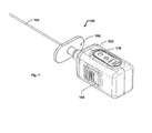
- FIG. 1 is a perspective view of an electronic syringe.
- FIG. 2 is an exploded view of the electronic syringe of FIG. 1 .
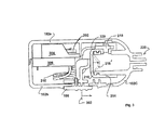
- FIG. 3 is a side cross-sectional view of the electronic syringe of FIG. 1 .
- FIG. 4 is a perspective view of the electronic syringe of FIG. 1 with part of the housing removed.
- FIG. 5 is a schematic diagram of the electronic circuit used in the electronic syringe in FIG. 1 .
- FIG. 6 is a flowchart illustrating operation of the electronic syringe.
- FIG. 1 is a perspective view of an example of an electronic syringe 100.
- the electronic syringe 100 in FIG. 1 includes a housing 102, which holds the internal components (described in more detail below with reference to FIGs. 2-5 ), and a needle 104.
- the electronic syringe 100 also includes a connector 106 for connecting the needle 104, which may include a stylet tip, a switch 108 for activating the electronic syringe 100, and an alarm 110 to indicate when the epidural space has been entered.
- the electronic syringe 100 is used by a doctor, or anesthesiologist or any other trained medical personnel, to locate the epidural space during an anesthesia procedure.
- the electronic syringe 100 facilitates the punction into the epidural space without advancing the needle too deep.
- a built-in safety mechanism in the form of a pneumatic sensor generates an alarm when the user is at risk of advancing too far.
- the user operates the electronic syringe 100 as an anatomical pressure localizer to precisely locate the epidural space advantageously minimizing complications such as a dura punction, which may lead to a release of spinal fluid during the anesthesia procedure.
- the electronic syringe 100 in FIG. 1 may be implemented in a size that would permit use as a hand-held device used by a single person.
- the electronic syringe 100 is approximately 36.5 W x 35.4 T x 59 L mm, however, any the electronic syringe 100 may be any suitable size.
- the examples of the electronic syringe 100 described here are battery-powered; however, any form of electrical power may be used.
- a feature may be included to ensure that the battery is only activated for an intended first use by avoiding accidental activation.
- a small piece of paper may be inserted between the battery and its contacts such that it extends out of the housing 102 through slit to allow the user to withdraw the paper to enable the first use. Once the paper is cleared, the user may initiate operation by sliding the switch 108 to activate the electronic syringe 100.
- the electronic syringe 100 includes two lithium 3V batteries in series that are capable of powering the device for around one hour.
- the alarm 110 in the example shown in FIG. 1 is a visual alarm 110 implemented as an LED, or other suitable light emitting device.
- the alarm 110 may also be implemented as an audible alarm, or any other device intended to attract the user's attention and indicate the occurrence of an event.
- the alarm 110 indicates when the user has reached the epidural space.
- the alarm 110 is implemented as an LED that changes to a red color to indicate that the needle may be advanced, and to a green color to indicate that the epidural space has been reached.
- the electronic syringe 100 includes the connector 106, which may be a Luer-type connector to provide a hermetically sealed connection to the needle 104. Although, any suitable connector 106 may be used.
- the examples of the electronic syringe 100 are described here as being compatible for use with a Tuohy needle 104; however, any suitable needle 104 may be used.
- the needle 104 used with the electronic syringe 100 may include a stylet tip; however, any suitable structure or technique may be used to prevent tissue from entering the needle 104 as it is inserted into the patient.
- a stylet tip may be integrated with the needle 104 and attached to the electronic syringe 100 as an assembly. In another example, no stylet tip is used with the needle 104 during the procedure.
- the electronic syringe 100 may be used as follows. The user first introduces the Tuohy needle 104 into the patient. Initially, the needle 100 may be inserted with a stylet tip. Neither the needle 104 nor stylet are attached to the electronic syringe 100 when first introduced into the patient. The user inserts the needle and stylet 104 until reaching the ligamentum flavum. When the ligamentum flavum is reached, the stylet tip is retired and the electronic syringe 100 is connected to the Tuohy needle 104 at the Luer connector 106 creating a hermetically sealed connection. The user then slides the switch 108 to the active state.
- a pressure sensor inside the housing 102 detects a pressure inside an air tank continuously, and the pressure is compared by an electronic circuit with a threshold pressure. As long as the measured pressure is greater than the threshold level, the alarm 110 is set to indicate that the user may continue to advance the needle into the patient. When the epidural space is reached, a sudden decrease in pressure below the threshold is detected and the electronic circuit causes the alarm 110 to indicate that the epidural space is reached.
- the alarm 110 provides built-in fault detection. For example, when the electronic syringe 100 is connected to the needle at the Luer connector 106, if the alarm 110 is not set to indicate that the user may advance the needle after the switch 108 is slid into the active state, the user does not advance the needle. If the alarm 110 does not change, there could be a problem with the devices or the needle may not have been inserted properly.
- the needle 104, and an attached stylet tip may be connected to the electronic syringe 100 prior to inserting into the patient.
- the user may then insert the needle attached to the electronic syringe 100 into the patient and slide the switch 108 to charge the electronic syringe 100.
- the alarm 110 indicates that the needle may be advanced
- the user continues the insertion of the needle until the alarm 110 changes to indicate that the epidural space has been reached.
- the stylet tip is fixed to the needle 104 as an attachment to the electronic syringe 100.
- the stylet tip has a smaller caliber than the Tuohy needle 104 and may include an airway to allow air to escape through the needle 104 when the needle 104 is first applied to the patient. The airway may then become obstructed when the needle 104 is introduced further into the patient thereby closing the space in the needle 104 and tank of the electronic syringe 100.
- the electronic syringe 100 may be used as one unit from the moment the needle 104 is inserted into the patient.
- FIG. 2 is an exploded view of the electronic syringe of FIG. 1 .
- the exploded view 200 in FIG. 2 shows the needle 104 removed from an exploded view of the housing 102 its contents.
- the housing 102 is shown in its component parts, a first side housing 102a, a second side housing 102b, and a housing cap 102c.
- Inside the housing 102 are components that include two batteries 202, a battery holder 204, a circuit board 206, an air tank 214, a diaphragm 216, a pressure sensor 218, an actuator 212, and an internal switch mechanism 210.
- the actuator 212 is mounted in the switch mechanism 210 to follow the motion of the switch 108.
- the actuator 212 is positioned to be in slight contact, or very close to, the diaphragm 216.
- the air tank 214 contains air and is hermetically sealed to the housing cap 102c.
- the diaphragm 216 may be made of a compressible material.
- the switch 108 is slid to activate the electronic syringe 100, the diaphragm 216 is compressed causing a reduction of volume in the closed space formed by the air tank 214 and needle 104.
- the compression of the diaphragm 216 charges the air tank 214 by increasing pressure that is higher than a threshold pressure.
- the threshold may be set to about 20 mm Hg within about ⁇ 2 mm Hg.
- the pressure sensor 218 continuously monitors the air pressure in the air tank 214. When the epidural space is found, it provides a slight escape for the air in the air tank 214 sufficient to decrease the air pressure in the air tank 214.
- the alarm 110 indicates when the epidural space has been found by changing color when the pressure drops in the air tank 214.
- FIG. 3 is a side cross-sectional view of the electronic syringe of FIG. 1 .
- the view in FIG. 3 is taken such that the switch 108 is on the bottom side of the housing 102b.
- the battery 202 sits in the battery holder 204 and the battery holder 204 is mounted on top of the circuit board 206.
- a set of wires 310 extends from the circuit board 206 to make electrical connection with the pressure sensor 218 and the alarm 110 (shown in FIGs. 1 and 2 ).
- the view in FIG. 3 illustrates the motion of the switch 108 at 302 when the electronic syringe 100 is activated.
- the switch 108 causes the actuator 212 to press on the diaphragm 216 to a compressed state at 216'.
- the switch 108 is activated while the needle (and/or stylet tip) has been inserted partially into the patient creating a higher pressure from the decreased volume in the air tank 214.
- the air in the air tank 214 is released into the epidural space causing a decrease in pressure in the air tank 214.
- the pressure sensor 218 senses the drop in pressure and generates a signal to the alarm 110 to indicate that the epidural space has been reached.
- FIG. 4 is a perspective view of the electronic syringe of FIG. 1 with part of the housing removed.
- FIG. 4 shows the battery 202, the battery holder 204, the circuit board 206, and the wires 310 from the circuit board 206 to the alarm 110 and the pressure sensor 218.
- the pressure sensor 218 is shown positioned on the air tank 214.
- the diaphragm 216 is shown sealing the rear end of the air tank 214.
- FIG. 5 is a schematic diagram of an electronic circuit 500 used in the electronic syringe in FIG. 1 .
- the electronic circuit 500 includes a Tuohy needle 502, an air vessel 504, a pressure sensor 506, an amplifier 508, a comparator 510, a threshold indicator 512, and a comparator output 512.
- the Tuohy needle 502 is inserted into the patient.
- an air vessel 504 is formed in the air tank 214 and the space of the needle 502.
- the air vessel 504 is a closed volume and when the electronic syringe 100 is charged by activating the switch 108, the pressure sensor 506 detects the increase in pressure over about 35 mm Hg.
- the pressure sensor 506 may have a range of about 0 to 300 mm Hg.
- the pressure sensor 506 outputs an electrical signal that is indicative of the pressure sensed to the amplifier 508.
- the amplifier 508 outputs an amplified signal to the comparator 510.
- the amplified signal is compared to the threshold 512, which is an electronic signal indicative of a threshold pressure below which is indicative of having reached the epidural space.
- the threshold 512 is an electronic signal indicative of a threshold pressure below which is indicative of having reached the epidural space.
- a red visual alarm 512a is output.
- the comparator 510 output changes states indicating that the pressure in the air vessel 504 has dropped below the threshold
- the comparator 510 output a signal that changes the visual alarm to a green color 512b.
- the user detects the change to the green color 512b, the user stops advancing the needle avoiding puncturing the dural.
- the color changes in the visual alarm are red and green to illustrate operation of the device. Any color change may be used.
- an audible alarm may be added to the device, or used instead of a visual alarm.
- FIG. 6 is a flowchart illustrating operation of the electronic syringe 100 (in FIG. 1 ).
- the method may begin by a test of the electronic syringe 100 to determine if it works at step 602. Any suitable test may be performed.
- the user presses the switch 108 ( FIG. 1 ) while blocking the air escape at the connector to prevent air escaping the air tank to activate the electronic syringe 100.
- the press of the switch 108 should turn on the alarm, which in this example is assumed to be a light, either green or red.
- the user may then unblock the air escape from the air tank, allowing the air to escape. As the air escapes the air tank the alarm should change color, for example, either red or green.
- the switch is returned to the inactive state once the test is complete.
- Decision block 606 checks if the indicator light went on when the switch was pressed. If the indicator light did not go 'on,' the electronic syringe 100 is discarded at step 604. If the indicator light went 'on,' the needle is placed into the patient and pressed until reaching the ligamentum flavum at step 608. At step 610, the needle is connected to the electronic syringe 100. In one example, a Luer connector is used to make the connection. At step 612, the button on the electronic syringe 100 is pressed to the active state. Decision block 618 checks if the alarm shows a color change, for example to red, or from green to red.
- the electronic syringe 100 is removed from the needle at step 614, and the button is returned to the inactive state. The electronic syringe 100 is then re-connected for another attempt at step 610.
- the needle is sunk into the patient at step 622.
- Decision block 624 checks for a color change in the alarm as the needle is sunk into the patient. If a color change is detected, the epidural space is found at step 626. The user may then continue the procedure. If no color change is detected, the user checks if the needle is in too deep at decision block 620. If the needle is too deep, the electronic syringe is removed at step 616 and discarded at step 604. If the needle is not too deep, the user continues to sink the needle into the patient at step 622.
- FIG. 6 illustrates one example of a method of using the electronic syringe to locate an epidural space. Modifications may be made.
- the needle may include an air escape. It may then be attached to the electronic syringe prior to inserting into the patient. When the ligamentum flavum is found, the method may then proceed as shown in FIG. 6 from step 612. Other modifications may be made as well.
- the electronic syringe 100 does involve injecting air into the patient.
- the amount of air injected to the patient is less than 0.1 mL, which is much less than prior techniques by as much as 30 time.
- the electronic syringe 100 is reusable within the same procedure, and may even be used repetitively if an epidural space is not located initially.
- the electronic syringe 100 may be used with any suitable needle and/or stylet tip.
- the switch 108 may be implemented as any mechanism configured to move an actuator to compress the diaphragm 216.
- the switch 108 may be implemented as a push-button, or any other suitable mechanism.
Landscapes
- Health & Medical Sciences (AREA)
- Surgery (AREA)
- Life Sciences & Earth Sciences (AREA)
- Heart & Thoracic Surgery (AREA)
- Nuclear Medicine, Radiotherapy & Molecular Imaging (AREA)
- Pathology (AREA)
- Engineering & Computer Science (AREA)
- Biomedical Technology (AREA)
- Anesthesiology (AREA)
- Medical Informatics (AREA)
- Molecular Biology (AREA)
- Animal Behavior & Ethology (AREA)
- General Health & Medical Sciences (AREA)
- Public Health (AREA)
- Veterinary Medicine (AREA)
- Infusion, Injection, And Reservoir Apparatuses (AREA)
Applications Claiming Priority (3)
| Application Number | Priority Date | Filing Date | Title |
|---|---|---|---|
| US98933307P | 2007-11-20 | 2007-11-20 | |
| US12/275,162 US20090131832A1 (en) | 2007-11-20 | 2008-11-20 | Electronic Syringe with Safety System for Spinal Injection |
| PCT/MX2008/000158 WO2009066972A1 (fr) | 2007-11-20 | 2008-11-20 | Seringue électronique avec système de sécurité pour injection rachidienne |
Publications (1)
| Publication Number | Publication Date |
|---|---|
| EP2223662A1 true EP2223662A1 (fr) | 2010-09-01 |
Family
ID=40642726
Family Applications (1)
| Application Number | Title | Priority Date | Filing Date |
|---|---|---|---|
| EP08852740A Withdrawn EP2223662A1 (fr) | 2007-11-20 | 2008-11-20 | Seringue électronique avec système de sécurité pour injection rachidienne |
Country Status (3)
| Country | Link |
|---|---|
| US (1) | US20090131832A1 (fr) |
| EP (1) | EP2223662A1 (fr) |
| WO (1) | WO2009066972A1 (fr) |
Cited By (6)
| Publication number | Priority date | Publication date | Assignee | Title |
|---|---|---|---|---|
| WO2017010795A1 (fr) * | 2015-07-13 | 2017-01-19 | 최형찬 | Point de contrôle épidural |
| CN106404257A (zh) * | 2016-11-30 | 2017-02-15 | 哈尔滨理工大学 | 一种柔性针穿刺力测量装置 |
| US10004450B2 (en) | 2016-05-03 | 2018-06-26 | Texas Medical Center | Tactile sensing device for lumbar punctures |
| CN108697325A (zh) * | 2016-02-23 | 2018-10-23 | 里程碑科技有限公司 | 用于识别目标区的设备和方法 |
| US10383610B2 (en) | 2017-10-27 | 2019-08-20 | Intuitap Medical, Inc. | Tactile sensing and needle guidance device |
| US11439353B2 (en) | 2016-06-13 | 2022-09-13 | Medtronic Holding Company Sàrl | Multi-cannula sensing device |
Families Citing this family (34)
| Publication number | Priority date | Publication date | Assignee | Title |
|---|---|---|---|---|
| US8641715B2 (en) | 2002-05-31 | 2014-02-04 | Vidacare Corporation | Manual intraosseous device |
| IL165224A0 (en) | 2002-05-31 | 2005-12-18 | Vidacare Corp | Apparatus and method to access bone marrow |
| US20070049945A1 (en) | 2002-05-31 | 2007-03-01 | Miller Larry J | Apparatus and methods to install, support and/or monitor performance of intraosseous devices |
| US10973545B2 (en) | 2002-05-31 | 2021-04-13 | Teleflex Life Sciences Limited | Powered drivers, intraosseous devices and methods to access bone marrow |
| US11337728B2 (en) | 2002-05-31 | 2022-05-24 | Teleflex Life Sciences Limited | Powered drivers, intraosseous devices and methods to access bone marrow |
| US8668698B2 (en) | 2002-05-31 | 2014-03-11 | Vidacare Corporation | Assembly for coupling powered driver with intraosseous device |
| US9504477B2 (en) | 2003-05-30 | 2016-11-29 | Vidacare LLC | Powered driver |
| US8944069B2 (en) | 2006-09-12 | 2015-02-03 | Vidacare Corporation | Assemblies for coupling intraosseous (IO) devices to powered drivers |
| US9987468B2 (en) | 2007-06-29 | 2018-06-05 | Actuated Medical, Inc. | Reduced force device for intravascular access and guidewire placement |
| US10219832B2 (en) | 2007-06-29 | 2019-03-05 | Actuated Medical, Inc. | Device and method for less forceful tissue puncture |
| US8328738B2 (en) * | 2007-06-29 | 2012-12-11 | Actuated Medical, Inc. | Medical tool for reduced penetration force with feedback means |
| US10463838B2 (en) | 2009-08-19 | 2019-11-05 | Medline Industries, Inc | Vascular access methods and devices |
| EP2467057B1 (fr) | 2009-08-19 | 2020-07-22 | Medline Industries, Inc., | Systèmes et dispositifs pour faciliter l accès à des sites ou des environnements anatomiques cibles |
| US8814807B2 (en) | 2009-08-19 | 2014-08-26 | Mirador Biomedical | Spinal canal access and probe positioning, devices and methods |
| CN101721205B (zh) * | 2010-02-02 | 2011-04-20 | 天津美迪斯医疗用品有限公司 | 一次性使用硬膜外穿刺负压指示器 |
| WO2011158227A2 (fr) * | 2010-06-13 | 2011-12-22 | Omeq - Innovative Medical Devices Ltd | Appareil et méthode de positionnement anatomique avec un dispositif expansible |
| WO2012089223A1 (fr) * | 2010-12-28 | 2012-07-05 | Mishail Ishak Ibrahim | Dispositif de détection de l'espace épidural et de positionnement précis d'un cathéter |
| US9956341B2 (en) | 2012-07-03 | 2018-05-01 | Milestone Scientific, Inc. | Drug infusion with pressure sensing and non-continuous flow for identification of and injection into fluid-filled anatomic spaces |
| US20140378905A1 (en) * | 2013-06-24 | 2014-12-25 | Thomas A. Senatore | Stylet-accommodating apparatuses for the detection of needle penetration into the epidural space |
| JP5467668B1 (ja) * | 2013-10-07 | 2014-04-09 | 応用電子工業株式会社 | 硬膜外腔識別装置 |
| TWI572387B (zh) | 2014-11-21 | 2017-03-01 | 羅文甫 | 注射器定位裝置 |
| CN105664302B (zh) * | 2014-11-21 | 2019-02-15 | 医盟科技股份有限公司 | 注射器定位装置 |
| US10940292B2 (en) | 2015-07-08 | 2021-03-09 | Actuated Medical, Inc. | Reduced force device for intravascular access and guidewire placement |
| US11793543B2 (en) | 2015-09-18 | 2023-10-24 | Obvius Robotics, Inc. | Device and method for automated insertion of penetrating member |
| US10220180B2 (en) | 2015-10-16 | 2019-03-05 | Milestone Scientific, Inc. | Method and apparatus for performing a peripheral nerve block |
| US10632255B2 (en) | 2017-02-15 | 2020-04-28 | Milestone Scientific, Inc. | Drug infusion device |
| WO2018172817A1 (fr) * | 2017-03-23 | 2018-09-27 | Velez Rivera Hector De Jesus | Aiguille d'administration d'anesthésique comportant un élément d'orientation lumineux |
| US11471595B2 (en) | 2017-05-04 | 2022-10-18 | Milestone Scientific, Inc. | Method and apparatus for performing a peripheral nerve block |
| CN109420220B (zh) * | 2017-08-24 | 2024-02-20 | 南京巨鲨显示科技有限公司 | 一种高压注射器自适应流速控制方法 |
| US20210236737A1 (en) * | 2018-04-26 | 2021-08-05 | Virginia Commonwealth University | Pressure sensitive needle positioning devices, release mechanisms, and methods |
| NL2022011B1 (en) * | 2018-11-16 | 2020-05-26 | Crea Ip B V | Vitrectome actuator |
| US10646660B1 (en) | 2019-05-16 | 2020-05-12 | Milestone Scientific, Inc. | Device and method for identification of a target region |
| US20210330349A1 (en) | 2020-04-24 | 2021-10-28 | Milestone Scientific, Inc. | Device and Method for Needle/Catheter Location Utilizing Correlation Analysis |
| US12426965B2 (en) | 2023-06-15 | 2025-09-30 | Obvius Robotics, Inc. | Image-guided robotic arm for inserting a penetrating member into a body lumen |
Family Cites Families (62)
| Publication number | Priority date | Publication date | Assignee | Title |
|---|---|---|---|---|
| US2007667A (en) * | 1933-04-18 | 1935-07-09 | Stubbs George Edwin | Boat anchor |
| US2409979A (en) * | 1946-03-14 | 1946-10-22 | Ralph L Huber | Hypodermic needle |
| US2646042A (en) * | 1951-05-18 | 1953-07-21 | Hu Quang Hsi | Medical apparatus |
| US3675722A (en) * | 1971-04-05 | 1972-07-11 | Gen Fire Extinguisher Corp | Pressure indicator |
| US3856009A (en) * | 1971-11-26 | 1974-12-24 | Johnson & Johnson | Catheter placement unit |
| US3920002A (en) * | 1971-12-22 | 1975-11-18 | Kendall & Co | Fluid sampling and measuring apparatus |
| US3780693A (en) * | 1972-05-15 | 1973-12-25 | E Parr | Visible fluid pressure indicator |
| US4000741A (en) * | 1975-11-03 | 1977-01-04 | The Kendall Company | Syringe assembly |
| MX144149A (es) * | 1976-04-28 | 1981-09-02 | Kendall & Co | Dispositivo mejorado para verificar la posicion de una aguja en el cuerpo de un paciente |
| US4178867A (en) * | 1978-01-19 | 1979-12-18 | Yin-Lung Yang | Rescue signal device |
| US4215699A (en) * | 1978-04-03 | 1980-08-05 | The Kendall Company | Position indicating device |
| US4284084A (en) * | 1979-06-29 | 1981-08-18 | The Kendall Company | Syringe assembly |
| US4737146A (en) * | 1979-12-25 | 1988-04-12 | Yoshikiyo Amaki | Multi-lumen epidural catheter |
| US4403988A (en) * | 1980-08-21 | 1983-09-13 | The Kendall Company | Syringe assembly |
| US4349023A (en) * | 1980-10-09 | 1982-09-14 | Abbott Laboratories | Epidural needle catheter and adapter |
| US4570640A (en) * | 1981-08-06 | 1986-02-18 | Barsa John E | Sensory monitoring apparatus and method |
| US4535773A (en) * | 1982-03-26 | 1985-08-20 | Inbae Yoon | Safety puncturing instrument and method |
| DE3327585A1 (de) * | 1982-08-06 | 1984-02-09 | John Martin Oxford Evans | Chirurgisches instrument fuer die epidurale und spinale anaesthesie |
| US4519403A (en) * | 1983-04-29 | 1985-05-28 | Medtronic, Inc. | Balloon lead and inflator |
| US4801293A (en) * | 1985-10-09 | 1989-01-31 | Anthony Jackson | Apparatus and method for detecting probe penetration of human epidural space and injecting a therapeutic substance thereinto |
| US4623335A (en) * | 1985-10-09 | 1986-11-18 | Anthony Jackson | Apparatus and methods for detecting probe penetration of human internal target tissue having predetermined internal pressure |
| US4944724A (en) * | 1987-06-05 | 1990-07-31 | Uresil Corporation | Apparatus for locating body cavities having signaling indicator |
| US4796641A (en) * | 1987-07-06 | 1989-01-10 | Data Sciences, Inc. | Device and method for chronic in-vivo measurement of internal body pressure |
| US4958901A (en) * | 1987-07-13 | 1990-09-25 | Neurodelivery Technology, Inc. | Method for making a multi-lumen epidural-spinal needle and tip and stock configuration for the same |
| ES2007667A6 (es) * | 1987-07-28 | 1989-07-01 | Espejo Martinez Antonio | Aparato localizador del espacio epidural |
| US4898080A (en) * | 1987-08-11 | 1990-02-06 | Lieberman Walter G | Fluid powered linear slide |
| US4828547A (en) * | 1987-09-28 | 1989-05-09 | Bio-Plexus, Inc. | Self-blunting needle assembly and device including the same |
| US4805605A (en) * | 1988-01-11 | 1989-02-21 | Glassman Medical Products, Inc. | Abduction pillow |
| US4940458A (en) * | 1989-02-02 | 1990-07-10 | Cohn Arnold K | Epidural needle placement system |
| US5018526A (en) * | 1989-02-28 | 1991-05-28 | Gaston Johansson Fannie | Apparatus and method for providing a multidimensional indication of pain |
| US5024662A (en) * | 1990-03-13 | 1991-06-18 | Menes Cesar M | Resistance syringe for epidural anesthesia |
| US5081990A (en) * | 1990-05-11 | 1992-01-21 | New York University | Catheter for spinal epidural injection of drugs and measurement of evoked potentials |
| US5270685A (en) * | 1991-07-02 | 1993-12-14 | Mallinckrodt Medical, Inc. | Syringe pressure monitor |
| US5188594A (en) * | 1991-07-22 | 1993-02-23 | Michael Zilberstein | Method of administering medication into epidural space |
| US5205828A (en) * | 1991-10-24 | 1993-04-27 | Dan Kedem | Epidural needle location indicator assembly |
| US5163904A (en) * | 1991-11-12 | 1992-11-17 | Merit Medical Systems, Inc. | Syringe apparatus with attached pressure gauge |
| US6540764B1 (en) * | 1992-06-02 | 2003-04-01 | General Surgical Innovations, Inc. | Apparatus and method for dissecting tissue layers |
| US5470316A (en) * | 1993-09-07 | 1995-11-28 | United States Surgical Corporation | Body tissue penetrating device having a vacuum indicator |
| US5531696A (en) * | 1993-12-13 | 1996-07-02 | Menes; Cesar M. | Elastomeric driver for epidural resistance syringe |
| US5397313A (en) * | 1994-01-27 | 1995-03-14 | The Kendall Company | Low friction syringe |
| US5725509A (en) * | 1994-04-05 | 1998-03-10 | Symbiosis Corporation | Air introduction system for medical needles |
| US5722955A (en) * | 1994-08-04 | 1998-03-03 | Epimed International, Inc. | Pressure sensing syringe |
| US5819950A (en) * | 1996-04-05 | 1998-10-13 | Mccloskey; James Paschal | Portable trommel |
| US5865184A (en) * | 1997-01-13 | 1999-02-02 | Takiguchi; Tetsuo | Combined spinal and epidural anesthesia |
| WO1999004705A1 (fr) * | 1997-07-25 | 1999-02-04 | Tsui Ban C H | Dispositifs, systemes et techniques permettant de determiner l'emplacement ideal des catheters epiduraux |
| US5902273A (en) * | 1997-10-15 | 1999-05-11 | Yang; Ian Y. | Pressurizable epidural space identification syringe |
| US6183442B1 (en) * | 1998-03-02 | 2001-02-06 | Board Of Regents Of The University Of Texas System | Tissue penetrating device and methods for using same |
| JP3825588B2 (ja) * | 1999-08-23 | 2006-09-27 | 住友商事株式会社 | 静電植毛装置、静電塗装装置に配する静電加工室 |
| AU2632001A (en) * | 2000-01-06 | 2001-07-16 | Raymond L. Bedell | Steerable fiberoptic epidural balloon catheter and scope |
| EP1280531B1 (fr) * | 2000-05-12 | 2007-01-24 | Novalar Pharmaceuticals, Inc. | Formulation consistée de phentolamine mesylate et son utilisation |
| GB2366729B (en) * | 2000-07-05 | 2004-10-06 | Mann Hasan | A device for identification of the epidural space |
| AU2001290582A1 (en) * | 2000-08-28 | 2002-03-13 | The United States Of America As Represented By The Administrator Of The National Aeronautics And Space Administration (Nasa) | Multiple sensor system for tissue characterization |
| US6890295B2 (en) * | 2002-10-31 | 2005-05-10 | Medtronic, Inc. | Anatomical space access tools and methods |
| US6786898B2 (en) * | 2003-01-15 | 2004-09-07 | Medtronic, Inc. | Methods and tools for accessing an anatomic space |
| USD455495S1 (en) * | 2001-01-30 | 2002-04-09 | Ronald E. Tinsley | Epidural stabilization device |
| US6565542B2 (en) * | 2001-06-22 | 2003-05-20 | Minnesota High-Tech Resources | Epidural needle having a distal flare |
| US6773417B2 (en) * | 2001-07-06 | 2004-08-10 | Ispg, Inc. | Epidural space locating device |
| US6860855B2 (en) * | 2001-11-19 | 2005-03-01 | Advanced Imaging Technologies, Inc. | System and method for tissue biopsy using ultrasonic imaging |
| US6810879B1 (en) * | 2002-05-31 | 2004-11-02 | Ronald E. Tinsley | Lateral epidural positioning device |
| US6769546B2 (en) * | 2002-07-03 | 2004-08-03 | L. John Busch | Epidural anesthesia kit |
| US7175608B2 (en) * | 2003-03-20 | 2007-02-13 | Maan Hasan | Device for the identification of the epidural space |
| US7186214B2 (en) * | 2004-02-12 | 2007-03-06 | Medtronic, Inc. | Instruments and methods for accessing an anatomic space |
-
2008
- 2008-11-20 EP EP08852740A patent/EP2223662A1/fr not_active Withdrawn
- 2008-11-20 WO PCT/MX2008/000158 patent/WO2009066972A1/fr not_active Ceased
- 2008-11-20 US US12/275,162 patent/US20090131832A1/en not_active Abandoned
Non-Patent Citations (1)
| Title |
|---|
| See references of WO2009066972A1 * |
Cited By (9)
| Publication number | Priority date | Publication date | Assignee | Title |
|---|---|---|---|---|
| WO2017010795A1 (fr) * | 2015-07-13 | 2017-01-19 | 최형찬 | Point de contrôle épidural |
| CN108697325A (zh) * | 2016-02-23 | 2018-10-23 | 里程碑科技有限公司 | 用于识别目标区的设备和方法 |
| US10004450B2 (en) | 2016-05-03 | 2018-06-26 | Texas Medical Center | Tactile sensing device for lumbar punctures |
| US11179097B2 (en) | 2016-05-03 | 2021-11-23 | Texas Medical Center | Tactile sensing device for lumbar punctures |
| US11439353B2 (en) | 2016-06-13 | 2022-09-13 | Medtronic Holding Company Sàrl | Multi-cannula sensing device |
| CN106404257A (zh) * | 2016-11-30 | 2017-02-15 | 哈尔滨理工大学 | 一种柔性针穿刺力测量装置 |
| CN106404257B (zh) * | 2016-11-30 | 2022-05-31 | 哈尔滨理工大学 | 一种柔性针穿刺力测量装置 |
| US10383610B2 (en) | 2017-10-27 | 2019-08-20 | Intuitap Medical, Inc. | Tactile sensing and needle guidance device |
| US11000311B2 (en) | 2017-10-27 | 2021-05-11 | Intuitap Medical, Inc. | Tactile sensing and needle guidance device |
Also Published As
| Publication number | Publication date |
|---|---|
| US20090131832A1 (en) | 2009-05-21 |
| WO2009066972A1 (fr) | 2009-05-28 |
Similar Documents
| Publication | Publication Date | Title |
|---|---|---|
| EP2223662A1 (fr) | Seringue électronique avec système de sécurité pour injection rachidienne | |
| CN107548310B (zh) | 硬膜外空间识别探测装置 | |
| US5817052A (en) | Apparatus for intraosseous infusion or aspiration | |
| JP6349311B2 (ja) | 圧力検出を伴う薬物の注入、ならびに、流体で満たされた体内空間の識別および流体で満たされた体内空間への注入のための不連続的な流れ | |
| US6190370B1 (en) | Devices, systems and methods for determining proper placement of epidural catheters | |
| CA1078287A (fr) | Indicateur de position et methode d'utilisation | |
| JP2009504316A (ja) | 注入圧力検出を使用した中枢・末梢神経組織の識別のための医薬注入装置 | |
| CN105232093B (zh) | 使用压力侦测医疗用针定位的方法及其装置和穿刺针套组 | |
| US20100152616A1 (en) | Devices and methods for safely accessing bone marrow and other tissues | |
| US9504790B1 (en) | Device and method for identification of a target region | |
| US20060173480A1 (en) | Safety penetrating method and apparatus into body cavities, organs, or potential spaces | |
| US11185245B2 (en) | Catheter for monitoring pressure for muscle compartment syndrome | |
| JP2004242936A (ja) | 穿刺針 | |
| KR20040102355A (ko) | 척추용 니들 시스템 | |
| US20050283092A1 (en) | Continuous compartment pressure monitoring device | |
| US20190159804A1 (en) | Medical Needle System | |
| US20220000381A1 (en) | Catheter for monitoring pressure for muscle compartment syndrome | |
| WO2025120408A1 (fr) | Aiguille épidurale équipée d'un capteur de pression pour améliorer le succès de l'anesthésie neuraxiale par l'intermédiaire de la méthode épidurale | |
| Capogna | Epidural technique | |
| US20190321072A1 (en) | Apparatuses and methods to guide placement of needles within the body | |
| EP3629892A1 (fr) | Cathéter pour surveiller la pression pour le syndrome de loge | |
| CN120036895B (zh) | 具有血肿监测功能的穿刺装置及系统 | |
| MX2010005507A (es) | Jeringa electronica con sistema de seguridad para inyeccion espinal. | |
| CA2260080A1 (fr) | Dispositifs, systemes et techniques permettant de determiner l'emplacement ideal des catheters epiduraux | |
| CN101219250A (zh) | 硬膜外导管 |
Legal Events
| Date | Code | Title | Description |
|---|---|---|---|
| PUAI | Public reference made under article 153(3) epc to a published international application that has entered the european phase |
Free format text: ORIGINAL CODE: 0009012 |
|
| 17P | Request for examination filed |
Effective date: 20100618 |
|
| AK | Designated contracting states |
Kind code of ref document: A1 Designated state(s): AT BE BG CH CY CZ DE DK EE ES FI FR GB GR HR HU IE IS IT LI LT LU LV MC MT NL NO PL PT RO SE SI SK TR |
|
| AX | Request for extension of the european patent |
Extension state: AL BA MK RS |
|
| DAX | Request for extension of the european patent (deleted) | ||
| STAA | Information on the status of an ep patent application or granted ep patent |
Free format text: STATUS: THE APPLICATION IS DEEMED TO BE WITHDRAWN |
|
| 18D | Application deemed to be withdrawn |
Effective date: 20120601 |