EP2323101A2 - Procédé pour fusion d'imagerie 3D du rachis et 3D intégrale du tronc en position debout - Google Patents
Procédé pour fusion d'imagerie 3D du rachis et 3D intégrale du tronc en position debout Download PDFInfo
- Publication number
- EP2323101A2 EP2323101A2 EP10306105A EP10306105A EP2323101A2 EP 2323101 A2 EP2323101 A2 EP 2323101A2 EP 10306105 A EP10306105 A EP 10306105A EP 10306105 A EP10306105 A EP 10306105A EP 2323101 A2 EP2323101 A2 EP 2323101A2
- Authority
- EP
- European Patent Office
- Prior art keywords
- spine
- reconstruction
- full
- trunk
- image
- Prior art date
- Legal status (The legal status is an assumption and is not a legal conclusion. Google has not performed a legal analysis and makes no representation as to the accuracy of the status listed.)
- Withdrawn
Links
Images


Classifications
-
- G—PHYSICS
- G06—COMPUTING OR CALCULATING; COUNTING
- G06T—IMAGE DATA PROCESSING OR GENERATION, IN GENERAL
- G06T17/00—Three-dimensional [3D] modelling for computer graphics
-
- G—PHYSICS
- G06—COMPUTING OR CALCULATING; COUNTING
- G06T—IMAGE DATA PROCESSING OR GENERATION, IN GENERAL
- G06T7/00—Image analysis
- G06T7/0002—Inspection of images, e.g. flaw detection
- G06T7/0012—Biomedical image inspection
-
- G—PHYSICS
- G06—COMPUTING OR CALCULATING; COUNTING
- G06T—IMAGE DATA PROCESSING OR GENERATION, IN GENERAL
- G06T2200/00—Indexing scheme for image data processing or generation, in general
- G06T2200/04—Indexing scheme for image data processing or generation, in general involving 3D image data
-
- G—PHYSICS
- G06—COMPUTING OR CALCULATING; COUNTING
- G06T—IMAGE DATA PROCESSING OR GENERATION, IN GENERAL
- G06T2207/00—Indexing scheme for image analysis or image enhancement
- G06T2207/10—Image acquisition modality
- G06T2207/10028—Range image; Depth image; 3D point clouds
-
- G—PHYSICS
- G06—COMPUTING OR CALCULATING; COUNTING
- G06T—IMAGE DATA PROCESSING OR GENERATION, IN GENERAL
- G06T2207/00—Indexing scheme for image analysis or image enhancement
- G06T2207/10—Image acquisition modality
- G06T2207/10116—X-ray image
-
- G—PHYSICS
- G06—COMPUTING OR CALCULATING; COUNTING
- G06T—IMAGE DATA PROCESSING OR GENERATION, IN GENERAL
- G06T2207/00—Indexing scheme for image analysis or image enhancement
- G06T2207/20—Special algorithmic details
- G06T2207/20212—Image combination
- G06T2207/20221—Image fusion; Image merging
-
- G—PHYSICS
- G06—COMPUTING OR CALCULATING; COUNTING
- G06T—IMAGE DATA PROCESSING OR GENERATION, IN GENERAL
- G06T2207/00—Indexing scheme for image analysis or image enhancement
- G06T2207/30—Subject of image; Context of image processing
- G06T2207/30004—Biomedical image processing
- G06T2207/30008—Bone
- G06T2207/30012—Spine; Backbone
Definitions
- the present invention is in the technical field of biomedical simulation and reconstruction software.
- the present invention is in the technical field of fusion of images between different imaging modalities. More particularly, the present invention is in the technical field of 3 dimensional imaging and simulation, and further processing and analysis of the information contained to obtain 3D parameters. More particularly, the present invention is in the technical field of the use of 3D parameters and models for medical applications.
- the present invention is related to a specific method for merging (or fusing) three dimensional reconstruction or images of set of bone joints and the full three dimensional image of the related external shape.
- This method allows further analysis of the 3 dimensional parameters and volumetric parameters for medical applications.
- the 3D reconstruction or image of the set of bone joints can be issued from any stereo x-ray system or rendering (hereafter called Stereo Acquisition) such as and not limited to Biomod 3S system or the EOS system.
- Stereo Acquisition any stereo x-ray system or rendering (hereafter called Stereo Acquisition) such as and not limited to Biomod 3S system or the EOS system.
- the 3D Trunk shape image or reconstruction can be issued from any 3D optical scan for trunk shape applications such as, but not limited to the ORTEN or RODIN4D systems.
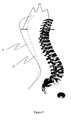
- Fig. 1 there is shown the devisl by witch a 3D spine reconstruction issued from any stereo X Ray system (1) is merged (or fused) with a 3D complete trunk shape reconstruction (2) to obtain 3D spine, trunk and rib cage reconstruction for volumetric assessment (3).
- the 3D spine reconstruction (1) can be issued form any stereotactic XR system, that could be either monomplane systems ( two separate acquisitions needed) or a by-plane system ( one acquisition system, such as in the EOS system, but not limited to this system).
- Fig. 2 shows how a stereo X ray acquisition system (4 and 5 is a simplified view of a XR emitting device and a XR digital Imaging Detector) can be used simultaneously or in sequences with an optical acquisition set up (6 is a simplified view of an optical acquisition system composed of a light source and a optical detector).
- the Optical Acquisition System 6 will create a 3D image (or shape) of back shape surface of the subject hereafter designed as "S1".
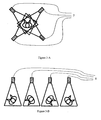
- Fig. 3-A explains how four optical acquisitions with equipment 6 can be performed simultaneously (7).
- One of theses four optical acquisitions set ups will generate a back shape surface of the subject hereafter designed as "S2".
- Fig. 3-B explains how four optical acquisitions performed successively (8).
- the subject is in standing position on a turn table or similar accessory to perform the four successive positions relating to the four optical acquisitions.
- One of theses four optical acquisitions will generate a complete 3D back shape surface of the subject hereafter designed as "S3".
- Fig. 4 and Fig. 5 illustrate the computerized principle used to evaluate, measure and correct the movements of the subject between the Optical Acquisition (9) performed as shown is Fig. 2 and the optical acquisition (10) as shown in Fig. 3-A or Fig. 3-B , prior to merging (or fusing) the 3D spine reconstruction and the 3D trunk shape reconstruction.
- an undeformable (or rigid) 3D calibration object (11) Prior to the acquisitions, an undeformable (or rigid) 3D calibration object (11) is placed on the object to image, and becomes a common part of S1 and S2 or S1 and S3.
- the aim of this 3D calibration object is to reposition both S1 and S2 or S1 and S3 in the same 3D / special referential system (12).
- the two back shapes ([S1] and [S2] or [S1] and [S3]) are placed in the same special reference system and the movements of the subject between the two acquisitions can be evaluated and corrected by the computing algorithm.
- the correction (13) is applied to the 3D spine reconstruction to be fitted to the actual subject positioning during the trunk shape acquisition, as illustrated on figure 4 .
- the reconstructed 3D spine and the 3D acquisition of the shape of the back are positioned in the same reference system, and their position is adjusted in reference of each other.
- the present invention guarantees this positioning.
- the three dimensional spine reconstruction and digitalized trunk shape are merged in the same axis system.
- the patient movements are taken into account and corrected during the merging process.
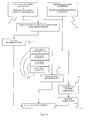
- Figure.6 is an illustration of the principle of the method and computed algorithms used to perform an accurate fusion of a 3D image or reconstruction of the spine with a full 3D image of the truck, which include specific algorithms to eliminate the patient movement between the different imaging sequences and accurately reposition the spine in the 3D volume of the trunk.
- the advantages of the present invention include, without limitation, (a) full compatibility with the different XRay stereotactic imaging apparatus and methods, (b) full compatibility with the different optical stereotactic imaging apparatus and methods (c) the elimination of patient movements between the different acquisition of images (d) the creation of a full 3D model of the trunk in witch a 3D model of the spine is accurately positioned (e) the subsequent analysis of this full 3D model to obtain an accurate analytical description that includes distances, angles in any 2D plane, (f) the subsequent analysis of this full 3D model to obtain an accurate volumetric of the trunck in relation to the position of the spine (g) that the algorithms are compatible with "of the self" hardware and/or clout computing platforms (h) that the 3D images and models that are elaborated and compatible with standard image exchange protocols in healthcare.
- the present invention is a method to fuse (merge) a full 3D image of the trunk and a 3D image (or reconstruction) of the spine into an accurate 3D image, for patients in standing position, to offer a 3D digital image that can be further processed to enables better delivery of care.
- the present invention is related to a specific
Landscapes
- Engineering & Computer Science (AREA)
- Physics & Mathematics (AREA)
- Theoretical Computer Science (AREA)
- General Physics & Mathematics (AREA)
- Medical Informatics (AREA)
- Radiology & Medical Imaging (AREA)
- Quality & Reliability (AREA)
- Computer Vision & Pattern Recognition (AREA)
- Nuclear Medicine, Radiotherapy & Molecular Imaging (AREA)
- Health & Medical Sciences (AREA)
- General Health & Medical Sciences (AREA)
- Computer Graphics (AREA)
- Geometry (AREA)
- Software Systems (AREA)
- Apparatus For Radiation Diagnosis (AREA)
Applications Claiming Priority (1)
| Application Number | Priority Date | Filing Date | Title |
|---|---|---|---|
| US25053909P | 2009-10-11 | 2009-10-11 |
Publications (2)
| Publication Number | Publication Date |
|---|---|
| EP2323101A2 true EP2323101A2 (fr) | 2011-05-18 |
| EP2323101A3 EP2323101A3 (fr) | 2011-06-22 |
Family
ID=43532616
Family Applications (1)
| Application Number | Title | Priority Date | Filing Date |
|---|---|---|---|
| EP10306105A Withdrawn EP2323101A3 (fr) | 2009-10-11 | 2010-10-11 | Procédé pour fusion d'imagerie 3D du rachis et 3D intégrale du tronc en position debout |
Country Status (2)
| Country | Link |
|---|---|
| US (1) | US20110135173A1 (fr) |
| EP (1) | EP2323101A3 (fr) |
Cited By (5)
| Publication number | Priority date | Publication date | Assignee | Title |
|---|---|---|---|---|
| CN105225271A (zh) * | 2015-11-09 | 2016-01-06 | 浙江海洋学院 | 一种水域浮游生物的远程实时图像采集与三维重建系统 |
| CN106570852A (zh) * | 2016-11-07 | 2017-04-19 | 中国航空无线电电子研究所 | 一种实时3d图像态势感知方法 |
| CN107451983A (zh) * | 2017-07-18 | 2017-12-08 | 中山大学附属第六医院 | Ct图像的三维融合方法和系统 |
| FR3071715A1 (fr) * | 2017-10-03 | 2019-04-05 | Proteor | Procede d'imagerie radiographique, dispositif de traitement d'image radiographique et dispositif d'imagerie radiographique. |
| CN120298415A (zh) * | 2025-06-13 | 2025-07-11 | 中国科学院合肥物质科学研究院 | 基于rgb图像与解剖约束的脊柱三维评估方法及系统 |
Families Citing this family (16)
| Publication number | Priority date | Publication date | Assignee | Title |
|---|---|---|---|---|
| US9824302B2 (en) | 2011-03-09 | 2017-11-21 | Siemens Healthcare Gmbh | Method and system for model-based fusion of multi-modal volumetric images |
| US9292917B2 (en) | 2011-11-23 | 2016-03-22 | Siemens Aktiengesellschaft | Method and system for model-based fusion of computed tomography and non-contrasted C-arm computed tomography |
| US9384546B2 (en) | 2012-02-22 | 2016-07-05 | Siemens Aktiengesellschaft | Method and system for pericardium based model fusion of pre-operative and intra-operative image data for cardiac interventions |
| US10846860B2 (en) | 2013-03-05 | 2020-11-24 | Nview Medical Inc. | Systems and methods for x-ray tomosynthesis image reconstruction |
| US10070828B2 (en) | 2013-03-05 | 2018-09-11 | Nview Medical Inc. | Imaging systems and related apparatus and methods |
| WO2018163499A1 (fr) * | 2017-03-10 | 2018-09-13 | ソニー・オリンパスメディカルソリューションズ株式会社 | Dispositif de commande d'affichage d'image médicale, dispositif d'affichage d'image médicale, système de traitement d'informations médicales et procédé de commande d'affichage d'image médicale |
| US10102682B1 (en) * | 2017-04-17 | 2018-10-16 | Raytheon Company | System and method for combining 3D images in color |
| CN107610218B (zh) * | 2017-08-25 | 2020-10-23 | 武汉工程大学 | 一种面向立体结构网点三维图像重建的层面数据获取方法 |
| US11610346B2 (en) | 2017-09-22 | 2023-03-21 | Nview Medical Inc. | Image reconstruction using machine learning regularizers |
| US11475558B2 (en) | 2019-11-13 | 2022-10-18 | Raytheon Company | Organ isolation in scan data |
| US11282209B2 (en) | 2020-01-10 | 2022-03-22 | Raytheon Company | System and method for generating contours |
| CN111772584B (zh) * | 2020-07-08 | 2022-08-09 | 莆田学院附属医院(莆田市第二医院) | 一种智能脊柱数字化手术装置 |
| US12347100B2 (en) | 2020-11-19 | 2025-07-01 | Mazor Robotics Ltd. | Systems and methods for generating virtual images |
| US11562512B2 (en) | 2020-12-09 | 2023-01-24 | Raytheon Company | System and method for generating and displaying contours |
| US11893745B2 (en) | 2020-12-09 | 2024-02-06 | Raytheon Company | System and method for generating and displaying contours |
| CN114732429A (zh) * | 2022-04-08 | 2022-07-12 | 深圳市安健科技股份有限公司 | 基于dr设备的三维图像的采集方法和系统 |
Citations (1)
| Publication number | Priority date | Publication date | Assignee | Title |
|---|---|---|---|---|
| WO2008012479A1 (fr) | 2006-07-27 | 2008-01-31 | Axs Ingenierie | Procede d'imagerie informatise permettant une reconstruction tridimensionnelle a partir d'images radiographiques bidimensionnelles; dispositif de mise en oeuvre |
-
2010
- 2010-10-11 EP EP10306105A patent/EP2323101A3/fr not_active Withdrawn
- 2010-10-12 US US12/902,708 patent/US20110135173A1/en not_active Abandoned
Patent Citations (1)
| Publication number | Priority date | Publication date | Assignee | Title |
|---|---|---|---|---|
| WO2008012479A1 (fr) | 2006-07-27 | 2008-01-31 | Axs Ingenierie | Procede d'imagerie informatise permettant une reconstruction tridimensionnelle a partir d'images radiographiques bidimensionnelles; dispositif de mise en oeuvre |
Non-Patent Citations (1)
| Title |
|---|
| AJEMBA P O ET AL: "A Torso-Imaging System to Quantify the Deformity Associated With Scoliosis", IEEE TRANSACTIONS ON INSTRUMENTATION AND MEASUREMENT, IEEE SERVICE CENTER, PISCATAWAY, NJ, US, vol. 56, no. 5, 1 October 2007 (2007-10-01), pages 1520 - 1526, XP011192288, ISSN: 0018-9456, DOI: 10.1109/TIM.2007.903592 * |
Cited By (8)
| Publication number | Priority date | Publication date | Assignee | Title |
|---|---|---|---|---|
| CN105225271A (zh) * | 2015-11-09 | 2016-01-06 | 浙江海洋学院 | 一种水域浮游生物的远程实时图像采集与三维重建系统 |
| CN106570852A (zh) * | 2016-11-07 | 2017-04-19 | 中国航空无线电电子研究所 | 一种实时3d图像态势感知方法 |
| CN106570852B (zh) * | 2016-11-07 | 2019-12-03 | 中国航空无线电电子研究所 | 一种实时3d图像态势感知方法 |
| CN107451983A (zh) * | 2017-07-18 | 2017-12-08 | 中山大学附属第六医院 | Ct图像的三维融合方法和系统 |
| FR3071715A1 (fr) * | 2017-10-03 | 2019-04-05 | Proteor | Procede d'imagerie radiographique, dispositif de traitement d'image radiographique et dispositif d'imagerie radiographique. |
| WO2019069001A1 (fr) * | 2017-10-03 | 2019-04-11 | Proteor | Procede d'imagerie radiographique, dispositif de traitement d'image radiographique et dispositif d'imagerie radiographique |
| US11430110B2 (en) | 2017-10-03 | 2022-08-30 | Proteor | Radiographic imaging method, radiographic image processing device, and radiographic imaging device |
| CN120298415A (zh) * | 2025-06-13 | 2025-07-11 | 中国科学院合肥物质科学研究院 | 基于rgb图像与解剖约束的脊柱三维评估方法及系统 |
Also Published As
| Publication number | Publication date |
|---|---|
| US20110135173A1 (en) | 2011-06-09 |
| EP2323101A3 (fr) | 2011-06-22 |
Similar Documents
| Publication | Publication Date | Title |
|---|---|---|
| EP2323101A2 (fr) | Procédé pour fusion d'imagerie 3D du rachis et 3D intégrale du tronc en position debout | |
| ES2716837T3 (es) | Detección automática de implantes a partir de artefactos de imagen | |
| Aubry et al. | Measurements of intrafraction motion and interfraction and intrafraction rotation of prostate by three-dimensional analysis of daily portal imaging with radiopaque markers | |
| ES2987933T3 (es) | Método para determinar la posición de un objeto mediante el uso de proyecciones de marcadores o puntales | |
| WO2017117517A1 (fr) | Système et procédé d'imagerie médicale | |
| De Silva et al. | Virtual fluoroscopy for intraoperative C-arm positioning and radiation dose reduction | |
| Galantucci et al. | Noninvasive computerized scanning method for the correlation between the facial soft and hard tissues for an integrated three-dimensional anthropometry and cephalometry | |
| Muhit et al. | Image-assisted non-invasive and dynamic biomechanical analysis of human joints | |
| JP2024526601A (ja) | 写真測量を使用して整形外科手術のための患者固有のガイドを作製するためのシステム及び方法 | |
| CN120284468A (zh) | 一种基于稀疏重建的成像设备摆位导引方法、系统和设备 | |
| Kadoury et al. | Three-dimensional reconstruction of the scoliotic spine and pelvis from uncalibrated biplanar x-ray images | |
| KR20160057024A (ko) | 마커리스 3차원 객체추적 장치 및 그 방법 | |
| US9254106B2 (en) | Method for completing a medical image data set | |
| US20250152118A1 (en) | Method To Superimpose Rendering Over Spine Hardware Implants On Images Produced By Cbct Scanner System | |
| Hocquelet et al. | Patient-specific 3D models created by 3D imaging system or bi-planar imaging coupled with Moiré–Fringe projections: a comparative study of accuracy and reliability on spinal curvatures and vertebral rotation data | |
| Douglas et al. | Three-dimensional point localisation in low-dose X-ray images using stereo-photogrammetry | |
| JP7738655B2 (ja) | 医用撮像の際に患者の位置を調整するための誘導 | |
| CN120769730A (zh) | 用于验证规程的系统 | |
| Broberg et al. | Radiostereometric analysis using clinical radiographic views: development of a universal calibration object | |
| Galantucci et al. | New 3D digitizer for human faces based on digital close range photogrammetry: Application to face symmetry analysis | |
| Rashed et al. | An interactive augmented reality imaging system for minimally invasive orthopedic surgery | |
| Zeng et al. | Low‐dose three‐dimensional reconstruction of the femur with unit free‐form deformation | |
| Yoo et al. | Three-dimensional localization of cochlear implant electrodes using epipolar stereophotogrammetry | |
| Albiol et al. | 3D measurements in conventional X-ray imaging with RGB-D sensors | |
| JP7841179B2 (ja) | 医療システムのための画像再構成方法 |
Legal Events
| Date | Code | Title | Description |
|---|---|---|---|
| PUAI | Public reference made under article 153(3) epc to a published international application that has entered the european phase |
Free format text: ORIGINAL CODE: 0009012 |
|
| AK | Designated contracting states |
Kind code of ref document: A2 Designated state(s): AL AT BE BG CH CY CZ DE DK EE ES FI FR GB GR HR HU IE IS IT LI LT LU LV MC MK MT NL NO PL PT RO RS SE SI SK SM TR |
|
| AX | Request for extension of the european patent |
Extension state: BA ME |
|
| PUAL | Search report despatched |
Free format text: ORIGINAL CODE: 0009013 |
|
| RIN1 | Information on inventor provided before grant (corrected) |
Inventor name: BLANCHARD, BERTRAND Inventor name: ELBAROUDI, FOUAD |
|
| AK | Designated contracting states |
Kind code of ref document: A3 Designated state(s): AL AT BE BG CH CY CZ DE DK EE ES FI FR GB GR HR HU IE IS IT LI LT LU LV MC MK MT NL NO PL PT RO RS SE SI SK SM TR |
|
| AX | Request for extension of the european patent |
Extension state: BA ME |
|
| RIC1 | Information provided on ipc code assigned before grant |
Ipc: G06T 17/00 20060101ALI20110517BHEP Ipc: G06T 7/00 20060101AFI20110210BHEP |
|
| 17P | Request for examination filed |
Effective date: 20111206 |
|
| 17Q | First examination report despatched |
Effective date: 20120216 |
|
| STAA | Information on the status of an ep patent application or granted ep patent |
Free format text: STATUS: THE APPLICATION IS DEEMED TO BE WITHDRAWN |
|
| 18D | Application deemed to be withdrawn |
Effective date: 20130409 |