EP2399612B1 - Composition pour marquer des tissus du système nerveux central, procédé pour marquer des tissus du système nerveux central, et procédé de criblage utilisant la composition pour marquer des tissus du système nerveux central - Google Patents
Composition pour marquer des tissus du système nerveux central, procédé pour marquer des tissus du système nerveux central, et procédé de criblage utilisant la composition pour marquer des tissus du système nerveux central Download PDFInfo
- Publication number
- EP2399612B1 EP2399612B1 EP10838989.1A EP10838989A EP2399612B1 EP 2399612 B1 EP2399612 B1 EP 2399612B1 EP 10838989 A EP10838989 A EP 10838989A EP 2399612 B1 EP2399612 B1 EP 2399612B1
- Authority
- EP
- European Patent Office
- Prior art keywords
- nervous system
- central nervous
- labeling
- system tissue
- compound
- Prior art date
- Legal status (The legal status is an assumption and is not a legal conclusion. Google has not performed a legal analysis and makes no representation as to the accuracy of the status listed.)
- Active
Links
- 0 *N(c(cccc1C(c2ccccc22)=O)c1C2=C1)C1=O Chemical compound *N(c(cccc1C(c2ccccc22)=O)c1C2=C1)C1=O 0.000 description 1
Images




Classifications
-
- A—HUMAN NECESSITIES
- A61—MEDICAL OR VETERINARY SCIENCE; HYGIENE
- A61K—PREPARATIONS FOR MEDICAL, DENTAL OR TOILETRY PURPOSES
- A61K49/00—Preparations for testing in vivo
- A61K49/001—Preparation for luminescence or biological staining
- A61K49/0013—Luminescence
- A61K49/0017—Fluorescence in vivo
- A61K49/0019—Fluorescence in vivo characterised by the fluorescent group, e.g. oligomeric, polymeric or dendritic molecules
- A61K49/0021—Fluorescence in vivo characterised by the fluorescent group, e.g. oligomeric, polymeric or dendritic molecules the fluorescent group being a small organic molecule
- A61K49/0032—Methine dyes, e.g. cyanine dyes
-
- A—HUMAN NECESSITIES
- A61—MEDICAL OR VETERINARY SCIENCE; HYGIENE
- A61K—PREPARATIONS FOR MEDICAL, DENTAL OR TOILETRY PURPOSES
- A61K49/00—Preparations for testing in vivo
- A61K49/001—Preparation for luminescence or biological staining
- A61K49/0013—Luminescence
- A61K49/0017—Fluorescence in vivo
- A61K49/0019—Fluorescence in vivo characterised by the fluorescent group, e.g. oligomeric, polymeric or dendritic molecules
- A61K49/0021—Fluorescence in vivo characterised by the fluorescent group, e.g. oligomeric, polymeric or dendritic molecules the fluorescent group being a small organic molecule
- A61K49/0039—Coumarin dyes
-
- A—HUMAN NECESSITIES
- A61—MEDICAL OR VETERINARY SCIENCE; HYGIENE
- A61K—PREPARATIONS FOR MEDICAL, DENTAL OR TOILETRY PURPOSES
- A61K49/00—Preparations for testing in vivo
- A61K49/001—Preparation for luminescence or biological staining
- A61K49/006—Biological staining of tissues in vivo, e.g. methylene blue or toluidine blue O administered in the buccal area to detect epithelial cancer cells, dyes used for delineating tissues during surgery
-
- C—CHEMISTRY; METALLURGY
- C07—ORGANIC CHEMISTRY
- C07D—HETEROCYCLIC COMPOUNDS
- C07D417/00—Heterocyclic compounds containing two or more hetero rings, at least one ring having nitrogen and sulfur atoms as the only ring hetero atoms, not provided for by group C07D415/00
- C07D417/02—Heterocyclic compounds containing two or more hetero rings, at least one ring having nitrogen and sulfur atoms as the only ring hetero atoms, not provided for by group C07D415/00 containing two hetero rings
- C07D417/06—Heterocyclic compounds containing two or more hetero rings, at least one ring having nitrogen and sulfur atoms as the only ring hetero atoms, not provided for by group C07D415/00 containing two hetero rings linked by a carbon chain containing only aliphatic carbon atoms
-
- G—PHYSICS
- G01—MEASURING; TESTING
- G01N—INVESTIGATING OR ANALYSING MATERIALS BY DETERMINING THEIR CHEMICAL OR PHYSICAL PROPERTIES
- G01N33/00—Investigating or analysing materials by specific methods not covered by groups G01N1/00 - G01N31/00
- G01N33/48—Biological material, e.g. blood, urine; Haemocytometers
- G01N33/50—Chemical analysis of biological material, e.g. blood, urine; Testing involving biospecific ligand binding methods; Immunological testing
-
- G—PHYSICS
- G01—MEASURING; TESTING
- G01N—INVESTIGATING OR ANALYSING MATERIALS BY DETERMINING THEIR CHEMICAL OR PHYSICAL PROPERTIES
- G01N33/00—Investigating or analysing materials by specific methods not covered by groups G01N1/00 - G01N31/00
- G01N33/48—Biological material, e.g. blood, urine; Haemocytometers
- G01N33/50—Chemical analysis of biological material, e.g. blood, urine; Testing involving biospecific ligand binding methods; Immunological testing
- G01N33/58—Chemical analysis of biological material, e.g. blood, urine; Testing involving biospecific ligand binding methods; Immunological testing involving labelled substances
-
- G—PHYSICS
- G01—MEASURING; TESTING
- G01T—MEASUREMENT OF NUCLEAR OR X-RADIATION
- G01T1/00—Measuring X-radiation, gamma radiation, corpuscular radiation, or cosmic radiation
- G01T1/16—Measuring radiation intensity
- G01T1/161—Applications in the field of nuclear medicine, e.g. in vivo counting
Definitions
- the present invention relates to a labeling composition capable of clearly labeling a central nervous system tissue, a method for labeling a central nervous system tissue using the central nervous system tissue-labeling composition, and a screening method using the central nervous system tissue-labeling composition.
- central nervous system diseases include Parkinson's disease, Alzheimer's disease, epilepsy, migraine, spinocerebellar degeneration, brain tumor, cerebral hemorrhage, and cerebral infarction.
- Central nervous system diseases often impair motor function and cognitive function, leading to a significant decrease in a patient's quality of life.
- an abnormality in the central nervous system tissue be accurately recognized for early detection of a disease so that therapy or a measure to retard the progression is provided.
- morphological assessments by imaging using computer tomography (CT) and magnetic resonance imaging (MRI), and radionuclide imaging diagnoses by a method such as positron emission tomography (PET) are employed.
- the brain is tissues containing neurons and glial cells.
- the brain performs advanced functions through complex intercellular networks and hierarchical structures.
- An imaging technology allows visualized measurement without impairing the function of the central nervous system, enabling more intuitive as well as dynamic and quantitative examination.
- Recently, development of not only the aforementioned imaging diagnosis techniques but also new techniques such as fluorescent imaging and near-infrared imaging is ongoing.
- SPECT single photon emission computed tomography
- a synthetic compound labeled with a radioactive isotope (ligand) is administered to the body, and then localized radioactivities in the brain are quantitated to analyze the distribution of the ligand in the body and the metabolic dynamics of the ligand for mapping of the functional localization.
- ligand radioactive isotope
- SPECT single photon emission computed tomography
- a compound that specifically binds to ⁇ -amyloid which is deposited in the brain tissue and that is used for diagnosis of Alzheimer's disease is disclosed (Patent Literatures 1 and 2).
- Non-Patent Literature 1 a compound capable of fluorescently labeling glial cells in the brain is disclosed in Non-Patent Literature 1.
- Non-Patent Literature 3 discloses 4-amino-1,8-napthalimide, a poly(ADP ribose) polymerase inhibitor in middle cerebral artery occlusion-induced focal cerebral ischemia in rat.
- Non-Patent Literature 4 discloses fluorescent markers for hypoxic cells of naphthalimides with two heterocyclic side chains of 2-nitroimidazole for bioreductive binding.
- Non-Patent Literature 5 discloses the synthesis and characterization of fluorescent antagonists for human 5-HT 4 receptors.
- the conventionally-employed probes utilize their properties such as accumulating in a site where a large amount of glucose is taken up or of specifically accumulating to ⁇ -amyloid; therefore, they are not suitable for morphological imaging of a specific site in the brain in diseases not associated with such a specific site. Also, there is a problem with the aforementioned glial cell-staining compound because the compound requires such a highly invasive treatment that involves direct administration to the brain.
- the transferability of a compound to the brain is regulated by the blood-brain barrier (BBB) and the blood-cerebrospinal fluid barrier (BCSFB), and many of the compounds that can migrate into a normal tissue may not be able to migrate into the brain.
- BBB blood-brain barrier
- BCSFB blood-cerebrospinal fluid barrier
- some of a compound capable of migrating into the brain in the juvenile stage loses its transferability to the brain once BBB is functioning.
- Patent Literatures 1 and 2 report a BBB-permeable coumarin compound
- the technologies disclosed therein only focus on the compound's specific binding ability to ⁇ -amyloid, while these literatures are silent on a property of labeling the central nervous system tissue in the brain.
- dye compounds represented by the following general formula (1) are capable of labeling the central nervous system tissue of the living body alive. That is, they have found that the compounds label at least any one of the tissues including optic nerve, optic tract, superior colliculus (optic tectum), pituitary gland, tectospinal (tectobulbar) tract, and reticular formation with high sensitivity, serving as a novel central nervous system tissue-labeling compound as defined in claim 1, enabling highly accurate diagnosis and screening of a drug, thereby completing the present invention.
- the present inventors have established a method for labeling the central nervous system tissue of the living body as defined in claim 5. Further, the present inventors have developed a screening method using a labeling composition of the present invention as defined in claim 6, thereby completing the present invention.
- the other claims relate to further developments.
- novel compound for the central nervous system tissue of the present invention is defined in claim 1, and is able to label at least any one of the tissues including optic nerve, optic tract, superior colliculus (optic tectum), pituitary gland, tectospinal (tectobulbar) tract, and reticular formation.
- central nervous system tissue-labeling composition of the present invention has enabled selective labeling of a brain tissue such as optic nerve, optic tract, superior colliculus (optic tectum), pituitary gland, tectospinal (tectobulbar) tract, and reticular formation, which has been conventionally difficult. This enables simple and highly precise assessment and analysis of the morphology and the state of cells of a specific site in central nervous system tissues. Further, a screening method using the central nervous system tissue-labeling composition of the present invention can be a novel, effective tool for the research and the discovery of a drug for the central nervous system.
- the central nervous system tissue-labeling composition according to a first embodiment of the present invention is characterized by containing, as an active ingredient, at least one of compounds represented by the general formula (1) wherein aromatic ring A represents a structure of the following formula (2).
- R 1 to R 2 each independently represent a hydrogen atom, an alkyl group, an aralkyl group, or an aryl group.
- an aromatic ring A represents a skeletal structure represented by the following general formulas (2) to (4), or an aromatic ring A represents, through binding to R 2 via N, a skeletal structure represented by the following general formulas (5) to (6):
- R 3 represents a hydrogen atom, an alkyl group, an aralkyl group, or an aryl group.
- R 4 represents an oxygen atom, a sulfur atom, or N(R 6 )
- R 5 represents a hydrogen atom, an alkyl group, an alkoxy group, or a sulfonic acid group
- R 6 represents a hydrogen atom, an alkyl group, or an aryl group.
- R 7 represents a hydrogen atom, an alkyl group, an aryl group, or a heterocyclic group.
- R 8 represents an alkyl group or an alkyl chain having a carboxy group at its end
- X and Y represent a hydrogen atom or an alkyl group
- Z represents a hydrogen atom or a halogen atom
- n represents an integer of 0 or 1.
- R 9 to R 10 represent an alkyl group or an aryl group
- R 11 represents a hydrogen atom, an alkyl group, an alkoxy group, a carboxylic acid group, or a sulfonic acid group.
- the alkyl group at R 1 to R 2 in the aforementioned general formula (1) examples thereof include a methyl group, an ethyl group, a propyl group, a butyl group, a cyclohexyl group, and a 3-hexanyl group.
- the alkyl group may further contain a substituent as long as the substituent does not markedly deteriorate the preservation stability of the dye compound.
- No particular limitation is imposed on the aralkyl group at R 1 to R 2 , and examples thereof include a benzyl group and a phenethyl group.
- the aralkyl group may contain a substituent.
- no particular limitation is imposed on the aryl group at R 1 to R 2 , and examples thereof include a phenyl group and a naphthyl group.
- the aryl group may contain a substituent.
- R 3 No particular limitation is imposed on the alkyl group at R 3 in the aforementioned general formula (2), and examples thereof include a methyl group, an ethyl group, a propyl group, a butyl group, and a cyclohexyl group.
- R 1 and R 2 are a hydrogen atom and the other is an alkyl group or an aralkyl group, intense fluorescence is obtained.
- R 3 can be a methyl group, a butyl group, and a cyclohexyl group for easiness of synthesis.
- alkyl group at R 5 to R 6 in the aforementioned general formula (3) No particular limitation is imposed on the alkyl group at R 5 to R 6 in the aforementioned general formula (3), and examples thereof include a methyl group, an ethyl group, a propyl group, and a butyl group.
- alkoxy group at R 5 No particular limitation is imposed on the alkoxy group at R 5 , and examples thereof include a methoxy group, an ethoxy group, a propoxy group, and a butoxy group.
- aryl group at R 6 No particular limitation is imposed on the aryl group at R 6 , and examples thereof include a phenyl group.
- alkyl group at R 7 in the aforementioned general formula (4) examples thereof include a methyl group, an ethyl group, a propyl group, and a butyl group.
- aryl group at R 7 examples thereof include a phenyl group.
- the aryl group may further contain a substituent as long as the substituent does not markedly deteriorate the preservation stability of the dye compound.
- heterocyclic group at R 7 and examples thereof include a pyridyl group, a pyrazyl group, and a morpholinyl group.
- the alkyl group at R 8 , X, and Y in the aforementioned general formula (5) examples thereof include a methyl group, an ethyl group, a propyl group, and a butyl group.
- the alkyl group may further contain a substituent as long as the substituent does not markedly deteriorate the preservation stability of the dye compound.
- the halogen atom at Z in the aforementioned general formula (5) examples thereof include a fluorine atom, a chlorine atom, a bromine atom, or an iodine atom.
- No particular limitation is imposed on the aryl group at R 9 to R 10 and examples thereof include a phenyl group.
- the aryl group may further contain a substituent as long as the substituent does not markedly deteriorate the preservation stability of the dye compound.
- the alkyl group at R 11 No particular limitation is imposed on the alkyl group at R 11 , and examples thereof include a methyl group, an ethyl group, a propyl group, and a butyl group.
- the alkoxy group at R 11 No particular limitation is imposed on the alkoxy group at R 11 , and examples thereof include a methoxy group, an ethoxy group, a propoxy group, and a butoxy group.
- the dye compound represented by the general formulas (1) to (6) is commercially obtainable, it can also be synthesized in accordance with a publicly known method.
- R 21 to R 24 each independently represent a hydrogen atom, an alkyl group, an alkoxy group, an amino group, or a halogen atom.
- R 21 and R 22 , R 22 and R 23 , or R 23 and R 24 may bind to each other to form a ring.
- R 25 to R 28 each independently represent a hydrogen atom, an alkyl group, an alkoxy group, or a halogen atom.
- B represents an oxygen atom or an NH group.
- Q represents an oxygen atom, a sulfur atom, and an N-R 29 group, wherein R 29 represents a hydrogen atom or an alkyl group.
- alkyl group at R 21 to R 29 in the aforementioned general formula (7) No particular limitation is imposed on the alkyl group at R 21 to R 29 in the aforementioned general formula (7), and examples thereof include a methyl group, an ethyl group, a propyl group, and a butyl group.
- alkoxy group at R 21 to R 28 No particular limitation is imposed on the alkoxy group at R 21 to R 28 , and examples thereof include a methoxy group, an ethoxy group, a propoxy group, and a butoxy group.
- amino group at R 21 to R 24 No particular limitation is imposed on the amino group at R 21 to R 24 , and examples thereof include an unsubstituted amino group; a mono-substituted amino group such as an N-methylamino group and an N-ethylamino group; and a di-substituted amino group such as an N,N-dimethylamino group, an N,N-diethylamino group, and an N,N-methylpropylamino group.
- halogen atom at R 21 to R 28 examples include a fluorine atom, a chlorine atom, a bromine atom, or an iodine atom.
- No particular limitation is imposed on the ring formed by binding of R 21 and R 22 , R 22 and R 23 , or R 23 and R 24 , and examples thereof include an aromatic ring such as a benzene ring, a saturated ring such as a cyclohexane ring, a partially-saturated ring such as a cyclopentene ring, and a hetero ring such as a piperidine ring.
- the ring may further contain a substituent as long as the substituent does not markedly deteriorate the preservation stability of the dye compound.
- R 22 when R 22 is an electron-donating substituent such as an amino group or an alkoxy group, the fluorescent intensity is increased. Thus, such a compound can be employed.
- R 22 is a di-substituted amino group such as an N,N-dimethylamino group and an N,N-diethylamino group, a high fluorescent intensity is attained; therefore, such a compound can be employed.
- Q is an oxygen atom or a sulfur atom from the viewpoint of the labeling property. Particularly, when Q is an oxygen atom, a high fluorescent intensity is attained and a specific site can be effectively labeled; therefore, such a compound can be employed.
- R 26 is an alkyl group such as methyl or a halogen atom such as a chlorine atom, a high fluorescent intensity is attained and a specific site can be effectively labeled; therefore, such a compound can be employed.
- a dye compound represented by the general formula (7) is commercially obtainable, it can also be synthesized in accordance with a publicly known method (for example, Dyes and pigments, Vol. 47 (Issues 1-2), pages 79-89 (2000 )).
- a publicly known method for example, Dyes and pigments, Vol. 47 (Issues 1-2), pages 79-89 (2000 )
- Preferable specific examples of the dye compound represented by the general formula (7) (compounds (15) to (28)) will be shown below.
- a central nervous system tissue-labeling composition of the present invention is characterized by containing a compound capable of labeling at least one cell type present in a central nervous system tissue.
- the central nervous system tissue-labeling composition of the present invention can contain a compound selectively labeling at least one of optic nerve, optic tract, superior colliculus (optic tectum), pituitary gland, tectospinal (tectobulbar) tract, and reticular formation of the central nervous system tissue.
- selective labeling refers to such a labeling property that at least a cell or a site clearly noted in the present invention is labeled while a tissue other than the aforementioned cell or site in the central nervous system tissue is not labeled, or the target cells or sites of the composition are labeled differently (labeled at a high or low level).
- the compound in consideration of migration of a compound represented by the general formula (1) encompassed by the central nervous system tissue-labeling compositions of the present invention into the target central nervous tissue, the compound can be a low molecular compound, and a compound having a molecular weight of 2000 or less is selected. Further, a compound having a molecular weight of 1000 or less, particularly 600 or less, can be employed.
- a compound of the present invention can be a fluorescent compound having a fluorescent property. Owing to a high sensitivity of a fluorescent compound, a low concentration of the compound is required for labeling, whereby the amount of the compound necessary for labeling can be relatively reduced. Also, selecting a combination of compounds with different labeling sites and fluorescent spectra enables multi-labeling. This is highly useful because more information can be obtained by single observation.
- a central nervous system tissue-labeling composition of the present invention can migrate into the central nervous system tissue without being blocked by BBB or BCSFB, the composition can be administered without damaging the central nervous system tissue or the tissue connected to the central nervous system tissue in a living organism.
- a central nervous system tissue or a tissue connected to the central nervous system tissue can be labeled without being damaged. That is, a living organism can be labeled with the central nervous system tissue-labeling composition without causing surgical injury such as an incision in the tissue near the central nerve and a puncture in the central nervous system tissue or in the nervous tissue connected to the central nervous system tissue. It is to be noted that the present invention does not exclude the aforementioned labeling method involving surgical injury.
- the labeling method not causing surgical injury includes a method of exposing the central nervous system tissue-labeling composition to a living organism locally or systemically, a method by oral contact, a method by pulmonary contact, a method by nasal contact, a method by contacting with the digestive tract, a method by mucosal contact, a method by contacting with a body fluid, a method by sublingual contact, a method by intravascular contact such as contacting with the vein or artery, a method by intraperitoneal contact, an infusion method such as an intravaginal, subcutaneous, intradermal, intravesical, or intratracheal (intrabronchial) infusion, and a method of contacting with the living body by, for example, spraying or applying.
- the dosage form, administration route, and dose of the composition are appropriately selected depending on the weight and the condition of the subject animal.
- a method for acquiring the information through visualization of the state of labeling is characterized by labeling a central nervous system tissue in the living body with the central nervous system tissue-labeling composition to acquire an image. That is, the method is characterized by administering the central nervous system tissue-labeling composition of the present invention by any method, and a certain time later, irradiating the observation site with light of excitation wavelength, and measuring fluorescence of longer wavelength thus generated to create an image.
- Examples of specific method of labeling of the present invention can include a method using probes such as a fluorescent probe and a radionuclide-labeled probe. Staining the central nervous system tissue with these probes enables imaging of the distribution and the orientation of a periphery nerve system tissue connected to the central nervous system tissue.
- staining the cell morphology of the central nervous system tissue refers to achieving a state in which at least one cell type present in the central nervous system tissue is stained so that the cell morphology of the cell type is clearly distinguished through, for example, the fluorescent color.
- the observation method of the present invention is characterized by using a central nervous system tissue-labeling composition of the present invention.
- the measurement and the detection of the central nervous system tissue-labeling composition are carried out by a method publicly known to those skilled in the art.
- the observation method employed in the present invention is a method of capturing the state and the change of a biological sample as an image.
- Examples thereof include visible light observation, near-infrared light observation, infrared light observation, or laser microscopic observation, in which the eye tissue is irradiated with visible light, near-infrared light, or infrared light and then observed by a camera, CCD, etc., or fluorescent observation, fluorescence microscopic observation, fluorescence endoscopic observation, confocal fluorescence microscopic observation, multiphoton-excited fluorescence microscopic observation, narrow band imaging, in which, by using fluorescence endoscopy and so on, a biological sample is irradiated with excitation light from the excitation light source and the fluorescence emitting from the biological sample is observed, or optical coherence tomography (OCT), and further, observation under a soft X-ray microscope.
- OCT optical coherence tomography
- the excitation wavelength is normally 200 to 1010 nm, or it can be 400 to 900 nm, and further, it can be 480 to 800 nm.
- the wavelength of 600 to 1000 nm is normally employed, and the wavelength of 680 to 900 nm can be used since the light within such a range of wavelength has excellent permeability through the living body.
- the fluorescence excitation source used in the present invention No particular limitation is imposed on the fluorescence excitation source used in the present invention, and various laser light sources can be used. Examples thereof include a dye laser, a semiconductor laser, an ion laser, a fiber laser, a halogen lamp, a xenon lamp, or a tungsten lamp. Furthermore, using various optical filters, a preferable excitation wavelength can be obtained or fluorescence only can be detected. As described above, the fluorescence is emitted inside the central nervous system tissue of an organism being irradiated with the excitation light, and imaging the central nervous system tissue, allowing easy detection of the light-emitting site.
- a bright-field image obtained by irradiation of visible light and a fluorescent image obtained by irradiation of excitation light can be combined by image processing to enable a more detailed observation of the central nervous system tissue.
- a confocal microscope can be used for acquisition of an optical image of a section.
- a multiphoton-excited fluorescence microscope can be used for an observation of the inside of the tissue for its high accessibility to a deep part and spatial resolution.
- a central nervous system tissue-labeling composition of the present invention can also be used as a radionuclide-labeled probe.
- the radionuclide type used for labeling can be appropriately selected depending on the manner in which it is used.
- a positron-emitting radionuclide such as 11 C, 14 C, 13 N, 15 O, 18 F, 19 F, 62 Cu, 68 Ga, or 78 Br can be used.
- 11 C, 13 N, 15 O, or 18 F can be used, of which 11 C or 18 F can particularly be used.
- a ⁇ -emitting nuclide such as 99 mTc, 111 In, 67 Ga, 201 Tl, 123 I, or 133 Xe can be used.
- 99 mTc or 123 I can be used.
- a radionuclide having a longer half-life such as 125 I can be used.
- GREI for example, 131 I, 85 Sr, and 65 Zn can be used.
- a central nervous system tissue-labeling composition labeled with a radionuclide can be imaged by, for example, autoradiography, positron emission tomography (PET) using a positron-emitting radionuclide, single photon emission computed tomography (SPECT) using various gamma-emitting nuclides.
- the composition can be detected by magnetic resonance imaging (MRI) utilizing an MR signal originating from the fluorine nucleus and 13C.
- MRI magnetic resonance imaging
- the compound can also be imaged by a Compton camera (GREI), which is capable of multiple molecular simultaneous imaging as a next-generation molecular imaging apparatus.
- GREI Compton camera
- a probe for the central nervous system tissue can be quantitated by using, for example, a liquid scintillation counter, an X-ray film, and an imaging plate.
- the concentration of the central nervous system tissue-labeling composition labeled with a radioisotope such as 14 C in blood (or in urine, or in feces) by a method such as accelerator mass spectrometry (AMS), the pharmacokinetic information (such as area under the blood drug concentration time curve (AUC), the blood half-life (T1/2), the maximum blood concentration (Cmax), the time to maximum blood concentration (Tmax), the volume of distribution, the first pass effect, the bioavailability, and the rate of excretion in feces and urine) of an unmodified form or a metabolite of the labeled composition can be acquired.
- AMS accelerator mass spectrometry
- the radionuclide may be contained in or bound to the compound represented by the general formula (1) or (7). No particular limitation is imposed on the method of labeling a radionuclide, and a method generally employed may be used. Also, a radionucleotide may substitute or be bound to at least a part of the elements constituting the compound represented by the general formula (1) or (7). When labeling the compound represented by the general formula (1) or (7) with a radionuclide, the resulting compound can have a radioactivity of approximately 1 to 100 ⁇ Ci per 1 mM. In this case, no particular limitation is imposed on the dose of the central nervous system tissue-labeling composition as long as the composition does not affect the subject, and the dose is appropriately selected depending on the compound type and the radionuclide used for labeling.
- the central nervous system tissue can be labeled with the central nervous system tissue-labeling compound of the present invention.
- examples thereof include, as a vertebrate, teleosts such as Takifugu rubripes, Takifugu niphobles, Tetraodon nigroviridis, Oryzias latipes, and zebrafish, amphibians such as African clawed frogs, birds such as chickens and quails, small animals such as rats, mice, and hamsters, large animals such as goats, pigs, dogs, cats, cows, and horses, monkeys, chimpanzees, and humans.
- the intraocular tissue of these organisms can be labeled alive.
- humans may be excluded.
- zebrafish can be used. Zebrafish expresses Claudin-5 and Zonula Occludens-1, which are the major constituent protein of the tight junctions of BBB, in an embryo at three days post fertilization (3 dpf) ( Brain Research Bulltein 75 (2008) 619-628 ). Major organs are formed on 6-7 dpf, and P-glycoprotein, which functions to excrete substances across BBB, is expressed by 8 dpf.
- zebrafish can be used for assessment of the central nervous system tissue. Further, there is such an advantage that, because zebrafish produces more than approximately 200 fertilized eggs per spawning, zebrafish having the identical genetic background can be obtained, which is convenient for screening.
- Examples of the central nervous system tissues that can be labeled with the central nervous system-labeling composition of the present invention include a central nervous system tissue composed of cerebrum (telencephalon), cerebral cortex, basal ganglia, midbrain, cerebellum, diencephalon, hindbrain (pallium), pons, medulla oblongata, spinal cord, optic tract, superior colliculus (optic tectum), pituitary gland, tectospinal (tectobulbar) tract, reticular formation, septal nuclei, amygdala, internal capsule, and optic nerve, these tissues in a pathological condition, or a neoplasm resulting from a disease and a cancer tissues.
- a central nervous system-labeling composition of the present invention can label optic nerve, optic tract, superior colliculus (optic tectum), pituitary gland, tectospinal (tectobulbar) tract, and reticular formation.
- a central nervous system tissue-labeling composition of the present invention can label the cranial nerve such as the optic nerve. Staining the cranial nerve enables imaging of the distribution and the orientation of a periphery nerve system tissue connected to the central nervous system tissue.
- labeling a central nervous system tissue namely labeling the cell morphology of the central nervous system tissue refers to achieving a state in which at least one cell type present in the central nervous system tissue is labeled so that the cell morphology of the cell type is clearly distinguished by an appropriate observation method.
- a central nervous system disease to be diagnosed by imaging using the central nervous system tissue-labeling composition of the present invention.
- Examples thereof include Parkinson's disease, Alzheimer's disease, Huntington's disease, motor neuron disease, tauopathy, corticobasal degeneration, depression, epilepsy, migraine, spinocerebellar degeneration, brain tumor, cerebral hemorrhage, and cerebral infarction.
- concentrations of the compound contained in a central nervous system tissue-labeling composition of the present invention are normally used in a concentration of 0.001 ng/mL or more and 100 ⁇ g/mL or less. It can also be used in a concentration of 0.001 ng/mL or more and 10 ⁇ g/mL or less, and further, in a concentration of 0.001 ng/mL or more and 5 ⁇ g/mL or less.
- a central nervous system tissue-labeling composition of the present invention is used by dissolving at least one of the dye compounds represented by the aforementioned general formula (1) or (7) in an appropriate solvent.
- the solvent No particular limitation is imposed on the solvent as long as it does not affect the living body.
- a highly biocompatible aqueous liquid can be used.
- ⁇ examples thereof include water; physiological saline; a buffer such as phosphate buffered saline (PBS) and Tris; an alcohol solvent such as methanol, ethanol, isopropanol, butanol, ethyleneglycol, and glycerin; an organic solvent such as N,N-dimethylsulfoxide (hereinbelow, abbreviated as DMSO) and N,N-dimethylformamide (hereinbelow, abbreviated as DMF); a cell culture medium such as D-MEM and HBSS, or an infusion solution such as a lactate Ringer's solution. Particularly, these solvents can contain more than 50% water. Also, a mixture of two or more kinds of these solvents can be used.
- PBS phosphate buffered saline
- Tris an alcohol solvent such as methanol, ethanol, isopropanol, butanol, ethyleneglycol, and glycerin
- an organic solvent such as N
- a production method of a central nervous system tissue-labeling composition of the present invention may be produced by diluting a concentrated solution of the compound in the aforementioned solvent.
- a low water-soluble compound can be dissolved in an appropriate solvent first, and then dissolved in purified water for use.
- purified water Particularly, methanol, ethanol, and DMSO can be used.
- an additive or a combination of two or more of additives can be added to the central nervous system tissue-labeling composition of the present invention.
- the additive used in the present invention includes humectants, surface tension-preparing agents, viscosity enhancers, salts such as sodium chloride, various pH-preparing agents, pH buffers, antiseptics, antimicrobial agents, sweeteners, or flavoring agents.
- the pH-preparing agent can be those that prepare pH to 5 to 9. No particular limitation is imposed on the pH-preparing agent, and examples thereof include hydrochloric acid, acetic acid, phosphoric acid, citric acid, malic acid, sodium hydroxide, or sodium hydrogen carbonate.
- Use of the central nervous system tissue-labeling composition of the present invention enables labeling of the central nervous system tissue without causing surgical injury such an incision in the tissue near the central nerve system and a puncture in the central nervous system tissue or in the nervous tissue connected to the central nervous system tissue.
- a screening method is characterized by detecting a compound acting on a central nervous system tissue in vivo by using the central nervous system tissue-labeling composition.
- the central nervous system tissue-labeling composition of the present invention labels the central nervous system tissue in an organism, for example zebrafish, which is a small teleost, alive.
- the composition's central nervous system-labeling property in a living organism i.e., the composition's in vivo labeling property, as an index
- the transferability of the compound-to-be-screened-for into the central nervous system tissue and the pharmacological effect of the compound-to-be-screened-for can be screened.
- live zebrafish a living organism, is used, the safety of the compound-to-be-screened-for can be simultaneously screened.
- zebrafish has been recognized as the third model animal after mice and rats in U.S. and U.K. It has been elucidated that the complete genome sequence of zebrafish has an 80% homology with that of humans, and also, zebrafish has nearly the same number of genes as humans, and the major organs, the development of tissues, and the structures are very similar between zebrafish and humans. Zebrafish can particularly be used for screening as a model animal because the process of differentiation and formation of each part (an organ and a part such as the heart, liver, kidney, and digestive tract) from a fertilized egg can be observed through a transparent body.
- Detecting a compound acting on a central nervous system tissue refers to measuring, using a central nervous system tissue-labeling composition of the present invention, the change in the labeling property when a compound of interest (a compound-to-be-screened-for) is allowed to act on the central nervous system to detect the presence or absence and the characteristics of the compound acting on the central nervous system tissue.
- a specific example thereof is a screening method in which a compound-to-be-screened-for and the central nervous system tissue-labeling composition of the present invention are contacted with zebrafish to observe the effect of the presence of the compound-to-be-screened-for on the condition of the labeling of the central nervous system tissue with the central nervous system tissue-labeling composition.
- No particular limitation is imposed on the method for contacting the compound-to-be-screened-for.
- the compound-to-be-screened-for is water-soluble
- a method of administering the compound-to-be-screened-for into the rearing water may be employed.
- the compound-to-be-screened-for is water-insoluble
- methods such as singly administering the compound-to-be-screened-for by dispersing it into the rearing water, administering it with a trace amount of surfactants and DMSO, orally administering it by mixing with the feed for zebrafish, or parenterally administering it by an injection may be employed.
- a method of administering the compound-to-be-screened-for into the rearing water can be employed for easiness.
- the effect, side effect, or safety of the compound-to-be-screened-for on the central nervous system tissue in an organism can be screened for. That is, the effect of the compound-to-be-screened-for on an organism can be screened in vivo using, for example, zebrafish.
- the central nervous system tissue-labeling composition used can be selected as desired depending on the target site, purpose, examination measures, etc. Further, owing to the labeling property of the central nervous system tissue-labeling composition, the application of the composition is expected to be expanded to, for example, the development of highly accurate diagnosis and treatment method of a disease. Thus, the central nervous system tissue-labeling composition can be used as a diagnostic composition.
- the aforementioned compound-to-be-screened-for refers to the generic term for compounds having chemical actions. No particular limitation is imposed on the compound, and examples thereof include pharmaceutical products, organic compounds, therapeutic agents, investigational new drugs, agricultural chemicals, cosmetics, environmental pollution substances, or endocrine disrupting substances.
- zebrafish is not limited to wild zebrafish, and various zebrafish disease models can be used. When using a disease model, the effect of a new drug candidate compound is found out through observation, which can then be applied to screening of a therapeutic or preventive drug for a disease.
- small teleosts can be employed in the screening method of the present invention.
- the small teleost used in the screening method of the present invention examples thereof include zebrafish, pufferfish, goldfish, Oryzias latipes, and giant rerio.
- Small teleosts can be used since they are highly excellent in terms of speed and cost compared to mice and rats.
- zebrafish can be used because the genome of the organism has been almost completely sequenced, and it can be easily reared and bred, and distributed at low cost, and further, the basic structures of the major organs and tissues are formed within 48 to 72 hours after fertilization.
- a central nervous system tissue-labeling composition of the present invention can be used, for example, for site-specifically and selectively labeling a cellular tissue at a pathological site and a region that is presumed to be tumor during brain surgery so as to distinguish those tissue and region from a normal cell, or for observing the change in the tissue caused by a disease.
- a cerebral endoscope (fiberscope) and a microscope for brain surgery can be used.
- the central nervous system tissue-labeling composition of the present invention can label the central nervous system tissue in a living organism without requiring highly invasive operations such as exposing the central nervous system tissue and infusing a labeling agent into the central nervous system tissue or the tissue connected to the central nervous system tissue.
- the composition can be applied as a diagnostic agent.
- the diagnostic agent the compound can be used as, for example, a diagnostic agent for examination of the brain function and for a brain disease.
- a central nervous system tissue-labeling composition of the present invention can be used as a probe for brain function imaging and mapping.
- the fluorescent characteristics of the central nervous system tissue-labeling composition of the present invention vary depending on the biomolecules to be interacting with and the environment of a solvent. Thus, by detecting a change in the fluorescent characteristics, a change in the state of activity of the brain neurons can be detected.
- a central nervous system tissue-labeling composition of the present invention can also be used as a photosensitizer.
- a photosensitizer is a chemical compound that is activated upon irradiation with photoactivating light and converted into a cytotoxic form, thereby killing the target cell or attenuating the proliferation ability of the target cell.
- a central nervous system tissue-labeling composition of the present invention can also be applied to humans.
- the extrapolation to humans can be confirmed by the general approximation based on the recognition of similarities and differences between the central nervous system tissues of humans and those of the experimental animals. Although some examples will be shown below, the confirmation of the extrapolation to humans is not limited thereto.
- the central nervous system tissue-labeling composition of the present invention can be confirmed to be applicable also to humans.
- the extrapolation to humans can be verified by administering an infinitesimal amount of radiolabeled central nervous system tissue-labeling composition of the present invention to the human body and confirming the localization of the composition to the central nervous system tissue. This technique is called microdosing test.
- the following method can be employed: (1) identifying the target biomolecule or mechanism of labeling of a central nervous system tissue-labeling composition of the present invention in a central nervous system tissue of a non-human biological sample, (2) identifying a biomolecule or mechanism of labeling in humans homologous to the aforementioned target biomolecule or molecular mechanism of labeling, (3) introducing the biomolecule or mechanism of labeling in humans into a non-human experimental animal by genetic modification, and (4) using the experimental animal thus obtained, confirming that labeling is achieved via the biomolecule or mechanism of labeling thus introduced.
- zebrafish As the non-human biological sample, particularly zebrafish can be used.
- BBB blood-brain barrier
- Use of zebrafish is highly advantageous as the cost of rearing is low compared to other organisms such as mice, and only a small amount of the compound is required. Further, not only morphological models but also models of a number of human diseases have been produced. In view of the foregoing, zebrafish can be used for confirmation of the extrapolation of the central nervous system tissue-labeling composition of the present invention to humans.
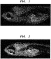
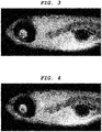
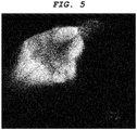
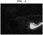
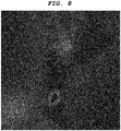
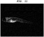
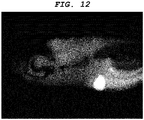
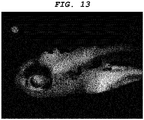
- juvenile zebrafish was removed from the well and embedded in 5% low melting point agarose gel on a slide glass so as to restrict movement, and the state of labeling in the central nervous system tissues were observed from the lateral side of zebrafish by a fluorescence stereomicroscope (manufactured by Leica Microsystems, MZ16FA). Also, the brain nerve tissues were observed from the parietal region by a confocal microscope (manufactured by Carl Zeiss, Inc., Pascal Exciter). As a result, fluorescence was observed in the brain nerve tissues of zebrafish.
- optic nerve, optic tract, superior colliculus (optic tectum), pituitary gland, tectospinal (tectobulbar) tract, and reticular formation were intensely labeled.
- Zebrafish was labeled and observed by similar operations to Example 1 except for changing the dye compound (8) of Example 1 to the dye compounds (9) to (14) listed in Table 1 and for using labeling solutions 2 to 7.
- Compounds (10) to (14) are not in the scope of the present claims.
- Zebrafish was labeled and observed by similar operations to Example 1 except for changing the dye compound (8) of Example 1 to fluorescein.
- the labeling properties (++: a central nervous system tissue(s) is intensely labeled, +: a central nervous system tissue(s) is weakly labeled, and -: not labeled) were assessed in the aforementioned Examples 1 to 7 and in Comparative Example 1.
- the results thus obtained are shown in Table 1. It is to be noted that the excitation wavelength and the fluorescence wavelength of the dye compounds of Examples 1 to 7 and Comparative Example 1 were obtained by measuring an aqueous solution prepared by diluting a 10 mg/mL DMSO solution 500-fold with distilled water by FL4500 fluorescence spectrophotometer, Hitachi High-Technologies Corporation.
- Zebrafish was labeled and observed by similar operations to Examples 6 and 7 except for changing a day-7 embryo (7 dpf) of juvenile zebrafish to a day-14 embryo (14 dpf) of juvenile zebrafish.
- fluorescence was observed also in the brain nerve tissues of 14 dpf juvenile zebrafish.
- the state of labeling in the brain varied depending on the site, and it was observed that optic nerve, optic tract, superior colliculus (optic tectum), pituitary gland, tectospinal (tectobulbar) tract, and reticular formation were intensely labeled.
- a 3-month-old B10 mouse was sacrificed by diethyl ether anesthesia, and the brain is collected.
- the brain thus removed is embedded in an OCT compound, and then frozen in isopentane cooled with liquid nitrogen.
- the resulting brain was sliced into thin sections of approximately 10 ⁇ m in thickness in a cryostat cooled to -20°C. The thin sections were then placed on a slide glass and dried, whereby a section of the brain tissue was prepared.
- a 1 ug/mL solution of the compound (13) in PBS was added, followed by incubation for one hour.
- the slide glass was washed with PBST (PBS containing 0.2% Triton-X100) three times, and then sealed with a cover glass.
- PBST PBS containing 0.2% Triton-X100
- the compound (13) was confirmed to exert a labeling property in a mouse brain tissue section.
- the compound (13) is added to an equimolar solution of NaOH so that a concentration of 10 mg/ml is reached, and the resulting mixture is centrifuged at 14 krpm for five minutes to obtain a supernatant. Then, 0.2 ml of the supernatant thus obtained is intraperitoneally administered to a 3-month-old B10 mouse in a single dose. After one hour, the animal thus treated is sacrificed by diethyl ether anesthesia, and the brain is collected. The brain thus removed is embedded in an OCT compound, and then frozen in isopentane cooled with liquid nitrogen. The resulting brain was sliced into thin sections of approximately 10 ⁇ m in thickness in a cryostat cooled to -20°C.
- the thin sections were then placed on a slide glass and dried, whereby a section of the brain tissue was prepared.
- the brain tissue section thus prepared was observed under a confocal microscope (manufactured by Carl Zeiss, Inc., Pascal Exciter). As a result, the compound was confirmed to exert a labeling property in a mouse brain by intraperitoneal administration.
- Zebrafish was labeled and observed by similar operations to Comparative Example 1 except for changing 7 dpf juvenile zebrafish used in Comparative Example 1 to 3 dpf juvenile zebrafish. As a result, the brain nerve tissues were not observed to be stained either in the 3 dpf juvenile zebrafish.
- Zebrafish was labeled and observed by similar operations to Example 1 except for changing the dye compound (8) of Example 1 to the dye compounds (15) to (28) listed in Table 2 and for using labeling solutions 12 to 25.
- fluorescence was observed also in the brain nerve tissues of 14 dpf juvenile zebrafish.
- the state of labeling in the brain varied depending on the site, and it was observed that optic nerve, optic tract, superior colliculus (optic tectum), pituitary gland, tectospinal (tectobulbar) tract, and reticular formation were intensely labeled.
- the labeling properties (++: a central nervous system tissue(s) is intensely labeled, +: a central nervous system tissue(s) is weakly labeled, and -: not labeled) and the fluorescence sensitivities (++: a central nervous system tissue(s) is intensely observed, +: a central nervous system tissue(s) is weakly observed, and -: not labeled) were assessed in the aforementioned Examples 12 to 25 and in Comparative Example 1.
- Example No. Compound No. Excitation wavelength ⁇ ex Fluorescence wavelength ⁇ em Stokes shift ⁇ ex- ⁇ em Labeling property Fluorescence sensitivity
- Example 12 15 469 555 86 ++ ++
- Example 13 16 380 470 90 + + Example 14 17 410 490 80 + + Example 15 18 464 514 50 ++ ++
- Example 16 19 459 520 62 ++ ++
- Example 17 20 380 470 90 + + Example 18 21 360 490 130 + + Example 19 22 360 510 150 + + + Example 20 23 422 476 54 ++ + Example 21 24 380 490 110 + + + Example 22 25 463 509 46 ++ ++
- Example 23 26 410 540 130 + + Example 24 27 472 504 32 ++ ++
- Example 25 28 410 540 130 + + Comparative Example 01 Fluorescein 494 521 27 Absence Absence
- Example 20 Into a 100 mL beaker, 30 mL of the labeling solution 20 prepared in Example 20 was poured. Then, 3-month-old zebrafish was placed and left there for one hour. Subsequently, the labeling solution 20 was removed and replaced by 50 mL of distilled water. This operation was repeated three times. Then, zebrafish was fixed in a phosphate buffer containing 4% paraformaldehyde and then embedded in 5% low melting point agarose gel. Using a linear slicer PRO7 (manufactured by Dosaka EM Co., Ltd.), a specimen of exposed brain was prepared.
- a linear slicer PRO7 manufactured by Dosaka EM Co., Ltd.
- the central nervous system tissue was observed to be stained also in 3-month-old adult zebrafish, based on which the labeling solution 20 was confirmed to exert a labeling property on the central nervous system tissues also in an organism in which the blood-brain barrier is operative.
- a 3-month-old B10 mouse was sacrificed by diethyl ether anesthesia, and the brain is collected.
- the brain thus removed is embedded in an OCT compound, and then frozen in isopentane cooled with liquid nitrogen.
- the resulting brain was sliced into thin sections of approximately 10 ⁇ m in thickness in a cryostat cooled to -20°C. The thin sections were then placed on a slide glass and dried, whereby a section of the brain tissue was prepared.
- a 1 ug/mL solution of the compound (27) in PBS was added, followed by incubation for one hour.
- the slide glass was washed with PBST (PBS containing 0.2% Triton-X100) three times, and then sealed with a cover glass.
- PBST PBS containing 0.2% Triton-X100
- the compound (27) was confirmed to exert a labeling property in a mouse brain tissue section.
- the compound (27) was dissolved in chloroform, to which concentrated hydrochloric acid was added while stirring to form a precipitate.
- the precipitate was collected by filtration under reduced pressure.
- the precipitate thus collected was dried in a vacuum oven at 50°C for 24 hours to give hydrochloride of the compound (27).
- the hydrochloride of compound 27 is dissolved in PBS so that a concentration of 1 mg/mL is reached, and 0.2 ml of this solution is intraperitoneally administered to a 3-month-old B10 mouse in a single dose. After one hour, the animal thus treated is sacrificed by diethyl ether anesthesia, and the brain is collected. The brain thus removed is embedded in an OCT compound, and then frozen in isopentane cooled with liquid nitrogen.
- the resulting brain was sliced into thin sections of approximately 10 ⁇ m in thickness in a cryostat cooled to -20°C. The thin sections were then placed on a slide glass and dried, whereby a section of the brain tissue was prepared. The brain tissue section thus prepared was observed under a confocal microscope (manufactured by Carl Zeiss, Inc., Pascal Exciter). As a result, the compound was confirmed to exert a labeling property in a mouse brain by intraperitoneal administration.
- Juvenile zebrafish was labeled by the same operations as Example 27 and then fixed in 4% PFA, and subsequently embedded in 5% low melting point agarose gel.
- a linear slicer Pro7 manufactured by Dosaka EM Co., Ltd.
- thin sections of the zebrafish were prepared, which were mounted on a slide glass.
- a confocal microscope manufactured by Carl Zeiss, Inc., Pascal Exciter
- optic tectum and reticular formation of the brain of the zebrafish were confirmed to be intensely labeled.
- Patent Literature 3 discloses a method for screening compounds for the central nervous system using zebrafish. According to this literature, it is described that because the expression of BBB transporter gene is incomplete in juvenile zebrafish, migration of dyes administered, namely Evans blue, fluorescein, and rhodamine 123, into the brain are confirmed up to 4 dpf, 8 dpf, and 5 dpf, respectively; however, because BBB is formed by 10 dpf, no dye will be observed to migrate into the brain any longer.
- Zebrafish was labeled and observed by similar operations to Example 1 except for changing the dye compound (8) of Example 1 to the dye compounds (29) to (45) listed in Table 3 and for using labeling solutions 29 to 45. It is to be noted that only the labeling solution 45 used in Example 46 had a concentration of the dye compound of 3 ⁇ g/mL. As a result, fluorescence was observed in the brain nerve tissues of 7 dpf juvenile zebrafish. The state of labeling in the brain varied depending on the site, and it was observed that optic nerve, optic tract, superior colliculus (optic tectum), pituitary gland, tectospinal (tectobulbar) tract, and reticular formation were intensely labeled.
- optic nerve, optic tract, superior colliculus (optic tectum), pituitary gland, tectospinal (tectobulbar) tract, and reticular formation were intensely labeled.
- the labeling properties (++: a central nervous system tissue(s) is intensely labeled, +: a central nervous system tissue(s) is weakly labeled, and -: not labeled) were assessed in the aforementioned Examples 30 to 46.
- the results thus obtained are shown in Table 3. It is to be noted that the excitation wavelength and the fluorescence wavelength of the dye compounds were obtained by measuring 5 ⁇ M chloroform solutions of the compounds in Examples 30 to 34, and 5 ⁇ M DMSO solutions of the compounds in Examples 35 to 46 by FL4500 fluorescence spectrophotometer, Hitachi High-Technologies Corporation.
- the present invention provides a central nervous system tissue-labeling composition capable of labeling a central nervous system tissue in a live biological sample and imaging the cell morphology of the central nervous system tissue with high sensitivity.
- the central nervous system tissue-labeling composition serves as a necessary material for a research in the area of the central nervous system and a technology pertaining to imaging of the central nervous system tissue.
- the central nervous system tissue-labeling composition allows chronological assessment of the central nervous system tissue, enabling highly accurate screening with high throughput at low cost. This dramatically progresses the development of new diagnostic and therapeutic methods for a disease, and further, expands research on the central nervous system, establishing a highly effective basic technology for not only industrial but also practical applications.
Landscapes
- Health & Medical Sciences (AREA)
- Life Sciences & Earth Sciences (AREA)
- Engineering & Computer Science (AREA)
- Biomedical Technology (AREA)
- Chemical & Material Sciences (AREA)
- General Health & Medical Sciences (AREA)
- Molecular Biology (AREA)
- Physics & Mathematics (AREA)
- Urology & Nephrology (AREA)
- Immunology (AREA)
- Hematology (AREA)
- Organic Chemistry (AREA)
- General Physics & Mathematics (AREA)
- Public Health (AREA)
- Animal Behavior & Ethology (AREA)
- Epidemiology (AREA)
- Veterinary Medicine (AREA)
- Pathology (AREA)
- Analytical Chemistry (AREA)
- Biotechnology (AREA)
- Cell Biology (AREA)
- Microbiology (AREA)
- Biochemistry (AREA)
- Food Science & Technology (AREA)
- Medicinal Chemistry (AREA)
- Optics & Photonics (AREA)
- Nuclear Medicine, Radiotherapy & Molecular Imaging (AREA)
- High Energy & Nuclear Physics (AREA)
- Medical Informatics (AREA)
- Spectroscopy & Molecular Physics (AREA)
- Biodiversity & Conservation Biology (AREA)
- Oncology (AREA)
- Medicines Containing Antibodies Or Antigens For Use As Internal Diagnostic Agents (AREA)
- Investigating Or Analysing Biological Materials (AREA)
- Nuclear Medicine (AREA)
Claims (6)
- Composition de marquage d'un tissu du système nerveux central destinée à être utilisée dans le diagnostic d'une maladie du système nerveux central in vivo comprenant, comme ingrédient actif, un composé représenté par la formule générale (1)


- Composition de marquage d'un tissu du système nerveux central destinée à être utilisée selon la revendication 1, dans laquelle le composé est capable de marquer au moins l'un quelconque d'un nerf optique, du tractus optique, du colliculus supérieur (tectum optique), de la glande pituitaire, du tractus tecto-spinal (tecto-bulbaire) et d'une formation réticulaire.
- Composition de marquage d'un tissu du système nerveux central destinée à être utilisée selon la revendication 1 ou 2, dans laquelle le composé est un composé fluorescent.
- Composition de marquage d'un tissu du système nerveux central destinée à être utilisée selon l'une quelconque des revendications 1 à 3, dans laquelle le composé est marqué avec un radionucléide.
- Procédé de marquage d'un tissu du système nerveux central comprenant l'utilisation de la composition de marquage d'un tissu de système nerveux central selon l'une quelconque des revendications 1 à 4 pour marquer un tissu du système nerveux central dans un corps vivant, où le procédé n'est pas un procédé pour le traitement du corps humain ou animal par chirurgie.
- Procédé de criblage comprenant l'utilisation de la composition de marquage d'un tissu du système nerveux central selon l'une quelconque des revendications 1 à 4 pour détecter un composé agissant sur un tissu du système nerveux central in vivo, où le procédé n'est pas un procédé pour le traitement du corps humain ou animal par chirurgie.
Applications Claiming Priority (2)
| Application Number | Priority Date | Filing Date | Title |
|---|---|---|---|
| JP2009296270 | 2009-12-25 | ||
| PCT/JP2010/007519 WO2011077751A1 (fr) | 2009-12-25 | 2010-12-24 | Composition pour marquer des tissus du système nerveux central, procédé pour marquer des tissus du système nerveux central, et procédé de criblage utilisant la composition pour marquer des tissus du système nerveux central |
Publications (3)
| Publication Number | Publication Date |
|---|---|
| EP2399612A1 EP2399612A1 (fr) | 2011-12-28 |
| EP2399612A4 EP2399612A4 (fr) | 2016-07-13 |
| EP2399612B1 true EP2399612B1 (fr) | 2018-02-21 |
Family
ID=44195304
Family Applications (1)
| Application Number | Title | Priority Date | Filing Date |
|---|---|---|---|
| EP10838989.1A Active EP2399612B1 (fr) | 2009-12-25 | 2010-12-24 | Composition pour marquer des tissus du système nerveux central, procédé pour marquer des tissus du système nerveux central, et procédé de criblage utilisant la composition pour marquer des tissus du système nerveux central |
Country Status (4)
| Country | Link |
|---|---|
| US (3) | US20110189096A1 (fr) |
| EP (1) | EP2399612B1 (fr) |
| JP (1) | JP5733716B2 (fr) |
| WO (1) | WO2011077751A1 (fr) |
Families Citing this family (19)
| Publication number | Priority date | Publication date | Assignee | Title |
|---|---|---|---|---|
| WO2010074326A1 (fr) | 2008-12-25 | 2010-07-01 | Canon Kabushiki Kaisha | Sonde pour échantillon biologique, procédé de marquage et procédé de criblage utilisant la sonde |
| JP2010168369A (ja) | 2008-12-25 | 2010-08-05 | Canon Inc | 眼内組織用標識組成物、眼内組織の標識方法及びスクリーニング方法 |
| JP5733716B2 (ja) * | 2009-12-25 | 2015-06-10 | キヤノン株式会社 | 中枢神経系組織標識用組成物、中枢神経系組織の標識方法、及び中枢神経系組織標識用組成物を用いたスクリーニング方法 |
| EP2517732B1 (fr) | 2009-12-25 | 2016-09-21 | Canon Kabushiki Kaisha | Composition transnasale de marquage pour des tissus du système nerveux central |
| JP5888904B2 (ja) | 2011-02-16 | 2016-03-22 | 国立大学法人三重大学 | 中枢神経移行性評価用プローブ、中枢神経移行性評価方法、及び、中枢神経移行性評価用プローブを用いたスクリーニング方法 |
| WO2013015433A1 (fr) * | 2011-07-27 | 2013-01-31 | Canon Kabushiki Kaisha | Composition liquide et composition de réserve pour filtre coloré, feuille d'impression par transfert thermique et encre qui utilisent chacune la composition liquide |
| US9134639B2 (en) * | 2011-07-27 | 2015-09-15 | Canon Kabushiki Kaisha | Magenta toner |
| US8926740B2 (en) | 2011-07-27 | 2015-01-06 | Canon Kabushiki Kaisha | Pigment dispersion, and resist composition for color filter and ink composition each using the pigment dispersion |
| WO2013051724A1 (fr) | 2011-10-04 | 2013-04-11 | Canon Kabushiki Kaisha | Composé de matériau colorant insoluble dans l'eau, encre utilisant le composé de matériau colorant insoluble dans l'eau, feuille d'enregistrement par transfert thermique et composition de réserve pour filtre coloré |
| JP6057574B2 (ja) * | 2012-07-11 | 2017-01-11 | キヤノン株式会社 | 造血幹細胞識別用プローブ |
| US20150182518A1 (en) | 2012-10-26 | 2015-07-02 | Canon Kabushiki Kaisha | Cancer cell inhibitory drug and cancer stem-cell detection probe |
| JP6327431B2 (ja) | 2012-10-26 | 2018-05-23 | 国立大学法人三重大学 | がん細胞阻害薬、がん幹細胞検出用プローブ |
| EP2948047B1 (fr) | 2013-01-24 | 2021-03-03 | Tylerton International Holdings Inc. | Système et méthode d'imagerie de structure corporelle pour localiser ganglion plexi |
| WO2014141247A1 (fr) * | 2013-03-11 | 2014-09-18 | Tylerton International Holdings Inc. | Modélisation du système nerveux végétatif et applications associées |
| EP2943584B1 (fr) | 2013-05-30 | 2019-08-14 | Canon Kabushiki Kaisha | Agent d'identification de macrophage, et procédé d'identification, procédé de tri, procédé d'évaluation, procédé de criblage et trousse utilisant l'agent d'identification de macrophage |
| US20160220835A1 (en) | 2013-09-08 | 2016-08-04 | Tylerton International Inc. | Apparatus and methods for diagnosis and treatment of patterns of nervous system activity affecting disease |
| US10646183B2 (en) | 2014-01-10 | 2020-05-12 | Tylerton International Inc. | Detection of scar and fibrous cardiac zones |
| WO2016016837A1 (fr) | 2014-07-30 | 2016-02-04 | Navix International Limited | Localisation de sonde |
| JP6749648B2 (ja) * | 2015-09-17 | 2020-09-02 | 国立研究開発法人科学技術振興機構 | 顕微鏡用光学モジュール、顕微鏡、顕微鏡用光学モジュール制御装置及び多光子励起顕微鏡 |
Family Cites Families (27)
| Publication number | Priority date | Publication date | Assignee | Title |
|---|---|---|---|---|
| JPS5917822B2 (ja) * | 1977-02-01 | 1984-04-24 | コニカ株式会社 | ハロゲン化銀写真感光材料 |
| JPS6010241A (ja) * | 1983-06-29 | 1985-01-19 | Fuji Photo Film Co Ltd | 内部潜像型直接ポジハロゲン化銀乳剤 |
| US5308604A (en) * | 1989-04-19 | 1994-05-03 | Deutsches Krebsforschungsinstitut | Conjugates for tumor localization and/or tumor therapy |
| EP0915090A1 (fr) * | 1992-09-10 | 1999-05-12 | Eli Lilly And Company | Thiazolidinones comme agents hypoglycemiants et pour le traitement de la maladie d'Alzheimer |
| JP2000344684A (ja) * | 1999-03-26 | 2000-12-12 | Bf Kenkyusho:Kk | ピロニンb類似化合物によるアミロイドが蓄積する疾患の画像診断プローブおよびそれを含む画像診断用組成物 |
| US6972287B1 (en) * | 1999-06-10 | 2005-12-06 | Pfizer Inc. | Method of inhibiting amyloid protein aggregation and imaging amyloid deposits |
| EP1192144A1 (fr) * | 1999-06-10 | 2002-04-03 | Warner-Lambert Company | Derives de la rhodanine et leur utilisation pour l'inhibition et la mise en images d'amyloides |
| US20040197268A1 (en) * | 1999-06-10 | 2004-10-07 | Augelli-Szafran Corinne Elizabeth | Method of imaging amyloid deposits using substituted rhodanine derivatives |
| JP2001305030A (ja) * | 2000-04-19 | 2001-10-31 | Olympus Optical Co Ltd | 試料調整法 |
| US20040256002A1 (en) * | 2002-07-29 | 2004-12-23 | Tamotsu Horiuchi | Organic dye, photoelectric transducing material, semiconductor electrode, and photoelectric transducing device |
| EP1547996A4 (fr) * | 2002-08-30 | 2006-08-02 | Bf Res Inst Inc | Sondes de diagnostic et remedes contre des maladies presentant une accumulation de la proteine du prion et methode de marquage |
| JP2004250411A (ja) * | 2003-02-21 | 2004-09-09 | Bf Kenkyusho:Kk | アミロイドβ蓄積性疾患の診断プローブおよび治療用化合物 |
| JP2004292571A (ja) * | 2003-03-26 | 2004-10-21 | Mitsui Chemicals Inc | 水系インク |
| GB0403490D0 (en) | 2004-02-17 | 2004-03-24 | Daniolabs Ltd | Screening methods |
| JP4695345B2 (ja) * | 2004-05-18 | 2011-06-08 | 株式会社 日立ディスプレイズ | 有機エレクトロルミネッセンス表示装置 |
| JP2007106755A (ja) * | 2005-09-13 | 2007-04-26 | Japan Health Science Foundation | アミロイドイメージング用プローブ |
| WO2009089537A2 (fr) * | 2008-01-11 | 2009-07-16 | Northwestern University | Composés anticancéreux |
| JP4514814B2 (ja) | 2008-06-04 | 2010-07-28 | 株式会社日本自動車部品総合研究所 | アンテナ装置 |
| WO2010074326A1 (fr) * | 2008-12-25 | 2010-07-01 | Canon Kabushiki Kaisha | Sonde pour échantillon biologique, procédé de marquage et procédé de criblage utilisant la sonde |
| JP5538715B2 (ja) * | 2008-12-25 | 2014-07-02 | キヤノン株式会社 | 網膜組織染色組成物、網膜組織の染色方法 |
| JP2010168369A (ja) * | 2008-12-25 | 2010-08-05 | Canon Inc | 眼内組織用標識組成物、眼内組織の標識方法及びスクリーニング方法 |
| JP5416450B2 (ja) * | 2009-03-27 | 2014-02-12 | 三菱製紙株式会社 | 光電変換材料、半導体電極並びにそれを用いた光電変換素子 |
| WO2011057229A2 (fr) * | 2009-11-09 | 2011-05-12 | University Of Miami | Analogues de neurotransmetteurs fluorescents, compositions les contenant et procédés pour les utiliser |
| JP5733716B2 (ja) * | 2009-12-25 | 2015-06-10 | キヤノン株式会社 | 中枢神経系組織標識用組成物、中枢神経系組織の標識方法、及び中枢神経系組織標識用組成物を用いたスクリーニング方法 |
| EP2517732B1 (fr) * | 2009-12-25 | 2016-09-21 | Canon Kabushiki Kaisha | Composition transnasale de marquage pour des tissus du système nerveux central |
| JP5888904B2 (ja) * | 2011-02-16 | 2016-03-22 | 国立大学法人三重大学 | 中枢神経移行性評価用プローブ、中枢神経移行性評価方法、及び、中枢神経移行性評価用プローブを用いたスクリーニング方法 |
| JP6057574B2 (ja) * | 2012-07-11 | 2017-01-11 | キヤノン株式会社 | 造血幹細胞識別用プローブ |
-
2010
- 2010-12-24 JP JP2010288573A patent/JP5733716B2/ja active Active
- 2010-12-24 WO PCT/JP2010/007519 patent/WO2011077751A1/fr not_active Ceased
- 2010-12-24 EP EP10838989.1A patent/EP2399612B1/fr active Active
-
2011
- 2011-04-12 US US13/084,997 patent/US20110189096A1/en not_active Abandoned
-
2013
- 2013-06-19 US US13/921,945 patent/US20130280169A1/en not_active Abandoned
-
2016
- 2016-08-23 US US15/244,835 patent/US20160361442A1/en not_active Abandoned
Non-Patent Citations (1)
| Title |
|---|
| None * |
Also Published As
| Publication number | Publication date |
|---|---|
| US20110189096A1 (en) | 2011-08-04 |
| JP2011148794A (ja) | 2011-08-04 |
| JP5733716B2 (ja) | 2015-06-10 |
| EP2399612A4 (fr) | 2016-07-13 |
| EP2399612A1 (fr) | 2011-12-28 |
| US20130280169A1 (en) | 2013-10-24 |
| WO2011077751A1 (fr) | 2011-06-30 |
| US20160361442A1 (en) | 2016-12-15 |
Similar Documents
| Publication | Publication Date | Title |
|---|---|---|
| EP2399612B1 (fr) | Composition pour marquer des tissus du système nerveux central, procédé pour marquer des tissus du système nerveux central, et procédé de criblage utilisant la composition pour marquer des tissus du système nerveux central | |
| EP2370110B1 (fr) | Composition de marquage pour tissu intra-oculaire, procédé de marquage de tissu intra-oculaire, et procédé de criblage | |
| US9649392B2 (en) | Central nervous system labelling composition for intranasal administration and labelling method and screening method using central nervous system labelling composition for intranasal administration | |
| JP5783673B2 (ja) | 有毛細胞標識剤、及び該標識剤を用いた有毛細胞標識方法 | |
| US9801960B2 (en) | Probe for a biological specimen and labelling method and screening method using the probe | |
| JP5538715B2 (ja) | 網膜組織染色組成物、網膜組織の染色方法 | |
| EP2489665B1 (fr) | Sonde d'évaluation de la perméabilité du système nerveux central, procédé d'évaluation de la perméabilité du système nerveux central et procédé de criblage utilisant une sonde d'évaluation de la perméabilité du système nerveux central | |
| US20030138374A1 (en) | Image diagnosis probe based on substituted azobenzene or analogue thereof for disease attributable to amyloid accumulation and composition for image diagnosis containing the same |
Legal Events
| Date | Code | Title | Description |
|---|---|---|---|
| PUAI | Public reference made under article 153(3) epc to a published international application that has entered the european phase |
Free format text: ORIGINAL CODE: 0009012 |
|
| 17P | Request for examination filed |
Effective date: 20110921 |
|
| AK | Designated contracting states |
Kind code of ref document: A1 Designated state(s): AL AT BE BG CH CY CZ DE DK EE ES FI FR GB GR HR HU IE IS IT LI LT LU LV MC MK MT NL NO PL PT RO RS SE SI SK SM TR |
|
| DAX | Request for extension of the european patent (deleted) | ||
| RIC1 | Information provided on ipc code assigned before grant |
Ipc: G01N 33/50 20060101ALI20160201BHEP Ipc: A61K 49/00 20060101AFI20160201BHEP Ipc: G01N 33/15 20060101ALI20160201BHEP Ipc: A61K 51/00 20060101ALI20160201BHEP Ipc: G01N 33/58 20060101ALI20160201BHEP |
|
| RA4 | Supplementary search report drawn up and despatched (corrected) |
Effective date: 20160613 |
|
| RIC1 | Information provided on ipc code assigned before grant |
Ipc: G01N 33/58 20060101ALI20160607BHEP Ipc: A61K 49/00 20060101AFI20160607BHEP Ipc: G01N 33/15 20060101ALI20160607BHEP Ipc: A61K 51/00 20060101ALI20160607BHEP Ipc: G01N 33/50 20060101ALI20160607BHEP |
|
| GRAP | Despatch of communication of intention to grant a patent |
Free format text: ORIGINAL CODE: EPIDOSNIGR1 |
|
| INTG | Intention to grant announced |
Effective date: 20170811 |
|
| GRAJ | Information related to disapproval of communication of intention to grant by the applicant or resumption of examination proceedings by the epo deleted |
Free format text: ORIGINAL CODE: EPIDOSDIGR1 |
|
| GRAS | Grant fee paid |
Free format text: ORIGINAL CODE: EPIDOSNIGR3 |
|
| GRAP | Despatch of communication of intention to grant a patent |
Free format text: ORIGINAL CODE: EPIDOSNIGR1 |
|
| INTC | Intention to grant announced (deleted) | ||
| GRAA | (expected) grant |
Free format text: ORIGINAL CODE: 0009210 |
|
| INTG | Intention to grant announced |
Effective date: 20180104 |
|
| AK | Designated contracting states |
Kind code of ref document: B1 Designated state(s): AL AT BE BG CH CY CZ DE DK EE ES FI FR GB GR HR HU IE IS IT LI LT LU LV MC MK MT NL NO PL PT RO RS SE SI SK SM TR |
|
| REG | Reference to a national code |
Ref country code: GB Ref legal event code: FG4D |
|
| REG | Reference to a national code |
Ref country code: CH Ref legal event code: EP Ref country code: CH Ref legal event code: NV Representative=s name: BOVARD AG PATENT- UND MARKENANWAELTE, CH |
|
| REG | Reference to a national code |
Ref country code: AT Ref legal event code: REF Ref document number: 971005 Country of ref document: AT Kind code of ref document: T Effective date: 20180315 |
|
| REG | Reference to a national code |
Ref country code: IE Ref legal event code: FG4D |
|
| REG | Reference to a national code |
Ref country code: DE Ref legal event code: R096 Ref document number: 602010048723 Country of ref document: DE |
|
| REG | Reference to a national code |
Ref country code: NL Ref legal event code: MP Effective date: 20180221 |
|
| REG | Reference to a national code |
Ref country code: LT Ref legal event code: MG4D |
|
| REG | Reference to a national code |
Ref country code: AT Ref legal event code: MK05 Ref document number: 971005 Country of ref document: AT Kind code of ref document: T Effective date: 20180221 |
|
| PG25 | Lapsed in a contracting state [announced via postgrant information from national office to epo] |
Ref country code: FI Free format text: LAPSE BECAUSE OF FAILURE TO SUBMIT A TRANSLATION OF THE DESCRIPTION OR TO PAY THE FEE WITHIN THE PRESCRIBED TIME-LIMIT Effective date: 20180221 Ref country code: NO Free format text: LAPSE BECAUSE OF FAILURE TO SUBMIT A TRANSLATION OF THE DESCRIPTION OR TO PAY THE FEE WITHIN THE PRESCRIBED TIME-LIMIT Effective date: 20180521 Ref country code: HR Free format text: LAPSE BECAUSE OF FAILURE TO SUBMIT A TRANSLATION OF THE DESCRIPTION OR TO PAY THE FEE WITHIN THE PRESCRIBED TIME-LIMIT Effective date: 20180221 Ref country code: CY Free format text: LAPSE BECAUSE OF FAILURE TO SUBMIT A TRANSLATION OF THE DESCRIPTION OR TO PAY THE FEE WITHIN THE PRESCRIBED TIME-LIMIT Effective date: 20180221 Ref country code: LT Free format text: LAPSE BECAUSE OF FAILURE TO SUBMIT A TRANSLATION OF THE DESCRIPTION OR TO PAY THE FEE WITHIN THE PRESCRIBED TIME-LIMIT Effective date: 20180221 Ref country code: ES Free format text: LAPSE BECAUSE OF FAILURE TO SUBMIT A TRANSLATION OF THE DESCRIPTION OR TO PAY THE FEE WITHIN THE PRESCRIBED TIME-LIMIT Effective date: 20180221 Ref country code: NL Free format text: LAPSE BECAUSE OF FAILURE TO SUBMIT A TRANSLATION OF THE DESCRIPTION OR TO PAY THE FEE WITHIN THE PRESCRIBED TIME-LIMIT Effective date: 20180221 |
|
| PG25 | Lapsed in a contracting state [announced via postgrant information from national office to epo] |
Ref country code: BG Free format text: LAPSE BECAUSE OF FAILURE TO SUBMIT A TRANSLATION OF THE DESCRIPTION OR TO PAY THE FEE WITHIN THE PRESCRIBED TIME-LIMIT Effective date: 20180521 Ref country code: GR Free format text: LAPSE BECAUSE OF FAILURE TO SUBMIT A TRANSLATION OF THE DESCRIPTION OR TO PAY THE FEE WITHIN THE PRESCRIBED TIME-LIMIT Effective date: 20180522 Ref country code: RS Free format text: LAPSE BECAUSE OF FAILURE TO SUBMIT A TRANSLATION OF THE DESCRIPTION OR TO PAY THE FEE WITHIN THE PRESCRIBED TIME-LIMIT Effective date: 20180221 Ref country code: SE Free format text: LAPSE BECAUSE OF FAILURE TO SUBMIT A TRANSLATION OF THE DESCRIPTION OR TO PAY THE FEE WITHIN THE PRESCRIBED TIME-LIMIT Effective date: 20180221 Ref country code: LV Free format text: LAPSE BECAUSE OF FAILURE TO SUBMIT A TRANSLATION OF THE DESCRIPTION OR TO PAY THE FEE WITHIN THE PRESCRIBED TIME-LIMIT Effective date: 20180221 Ref country code: AT Free format text: LAPSE BECAUSE OF FAILURE TO SUBMIT A TRANSLATION OF THE DESCRIPTION OR TO PAY THE FEE WITHIN THE PRESCRIBED TIME-LIMIT Effective date: 20180221 |
|
| PG25 | Lapsed in a contracting state [announced via postgrant information from national office to epo] |
Ref country code: EE Free format text: LAPSE BECAUSE OF FAILURE TO SUBMIT A TRANSLATION OF THE DESCRIPTION OR TO PAY THE FEE WITHIN THE PRESCRIBED TIME-LIMIT Effective date: 20180221 Ref country code: IT Free format text: LAPSE BECAUSE OF FAILURE TO SUBMIT A TRANSLATION OF THE DESCRIPTION OR TO PAY THE FEE WITHIN THE PRESCRIBED TIME-LIMIT Effective date: 20180221 Ref country code: AL Free format text: LAPSE BECAUSE OF FAILURE TO SUBMIT A TRANSLATION OF THE DESCRIPTION OR TO PAY THE FEE WITHIN THE PRESCRIBED TIME-LIMIT Effective date: 20180221 Ref country code: RO Free format text: LAPSE BECAUSE OF FAILURE TO SUBMIT A TRANSLATION OF THE DESCRIPTION OR TO PAY THE FEE WITHIN THE PRESCRIBED TIME-LIMIT Effective date: 20180221 Ref country code: PL Free format text: LAPSE BECAUSE OF FAILURE TO SUBMIT A TRANSLATION OF THE DESCRIPTION OR TO PAY THE FEE WITHIN THE PRESCRIBED TIME-LIMIT Effective date: 20180221 |
|
| REG | Reference to a national code |
Ref country code: DE Ref legal event code: R097 Ref document number: 602010048723 Country of ref document: DE |
|
| PG25 | Lapsed in a contracting state [announced via postgrant information from national office to epo] |
Ref country code: CZ Free format text: LAPSE BECAUSE OF FAILURE TO SUBMIT A TRANSLATION OF THE DESCRIPTION OR TO PAY THE FEE WITHIN THE PRESCRIBED TIME-LIMIT Effective date: 20180221 Ref country code: SK Free format text: LAPSE BECAUSE OF FAILURE TO SUBMIT A TRANSLATION OF THE DESCRIPTION OR TO PAY THE FEE WITHIN THE PRESCRIBED TIME-LIMIT Effective date: 20180221 Ref country code: SM Free format text: LAPSE BECAUSE OF FAILURE TO SUBMIT A TRANSLATION OF THE DESCRIPTION OR TO PAY THE FEE WITHIN THE PRESCRIBED TIME-LIMIT Effective date: 20180221 Ref country code: DK Free format text: LAPSE BECAUSE OF FAILURE TO SUBMIT A TRANSLATION OF THE DESCRIPTION OR TO PAY THE FEE WITHIN THE PRESCRIBED TIME-LIMIT Effective date: 20180221 |
|
| PLBE | No opposition filed within time limit |
Free format text: ORIGINAL CODE: 0009261 |
|
| STAA | Information on the status of an ep patent application or granted ep patent |
Free format text: STATUS: NO OPPOSITION FILED WITHIN TIME LIMIT |
|
| 26N | No opposition filed |
Effective date: 20181122 |
|
| PG25 | Lapsed in a contracting state [announced via postgrant information from national office to epo] |
Ref country code: SI Free format text: LAPSE BECAUSE OF FAILURE TO SUBMIT A TRANSLATION OF THE DESCRIPTION OR TO PAY THE FEE WITHIN THE PRESCRIBED TIME-LIMIT Effective date: 20180221 |
|
| GBPC | Gb: european patent ceased through non-payment of renewal fee |
Effective date: 20181224 |
|
| PG25 | Lapsed in a contracting state [announced via postgrant information from national office to epo] |
Ref country code: MC Free format text: LAPSE BECAUSE OF FAILURE TO SUBMIT A TRANSLATION OF THE DESCRIPTION OR TO PAY THE FEE WITHIN THE PRESCRIBED TIME-LIMIT Effective date: 20180221 Ref country code: LU Free format text: LAPSE BECAUSE OF NON-PAYMENT OF DUE FEES Effective date: 20181224 |
|
| REG | Reference to a national code |
Ref country code: IE Ref legal event code: MM4A |
|
| REG | Reference to a national code |
Ref country code: BE Ref legal event code: MM Effective date: 20181231 |
|
| PG25 | Lapsed in a contracting state [announced via postgrant information from national office to epo] |
Ref country code: FR Free format text: LAPSE BECAUSE OF NON-PAYMENT OF DUE FEES Effective date: 20181231 Ref country code: IE Free format text: LAPSE BECAUSE OF NON-PAYMENT OF DUE FEES Effective date: 20181224 |
|
| PG25 | Lapsed in a contracting state [announced via postgrant information from national office to epo] |
Ref country code: BE Free format text: LAPSE BECAUSE OF NON-PAYMENT OF DUE FEES Effective date: 20181231 |
|
| PG25 | Lapsed in a contracting state [announced via postgrant information from national office to epo] |
Ref country code: GB Free format text: LAPSE BECAUSE OF NON-PAYMENT OF DUE FEES Effective date: 20181224 |
|
| PG25 | Lapsed in a contracting state [announced via postgrant information from national office to epo] |
Ref country code: MT Free format text: LAPSE BECAUSE OF NON-PAYMENT OF DUE FEES Effective date: 20181224 |
|
| PG25 | Lapsed in a contracting state [announced via postgrant information from national office to epo] |
Ref country code: TR Free format text: LAPSE BECAUSE OF FAILURE TO SUBMIT A TRANSLATION OF THE DESCRIPTION OR TO PAY THE FEE WITHIN THE PRESCRIBED TIME-LIMIT Effective date: 20180221 |
|
| PG25 | Lapsed in a contracting state [announced via postgrant information from national office to epo] |
Ref country code: PT Free format text: LAPSE BECAUSE OF FAILURE TO SUBMIT A TRANSLATION OF THE DESCRIPTION OR TO PAY THE FEE WITHIN THE PRESCRIBED TIME-LIMIT Effective date: 20180221 |
|
| PG25 | Lapsed in a contracting state [announced via postgrant information from national office to epo] |
Ref country code: HU Free format text: LAPSE BECAUSE OF FAILURE TO SUBMIT A TRANSLATION OF THE DESCRIPTION OR TO PAY THE FEE WITHIN THE PRESCRIBED TIME-LIMIT; INVALID AB INITIO Effective date: 20101224 Ref country code: MK Free format text: LAPSE BECAUSE OF NON-PAYMENT OF DUE FEES Effective date: 20180221 |
|
| PG25 | Lapsed in a contracting state [announced via postgrant information from national office to epo] |
Ref country code: IS Free format text: LAPSE BECAUSE OF FAILURE TO SUBMIT A TRANSLATION OF THE DESCRIPTION OR TO PAY THE FEE WITHIN THE PRESCRIBED TIME-LIMIT Effective date: 20180621 |
|
| PGFP | Annual fee paid to national office [announced via postgrant information from national office to epo] |
Ref country code: DE Payment date: 20231121 Year of fee payment: 14 |
|
| PGFP | Annual fee paid to national office [announced via postgrant information from national office to epo] |
Ref country code: CH Payment date: 20240101 Year of fee payment: 14 |
|
| REG | Reference to a national code |
Ref country code: DE Ref legal event code: R119 Ref document number: 602010048723 Country of ref document: DE |
|
| REG | Reference to a national code |
Ref country code: CH Ref legal event code: PL |
|
| PG25 | Lapsed in a contracting state [announced via postgrant information from national office to epo] |
Ref country code: DE Free format text: LAPSE BECAUSE OF NON-PAYMENT OF DUE FEES Effective date: 20250701 |
|
| PG25 | Lapsed in a contracting state [announced via postgrant information from national office to epo] |
Ref country code: CH Free format text: LAPSE BECAUSE OF NON-PAYMENT OF DUE FEES Effective date: 20241231 |