EP2410046B1 - Procédé d'isolement et procédé de culture en masse pour des cellules souches pluripotentes dérivées de sang de cordon ombilical exprimant znf281 - Google Patents
Procédé d'isolement et procédé de culture en masse pour des cellules souches pluripotentes dérivées de sang de cordon ombilical exprimant znf281 Download PDFInfo
- Publication number
- EP2410046B1 EP2410046B1 EP10753642.7A EP10753642A EP2410046B1 EP 2410046 B1 EP2410046 B1 EP 2410046B1 EP 10753642 A EP10753642 A EP 10753642A EP 2410046 B1 EP2410046 B1 EP 2410046B1
- Authority
- EP
- European Patent Office
- Prior art keywords
- stem cells
- cells
- cord blood
- umbilical cord
- culture
- Prior art date
- Legal status (The legal status is an assumption and is not a legal conclusion. Google has not performed a legal analysis and makes no representation as to the accuracy of the status listed.)
- Active
Links
Images




Classifications
-
- C—CHEMISTRY; METALLURGY
- C12—BIOCHEMISTRY; BEER; SPIRITS; WINE; VINEGAR; MICROBIOLOGY; ENZYMOLOGY; MUTATION OR GENETIC ENGINEERING
- C12N—MICROORGANISMS OR ENZYMES; COMPOSITIONS THEREOF; PROPAGATING, PRESERVING, OR MAINTAINING MICROORGANISMS; MUTATION OR GENETIC ENGINEERING; CULTURE MEDIA
- C12N5/00—Undifferentiated human, animal or plant cells, e.g. cell lines; Tissues; Cultivation or maintenance thereof; Culture media therefor
- C12N5/06—Animal cells or tissues; Human cells or tissues
- C12N5/0602—Vertebrate cells
-
- C—CHEMISTRY; METALLURGY
- C12—BIOCHEMISTRY; BEER; SPIRITS; WINE; VINEGAR; MICROBIOLOGY; ENZYMOLOGY; MUTATION OR GENETIC ENGINEERING
- C12N—MICROORGANISMS OR ENZYMES; COMPOSITIONS THEREOF; PROPAGATING, PRESERVING, OR MAINTAINING MICROORGANISMS; MUTATION OR GENETIC ENGINEERING; CULTURE MEDIA
- C12N5/00—Undifferentiated human, animal or plant cells, e.g. cell lines; Tissues; Cultivation or maintenance thereof; Culture media therefor
- C12N5/06—Animal cells or tissues; Human cells or tissues
- C12N5/0602—Vertebrate cells
- C12N5/0603—Embryonic cells ; Embryoid bodies
- C12N5/0605—Cells from extra-embryonic tissues, e.g. placenta, amnion, yolk sac, Wharton's jelly
-
- A—HUMAN NECESSITIES
- A61—MEDICAL OR VETERINARY SCIENCE; HYGIENE
- A61P—SPECIFIC THERAPEUTIC ACTIVITY OF CHEMICAL COMPOUNDS OR MEDICINAL PREPARATIONS
- A61P19/00—Drugs for skeletal disorders
-
- A—HUMAN NECESSITIES
- A61—MEDICAL OR VETERINARY SCIENCE; HYGIENE
- A61P—SPECIFIC THERAPEUTIC ACTIVITY OF CHEMICAL COMPOUNDS OR MEDICINAL PREPARATIONS
- A61P25/00—Drugs for disorders of the nervous system
-
- A—HUMAN NECESSITIES
- A61—MEDICAL OR VETERINARY SCIENCE; HYGIENE
- A61P—SPECIFIC THERAPEUTIC ACTIVITY OF CHEMICAL COMPOUNDS OR MEDICINAL PREPARATIONS
- A61P9/00—Drugs for disorders of the cardiovascular system
-
- C—CHEMISTRY; METALLURGY
- C12—BIOCHEMISTRY; BEER; SPIRITS; WINE; VINEGAR; MICROBIOLOGY; ENZYMOLOGY; MUTATION OR GENETIC ENGINEERING
- C12N—MICROORGANISMS OR ENZYMES; COMPOSITIONS THEREOF; PROPAGATING, PRESERVING, OR MAINTAINING MICROORGANISMS; MUTATION OR GENETIC ENGINEERING; CULTURE MEDIA
- C12N5/00—Undifferentiated human, animal or plant cells, e.g. cell lines; Tissues; Cultivation or maintenance thereof; Culture media therefor
-
- C—CHEMISTRY; METALLURGY
- C12—BIOCHEMISTRY; BEER; SPIRITS; WINE; VINEGAR; MICROBIOLOGY; ENZYMOLOGY; MUTATION OR GENETIC ENGINEERING
- C12N—MICROORGANISMS OR ENZYMES; COMPOSITIONS THEREOF; PROPAGATING, PRESERVING, OR MAINTAINING MICROORGANISMS; MUTATION OR GENETIC ENGINEERING; CULTURE MEDIA
- C12N5/00—Undifferentiated human, animal or plant cells, e.g. cell lines; Tissues; Cultivation or maintenance thereof; Culture media therefor
- C12N5/06—Animal cells or tissues; Human cells or tissues
-
- C—CHEMISTRY; METALLURGY
- C12—BIOCHEMISTRY; BEER; SPIRITS; WINE; VINEGAR; MICROBIOLOGY; ENZYMOLOGY; MUTATION OR GENETIC ENGINEERING
- C12N—MICROORGANISMS OR ENZYMES; COMPOSITIONS THEREOF; PROPAGATING, PRESERVING, OR MAINTAINING MICROORGANISMS; MUTATION OR GENETIC ENGINEERING; CULTURE MEDIA
- C12N5/00—Undifferentiated human, animal or plant cells, e.g. cell lines; Tissues; Cultivation or maintenance thereof; Culture media therefor
- C12N5/06—Animal cells or tissues; Human cells or tissues
- C12N5/0602—Vertebrate cells
- C12N5/0634—Cells from the blood or the immune system
- C12N5/0647—Haematopoietic stem cells; Uncommitted or multipotent progenitors
Definitions
- the present invention relates to a method for isolating pluripotent/multipotent stem cells expressing ZNF281 derived from umbilical cord blood, characterized by culturing mononuclear cells isolated from umbilical cord blood in a culture vessel coated with fibronectin and then harvesting stem cells from the culture, wherein the stem cell is in an undifferentiated state, the umbilical cord blood-derived pluripotent/multipotent stem cells isolated thereby.
- the present invention also relates to the use of a novel culture media for stem cells, a culture method for stem cells which is characterized by culturing and proliferating stem cells in the culture media, and a method for increasing stemness of stem cells which is characterized by a sphere culture or a three-dimensional culture of stem cells.
- stem cells Characterized by being self-renewing, undergoing differentiation and being immortal, stem cells have been proposed to be a solution for the problems of regenerative medicine and tissue replacement so that they can be used to treat various degenerative diseases as well as to provide deep insight into cellular biology.
- Adult stem cells obtainable from various tissues, are far more attractive than embryonic stem cells because of the ability to come from an unlimited number of sources and the ethical objections to using human embryos as a source of cells are rendered irrelevant.
- stem cells isolated from umbilical cord blood have advantages over other adult stem cells in that the donors of umbilical cord blood are not injured, unlike the donors of bone marrow or adipose tissue.
- Umbilical cord blood-derived mesenchymal stem cells have been successfully introduced into various kinds of cells including neural cells, hepatocytes, osteocytes, etc., in vitro ( Sun, W. et al., Stem cells, 23:931, 2005 ; Hong SH. et al., Biochem. Biophys. Res. Commun., 30:1153, 2005 ; Hutson EL. et al., Tissue Engineering, 11:1407, 2005 ).
- Successful in vivo transplantation of umbilical cord blood-derived mesenchymal stem cells for injuries, diabetes mellitus and heart infarction were also reported ( Nonome, K. et al., Am, J. Physiol. Gastrointest.
- ZHAO ET AL "Identification of stem cells from human umbilical cord blood with embryonic and hematopoietic characteristics", EXPERIMENTAL CELL RESEARCH, vol. 312, no. 13, 1 August 2006, pages 2454-2464, XP005541900 discloses stem cells from the umbilical cord blood which display important embryonic stem cell characteristics including expression of embryonic-stem-cell-specific molecular markers including transcription factors OCT-4 and Nanog along stage-specific embryonic antigens.
- KOGLER GESINE ET AL "A new human somatic stem cell from placental cord blood with intrinsic pluripotent differentiation potential", THE JOURNAL OF EXPERIMENTAL MEDICINE, vol. 200, no. 2, 19 July 2004, pages 123-135 , XP002559665 discloses an intrinsically pluripotent CD45-negative population from human cord blood termed unrestricted somatic stem cells.
- mesenchymal stem cells from umbilical cord blood.
- the isolation rate remains at around 20%. As much as 50% of the mesenchymal stem cells may be isolated from flesh blood which has been taken 5 hours before the isolation. However, the isolation rate is decreased to 20% or less when the blood has been taken over 5 hours before doing the isolation and the cells, although isolated, do not proliferate well.
- the present invention pertains to a method for isolating umbilical cord blood-derived pluripotent or multipotent stem cells expressing ZNF281, comprising the steps of
- the step of harvesting the stem cells from the culture comprises utilizing an immunological property of stem cells to separate the stem cells.
- the isolation of mononuclear cells from umbilical cord blood may be achieved using a typical method. After mixing the umbilical cord blood with HetasepTM to deplete erythrocytes, monocytes are isolated using Ficoll®-paque.
- HetasepTM is preferably used in an amount of 0.5 ⁇ 2 mL per 5 mL of HetasepTM.
- HetasepTM is an erythrocyte aggregation agent used to quickly separate nucleated cells from red blood cells (RBCs) in whole blood.
- Ficoll®-paque is a ready to use sterile medium for isolation of lymphocytes in high yield. It is normally placed at the bottom of a conical tube, and blood is slowly layered above Ficoll®-paque.
- the umbilical cord blood used in the present invention is preferably one that is recovered immediately after childbirth, one that is stored at room temperature for 12 ⁇ 48 hours after the recovery, or one that is stored at 3 ⁇ 5°C for 6 ⁇ 72 hours after the recovery.
- the isolation of stem cells from the umbilical cord blood-derived mononuclear cells is characterized by the use of fibronectin.
- culture vessel containing fibronectin is intended to mean a condition in which mononuclear cells can be brought into contact with fibronectin.
- fibronectin may be layered on a culture vessel or may be contained in the form of spheres or three-dimensional structures in a culture medium.
- fibronectin when the culture vessel is coated therewith, fibronectin may be contained at a density of from 0.1 to 1 mg/mL.
- the fibronectin useful in the present invention may be derived from animals without limitation and preferably from humans.
- the fibronectin may be prepared by artificial synthesis (e.g., chemical synthesis, synthesis using a peptide synthesizer, etc.) or biosynthesis (e.g., recombinant DNA technology, fibroblast culture, etc.) or may be separated from the plasma of animals including humans from the extracellular matrices.
- the fibronectin may be a fragment or peptide sequence of fibronectin or may contain the fragment or peptide.
- SNU-1 or EGM-2 is used as a fundamental medium for culturing the monocytes.
- the SNU-1 medium contains the following composition (Table 1). TABLE 1 SNU-1 (mg/l) SNU-1 (mg/l) SNU-1 (mg/l) CaCl 2 (anhyd.) 200 L-Isoleucine 78 i-Inositol 3 KCL 400 L-Leucine 78 Riboflavin 0.15 MgSO 4 (anhyd.) 97.67 L-Lysine HCl 108.75 Thiamine HCl 1.5 NaCl 7635 L-Methionine 22.5 L-Alanine 17.8 NaH 2 PO 4 H 2 O 140 L-Phenylalanine 48 L-Asparagine H 2 O 30 D-Glucose 1000 L-Serine 21 L-Aspartic Acid 26.6 Phenol Red 10 L-Threonine 72 L-Glutamic acid 29.4 Sodium Pyruvate 110 L-Tryptophan 15 L-Proline 23 L-Arginine HCl 189 L-Ty
- the fundamental medium is supplemented with FGF-B (Fibroblast Growth Factor), ascorbic acid, EGF (Epidermal Growth Factor), hydrocortisone, IGF-1 (Insulin-like Growth Factor-1) or VEGF (Vascular Endothelial Growth Factor), and heparin and optionally with GA-1000 (Gentamycin Sulfate, Amphotericin-B)if needed.
- FGF-B Fibroblast Growth Factor
- EGF Epidermatitis
- hydrocortisone hydrocortisone
- IGF-1 Insulin-like Growth Factor-1
- VEGF Vascular Endothelial Growth Factor
- GA-1000 Genetamycin Sulfate, Amphotericin-B
- the fundamental medium is supplemented with fetal bovine serum (FBS) 20%, bFGF (Fibroblast Growth Factor) 1 ⁇ 40 ng/ml, ascorbic acid 0.1 ⁇ 5.0 ⁇ g/ml, EGF (Epidermal Growth Factor) 1 ⁇ 40 ng/ml, hydrocortisone 0.1 ⁇ 1 ⁇ g/ml, IGF-I (Insulin-like Growth Factor-1) 1 ⁇ 40 ng/ml or VEGF (Vascular Endothelial Growth Factor) 1 ⁇ 5 ng/ml and heparin 20 ⁇ 25 ⁇ g/ml and optionally with GA-1000 (Gentamycin Sulfate, Amphotericin-B) if needed.
- FBS fetal bovine serum
- bFGF Fibroblast Growth Factor 1 ⁇ 40 ng/ml
- ascorbic acid 0.1 ⁇ 5.0 ⁇ g/ml
- EGF Epidermatitis-1
- hydrocortisone 0.1 ⁇ 1 ⁇ g/
- the mononuclear cells which remain suspended are removed while only adherent cells are cultured.
- the mononuclear cells adherent to the vessel only stem cells are proliferated. From 12 to 20 days after the isolation, rapid proliferation of the stem cells can be observed.
- the medium is preferably replaced with a fresh one every two or three days.
- Flow cytometry sorting may be performed by electrostatic droplet charging or cell capture.
- an antibody specifically recognizing a surface antigen is fluorescence labeled after which the fluorescent intensity is measured from the labeled antibody-antigen conjugate and converted into electric signals to determine the expression level of the antigen of the cells.
- fluorophores may be used in combination to separate cells expressing different surface antigens.
- fluorophores are FITC (fluorescein isothiocyanate), PE (phycoerythrin), APC (allo-phycocyanin), TR (TexasRed), Cy3, CyChrome, Red613, Red670, TRI-Color, and QuantumRed.
- the stem cells harvested from the culture by, for example, centrifugation may be immunostained directly with an antibody or may be proliferated in a suitable medium before immunostaining with an antibody.
- a target cell sample is mixed with a primary antibody specific for a surface antigen and incubated for 0.5 to 1 hour on ice. If the primary antibody is labeled with a fluorophore, the cell sample is washed and separated with a flow cytometer. If the primary antibody is not labeled with a fluorophore, the cell sample treated with the primary antibody is washed and mixed with a fluorescence-labeled secondary antibody that can bind to the primary antibody. Then, the immunostained cells are incubated again for 0.5 to 1 hour on ice and washed before being separated by a flow cytometer.
- the umbilical cord blood-derived pluripotent/multipotent stem cells isolated according to the present invention have at least one of the following properties:
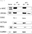
- umbilical cord blood-derived pluripotent/multipotent stem cells of the present invention remain undifferentiated is evident from the expression of Oct-4, Sox-2, Rex-1, c-myc, and ZNF281 in the stem cells.
- the umbilical cord blood-derived pluripotent/multipotent stem cells of the present invention is highly proliferative.
- a karyotype analysis demonstrated that the cells of the present invention rapidly proliferate, but have a normal chromosomal structure.
- the umbilical cord blood-derived pluripotent/multipotent stem cells of the present invention are immunologically negative to CD14, CD31, CD34, CD45 and HLA-DR, all known as hematopoietic stem cell markers or immunorejection-related markers. Due to the lack of such hematopoiesis- or immunorejection-related markers, the umbilical cord blood-derived stem cells of the present invention can be transplanted with minimal vascularization and immunorejection and thus can be effectively used in allogenic transplantation.
- the umbilical cord blood-derived pluripotent/multipotent stem cells of the present invention also may differentiate into hepatocytes from the mesoderm and neurons and retina-related cells from the ectoderm as well as osteoblasts, chondrocytes and adipose cells from the mesoderm. Therefore, the umbilical cord blood-derived pluripotent/multipotent stem cells of the present invention can be used to treat a variety of diseases.
- the umbilical cord blood-derived pluripotent /multipotent stem cells of the present invention may secrete various cytokines or chemokines including TIMP-2, TGF- ⁇ , RANTES CINC-3, EOTAXIN, GM-CSF, IFN- ⁇ , IL-1b, IL-3, IL-6, IL-8, IL-10, IL12p40, IL13, IL-16, IP-10, Leptin, MCP-2, MIG, MIP-3a, b-NGFm, sTNFRI, and PFGF-bb.
- the umbilical cord blood-derived pluripotent/multipotent stem cells of the present invention can be applied to the treatment of various diseases.
- the novel nature of the umbilical cord blood-derived pluripotent/multipotent stem cells of the present invention comes from their having these characteristics.
- the umbilical cord blood-derived pluripotent/multipotent stem cells of the present invention can be differentiated into various types of cells including osteoblasts, chondrocytes, adipose cells, hepatocytes and neurons, and thus find applications in therapies for various corresponding diseases. Therefore, the present invention provides a cell therapeutic agent comprising the umbilical cord blood-derived pluripotent/multipotent stem cells of the present invention or the cells differentiated therefrom.
- the cell therapeutic agent of the present invention can be applied to the treatment of various diseases including, for instance, neural diseases (e.g., degenerative neural diseases), osteoarthritis (e.g., degenerative arthritis, rheumatoid arthritis), bone loss (e.g., osteoporosis), hepatic diseases (e.g., hepatocirrhosis), and cardiovascular diseases.
- neural diseases e.g., degenerative neural diseases
- osteoarthritis e.g., degenerative arthritis, rheumatoid arthritis
- bone loss e.g., osteoporosis
- hepatic diseases e.g., hepatocirrhosis
- cardiovascular diseases e.g., cardiovascular diseases.
- the cell therapeutic agent as described contains at least one diluent that can protect and maintain the cells.
- Buffered solutions such as physiological saline, PBS (Phosphate Buffered Saline), HBSS (Hank's balanced salt solution), and plasma or serum components may be used as the diluents.
- the present invention relates to the use of a medium for culturing the novel stem cells.
- the medium is based on the EGM-2 or SNU-1 medium and supplemented with fetal bovine serum (FBS) 20%, bFGF (Fibroblast Growth Factor) 1 ⁇ 40ng/ml, ascorbic acid 0.1 ⁇ 5.0 ⁇ g/ml, EGF (Epidermal Growth Factor) 1 ⁇ 40ng/ml, hydrocortisone 0.1 ⁇ 1 ⁇ g/ml, IGF-I (Insulin-like Growth Factor-1) 1 ⁇ 40ng/ml or VEGF (Vascular Endothelial Growth Factor) 1 ⁇ 5ng/ml and heparin 20 ⁇ 25 ⁇ g/ml, and optionally with GA-1000 (Gentamycin Sulfate, Amphotericin-B) if needed.
- FBS fetal bovine serum
- bFGF Fibroblast Growth Factor 1 ⁇ 40ng/ml
- ascorbic acid 0.1 ⁇ 5.0
- the culturing medium for stem cells is novel and is used in the method for isolating umbilical cord blood-derived pluripotent/multipotent stem cells according to the present invention. Further, the culture medium of the present invention is useful for proliferating all adult stem cells including umbilical cord blood-derived stem cells and is applicable to the culturing of adult stem cells.
- the present invention addresses a method for culturing stem cells comprising culturing and proliferating stem cells in the medium of the present invention.
- the stem cells may be adult stem cells.
- the medium for culturing stem cells in accordance with the present invention may be used to culture the umbilical cord blood-derived pluripotent/multipotent stem cells of the present invention.
- the umbilical cord blood-derived pluripotent/multipotent stem cells of the present invention may be preferably passaged 3-5 days after the formation of cell colonies in a spindle form. Cultivation is preferably performed in a 5% CO 2 condition and may be carried out for 5 ⁇ 30 days, but the present invention is not limited to these.
- stem cells characterized by a sphere culture or a three-dimensional culture of stem cells whereby stem cells are cultured.
- MEF Mae embryonic fibroblast cell
- the stem cells may preferably be adult stem cells.
- increasing sternness means forming an embryonic stem cell-like colony or expressing transcription factors such as Oct4, Sox2, etc. at a higher level.
- the human umbilical cord blood-derived pluripotent/multipotent stem cells of the present invention When cultured in the presence of fibronectin, as explained in detail above, the human umbilical cord blood-derived pluripotent/multipotent stem cells of the present invention actively proliferate for a longer period of time without differentiating, compared to conventional adult stem cells.
- the pluripotent/multipotent stem cells of the present invention can be effectively applied to the treatment of conventionally incurable diseases, as well as neural diseases, cardiovascular diseases, and diseases of the skeletal system.
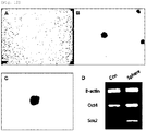
- FIGS. 12 and 13 are photographs showing three-dimensional cultures of the umbilical cord blood-derived pluripotent/multipotent stem cells using a sphere culture process and STO cells and the expression patterns of the transcription factors Oct4 and Sox2 as measured by RT-PCR
- FIG. 12 a three-dimensional culture of umbilical cord blood-derived pluripotent/multipotent stem cells by a sphere culture process
- FIG. 13 a three-dimensional culture of umbilical cord blood-derived pluripotent/multipotent stem cells on STO cells.
- EXAMPLE 1 Culturing, Proliferation and Karyotype Analysis of the Umbilical Cord Blood-Derived Pluripotent/Multipotent Stem Cells
- the full term UCB was mixed with HetaSep (Stem cells Technologies INC, Vancouver, BC) to deplete erythrocyte counts. Then, mononuclear cells were obtained by a typical Ficoll density gradient centrifugation. The monocytes were seeded at a density of 1 x 10 5 ⁇ 8 cells/well on 6-well plates coated with 0.1mg/ml - 1 mg/ml fibronectin and maintained in SNU-1 or EGM-2 (Lonza) supplemented with EGM-2 SingleQuots containing 20% FBS.
- the EGM-2 SingQouts contained heparin, Ascorbic acid, rhEGF, hydrocortisone, VEGF, rhFGF-B, R 3 -IGF-1, and GA-1000.
- the monocytes which still remained non-adherent three days after culturing were removed while the medium was replaced with a fresh one every two or three days.
- FIG. 1A The adherent cells were observed to form colonies, exhibiting spindle-shaped morphology 5-30 days after being cultured under the condition of 5% CO 2 ( FIG. 1A ). Once formed, the colonies rapidly proliferated. The cells were suspended with 0.125% Trypsin-EDTA 3 ⁇ 7 days after the formation of colonies, and transferred to fresh dishes where the cells continued to be maintained ( FIGS. 1B, 1C, 1D, 1E and 1F).
- FIG. 1 is of photographs showing the culture progress of the cells. As seen in FIG. 1 , the cell colony grew larger with the lapse of time. In addition even after passage, the morphology of the cells stayed uniform ( FIG.1 . isolation and proliferation of the umbilical cord blood-derived pluripotent/multipotent stem cells. Colonies formed after the mononuclear cells were cultured for 14 days (A), 15 days (B), 16 days (C), 17 days (D) and 18 days (E), and cells after passage 3 (F)).
- umbilical cord blood-derived pluripotent/multipotent stem cells of three kinds were isolated from different samples of umbilical cord blood and were seeded at a density of 2x10 5 cells on 100 dishes and passaged at regular intervals of three to four days while being counted using a cytometer. Cell counting was performed until the cells stopped growing.
- CPDL values were calculated while the cells continued to be maintained.
- hUCB-EPC human umbilical cord blood derived endothelial progenitor cells
- FIG. 2 The results are shown in FIG. 2 .
- hUCB-EPC had a CPDL of about 20 over two months whereas the CPDL of all the umbilical cord blood-derived pluripotent/multipotent stem cells from three different samples were observed to range between 40 and 45.
- chromosomes of the umbilical cord blood-derived pluripotent/multipotent stem cells isolated according to the present invention are normal.
- a karyotype analysis was performed to examine the chromosomes of the cells. As can be seen in FIG. 3 , the cells were found to have a normal chromosomal structure even after passage 10.
- Flow cytometry was carried out to analyze the characteristics of the cells suspended in media.
- FITC fluorescein isothiocyanate
- PE phycoerythrin
- the surface antigens used to analyze the characteristics of the umbilical cord blood-derived pluripotent/multipotent stem cells (partially pluripotent/multipotent stem cell) isolated in the present invention included CD10 (T cell marker), CD14 (monocyte marker), CD24 (epithelial cell marker), CD29 (monocyte marker), CD31 (endothelial cell marker), CD34 (hematopoietic stem cell marker), CD44 (mesenchymal stem cell marker), CD45 (non-hematopoietic stem cell marker), CD51/61 (osteoclast marker), CD73 (mesenchymal stem cell marker), CD90 (mesenchymal stem cell marker), CD105 (mesenchymal stem cell marker), CD133 (hematopoietic stem cell marker), and HLA-DR(immunorejection-related marker), and were analyzed using a flow cytometer.
- EXAMPLE 3 Expression Pattern of ZNF281 and Core transcription factor in the Umbilical Cord Blood-Derived Pluripotent/Multipotent Stem Cells
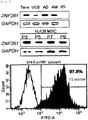
- ZNF281 (Zinc finger protein 281) is one of the core transcription factors of ESC ( Wang J et al. (2006) Nature 444, 364-368 ). ZNF281, also called ZBP-99, contains four Kruppel-type zinc fingers that collectively share 91% amino acid sequence similarity and 79% sequence identity with those found in ZBP-89. In addition, there are highly conserved amino acid sequences in the carboxy-terminal segments of the two genes. The predicted open reading frame of ZNF281 cDNA encodes a 99-kDa protein.
- EMSA Electrophoretic mobility shift as say
- ZNF281 protein specifically binds to the GC-rich promoter elements of GASTRIN and ORNITHINE DECARBOXYLASE genes ( Law DJ et al. (1999) Biochem Biophys Res Commun 262, 113-120 ; Lisowsky T et a 1. (1999) FEBS Lett 453, 369-374 ).
- ZNF281 was identified as a c-MYC-associated protein by mass spectral multidimensional protein identification technology and tandem affinity purification ( Koch HB et al. (2007) Cell Cycle 6, 205-217 .).
- Oct3/4 genes such as POU family transcription factors, are known to not exist in differentiated tissues, but to be expressed particularly in undifferentiated stem cells which have a high proliferative capacity ( Tai M-H. et al., Carcinogenesis 26:4 95, 2005 ; Tondreau T. et al., Stem cells, 23:1105, 2005 ).
- Oct3/4 are therefore used as markers for embryonic stem cells and also as markers indicative of undifferentiation.
- the cell colonies were stained using Oct4 as a marker for stemness. As a result, many cells were found to have Oct4 stained around the nucleus. Flow cytometry also demonstrated the Oct4 expression in the cells.
- the cells were fixed at 4°C overnight with 4% formaldehyde and permeabilized for 10 min with 0.1% Triton X-100 (Sigma-Aldrich).
- the slides and the dishes were incubated for one hour with an anti-human Oct4 mouse primary antibody (1:200), washed with PBS (phosphate buffered saline; Gibco), immunostained for one hour with an Alexa594-conjugated, goat anti-mouse IgG secondary antibody (Invitrogen) while the nucleus was countstained with DAPI.
- FIGS. 5 and 6 many of the umbilical cord blood-derived pluripotent/multipotent stem cells were found to express Oct4 ( FIG. 5A : flow cytogram of Oct4-expressing cells, B: image of immunostained Oct4 C: image of the nucleus stained in Oct4 expressed cells, D: overlapped image of Oct4 expression and nucleus staining).
- the cells were induced to differentiate into osteoblasts.
- the cells were allowed to adhere to culture dishes and incubated to about 70-80% confluency.
- the culture medium was replaced with an osteogenic differentiation-inducing medium.
- the osteogenic differentiation-inducing medium was prepared by supplementing DMEM low glucose with 10% FBS plus 10 mM beta-glycerophosphate (Sigma-Aldrich), 0.1 ⁇ M D examethasone (Sigma-Aldrich), and 50 ⁇ M ascorbate (Sigma-Aldrich).
- the medium was replaced with a fresh one every third day while differentiation was induced for about two weeks.
- the calcium mineralization attributed to the osteogenic differentiation was examined by Alizarin red S staining.
- the staining was conducted as follows. After the medium was removed, the cells were washed twice with distilled water and fixed at 4°C for one hour in cold 70% EtOH. Then, the cells were washed again twice with distilled water and stained at room temperature for 10 min with 40mM Alizarin red S, followed by washing five times with distilled water.
- FIGS. 8A and 8B in FIG. 8A no calcium was detected by Alizarin red S in the absence of differentiation whereas in FIG. 8B calcium appeared red when the cells were induced to differentiate into osteoblasts, which indicates that the umbilical cord blood-derived pluripotent/multipotent stem cells differentiated into osteoblasts releasing calcium.
- the cells were induced to differentiate into adipose cells.
- the cells were allowed to adhere to culture dishes and cultured to about 70-80% confluency.
- the medium was replaced with an adipocyte differentiation medium.
- the adipocyte differentiation medium was prepared by supplementing DMEM low glucose with 10% FBS plus 1 ⁇ M Dexamethasone, 10 ⁇ g/ml insulin (Sigma-Aldrich), 0.5 mM 3-isobutyl-1-methylxanthine (Sigma-Aldrich), and 0.2 mM indomethacin (Sigma-Aldrich).
- the differentiation medium was replaced with a fresh one every third day while differentiation was induced for about 2-3 weeks.
- adipocyte differentiation was examined by Oil red O staining.
- the medium was removed and the cells were washed in PBS and incubated at room temperature for 5 min in 10% formalin.
- the formalin was replaced with the same volume of a fresh one, followed by fixing the cells at room temperature for at least one hour.
- the cells were washed with 60% isopropanol.
- the alcohol was completely evaporated before the cells were stained at room temperature for 10 min with Oil Red O. Immediately after this dye was removed, the cells were washed in distilled water.
- Adipocytes appear red upon treatment with Oil red O because it stains lipid droplets red.
- Oil red O because it stains lipid droplets red.
- FIGS. 8C and 8D in FIG. 8C neither lipid droplets nor the red appearance was detected in the absence of adipocyte differentiation whereas in FIG. 8D when induced to differentiate into adipocytes, the cells were observed to have many lipid droplets and thus turn red).
- the cells were treated for three weeks with the rTGF-beta 3-containing chondrogenic differentiation medium (PT-3003) from Lonza, while the medium was replaced twice a week with a fresh one. Chondrogenesis was measured once a week.
- PT-3003 the rTGF-beta 3-containing chondrogenic differentiation medium
- toluidine blue staining was conducted. The cells were fixed for 10 hours with 4% formaldehyde and then for an additional 10 hours with picric acid. After cryosection, the cells were stained for 3 min with toluidine blue and counterstained for 3 sec staining with hematoxilin.
- a pellet of chondrocytes does not collapse, but maintains a constant morphology and appears blue when stained with toluidine blue.
- FIGS. 8E and 8F the cells subjected to chondrogenic differentiation were observed to be stained blue and maintain their morphology.
- EXAMPLE 7 Change in Gene Expression Level after Differentiation of the Umbilical Cord Blood-Derived Pluripotent/Multipotent Stem Cells into Osteoblasts and Adipocytes
- the cells were induced to differentiate into neurons.
- the cells were pre-incubated for 24 hours in DMEM supplemented with 5% FBS and 10ng/ml bFGF (basic Fibroblast Growth Factor). Thereafter, the cells were treated for 24 hours in DMEM containing 1% DMSO, 100 ⁇ M BHA, 0.5mM VPA, 10mM KCl, and 10 ng/ml NGF, and B27 to induce neural differentiation.
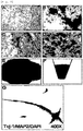
- the cells were fixed with 4% paraformaldehyde and immunostained for the neural markers Tuj-1, MAP-2, GFAP, and Neurofilament-160. As a result, the four markers were observed to be expressed ( FIG. 8G ).
- the retina-related characteristics of the umbilical cord blood-derived pluripotent/multipotent stem cells were examined using immunofluorescence.
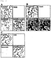
- FIG. 10 is of photographs showing expression patterns of retina-specific proteins as measured by immunofluorescence.
- the expression patterns of PAX6 and Hu protein are shown in FIG. 10A .
- PAX6 is known as a retina progenitor marker and Hu protein is expressed specifically in gaglion cells and amacrine cells, which are constituents of the retina. They were not detected in a normal culture.
- opsin and rhodopsin are detected. Opsin is expressed specifically in cone cells while rhodposin is specific for rod cells. In a normal culture, opsin was not detected, but expression of rhodopsin was observed.
- CRX is known as a pan-photoreceptor marker
- the therapeutic capacity of stem cells is largely attributable to two effects: first, direct differentiation of stem cells into impaired cells; and secondly, the ability to secrete various cytokines or growth factors that induce positive alterations that lead to a therapeutic effect on pre-existing cells.
- stem cells are known to secrete various cytokines or growth factors, showing so-called paracrine effects ( Kim et al. Cytokine. 2005 ).
- a human Cytokine antibody array (RaybioTech. Norc ross, USA) was used.
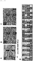
- FIG. 11 shows array analysis results of hUCB-MSC1(A), hUCB-MSC2 (B) and hUCB-MSC3 (C) and an arrangement of antibodies (D)).
- EXAMPLE 11 3-Dimensional Culture of the Umbilical Cord Blood-Derived Pluripotent/Multipotent Stem Cells
- the culture dishes were coated with 0.7% agarose to a thickness of 5 mm or more so that the cells could not adhere to the bottom, but formed spheres.
- the cells were seeded at a density of less than 2000 cells/cm 2 to prevent adhesion between single cells.
- the spheres thus formed were separated from single cells using a 40 ⁇ m strainer. As can been seen in FIG.
- the stem cells did not undergo cell death, but formed spheres, maintaining the characteristics of stem cells when they were cultured in a sphere culture system.
- the expression levels of embryonic markers such as OCT4, SOX2 and the like in cells that were maintained in a sphere culture were observed to be higher compared to those which were maintained as a monolayer.
- Embryonic stem cells are typically cultured on a layer of mouse embryonic fibroblast cells because the kemokines, such as LIF, from the mouse embryonic fibroblast cells play an important role in maintaining the morphology of ES cells and preventing the differentiation of ES cells.
- the umbilical cord blood-derived pluripotent/multipotent stem cells did not grow in a flat shape like typical adult stem cells, but formed 3-dimensional colonies.
- STO cells were seeded at a density of 2 x 10 5 cells/ml on 0.1% gelatin-coated dishes and incubated for 24 hours, followed by seeding the umbilical cord blood-derived pluripotent/multipotent stem cells on the STO cell layer. As seen in FIG. 13 , the cells were observed to proliferate on the STO cells in a pattern which was similar to that of embryonic stem cell with the lapse of time.
- the human umbilical cord blood-derived pluripotent/multipotent stem cells of the present invention When cultured in the presence of fibronectin, the human umbilical cord blood-derived pluripotent/multipotent stem cells of the present invention actively proliferate for a longer period of time without differentiating, compared to conventional adult stem cells.
- the pluripotent/multipotent stem cells of the present invention can be effectively applied to the treatment of conventionally incurable diseases, as well as neural diseases, cardiovascular diseases, and diseases of the skeletal system.
Landscapes
- Health & Medical Sciences (AREA)
- Life Sciences & Earth Sciences (AREA)
- Engineering & Computer Science (AREA)
- Biomedical Technology (AREA)
- Organic Chemistry (AREA)
- Bioinformatics & Cheminformatics (AREA)
- Chemical & Material Sciences (AREA)
- Genetics & Genomics (AREA)
- Zoology (AREA)
- Wood Science & Technology (AREA)
- Biotechnology (AREA)
- General Health & Medical Sciences (AREA)
- Microbiology (AREA)
- Cell Biology (AREA)
- General Engineering & Computer Science (AREA)
- Biochemistry (AREA)
- Developmental Biology & Embryology (AREA)
- Hematology (AREA)
- Reproductive Health (AREA)
- Gynecology & Obstetrics (AREA)
- Nuclear Medicine, Radiotherapy & Molecular Imaging (AREA)
- Medicinal Chemistry (AREA)
- Chemical Kinetics & Catalysis (AREA)
- General Chemical & Material Sciences (AREA)
- Pharmacology & Pharmacy (AREA)
- Veterinary Medicine (AREA)
- Public Health (AREA)
- Animal Behavior & Ethology (AREA)
- Pregnancy & Childbirth (AREA)
- Immunology (AREA)
- Neurology (AREA)
- Physical Education & Sports Medicine (AREA)
- Neurosurgery (AREA)
- Heart & Thoracic Surgery (AREA)
- Cardiology (AREA)
- Micro-Organisms Or Cultivation Processes Thereof (AREA)
- Medicines Containing Material From Animals Or Micro-Organisms (AREA)
Claims (10)
- Procédé d'isolement de cellules souches pluripotentes ou multipotentes dérivées de sang de cordon ombilical exprimant ZNF281, comprenant les étapes consistant à :(a) mettre en culture des cellules mononucléées isolées à partir de sang de cordon ombilical dans un récipient de culture recouvert de fibronectine et contenant un milieu EGM-2™ ou SNU-1 tel que défini dans le tableau 1 avec du sérum bovin foetal (FBS), du bFGF, de l'acide ascorbique, de l'EGF, de l'hydrocortisone, de l'IGF-I, VEGF et de l'héparine ; et(b) récolter les cellules souches cultivées à partir de la culture, dans lequel la cellule souche est dans un état indifférencié.
- Procédé selon la revendication 1, dans lequel les cellules mononucléées sont isolées en mélangeant du sang de cordon ombilical avec de l'Hetasep™ pour éliminer les érythrocytes et ensuite en utilisant du Ficoll®-paque.
- Procédé selon la revendication 1, dans lequel les cellules souches sont récoltées à partir de colonies de cellules formées 5 à 30 jours après leur mise en culture.
- Procédé selon la revendication 1, dans lequel la fibronectine est déposée en couches sur le récipient de culture ou contenue sous la forme de sphères ou de structures tridimensionnelles dans un milieu de culture.
- Procédé selon la revendication 1, dans lequel les cellules mononucléées sont cultivées dans un milieu EGM-2™ ou SNU-1 tel que défini dans le tableau 1 additionné de sérum bovin foetal (FBS), de 1 à 40 ng/ml de bFGF, de 0,1 à 5,0 µg/ml d'acide ascorbique, de 1 à 40 ng/ml d'EGF, de 0,1 à 1 µg/ml d'hydrocortisone, de 1 à 40 ng/ml d'IGF-I, de 1 à 5 ng/ml de VEGF et de 20 à 25 µg/ml d'héparine.
- Cellules souches pluripotentes ou multipotentes dérivées de sang de cordon ombilical présentant une caractéristique immunologique positive au facteur de transcription ZNF281 et une caractéristique immunologique négative au CD14, dans lesquelles les cellules souches peuvent être obtenues par culture dans un milieu EGM-2™ ou SNU-1 tel que défini dans le tableau 1 additionné de sérum bovin foetal (FBS), de 1 à 40 ng/ml de bFGF, de 0,1 à 5,0 µg/ml d'acide ascorbique, de 1 à 40 ng/ml d'EGF, de 0,1 à 1 µg/ml d'hydrocortisone, de 1 à 40 ng/ml d'IGF-I, de 1 à 5 ng/ml de VEGF et de 20 à 25 µg/ml d'héparine.
- Cellules souches dérivées de sang de cordon ombilical selon la revendication 6, isolées en utilisant le procédé selon l'une quelconque des revendications 1 à 5, ayant au moins l'une des propriétés suivantes :(a) montrant une caractéristique immunologique positive au facteur de transcription c-myc ;(b) adhérant à une surface recouverte de matrice extracellulaire et formant des colonies de cellules sous une forme de tige ou sphérique 5 à 30 jours après l'adhésion ;(c) montrant un CPDL (niveau de doublement cumulatif de population) de 30 à 45 ;(d) présentant une caractéristique immunologique négative vis-à-vis de CD31, CD34, CD45 et HLA-DR ;(e) aptitude à se différencier en cellules mésodermiques, endodermiques et ectodermiques ; et(f) sécrétant au moins une cytokine ou une chimiokine choisie dans le groupe comprenant TIMP-2, TGF-β, RANTES CINC-3, EOTAXIN, GM-CSF, IFN-γ, IL-1b, IL-3, IL-6, IL-8, IL-10, IL12p40, IL13, IL-16, IP-10, leptine, MCP-2, MIG, MIP-3a, b-NGFm, sTNFRI et PFGF-bb.
- Utilisation d'un milieu de culture pour cellules souches comprenant un milieu EGM-2™ ou SNU-1 tel que défini dans le tableau 1, additionnée avec du sérum bovin foetal (FBS), de 1 à 40 ng/ml de bFGF, de 0,1 à 5,0 µg/ml d'acide ascorbique, de 1 à 40 ng/ml d'EGF, de 0,1 à 1 µg/ml d'hydrocortisone, de 1 à 40 ng/ml d'IGF-I, de 1 à 5 ng/ml de VEGF et 20 à 25 µg/ml d'héparine dans un procédé selon la revendication 1.
- Procédé selon la revendication 1, comprenant en outre l'étape consistant à augmenter le caractère souche de cellules souches, en utilisant une culture en sphère ou une culture en trois dimensions de cellules souches.
- Procédé selon la revendication 9, dans lequel une MEF (cellule fibroblaste d'embryon de souris) est utilisée pour la culture en trois dimensions.
Priority Applications (1)
| Application Number | Priority Date | Filing Date | Title |
|---|---|---|---|
| PL10753642T PL2410046T3 (pl) | 2009-03-20 | 2010-03-03 | Sposób izolowania pluripotencjalnych komórek macierzystych pochodzących z krwi pępowinowej wykazujących ekspresję ZNF281 |
Applications Claiming Priority (2)
| Application Number | Priority Date | Filing Date | Title |
|---|---|---|---|
| KR1020090023821A KR100950195B1 (ko) | 2009-03-20 | 2009-03-20 | Znf281을 발현하는 제대혈 유래 만능 줄기세포의 분리방법 |
| PCT/KR2010/001338 WO2010107192A2 (fr) | 2009-03-20 | 2010-03-03 | Procédé d'isolement et procédé de culture en masse pour des cellules souches pluripotentes dérivées de sang de cordon ombilical exprimant znf281 |
Publications (3)
| Publication Number | Publication Date |
|---|---|
| EP2410046A2 EP2410046A2 (fr) | 2012-01-25 |
| EP2410046A4 EP2410046A4 (fr) | 2013-02-27 |
| EP2410046B1 true EP2410046B1 (fr) | 2019-01-23 |
Family
ID=42183944
Family Applications (1)
| Application Number | Title | Priority Date | Filing Date |
|---|---|---|---|
| EP10753642.7A Active EP2410046B1 (fr) | 2009-03-20 | 2010-03-03 | Procédé d'isolement et procédé de culture en masse pour des cellules souches pluripotentes dérivées de sang de cordon ombilical exprimant znf281 |
Country Status (11)
| Country | Link |
|---|---|
| US (1) | US10584312B2 (fr) |
| EP (1) | EP2410046B1 (fr) |
| JP (2) | JP5995720B2 (fr) |
| KR (1) | KR100950195B1 (fr) |
| CN (1) | CN102395673B (fr) |
| AU (1) | AU2010225612B2 (fr) |
| ES (1) | ES2722930T3 (fr) |
| MY (1) | MY160164A (fr) |
| PL (1) | PL2410046T3 (fr) |
| RU (1) | RU2511417C2 (fr) |
| WO (2) | WO2010107159A1 (fr) |
Cited By (1)
| Publication number | Priority date | Publication date | Assignee | Title |
|---|---|---|---|---|
| US11674090B1 (en) | 2021-11-30 | 2023-06-13 | Honeywell International Inc. | Energy optimization in fluid catalytic cracking and dehydrogenation units |
Families Citing this family (23)
| Publication number | Priority date | Publication date | Assignee | Title |
|---|---|---|---|---|
| KR100950195B1 (ko) * | 2009-03-20 | 2010-03-29 | 서울대학교산학협력단 | Znf281을 발현하는 제대혈 유래 만능 줄기세포의 분리방법 |
| JP5833126B2 (ja) * | 2010-09-08 | 2015-12-16 | カンステム バイオテック カンパニー リミテッド | ウマ科動物の羊水由来の多分化能幹細胞及びそれを製造する方法 |
| US8940294B2 (en) | 2012-03-02 | 2015-01-27 | Tissuetech, Inc. | Methods of isolating and culturing stem cells |
| CN103396995B (zh) * | 2013-07-16 | 2015-09-23 | 广州赛哲生物科技有限公司 | 一种筛选乳腺癌干细胞的三维培养方法 |
| PL3064572T3 (pl) * | 2013-11-01 | 2020-07-13 | Bbhc Co. Ltd. | Sposób wytwarzania indukowanych pluripotencjalnych komórek macierzystych z mezenchymalnych komórek macierzystych i indukowane pluripotencjalne komórki macierzyste wytworzone tym sposobem |
| RU2729396C2 (ru) * | 2015-07-14 | 2020-08-06 | МЕДИММЬЮН, ЭлЭлСи | Композиции и способы лечения рака |
| US10384207B2 (en) | 2015-07-21 | 2019-08-20 | Neuro Probe Incorporated | Assay apparatus and methods |
| US20210355437A1 (en) | 2015-09-02 | 2021-11-18 | Restem Llc | Factor rich product from umbilical cord mesenchymal stem cells |
| CN105087494A (zh) * | 2015-09-11 | 2015-11-25 | 中国人民解放军第四军医大学 | 一种乳腺癌干细胞的培养方法 |
| KR102676761B1 (ko) * | 2015-12-03 | 2024-06-18 | 다카라 바이오 가부시키가이샤 | 중간엽 줄기세포의 제조방법 |
| JP6912789B2 (ja) * | 2016-03-16 | 2021-08-04 | 株式会社日本触媒 | 神経幹細胞の培養方法、およびニューロスフェロイドの形成方法 |
| EP3490572A4 (fr) * | 2016-08-01 | 2020-05-13 | Embryogenesis Pty Ltd | Formulation pour le soin pour la peau |
| EP4600348A3 (fr) | 2016-08-26 | 2025-12-10 | Restem Llc | Composition et procédés d'utilisation de cellules souches de revêtement de cordon ombilical |
| KR101798217B1 (ko) | 2016-10-21 | 2017-11-15 | 차의과학대학교 산학협력단 | 만능줄기세포로부터 유래된 중간엽줄기세포를 선별하는 방법 |
| RU2645255C1 (ru) * | 2016-12-16 | 2018-02-19 | Федеральное государственное бюджетное учреждение "Национальный медицинский исследовательский центр акушерства, гинекологии и перинатологии имени академика В.И.Кулакова" Министерства здравоохранения Российской Федерации | Способ получения биобезопасной культуры мезенхимальных стволовых клеток из ворсин хориона человека |
| CN109666630A (zh) * | 2017-10-17 | 2019-04-23 | 澳门大学 | 多能干细胞分化为间充质干细胞的方法及其培养基和应用 |
| CN109112099A (zh) * | 2018-08-30 | 2019-01-01 | 丰泽康生物医药(深圳)有限公司 | 一种提高单核细胞向多潜能细胞转化的无血清培养基 |
| CN111424014B (zh) * | 2019-11-22 | 2022-03-29 | 上海交通大学医学院附属第九人民医院 | 人颈静脉球副神经节瘤永生化细胞株培养基 |
| TW202208613A (zh) * | 2020-05-05 | 2022-03-01 | 美商加速生物科學有限公司 | 非永生的多潛能幹細胞 |
| CN112430626A (zh) * | 2020-11-27 | 2021-03-02 | 成都康景生物科技有限公司 | 一种基因修饰的脐间充质干细胞、制备方法及应用 |
| EP4257674A4 (fr) * | 2020-12-04 | 2024-11-27 | Primoris Therapeutics Co., Ltd | Procédé d'isolement et de culture de cellules souches de sang de cordon exprimant gdf-3 à un niveau élevé, et utilisation de gdf -3 |
| WO2023163560A1 (fr) | 2022-02-25 | 2023-08-31 | 주식회사 강스템바이오텍 | Procédé de culture de cellules souches à l'aide d'un microsupport |
| JP2025508482A (ja) | 2022-02-25 | 2025-03-26 | カンステム バイオテック カンパニー リミテッド | 無血清培地を用いた幹細胞の培養方法 |
Family Cites Families (16)
| Publication number | Priority date | Publication date | Assignee | Title |
|---|---|---|---|---|
| US6642049B1 (en) * | 1998-12-04 | 2003-11-04 | The United States Of America As Represented By The Secretary Of The Navy | Human brain endothelial cells and growth medium and method for expansion of primitive CD34+CD38-bone marrow stem cells |
| WO2002064748A2 (fr) * | 2001-02-14 | 2002-08-22 | Furcht Leo T | Cellules souches adultes totipotentes, sources de ces cellules, procedes d'obtention et de maintien de ces dernieres, procedes de differentiation de ces cellules, procedes d'utilisation correspondants et cellules derivees des cellules susmentionnees |
| US6610535B1 (en) | 2000-02-10 | 2003-08-26 | Es Cell International Pte Ltd. | Progenitor cells and methods and uses related thereto |
| US20040005559A1 (en) * | 2000-07-24 | 2004-01-08 | Loring Jeanne F. | Markers of neuronal differentiation and morphogenesis |
| US7736892B2 (en) * | 2002-02-25 | 2010-06-15 | Kansas State University Research Foundation | Cultures, products and methods using umbilical cord matrix cells |
| TWI288779B (en) * | 2002-03-28 | 2007-10-21 | Blasticon Biotech Forschung | Dedifferentiated, programmable stem cells of monocytic origin, and their production and use |
| WO2004033396A2 (fr) * | 2002-10-08 | 2004-04-22 | American National Red Cross | Procede d'enrichissement de populations de monocytes adherents |
| US20030224411A1 (en) * | 2003-03-13 | 2003-12-04 | Stanton Lawrence W. | Genes that are up- or down-regulated during differentiation of human embryonic stem cells |
| WO2006071794A2 (fr) * | 2004-12-23 | 2006-07-06 | Ethicon Incorporated | Cellules postnatales derivees de tissus du cordon ombilical, et procedes de production et d'utilisation de celles-ci |
| WO2005056778A1 (fr) | 2003-12-11 | 2005-06-23 | Riken | Procede de regulation de differenciation des cellules souches hematopoietiques ou de leur proliferation |
| WO2006012404A2 (fr) * | 2004-07-20 | 2006-02-02 | Case Western Reserve University | Nouvelles populations cellulaires et utilisations de celles-ci |
| KR100697326B1 (ko) * | 2005-12-02 | 2007-03-20 | 재단법인서울대학교산학협력재단 | Oct4 발현능을 가지는 제대혈 유래 다분화능 성체줄기세포 및 그의 제조방법 |
| WO2008110570A1 (fr) | 2007-03-13 | 2008-09-18 | Medizinische Universität Graz | Procédé pour étudier la prolifération de cellules progénitrices endothéliales et l'influence potentielle de composés sur leur comportement de prolifération |
| RU2343928C1 (ru) * | 2007-04-03 | 2009-01-20 | Открытое акционерное общество "Институт стволовых клеток человека" | Способ получения ядросодержащих клеток из пуповинной крови |
| TW200916583A (en) * | 2007-06-08 | 2009-04-16 | Novathera Ltd | Cell expansion |
| KR100950195B1 (ko) | 2009-03-20 | 2010-03-29 | 서울대학교산학협력단 | Znf281을 발현하는 제대혈 유래 만능 줄기세포의 분리방법 |
-
2009
- 2009-03-20 KR KR1020090023821A patent/KR100950195B1/ko active Active
- 2009-05-01 WO PCT/KR2009/002333 patent/WO2010107159A1/fr not_active Ceased
-
2010
- 2010-03-03 US US13/257,961 patent/US10584312B2/en active Active
- 2010-03-03 RU RU2011142170/10A patent/RU2511417C2/ru active
- 2010-03-03 CN CN201080016872.7A patent/CN102395673B/zh active Active
- 2010-03-03 EP EP10753642.7A patent/EP2410046B1/fr active Active
- 2010-03-03 JP JP2012500708A patent/JP5995720B2/ja active Active
- 2010-03-03 PL PL10753642T patent/PL2410046T3/pl unknown
- 2010-03-03 WO PCT/KR2010/001338 patent/WO2010107192A2/fr not_active Ceased
- 2010-03-03 AU AU2010225612A patent/AU2010225612B2/en active Active
- 2010-03-03 ES ES10753642T patent/ES2722930T3/es active Active
- 2010-03-03 MY MYPI2011004459A patent/MY160164A/en unknown
-
2015
- 2015-01-05 JP JP2015000016A patent/JP6038962B2/ja active Active
Non-Patent Citations (1)
| Title |
|---|
| KWANG-WON SEO ET AL: "ZNF281 Knockdown Induced Osteogenic Differentiation of Human Multipotent Stem Cells In Vivo and In Vitro", CELL TRANSPLANTATION, vol. 22, no. 1, 1 January 2013 (2013-01-01), pages 29 - 40, XP055099615, ISSN: 0963-6897, DOI: 10.3727/096368912X654948 * |
Cited By (1)
| Publication number | Priority date | Publication date | Assignee | Title |
|---|---|---|---|---|
| US11674090B1 (en) | 2021-11-30 | 2023-06-13 | Honeywell International Inc. | Energy optimization in fluid catalytic cracking and dehydrogenation units |
Also Published As
| Publication number | Publication date |
|---|---|
| JP5995720B2 (ja) | 2016-09-21 |
| AU2010225612B2 (en) | 2014-06-26 |
| JP6038962B2 (ja) | 2016-12-07 |
| PL2410046T3 (pl) | 2019-10-31 |
| KR100950195B1 (ko) | 2010-03-29 |
| JP2012520671A (ja) | 2012-09-10 |
| CN102395673B (zh) | 2015-07-22 |
| RU2011142170A (ru) | 2013-04-27 |
| EP2410046A2 (fr) | 2012-01-25 |
| JP2015091254A (ja) | 2015-05-14 |
| WO2010107192A2 (fr) | 2010-09-23 |
| ES2722930T3 (es) | 2019-08-20 |
| RU2511417C2 (ru) | 2014-04-10 |
| AU2010225612A1 (en) | 2011-11-10 |
| US10584312B2 (en) | 2020-03-10 |
| EP2410046A4 (fr) | 2013-02-27 |
| WO2010107192A3 (fr) | 2011-02-24 |
| US20120021509A1 (en) | 2012-01-26 |
| CN102395673A (zh) | 2012-03-28 |
| MY160164A (en) | 2017-02-28 |
| WO2010107159A1 (fr) | 2010-09-23 |
Similar Documents
| Publication | Publication Date | Title |
|---|---|---|
| EP2410046B1 (fr) | Procédé d'isolement et procédé de culture en masse pour des cellules souches pluripotentes dérivées de sang de cordon ombilical exprimant znf281 | |
| Trivedi et al. | Derivation and immunological characterization of mesenchymal stromal cells from human embryonic stem cells | |
| Kang et al. | Advanced properties of urine derived stem cells compared to adipose tissue derived stem cells in terms of cell proliferation, immune modulation and multi differentiation | |
| EP2374871B1 (fr) | Cellules souches pluripotentes, procédé pour la préparation de celles-ci et utilisations de celles-ci | |
| GUO et al. | Biological features of mesenchymal stem cells from human bone marrow | |
| CN104204193B (zh) | 间充质干细胞的培养 | |
| US20100105132A1 (en) | Human Mesenchymal stem cells and preparation thereof | |
| Hsu et al. | Isolation of the multipotent MSC subpopulation from human gingival fibroblasts by culturing on chitosan membranes | |
| WO1997039104A1 (fr) | Cryoconservation et repiquage extensif de cellules souches mesenchymateuses d'origine humaine | |
| Sreejit et al. | Generation of mesenchymal stem cell lines from murine bone marrow | |
| US20170226472A1 (en) | Culture medium and method for enriching and maintaining cancer stem cells (cscs) using said medium | |
| Sato et al. | Serum-free isolation and culture system to enhance the proliferation and bone regeneration of adipose tissue-derived mesenchymal stem cells | |
| CN102884177A (zh) | Cd49f通过pi3k/akt/gsk3途径促进成体干细胞的增殖、专能性和重编程 | |
| JP7757793B2 (ja) | 間葉系幹細胞を含む生体由来細胞試料から間葉系幹細胞を製造する方法 | |
| Kim et al. | Characterization of human fetal cartilage progenitor cells during long-term expansion in a xeno-free medium | |
| US20130005036A1 (en) | Manufacturing process for fresh and frozen stem cells | |
| US20200131480A1 (en) | Method For Enhancing Proliferation Capability Of Stem Cells Using Ethionamide | |
| Kannaiyan et al. | Villous chorion: a potential source for pluripotent-like stromal cells | |
| KR101627907B1 (ko) | Znf281을 발현하는 제대혈 유래 줄기세포의 대량 배양방법 | |
| CN100453640C (zh) | 一种从脐带血中分离多能成体祖细胞的方法 | |
| KR20120030515A (ko) | Znf281을 발현하는 제대혈 유래 줄기세포의 대량 배양방법 | |
| US20080299656A1 (en) | Isolation of multi-lineage stem cells | |
| KR101158664B1 (ko) | Znf281을 발현하는 제대혈 유래 줄기세포의 대량 배양방법 | |
| Zhou et al. | Optimization of culture condition of human bone marrow stromal cells in terms of purification, proliferation, and pluripotency: Zhou et al. | |
| Singh et al. | In vitro Culture and Morphometry of Porcine Adipose Derived Mesenchymal Stem Cells (pAD-MSCs) |
Legal Events
| Date | Code | Title | Description |
|---|---|---|---|
| PUAI | Public reference made under article 153(3) epc to a published international application that has entered the european phase |
Free format text: ORIGINAL CODE: 0009012 |
|
| 17P | Request for examination filed |
Effective date: 20111019 |
|
| AK | Designated contracting states |
Kind code of ref document: A2 Designated state(s): AT BE BG CH CY CZ DE DK EE ES FI FR GB GR HR HU IE IS IT LI LT LU LV MC MK MT NL NO PL PT RO SE SI SK SM TR |
|
| DAX | Request for extension of the european patent (deleted) | ||
| A4 | Supplementary search report drawn up and despatched |
Effective date: 20130128 |
|
| RIC1 | Information provided on ipc code assigned before grant |
Ipc: C12N 5/07 20100101ALI20130122BHEP Ipc: C12N 5/02 20060101ALI20130122BHEP Ipc: C12N 5/071 20100101AFI20130122BHEP |
|
| 17Q | First examination report despatched |
Effective date: 20140206 |
|
| STAA | Information on the status of an ep patent application or granted ep patent |
Free format text: STATUS: EXAMINATION IS IN PROGRESS |
|
| GRAP | Despatch of communication of intention to grant a patent |
Free format text: ORIGINAL CODE: EPIDOSNIGR1 |
|
| STAA | Information on the status of an ep patent application or granted ep patent |
Free format text: STATUS: GRANT OF PATENT IS INTENDED |
|
| GRAS | Grant fee paid |
Free format text: ORIGINAL CODE: EPIDOSNIGR3 |
|
| INTG | Intention to grant announced |
Effective date: 20181121 |
|
| GRAA | (expected) grant |
Free format text: ORIGINAL CODE: 0009210 |
|
| STAA | Information on the status of an ep patent application or granted ep patent |
Free format text: STATUS: THE PATENT HAS BEEN GRANTED |
|
| AK | Designated contracting states |
Kind code of ref document: B1 Designated state(s): AT BE BG CH CY CZ DE DK EE ES FI FR GB GR HR HU IE IS IT LI LT LU LV MC MK MT NL NO PL PT RO SE SI SK SM TR |
|
| REG | Reference to a national code |
Ref country code: GB Ref legal event code: FG4D |
|
| REG | Reference to a national code |
Ref country code: CH Ref legal event code: EP |
|
| REG | Reference to a national code |
Ref country code: DE Ref legal event code: R096 Ref document number: 602010056731 Country of ref document: DE |
|
| REG | Reference to a national code |
Ref country code: AT Ref legal event code: REF Ref document number: 1091477 Country of ref document: AT Kind code of ref document: T Effective date: 20190215 |
|
| REG | Reference to a national code |
Ref country code: IE Ref legal event code: FG4D |
|
| REG | Reference to a national code |
Ref country code: DE Ref legal event code: R088 Ref document number: 602010056731 Country of ref document: DE |
|
| REG | Reference to a national code |
Ref country code: CH Ref legal event code: NV Representative=s name: FREI PATENTANWALTSBUERO AG, CH |
|
| REG | Reference to a national code |
Ref country code: NL Ref legal event code: FP |
|
| PG25 | Lapsed in a contracting state [announced via postgrant information from national office to epo] |
Ref country code: PT Free format text: LAPSE BECAUSE OF FAILURE TO SUBMIT A TRANSLATION OF THE DESCRIPTION OR TO PAY THE FEE WITHIN THE PRESCRIBED TIME-LIMIT Effective date: 20190523 Ref country code: NO Free format text: LAPSE BECAUSE OF FAILURE TO SUBMIT A TRANSLATION OF THE DESCRIPTION OR TO PAY THE FEE WITHIN THE PRESCRIBED TIME-LIMIT Effective date: 20190423 Ref country code: LT Free format text: LAPSE BECAUSE OF FAILURE TO SUBMIT A TRANSLATION OF THE DESCRIPTION OR TO PAY THE FEE WITHIN THE PRESCRIBED TIME-LIMIT Effective date: 20190123 Ref country code: FI Free format text: LAPSE BECAUSE OF FAILURE TO SUBMIT A TRANSLATION OF THE DESCRIPTION OR TO PAY THE FEE WITHIN THE PRESCRIBED TIME-LIMIT Effective date: 20190123 Ref country code: SE Free format text: LAPSE BECAUSE OF FAILURE TO SUBMIT A TRANSLATION OF THE DESCRIPTION OR TO PAY THE FEE WITHIN THE PRESCRIBED TIME-LIMIT Effective date: 20190123 |
|
| REG | Reference to a national code |
Ref country code: AT Ref legal event code: MK05 Ref document number: 1091477 Country of ref document: AT Kind code of ref document: T Effective date: 20190123 |
|
| REG | Reference to a national code |
Ref country code: ES Ref legal event code: FG2A Ref document number: 2722930 Country of ref document: ES Kind code of ref document: T3 Effective date: 20190820 |
|
| PG25 | Lapsed in a contracting state [announced via postgrant information from national office to epo] |
Ref country code: BG Free format text: LAPSE BECAUSE OF FAILURE TO SUBMIT A TRANSLATION OF THE DESCRIPTION OR TO PAY THE FEE WITHIN THE PRESCRIBED TIME-LIMIT Effective date: 20190423 Ref country code: HR Free format text: LAPSE BECAUSE OF FAILURE TO SUBMIT A TRANSLATION OF THE DESCRIPTION OR TO PAY THE FEE WITHIN THE PRESCRIBED TIME-LIMIT Effective date: 20190123 Ref country code: IS Free format text: LAPSE BECAUSE OF FAILURE TO SUBMIT A TRANSLATION OF THE DESCRIPTION OR TO PAY THE FEE WITHIN THE PRESCRIBED TIME-LIMIT Effective date: 20190523 Ref country code: LV Free format text: LAPSE BECAUSE OF FAILURE TO SUBMIT A TRANSLATION OF THE DESCRIPTION OR TO PAY THE FEE WITHIN THE PRESCRIBED TIME-LIMIT Effective date: 20190123 Ref country code: GR Free format text: LAPSE BECAUSE OF FAILURE TO SUBMIT A TRANSLATION OF THE DESCRIPTION OR TO PAY THE FEE WITHIN THE PRESCRIBED TIME-LIMIT Effective date: 20190424 |
|
| REG | Reference to a national code |
Ref country code: DE Ref legal event code: R097 Ref document number: 602010056731 Country of ref document: DE |
|
| PG25 | Lapsed in a contracting state [announced via postgrant information from national office to epo] |
Ref country code: RO Free format text: LAPSE BECAUSE OF FAILURE TO SUBMIT A TRANSLATION OF THE DESCRIPTION OR TO PAY THE FEE WITHIN THE PRESCRIBED TIME-LIMIT Effective date: 20190123 Ref country code: SK Free format text: LAPSE BECAUSE OF FAILURE TO SUBMIT A TRANSLATION OF THE DESCRIPTION OR TO PAY THE FEE WITHIN THE PRESCRIBED TIME-LIMIT Effective date: 20190123 Ref country code: CZ Free format text: LAPSE BECAUSE OF FAILURE TO SUBMIT A TRANSLATION OF THE DESCRIPTION OR TO PAY THE FEE WITHIN THE PRESCRIBED TIME-LIMIT Effective date: 20190123 Ref country code: DK Free format text: LAPSE BECAUSE OF FAILURE TO SUBMIT A TRANSLATION OF THE DESCRIPTION OR TO PAY THE FEE WITHIN THE PRESCRIBED TIME-LIMIT Effective date: 20190123 Ref country code: MC Free format text: LAPSE BECAUSE OF FAILURE TO SUBMIT A TRANSLATION OF THE DESCRIPTION OR TO PAY THE FEE WITHIN THE PRESCRIBED TIME-LIMIT Effective date: 20190123 Ref country code: EE Free format text: LAPSE BECAUSE OF FAILURE TO SUBMIT A TRANSLATION OF THE DESCRIPTION OR TO PAY THE FEE WITHIN THE PRESCRIBED TIME-LIMIT Effective date: 20190123 |
|
| PG25 | Lapsed in a contracting state [announced via postgrant information from national office to epo] |
Ref country code: SM Free format text: LAPSE BECAUSE OF FAILURE TO SUBMIT A TRANSLATION OF THE DESCRIPTION OR TO PAY THE FEE WITHIN THE PRESCRIBED TIME-LIMIT Effective date: 20190123 Ref country code: LU Free format text: LAPSE BECAUSE OF NON-PAYMENT OF DUE FEES Effective date: 20190303 |
|
| PLBE | No opposition filed within time limit |
Free format text: ORIGINAL CODE: 0009261 |
|
| STAA | Information on the status of an ep patent application or granted ep patent |
Free format text: STATUS: NO OPPOSITION FILED WITHIN TIME LIMIT |
|
| PG25 | Lapsed in a contracting state [announced via postgrant information from national office to epo] |
Ref country code: AT Free format text: LAPSE BECAUSE OF FAILURE TO SUBMIT A TRANSLATION OF THE DESCRIPTION OR TO PAY THE FEE WITHIN THE PRESCRIBED TIME-LIMIT Effective date: 20190123 |
|
| 26N | No opposition filed |
Effective date: 20191024 |
|
| PG25 | Lapsed in a contracting state [announced via postgrant information from national office to epo] |
Ref country code: IE Free format text: LAPSE BECAUSE OF NON-PAYMENT OF DUE FEES Effective date: 20190303 |
|
| PG25 | Lapsed in a contracting state [announced via postgrant information from national office to epo] |
Ref country code: SI Free format text: LAPSE BECAUSE OF FAILURE TO SUBMIT A TRANSLATION OF THE DESCRIPTION OR TO PAY THE FEE WITHIN THE PRESCRIBED TIME-LIMIT Effective date: 20190123 |
|
| PG25 | Lapsed in a contracting state [announced via postgrant information from national office to epo] |
Ref country code: TR Free format text: LAPSE BECAUSE OF FAILURE TO SUBMIT A TRANSLATION OF THE DESCRIPTION OR TO PAY THE FEE WITHIN THE PRESCRIBED TIME-LIMIT Effective date: 20190123 |
|
| PG25 | Lapsed in a contracting state [announced via postgrant information from national office to epo] |
Ref country code: MT Free format text: LAPSE BECAUSE OF NON-PAYMENT OF DUE FEES Effective date: 20190303 |
|
| PG25 | Lapsed in a contracting state [announced via postgrant information from national office to epo] |
Ref country code: CY Free format text: LAPSE BECAUSE OF FAILURE TO SUBMIT A TRANSLATION OF THE DESCRIPTION OR TO PAY THE FEE WITHIN THE PRESCRIBED TIME-LIMIT Effective date: 20190123 |
|
| PG25 | Lapsed in a contracting state [announced via postgrant information from national office to epo] |
Ref country code: HU Free format text: LAPSE BECAUSE OF FAILURE TO SUBMIT A TRANSLATION OF THE DESCRIPTION OR TO PAY THE FEE WITHIN THE PRESCRIBED TIME-LIMIT; INVALID AB INITIO Effective date: 20100303 |
|
| PG25 | Lapsed in a contracting state [announced via postgrant information from national office to epo] |
Ref country code: MK Free format text: LAPSE BECAUSE OF FAILURE TO SUBMIT A TRANSLATION OF THE DESCRIPTION OR TO PAY THE FEE WITHIN THE PRESCRIBED TIME-LIMIT Effective date: 20190123 |
|
| P01 | Opt-out of the competence of the unified patent court (upc) registered |
Effective date: 20230522 |
|
| PGFP | Annual fee paid to national office [announced via postgrant information from national office to epo] |
Ref country code: ES Payment date: 20250414 Year of fee payment: 16 |
|
| PGFP | Annual fee paid to national office [announced via postgrant information from national office to epo] |
Ref country code: CH Payment date: 20250401 Year of fee payment: 16 |
|
| REG | Reference to a national code |
Ref country code: CH Ref legal event code: U11 Free format text: ST27 STATUS EVENT CODE: U-0-0-U10-U11 (AS PROVIDED BY THE NATIONAL OFFICE) Effective date: 20260401 |
|
| PGFP | Annual fee paid to national office [announced via postgrant information from national office to epo] |
Ref country code: GB Payment date: 20260303 Year of fee payment: 17 |
|
| PGFP | Annual fee paid to national office [announced via postgrant information from national office to epo] |
Ref country code: DE Payment date: 20260225 Year of fee payment: 17 |
|
| PGFP | Annual fee paid to national office [announced via postgrant information from national office to epo] |
Ref country code: BE Payment date: 20260317 Year of fee payment: 17 Ref country code: IT Payment date: 20260226 Year of fee payment: 17 |
|
| PGFP | Annual fee paid to national office [announced via postgrant information from national office to epo] |
Ref country code: NL Payment date: 20260317 Year of fee payment: 17 |
|
| PGFP | Annual fee paid to national office [announced via postgrant information from national office to epo] |
Ref country code: FR Payment date: 20260309 Year of fee payment: 17 |
|
| PGFP | Annual fee paid to national office [announced via postgrant information from national office to epo] |
Ref country code: PL Payment date: 20260302 Year of fee payment: 17 |