EP2455454A1 - Processus d'introduction d'un polynucléotide dans des cellules végétables de protoplastes - Google Patents
Processus d'introduction d'un polynucléotide dans des cellules végétables de protoplastes Download PDFInfo
- Publication number
- EP2455454A1 EP2455454A1 EP10191985A EP10191985A EP2455454A1 EP 2455454 A1 EP2455454 A1 EP 2455454A1 EP 10191985 A EP10191985 A EP 10191985A EP 10191985 A EP10191985 A EP 10191985A EP 2455454 A1 EP2455454 A1 EP 2455454A1
- Authority
- EP
- European Patent Office
- Prior art keywords
- polynucleotide
- protoplast cells
- cells
- plant
- solid support
- Prior art date
- Legal status (The legal status is an assumption and is not a legal conclusion. Google has not performed a legal analysis and makes no representation as to the accuracy of the status listed.)
- Withdrawn
Links
- 210000001938 protoplast Anatomy 0.000 title claims abstract description 156
- 238000000034 method Methods 0.000 title claims abstract description 63
- 108091033319 polynucleotide Proteins 0.000 title claims abstract description 48
- 102000040430 polynucleotide Human genes 0.000 title claims abstract description 48
- 239000002157 polynucleotide Substances 0.000 title claims abstract description 48
- 239000007787 solid Substances 0.000 claims abstract description 31
- 238000005406 washing Methods 0.000 claims abstract description 13
- 150000007523 nucleic acids Chemical group 0.000 claims description 49
- 230000009466 transformation Effects 0.000 claims description 40
- 108091028043 Nucleic acid sequence Proteins 0.000 claims description 37
- 239000002679 microRNA Substances 0.000 claims description 37
- 230000014509 gene expression Effects 0.000 claims description 34
- 108090000623 proteins and genes Proteins 0.000 claims description 32
- 108020004414 DNA Proteins 0.000 claims description 28
- 108091026821 Artificial microRNA Proteins 0.000 claims description 27
- 108700011259 MicroRNAs Proteins 0.000 claims description 22
- 239000003550 marker Substances 0.000 claims description 19
- 238000012216 screening Methods 0.000 claims description 16
- 239000013612 plasmid Substances 0.000 claims description 15
- 239000002202 Polyethylene glycol Substances 0.000 claims description 14
- 229920001223 polyethylene glycol Polymers 0.000 claims description 14
- 239000002356 single layer Substances 0.000 claims description 13
- 239000000126 substance Substances 0.000 claims description 13
- 239000000725 suspension Substances 0.000 claims description 13
- 239000003795 chemical substances by application Substances 0.000 claims description 9
- XLYOFNOQVPJJNP-UHFFFAOYSA-N water Substances O XLYOFNOQVPJJNP-UHFFFAOYSA-N 0.000 claims description 9
- 229910001868 water Inorganic materials 0.000 claims description 9
- 238000000386 microscopy Methods 0.000 claims description 7
- 102000034287 fluorescent proteins Human genes 0.000 claims description 6
- 108091006047 fluorescent proteins Proteins 0.000 claims description 6
- 239000010410 layer Substances 0.000 claims description 6
- 108091026890 Coding region Proteins 0.000 claims description 5
- 238000012258 culturing Methods 0.000 claims description 4
- 230000003100 immobilizing effect Effects 0.000 claims description 3
- 230000002401 inhibitory effect Effects 0.000 claims description 3
- 238000007431 microscopic evaluation Methods 0.000 claims description 3
- 230000005764 inhibitory process Effects 0.000 claims description 2
- 238000004020 luminiscence type Methods 0.000 claims 1
- 210000004027 cell Anatomy 0.000 description 96
- 241000196324 Embryophyta Species 0.000 description 73
- 239000002609 medium Substances 0.000 description 39
- 108091070501 miRNA Proteins 0.000 description 25
- 241000219194 Arabidopsis Species 0.000 description 18
- 244000061176 Nicotiana tabacum Species 0.000 description 13
- 235000002637 Nicotiana tabacum Nutrition 0.000 description 13
- 108091032973 (ribonucleotides)n+m Proteins 0.000 description 12
- 229920000936 Agarose Polymers 0.000 description 12
- 229920000615 alginic acid Polymers 0.000 description 12
- 230000000694 effects Effects 0.000 description 12
- 239000005090 green fluorescent protein Substances 0.000 description 12
- 238000002955 isolation Methods 0.000 description 12
- 239000000203 mixture Substances 0.000 description 12
- 239000013598 vector Substances 0.000 description 11
- 108010043121 Green Fluorescent Proteins Proteins 0.000 description 10
- 102000004144 Green Fluorescent Proteins Human genes 0.000 description 10
- 235000010443 alginic acid Nutrition 0.000 description 10
- 230000029087 digestion Effects 0.000 description 10
- 239000000783 alginic acid Substances 0.000 description 9
- 229960001126 alginic acid Drugs 0.000 description 9
- 150000004781 alginic acids Chemical class 0.000 description 9
- 150000001875 compounds Chemical class 0.000 description 9
- 102000039446 nucleic acids Human genes 0.000 description 9
- 108020004707 nucleic acids Proteins 0.000 description 9
- 238000004458 analytical method Methods 0.000 description 8
- 239000002773 nucleotide Substances 0.000 description 8
- 230000001052 transient effect Effects 0.000 description 8
- 229920002535 Polyethylene Glycol 1500 Polymers 0.000 description 7
- 238000011161 development Methods 0.000 description 7
- 230000018109 developmental process Effects 0.000 description 7
- 239000006228 supernatant Substances 0.000 description 7
- SXGZJKUKBWWHRA-UHFFFAOYSA-N 2-(N-morpholiniumyl)ethanesulfonate Chemical compound [O-]S(=O)(=O)CC[NH+]1CCOCC1 SXGZJKUKBWWHRA-UHFFFAOYSA-N 0.000 description 6
- TWRXJAOTZQYOKJ-UHFFFAOYSA-L Magnesium chloride Chemical compound [Mg+2].[Cl-].[Cl-] TWRXJAOTZQYOKJ-UHFFFAOYSA-L 0.000 description 6
- 230000015572 biosynthetic process Effects 0.000 description 6
- 230000001404 mediated effect Effects 0.000 description 6
- 238000002844 melting Methods 0.000 description 6
- 230000008018 melting Effects 0.000 description 6
- 125000003729 nucleotide group Chemical group 0.000 description 6
- 239000008188 pellet Substances 0.000 description 6
- 239000002243 precursor Substances 0.000 description 6
- 238000004062 sedimentation Methods 0.000 description 6
- 238000012360 testing method Methods 0.000 description 6
- FBPFZTCFMRRESA-KVTDHHQDSA-N D-Mannitol Chemical compound OC[C@@H](O)[C@@H](O)[C@H](O)[C@H](O)CO FBPFZTCFMRRESA-KVTDHHQDSA-N 0.000 description 5
- 229930195725 Mannitol Natural products 0.000 description 5
- 238000005119 centrifugation Methods 0.000 description 5
- 230000030279 gene silencing Effects 0.000 description 5
- 239000001963 growth medium Substances 0.000 description 5
- 230000001965 increasing effect Effects 0.000 description 5
- 239000000594 mannitol Substances 0.000 description 5
- 235000010355 mannitol Nutrition 0.000 description 5
- 230000001105 regulatory effect Effects 0.000 description 5
- 210000001519 tissue Anatomy 0.000 description 5
- 238000011282 treatment Methods 0.000 description 5
- 229920001817 Agar Polymers 0.000 description 4
- 108010059419 NIMA-Interacting Peptidylprolyl Isomerase Proteins 0.000 description 4
- 239000008272 agar Substances 0.000 description 4
- 239000011575 calcium Substances 0.000 description 4
- 229910001424 calcium ion Inorganic materials 0.000 description 4
- ZCCIPPOKBCJFDN-UHFFFAOYSA-N calcium nitrate Chemical compound [Ca+2].[O-][N+]([O-])=O.[O-][N+]([O-])=O ZCCIPPOKBCJFDN-UHFFFAOYSA-N 0.000 description 4
- 238000003197 gene knockdown Methods 0.000 description 4
- 239000000047 product Substances 0.000 description 4
- 230000001629 suppression Effects 0.000 description 4
- 108091005957 yellow fluorescent proteins Proteins 0.000 description 4
- BHPQYMZQTOCNFJ-UHFFFAOYSA-N Calcium cation Chemical compound [Ca+2] BHPQYMZQTOCNFJ-UHFFFAOYSA-N 0.000 description 3
- UXVMQQNJUSDDNG-UHFFFAOYSA-L Calcium chloride Chemical compound [Cl-].[Cl-].[Ca+2] UXVMQQNJUSDDNG-UHFFFAOYSA-L 0.000 description 3
- 102000005591 NIMA-Interacting Peptidylprolyl Isomerase Human genes 0.000 description 3
- 229940072056 alginate Drugs 0.000 description 3
- 239000001110 calcium chloride Substances 0.000 description 3
- 229910001628 calcium chloride Inorganic materials 0.000 description 3
- 238000004113 cell culture Methods 0.000 description 3
- 230000000295 complement effect Effects 0.000 description 3
- 229910001629 magnesium chloride Inorganic materials 0.000 description 3
- 239000000463 material Substances 0.000 description 3
- 210000003463 organelle Anatomy 0.000 description 3
- 229920001184 polypeptide Polymers 0.000 description 3
- -1 polypropylene Polymers 0.000 description 3
- 108091007428 primary miRNA Proteins 0.000 description 3
- 102000004196 processed proteins & peptides Human genes 0.000 description 3
- 108090000765 processed proteins & peptides Proteins 0.000 description 3
- 238000012545 processing Methods 0.000 description 3
- 102000004169 proteins and genes Human genes 0.000 description 3
- 239000000523 sample Substances 0.000 description 3
- 239000013049 sediment Substances 0.000 description 3
- 241000894007 species Species 0.000 description 3
- 230000005026 transcription initiation Effects 0.000 description 3
- YBJHBAHKTGYVGT-ZKWXMUAHSA-N (+)-Biotin Chemical compound N1C(=O)N[C@@H]2[C@H](CCCCC(=O)O)SC[C@@H]21 YBJHBAHKTGYVGT-ZKWXMUAHSA-N 0.000 description 2
- 102000040650 (ribonucleotides)n+m Human genes 0.000 description 2
- YMHOBZXQZVXHBM-UHFFFAOYSA-N 2,5-dimethoxy-4-bromophenethylamine Chemical compound COC1=CC(CCN)=C(OC)C=C1Br YMHOBZXQZVXHBM-UHFFFAOYSA-N 0.000 description 2
- 108091032955 Bacterial small RNA Proteins 0.000 description 2
- 240000002791 Brassica napus Species 0.000 description 2
- 235000004977 Brassica sinapistrum Nutrition 0.000 description 2
- 241000701489 Cauliflower mosaic virus Species 0.000 description 2
- 108010059892 Cellulase Proteins 0.000 description 2
- 229920000742 Cotton Polymers 0.000 description 2
- 108090000790 Enzymes Proteins 0.000 description 2
- 102000004190 Enzymes Human genes 0.000 description 2
- WQZGKKKJIJFFOK-GASJEMHNSA-N Glucose Natural products OC[C@H]1OC(O)[C@H](O)[C@@H](O)[C@@H]1O WQZGKKKJIJFFOK-GASJEMHNSA-N 0.000 description 2
- 244000068988 Glycine max Species 0.000 description 2
- 235000010469 Glycine max Nutrition 0.000 description 2
- 241000219146 Gossypium Species 0.000 description 2
- 244000020551 Helianthus annuus Species 0.000 description 2
- 235000003222 Helianthus annuus Nutrition 0.000 description 2
- 235000007340 Hordeum vulgare Nutrition 0.000 description 2
- 240000005979 Hordeum vulgare Species 0.000 description 2
- 235000004431 Linum usitatissimum Nutrition 0.000 description 2
- 240000006240 Linum usitatissimum Species 0.000 description 2
- CSNNHWWHGAXBCP-UHFFFAOYSA-L Magnesium sulfate Chemical compound [Mg+2].[O-][S+2]([O-])([O-])[O-] CSNNHWWHGAXBCP-UHFFFAOYSA-L 0.000 description 2
- PVNIIMVLHYAWGP-UHFFFAOYSA-N Niacin Chemical compound OC(=O)C1=CC=CN=C1 PVNIIMVLHYAWGP-UHFFFAOYSA-N 0.000 description 2
- 244000046052 Phaseolus vulgaris Species 0.000 description 2
- 235000010627 Phaseolus vulgaris Nutrition 0.000 description 2
- 240000004713 Pisum sativum Species 0.000 description 2
- 235000010582 Pisum sativum Nutrition 0.000 description 2
- 108091030071 RNAI Proteins 0.000 description 2
- 108700008625 Reporter Genes Proteins 0.000 description 2
- FAPWRFPIFSIZLT-UHFFFAOYSA-M Sodium chloride Chemical compound [Na+].[Cl-] FAPWRFPIFSIZLT-UHFFFAOYSA-M 0.000 description 2
- 229930006000 Sucrose Natural products 0.000 description 2
- CZMRCDWAGMRECN-UGDNZRGBSA-N Sucrose Chemical compound O[C@H]1[C@H](O)[C@@H](CO)O[C@@]1(CO)O[C@@H]1[C@H](O)[C@@H](O)[C@H](O)[C@@H](CO)O1 CZMRCDWAGMRECN-UGDNZRGBSA-N 0.000 description 2
- 241000545067 Venus Species 0.000 description 2
- 241000700605 Viruses Species 0.000 description 2
- 240000008042 Zea mays Species 0.000 description 2
- 235000016383 Zea mays subsp huehuetenangensis Nutrition 0.000 description 2
- 235000002017 Zea mays subsp mays Nutrition 0.000 description 2
- 230000002411 adverse Effects 0.000 description 2
- 210000004102 animal cell Anatomy 0.000 description 2
- 230000011712 cell development Effects 0.000 description 2
- 210000002421 cell wall Anatomy 0.000 description 2
- 229940106157 cellulase Drugs 0.000 description 2
- 230000008045 co-localization Effects 0.000 description 2
- 108010081495 driselase Proteins 0.000 description 2
- 229940088598 enzyme Drugs 0.000 description 2
- 241001233957 eudicotyledons Species 0.000 description 2
- 238000001704 evaporation Methods 0.000 description 2
- 230000008020 evaporation Effects 0.000 description 2
- 238000002474 experimental method Methods 0.000 description 2
- 238000001914 filtration Methods 0.000 description 2
- 238000005188 flotation Methods 0.000 description 2
- 239000012634 fragment Substances 0.000 description 2
- 230000009368 gene silencing by RNA Effects 0.000 description 2
- 238000012226 gene silencing method Methods 0.000 description 2
- 239000008103 glucose Substances 0.000 description 2
- 238000001727 in vivo Methods 0.000 description 2
- 230000001939 inductive effect Effects 0.000 description 2
- 239000011229 interlayer Substances 0.000 description 2
- 235000009973 maize Nutrition 0.000 description 2
- 108020004999 messenger RNA Proteins 0.000 description 2
- 238000002156 mixing Methods 0.000 description 2
- 230000021616 negative regulation of cell division Effects 0.000 description 2
- FGIUAXJPYTZDNR-UHFFFAOYSA-N potassium nitrate Chemical compound [K+].[O-][N+]([O-])=O FGIUAXJPYTZDNR-UHFFFAOYSA-N 0.000 description 2
- 238000002360 preparation method Methods 0.000 description 2
- 230000004850 protein–protein interaction Effects 0.000 description 2
- 108010054624 red fluorescent protein Proteins 0.000 description 2
- 238000007711 solidification Methods 0.000 description 2
- 230000008023 solidification Effects 0.000 description 2
- 239000008223 sterile water Substances 0.000 description 2
- 239000005720 sucrose Substances 0.000 description 2
- VUYXVWGKCKTUMF-UHFFFAOYSA-N tetratriacontaethylene glycol monomethyl ether Chemical compound COCCOCCOCCOCCOCCOCCOCCOCCOCCOCCOCCOCCOCCOCCOCCOCCOCCOCCOCCOCCOCCOCCOCCOCCOCCOCCOCCOCCOCCOCCOCCOCCOCCOCCO VUYXVWGKCKTUMF-UHFFFAOYSA-N 0.000 description 2
- 230000009261 transgenic effect Effects 0.000 description 2
- 210000005253 yeast cell Anatomy 0.000 description 2
- HZLCGUXUOFWCCN-UHFFFAOYSA-N 2-hydroxynonadecane-1,2,3-tricarboxylic acid Chemical compound CCCCCCCCCCCCCCCCC(C(O)=O)C(O)(C(O)=O)CC(O)=O HZLCGUXUOFWCCN-UHFFFAOYSA-N 0.000 description 1
- 108020005075 5S Ribosomal RNA Proteins 0.000 description 1
- FHVDTGUDJYJELY-UHFFFAOYSA-N 6-{[2-carboxy-4,5-dihydroxy-6-(phosphanyloxy)oxan-3-yl]oxy}-4,5-dihydroxy-3-phosphanyloxane-2-carboxylic acid Chemical compound O1C(C(O)=O)C(P)C(O)C(O)C1OC1C(C(O)=O)OC(OP)C(O)C1O FHVDTGUDJYJELY-UHFFFAOYSA-N 0.000 description 1
- 241000589155 Agrobacterium tumefaciens Species 0.000 description 1
- 241000219195 Arabidopsis thaliana Species 0.000 description 1
- 101100190527 Arabidopsis thaliana PIN5 gene Proteins 0.000 description 1
- 108010001572 Basic-Leucine Zipper Transcription Factors Proteins 0.000 description 1
- 102000000806 Basic-Leucine Zipper Transcription Factors Human genes 0.000 description 1
- 241000167854 Bourreria succulenta Species 0.000 description 1
- 241000195940 Bryophyta Species 0.000 description 1
- OYPRJOBELJOOCE-UHFFFAOYSA-N Calcium Chemical compound [Ca] OYPRJOBELJOOCE-UHFFFAOYSA-N 0.000 description 1
- 229910021580 Cobalt(II) chloride Inorganic materials 0.000 description 1
- 241000195493 Cryptophyta Species 0.000 description 1
- 239000005504 Dicamba Substances 0.000 description 1
- 241000006271 Discosoma sp. Species 0.000 description 1
- 229920002148 Gellan gum Polymers 0.000 description 1
- SQUHHTBVTRBESD-UHFFFAOYSA-N Hexa-Ac-myo-Inositol Natural products CC(=O)OC1C(OC(C)=O)C(OC(C)=O)C(OC(C)=O)C(OC(C)=O)C1OC(C)=O SQUHHTBVTRBESD-UHFFFAOYSA-N 0.000 description 1
- 206010020649 Hyperkeratosis Diseases 0.000 description 1
- 108091092195 Intron Proteins 0.000 description 1
- 239000007836 KH2PO4 Substances 0.000 description 1
- 108091026898 Leader sequence (mRNA) Proteins 0.000 description 1
- 108060001084 Luciferase Proteins 0.000 description 1
- 239000005089 Luciferase Substances 0.000 description 1
- 102000006830 Luminescent Proteins Human genes 0.000 description 1
- 108010047357 Luminescent Proteins Proteins 0.000 description 1
- 102000003939 Membrane transport proteins Human genes 0.000 description 1
- 108090000301 Membrane transport proteins Proteins 0.000 description 1
- 241001465754 Metazoa Species 0.000 description 1
- 229910004619 Na2MoO4 Inorganic materials 0.000 description 1
- 241000208125 Nicotiana Species 0.000 description 1
- 238000000636 Northern blotting Methods 0.000 description 1
- 108091034117 Oligonucleotide Proteins 0.000 description 1
- 108700026244 Open Reading Frames Proteins 0.000 description 1
- 101150011456 PIN8 gene Proteins 0.000 description 1
- 241000195887 Physcomitrella patens Species 0.000 description 1
- 108700001094 Plant Genes Proteins 0.000 description 1
- 239000004743 Polypropylene Substances 0.000 description 1
- 239000004793 Polystyrene Substances 0.000 description 1
- 102000009572 RNA Polymerase II Human genes 0.000 description 1
- 108010009460 RNA Polymerase II Proteins 0.000 description 1
- 108010003581 Ribulose-bisphosphate carboxylase Proteins 0.000 description 1
- 101710199392 TATA-box-binding protein 1 Proteins 0.000 description 1
- 108091036066 Three prime untranslated region Proteins 0.000 description 1
- 108020004566 Transfer RNA Proteins 0.000 description 1
- 108090000848 Ubiquitin Proteins 0.000 description 1
- 102000044159 Ubiquitin Human genes 0.000 description 1
- JLCPHMBAVCMARE-UHFFFAOYSA-N [3-[[3-[[3-[[3-[[3-[[3-[[3-[[3-[[3-[[3-[[3-[[5-(2-amino-6-oxo-1H-purin-9-yl)-3-[[3-[[3-[[3-[[3-[[3-[[5-(2-amino-6-oxo-1H-purin-9-yl)-3-[[5-(2-amino-6-oxo-1H-purin-9-yl)-3-hydroxyoxolan-2-yl]methoxy-hydroxyphosphoryl]oxyoxolan-2-yl]methoxy-hydroxyphosphoryl]oxy-5-(5-methyl-2,4-dioxopyrimidin-1-yl)oxolan-2-yl]methoxy-hydroxyphosphoryl]oxy-5-(6-aminopurin-9-yl)oxolan-2-yl]methoxy-hydroxyphosphoryl]oxy-5-(6-aminopurin-9-yl)oxolan-2-yl]methoxy-hydroxyphosphoryl]oxy-5-(6-aminopurin-9-yl)oxolan-2-yl]methoxy-hydroxyphosphoryl]oxy-5-(6-aminopurin-9-yl)oxolan-2-yl]methoxy-hydroxyphosphoryl]oxyoxolan-2-yl]methoxy-hydroxyphosphoryl]oxy-5-(5-methyl-2,4-dioxopyrimidin-1-yl)oxolan-2-yl]methoxy-hydroxyphosphoryl]oxy-5-(4-amino-2-oxopyrimidin-1-yl)oxolan-2-yl]methoxy-hydroxyphosphoryl]oxy-5-(5-methyl-2,4-dioxopyrimidin-1-yl)oxolan-2-yl]methoxy-hydroxyphosphoryl]oxy-5-(5-methyl-2,4-dioxopyrimidin-1-yl)oxolan-2-yl]methoxy-hydroxyphosphoryl]oxy-5-(6-aminopurin-9-yl)oxolan-2-yl]methoxy-hydroxyphosphoryl]oxy-5-(6-aminopurin-9-yl)oxolan-2-yl]methoxy-hydroxyphosphoryl]oxy-5-(4-amino-2-oxopyrimidin-1-yl)oxolan-2-yl]methoxy-hydroxyphosphoryl]oxy-5-(4-amino-2-oxopyrimidin-1-yl)oxolan-2-yl]methoxy-hydroxyphosphoryl]oxy-5-(4-amino-2-oxopyrimidin-1-yl)oxolan-2-yl]methoxy-hydroxyphosphoryl]oxy-5-(6-aminopurin-9-yl)oxolan-2-yl]methoxy-hydroxyphosphoryl]oxy-5-(4-amino-2-oxopyrimidin-1-yl)oxolan-2-yl]methyl [5-(6-aminopurin-9-yl)-2-(hydroxymethyl)oxolan-3-yl] hydrogen phosphate Polymers Cc1cn(C2CC(OP(O)(=O)OCC3OC(CC3OP(O)(=O)OCC3OC(CC3O)n3cnc4c3nc(N)[nH]c4=O)n3cnc4c3nc(N)[nH]c4=O)C(COP(O)(=O)OC3CC(OC3COP(O)(=O)OC3CC(OC3COP(O)(=O)OC3CC(OC3COP(O)(=O)OC3CC(OC3COP(O)(=O)OC3CC(OC3COP(O)(=O)OC3CC(OC3COP(O)(=O)OC3CC(OC3COP(O)(=O)OC3CC(OC3COP(O)(=O)OC3CC(OC3COP(O)(=O)OC3CC(OC3COP(O)(=O)OC3CC(OC3COP(O)(=O)OC3CC(OC3COP(O)(=O)OC3CC(OC3COP(O)(=O)OC3CC(OC3COP(O)(=O)OC3CC(OC3COP(O)(=O)OC3CC(OC3COP(O)(=O)OC3CC(OC3CO)n3cnc4c(N)ncnc34)n3ccc(N)nc3=O)n3cnc4c(N)ncnc34)n3ccc(N)nc3=O)n3ccc(N)nc3=O)n3ccc(N)nc3=O)n3cnc4c(N)ncnc34)n3cnc4c(N)ncnc34)n3cc(C)c(=O)[nH]c3=O)n3cc(C)c(=O)[nH]c3=O)n3ccc(N)nc3=O)n3cc(C)c(=O)[nH]c3=O)n3cnc4c3nc(N)[nH]c4=O)n3cnc4c(N)ncnc34)n3cnc4c(N)ncnc34)n3cnc4c(N)ncnc34)n3cnc4c(N)ncnc34)O2)c(=O)[nH]c1=O JLCPHMBAVCMARE-UHFFFAOYSA-N 0.000 description 1
- 101150116184 abi gene Proteins 0.000 description 1
- 238000002835 absorbance Methods 0.000 description 1
- 239000002253 acid Substances 0.000 description 1
- 229940040563 agaric acid Drugs 0.000 description 1
- 239000011543 agarose gel Substances 0.000 description 1
- BFNBIHQBYMNNAN-UHFFFAOYSA-N ammonium sulfate Chemical compound N.N.OS(O)(=O)=O BFNBIHQBYMNNAN-UHFFFAOYSA-N 0.000 description 1
- 229910052921 ammonium sulfate Inorganic materials 0.000 description 1
- 230000000692 anti-sense effect Effects 0.000 description 1
- 238000013459 approach Methods 0.000 description 1
- 230000001580 bacterial effect Effects 0.000 description 1
- 229960002685 biotin Drugs 0.000 description 1
- 235000020958 biotin Nutrition 0.000 description 1
- 239000011616 biotin Substances 0.000 description 1
- KGBXLFKZBHKPEV-UHFFFAOYSA-N boric acid Chemical compound OB(O)O KGBXLFKZBHKPEV-UHFFFAOYSA-N 0.000 description 1
- 229910052791 calcium Inorganic materials 0.000 description 1
- 230000030833 cell death Effects 0.000 description 1
- 230000032823 cell division Effects 0.000 description 1
- 239000013553 cell monolayer Substances 0.000 description 1
- 230000009087 cell motility Effects 0.000 description 1
- 230000036978 cell physiology Effects 0.000 description 1
- 238000006243 chemical reaction Methods 0.000 description 1
- 235000019693 cherries Nutrition 0.000 description 1
- 229930002875 chlorophyll Natural products 0.000 description 1
- 235000019804 chlorophyll Nutrition 0.000 description 1
- ATNHDLDRLWWWCB-AENOIHSZSA-M chlorophyll a Chemical compound C1([C@@H](C(=O)OC)C(=O)C2=C3C)=C2N2C3=CC(C(CC)=C3C)=[N+]4C3=CC3=C(C=C)C(C)=C5N3[Mg-2]42[N+]2=C1[C@@H](CCC(=O)OC\C=C(/C)CCC[C@H](C)CCC[C@H](C)CCCC(C)C)[C@H](C)C2=C5 ATNHDLDRLWWWCB-AENOIHSZSA-M 0.000 description 1
- 210000003763 chloroplast Anatomy 0.000 description 1
- 235000020415 coconut juice Nutrition 0.000 description 1
- 230000005757 colony formation Effects 0.000 description 1
- 230000001086 cytosolic effect Effects 0.000 description 1
- 230000003247 decreasing effect Effects 0.000 description 1
- 238000013461 design Methods 0.000 description 1
- IWEDIXLBFLAXBO-UHFFFAOYSA-N dicamba Chemical compound COC1=C(Cl)C=CC(Cl)=C1C(O)=O IWEDIXLBFLAXBO-UHFFFAOYSA-N 0.000 description 1
- 230000003828 downregulation Effects 0.000 description 1
- 229940079593 drug Drugs 0.000 description 1
- 239000003814 drug Substances 0.000 description 1
- 238000003255 drug test Methods 0.000 description 1
- 210000002257 embryonic structure Anatomy 0.000 description 1
- 239000003623 enhancer Substances 0.000 description 1
- 230000002255 enzymatic effect Effects 0.000 description 1
- 238000006911 enzymatic reaction Methods 0.000 description 1
- 229910052564 epsomite Inorganic materials 0.000 description 1
- 238000011049 filling Methods 0.000 description 1
- 238000001917 fluorescence detection Methods 0.000 description 1
- 238000002292 fluorescence lifetime imaging microscopy Methods 0.000 description 1
- 238000000799 fluorescence microscopy Methods 0.000 description 1
- 230000002538 fungal effect Effects 0.000 description 1
- 230000004927 fusion Effects 0.000 description 1
- 239000000499 gel Substances 0.000 description 1
- 238000010353 genetic engineering Methods 0.000 description 1
- 230000035784 germination Effects 0.000 description 1
- 239000011521 glass Substances 0.000 description 1
- 230000005484 gravity Effects 0.000 description 1
- 239000003630 growth substance Substances 0.000 description 1
- 238000005734 heterodimerization reaction Methods 0.000 description 1
- 238000012203 high throughput assay Methods 0.000 description 1
- 238000009396 hybridization Methods 0.000 description 1
- 238000010191 image analysis Methods 0.000 description 1
- 238000003384 imaging method Methods 0.000 description 1
- 238000000338 in vitro Methods 0.000 description 1
- 238000011534 incubation Methods 0.000 description 1
- 229960000367 inositol Drugs 0.000 description 1
- CDAISMWEOUEBRE-GPIVLXJGSA-N inositol Chemical compound O[C@H]1[C@H](O)[C@@H](O)[C@H](O)[C@H](O)[C@@H]1O CDAISMWEOUEBRE-GPIVLXJGSA-N 0.000 description 1
- 238000003780 insertion Methods 0.000 description 1
- 230000037431 insertion Effects 0.000 description 1
- 230000003993 interaction Effects 0.000 description 1
- 238000012482 interaction analysis Methods 0.000 description 1
- 229940043355 kinase inhibitor Drugs 0.000 description 1
- 238000011068 loading method Methods 0.000 description 1
- 230000005923 long-lasting effect Effects 0.000 description 1
- 238000000504 luminescence detection Methods 0.000 description 1
- 229910052943 magnesium sulfate Inorganic materials 0.000 description 1
- 235000019341 magnesium sulphate Nutrition 0.000 description 1
- SQQMAOCOWKFBNP-UHFFFAOYSA-L manganese(II) sulfate Chemical compound [Mn+2].[O-]S([O-])(=O)=O SQQMAOCOWKFBNP-UHFFFAOYSA-L 0.000 description 1
- 229910000357 manganese(II) sulfate Inorganic materials 0.000 description 1
- 238000004519 manufacturing process Methods 0.000 description 1
- 239000011159 matrix material Substances 0.000 description 1
- 230000009061 membrane transport Effects 0.000 description 1
- 230000000442 meristematic effect Effects 0.000 description 1
- 230000002438 mitochondrial effect Effects 0.000 description 1
- 229910000402 monopotassium phosphate Inorganic materials 0.000 description 1
- 229960003512 nicotinic acid Drugs 0.000 description 1
- 235000001968 nicotinic acid Nutrition 0.000 description 1
- 239000011664 nicotinic acid Substances 0.000 description 1
- 108091027963 non-coding RNA Proteins 0.000 description 1
- 102000042567 non-coding RNA Human genes 0.000 description 1
- 235000015097 nutrients Nutrition 0.000 description 1
- 230000003287 optical effect Effects 0.000 description 1
- 210000000056 organ Anatomy 0.000 description 1
- 239000003757 phosphotransferase inhibitor Substances 0.000 description 1
- 230000008635 plant growth Effects 0.000 description 1
- 238000007747 plating Methods 0.000 description 1
- 230000008488 polyadenylation Effects 0.000 description 1
- 229920000515 polycarbonate Polymers 0.000 description 1
- 239000004417 polycarbonate Substances 0.000 description 1
- 229920001155 polypropylene Polymers 0.000 description 1
- 229920002223 polystyrene Polymers 0.000 description 1
- GNSKLFRGEWLPPA-UHFFFAOYSA-M potassium dihydrogen phosphate Chemical compound [K+].OP(O)([O-])=O GNSKLFRGEWLPPA-UHFFFAOYSA-M 0.000 description 1
- 230000004853 protein function Effects 0.000 description 1
- 230000026447 protein localization Effects 0.000 description 1
- ZUFQODAHGAHPFQ-UHFFFAOYSA-N pyridoxine hydrochloride Chemical compound Cl.CC1=NC=C(CO)C(CO)=C1O ZUFQODAHGAHPFQ-UHFFFAOYSA-N 0.000 description 1
- 235000019171 pyridoxine hydrochloride Nutrition 0.000 description 1
- 239000011764 pyridoxine hydrochloride Substances 0.000 description 1
- 230000005855 radiation Effects 0.000 description 1
- 238000011160 research Methods 0.000 description 1
- 210000003660 reticulum Anatomy 0.000 description 1
- CDAISMWEOUEBRE-UHFFFAOYSA-N scyllo-inosotol Natural products OC1C(O)C(O)C(O)C(O)C1O CDAISMWEOUEBRE-UHFFFAOYSA-N 0.000 description 1
- 230000001932 seasonal effect Effects 0.000 description 1
- 238000000926 separation method Methods 0.000 description 1
- 239000011780 sodium chloride Substances 0.000 description 1
- AJPJDKMHJJGVTQ-UHFFFAOYSA-M sodium dihydrogen phosphate Chemical compound [Na+].OP(O)([O-])=O AJPJDKMHJJGVTQ-UHFFFAOYSA-M 0.000 description 1
- 239000011684 sodium molybdate Substances 0.000 description 1
- TVXXNOYZHKPKGW-UHFFFAOYSA-N sodium molybdate (anhydrous) Chemical compound [Na+].[Na+].[O-][Mo]([O-])(=O)=O TVXXNOYZHKPKGW-UHFFFAOYSA-N 0.000 description 1
- 229910000162 sodium phosphate Inorganic materials 0.000 description 1
- RPACBEVZENYWOL-XFULWGLBSA-M sodium;(2r)-2-[6-(4-chlorophenoxy)hexyl]oxirane-2-carboxylate Chemical compound [Na+].C=1C=C(Cl)C=CC=1OCCCCCC[C@]1(C(=O)[O-])CO1 RPACBEVZENYWOL-XFULWGLBSA-M 0.000 description 1
- 230000000392 somatic effect Effects 0.000 description 1
- 238000007619 statistical method Methods 0.000 description 1
- 238000004114 suspension culture Methods 0.000 description 1
- 230000035897 transcription Effects 0.000 description 1
- 238000013518 transcription Methods 0.000 description 1
- 230000005030 transcription termination Effects 0.000 description 1
- 238000012546 transfer Methods 0.000 description 1
- 238000011426 transformation method Methods 0.000 description 1
- 230000010474 transient expression Effects 0.000 description 1
- 238000013519 translation Methods 0.000 description 1
- 238000010396 two-hybrid screening Methods 0.000 description 1
- 229910021642 ultra pure water Inorganic materials 0.000 description 1
- 239000012498 ultrapure water Substances 0.000 description 1
- 238000010200 validation analysis Methods 0.000 description 1
- 229940011671 vitamin b6 Drugs 0.000 description 1
- NWONKYPBYAMBJT-UHFFFAOYSA-L zinc sulfate Chemical compound [Zn+2].[O-]S([O-])(=O)=O NWONKYPBYAMBJT-UHFFFAOYSA-L 0.000 description 1
- 229910000368 zinc sulfate Inorganic materials 0.000 description 1
- 239000011686 zinc sulphate Substances 0.000 description 1
Images


Classifications
-
- C—CHEMISTRY; METALLURGY
- C12—BIOCHEMISTRY; BEER; SPIRITS; WINE; VINEGAR; MICROBIOLOGY; ENZYMOLOGY; MUTATION OR GENETIC ENGINEERING
- C12N—MICROORGANISMS OR ENZYMES; COMPOSITIONS THEREOF; PROPAGATING, PRESERVING, OR MAINTAINING MICROORGANISMS; MUTATION OR GENETIC ENGINEERING; CULTURE MEDIA
- C12N15/00—Mutation or genetic engineering; DNA or RNA concerning genetic engineering, vectors, e.g. plasmids, or their isolation, preparation or purification; Use of hosts therefor
- C12N15/09—Recombinant DNA-technology
- C12N15/63—Introduction of foreign genetic material using vectors; Vectors; Use of hosts therefor; Regulation of expression
- C12N15/79—Vectors or expression systems specially adapted for eukaryotic hosts
- C12N15/82—Vectors or expression systems specially adapted for eukaryotic hosts for plant cells, e.g. plant artificial chromosomes (PACs)
- C12N15/8201—Methods for introducing genetic material into plant cells, e.g. DNA, RNA, stable or transient incorporation, tissue culture methods adapted for transformation
- C12N15/8206—Methods for introducing genetic material into plant cells, e.g. DNA, RNA, stable or transient incorporation, tissue culture methods adapted for transformation by physical or chemical, i.e. non-biological, means, e.g. electroporation, PEG mediated
-
- C—CHEMISTRY; METALLURGY
- C12—BIOCHEMISTRY; BEER; SPIRITS; WINE; VINEGAR; MICROBIOLOGY; ENZYMOLOGY; MUTATION OR GENETIC ENGINEERING
- C12N—MICROORGANISMS OR ENZYMES; COMPOSITIONS THEREOF; PROPAGATING, PRESERVING, OR MAINTAINING MICROORGANISMS; MUTATION OR GENETIC ENGINEERING; CULTURE MEDIA
- C12N15/00—Mutation or genetic engineering; DNA or RNA concerning genetic engineering, vectors, e.g. plasmids, or their isolation, preparation or purification; Use of hosts therefor
- C12N15/09—Recombinant DNA-technology
- C12N15/63—Introduction of foreign genetic material using vectors; Vectors; Use of hosts therefor; Regulation of expression
- C12N15/79—Vectors or expression systems specially adapted for eukaryotic hosts
- C12N15/82—Vectors or expression systems specially adapted for eukaryotic hosts for plant cells, e.g. plant artificial chromosomes (PACs)
- C12N15/8216—Methods for controlling, regulating or enhancing expression of transgenes in plant cells
- C12N15/8218—Antisense, co-suppression, viral induced gene silencing [VIGS], post-transcriptional induced gene silencing [PTGS]
-
- C—CHEMISTRY; METALLURGY
- C12—BIOCHEMISTRY; BEER; SPIRITS; WINE; VINEGAR; MICROBIOLOGY; ENZYMOLOGY; MUTATION OR GENETIC ENGINEERING
- C12N—MICROORGANISMS OR ENZYMES; COMPOSITIONS THEREOF; PROPAGATING, PRESERVING, OR MAINTAINING MICROORGANISMS; MUTATION OR GENETIC ENGINEERING; CULTURE MEDIA
- C12N5/00—Undifferentiated human, animal or plant cells, e.g. cell lines; Tissues; Cultivation or maintenance thereof; Culture media therefor
- C12N5/10—Cells modified by introduction of foreign genetic material
- C12N5/12—Fused cells, e.g. hybridomas
- C12N5/14—Plant cells
Definitions
- Plant as well as bacterial or fungal protoplasts are cells in which the cell wall was partially or completely removed by either mechanical or enzymatic treatment. Since 1961, when enzymatic methods of protoplast isolation from plant tissues were reported, these cell-wall-less cells faced various periods of "popularity”. In 1970s totipotency of plant protoplasts was demonstrated by generating fertile plants from these cells. This led to a "golden age” period during which major methods and techniques for cell preparation, handling and treatments including approaches for DNA uptake were developed. Further expectations were raised with respect to generation of plants with novel properties using genetic manipulation of protoplasts (e.g. nuclear and organelle transformation, or generation of hybrids and cybrids).
- genetic manipulation of protoplasts e.g. nuclear and organelle transformation, or generation of hybrids and cybrids.
- Protoplasts were used as a versatile system to study plant cell development and physiology, particularly cytodifferentiation, organellogenesis, membrane transport and plant virus function and interaction of viruses with plant cells. Recent advances in genomics, transcriptomics, proteomics and discovery of fluorescent proteins led to "renaissance" of protoplasts in modern science. Despite regular exploitation of protoplasts to study gene and protein function application of protoplasts in high-throughput assays are rather rare. Reasons for this are absence of efficient, practical and economical methods to handle and maintain cell cultures at large scales.
- Protoplast isolation is now routine from a wide range of plant species.
- a protoplast isolation procedure consists of a filtration step to remove large debris after cell wall digestion and one or several centrifugation steps using solutions osmotically and ionically adjusted for a given species to further purify intact and, in special cases, specific cell types likeguard cells.
- Numerous factors such as different plant material, pre-isolation, isolation and post-isolation physical and chemical requirements and nutrient composition of media used and combination of growth regulators, influence division frequencies of protoplasts and subsequent development of protoplast-derived colonies. Seasonal and internal clock conditions may influence cell behaviour even in vitro, but very likely is species specific.
- Protoplasts can be used, for example, for drug assays, transient and/or stable transformation or somatic hybridisation. However, as freshly prepared plant protoplasts do not easily take up foreign nucleic acids protocols need to be developed for efficient DNA uptake at large scale.
- Immobilisation of non-adhesively growing cells is necessary to prevent non-predictable and uncontrolled cell movement, which is not avoidable if cells float freely in the culture medium.
- Protoplast embedding into semi-solid matrixes allows developing cells to generate microenvironments.
- effect of drugs and/or physiologically active compounds can be easily investigated by replacing of incubation/culture media. Immobilised cells or surface growing cells could be subjected to automated microscopy to generate image data suitable for statistical analysis afterwards.
- the present invention provides an efficient method for high-throughput single cell analysis using robotic handling and automated microscopy.
- a first aspect of the present invention is a method for introducing a polynucleotide into plant protoplast cells, comprising the following steps:
- Another aspect of this invention is an automated method for analyzing cells, comprising the following steps:
- the non-adhesively growing cells include animal cells, yeast cells and plant cells.
- the non-adhesively growing cells are plant protoplast cells.
- Yet another aspect of the invention is a screening method to identify efficient artificial microRNA sequences, comprising the following steps:
- microRNA or miRNA refers to oligoribonucleic acid, which regulates expression of a polynucleotide comprising the target sequence.
- microRNAs are noncoding RNAs of about 19 to about 24 nucleotides (nt) in length that have been identified in both animals and plants which regulate expression of a polynucleotide comprising the target sequence. They are processed from longer precursor transcripts that range in size from approximately 70 to 2000 nt or longer, and these precursor transcripts have the ability to form stable hairpin structures. In plants, miRNAs usually have single, highly complementary target sites that mostly locate to coding regions.
- pri-miRNAs or “primary miRNAs” are long, polyadenylated RNAs transcribed by RNA polymerase II that encode miRNAs.
- pre-miRNAs are primary miRNAs that have been processed to form a shorter sequence that has the capacity to form a stable hairpin and is further processed to release a miRNA.
- a “target gene” refers to a gene that encodes a target RNA, ie., a gene from which a target RNA is transcribed.
- the gene may encode mRNA, tRNA, small RNA, etc.
- a “target sequence” refers to an RNA whose expression is to be modulated, e.g., down-regulated.
- the target sequence may be a portion of an open reading frame, 5' or 3' untranslated region, exon(s), intron(s), flanking region, etc.
- a “star sequence” or " miRNA* strand” is the complementary sequence within a miRNA precursor that forms a duplex with the miRNA. The complementarity of the star sequence does not need to be perfect.
- a recombinant construct comprises an artificial combination of nucleic acid fragments, e.g., regulatory and coding sequences that are not found together in nature.
- the construct may be transcribed to form an RNA, wherein the RNA may be capable of forming a doublestranded RNA and/or hairpin structure.
- This construct may be expressed in the cell, or isolated or synthetically produced.
- the construct may further comprise a promoter, or other sequences which facilitate manipulation or expression of the construct.
- suppression or “silencing” or “inhibition” are used interchangeably to denote the down-regulation of the expression of a product of a target sequence. If the suppression by an artificial miRNA is concerned, the degree of suppression by this artificial miRNA is determined relative to the same organism lacking the nucleic acid encoding the artificial miRNA (e.g. relative to a cell comprising the same target sequence which, however, lacks the nucleic acid sequence encoding the artificial miRNA).
- This "same organism” e.g. a cell
- Suppression includes expression that is decreased by at least about 10%, preferably by at least about 25%, more preferably by at least about 50%, more preferably by at least about 75%, most preferably by at least about 90%, e.g. by about 95% or about 100% relative to the same organism (e.g. a cell) which lacks the nucleic acid sequence encoding the artificial microRNA.
- encodes or “encoding” refers to a DNA sequence which can be processed to generate an RNA and/or polypeptide.
- expression refers to production of a functional product, such as, the generation of an RNA transcript from a DNA sequence.
- the term may also refer to a polypeptide produced from an mRNA generated from a DNA precursor.
- expression of a nucleic acid fragment may refer to transcription of the nucleic acid fragment and/or translation of RNA into a polypeptide.
- Plant includes reference to whole plants, plant organs, plant tissues, seeds and plant cells and progeny of same.
- Plant cells include, without limitation, cells from seeds, suspension cultures, embryos, meristematic regions, callus tissue, leaves, roots, shoots, gametophytes, sporophytes, pollen, and microspores.
- the plant cells used herein are monocotyledonous or dicotyledonous plant cells, but also lower plants such as algae or mosses like Physcomitrella patens or else.
- a monocotyledonous cell is a maize cell.
- the plant cell is a dicot plant cell.
- dicot plant cells include soybean, rapeseed, sunflower, flax, cotton, barley, bean, pea, tobacco, and Arabidopsis.
- introduced means providing a nucleic acid (e.g., expression construct) or protein into a cell.
- Introduction of nucleic acid into plant cells is referred to herein also as “transformation”.
- the transformation may be transient or stable.
- the present invention pertains to a method for introducing a polynucleotide into plant protoplast cells, comprising the following steps: (a) providing a solid support having immobilized thereto the polynucleotide in dry state; (b) contacting the plant protoplasts with the polynucleotide on the solid support so as to obtain transformed plant protoplast cells; and (c) optionally washing the plant protoplasts.
- the plant protoplast cells used herein are derived from monocotyledonous or dicotyledonous plants or lower plants.
- the phrase "derived from” means “obtained from” or “isolated from”.
- An example of a monocotyledonous plant is maize.
- the plant protoplast cells are derived from dicotyledonous plants. Examples of dicotyledonous plants include soybean, rapeseed, sunflower, flax, cotton, barley, bean, pea, tobacco, and Arabidopsis.
- the plant protoplast cells are derived from Arabidopsis, e.g. Arabidopsis thaliana.
- the solid support may be made of any material which does not adversely affect the growth of plant protoplast cells. Suitable materials include, but are not limited to, glass, polystyrene, polypropylene, polycarbonate. Preferably, the solid support is suitable to allow one or more of optical absorbance, fluorescence and luminescence detection. In a preferred embodiment, the solid support has a plurality of locations, preferably cavities, where different polynucleotides may be immobilized. This embodiment is preferably a multi-well plate having a plurality of "wells" or "cavities". Suitable types include multi-well culture plates in 6-, 12-, 24-, 48-, 96-, 384- or higher well formats. Preferred are 24-well plates, more preferred are 48-well plates, most preferred are 96-well plates. The nominal volume of each well is preferably from 0.1 ml to about 2 ml, most preferably it is about 0.5-1 ml.
- the polynucleotide is immobilized on the solid support by adding a solution containing the polynucleotide onto the solid support and removing the water from the solution on the solid support.
- the removal of the water can be achieved by letting evaporate the water over about 6-48h, preferably over about 12-36 h, e.g. 24h. This is preferably done under sterile conditions, e.g. under a sterile flow hood.
- the water may be removed by vacuum exsiccation.
- 0.1 ⁇ g to 10 ⁇ g, preferably 0.2 ⁇ g to 5 ⁇ g, more preferably 0.3 ⁇ g to 2 ⁇ g, most preferably 0.5 ⁇ g to 1.5 ⁇ g of (each) polynucleotide is added to the solid support or to each separate location of the solid support.
- the dried DNA immobilized on the solid support can be stored, e.g. at -20°C or lower, for at least 1 month, e.g. for at least 2 or 3 or 4 or 5 or 6 months or 12 months or longer.
- the transformation step may comprise adding to the solid support a suspension comprising the plant protoplasts.
- the protoplasts are preferably suspended in a suitable medium that does not adversely affect or inhibit later transformation, e.g TM550 (see Table 1 infra).
- TM550 see Table 1 infra
- the following medium can be used: 0.5 mM MES (salt-free), 15 mM MgCl 2 , 0.48 mM mannitol, pH 5.8 (TM550).
- the cell density in the protoplast suspension may range from about 1x10 4 to about 1x10 8 , preferably from about 1x10 5 to about 1x10 7 , more preferably from about 5x10 5 to about 2x10 5 , most preferably it is about 1x10 6 protoplast cells per ml.
- the protoplast suspension may be added directly to the dried DNA on the solid support. Alternatively, the dried DNA may first be re-dissolved in a suitable solution, followed by addition of the protoplast suspension.
- 10 ⁇ l to 500 ⁇ l, more preferably 20 ⁇ l to 200 ⁇ l, still more preferably 25 ⁇ l to 100 ⁇ l, most preferably 30 ⁇ l to 50 ⁇ l of protoplast suspension is added to the DNA, e.g. in a cavity of a multi-well plate.
- cells may be left to sediment for about 0.3 to 60 min, preferably for about 0.5 to 10 min, most preferably for about 1 to 2 minutes.
- protoplasts could be centrifuged for at least 30 seconds at least 2g, e.g. for 1 min at 10g.
- the cells are arranged in a layer, preferably a monolayer, on the solid support, e.g. at the bottom of a well or cavity of a multi-well plate.
- a transformation agent is added to the protoplast suspension comprising the polynucleotide in order to induce nucleic acid uptake.
- the transformation agent may be polyethylene glycol (PEG) or another suitable agent which induces DNA uptake into protoplasts.
- PEG polyethylene glycol
- the PEG is PEG 1500.
- the transformation agent is usually comprised in a solution which is added to protoplast suspension. For example, an equal volume of 40% PEG 1500 may be added to the protoplast suspension.
- a preferred composition to be added to the protoplast suspension is as follows: 67 mM Ca(NO 3 ) 2 ⁇ 4H 2 O, 270 mM Mannitol, 384 g/l PEG1500, pH 9.75 (see also Table 1 infra).
- the suspension is preferably incubated for about 7 to 10 minutes.
- TM550 may be added, preferably about 40 to 60 % of the volume of the suspension (protoplasts + transformation agent composition) already present in the well.
- a suitable solution e.g. TM550
- the protoplasts may be washed once or several times with a suitable medium, e.g. TM550, in order to remove the transformation agent, e.g. PEG, and Ca 2+ ions. After the washing, the transformed protoplasts may be resuspended in a suitable solution, e.g. PCA (see Table 2 infra).
- the solid support is preferably a multi-well plate. Accordingly, it is preferred that a plurality of different polynucleotides are immobilized in different wells of the plate, respectively.
- the number of different polynucleotides on the same solid support is at least 2, more preferably at least 6, more preferably at least 12, most preferably at least 24, e.g. 48 or 96. It is important that each polynucleotide is spatially separated from the other polynucleotides. This is of course accomplished if each polynucleotide is immobilized at the bottom of a different well of a multi-well plate.
- the polynucleotide is preferably plasmid DNA.
- the polynucleotide may comprise various nucleic acid sequences encoding different products.
- the polynucleotide comprises a nucleic acid sequence encoding a transformation marker. Suitable transformation markers include fluorescent proteins, e.g. red fluorescent protein ("Cherry") or green fluorescent protein (GFP).
- the polynucleotide comprises a nucleic acid sequence encoding plant microRNA.
- the polynucleotide comprises a nucleic acid sequence encoding a plant microRNA, and a nucleic acid sequence encoding the corresponding target gene.
- the polynucleotide comprises a nucleic acid sequence encoding an artificial microRNA, a nucleic acid sequence representing the target gene of the artificial microRNA, and a nucleic acid sequence encoding a transformation marker.
- These nucleic acid sequences are preferably present on a single DNA plasmid.
- Preferred embodiments of the nucleic acid sequences, of the vectors and plasmids that may be used are disclosed infra in respect of the method of screening. These embodiments apply to this first aspect of the invention mutatis mutandis.
- the DNA uptake method of this invention can be carried out in a fully automated manner.
- a particular advantage is that the plates having the dried DNA immobilized thereto can be stored and shipped for later use, without loss in transformation efficiency.
- Another important advantage is that only small amounts of a plasmid are required for foreign polynucleotides uptake.
- this invention relates to an automated method for analyzing cells, comprising the following steps
- the non-adhesively growing cells include animal cells, yeast cells and plant cells.
- the non-adhesively growing cells are plant protoplast cells.
- Protoplasts can be isolated and cultured by known methods, see supra.
- the protoplast culture may be provided in wells of a multi-well plate as described supra with respect to the transformation method of the invention.
- the protoplasts are then suspended in a suitable immobilization medium which comprises at least one gelling substance.
- Gelling substances include but are not limited to low melting temperature agarose, agar and alginic acid (ratios and concentrations may vary upon species used, but are preferably Ca 2+ free).
- the immobilization medium preferably contains mannitol and MES (2-[N-morpholino] ethane-sulfonic acid).
- concentration of MES may range from 1 mM to about 100 mM, preferably from about 5 mM to about 50 mM, most preferably from about 10 mM to about 20 mM.
- the concentration of mannitol in the immobilization medium may range from 10 mM to 1 M, preferably it is from 100 mM to 500 mM.
- the immobilization medium may further comprise calcium chloride, magnesium chloride and/or magnesium sulfate at suitable concentrations. Preferred immobilization media are disclosed in the examples section.
- the immobilization medium should not comprise Ca 2+ ions.
- a suitable immobilization medium for alginic acid mediated embedding is described supra.
- the immobilization medium may contain calcium ions, e.g. at a concentration from 1 mM to 1 M preferably from 10 mM to 100 mM.
- the concentration of agarose in the immobilization medium is preferably from 1 % (w/w) to 5 % (w/w), preferably it is about 2 % (w/w). If low melting temperature agarose is used, it is important to maintain the temperature above 30 °C, preferably above 35°C in order to avoid generation of agarose aggregates.
- the concentration of alginic acid in the immobilization medium is preferably from 0.5% (w/v) to 5% (w/v), more preferably from 1.5% (w(v) to 3% (w/v).
- a gravity force is applied to form a cell monolayer at the bottom of the multi-well slide or plate. This can be achieved either by sedimentation, e.g. for at least 5 minutes, or by centrifugation for at least 30 seconds at 2g or more, e.g. for 1 min at 10g.
- the cells are then trapped at the bottom of the solid support (multi-well plate or slide) by solidification of the gelling substance.
- This may be achieved by lowering the temperature to below 40°C, preferably to below 35°C, preferably to below 30°C (in case of agar or agarose), or by adding a solution containing at least 10mM Ca 2+ salts at the top of the protoplasts mixed with the alginic acid containing medium as microdrops ( ⁇ 5 ⁇ l or smaller) (in case of alginic acid as gelling substance).
- the gelling is induced by increasing the calcium concentration to at least 10 mM, preferably to at least 50 mM, more preferably to at least 125 mM.
- a suitable culture medium may be added on top of the gels formed, e.g. to the nominal filling volume of the well.
- the immobilized protoplast cells can be further cultured in their immobilized state by adding suitable media on top of the solidified protoplast composition. Similarly, the immobilized in that way protoplasts are accessible to exposure of any test substances in this stage, i.e. prior to analysis.
- the method according to this aspect of the invention therefore comprises in a specific embodiment the step of contacting the immobilized protoplast cells with a test compound, and determining the effect of the test compound on the protoplast cells. This is preferably done by (i) determining at least one parameter of the cells in the presence of test compound, (ii) determining at least one parameter of the cells in the absence of test compound, and (iii) comparing the parameters determined in (i) and (ii).
- the parameters are usually determined by microscopy and include, but are not limited to, fluorescence, morphology and combinations thereof.
- Methods of microscopy include, but are not limited to, those described in, e.g., Hasek J, Streiblová E. Fluorescence microscopy methods.Methods Mol Biol 53, 391-405 (1996 ), Ehlert A, Weltmeier F, Wang X, Mayer CS, Smeekens S, Vicente-Carbajosa J, Dröge-Laser W. Two-hybrid protein-protein interaction analysis in Arabidopsis protoplasts: establishment of a heterodimerization map of group C and group S BZIP transcription factors. Plant J 46, 890-900 (2006 ), locationsl C, Aker J, de Vries S, Borst JW. Probing protein-protein Interactions with FRET-FLIM. Methods Mol Biol 655, 389-399 (2010 ).
- the method of this aspect of the invention is preferably combined with a preceding transformation of the protoplast cells prior to embedding / immobilization.
- the method may comprise expression of transformed nucleic acid sequence(s) and determining the effect of that expression on the cells.
- the polynucleotide transformed is one as defined above in respect of the method for introducing polynucleotides into plant protoplast cells according to the first aspect of the invention.
- All steps can be carried out in a fully automated manner, e.g. by using pipetting and dispersing robots known in the art.
- Yet another aspect of the invention is a screening method to identify efficient plant microRNA sequences, comprising the following steps:
- the method is preferably carried out in combination with the method for introducing polynucleotides into plant protoplast cells described herein. All embodiments described above apply to this screening method as well.
- the screening method uses a vector comprising a nucleic acid sequence encoding a candidate artificial microRNA, a nucleic acid sequence representing the target gene of the candidate microRNA, and a nucleic acid sequence encoding a transformation marker. These components are present on a single vector. This avoids the need for transformation of multiple plasmids or the like. Typical elements of plant transformation plasmids may also be included in the plasmid used in accordance with this invention.
- the three main elements of the vector (i) the nucleic acid sequence encoding the candidate artificial microRNA, (ii) the nucleic acid sequence representing the target gene of the candidate microRNA ("the target sequence"), and (iii) the nucleic acid sequence encoding the transformation marker are expressed in the protoplast cell. Efficient expression can be achieved by using a suitable promoter operably linked to the respective nucleic acid sequence. Various promoters can be used.
- the promoter may also contain, if desired, a promoter regulatory region (e.g., one conferring inducible, constitutive, environmentally- or developmentally-regulated, or cell- or tissue-specific/selective expression), a transcription initiation start site, a ribosome binding site, an RNA processing signal, a transcription termination site, and/or a polyadenylation signal.
- a promoter regulatory region e.g., one conferring inducible, constitutive, environmentally- or developmentally-regulated, or cell- or tissue-specific/selective expression
- a transcription initiation start site e.g., one conferring inducible, constitutive, environmentally- or developmentally-regulated, or cell- or tissue-specific/selective expression
- a transcription initiation start site e.g., one conferring inducible, constitutive, environmentally- or developmentally-regulated, or cell- or tissue-specific/selective expression
- RNA processing signal e.g., RNA processing signal
- transcription termination site
- constitutive promoters include the cauliflower mosaic virus (CaMV) 35S transcription initiation region, the 1'- or 2'-promoter derived from T-DNA of Agrobacterium tumefaciens, the ubiquitin 1 promoter, the Smas promoter, the Nos promoter, the pEmu promoter, the rubisco promoter, the GRP1-8 promoter and other transcription initiation regions from various plant genes known to those of skill.
- CaMV cauliflower mosaic virus
- 1'- or 2'-promoter derived from T-DNA of Agrobacterium tumefaciens
- the ubiquitin 1 promoter the Smas promoter
- the Nos promoter the Nos promoter
- the pEmu promoter the pEmu promoter
- rubisco promoter the GRP1-8 promoter and other transcription initiation regions from various plant genes known to those of skill.
- the artificial candidate microRNA may be designed on the basis of known design tools, e.g. that described in Ossowski, S., Schwab, R. & Weigel, D. Gene silencing in plants using artificial microRNAs and other small RNAs. Plant J 53, 674-690 (2008 ). However, it surprisingly turned out that only a small fraction of the so designed microRNAs were efficient in silencing the target gene. Thus, there is a need for a screening procees in order to identify microRNA sequences which are actually efficient in silencing a given target molecule.
- Target sequences may include coding regions and non-coding regions such as promoters, enhancers, terminators, introns and the like. Preferably, target sequences are located within coding regions.
- transformation marker is used to label cells that have been successfully transformed.
- Suitable transformation markers used in the invention include, but are not limited to, any flurorescent proteins, e.g. those described in Shaner, N.C. et al. Improved monomeric red, orange and yellow fluorescent proteins derived from Discosoma sp. red fluorescent protein. Nat Biotechnol 22, 1567-1572 (2004 ).
- the nucleic acid encoding the target sequence is preferably fused to a nucleic acid encoding a marker, e.g. a fluorescent protein such as green fluorescent protein, a luminescent protein such as luciferase, or an enzyme which catalyzes a detectable reaction.
- a marker e.g. a fluorescent protein such as green fluorescent protein, a luminescent protein such as luciferase, or an enzyme which catalyzes a detectable reaction.
- This "target marker” can then be detected as a measure of the level of expression of the target sequence. If the target gene is an enzyme, it may be possible that the expression product of the target gene is the target marker itself.
- the present screening method can be advantageously combined with the other aspects of the invention, namely the method for introducing a polynucleotide into plant protoplast cells, and the immobilization / embedding technique described supra.
- Example 1 Monolayer embedding of tobacco leaf protoplasts for continuous cell tracking.
- Digestion medium was further filtered through 100 ⁇ m sieves in 12ml tube (Greiner, Germany) to remove non-digested tissues. Afterwards 2ml of TM550 were overlaid on a top of filtered F-PIN containing protoplasts. Intact protoplasts were collected from the interlayer between TM550 and protoplast/digestion mixture after 10min flotation at 100g and transferred to a new tube. Total volume was adjusted to 10ml with W5 medium and protoplast number was estimated. Protoplasts were further pelleted for 5min at 100g and supernatant was discarded. After the last centrifugation step protoplast pellet was mixed with TM550 to achieve density of 2.10 4 cells per 1ml.
- Protoplasts/TM550 mixture was further mixed at ratio 1:1 with Alg-A medium for Ca 2+ -alginate embedding or with TM550 containing 2% of low melting temperature agarose for agarose embedding. It is important to maintain temperature above 30°C (recommended 35°C) until formation of a cell layer at a well bottom and to avoid generation of agarose aggregates.
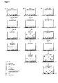
- Fig. 1A1 For cell immobilisation using low melting temperature agarose 100 ⁇ l or 200 ⁇ l of protoplast embedding mixture per were transferred into 96-well plates or 8-well slides respectively ( Fig. 1A1 ). After formation of cell layers at 30-35°C achieved either by centrifugation for at least 1min at 10g or by sedimentation for at least 20-30min ( Fig 1A2 ) plates can be placed at room temperature for agarose solidification ( Fig. 1A3 ). Embedded cells should be washed twice for 15min with the culture F-PCN medium and 200 ⁇ l of fresh F-PCN were added after the last washing ( Fig. 1A4 ). It is important to note, that within first 24h of culture mild expansion of agarose gel with embedded cells was not avoidable due to long lasting water uptake from the culture medium.
- Fig. 1B1 For cell immobilisation using Ca 2+ -alginate embedding 100 ⁇ l or 200 ⁇ l of protoplast embedding mixture per were transferred into 96-well plates or 8-well slides respectively ( Fig. 1B1 ). To form a cell layer protoplasts could be either centrifuged at 10g for at least 1 min or sedimented for at least 20-30min ( Fig. 1B2 ).
- the upper surface of the protoplast embedding mixture was covered with 20 ⁇ l or 40 ⁇ l for 96-well plates or 8-well slides respectively of W5 or another osmotically adjusted solution with a high Ca 2+ -salt ( ⁇ 20mM) content in a form of micro-droplets ( ⁇ 1 ⁇ l droplets if manually or 100-500nl droplets if using dispersing robots, Fig. 1B3 , Fig. 2 ).
- W5 medium was replaced with 200 ⁇ l of the culture F-PCN medium for washing.
- Fig. 1B5 After two washings, 200 ⁇ l of fresh F-PCN were added for subsequent protoplast culture (Fig. 1B5 ). Embedded cells were observed with 24h interval and their development was documented by imaging of the same area using inverted microscope (Axiovert 200M, Zeiss) for a period of 1 week ( Fig. 3 ). Due to formation of a single cell layer at the bottom of culture wells and subsequent immobilization this technique was named p rotoplast- m onolayer- e mbedding technique (PME).
- PME p rotoplast- m onolayer- e mbedding technique
- Protoplast isolation was done as described in the example 1 prior the washing with the culture medium.
- Cell immobilisation was performed in 8-well slides (IBIDI). After the removal of W5 medium, F-PCN with various concentration of compound R113 per well was used for washing and further cell culture. Analysis of cell division efficiency ( Fig. 4 ) was performed using inverted microscope (Axiovert 200M, Zeiss).
- Example 3 Monolayer embedding of Arabidopsis protoplasts for continuous cell tracking.
- Arabidopsis protoplasts were isolated from hypocotyls of 7-days old Arabidopsis seedlings (Col-0) germinated on SCA medium according to Dovzhenko et al. (2003) Protoplasma 222, 107-111 . Explants were cut in 0.5-1 mm fragments and preplasmolysed in MMC600 for 1 h. Afterwards the medium was substituted with fresh MMC600 supplemented with 0.5% Cellulase Onozuka R-10 (DUCHEFA), 0.5% Macerozyme Onozuka R-10 and 0.05% Driselase (SIGMA). After 14h of digestion, protoplast containing medium was filtered through 32 ⁇ m sieves.
- DUCHEFA Cellulase Onozuka R-10
- SIGMA Driselase
- Protoplasts containing medium was further mixed with an equal volume of TM550 and collected by centrifugation for 10min at 100g. Supernatant was removed and the pellet was mixed with TM550:Alg-A mixture (1:1) to achieve density 1 ⁇ 10 4 protoplasts per 100 ⁇ l. 100 ⁇ l aliquots of protoplast embedding mixture were transferred in 96-well plate and immobilized as described in the example 1. After the removal of W5 medium, PCA medium was used for washing and culture steps. Cell observations ( Fig. 5 ) were done using inverted microscope (Axiovert 200M, Zeiss).
- Cotyledons from 7-days old Arabidopsis seedlings were removed and cut in 0.5-1mm fragments. Preplasmolysis and digestion was performed as describe in the example 3, however no Driselase was used for digestion. After filtration through 56 ⁇ m sieves, protoplasts were collected in 12ml tube and pelleted for 10min at 100g. Supernatant was discarded and 10ml of MSC600 was added. For flotation 2ml of TM550 were overlayed on a top of MSC600 and centrifuged for 10min at 100g. Interlayer was collected and transferred into a new 12ml tube. Total volume was adjusted to 10ml with W5 medium.
- Protoplasts were washed for 5min at 50g and afterwards pellet was resuspended in TM550 to achieve density 2 ⁇ 10 4 protoplasts per 100 ⁇ l. After mixing 1:1 with Alg-A medium, 100 ⁇ l aliquots of protoplast embedding mixture were used for cell immobilisation in 96-well plates. Immobilisation and subsequent culture procedure were performed as described in example 3. While GFP fluorescence was detected only in 3% of freshly isolated protoplasts, it was observed in over than 95% of intact cells after 48h of culture thus demonstrating activity of DR5 promoter ( Fig. 6 ).
- plasmid DNA was dissolved in ultra-pure sterile water. Aliquots of aqueous DNA solution at various concentrations (0, 0.1, 0.5, 1 and 5 ⁇ g per 10 ⁇ l) were transferred inside 96-well plate (ABIGene). Water evaporation took place under the sterile bench overnight ( Fig. 1C1 , 2 ). After complete evaporation plates could be used either immediately or stored at -20°C.
- Arabidopsis shoots from 3-weeks old seedlings were used. Digestion and isolation were performed as described in example 4. After last washing step cell density was adjusted to 1x10 5 and aliquots of 30 ⁇ l were transferred into the wells containing dried DNA ( Fig. 1C3 ). Afterwards cells were left to sediment for at least 2min ( Fig. 1C4 ). Alternatively, protoplasts could be centrifuged for 1min at 10g. Further PEG-mediated DNA uptake was performed by adding an equal volume of 40% PEG1500 solution to protoplasts in wells ( Fig. 1C5 ).
- TM550 After 8min of the PEG treatment, a half of the total volume of TM550 was added and after additional 2min total volume was increased to 1ml using TM550 ( Fig. 1C6 ). To avoid formation of protoplast-aggregates, well contents were mixed using 1ml 8-well pipette. Sedimentation for at least 30min was performed to collect cells. Alternatively, protoplasts could be centrifuged for 10min at 50g. Supernatant (950 ⁇ l) was discarded and protoplast pellet was resuspended in 300 ⁇ l of PCA. Transformation efficiencies ( Fig. 7 ) were estimated after 30 hours using inverted microscope (Axiovert 200M, Zeiss).
- Tobacco leaf protoplasts were isolated as described in the Example 1 prior the embedding procedure. After protoplast pelleting in W5 medium and removal of the supernatant, cell density was adjusted to 2 ⁇ 10 6 cells/ml with TM550. Aliquots of 30 ⁇ l (2-2.5 ⁇ 10 5 cells) were transferred into the wells using 8-channel pipette. Afterwards cells were left to sediment for at least 2min. Alternatively, protoplasts could be centrifuged for 1 min at 10g. Further PEG-mediated DNA uptake was performed by adding an equal volume of 40% PEG1500 solution to protoplasts in wells. After 8min of the PEG treatment, a half of the total volume of TM550 was added and after additional 2min total volume was increased to 1ml using TM550.
- Arabidopsis shoots from 3-weeks old seedlings (35S::GFP in Col-0 background) were used. Digestion and isolation were performed as described in example 4. After last washing step cell density was adjusted to 1 ⁇ 10 5 and aliquots of 30 ⁇ l were transferred into 96-well plate. 1-2 ⁇ g DNA in 5 ⁇ l of ultra-pure sterile water was mixed with protoplasts. PEG-mediated DNA uptake was performed by adding an equal (35 ⁇ l) volume of 40% PEG1500 solution. After 8min of the PEG treatment, a half of the total volume of TM550 was added and after additional 2min total volume was increased to 1ml using TM550.
- amiRNAs were estimated as a read-out of fluorescence intensity of the target gene translationally fused with the reporter gene ( Fig. 11 ). Randomly selected amiRNA constructs were further used for generation of transgenic plants which exhibited phenotypes confirming microscopy-based conclusions on amiRNA efficiency ( Fig. 12 ).
Landscapes
- Health & Medical Sciences (AREA)
- Life Sciences & Earth Sciences (AREA)
- Genetics & Genomics (AREA)
- Engineering & Computer Science (AREA)
- Biomedical Technology (AREA)
- Biotechnology (AREA)
- Wood Science & Technology (AREA)
- Organic Chemistry (AREA)
- Chemical & Material Sciences (AREA)
- Zoology (AREA)
- Bioinformatics & Cheminformatics (AREA)
- General Engineering & Computer Science (AREA)
- Molecular Biology (AREA)
- Cell Biology (AREA)
- Microbiology (AREA)
- Biochemistry (AREA)
- General Health & Medical Sciences (AREA)
- Plant Pathology (AREA)
- Biophysics (AREA)
- Physics & Mathematics (AREA)
- Virology (AREA)
- Botany (AREA)
- Micro-Organisms Or Cultivation Processes Thereof (AREA)
- Measuring Or Testing Involving Enzymes Or Micro-Organisms (AREA)
Priority Applications (6)
| Application Number | Priority Date | Filing Date | Title |
|---|---|---|---|
| EP10191985A EP2455454A1 (fr) | 2010-11-19 | 2010-11-19 | Processus d'introduction d'un polynucléotide dans des cellules végétables de protoplastes |
| EP11787849.6A EP2640830B1 (fr) | 2010-11-19 | 2011-11-21 | Procédé d'introduction d'un polynucléotide à l'intérieur de cellules végétales non adhésives en croissance |
| EP17171376.1A EP3231865B1 (fr) | 2010-11-19 | 2011-11-21 | Procédé d'introduction d'un polynucléotide à l'intérieur de cellules végétales non adhésives en croissance |
| US13/988,276 US10301635B2 (en) | 2010-11-19 | 2011-11-21 | Method for introducing a polynucleotide into non-adhesively growing plant cells |
| PCT/EP2011/070602 WO2012066147A1 (fr) | 2010-11-19 | 2011-11-21 | Procédé d'introduction d'un polynucléotide à l'intérieur de cellules végétales non adhésives en croissance |
| US16/379,197 US11261454B2 (en) | 2010-11-19 | 2019-04-09 | Method for introducing a polynucleotide into non-adhesively growing plant cells |
Applications Claiming Priority (1)
| Application Number | Priority Date | Filing Date | Title |
|---|---|---|---|
| EP10191985A EP2455454A1 (fr) | 2010-11-19 | 2010-11-19 | Processus d'introduction d'un polynucléotide dans des cellules végétables de protoplastes |
Publications (1)
| Publication Number | Publication Date |
|---|---|
| EP2455454A1 true EP2455454A1 (fr) | 2012-05-23 |
Family
ID=43530211
Family Applications (3)
| Application Number | Title | Priority Date | Filing Date |
|---|---|---|---|
| EP10191985A Withdrawn EP2455454A1 (fr) | 2010-11-19 | 2010-11-19 | Processus d'introduction d'un polynucléotide dans des cellules végétables de protoplastes |
| EP17171376.1A Active EP3231865B1 (fr) | 2010-11-19 | 2011-11-21 | Procédé d'introduction d'un polynucléotide à l'intérieur de cellules végétales non adhésives en croissance |
| EP11787849.6A Active EP2640830B1 (fr) | 2010-11-19 | 2011-11-21 | Procédé d'introduction d'un polynucléotide à l'intérieur de cellules végétales non adhésives en croissance |
Family Applications After (2)
| Application Number | Title | Priority Date | Filing Date |
|---|---|---|---|
| EP17171376.1A Active EP3231865B1 (fr) | 2010-11-19 | 2011-11-21 | Procédé d'introduction d'un polynucléotide à l'intérieur de cellules végétales non adhésives en croissance |
| EP11787849.6A Active EP2640830B1 (fr) | 2010-11-19 | 2011-11-21 | Procédé d'introduction d'un polynucléotide à l'intérieur de cellules végétales non adhésives en croissance |
Country Status (3)
| Country | Link |
|---|---|
| US (2) | US10301635B2 (fr) |
| EP (3) | EP2455454A1 (fr) |
| WO (1) | WO2012066147A1 (fr) |
Cited By (1)
| Publication number | Priority date | Publication date | Assignee | Title |
|---|---|---|---|---|
| WO2014039970A1 (fr) * | 2012-09-07 | 2014-03-13 | Dow Agrosciences Llc | Enrichissement par tri cellulaire par fluorescence (facs) pour générer des plantes |
Families Citing this family (5)
| Publication number | Priority date | Publication date | Assignee | Title |
|---|---|---|---|---|
| EP2455454A1 (fr) | 2010-11-19 | 2012-05-23 | Albert-Ludwigs-Universität Freiburg | Processus d'introduction d'un polynucléotide dans des cellules végétables de protoplastes |
| WO2016164746A1 (fr) | 2015-04-08 | 2016-10-13 | Alnylam Pharmaceuticals, Inc. | Compositions et méthodes d'inhibition de l'expression du gène lect2 |
| GB201508302D0 (en) * | 2015-05-14 | 2015-06-24 | Synpromics Ltd | Method of screening synthetic promotors |
| EP3836943A4 (fr) * | 2018-08-14 | 2022-10-19 | Inscripta, Inc. | Détection améliorée de séquences à édition par nucléase dans des modules automatisés et des instruments par l'intermédiaire d'une culture en masse de cellules |
| IL294109B2 (en) | 2019-12-19 | 2025-11-01 | Pioneer Hi Bred Int | Microspore culture predictive model generation and use |
Citations (2)
| Publication number | Priority date | Publication date | Assignee | Title |
|---|---|---|---|---|
| EP0465875A1 (fr) * | 1990-06-23 | 1992-01-15 | Hoechst Schering AgrEvo GmbH | Génotypes améliorés de Zea mays (L.) avec capacité de régénération en plantes de haute efficacité de longue durée |
| US20020173037A1 (en) * | 2001-03-29 | 2002-11-21 | Pati Pratap Kumar | Efficient method of protoplast culture |
Family Cites Families (2)
| Publication number | Priority date | Publication date | Assignee | Title |
|---|---|---|---|---|
| EP2311934B1 (fr) * | 2001-09-06 | 2013-06-05 | Rapid Micro Biosystems, Inc. | Détection rapide de cellules proliférantes |
| EP2455454A1 (fr) | 2010-11-19 | 2012-05-23 | Albert-Ludwigs-Universität Freiburg | Processus d'introduction d'un polynucléotide dans des cellules végétables de protoplastes |
-
2010
- 2010-11-19 EP EP10191985A patent/EP2455454A1/fr not_active Withdrawn
-
2011
- 2011-11-21 US US13/988,276 patent/US10301635B2/en active Active
- 2011-11-21 WO PCT/EP2011/070602 patent/WO2012066147A1/fr not_active Ceased
- 2011-11-21 EP EP17171376.1A patent/EP3231865B1/fr active Active
- 2011-11-21 EP EP11787849.6A patent/EP2640830B1/fr active Active
-
2019
- 2019-04-09 US US16/379,197 patent/US11261454B2/en active Active
Patent Citations (2)
| Publication number | Priority date | Publication date | Assignee | Title |
|---|---|---|---|---|
| EP0465875A1 (fr) * | 1990-06-23 | 1992-01-15 | Hoechst Schering AgrEvo GmbH | Génotypes améliorés de Zea mays (L.) avec capacité de régénération en plantes de haute efficacité de longue durée |
| US20020173037A1 (en) * | 2001-03-29 | 2002-11-21 | Pati Pratap Kumar | Efficient method of protoplast culture |
Non-Patent Citations (19)
| Title |
|---|
| "Detecting environmental pollutants with protoplasts in alginate matrix", INTERNATIONAL JOURNAL OF BIOCHEMISTRY, PERGAMON, vol. 20, no. 6, 1 January 1988 (1988-01-01), pages X, XP025218004, ISSN: 0020-711X, [retrieved on 19880101], DOI: 10.1016/0020-711X(88)90148-6 * |
| "Dry-state, room-temperature storage of DNA and RNA", NATURE METHODS, vol. 5, September 2008 (2008-09-01), XP002621445, Retrieved from the Internet <URL:http://www.nature.com/nmeth/journal/v5/n9/full/nmeth.f.219.html> [retrieved on 20110209] * |
| "Methods in molecular biology", 2005, article "Arabidopsis protocols" |
| BART REBECCA ET AL: "A novel system for gene silencing using siRNAs in rice leaf and stem-derived protoplasts", PLANT METHODS, BIOMED CENTRAL, LONDON, GB, vol. 2, no. 1, 29 June 2006 (2006-06-29), pages 13, XP021020117, ISSN: 1746-4811, DOI: 10.1186/1746-4811-2-13 * |
| BUCHERL C; AKER J; DE VRIES S; BORST JW: "Probing protein-protein Interactions with FRET-FLIM", METHODS MOL BIOL, vol. 655, 2010, pages 389 - 399 |
| CRAIG W ET AL: "Direct gene transfer in potato: A comparison of particle bombardment of leaf explants and PEG-mediated transformation of protoplasts", PLANT CELL REPORTS, SPRINGER, BERLIN, DE, vol. 24, no. 10, 1 December 2005 (2005-12-01), pages 603 - 611, XP019335439, ISSN: 1432-203X, DOI: 10.1007/S00299-005-0018-0 * |
| DAVEY; ANTHONY: "Plant Cell Culture: Essential Methods", 2010 |
| DOVZHENKO A ET AL: "Thin-alginate-layer technique for protoplast culture of tobacco leaf protoplasts: Shoot formation in less than two weeks", PROTOPLASMA, vol. 204, no. 1-2, 1998, pages 114 - 118, XP002621446, ISSN: 0033-183X * |
| DOVZHENKO ET AL., PROTOPLASMA, vol. 204, 1998, pages 114 - 118 |
| DOVZHENKO ET AL., PROTOPLASMA, vol. 222, 2003, pages 107 - 111 |
| EHLERT A; WELTMEIER F; WANG X; MAYER CS; SMEEKENS S; VICENTE-CARBAJOSA J; DR6GE-LASER W: "Two-hybrid protein-protein interaction analysis in Arabidopsis protoplasts: establishment of a heterodimerization map of group C and group S bZIP transcription factors", PLANT J, vol. 46, 2006, pages 890 - 900 |
| HASEK J; STREIBTOVA E: "Fluorescence microscopy methods", METHODS MOL BIOL, vol. 53, 1996, pages 391 - 405 |
| NELSON ET AL.: "A multicoloured set of in vivo organelle markers for co-localization studies in Arabidopsis and other plants", PLANT J, vol. 51, 2006, pages 1126 - 1136 |
| OSSOWSKI, S.; SCHWAB, R.; WEIGEL, D.: "Gene silencing in plants using artificial microRNAs and other small RNAs", PLANT J, vol. 53, 2008, pages 674 - 690 |
| PATI P K ET AL: "Extra thin alginate film: an efficient technique for protoplast culture", PROTOPLASMA ; AN INTERNATIONAL JOURNAL OF CELL BIOLOGY, SPRINGER-VERLAG, VI, vol. 226, no. 3-4, 1 December 2005 (2005-12-01), pages 217 - 221, XP019379124, ISSN: 1615-6102, DOI: 10.1007/S00709-005-0096-4 * |
| SHANER, N.C. ET AL.: "Improved monomeric red, orange and yellow fluorescent proteins derived from Discosoma sp. red fluorescent protein", NAT BIOTECHNOL, vol. 22, 2004, pages 1567 - 1572 |
| TRAUD WINKELMANN ET AL: "Efficient plant regeneration from protoplasts isolated from embryogenic suspension cultures of Cyclamen persicum Mill", PLANT CELL, TISSUE AND ORGAN CULTURE, KLUWER ACADEMIC PUBLISHERS, DO, vol. 86, no. 3, 20 July 2006 (2006-07-20), pages 337 - 347, XP019409385, ISSN: 1573-5044, DOI: 10.1007/S11240-006-9129-8 * |
| YAMADA YASUYUKI ET AL: "Identification of regulatory protein genes involved in alkaloid biosynthesis using a transient RNAi system.", METHODS IN MOLECULAR BIOLOGY (CLIFTON, N.J.) 2010 LNKD- PUBMED:20552442, vol. 643, 2010, pages 33 - 45, XP001525690, ISSN: 1940-6029 * |
| YOO SANG-DONG ET AL: "Arabidopsis mesophyll protoplasts: a versatile cell system for transient gene expression analysis.", NATURE PROTOCOLS 2007 LNKD- PUBMED:17585298, vol. 2, no. 7, 2007, pages 1565 - 1572, XP002621444, ISSN: 1750-2799 * |
Cited By (1)
| Publication number | Priority date | Publication date | Assignee | Title |
|---|---|---|---|---|
| WO2014039970A1 (fr) * | 2012-09-07 | 2014-03-13 | Dow Agrosciences Llc | Enrichissement par tri cellulaire par fluorescence (facs) pour générer des plantes |
Also Published As
| Publication number | Publication date |
|---|---|
| EP2640830B1 (fr) | 2017-05-17 |
| EP2640830A1 (fr) | 2013-09-25 |
| EP3231865A3 (fr) | 2017-11-29 |
| US11261454B2 (en) | 2022-03-01 |
| EP3231865A2 (fr) | 2017-10-18 |
| EP3231865B1 (fr) | 2021-01-27 |
| US20130330717A1 (en) | 2013-12-12 |
| US10301635B2 (en) | 2019-05-28 |
| WO2012066147A1 (fr) | 2012-05-24 |
| US20190292552A1 (en) | 2019-09-26 |
Similar Documents
| Publication | Publication Date | Title |
|---|---|---|
| US11261454B2 (en) | Method for introducing a polynucleotide into non-adhesively growing plant cells | |
| AU2018212986B2 (en) | Novel plant cells, plants, and seeds | |
| JP6990653B2 (ja) | 迅速な植物形質転換のための方法および組成物 | |
| EP2308986B1 (fr) | Mini-chromosomes de plantes artificielles | |
| Eriksson et al. | Genetics applied to forestry | |
| US9957514B2 (en) | Transformation of duckweed and uses thereof | |
| JP2022534381A (ja) | ゲノム編集を使用してドミナントアレルを生成する方法及び組成物 | |
| Huang et al. | A DEAD-box protein, AtRH36, is essential for female gametophyte development and is involved in rRNA biogenesis in Arabidopsis | |
| Sánchez-Simarro et al. | Loss of Arabidopsis β-COP function affects Golgi structure, plant growth and tolerance to salt stress | |
| Raji et al. | Multiple fungal diseases resistance induction in Cucumis melo through co-transformation of different pathogenesis related (PR) protein genes | |
| Van Eck et al. | Agrobacterium tumefaciens-mediated transformation of Setaria viridis | |
| Yang et al. | Essential role of conserved DUF177A protein in plastid 23S rRNA accumulation and plant embryogenesis | |
| US20110119795A1 (en) | Artificial plant minichromosomes | |
| US20090100550A1 (en) | Artificial Plant Minichromosomes | |
| CN1654662A (zh) | 一种培育无选择标记转基因植物的方法及其专用表达载体 | |
| US20240279672A1 (en) | Compositions and methods for improved agronomic performance in plants | |
| KR102103189B1 (ko) | 염분 저항성을 가지는 형질전환 식물체의 조기 검정 방법 | |
| Charles Marie Roumégoux | Toward a role for LOTUS domain containing proteins during sexual reproduction in Arabidopsis thaliana | |
| Roumégoux | Toward a Role for LOTUS Domain Containing Proteins During Sexual Reproduction in Arabidopsis Thaliana | |
| CN121801954A (zh) | 一种供体dna的转基因方法 | |
| Ji | Characterization of a Mutator Organelle DNA Polymerase and Its Application to Mutate the Chloroplast Genome | |
| Dimos | Functional analysis of the cellulose synthase-like D (CSLD) gene family in Physcomitrella patens | |
| Peters | Investigation into the transcriptional regulation of DUO1: determining the fate of the male germline in flowering plants | |
| Minogue | The identification and characterisation of key genes involved in somatic embryogenesis in wheat (Triticum, aestivum L.) | |
| KR20150026420A (ko) | 형질전환 억새 식물체의 제조 방법 및 그에 따른 식물체 |
Legal Events
| Date | Code | Title | Description |
|---|---|---|---|
| PUAI | Public reference made under article 153(3) epc to a published international application that has entered the european phase |
Free format text: ORIGINAL CODE: 0009012 |
|
| AK | Designated contracting states |
Kind code of ref document: A1 Designated state(s): AL AT BE BG CH CY CZ DE DK EE ES FI FR GB GR HR HU IE IS IT LI LT LU LV MC MK MT NL NO PL PT RO RS SE SI SK SM TR |
|
| AX | Request for extension of the european patent |
Extension state: BA ME |
|
| STAA | Information on the status of an ep patent application or granted ep patent |
Free format text: STATUS: THE APPLICATION IS DEEMED TO BE WITHDRAWN |
|
| 18D | Application deemed to be withdrawn |
Effective date: 20121124 |