EP2505162B1 - Procédé et appareil de génération d'images médicales d'organes corporels en utilisant un modèle 3-D - Google Patents
Procédé et appareil de génération d'images médicales d'organes corporels en utilisant un modèle 3-D Download PDFInfo
- Publication number
- EP2505162B1 EP2505162B1 EP12161756.7A EP12161756A EP2505162B1 EP 2505162 B1 EP2505162 B1 EP 2505162B1 EP 12161756 A EP12161756 A EP 12161756A EP 2505162 B1 EP2505162 B1 EP 2505162B1
- Authority
- EP
- European Patent Office
- Prior art keywords
- images
- organ
- image
- model
- patient
- Prior art date
- Legal status (The legal status is an assumption and is not a legal conclusion. Google has not performed a legal analysis and makes no representation as to the accuracy of the status listed.)
- Active
Links
Images


Classifications
-
- A—HUMAN NECESSITIES
- A61—MEDICAL OR VETERINARY SCIENCE; HYGIENE
- A61B—DIAGNOSIS; SURGERY; IDENTIFICATION
- A61B34/00—Computer-aided surgery; Manipulators or robots specially adapted for use in surgery
- A61B34/10—Computer-aided planning, simulation or modelling of surgical operations
-
- G—PHYSICS
- G06—COMPUTING OR CALCULATING; COUNTING
- G06T—IMAGE DATA PROCESSING OR GENERATION, IN GENERAL
- G06T7/00—Image analysis
- G06T7/10—Segmentation; Edge detection
- G06T7/12—Edge-based segmentation
-
- G—PHYSICS
- G06—COMPUTING OR CALCULATING; COUNTING
- G06T—IMAGE DATA PROCESSING OR GENERATION, IN GENERAL
- G06T7/00—Image analysis
- G06T7/10—Segmentation; Edge detection
- G06T7/149—Segmentation; Edge detection involving deformable models, e.g. active contour models
-
- G—PHYSICS
- G06—COMPUTING OR CALCULATING; COUNTING
- G06T—IMAGE DATA PROCESSING OR GENERATION, IN GENERAL
- G06T7/00—Image analysis
- G06T7/30—Determination of transform parameters for the alignment of images, i.e. image registration
- G06T7/38—Registration of image sequences
-
- A—HUMAN NECESSITIES
- A61—MEDICAL OR VETERINARY SCIENCE; HYGIENE
- A61B—DIAGNOSIS; SURGERY; IDENTIFICATION
- A61B34/00—Computer-aided surgery; Manipulators or robots specially adapted for use in surgery
- A61B34/10—Computer-aided planning, simulation or modelling of surgical operations
- A61B2034/101—Computer-aided simulation of surgical operations
- A61B2034/105—Modelling of the patient, e.g. for ligaments or bones
-
- G—PHYSICS
- G06—COMPUTING OR CALCULATING; COUNTING
- G06T—IMAGE DATA PROCESSING OR GENERATION, IN GENERAL
- G06T2207/00—Indexing scheme for image analysis or image enhancement
- G06T2207/10—Image acquisition modality
- G06T2207/10072—Tomographic images
-
- G—PHYSICS
- G06—COMPUTING OR CALCULATING; COUNTING
- G06T—IMAGE DATA PROCESSING OR GENERATION, IN GENERAL
- G06T2207/00—Indexing scheme for image analysis or image enhancement
- G06T2207/20—Special algorithmic details
- G06T2207/20112—Image segmentation details
- G06T2207/20124—Active shape model [ASM]
-
- G—PHYSICS
- G06—COMPUTING OR CALCULATING; COUNTING
- G06T—IMAGE DATA PROCESSING OR GENERATION, IN GENERAL
- G06T2207/00—Indexing scheme for image analysis or image enhancement
- G06T2207/30—Subject of image; Context of image processing
- G06T2207/30004—Biomedical image processing
- G06T2207/30056—Liver; Hepatic
Definitions
- the present disclosure relates to a method and apparatus for generating a medical image of a body organ by using a three-dimensional (3-D) model.
- apparatuses of generating the right image for fast and accurate tracing of an internal organ in a real time medical image of a patient are provided.
- a method of generating an image of an organ includes: generating a three-dimensional (3-D) model of at least one organ of a patient, based on a medical image of the at least one organ; generating a plurality of matched images by matching a plurality of images, which show a change of a shape of the at least one organ due to a body activity of the patient, and the 3-D model of the at least one organ; and selecting one of the plurality of matched images based on the current body condition of the patient and outputting a selected matched image.
- 3-D three-dimensional
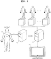
- FIG. 1 is a diagram illustrating a configuration of a system for generating an image of a body organ, according to an embodiment of the present invention.
- the system includes an image detection device 10, an image matching device 20, and an image display device 30.
- the image detection device 10 generates image data by using a response that is generated by transmitting a source signal generated from a probe 11 installed therein to a target part of a patient's body.
- the source signal may be a signal such as an ultrasound signal, an X-ray, or the like.
- the image detection device 10 is an ultrasonography machine which captures three-dimensional (3-D) images of the patient's body by using ultrasound is explained below.
- the probe 11 is generally in the form of a piezoelectric transducer. If an ultrasound signal in the range of 2 to 18 MHz is transmitted from the probe 11 of the image detection device 10 to a part of the inside of the patient's body, this ultrasound signal is partially reflected from strata between various different tissues. In particular, the ultrasound is reflected from parts of which densities are changed in the inside of the body, for example, blood cells of blood plasma, small structures of organs, and the like. The reflected ultrasound vibrates the piezoelectric transducer of the probe 11, and the piezoelectric transducer outputs electrical pulses due to the vibration. The electrical pulses are converted into images.
- the image detection device 10 may output two-dimensional (2-D) images and may also output 3-D images.
- a method in which the image detection device 10 outputs 3-D images is as follows.
- the image detection device 10 captures a plurality of cross-sectional images of a part of the patient's body while changing a location and orientation of the probe 11 over the patient's body.
- the image detection device 10 accumulates the cross-sectional images and thereby generates 3-D volume image data indicating three-dimensionally the part of the patient's body.
- MPR multi-planar reconstruction
- images which may be obtained by the image detection device 10 may be obtained in real time, but it is difficult to clearly identify an outline and internal structure of an organ or a lesion through the ultrasound images.
- CT images computed tomography (CT) images or magnetic resonance (MR) images
- MR images magnetic resonance
- a location of an organ or a location of a lesion may be clearly identified.
- CT images may not be output in real time because the CT images are obtained by using radiation and thus require short time photographing due to a danger to the patient or surgeon of prolonged radiation exposure.
- the MR images may not be output in real time because it takes a long time to capture them.
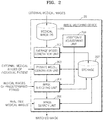
- FIG. 2 is a block diagram illustrating a configuration of the image matching device 20 of FIG. 1 .
- the image matching device 20 includes a medical image database (DB) 201, an average model generation unit 202, a private model generation unit 203, an image matching unit 204, an image search unit 205, an additional adjustment unit 206, and a storage 207.
- DB medical image database
- the average model generation unit 202 generates an average model of an organ by receiving various medical images of a patient and then processing them.
- an organ of a patient is traced by using a private model, i.e., a personalized model of the patient.
- the average model is generated by the average model generation unit 202 as a preparatory step for generating the private model. This is because, since characteristics of an organ, such as shape and size, are different for each individual person, it is necessary to reflect the characteristics of each individual to provide an accurate surgical operation environment. Various pieces of image information of each individual may be used to obtain an accurate average model. In addition, images at various points of breathing may be obtained to reflect a form of an organ which is changed according to the breathing.
- the average model generation unit 202 receives images (hereinafter, referred to as "external medical images"), which a medical expert has captured for diagnosis of a patient, directly from a photographing apparatus or from an image storage medium.
- images which make it possible to easily analyze outlines of an organ or a lesion or characteristics of the inside of the organ, as the external medical images.
- CT images or MR images may be input as the external medical images.
- the external medical images may be stored in the medical image DB 201, and the average model generation unit 202 may receive the external medical images stored in the medical image DB 201.
- the medical image DB 201 may store medical images of various individuals, which may be captured by the photographing apparatus or may be input from the image storage medium.
- the average model generation unit 202 may receive all or some of the external medical images from the medical image DB 201 depending on a selection of a user.
- the average model generation unit 202 may apply a 3-D active shape model (ASM) algorithm based on the received external medical images.
- ASM active shape model
- the average model generation unit 202 extracts shape, size, and anatomic features of an organ from the received external medical images by analyzing the received external medical images, and generates an average model of the organ by averaging them.
- the 3-D ASM algorithm is disclosed in the paper " The Use of Active Shape Models For Locating Structure in Medical Images" published in 1994 by T.F.Cootes, A.Hill, C.J.Taylor and J.Haslam . It is possible to obtain an average shape of the organ by applying the 3-D ASM algorithm, and the average shape of the organ may be transformed by modifying variables.
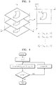
- FIG. 3 is a diagram for explaining a process of analyzing the external medical images, which is performed by the average model generation unit 202.
- FIG. 4 roughly illustrates a process of extracting location coordinate information of a boundary and internal structure of an organ from the external medical images, for example, the CT or MR images.
- the average model generation unit 202 performs an operation of extracting the location coordinate information of the boundary and internal structure of the organ by using different methods depending on whether the external medical images are 2-D images or 3-D images.
- an internal structure of a liver may include a hepatic artery, a hepatic vein, and a hepatic duct and boundaries between them.
- the average model generation unit 202 obtains 3-D volume images indicating three-dimensionally a target part by accumulating a plurality of cross-sectional images to generate a 3-D model. This method of obtaining the 3-D volume images is illustrated in the left side of FIG. 3 .
- the location coordinate information of the boundary and internal structure of the organ is extracted from each of the plurality of cross-sectional images. Then, it is possible to obtain 3-D coordinate information by adding coordinate information of an axis of direction, in which the plurality of cross-sectional images are accumulated, to the extracted information. For example, since the image illustrated in the right of FIG.
- 3-D coordinate information of the image illustrated in the right of FIG. 3 is [X,Y,1].
- 3-D coordinate information of the image illustrated in the right of FIG. 3 is [X,Y,1].
- 2-D coordinate information [X,Y] both a coordinate value of the Z-axis and the 2-D coordinate information [X,Y] are extracted to obtain the location coordinate information of the images illustrated in the left of FIG. 3 .
- the location coordinate information of the images will be 3-D coordinate information [X,Y,Z].
- location coordinate information of a boundary of an organ in 2-D images may be automatically or semi-automatically obtained by using an algorithm, and may also be manually input by a user with reference to output image information.
- a method of automatically obtaining the location coordinate information of the boundary of the organ it is possible to obtain location coordinate information of a part in which brightness of an image is abruptly changed, and also it is possible to extract a location of which a frequency value is largest, as a boundary location by using a discrete time Fourier transform (DTFT).
- DTFT discrete time Fourier transform
- a method of semi-automatically obtaining the location coordinate information of the boundary of the organ if information about a boundary point of an image is input by a user, it is possible to extract the location coordinate of a boundary based on the boundary point, similar to the method of automatically obtaining the location coordinate information. Since the boundary of the organ is continuous and has a looped curve shape, it is possible to obtain information about the whole boundary of the organ by using this characteristic. Since the method of semi-automatically obtaining the location coordinate information does not require searching for the whole of an image, it is possible to rapidly obtain a result compared to the method of automatically obtaining the location coordinate information.
- a user may directly designate coordinates of a boundary while viewing the image. At this time, since an interval at which the coordinates of the boundary is designated may not be continuous, it is possible to continuously extract the boundary by performing interpolation with respect to discontinuous sections. If the location coordinate information of the organ or a lesion, obtained by using the above methods, is output after setting a brightness value of a voxel corresponding to the location coordinate to a predetermined value, the user may confirm shapes of the organ or the lesion expressed three-dimensionally and graphically.
- a brightness value of boundary coordinates of a target organ is set to a minimum value, namely the darkest value
- an image of the target organ will have a dark form in an output image.
- the brightness value of the target organ is set to a medium value between a white color and a black color and the brightness value of a lesion is set to the black color, it is possible to easily distinguish the lesion from the target organ with the naked eye.
- the location coordinate information of boundaries and internal structures of a plurality of organs obtained by using the above methods, may be defined as a data set and be used to perform the 3-D ASM algorithm.
- the 3-D ASM algorithm is explained below.
- coordinate axes of location coordinates of the boundaries and internal structures of the plurality of organs are fit to each other. Fitting the coordinate axes to each other means fitting the centers of gravities of the plurality of organs to one origin and aligning directions of the plurality of.
- landmark points are determined in the location coordinate information of the boundaries and internal structures of the plurality of organs.
- the landmark points are basic points used to apply the 3-D ASM algorithm. The landmark points are determined by using following method.
- the points may include division points of blood vessels of a liver, a boundary between the right atrium and the left atrium in a heart, a boundary between a main vein and an outer wall of the heart, and the like.
- the highest points or the lowest points of a target in a predetermined coordinate system are determined as the landmark points.
- points for interpolating between the first determined points and the second determined points are determined as the landmark points along a boundary at predetermined intervals.
- the determined landmark points may be represented by using coordinates of the X and Y axes in two dimensions, and may be represented by using coordinates of the X, Y, and Z axes in three dimensions.
- coordinates of each of the landmark points are indicated as vectors x 0 , x 1 , ⁇ x n -1 in three dimensions (here, n is the number of landmark points)
- the vectors x 0 , x 1 , ⁇ x n -1 may be represented by the following equation 1.
- x i 0 x i 0 y i 0 z i 0
- the subscript i indicates location coordinate information of a boundary and internal structure of an organ, obtained in an i-th image.
- the number of the location coordinate information may be increased in some cases, and thus, the location coordinate information may be represented as a single vector to facilitate a calculation thereof.
- a landmark point vector which expresses all the landmark points with a single vector may be defined by the following equation 2.
- the average model generation unit 202 obtains the average x of the landmark points by calculating equation 3, and, if a model is generated based on the average x of the landmark points, the model may become an average organ model.
- the 3-D ASM algorithm may not only generate the average organ model, but may change a form of the average organ model only by adjusting a plurality of parameters.
- the average model generation unit 202 calculates not only the average organ model but also an equation so that the plurality of parameters may be applied. Below, an equation for applying the plurality of parameters is explained.
- equation 4 the average x of the landmark points and differences between data may be represented.
- the subscript i indicates an i-th image.
- equation 4 indicates a difference between the landmark points of each image and the average of all images.
- a covariance matrix for three variables x, y, and z may be defined as the following equation 5.
- the reason for obtaining the covariance matrix is to obtain a unit eigen-vector for the plurality of parameters to apply the 3-D ASM algorithm.
- the vector p k indicates a change of a model generated by using the 3-D ASM algorithm. For example, if a parameter b 1 multiplied by a vector p 1 is changed within the range of ⁇ 2 ⁇ 1 ⁇ b 1 ⁇ 2 ⁇ 1 , a width of the model may be changed. If a parameter b 2 multiplied by a vector p 2 is changed within the range of ⁇ 2 ⁇ 2 ⁇ b 2 ⁇ 2 ⁇ 2 , a height of the model may be changed.
- the private model generation unit 203 receives external medical images of the individual patient from an external image photographing apparatus or a storage medium 207, analyzes a shape, size, and location of an organ of the individual patient, and, if there is a lesion, analyzes a shape, size, and location of the lesion. Below, this operation of the private model generation unit 203 is explained.
- the private model generation unit 203 determines weights (the vector b) of eigen-vectors of the 3-D ASM algorithm for the individual patient, based on the medical images such as the CT or MR images in which a shape, size, and location of an organ may be clearly captured.
- the private model generation unit 203 receives the external medical images of the individual patient and obtains location coordinate information of a boundary and internal structure of an organ.
- the private model generation unit 203 uses the process of FIG. 3 , namely the process of analyzing the external medical images, which is performed by the average model generation unit 202.
- the vectors x and p determined by the average model generation unit 202 may be stored in the storage 207 as a database of an average model for a target organ, and may be repeatedly used if necessary.
- the external medical images of the individual patient, input to the private model generation unit 202 may be additionally used when determining the average model stored in the database during a medical examination and treatment of another patient.
- the image matching unit 204 may match the vectors with a patient's medical images received during a predetermined period.
- This matching means that a model using the 3-D ASM algorithm is overlapped with a location of an organ in an ultrasound medical image to output an output image.
- the matching means that it is possible to replace or overlap pixel or voxel values corresponding to coordinate information of a model formed by the 3-D ASM algorithm with a predetermined brightness. If the replacement operation is performed, an organ part is removed from an original ultrasound medical image and only a private model is output.
- an image in which the original ultrasound medical image is overlapped with the private model may be output.
- the overlapped image may be easily identified with the naked eye by differentiating a color thereof from that of another image. For example, it may be easy to identify a graphic figure with the naked eye by overlapping a private model with a black and white ultrasound image by using a blue color.
- the medical images may be images captured in real time and, for example, may be the ultrasound images.
- the medical images may be 2-D or 3-D images.
- the predetermined period may be one breathing cycle. This is because a change of an organ also is generated during a breathing cycle of the body. For example, if one breathing cycle of a patient is 5 seconds, ultrasound images having 100 frames may be generated when ultrasound images are generated 20 frames per 1 second.
- a process of matching which is performed in the image matching unit 204, may be divided into two operations.
- the two operations include an operation of reflecting a change of an organ due to breathing to a 3-D organ model in ultrasound images input during a predetermined period and an operation of aligning the changed 3-D organ model to a target organ in the ultrasound images by performing scale control, axis rotation, and axis movement.
- the operation of reflecting a change of an organ due to breathing to a 3-D organ model is as follows. Before matching the ultrasound images with medical images, a value of the vector b which is a weight of a parameter of the 3-D ASM algorithm is controlled by obtaining a location and change of an organ for each frame of the ultrasound images. A value of the vector b determined at this time does not have a large difference from a value of the vector b determined in the average model generation unit 202. This is because only a change due to the breathing is reflected in the image matching unit 204 and this change due to the breathing is smaller compared to changes in other individuals.
- a modification is performed within a predetermined limited range based on the value of the vector b determined in the average model generation unit 202.
- a vector b of a previous frame may be reflected in a determination of a vector b of a next frame. This is because there is no large change during a short period between frames since a change of an organ during the breathing is continuous. If the value of the vector b is determined, it is possible to generate a private model, for each frame, in which a modification of an organ is reflected in each ultrasound image by using a calculation of the 3-D ASM algorithm.
- FIG. 4 is a flowchart illustrating a process in which the image matching unit 204 fits a private model, in which a transformation of an organ is reflected for each image, to a location of an organ in an ultrasound image through rotation, scale control, and parallel displacement.
- FIG. 4 is a flowchart illustrating a process of performing one-to-one affine registration for each frame when the vector b, which is a weight value of an eigen-vector for each frame, is determined. If the number of frames is N and n is a frame number, a one-to-one match is performed from when n is 1 to when n becomes N.
- An affine transformation function is obtained by performing an iterative closest point (ICP) algorithm for each frame by using a landmark point set of an ultrasound image and a landmark point set of a model, and a 3-D body organ model image is obtained by using the affine transformation function.
- the ICP algorithm is an algorithm for rotating and parallel displacing other images and controlling scales of the other images based on an image to align a target in a plurality of images.
- the ICP algorithm is disclosed in detail in "Iterative point matching for registration of free-form curves and surfaces" by Zhengyou Zhang.
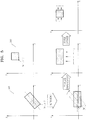
- FIG. 5 illustrates a process of obtaining the affine transformation function in a 2-D image.
- a diagram 501 illustrates a state before applying the affine transformation
- a diagram 502 illustrates a state after applying the affine transformation.
- the rotation, the parallel displacement, and the scale control should be performed to apply the transformation, it is possible to determine coefficients of a matrix T affine of the affine transformation function by obtaining first coordinates and last coordinates through the following equation 9 in consideration that the affine transformation uses one to one point correspondence.
- Equation 10 is an equation for applying an affine transformation function obtained in three-dimensions to each frame.
- x ICP n T affine n ⁇ x ASM n
- n is an integer indicating an n-th frame (1 ⁇ n ⁇ N ).
- x ASM ( n ) indicates a landmark point vector in which the vector b that is the weight value is changed in the image matching unit 204.
- x ICP ( n ) includes location coordinate information of organ boundaries and internal structures in which a modification is reflected for each frame. It is possible to confirm a graphic figure of an organ with the naked eye if a voxel value corresponding to location coordinates is replaced or overlapped with a predetermined brightness value in an ulrasonic image when matching the location coordinate information with the ultrasound image.
- FIG. 6 illustrates a process of matching an image in the image matching unit 204.
- FIG. 6 illustrate a process in which the image matching unit 204 generates a matched image between a ultrasound image input during a predetermined period and a body organ model based on an ultrasound image input during one breathing cycle.
- the input ultrasound image is disposed in a left edge portion, and a mark * illustrated in the input ultrasound image indicates a landmark point.
- the input ultrasound image may reflect various forms of breathing from inspiration to expiration.
- a private model generated in the private model generation unit 203 will be modified according to breathing. However, a modification according to the respiration will be smaller than that due to diversity between individuals. Thus, when reflecting a modification according to breathing, it may be faster and easier to adjust parameter values determined by the private model generation unit 203 compared to newly obtaining 3-D ASM algorithm.
- the affine transformation function T affine is applied through the ICP algorithm by using a landmark point in which the modification has been reflected and a landmark point of an organ of the ultrasound image. Through the affine transformation, a size and location of a 3-D organ model may be modified to be matched with a size and location of an organ of the ultrasound image.
- Combining a modified model with the ultrasound image may be performed through a method of replacing or overlapping a pixel or voxel value of the ultrasound image corresponding to a location of a model with a predetermined value.
- a matched image is referred to as an ultrasound-model matched image and may be stored in the storage 207.
- the image search unit 205 performs processes of a surgical operation.
- a graphic shape of an organ is output in an ultrasound image, which is input in real time, on a screen, and then a surgeon performs the surgical operation while confirming the graphic shape of the organ with the naked eye.
- Detailed operations of this process are as follow.
- a real time medical image of a patient is received.
- the real time medical image may be an image which is the same as that received by the image matching unit 204.
- a real time ultrasound image is received, by comparing the real time ultrasound image with medical images input to the image matching unit 204 during a predetermined, an image which is most similar to the real time ultrasound image is determined, and an ultrasound-model matched image corresponding to the determined image is searched in the storage and then a searched ultrasound-model matched image is output.
- the image search unit 205 searches for a similar image in the ultrasound image
- FIG. 7 is a graph illustrating an up and down movement of an absolute location of the diaphragm.
- a location of a probe 11 and a location of a patient may be fixed when capturing the medical images, which are input to the image matching unit 204 during the predetermined period, and the real time medical image, which are input to the image search unit 205.
- the reason is that a relative location of an organ in the image may be changed if the location of the probe 11 or the location of the patient is changed and it is not possible to accurately and rapidly perform a search operation when comparing images if the relative location is changed.
- the image search unit 205 searches for a similar image in the ultrasound image
- this method involves using the fact that a brightness difference between the most similar images is the smallest.
- a brightness difference between pixels of one of the first images and pixels of the second image is calculated and then a dispersion for the brightness difference is obtained.
- brightness differences between pixels of the other images of the first images and pixels of the second image also are calculated and then dispersions for the brightness differences are obtained. Then, an image whose dispersion is the smallest may be determined as the most similar image.
- the additional adjustment unit 206 may output an adjusted final result if a user adjusts the affine transformation function T affine and the parameters of the 3-D ASM algorithm while viewing an output image. That is, the user may perform accurate transformation while viewing the output image with the naked eye.
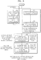
- FIG. 8 is a flowchart illustrating a method of tracing a dynamic organ and a lesion based on a 3-D organ model.
- Results of operations 802 and 803 may be stored in the medical image database (DB) 201 of FIG. 2 .
- DB medical image database
- CT or MR images for various breathing cycles of individuals are received.
- a 3-D body organ model is generated based on the received images.
- the 3-D ASM algorithm may be used.
- a CT or MR image of an individual patient is received.
- the 3-D body organ model generated in the operation 803 is modified based on the received image of the individual patient.
- a process of generating the modified 3-D body organ model, namely a private 3-D body organ model may be performed in the exterior of a surgical operation room as a preparatory process.
- ultrasound images (first ultrasound images) captured during one breathing cycle of a patient are received, and the first ultrasound images are matched with the private 3-D body organ model.
- a matched image is referred to as an ultrasound-model matched image, and may be stored in a temporary memory or in a storage medium such as storage.
- Operation 805 may be performed as a preparatory process in a surgical operation room.
- a location of the patient may be fixed.
- a location of a probe may be fixed.
- an ultrasound image (a second ultrasound image) of the patient is input in real time, an image, which is most similar to the second ultrasound image, from among the first ultrasound images is determined and then an ultrasound-model matched image corresponding to the determined first ultrasound image is output.
- the embodiments of the present general inventive concept can be written as computer programs and can be implemented in general-use digital computers that execute the programs using a computer readable recording medium.
- Examples of the computer readable recording medium include magnetic storage media (e.g., ROM, floppy disks, hard disks, etc.), optical recording media (e.g., CD-ROMs, or DVDs), etc.
Landscapes
- Engineering & Computer Science (AREA)
- Health & Medical Sciences (AREA)
- Theoretical Computer Science (AREA)
- General Physics & Mathematics (AREA)
- Physics & Mathematics (AREA)
- Computer Vision & Pattern Recognition (AREA)
- Surgery (AREA)
- Life Sciences & Earth Sciences (AREA)
- Public Health (AREA)
- Nuclear Medicine, Radiotherapy & Molecular Imaging (AREA)
- Animal Behavior & Ethology (AREA)
- General Health & Medical Sciences (AREA)
- Medical Informatics (AREA)
- Veterinary Medicine (AREA)
- Heart & Thoracic Surgery (AREA)
- Biomedical Technology (AREA)
- Robotics (AREA)
- Molecular Biology (AREA)
- Software Systems (AREA)
- Apparatus For Radiation Diagnosis (AREA)
- Magnetic Resonance Imaging Apparatus (AREA)
- Ultra Sonic Daignosis Equipment (AREA)
- Measuring And Recording Apparatus For Diagnosis (AREA)
- Image Processing (AREA)
- Image Analysis (AREA)
Claims (16)
- Procédé de production d'une image d'un organe, le procédé comprenant :la production d'un modèle tridimensionnel (3-D) d'au moins un organe d'un patient, sur la base des images médicales du au moins un organe ;la production d'une pluralité d'images qui correspondent en faisant correspondre une pluralité des images, qui montrent un changement d'une forme du au moins un organe dû à une activité corporelle du patient, et le modèle 3-D du au moins un organe ;la réception d'une image médicale en temps réel du patient ; etla sélection de l'une de la pluralité des images qui correspondent sur la base de l'état corporel présent du patient et l'émission de l'image sélectionnée qui correspond; caractérisée en ce que la sélection et l'émission comprennent la sélection d'une image qui correspond correspondant à une image, qui est la plus semblable à une image médicale en temps réel, parmi la pluralité des images.
- Procédé selon la revendication 1, dans lequel la production du modèle 3-D comprend la production du modèle 3-D montrant une forme du au moins un organe du patient sur la base de l'image médicale du patient.
- Procédé selon la revendication 1 ou 2, dans lequel la production de la pluralité des images qui correspondent comprend :la modification du modèle 3-D sur la base du changement de forme du au moins un organe ; etl'ajustement d'un axe de coordonnées du modèle 3-D à un axe de coordonnées de la pluralité des images ;en particulier, la production de la pluralité des images qui correspondent comprenant en outre la production de la pluralité des images qui correspondent par des pixels ou des voxels chevauchant de la pluralité des images avec des pixels ou des voxels d'une brillance prédéterminée.
- Procédé selon l'une quelconque des revendications 1 à 3, dans lequel la sélection et l'émission comprennent le calcul d'une différence entre un emplacement d'un diaphragme dans la pluralité des images et un emplacement d'un diaphragme dans l'image médicale en temps réel, et la sélection d'une image ayant la plus petite différence.
- Procédé selon l'une quelconque des revendications 1 à 4, dans lequel la production du modèle 3-D comprend :l'extraction de l'information des coordonnées de localisation d'une limite et d'une structure interne du au moins un organe des images médicales ;la désignation des coordonnées des points de repère dans l'information des coordonnées de localisation ; etla production d'un modèle d'apparence externe statistique.
- Procédé selon la revendication 5, dans lequel la production du modèle 3-D comprend en outre le changement du modèle d'apparence externe statistique en un modèle reflétant une forme caractéristique du au moins un organe du patient ;
en particulier, où la production du modèle 3-D comprend la réflexion de la forme caractéristique du au moins un organe du patient sur les images médicales des patients. - Procédé selon la revendication 5 ou 6, dans lequel l'extraction de l'information des coordonnées de localisation comprend la détermination d'une position, dont un changement d'une valeur de brillance est maximal dans l'image médicale, comme l'information des coordonnées de localisation de la limite et de la structure interne du au moins un organe ;
en particulier, où l'extraction de l'information des coordonnées de localisation comprend la détermination d'une position, dont une valeur de fréquence d'une transformée de Fourier à temps discret (DTFT) est maximale dans l'image médicale, comme l'information des coordonnées de localisation de la limite et de la structure interne du au moins un organe. - Procédé selon la revendication 5 ou 6, dans lequel l'extraction de l'information des coordonnées de localisation comprend la détermination de l'information des coordonnées de localisation de la limite et de la structure interne du au moins un organe sur la base des coordonnées saisies par un utilisateur.
- Procédé selon l'une quelconque des revendications 1 à 8, dans lequel la pluralité des images sont des images capturées à des intervalles prédéterminés dans un cycle de respiration du patient.
- Procédé selon l'une quelconque des revendications 1 à 9, dans lequel l'image médicale du au moins un organe sont des images capturées en utilisant un procédé de tomographie assistée par ordinateur (CT) ; ou
dans lequel les images médicales du au moins un organe sont des images capturées en utilisant un procédé de résonance magnétique (MR). - Procédé selon l'une quelconque des revendications 1 à 10, dans lequel la pluralité des images et l'image en temps réel du patient sont des images par ultrasons.
- Procédé selon l'une quelconque des revendications 1 à 11, dans lequel, au lieu de la production d'un modèle 3-D, un modèle 3-D pré-généré stocké dans une base de données est reçu.
- Procédé selon la revendication 6, dans lequel la forme caractéristique comprend une forme et une localisation d'une lésion.
- Appareil de production d'une image d'un organe, l'appareil comprenant :une unité de production d'un modèle d'organe (202) pour la production d'un modèle 3-D d'au moins un organe d'un patient, sur la base des images médicales du au moins un organe ;une unité de correspondance d'image (204) pour produire une pluralité d'images qui correspondent en faisant correspondre une pluralité d'images, qui montre un changement d'une forme du au moins un organe dû à une activité corporelle du patient, et du modèle 3-D du au moins un organe ;un moyen de capture d'une image médicale en temps réel du patient ; etune unité de recherche d'image (205) pour sélectionner l'une de la pluralité des images qui correspondent sur la base de l'état corporel présent du patient ;caractérisé en ce que l'unité de recherche d'image sélectionne une image qui correspond correspondant à une image, qui est la plus semblable à une image médicale en temps réel, parmi la pluralité des images.
- Appareil selon la revendication 21, comprenant en outre une unité d'ajustement additionnel (206) pour l'ajustement de manière supplémentaire de la pluralité des images qui correspondent, selon une saisie d'un utilisateur.
- Milieu d'enregistrement pouvant être lu par ordinateur présentant enregistré dessus un programme d'exécution du procédé selon l'une quelconque des revendications 1 à 13 lorsqu'exécuté sur l'appareil selon l'une des revendications 14 ou 15.
Applications Claiming Priority (2)
| Application Number | Priority Date | Filing Date | Title |
|---|---|---|---|
| US201161468754P | 2011-03-29 | 2011-03-29 | |
| KR1020110086694A KR20120111871A (ko) | 2011-03-29 | 2011-08-29 | 3차원적 모델을 이용한 신체 장기의 영상 생성 방법 및 장치 |
Publications (2)
| Publication Number | Publication Date |
|---|---|
| EP2505162A1 EP2505162A1 (fr) | 2012-10-03 |
| EP2505162B1 true EP2505162B1 (fr) | 2017-11-01 |
Family
ID=45976715
Family Applications (1)
| Application Number | Title | Priority Date | Filing Date |
|---|---|---|---|
| EP12161756.7A Active EP2505162B1 (fr) | 2011-03-29 | 2012-03-28 | Procédé et appareil de génération d'images médicales d'organes corporels en utilisant un modèle 3-D |
Country Status (4)
| Country | Link |
|---|---|
| US (1) | US20120253170A1 (fr) |
| EP (1) | EP2505162B1 (fr) |
| JP (1) | JP2012205899A (fr) |
| CN (1) | CN102727236B (fr) |
Families Citing this family (29)
| Publication number | Priority date | Publication date | Assignee | Title |
|---|---|---|---|---|
| JP5919665B2 (ja) * | 2011-07-19 | 2016-05-18 | 日本電気株式会社 | 情報処理装置、物体追跡方法および情報処理プログラム |
| KR101916855B1 (ko) * | 2011-10-17 | 2019-01-25 | 삼성전자주식회사 | 병변 수정 장치 및 방법 |
| EP2876607B1 (fr) * | 2012-07-23 | 2019-07-31 | Fujitsu Limited | Procédé et dispositif de production de données de forme |
| KR102070427B1 (ko) | 2012-08-08 | 2020-01-28 | 삼성전자주식회사 | 종양의 위치를 추적하는 방법 및 장치 |
| WO2014039935A1 (fr) * | 2012-09-08 | 2014-03-13 | Wayne State University | Appareil et méthode d'échocardiographie fœtale à navigation intelligente |
| JP6004875B2 (ja) * | 2012-10-02 | 2016-10-12 | キヤノン株式会社 | 医用画像表示装置、医用画像表示方法及びプログラム |
| KR102001219B1 (ko) | 2012-11-26 | 2019-07-17 | 삼성전자주식회사 | 의료 영상들의 정합 방법 및 장치 |
| RU2530220C1 (ru) | 2013-03-18 | 2014-10-10 | Корпорация "САМСУНГ ЭЛЕКТРОНИКС Ко., Лтд." | Система и способ для автоматической регистрации анатомических точек в объемных медицинских изображениях |
| JP6131161B2 (ja) * | 2013-09-27 | 2017-05-17 | 富士フイルム株式会社 | 画像位置合わせ装置、方法、およびプログラム、並びに3次元変形モデル生成方法 |
| CN104545987A (zh) * | 2013-10-10 | 2015-04-29 | 深圳迈瑞生物医疗电子股份有限公司 | 一种监测膈肌运动情况的监护仪 |
| KR20150074304A (ko) * | 2013-12-23 | 2015-07-02 | 삼성전자주식회사 | 의료 영상 정보를 제공하는 방법 및 그 장치 |
| KR102250086B1 (ko) * | 2014-05-16 | 2021-05-10 | 삼성전자주식회사 | 의료 영상 정합 방법, 이를 포함하는 장치 및 컴퓨터 기록 매체 |
| CN105989092A (zh) * | 2015-02-12 | 2016-10-05 | 东芝医疗系统株式会社 | 医学图像处理设备、医学图像处理方法以及医学成像系统 |
| JP7122115B2 (ja) * | 2015-07-17 | 2022-08-19 | コーニンクレッカ フィリップス エヌ ヴェ | 肺癌放射線のためのガイダンス |
| JP6493877B2 (ja) | 2015-08-25 | 2019-04-03 | 富士フイルム株式会社 | 基準点評価装置、方法およびプログラム、並びに位置合せ装置、方法およびプログラム |
| JP6392190B2 (ja) | 2015-08-31 | 2018-09-19 | 富士フイルム株式会社 | 画像位置合せ装置、画像位置合せ装置の作動方法およびプログラム |
| GB2542114B (en) * | 2015-09-03 | 2018-06-27 | Heartfelt Tech Limited | Method and apparatus for determining volumetric data of a predetermined anatomical feature |
| JP6392192B2 (ja) | 2015-09-29 | 2018-09-19 | 富士フイルム株式会社 | 画像位置合せ装置、画像位置合せ装置の作動方法およびプログラム |
| KR102532287B1 (ko) * | 2015-10-08 | 2023-05-15 | 삼성메디슨 주식회사 | 초음파 장치 및 그 제어방법 |
| WO2017109685A1 (fr) * | 2015-12-22 | 2017-06-29 | Koninklijke Philips N.V. | Appareil d'imagerie médicale et procédé d'imagerie médicale pour l'inspection d'un volume d'un sujet |
| JP6461024B2 (ja) | 2016-02-05 | 2019-01-30 | 富士フイルム株式会社 | 画像位置合せ装置、方法およびプログラム |
| JP6493885B2 (ja) * | 2016-03-15 | 2019-04-03 | 富士フイルム株式会社 | 画像位置合せ装置、画像位置合せ装置の作動方法および画像位置合せプログラム |
| DE102016113068B4 (de) * | 2016-07-15 | 2024-12-05 | Carl Zeiss Microscopy Gmbh | Verfahren und Vorrichtung zum Bestimmen der Lage einer optischen Grenzfläche entlang einer ersten Richtung |
| CN108537893A (zh) * | 2017-03-02 | 2018-09-14 | 南京同仁医院有限公司 | 一种甲状腺占位病变的三维可视化模型生成方法 |
| EP3422048A1 (fr) | 2017-06-26 | 2019-01-02 | Koninklijke Philips N.V. | Système et procédé d'imagerie |
| EP3432262A1 (fr) * | 2017-07-18 | 2019-01-23 | Koninklijke Philips N.V. | Procédé et système d'images multidimensionnelles dynamiques d'un objet |
| US10959696B1 (en) * | 2019-11-25 | 2021-03-30 | Robert Edwin Douglas | Method and apparatus for an improved localizer for 3D imaging |
| JP7165541B2 (ja) * | 2018-09-14 | 2022-11-04 | 富士フイルムヘルスケア株式会社 | ボリュームデータ処理装置、方法及びプログラム |
| CN113728398B (zh) * | 2020-01-21 | 2025-01-28 | 京东方科技集团股份有限公司 | 疾病位置获取方法、装置、设备以及计算机可读存储介质 |
Citations (3)
| Publication number | Priority date | Publication date | Assignee | Title |
|---|---|---|---|---|
| US20090163799A1 (en) * | 2007-12-13 | 2009-06-25 | Stephan Erbel | Detection of the position of a moving object and treatment method |
| EP2277441A1 (fr) * | 2009-07-22 | 2011-01-26 | Surgica Robotica S.p.A. | Méthode de génération d'images d'une zone du corps humain pendant une opération chirurgicale au moyen d'un appareil pour procédures chirurgicales minimalement invasives. |
| US20110028843A1 (en) * | 2009-07-31 | 2011-02-03 | Dong Gyu Hyun | Providing a 2-dimensional ct image corresponding to a 2-dimensional ultrasound image |
Family Cites Families (29)
| Publication number | Priority date | Publication date | Assignee | Title |
|---|---|---|---|---|
| AU5112898A (en) * | 1996-11-29 | 1998-06-22 | Life Imaging Systems Inc. | System, employing three-dimensional ultrasonographic imaging, for assisting in guiding and placing medical instruments |
| US6106466A (en) * | 1997-04-24 | 2000-08-22 | University Of Washington | Automated delineation of heart contours from images using reconstruction-based modeling |
| US6556695B1 (en) * | 1999-02-05 | 2003-04-29 | Mayo Foundation For Medical Education And Research | Method for producing high resolution real-time images, of structure and function during medical procedures |
| US7079674B2 (en) * | 2001-05-17 | 2006-07-18 | Siemens Corporate Research, Inc. | Variational approach for the segmentation of the left ventricle in MR cardiac images |
| JP3878462B2 (ja) * | 2001-11-22 | 2007-02-07 | ジーイー・メディカル・システムズ・グローバル・テクノロジー・カンパニー・エルエルシー | 画像診断支援システム |
| US7117026B2 (en) * | 2002-06-12 | 2006-10-03 | Koninklijke Philips Electronics N.V. | Physiological model based non-rigid image registration |
| JP4467987B2 (ja) * | 2004-01-05 | 2010-05-26 | 株式会社東芝 | 核医学診断装置 |
| US8989349B2 (en) * | 2004-09-30 | 2015-03-24 | Accuray, Inc. | Dynamic tracking of moving targets |
| US8352013B2 (en) * | 2005-01-18 | 2013-01-08 | Siemens Medical Solutions Usa, Inc. | Method and system for motion compensation in magnetic resonance (MR) imaging |
| US7604601B2 (en) * | 2005-04-26 | 2009-10-20 | Biosense Webster, Inc. | Display of catheter tip with beam direction for ultrasound system |
| EP2005388B1 (fr) * | 2006-03-24 | 2010-08-11 | Exini Diagnostics Ab | Interprétation automatique d'images médicales 3d du cerveau et procédé de production de résultats immédiats |
| US7841986B2 (en) * | 2006-05-10 | 2010-11-30 | Regents Of The University Of Minnesota | Methods and apparatus of three dimensional cardiac electrophysiological imaging |
| US8301226B2 (en) * | 2007-04-24 | 2012-10-30 | Medtronic, Inc. | Method and apparatus for performing a navigated procedure |
| US8565856B2 (en) * | 2007-09-18 | 2013-10-22 | Siemens Medical Solutions Usa, Inc. | Ultrasonic imager for motion measurement in multi-modality emission imaging |
| WO2009052497A2 (fr) * | 2007-10-18 | 2009-04-23 | The University Of North Carolina At Chapel Hill | Procédés, systèmes et supports lisibles par ordinateur servant à cartographier des régions dans un modèle d'un objet comprenant une structure anatomique à partir d'un ensemble de données d'images utilisé dans un diagnostic ou une intervention thérapeutique |
| CN102077108B (zh) * | 2008-04-28 | 2015-02-25 | 康奈尔大学 | 分子mri中的磁敏度精确量化 |
| JP5335280B2 (ja) * | 2008-05-13 | 2013-11-06 | キヤノン株式会社 | 位置合わせ処理装置、位置合わせ方法、プログラム、及び記憶媒体 |
| US8111892B2 (en) * | 2008-06-04 | 2012-02-07 | Medison Co., Ltd. | Registration of CT image onto ultrasound images |
| EP2189945A1 (fr) * | 2008-11-21 | 2010-05-26 | A&P ip B.V. | Procédé et agencement pour lier des coordonnées d'image sur les coordonnées d'un modèle de référence |
| JP5355110B2 (ja) * | 2009-01-27 | 2013-11-27 | キヤノン株式会社 | 診断支援装置及び診断支援方法 |
| US8311791B1 (en) * | 2009-10-19 | 2012-11-13 | Surgical Theater LLC | Method and system for simulating surgical procedures |
| US10580325B2 (en) * | 2010-03-24 | 2020-03-03 | Simbionix Ltd. | System and method for performing a computerized simulation of a medical procedure |
| US20110161013A1 (en) * | 2010-05-18 | 2011-06-30 | Saeed Ranjbar | Method for determining heat boundary value conditions of red blood cells in the neighborhood of myocardium |
| EP2584990B1 (fr) * | 2010-06-24 | 2019-04-24 | UC-Care Ltd. | Système de traitement du cancer de la prostate focalisé |
| US8600132B2 (en) * | 2011-05-03 | 2013-12-03 | General Electric Company | Method and apparatus for motion correcting medical images |
| CN102208117A (zh) * | 2011-05-04 | 2011-10-05 | 西安电子科技大学 | 脊椎的三维几何与有限元混合模型的构建方法 |
| US9265468B2 (en) * | 2011-05-11 | 2016-02-23 | Broncus Medical, Inc. | Fluoroscopy-based surgical device tracking method |
| CN103222874B (zh) * | 2012-01-31 | 2016-12-07 | Ge医疗系统环球技术有限公司 | 拣选ct切片图像的方法和构建ct三维图像的方法 |
| US9078622B2 (en) * | 2013-03-13 | 2015-07-14 | General Electric Company | Method and apparatus for data selection for positron emission tomogrpahy (PET) image reconstruction |
-
2012
- 2012-03-21 US US13/425,597 patent/US20120253170A1/en not_active Abandoned
- 2012-03-28 JP JP2012073631A patent/JP2012205899A/ja active Pending
- 2012-03-28 EP EP12161756.7A patent/EP2505162B1/fr active Active
- 2012-03-29 CN CN201210089147.3A patent/CN102727236B/zh active Active
Patent Citations (3)
| Publication number | Priority date | Publication date | Assignee | Title |
|---|---|---|---|---|
| US20090163799A1 (en) * | 2007-12-13 | 2009-06-25 | Stephan Erbel | Detection of the position of a moving object and treatment method |
| EP2277441A1 (fr) * | 2009-07-22 | 2011-01-26 | Surgica Robotica S.p.A. | Méthode de génération d'images d'une zone du corps humain pendant une opération chirurgicale au moyen d'un appareil pour procédures chirurgicales minimalement invasives. |
| US20110028843A1 (en) * | 2009-07-31 | 2011-02-03 | Dong Gyu Hyun | Providing a 2-dimensional ct image corresponding to a 2-dimensional ultrasound image |
Also Published As
| Publication number | Publication date |
|---|---|
| EP2505162A1 (fr) | 2012-10-03 |
| CN102727236B (zh) | 2016-08-03 |
| US20120253170A1 (en) | 2012-10-04 |
| CN102727236A (zh) | 2012-10-17 |
| JP2012205899A (ja) | 2012-10-25 |
Similar Documents
| Publication | Publication Date | Title |
|---|---|---|
| EP2505162B1 (fr) | Procédé et appareil de génération d'images médicales d'organes corporels en utilisant un modèle 3-D | |
| US12042232B2 (en) | Left atrial appendage closure guidance in medical imaging | |
| KR101982149B1 (ko) | 의료 영상의 일부 정보를 활용한 장기 영상 생성 방법 및 장치 | |
| KR20120111871A (ko) | 3차원적 모델을 이용한 신체 장기의 영상 생성 방법 및 장치 | |
| KR102114415B1 (ko) | 의료 영상 정합 방법 및 장치 | |
| EP3100236B1 (fr) | Procédé et système de construction d'avatars personnalisés à l'aide d'un maillage déformable paramétré | |
| US9687204B2 (en) | Method and system for registration of ultrasound and physiological models to X-ray fluoroscopic images | |
| US9230334B2 (en) | X-ray CT apparatus and image processing method | |
| US10945708B2 (en) | Method and apparatus for registration of medical images | |
| US20130346050A1 (en) | Method and apparatus for determining focus of high-intensity focused ultrasound | |
| US20180150929A1 (en) | Method and system for registration of 2d/2.5d laparoscopic and endoscopic image data to 3d volumetric image data | |
| US20150051480A1 (en) | Method and system for tracing trajectory of lesion in a moving organ using ultrasound | |
| US20140018676A1 (en) | Method of generating temperature map showing temperature change at predetermined part of organ by irradiating ultrasound wave on moving organs, and ultrasound system using the same | |
| CN109152566B (zh) | 校正超声融合成像系统中的探头引起的变形 | |
| MX2007003312A (es) | Registro de imagenes utilizando ajuste pesado localmente. | |
| KR20140105101A (ko) | 의료 영상들의 정합 방법 및 장치 | |
| CN114820855A (zh) | 一种基于患者4d-ct的肺部呼吸过程图像重构方法及装置 | |
| JPWO2020138136A1 (ja) | 画像処理装置、画像処理方法及びプログラム | |
| US11138736B2 (en) | Information processing apparatus and information processing method | |
| US9636076B2 (en) | X-ray CT apparatus and image processing method | |
| KR20140021109A (ko) | 초음파를 이용하여 움직이는 장기 내부에 위치한 병변의 이동 궤적을 추적하는 방법 및 시스템 | |
| US20240338941A1 (en) | Image processing apparatus, and image processing method | |
| US20220084206A1 (en) | Information processing apparatus, information processing method, and non-transitory storage medium | |
| CN121647723A (zh) | 用于实时校正传导延迟患者的应变迹线的系统和方法 |
Legal Events
| Date | Code | Title | Description |
|---|---|---|---|
| PUAI | Public reference made under article 153(3) epc to a published international application that has entered the european phase |
Free format text: ORIGINAL CODE: 0009012 |
|
| AK | Designated contracting states |
Kind code of ref document: A1 Designated state(s): AL AT BE BG CH CY CZ DE DK EE ES FI FR GB GR HR HU IE IS IT LI LT LU LV MC MK MT NL NO PL PT RO RS SE SI SK SM TR |
|
| AX | Request for extension of the european patent |
Extension state: BA ME |
|
| 17P | Request for examination filed |
Effective date: 20130403 |
|
| REG | Reference to a national code |
Ref country code: DE Ref legal event code: R079 Ref document number: 602012039122 Country of ref document: DE Free format text: PREVIOUS MAIN CLASS: A61B0019000000 Ipc: A61B0034100000 |
|
| GRAP | Despatch of communication of intention to grant a patent |
Free format text: ORIGINAL CODE: EPIDOSNIGR1 |
|
| RIC1 | Information provided on ipc code assigned before grant |
Ipc: G06T 7/38 20170101ALI20170622BHEP Ipc: A61B 34/10 20160101AFI20170622BHEP Ipc: A61B 90/00 20160101ALI20170622BHEP |
|
| INTG | Intention to grant announced |
Effective date: 20170711 |
|
| GRAS | Grant fee paid |
Free format text: ORIGINAL CODE: EPIDOSNIGR3 |
|
| GRAA | (expected) grant |
Free format text: ORIGINAL CODE: 0009210 |
|
| AK | Designated contracting states |
Kind code of ref document: B1 Designated state(s): AL AT BE BG CH CY CZ DE DK EE ES FI FR GB GR HR HU IE IS IT LI LT LU LV MC MK MT NL NO PL PT RO RS SE SI SK SM TR |
|
| REG | Reference to a national code |
Ref country code: GB Ref legal event code: FG4D |
|
| REG | Reference to a national code |
Ref country code: CH Ref legal event code: EP Ref country code: AT Ref legal event code: REF Ref document number: 941221 Country of ref document: AT Kind code of ref document: T Effective date: 20171115 |
|
| REG | Reference to a national code |
Ref country code: IE Ref legal event code: FG4D |
|
| REG | Reference to a national code |
Ref country code: DE Ref legal event code: R096 Ref document number: 602012039122 Country of ref document: DE |
|
| REG | Reference to a national code |
Ref country code: NL Ref legal event code: FP |
|
| REG | Reference to a national code |
Ref country code: LT Ref legal event code: MG4D |
|
| REG | Reference to a national code |
Ref country code: AT Ref legal event code: MK05 Ref document number: 941221 Country of ref document: AT Kind code of ref document: T Effective date: 20171101 |
|
| PG25 | Lapsed in a contracting state [announced via postgrant information from national office to epo] |
Ref country code: FI Free format text: LAPSE BECAUSE OF FAILURE TO SUBMIT A TRANSLATION OF THE DESCRIPTION OR TO PAY THE FEE WITHIN THE PRESCRIBED TIME-LIMIT Effective date: 20171101 Ref country code: SE Free format text: LAPSE BECAUSE OF FAILURE TO SUBMIT A TRANSLATION OF THE DESCRIPTION OR TO PAY THE FEE WITHIN THE PRESCRIBED TIME-LIMIT Effective date: 20171101 Ref country code: LT Free format text: LAPSE BECAUSE OF FAILURE TO SUBMIT A TRANSLATION OF THE DESCRIPTION OR TO PAY THE FEE WITHIN THE PRESCRIBED TIME-LIMIT Effective date: 20171101 Ref country code: ES Free format text: LAPSE BECAUSE OF FAILURE TO SUBMIT A TRANSLATION OF THE DESCRIPTION OR TO PAY THE FEE WITHIN THE PRESCRIBED TIME-LIMIT Effective date: 20171101 Ref country code: NO Free format text: LAPSE BECAUSE OF FAILURE TO SUBMIT A TRANSLATION OF THE DESCRIPTION OR TO PAY THE FEE WITHIN THE PRESCRIBED TIME-LIMIT Effective date: 20180201 |
|
| PG25 | Lapsed in a contracting state [announced via postgrant information from national office to epo] |
Ref country code: AT Free format text: LAPSE BECAUSE OF FAILURE TO SUBMIT A TRANSLATION OF THE DESCRIPTION OR TO PAY THE FEE WITHIN THE PRESCRIBED TIME-LIMIT Effective date: 20171101 Ref country code: GR Free format text: LAPSE BECAUSE OF FAILURE TO SUBMIT A TRANSLATION OF THE DESCRIPTION OR TO PAY THE FEE WITHIN THE PRESCRIBED TIME-LIMIT Effective date: 20180202 Ref country code: BG Free format text: LAPSE BECAUSE OF FAILURE TO SUBMIT A TRANSLATION OF THE DESCRIPTION OR TO PAY THE FEE WITHIN THE PRESCRIBED TIME-LIMIT Effective date: 20180201 Ref country code: HR Free format text: LAPSE BECAUSE OF FAILURE TO SUBMIT A TRANSLATION OF THE DESCRIPTION OR TO PAY THE FEE WITHIN THE PRESCRIBED TIME-LIMIT Effective date: 20171101 Ref country code: IS Free format text: LAPSE BECAUSE OF FAILURE TO SUBMIT A TRANSLATION OF THE DESCRIPTION OR TO PAY THE FEE WITHIN THE PRESCRIBED TIME-LIMIT Effective date: 20180301 Ref country code: LV Free format text: LAPSE BECAUSE OF FAILURE TO SUBMIT A TRANSLATION OF THE DESCRIPTION OR TO PAY THE FEE WITHIN THE PRESCRIBED TIME-LIMIT Effective date: 20171101 Ref country code: RS Free format text: LAPSE BECAUSE OF FAILURE TO SUBMIT A TRANSLATION OF THE DESCRIPTION OR TO PAY THE FEE WITHIN THE PRESCRIBED TIME-LIMIT Effective date: 20171101 |
|
| PG25 | Lapsed in a contracting state [announced via postgrant information from national office to epo] |
Ref country code: EE Free format text: LAPSE BECAUSE OF FAILURE TO SUBMIT A TRANSLATION OF THE DESCRIPTION OR TO PAY THE FEE WITHIN THE PRESCRIBED TIME-LIMIT Effective date: 20171101 Ref country code: CY Free format text: LAPSE BECAUSE OF FAILURE TO SUBMIT A TRANSLATION OF THE DESCRIPTION OR TO PAY THE FEE WITHIN THE PRESCRIBED TIME-LIMIT Effective date: 20171101 Ref country code: DK Free format text: LAPSE BECAUSE OF FAILURE TO SUBMIT A TRANSLATION OF THE DESCRIPTION OR TO PAY THE FEE WITHIN THE PRESCRIBED TIME-LIMIT Effective date: 20171101 Ref country code: SK Free format text: LAPSE BECAUSE OF FAILURE TO SUBMIT A TRANSLATION OF THE DESCRIPTION OR TO PAY THE FEE WITHIN THE PRESCRIBED TIME-LIMIT Effective date: 20171101 Ref country code: CZ Free format text: LAPSE BECAUSE OF FAILURE TO SUBMIT A TRANSLATION OF THE DESCRIPTION OR TO PAY THE FEE WITHIN THE PRESCRIBED TIME-LIMIT Effective date: 20171101 |
|
| REG | Reference to a national code |
Ref country code: DE Ref legal event code: R097 Ref document number: 602012039122 Country of ref document: DE |
|
| PG25 | Lapsed in a contracting state [announced via postgrant information from national office to epo] |
Ref country code: IT Free format text: LAPSE BECAUSE OF FAILURE TO SUBMIT A TRANSLATION OF THE DESCRIPTION OR TO PAY THE FEE WITHIN THE PRESCRIBED TIME-LIMIT Effective date: 20171101 Ref country code: SM Free format text: LAPSE BECAUSE OF FAILURE TO SUBMIT A TRANSLATION OF THE DESCRIPTION OR TO PAY THE FEE WITHIN THE PRESCRIBED TIME-LIMIT Effective date: 20171101 Ref country code: RO Free format text: LAPSE BECAUSE OF FAILURE TO SUBMIT A TRANSLATION OF THE DESCRIPTION OR TO PAY THE FEE WITHIN THE PRESCRIBED TIME-LIMIT Effective date: 20171101 Ref country code: PL Free format text: LAPSE BECAUSE OF FAILURE TO SUBMIT A TRANSLATION OF THE DESCRIPTION OR TO PAY THE FEE WITHIN THE PRESCRIBED TIME-LIMIT Effective date: 20171101 |
|
| PLBE | No opposition filed within time limit |
Free format text: ORIGINAL CODE: 0009261 |
|
| STAA | Information on the status of an ep patent application or granted ep patent |
Free format text: STATUS: NO OPPOSITION FILED WITHIN TIME LIMIT |
|
| 26N | No opposition filed |
Effective date: 20180802 |
|
| REG | Reference to a national code |
Ref country code: CH Ref legal event code: PL |
|
| GBPC | Gb: european patent ceased through non-payment of renewal fee |
Effective date: 20180328 |
|
| PG25 | Lapsed in a contracting state [announced via postgrant information from national office to epo] |
Ref country code: MC Free format text: LAPSE BECAUSE OF FAILURE TO SUBMIT A TRANSLATION OF THE DESCRIPTION OR TO PAY THE FEE WITHIN THE PRESCRIBED TIME-LIMIT Effective date: 20171101 Ref country code: SI Free format text: LAPSE BECAUSE OF FAILURE TO SUBMIT A TRANSLATION OF THE DESCRIPTION OR TO PAY THE FEE WITHIN THE PRESCRIBED TIME-LIMIT Effective date: 20171101 |
|
| REG | Reference to a national code |
Ref country code: BE Ref legal event code: MM Effective date: 20180331 |
|
| REG | Reference to a national code |
Ref country code: IE Ref legal event code: MM4A |
|
| PG25 | Lapsed in a contracting state [announced via postgrant information from national office to epo] |
Ref country code: LU Free format text: LAPSE BECAUSE OF NON-PAYMENT OF DUE FEES Effective date: 20180328 |
|
| PG25 | Lapsed in a contracting state [announced via postgrant information from national office to epo] |
Ref country code: IE Free format text: LAPSE BECAUSE OF NON-PAYMENT OF DUE FEES Effective date: 20180328 |
|
| PG25 | Lapsed in a contracting state [announced via postgrant information from national office to epo] |
Ref country code: BE Free format text: LAPSE BECAUSE OF NON-PAYMENT OF DUE FEES Effective date: 20180331 Ref country code: GB Free format text: LAPSE BECAUSE OF NON-PAYMENT OF DUE FEES Effective date: 20180328 Ref country code: LI Free format text: LAPSE BECAUSE OF NON-PAYMENT OF DUE FEES Effective date: 20180331 Ref country code: CH Free format text: LAPSE BECAUSE OF NON-PAYMENT OF DUE FEES Effective date: 20180331 |
|
| PG25 | Lapsed in a contracting state [announced via postgrant information from national office to epo] |
Ref country code: FR Free format text: LAPSE BECAUSE OF NON-PAYMENT OF DUE FEES Effective date: 20180331 |
|
| PG25 | Lapsed in a contracting state [announced via postgrant information from national office to epo] |
Ref country code: MT Free format text: LAPSE BECAUSE OF NON-PAYMENT OF DUE FEES Effective date: 20180328 |
|
| PG25 | Lapsed in a contracting state [announced via postgrant information from national office to epo] |
Ref country code: TR Free format text: LAPSE BECAUSE OF FAILURE TO SUBMIT A TRANSLATION OF THE DESCRIPTION OR TO PAY THE FEE WITHIN THE PRESCRIBED TIME-LIMIT Effective date: 20171101 |
|
| PG25 | Lapsed in a contracting state [announced via postgrant information from national office to epo] |
Ref country code: HU Free format text: LAPSE BECAUSE OF FAILURE TO SUBMIT A TRANSLATION OF THE DESCRIPTION OR TO PAY THE FEE WITHIN THE PRESCRIBED TIME-LIMIT; INVALID AB INITIO Effective date: 20120328 Ref country code: PT Free format text: LAPSE BECAUSE OF FAILURE TO SUBMIT A TRANSLATION OF THE DESCRIPTION OR TO PAY THE FEE WITHIN THE PRESCRIBED TIME-LIMIT Effective date: 20171101 |
|
| PG25 | Lapsed in a contracting state [announced via postgrant information from national office to epo] |
Ref country code: MK Free format text: LAPSE BECAUSE OF NON-PAYMENT OF DUE FEES Effective date: 20171101 |
|
| PG25 | Lapsed in a contracting state [announced via postgrant information from national office to epo] |
Ref country code: AL Free format text: LAPSE BECAUSE OF FAILURE TO SUBMIT A TRANSLATION OF THE DESCRIPTION OR TO PAY THE FEE WITHIN THE PRESCRIBED TIME-LIMIT Effective date: 20171101 |
|
| PGFP | Annual fee paid to national office [announced via postgrant information from national office to epo] |
Ref country code: NL Payment date: 20260220 Year of fee payment: 15 |
|
| PGFP | Annual fee paid to national office [announced via postgrant information from national office to epo] |
Ref country code: DE Payment date: 20260220 Year of fee payment: 15 |