EP2543740A2 - Séquences de codage de consensus des cancers colorectaux humains - Google Patents
Séquences de codage de consensus des cancers colorectaux humains Download PDFInfo
- Publication number
- EP2543740A2 EP2543740A2 EP12176467A EP12176467A EP2543740A2 EP 2543740 A2 EP2543740 A2 EP 2543740A2 EP 12176467 A EP12176467 A EP 12176467A EP 12176467 A EP12176467 A EP 12176467A EP 2543740 A2 EP2543740 A2 EP 2543740A2
- Authority
- EP
- European Patent Office
- Prior art keywords
- genes
- group
- gene
- cancer
- colorectal
- Prior art date
- Legal status (The legal status is an assumption and is not a legal conclusion. Google has not performed a legal analysis and makes no representation as to the accuracy of the status listed.)
- Withdrawn
Links
Images


Classifications
-
- C—CHEMISTRY; METALLURGY
- C12—BIOCHEMISTRY; BEER; SPIRITS; WINE; VINEGAR; MICROBIOLOGY; ENZYMOLOGY; MUTATION OR GENETIC ENGINEERING
- C12Q—MEASURING OR TESTING PROCESSES INVOLVING ENZYMES, NUCLEIC ACIDS OR MICROORGANISMS; COMPOSITIONS OR TEST PAPERS THEREFOR; PROCESSES OF PREPARING SUCH COMPOSITIONS; CONDITION-RESPONSIVE CONTROL IN MICROBIOLOGICAL OR ENZYMOLOGICAL PROCESSES
- C12Q1/00—Measuring or testing processes involving enzymes, nucleic acids or microorganisms; Compositions therefor; Processes of preparing such compositions
- C12Q1/68—Measuring or testing processes involving enzymes, nucleic acids or microorganisms; Compositions therefor; Processes of preparing such compositions involving nucleic acids
- C12Q1/6876—Nucleic acid products used in the analysis of nucleic acids, e.g. primers or probes
- C12Q1/6883—Nucleic acid products used in the analysis of nucleic acids, e.g. primers or probes for diseases caused by alterations of genetic material
- C12Q1/6886—Nucleic acid products used in the analysis of nucleic acids, e.g. primers or probes for diseases caused by alterations of genetic material for cancer
-
- C—CHEMISTRY; METALLURGY
- C12—BIOCHEMISTRY; BEER; SPIRITS; WINE; VINEGAR; MICROBIOLOGY; ENZYMOLOGY; MUTATION OR GENETIC ENGINEERING
- C12Q—MEASURING OR TESTING PROCESSES INVOLVING ENZYMES, NUCLEIC ACIDS OR MICROORGANISMS; COMPOSITIONS OR TEST PAPERS THEREFOR; PROCESSES OF PREPARING SUCH COMPOSITIONS; CONDITION-RESPONSIVE CONTROL IN MICROBIOLOGICAL OR ENZYMOLOGICAL PROCESSES
- C12Q2600/00—Oligonucleotides characterized by their use
- C12Q2600/106—Pharmacogenomics, i.e. genetic variability in individual responses to drugs and drug metabolism
-
- C—CHEMISTRY; METALLURGY
- C12—BIOCHEMISTRY; BEER; SPIRITS; WINE; VINEGAR; MICROBIOLOGY; ENZYMOLOGY; MUTATION OR GENETIC ENGINEERING
- C12Q—MEASURING OR TESTING PROCESSES INVOLVING ENZYMES, NUCLEIC ACIDS OR MICROORGANISMS; COMPOSITIONS OR TEST PAPERS THEREFOR; PROCESSES OF PREPARING SUCH COMPOSITIONS; CONDITION-RESPONSIVE CONTROL IN MICROBIOLOGICAL OR ENZYMOLOGICAL PROCESSES
- C12Q2600/00—Oligonucleotides characterized by their use
- C12Q2600/156—Polymorphic or mutational markers
Definitions
- This invention is related to the area of cancer characterization. In particular, it relates to breast and colorectal cancers.
- a method for diagnosing breast cancer in a human.
- a somatic mutation in a gene or its encoded cDNA or protein is determined in a test sample relative to a normal sample of the human.
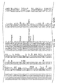
- the gene is selected from the group consisting of those listed in Fig. 13 (Table S5).
- the sample is identified as breast cancer when the somatic mutation is determined.
- a method for diagnosing colorectal cancer in a human is provided.
- a somatic mutation in a gene or its encoded cDNA or protein is determined in a test sample relative to a normal sample of the human.
- the gene is selected from the group consisting of those listed in Fig. 14 . (Table S6).
- the sample is identified as colorectal cancer if the somatic mutation is determined.
- a method for stratifying breast cancers for testing candidate or known anti-cancer therapeutics is provided.
- a CAN-gene mutational signature for a breast cancer is determined by determining at least one somatic mutation in a test sample relative to a normal sample of a human. The at least one somatic mutation is in one or more genes selected from the group consisting of Fig. 13 (Table S5).
- a first group of breast cancers that have the CAN-gene mutational signature is formed. Efficacy of a candidate or known anti-cancer therapeutic on the first group is compared to efficacy on a second group of breast cancers that has a different CAN-gene mutational signature.
- a CAN gene mutational signature which correlates with increased or decreased efficacy of the candidate or known anti-cancer therapeutic relative to other groups is identified.
- a method for stratifying colorectal cancers for testing candidate or known anti-cancer therapeutics is provided.
- a CAN-gene mutational signature for a colorectal cancer is determined by determining at least one somatic mutation in a test sample relative to a normal sample of the human. The at least one somatic mutation is in one or more genes selected from the group consisting of Fig. 14 . (Table S6).
- a first group of colorectal cancers that have the CAN-gene mutational signature is formed. Efficacy of a candidate or known anti-cancer therapeutic on the first group is compared to efficacy on a second group of colorectal cancers that has a different CAN-gene mutational signature.
- a CAN gene mutational signature is identified which correlates with increased or decreased efficacy of the candidate or known anti-cancer therapeutic relative to other groups.
- a method for characterizing a breast cancer in a human is provided.
- a somatic mutation in a gene or its encoded cDNA or protein is determined in a test sample relative to a normal sample of the human.
- the gene is selected from the group consisting of those listed in Fig. 13 (Table S5).
- Another method provided is for characterizing a colorectal cancer in a human.
- a somatic mutation in a gene or its encoded cDNA or protein is determined in a test sample relative to a normal sample of the human.
- the gene is selected from the group consisting of those listed in Fig. 14 (Table S6).
- Fig. 1A and 1B Schematic of Mutation Discovery and Validation Screens.
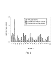
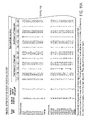
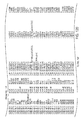
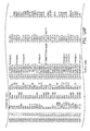
- Fig. 2 Mutation frequency of CAN -gene groups.
- CAN -genes were grouped by function using Gene Ontology groups, INTERPR® domains, and available literature. Bars indicate the fraction of tumors (35 breast or 35 colorectal) with at least one mutated gene in the functional group.
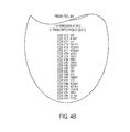
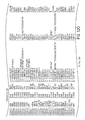
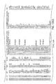
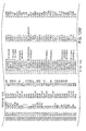
- Fig. 4 (Fig. S2) CCDS genes excluded from analysis.
- Fig. S2 CCDS genes excluded from analysis.
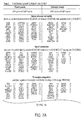
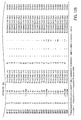
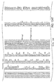
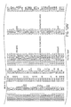
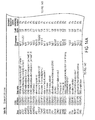
- Fig. 8 (Table S1.) Primers used for PCR amplification and sequencing (page 1 of 1333 only; all primer sequences are publicly available in a downloadable file (1133427_som_tables.zip) at the website of the journal Science (www.sciencemag.org) under Supporting Online Material located at the webpage /cgi/content/full/sci;l 133427/DC1)
- the inventors have developed methods for characterizing breast and colorectal cancers on the basis of gene signatures. These signatures comprise one or more genes which are mutated in a particular cancer.
- the signatures can be used as a means of diagnosis, prognosis, identification of metastasis, stratification for drug studies, and for assigning an appropriate treatment.
- a mutation typically a somatic mutation
- a somatic mutation can be determined by testing either a gene, its mRNA (or derived cDNA), or its encoded protein.
- Any method known in the art for determining a somatic mutation can be used.
- the method may involve sequence determination of all or part of a gene, cDNA, or protein.
- the method may involve mutation-specific reagents such as probes, primers, or antibodies.
- the method may be based on amplification, hybridization, antibody-antigen reactions, primer extension, etc. Any technique or method known in the art for determining a sequence-based feature may be used.
- Samples for testing may be tissue samples from breast or colorectal tissue or body, fluids or products that contain sloughed off cells or genes or mRNA or proteins.
- Such fluids or products include breast milk, stool, breast discharge, intestinal fluid.
- the same type of tissue or fluid is used for the test sample and the normal sample.
- the test sample is, however, suspected of possible neoplastic abnormality, while the normal sample is not suspect.
- Somatic mutations are determined by finding a difference between a test sample and a normal sample of a human. This criterion eliminates the possibility of germline differences confounding the analysis.
- the gene (or cDNA or protein) to be tested is any of those shown in Fig. 13 (Table S5). Particular genes which may be tested and useful are gelsolin GSN, cadherin genes CDH10 and CDH20 , actin and SMAD binding protein filamin B FLNB , and autocrine motility factor receptor AMFR. Additional useful genes include ATP-dependent transporter ATP8BI , intrinsic factor-cobalamin receptor CUBN, actin binding protein DBN1 , and tectorin alpha TECTA.
- the gene (or cDNA or protein) to be tested is any of those shown in Fig. 14 .
- Particular genes which may be tested and useful are ephrin receptor EPHB6, mixed lineage leukemia 3 gene (MLL3), and protein tyrosine phosphatase receptor PTPRD.
- Other genes which may be tested and useful are polycystic kidney and hepatic disease 1 gene PKHD1, guanylate cyclase 1 GUCYIA2, transcription factor TBX22, exocyst complex component SECBL1 , and tubulin tyrosine ligase TTLL3 . Any somatic mutation may be informative.
- Particular mutations which may be used are shown in Fig. 12 (Table S4).
- the number of genes or mutations that may be useful in forming a signature of a breast or colorectal cancer may vary from one to twenty-five. At least two, three, four, five, six, seven or more genes may be used.
- the mutations are typically somatic mutations and non-synonymous mutations. Those mutations described here are within coding regions. Other non-coding region mutations may also be found and may be informative.
- stratification on the basis of signatures can be used.
- One or more groups with a similar mutation signature will be formed and the effect of the therapeutic agent on the group will be compared to the effect of patients whose tumors do not share the signature of the group formed.
- the group of patients who do not share the signature may share a different signature or they may be a mixed population of tumor-bearing patients whose tumors bear a variety of signatures.
- Efficacy can be determined by any of the standard means known in the art. Any index of efficacy can be used. The index may be life span, disease free remission period, tumor shrinkage, tumor growth arrest, improvement of quality of life, decreased side effects, decreased pain, etc. Any useful measure of patient health and well-being can be used. In addition, in vitro testing may be done on tumor cells that have particular signatures. Tumor cells with particular signatures can also be tested in animal models.
- a signature Once a signature has been correlated with sensitivity or resistance to a particular therapeutic regimen, that signature can be used for prescribing a treatment to a patient. Thus determining a signature is useful for making therapeutic decisions.
- the signature can also be combined with other physical or biochemical findings regarding the patient to arrive at a therapeutic decision.
- a signature need not be the sole basis for making a therapeutic decision.
- An anti-cancer agent associated with a signature may be, for example, docetaxel, paclitaxel, topotecan, adriamycin, etoposide, fluorouracil (5-FU), or cyclophosphamide.
- the agent may be an alkylating agent (e.g., nitrogen mustards), antimetabolites (e.g., pyrimidine analogs), radioactive isotopes (e.g., phosphorous and iodine), miscellaneous agents (e.g., substituted ureas) and natural products (e.g., vinca alkyloids and antibiotics).
- the therapeutic agent may be allopurinol sodium, dolasetron mesylate, pamidronate disodium, etidronate, fluconazole, epoetin alfa, levamisole HCL, amifostine, granisetron HCL, leucovorin calcium, sargramostim, dronabinol, mesna, filgrastim, pilocarpine HCL, octreotide acetate, dexrazoxane, ondansetron HCL, ondansetron, busulfan, carboplatin, cisplatin, thiotepa, melphalan HCL, melphalan, cyclophosphamide, ifosfamide, chlorambucil, mechlorethamine HCL, carmustine, lomustine, polifeprosan 20 with carmustine implant, streptozocin, doxorubicin HCL, bleomycin sulf
- coli L-asparaginase Erwinia L-asparaginase, vincristine sulfate, denileukin diftitox, aldesleukin, rituximab, interferon alpha-2a, paclitaxel, docetaxel, BCG live (intravesical), vinblastine sulfate, etoposide, tretinoin, teniposide, porfimer sodium, fluorouracil, betamethasone sodium phosphate and betamethasone acetate, letrozole, etoposide citrororum factor, folinic acid, calcium leucouorin, 5-fluorouricil, adriamycin, cytoxan, or diamino-dichloro-platinum.
- the signatures of CAN genes according to the present invention can be used to determine an appropriate therapy for an individual.
- a sample of a tumor e.g., a tissue obtained by a biopsy procedure, such as a needle biopsy
- the gene expression profile of the tumor can be determined, such as by a nucleic acid array (or protein array) technology, and the expression profile can be compared to a database correlating signatures with treatment outcomes.
- Other information relating to the human e.g ., age, gender, family history, etc.
- a healthcare provider can make a decision to administer or prescribe a particular drug based on the comparison of the CAN gene signature of the tumor and information in the database.
- Exemplary healthcare providers include doctors, nurses, and nurse practitioners. Diagnostic laboratories can also provide a recommended therapy based on signatures and other information about the patient.
- a tumor tissue sample (such as a biopsy) can be taken at any stage of treatment.
- a tumor tissue sample can be taken upon tumor progression, which can be determined by tumor growth or metastasis.
- a CAN gene signature can be determined, and one or more secondary therapeutic agents can be administered to increase, or restore, the sensitivity of the tumor to the primary therapy.
- Treatment predictions may be based on pre-treatment gene signatures. Secondary or subsequent therapeutics can be selected based on the subsequent assessments of the patient and the later signatures of the tumor. The patient will typically be monitored for the effect on tumor progression.
- a medical intervention can be selected based on the identity of the CAN gene signature. For example, individuals can be sorted into subpopulations according to their genotype. Genotype-specific drug therapies can then be prescribed.
- Medical interventions include interventions that are widely practiced, as well as less conventional interventions.
- medical interventions include, but are not limited to, surgical procedures, administration of particular drugs or dosages of particular drugs (e.g., small molecules, bioengineered proteins, and gene-based drugs such as antisense oligonucleotides, ribozymes, gene replacements, and DNA-or RNA-based vaccines), including FDA-approved drugs, FDA-approved drugs used for off-label purposes, and experimental agents.
- Other medical interventions include nutritional therapy, holistic regimens, acupuncture, meditation, electrical or magnetic stimulation, osteopathic remedies, chiropractic treatments, naturopathic treatments, and exercise.
- TP53 and MDM2 mutations exert comparable effects on cells, as do mutations in RB1, CDKN2A (p16), CCND1 and CDK4. It will be of interest to determine whether a limited number of pathways include most CAN-genes, a possibility consistent with the groupings in Fig. 2 and Fig. 7 (Table 3).
- CAN-genes may also be dysregulated in tumors through changes in chromatin or DNA methylation rather than through mutation.
- the CAN-genes define a relatively small subset of genes that could prove useful as markers for neoplasia.
- some of these genes, particularly those on the cell surface or those with enzymatic activity, may prove to be good targets for therapeutic development.
- CCDS consensus coding sequences
- the initial step toward achieving these goals was the development of methods for high-throughput identification of somatic mutations in cancers. These methods included those for primer design, polymerase chain reaction (PCR), sequencing, and mutational analysis ( Fig. 1 ).
- the first component involved extraction of all protein coding sequences from the CCDS genes. A total of 120,839 non-redundant exons and adjacent intronic sequences were obtained from 14,661 different transcripts in CCDS. These sequences were used to design primers for PCR amplification and sequencing of exons and adjacent splice sites. Primers were designed using a number of criteria to ensure robust amplification and sequencing of template regions (7).
- Sequence data were assembled for each amplicon and evaluated for quality within the target region using software specifically designed for this purpose (7).
- the target region of each exon included all coding bases as well as the four intronic bases at both the 5' and 3' ends that serve as the major splice recognition sites.
- ⁇ 90% of bases in the target region have a Phred quality score (defined as - 10[log 10 (raw per-base error)]) of at least 20 in at least three quarters of the tumor samples analyzed ( 8 ). This quality cutoff was chosen to provide high sensitivity for mutation detection while minimizing false positives. Using these criteria, 93% of the 135,483 amplicons and 91% of the total targeted bases in CCDS were successfully analyzed for potential alterations.
- the regions containing them were independently re-amplified and re-sequenced in the corresponding tumors. This step removed 9,295 alterations. The regions containing the putative mutations were then sequenced in matched normal DNA samples to determine whether the mutations were truly somatic: 18,414 changes were observed to be present in the germline of these patients, representing variants not currently annotated in SNP databases, and were excluded. As a final step, the remaining 1,572 putative somatic mutations were carefully examined in silico to ensure that the alterations did not arise from mistargeted sequencing of highly related regions occurring elsewhere in the genome ( 7 ).
- Alterations in such duplicated regions may appear to be somatic when there is loss of one or both alleles of the target region in the tumor and when the selected primers closely match and therefore amplify similar areas of the genome.
- a total of 265 changes in closely related regions were excluded in this fashion, resulting in a total of 1,307 confirmed somatic mutations in 1,149 genes ( Fig. 5 ; Table 1).
- Somatic mutations in human tumors can arise either through selection of functionally important alterations via their effect on net cell growth or through accumulation of non-functional "passenger” alterations that arise during repeated rounds of cell division in the tumor or in its progenitor stem cell.
- distinction between selected and passenger mutations is generally not required when the number of genes and tumors analyzed is small. In large-scale studies, however, such distinctions are of paramount importance ( 11 , 12 ). For example, it has been estimated that nonsynonymous passenger mutations are present at a frequency no higher than ⁇ 1.2 per Mb of DNA in cancers of the breast or colon ( 13 - 15 ).
- the output of this analysis was a ca ncer m utation p revalence (CaMP) score for each gene analyzed.
- the CaMP score reflects the probability that the number of mutations actually observed in a gene is higher than that expected to be observed by chance given the background mutation rate; its derivation is based on principles described in the Supporting Online Material.
- the use of the CaMP score for analysis of somatic mutations is analogous to the use of the LOD score for linkage analysis in familial genetic settings. For example, 90% of the genes with CaMP scores > 1.0 are predicted to have mutation frequencies higher than the background mutation frequency.
- CAN -genes could be divided into three classes: (a) genes previously observed to be mutationally altered in human cancers; (b) genes in which no previous mutations in human cancers had been discovered but had been linked to cancer through functional studies; and (c) genes with no previous strong connections to neoplasia.
- nucleoporin NUP214 (2) , kinesin receptor KTN1 (27) , DEAD box polypeptide 10 DDX10 (28), glioma-associated oncogene homolog 1 GLII (29) , and the translocation target gene of the runt related transcription factor 1 RUNX1 T1 (MTG8) ( 2 ).
- MTG8 translocation target gene of the runt related transcription factor 1 RUNX1 T1
- At least one of these genes was mutated in more than 80% of the tumors of each type.
- Zinc-finger transcription factors were particularly highly represented (8 genes mutated collectively in 43% of breast cancer samples).
- genes involved in cell adhesion represented ⁇ 22% of CAN -genes and affected more than two thirds of tumors of either type.
- Genes involved in signal transduction represented ⁇ 23% of CAN -genes and at least one such gene was mutated in 77% and 94% of the breast and colorectal cancer samples, respectively.
- CCOS Consensus Coding DNA Sequence database
- CCOSI Consensus Coding DNA Sequence database
- genomic coordinates defining the transcript coding sequence must be identical in Ensembl and RefSeq databases.
- the transcripts must have canonical start and stop codons and consensus splice sites, not have in-frame stop codons, and be translatable from the reference genome sequence without frameshifts.
- CCOS transcripts must be supported by transcript and protein homology and inter-species conservation.
- Bioinformatic resources CCOS gene and transcript coordinates (release 1, 3 / 02 / 05), human genome sequences, and single nucleotide polymorphisms were obtained from the UCSC Santa Cruz Genome Bioinformatics Site (http://genome.ucsc.edu). Homology searches in the human and mouse genomes were performed using the BLAST-like alignment tool BLAT (S1) and In Silico PCR (http://genome.ucsc.edu/cqi-bin/hqPcr), All genomic positions correspond to UCSC Santa Cruz hgl7 build 35.1 human genome sequence.
- S1 BLAST-like alignment tool
- S2 In Silico PCR
- Primer design For each transcript, genomic sequences comprising the entire coding region of each exon as well as flanking intronic sequences and 5' UTR and 3' UTR sequences were extracted. Primer pairs for PCR amplification and sequencing of each coding exon were generated using Primer3 (http://frodo.wi.mit.edu/cqi-bin/primer3/primer3 www. cqi ) (S3). Forward and reverse PCR primers were required to be located no closer than 50 bp to the target exon boundaries, and genomic positions with known polymorph isms were avoided in the five 3'-most bases of the primers. Exons larger than 350 bp were analyzed as multiple overlapping amplicons.
- PCR products were designed to range in size from 300 to 600 bp, which was considered optimal for amplification, purification, and sequencing.
- primer pairs were filtered using UCSC In Silico PCR and only pairs yielding a single product were used. 0.33 Mb (-1.5%) of target genomic sequence was excluded from further analysis due to a lack of suitable amplification and sequencing primers. A total of 135,483 primer pairs encompassing -21 Mb of target sequence were successfully designed.
- a universal sequencing primer (M13 forward, 5'-GTAAAACGACGGCCAGT-3'; SEQ ID NO: 1) was appended to the 5' end of the primer in the pair with the smallest number of mono- and dinucleotide repeats between itself and the target exon. Primer sequences are listed in Fig. 8 ; Table S1.
- Tumor samples DNA samples from ductal breast carcinoma cell lines and matched normal mammary tissue or peripheral blood lines were obtained from American Type Culture Collection (Manassas, VA) or from A. Gazdar (S4, S5). Primary breast tumor and surrounding normal surgical tissue specimens isolated from node positive patients at Palmetto Health Richland or Institution Hospitals were obtained through the South Carolina Cancer Center Tissue Bank. Each tissue sample was flash frozen within 30 minutes of excision, and stored at -80 ° C . Surgically removed colorectal tumors were disaggregated and implanted into nude mice or into in vitro culture conditions as described previously (S6, 57). DNA was prepared within 3 passages after xenograft establishment.
- Laser capture microdissection 20 ⁇ m sections of snap frozen primary breast tumor tissues embedded in OCT were deposited on Sigma silane-prepTM slides and stained with hematoxylin and eosin. Tumor cells were separated from surrounding tissue and recovered on transfer film by laser-capture microdissection (PixCelle® lie, Arcturus). Genomic DNA was purified from approximately 20 slides for each sample using the QiagenTM QIAamp® DNA Micro kit according to the manufacturer's protocol.
- Whole Genome Amplification Whole genome amplification was used to provide sufficient quantities of DNA for the Validation Screen. Briefly, 5-20 ng template DNA was denatured with 5 M KOH, neutralized and incubated at 30°C for 16-24 hours with 4x REPLI-g buffer and REPLI-g DNA polymerase according to the manufacturer's instructions (Qiagen, Valencia, CA). Samples were incubated at 65°C for 3 min to inactivate the enzyme before storage at20°C. For each sample, a minimum of 5 independent WGA reactions were pooled to reduce the effects of any allelic or locus bias that may have occurred during amplification.
- PCR amplification and sequencing All primers were synthesized by Invitrogen (San Diego, CA). PCR was performed in 5 III reactions containing 1 x PCR Buffer (67 mM TrisHCl, pH 8.8, 6.7 mM MgCb, 16.6 mM NH4S04, 10 mM 2-mercaptoethanol), 1 mM dNTPs (Invitrogen, San Diego, CA), 1 11 M forward and 1 11 M reverse primers, 6% DMSO, 2 mM ATP, 0.25 U Platinum Taq (Invitrogen, San Diego, CA) and 3 ng DNA.
- 1 PCR Buffer 67 mM TrisHCl, pH 8.8, 6.7 mM MgCb, 16.6 mM NH4S04, 10 mM 2-mercaptoethanol
- 1 mM dNTPs Invitrogen, San Diego, CA
- 1 11 M forward and 1 11 M reverse primers 6% DMSO
- 2 mM ATP 0.25 U
- Reactions were carried out in 384-well AB19700 thermocyclers (Applied Biosystems, Foster City, CA) using a touchdown PCR protocol (1 cycle of 96°C for 2 min; 3 cycles of 96°C for 10 see, 64°C for 10 see, 70°C for 30 see; 3 cycles of 96°C for 10 see, 61°C for 10 see, 70°C for 30 see; 3 cycles of 96°C for 10 see, 58°C for 10 see, 70°C for 30 see; 41 cycles of 96°C for 10 see, 57°C for 10 see, 70°C for 30 see; 1 cycle of 70°C for 5 min).
- Templates were purified using AMPure (Agencourt Biosciences, Beverly, MA) and sequencing carried out with M13 forward primer (5'GTAAAACGACGGCCAGT-3'; SEQ ID NO: 1) and Big Dye Terminator Kit v.3.1 (Applied Biosystems, Foster City, CA). 1 % DMSO was included in sequencing reactions when the GC content of the template exceeded 65%. Dye terminators were removed using the CleanSEQ kit (Agencourt Biosciences, Beverly, MA) and sequence reactions were delineated on ABI PRISM 3730x1 sequencing apparatuses (Applied Biosystems, Foster City, CA).
- Sequence assembly and analysis of mutations Sequence traces from tumor and normal DNA samples were aligned to the genomic reference sequences. To consider an amplicon successfully sequenced, at least three quarters of the tumors were required to have 2':90% of the bases in the target region with a Phred quality score of 20 or better. Amplicons not meeting these criteria were not analyzed further. Mutational analysis was performed for all coding exonic sequences and the flanking 4 bp of intronic or UTR sequences using Mutation Surveyor (Softgenetics, State College, PA) coupled to a relational database (Microsoft SQL Server). For both Mutation Discovery and Validation Screens, the following basic steps were employed to identify mutations of interest. First, synonymous changes were identified and excluded from further analysis.
- BLAT 58 was used to search these sequences against the human genome. This examination ensured that the nucleotide change was not present in a highly related region in the human genome. For putative somatic mutations found in xenografted tumors, BLAT was used to similarly search the mouse genome to exclude the contribution of homologous mouse sequences.
- Mutation Validation Screen Every gene found mutated in the Discovery Screen was further analyzed by amplification and sequencing of 24 additional tumor samples of the same tissue type. Because of limiting amounts of sample DNA, the set of 24 tumors evaluated changed over time. All CCDS transcript variants of the gene of interest were investigated using primer pairs that yielded informative sequences in the Discovery Screen. Mutation detection, confirmation of alterations, and determination of somatic status was performed as above, with the exception that all germ line variants previously observed in the normal DNA samples of the Discovery Screen were considered to be known variants ( Fig. 1 ).
- NcpG, NTpC, NA, Nc, NG, and NT were designated NcpG, NTpC, NA, Nc, NG, and NT, respectively.
- N c did not include those C's within 5'-CpG or 5 ⁇ -TpC dinucleotides and NG did not include those G's within 5'-CpG-3' or 5'GpA-3 dinucleotides.
- the parameters for this calculation for the 5'-CpG-3' category used the observed number of mutations at 5'-CpG-3' sites as the number of positive events, NcpG as the number of independent trials, and the background mutation frequencies for NcpG listed in the table above (7.73 x 10⁻⁶ for colorectal cancers) as the probability of a positive result in each trial.
- the probabilities of a gene having the observed number of mutations at each of the other five dinucleotide or mononucleotides were similarly calculated.
- the probability of a gene containing the observed number of insertions, deletions, or duplications was calculated by using a binomial distribution with the following parameters: observed number of INS/DEL/DUP events as the number of positive events, total nucleotides successfully sequenced within the gene as the number of independent trials, and 0.55 x 10 -6 as the probability of a positive result in each trial. Note that each of these seven probabilities was considered to be independent.
- the probability of a gene having the observed number of mutations at the observed positions was then calculated to be the product of the seven nucleotide context-specific probabilities.
Landscapes
- Chemical & Material Sciences (AREA)
- Life Sciences & Earth Sciences (AREA)
- Health & Medical Sciences (AREA)
- Organic Chemistry (AREA)
- Proteomics, Peptides & Aminoacids (AREA)
- Engineering & Computer Science (AREA)
- Immunology (AREA)
- Pathology (AREA)
- Analytical Chemistry (AREA)
- Zoology (AREA)
- Wood Science & Technology (AREA)
- Genetics & Genomics (AREA)
- Hospice & Palliative Care (AREA)
- Biochemistry (AREA)
- Microbiology (AREA)
- Molecular Biology (AREA)
- Biophysics (AREA)
- Physics & Mathematics (AREA)
- Oncology (AREA)
- Biotechnology (AREA)
- Bioinformatics & Cheminformatics (AREA)
- General Engineering & Computer Science (AREA)
- General Health & Medical Sciences (AREA)
- Measuring Or Testing Involving Enzymes Or Micro-Organisms (AREA)
- Investigating Or Analysing Biological Materials (AREA)
- Apparatus For Radiation Diagnosis (AREA)
Applications Claiming Priority (3)
| Application Number | Priority Date | Filing Date | Title |
|---|---|---|---|
| US83694406P | 2006-08-11 | 2006-08-11 | |
| US84236306P | 2006-09-06 | 2006-09-06 | |
| EP07811279.4A EP2069535B1 (fr) | 2006-08-11 | 2007-08-13 | Séquences codantes consensus de cancers du sein et colorectaux humains |
Related Parent Applications (1)
| Application Number | Title | Priority Date | Filing Date |
|---|---|---|---|
| EP07811279.4 Division | 2007-08-13 |
Publications (2)
| Publication Number | Publication Date |
|---|---|
| EP2543740A2 true EP2543740A2 (fr) | 2013-01-09 |
| EP2543740A3 EP2543740A3 (fr) | 2013-03-27 |
Family
ID=39082658
Family Applications (4)
| Application Number | Title | Priority Date | Filing Date |
|---|---|---|---|
| EP12176466.6A Revoked EP2543739B1 (fr) | 2006-08-11 | 2007-08-13 | Séquences codantes consensus des cancer colorectaux humains |
| EP12176467A Withdrawn EP2543740A3 (fr) | 2006-08-11 | 2007-08-13 | Séquences de codage de consensus des cancers colorectaux humains |
| EP07811279.4A Active EP2069535B1 (fr) | 2006-08-11 | 2007-08-13 | Séquences codantes consensus de cancers du sein et colorectaux humains |
| EP15175950.3A Withdrawn EP2949763A3 (fr) | 2006-08-11 | 2007-08-13 | Séquences de codage de consensus des cancers du sein humains |
Family Applications Before (1)
| Application Number | Title | Priority Date | Filing Date |
|---|---|---|---|
| EP12176466.6A Revoked EP2543739B1 (fr) | 2006-08-11 | 2007-08-13 | Séquences codantes consensus des cancer colorectaux humains |
Family Applications After (2)
| Application Number | Title | Priority Date | Filing Date |
|---|---|---|---|
| EP07811279.4A Active EP2069535B1 (fr) | 2006-08-11 | 2007-08-13 | Séquences codantes consensus de cancers du sein et colorectaux humains |
| EP15175950.3A Withdrawn EP2949763A3 (fr) | 2006-08-11 | 2007-08-13 | Séquences de codage de consensus des cancers du sein humains |
Country Status (5)
| Country | Link |
|---|---|
| US (7) | US8741573B2 (fr) |
| EP (4) | EP2543739B1 (fr) |
| AU (1) | AU2007284649B2 (fr) |
| ES (2) | ES2548690T3 (fr) |
| WO (1) | WO2008021288A2 (fr) |
Families Citing this family (13)
| Publication number | Priority date | Publication date | Assignee | Title |
|---|---|---|---|---|
| EP1871809A2 (fr) | 2005-04-22 | 2008-01-02 | Morphotek, Inc. | Anticorps a activite d'effecteur immunitaire et internalises dans des cellules positives vis-a-vis de l'endosialine |
| EP2543739B1 (fr) | 2006-08-11 | 2015-07-22 | Johns Hopkins University | Séquences codantes consensus des cancer colorectaux humains |
| WO2008100913A2 (fr) * | 2007-02-12 | 2008-08-21 | The Johns Hopkins University | Détection précoce et pronostic de cancers du côlon |
| CA2682726C (fr) | 2007-04-05 | 2017-05-09 | Morphotek, Inc. | Procedes pour inhiber la liaison de l'endosialine a des ligands |
| EP2164990A4 (fr) * | 2007-05-21 | 2011-02-23 | Dana Farber Cancer Inst Inc | Compositions, trousses et procédés d'identification, d'évaluation, de prévention et de thérapie du cancer |
| US20090123928A1 (en) * | 2007-10-11 | 2009-05-14 | The Johns Hopkins University | Genomic Landscapes of Human Breast and Colorectal Cancers |
| JP5428527B2 (ja) * | 2008-06-03 | 2014-02-26 | 住友化学株式会社 | 化学物質が有する発生毒性の予測方法 |
| WO2011024433A1 (fr) * | 2009-08-24 | 2011-03-03 | Oncotherapy Science, Inc. | Gène adamts18 lié au cancer du poumon et au cancer de l'sophage |
| WO2013130748A1 (fr) * | 2012-02-29 | 2013-09-06 | The Regents Of The University Of Michigan | Marqueurs du cancer de la prostate et leurs utilisations |
| US9957557B2 (en) | 2012-08-13 | 2018-05-01 | Life Genetics Lab, Llc | Development of a highly sensitive quantification system for assessing DNA degradation and quality in forensic samples |
| US10993418B2 (en) | 2012-08-13 | 2021-05-04 | Life Genetics Lab, Llc | Method for measuring tumor burden in patient derived xenograft (PDX) mice |
| WO2014059381A1 (fr) * | 2012-10-11 | 2014-04-17 | Creighton University | Biomarqueurs du cancer du sein de la lignée germinale mll3 et leurs utilisations |
| WO2020136133A1 (fr) | 2018-12-23 | 2020-07-02 | F. Hoffmann-La Roche Ag | Classification de tumeur basée sur une charge mutationnelle tumorale prédite |
Family Cites Families (13)
| Publication number | Priority date | Publication date | Assignee | Title |
|---|---|---|---|---|
| US5605799A (en) * | 1990-07-12 | 1997-02-25 | University Of Utah Research Foundation | Somatic mutations in neurofibromatosis type 1 gene in human tumors |
| AU1366992A (en) * | 1991-01-16 | 1992-08-27 | Cancer Institute, The | Inherited and somatic mutations of apc gene in colorectal cancer of humans |
| DE69625678T3 (de) * | 1995-12-18 | 2006-11-09 | The University Of Utah Research Foundation, Salt Lake City | Chromosom 13 verbundene Brustkrebsempfindlichkeitsgen BRCA2 |
| US6630301B1 (en) | 1997-03-14 | 2003-10-07 | The Penn State Research Foundation | Detection of extracellular tumor-associated nucleic acid in blood plasma or serum |
| CA2273051A1 (fr) * | 1996-12-03 | 1998-06-11 | Prasanna Athma | Predisposition au cancer du sein liee a des mutations affectant le locus du gene d'ataxie telangiectasie |
| EP1141726B1 (fr) * | 1999-01-14 | 2004-08-04 | CA*TX, Inc. | Analyses immunologiques destinees a detecter des maladies ou des traits de predisposition a des maladies |
| WO2004082458A2 (fr) | 2003-02-21 | 2004-09-30 | The Johns Hopkins University | Tyrosine kinome |
| PE20051046A1 (es) * | 2003-11-28 | 2006-01-11 | Novartis Ag | Derivados de diaril-urea en el tratamiento de enfermedades dependientes de la quinasa de proteina |
| US8039210B2 (en) * | 2004-05-14 | 2011-10-18 | The Johns Hopkins University | Protein tyrosine phosphatase mutations in cancers |
| US7384556B2 (en) | 2005-07-19 | 2008-06-10 | The University Of Wyoming Research Corporation | Methods of enhancing biodegradation of groundwater contaminants |
| US8783397B2 (en) | 2005-07-19 | 2014-07-22 | Bsst Llc | Energy management system for a hybrid-electric vehicle |
| EP2543739B1 (fr) | 2006-08-11 | 2015-07-22 | Johns Hopkins University | Séquences codantes consensus des cancer colorectaux humains |
| US20140199405A1 (en) | 2013-01-11 | 2014-07-17 | Abraxis Bioscience, Llc | Method for treating cancer based on mutation status of k-ras |
-
2007
- 2007-08-13 EP EP12176466.6A patent/EP2543739B1/fr not_active Revoked
- 2007-08-13 WO PCT/US2007/017866 patent/WO2008021288A2/fr not_active Ceased
- 2007-08-13 EP EP12176467A patent/EP2543740A3/fr not_active Withdrawn
- 2007-08-13 US US12/377,073 patent/US8741573B2/en active Active
- 2007-08-13 AU AU2007284649A patent/AU2007284649B2/en active Active
- 2007-08-13 EP EP07811279.4A patent/EP2069535B1/fr active Active
- 2007-08-13 ES ES12176466.6T patent/ES2548690T3/es active Active
- 2007-08-13 EP EP15175950.3A patent/EP2949763A3/fr not_active Withdrawn
- 2007-08-13 ES ES07811279T patent/ES2421354T3/es active Active
-
2014
- 2014-03-25 US US14/224,102 patent/US9551037B2/en active Active
-
2017
- 2017-01-24 US US15/413,903 patent/US20170362659A1/en not_active Abandoned
-
2019
- 2019-10-25 US US16/664,505 patent/US20200048719A1/en not_active Abandoned
-
2020
- 2020-03-19 US US16/824,052 patent/US10787712B2/en active Active
-
2023
- 2023-11-22 US US18/518,055 patent/US20240344137A1/en not_active Abandoned
-
2025
- 2025-09-18 US US19/332,261 patent/US20260062759A1/en active Pending
Non-Patent Citations (28)
| Title |
|---|
| "Tumor Cell Heterogeneity", 1982, ACADEMIC PRESS, pages: 441 - 460 |
| A. DUVAL ET AL., CANCER RES, vol. 59, 1999, pages 4213 |
| A. SASAKI; Y. MASUDA; Y. OHTA; K. IKEDA; K. WATANABE, J BIOL CHEM, vol. 276, 2001, pages 17871 |
| B. PADMANABHAN ET AL., MOL CELL, vol. 21, 2006, pages 689 |
| C. A. LANDIS ET AL., NATURE, vol. 340, 1989, pages 692 |
| H. VARMUS, SCIENCE, vol. 312, 2006, pages 1162 |
| J. F. COSTELLO ET AL., NAT GENET, vol. 24, 2000, pages 132 |
| J. L. BOS ET AL., NATURE, vol. 327, 1987, pages 293 |
| J. L. KU ET AL., CANCER LETT, 5 July 2006 (2006-07-05) |
| K. D. PRUITT; T. TATUSOVA; D. R. MAGLOTT, NUCLEIC ACIDS RES, vol. 33, 2005, pages D501 |
| K. EPPERT ET AL., CELL, vol. 86, 1996, pages 543 |
| K. SALASSIDIS ET AL., CANCER RES, vol. 60, 2000, pages 2786 |
| K. W. KINZLER ET AL., SCIENCE, vol. 236, 1987, pages 70 |
| M. HOLLSTEIN; D. SIDRANSKY; B. VOGELSTEIN; C. C. HARRIS, SCIENCE, vol. 253, 1991, pages 49 |
| M. OLIVIER; S. P. HUSSAIN; C. CARON DE FROMCNTEL; P. HAINAUT; C. C. HARRIS, IARC SCI PUBL, vol. 247, 2004 |
| M. RUAULT; M. E. BRUN; M. VENTURA; G. ROIZES; A. DE SARIO, GENE, vol. 284, 2002, pages 73 |
| M. SATO ET AL., GENES CHROMOSOMES CANCER, vol. 44, 2005, pages 405 |
| M. TANAKA ET AL., CANCER RES, vol. 55, 1995, pages 3228 |
| O. BLUTEAU ET AL., NAT GENET, vol. 32, 2002, pages 312 |
| R. BRENT, CELL, vol. 100, 2000, pages 169 |
| S. L. OOI ET AL., TRENDS GENET, vol. 22, 2006, pages 56 |
| S. MARKOWITZ ET AL., SCIENCE, vol. 268, 1995, pages 1336 |
| T. IDEKER ET AL., SCIENCE, vol. 292, 2001, pages 929 |
| T. SOUSSI; G. LOZANO, BIOCHEM BIOPHYS RES COMMUN, vol. 331, 2005, pages 834 |
| X. X. TANG; G. M. BRODEUR; B. G. CAMPLING; N. IKEGAKI, CLIN CANCER RES, vol. 5, 1999, pages 455 |
| Y. ARAI ET AL., BLOOD, vol. 89, 1997, pages 3936 |
| Y. H. ROGERS; J. C. VENTER, NATURE, vol. 437, 2005, pages 326 |
| Y. ONISHI; K. TSUKADA; J. YOKOTA; A. RAZ, CLIN EXP METASTASIS, vol. 20, 2003, pages 51 |
Also Published As
| Publication number | Publication date |
|---|---|
| US20260062759A1 (en) | 2026-03-05 |
| US8741573B2 (en) | 2014-06-03 |
| WO2008021288A3 (fr) | 2008-11-13 |
| US9551037B2 (en) | 2017-01-24 |
| US10787712B2 (en) | 2020-09-29 |
| US20170362659A1 (en) | 2017-12-21 |
| EP2069535A4 (fr) | 2010-04-21 |
| EP2069535B1 (fr) | 2013-04-10 |
| ES2421354T3 (es) | 2013-08-30 |
| AU2007284649B2 (en) | 2013-09-26 |
| US20200048719A1 (en) | 2020-02-13 |
| ES2548690T3 (es) | 2015-10-20 |
| US20150167095A1 (en) | 2015-06-18 |
| EP2543739B1 (fr) | 2015-07-22 |
| US20100316995A1 (en) | 2010-12-16 |
| HK1180731A1 (en) | 2013-10-25 |
| WO2008021288A2 (fr) | 2008-02-21 |
| AU2007284649A1 (en) | 2008-02-21 |
| US20240344137A1 (en) | 2024-10-17 |
| EP2069535A2 (fr) | 2009-06-17 |
| EP2543740A3 (fr) | 2013-03-27 |
| EP2949763A3 (fr) | 2016-03-09 |
| US20200239970A1 (en) | 2020-07-30 |
| EP2949763A2 (fr) | 2015-12-02 |
| EP2543739A1 (fr) | 2013-01-09 |
Similar Documents
| Publication | Publication Date | Title |
|---|---|---|
| US20260062759A1 (en) | Consensus coding sequences of human breast and colorectal cancer | |
| CN101248180B (zh) | 作为检测日光照射、前列腺癌和其它癌症的诊断工具的线粒体突变及重排 | |
| US20090123928A1 (en) | Genomic Landscapes of Human Breast and Colorectal Cancers | |
| AU2018298443B2 (en) | Target-enriched multiplexed parallel analysis for assessment of tumor biomarkers | |
| Behdad et al. | A clinical grade sequencing-based assay for CEBPA mutation testing: report of a large series of myeloid neoplasms | |
| Favis et al. | Harmonized microarray/mutation scanning analysis of TP53 mutations in undissected colorectal tumors | |
| EP3612643A1 (fr) | Stratification et pronostic de cancer | |
| HK1180730A (en) | Consensus coding sequences of human colorectal cancers | |
| US9695479B2 (en) | Medulloblastoma genes as targets for diagnosis and therapeutics | |
| HK1180731B (en) | Consensus coding sequences of human colorectal cancers | |
| KR102063240B1 (ko) | 폐암 환자의 변증 구분용 snp 마커 및 이의 용도 | |
| CN109825590A (zh) | 肿瘤驱动基因突变和微卫星不稳定性联合检测的方法、试剂盒和引物 | |
| Pepek et al. | Results of Polish Adult Leukemia Study Group (PALG) project assessing TP53 mutations with next-generation sequencing technology in relapsed and refractory chronic lymphocytic leukemia patients—an 18-month update | |
| KR20250157590A (ko) | 단일염기변이를 이용한 개의 종양 예측 또는 진단용 조성물 | |
| Lung | Identification of candidate genes predisposing to familial colorectal cancer by germline whole exome sequencing | |
| Liquori et al. | A Single-Run Next-Generation Sequencing (NGS) Assay for the Simultaneous Detection of Both Gene Mutations and Large Chromosomal Abnormalities in Patients with Myelodysplastic Syndromes (MDS) and Related Myeloid Neoplasms. Cancers 2021, 13, 1947 | |
| EP1384777B1 (fr) | Adn genomiques participant a la polyarthrite rhumatoide, son procede de diagnostic, son procede d'estimation du risque d'apparition et trousse de diagnostic de detection associee | |
| CN117327796A (zh) | 用于检测尿路上皮癌的组合物及其用途 | |
| AU2011242123B2 (en) | Mitochondrial mutations and rearrangements as a diagnostic tool for the detection of sun exposure, prostate cancer and other cancers | |
| HK40030208A (en) | Target-enriched multiplexed parallel analysis for assessment of tumor biomarkers | |
| HK40030208B (en) | Target-enriched multiplexed parallel analysis for assessment of tumor biomarkers | |
| Wood | The genomic landscapes of human cancer |
Legal Events
| Date | Code | Title | Description |
|---|---|---|---|
| PUAI | Public reference made under article 153(3) epc to a published international application that has entered the european phase |
Free format text: ORIGINAL CODE: 0009012 |
|
| AC | Divisional application: reference to earlier application |
Ref document number: 2069535 Country of ref document: EP Kind code of ref document: P |
|
| AK | Designated contracting states |
Kind code of ref document: A2 Designated state(s): AT BE BG CH CY CZ DE DK EE ES FI FR GB GR HU IE IS IT LI LT LU LV MC MT NL PL PT RO SE SI SK TR |
|
| PUAL | Search report despatched |
Free format text: ORIGINAL CODE: 0009013 |
|
| AK | Designated contracting states |
Kind code of ref document: A3 Designated state(s): AT BE BG CH CY CZ DE DK EE ES FI FR GB GR HU IE IS IT LI LT LU LV MC MT NL PL PT RO SE SI SK TR |
|
| RIC1 | Information provided on ipc code assigned before grant |
Ipc: C12Q 1/68 20060101AFI20130218BHEP |
|
| REG | Reference to a national code |
Ref country code: HK Ref legal event code: DE Ref document number: 1180730 Country of ref document: HK |
|
| 17P | Request for examination filed |
Effective date: 20130927 |
|
| RBV | Designated contracting states (corrected) |
Designated state(s): AT BE BG CH CY CZ DE DK EE ES FI FR GB GR HU IE IS IT LI LT LU LV MC MT NL PL PT RO SE SI SK TR |
|
| STAA | Information on the status of an ep patent application or granted ep patent |
Free format text: STATUS: THE APPLICATION IS DEEMED TO BE WITHDRAWN |
|
| 18D | Application deemed to be withdrawn |
Effective date: 20130928 |
|
| REG | Reference to a national code |
Ref country code: HK Ref legal event code: WD Ref document number: 1180730 Country of ref document: HK |