EP2632359B1 - Blutstillungsinstrument - Google Patents
Blutstillungsinstrument Download PDFInfo
- Publication number
- EP2632359B1 EP2632359B1 EP11773008.5A EP11773008A EP2632359B1 EP 2632359 B1 EP2632359 B1 EP 2632359B1 EP 11773008 A EP11773008 A EP 11773008A EP 2632359 B1 EP2632359 B1 EP 2632359B1
- Authority
- EP
- European Patent Office
- Prior art keywords
- hemostasis
- tissue
- coagulation
- electrode
- patient
- Prior art date
- Legal status (The legal status is an assumption and is not a legal conclusion. Google has not performed a legal analysis and makes no representation as to the accuracy of the status listed.)
- Active
Links
Images


Classifications
-
- A—HUMAN NECESSITIES
- A61—MEDICAL OR VETERINARY SCIENCE; HYGIENE
- A61B—DIAGNOSIS; SURGERY; IDENTIFICATION
- A61B18/00—Surgical instruments, devices or methods for transferring non-mechanical forms of energy to or from the body
- A61B18/04—Surgical instruments, devices or methods for transferring non-mechanical forms of energy to or from the body by heating
- A61B18/08—Surgical instruments, devices or methods for transferring non-mechanical forms of energy to or from the body by heating by means of electrically-heated probes
-
- A—HUMAN NECESSITIES
- A61—MEDICAL OR VETERINARY SCIENCE; HYGIENE
- A61B—DIAGNOSIS; SURGERY; IDENTIFICATION
- A61B18/00—Surgical instruments, devices or methods for transferring non-mechanical forms of energy to or from the body
- A61B18/04—Surgical instruments, devices or methods for transferring non-mechanical forms of energy to or from the body by heating
- A61B18/12—Surgical instruments, devices or methods for transferring non-mechanical forms of energy to or from the body by heating by passing a current through the tissue to be heated, e.g. high-frequency current
-
- A—HUMAN NECESSITIES
- A61—MEDICAL OR VETERINARY SCIENCE; HYGIENE
- A61B—DIAGNOSIS; SURGERY; IDENTIFICATION
- A61B18/00—Surgical instruments, devices or methods for transferring non-mechanical forms of energy to or from the body
- A61B18/04—Surgical instruments, devices or methods for transferring non-mechanical forms of energy to or from the body by heating
- A61B18/042—Surgical instruments, devices or methods for transferring non-mechanical forms of energy to or from the body by heating using additional gas becoming plasma
-
- A—HUMAN NECESSITIES
- A61—MEDICAL OR VETERINARY SCIENCE; HYGIENE
- A61B—DIAGNOSIS; SURGERY; IDENTIFICATION
- A61B5/00—Measuring for diagnostic purposes; Identification of persons
- A61B5/48—Other medical applications
- A61B5/4836—Diagnosis combined with treatment in closed-loop systems or methods
-
- A—HUMAN NECESSITIES
- A61—MEDICAL OR VETERINARY SCIENCE; HYGIENE
- A61M—DEVICES FOR INTRODUCING MEDIA INTO, OR ONTO, THE BODY; DEVICES FOR TRANSDUCING BODY MEDIA OR FOR TAKING MEDIA FROM THE BODY; DEVICES FOR PRODUCING OR ENDING SLEEP OR STUPOR
- A61M5/00—Devices for bringing media into the body in a subcutaneous, intra-vascular or intramuscular way; Accessories therefor, e.g. filling or cleaning devices, arm-rests
-
- H—ELECTRICITY
- H05—ELECTRIC TECHNIQUES NOT OTHERWISE PROVIDED FOR
- H05H—PLASMA TECHNIQUE; PRODUCTION OF ACCELERATED ELECTRICALLY-CHARGED PARTICLES OR OF NEUTRONS; PRODUCTION OR ACCELERATION OF NEUTRAL MOLECULAR OR ATOMIC BEAMS
- H05H1/00—Generating plasma; Handling plasma
- H05H1/24—Generating plasma
- H05H1/2406—Generating plasma using dielectric barrier discharges, i.e. with a dielectric interposed between the electrodes
-
- A—HUMAN NECESSITIES
- A61—MEDICAL OR VETERINARY SCIENCE; HYGIENE
- A61B—DIAGNOSIS; SURGERY; IDENTIFICATION
- A61B18/00—Surgical instruments, devices or methods for transferring non-mechanical forms of energy to or from the body
- A61B18/04—Surgical instruments, devices or methods for transferring non-mechanical forms of energy to or from the body by heating
- A61B18/12—Surgical instruments, devices or methods for transferring non-mechanical forms of energy to or from the body by heating by passing a current through the tissue to be heated, e.g. high-frequency current
- A61B18/14—Probes or electrodes therefor
- A61B18/1477—Needle-like probes
-
- A—HUMAN NECESSITIES
- A61—MEDICAL OR VETERINARY SCIENCE; HYGIENE
- A61B—DIAGNOSIS; SURGERY; IDENTIFICATION
- A61B18/00—Surgical instruments, devices or methods for transferring non-mechanical forms of energy to or from the body
- A61B2018/00315—Surgical instruments, devices or methods for transferring non-mechanical forms of energy to or from the body for treatment of particular body parts
- A61B2018/00559—Female reproductive organs
-
- A—HUMAN NECESSITIES
- A61—MEDICAL OR VETERINARY SCIENCE; HYGIENE
- A61B—DIAGNOSIS; SURGERY; IDENTIFICATION
- A61B18/00—Surgical instruments, devices or methods for transferring non-mechanical forms of energy to or from the body
- A61B2018/00571—Surgical instruments, devices or methods for transferring non-mechanical forms of energy to or from the body for achieving a particular surgical effect
- A61B2018/00589—Coagulation
-
- A—HUMAN NECESSITIES
- A61—MEDICAL OR VETERINARY SCIENCE; HYGIENE
- A61B—DIAGNOSIS; SURGERY; IDENTIFICATION
- A61B18/00—Surgical instruments, devices or methods for transferring non-mechanical forms of energy to or from the body
- A61B2018/00571—Surgical instruments, devices or methods for transferring non-mechanical forms of energy to or from the body for achieving a particular surgical effect
- A61B2018/0063—Sealing
-
- A—HUMAN NECESSITIES
- A61—MEDICAL OR VETERINARY SCIENCE; HYGIENE
- A61B—DIAGNOSIS; SURGERY; IDENTIFICATION
- A61B18/00—Surgical instruments, devices or methods for transferring non-mechanical forms of energy to or from the body
- A61B2018/00994—Surgical instruments, devices or methods for transferring non-mechanical forms of energy to or from the body combining two or more different kinds of non-mechanical energy or combining one or more non-mechanical energies with ultrasound
-
- A—HUMAN NECESSITIES
- A61—MEDICAL OR VETERINARY SCIENCE; HYGIENE
- A61B—DIAGNOSIS; SURGERY; IDENTIFICATION
- A61B18/00—Surgical instruments, devices or methods for transferring non-mechanical forms of energy to or from the body
- A61B18/04—Surgical instruments, devices or methods for transferring non-mechanical forms of energy to or from the body by heating
- A61B18/12—Surgical instruments, devices or methods for transferring non-mechanical forms of energy to or from the body by heating by passing a current through the tissue to be heated, e.g. high-frequency current
- A61B18/14—Probes or electrodes therefor
- A61B2018/1405—Electrodes having a specific shape
- A61B2018/1425—Needle
-
- A—HUMAN NECESSITIES
- A61—MEDICAL OR VETERINARY SCIENCE; HYGIENE
- A61B—DIAGNOSIS; SURGERY; IDENTIFICATION
- A61B18/00—Surgical instruments, devices or methods for transferring non-mechanical forms of energy to or from the body
- A61B18/04—Surgical instruments, devices or methods for transferring non-mechanical forms of energy to or from the body by heating
- A61B18/12—Surgical instruments, devices or methods for transferring non-mechanical forms of energy to or from the body by heating by passing a current through the tissue to be heated, e.g. high-frequency current
- A61B18/14—Probes or electrodes therefor
- A61B2018/147—Electrodes transferring energy by capacitive coupling, i.e. with a dielectricum between electrode and target tissue
-
- H—ELECTRICITY
- H05—ELECTRIC TECHNIQUES NOT OTHERWISE PROVIDED FOR
- H05H—PLASMA TECHNIQUE; PRODUCTION OF ACCELERATED ELECTRICALLY-CHARGED PARTICLES OR OF NEUTRONS; PRODUCTION OR ACCELERATION OF NEUTRAL MOLECULAR OR ATOMIC BEAMS
- H05H2245/00—Applications of plasma devices
- H05H2245/30—Medical applications
- H05H2245/32—Surgery, e.g. scalpels, blades or bistoury; Treatments inside the body
Definitions
- the invention relates to a hemostasis instrument according to the preamble of claim 1 and to a surgical device for stopping bleeding according to the preamble of claim 9.
- the mechanical hemostasis is compared to electrosurgical and laser surgery hemostasis with a significantly greater amount of time and more difficult handling.
- suture and hemoclip incompatibilities can occur due to foreign material, in particular due to thread material and metallic materials. Hemoclips can migrate, open, and in the worst case, perforate organs or incorrectly "lock" neighboring structures. Sutures can cause adhesions or adhesions and abscesses. Especially endoscopically but also laparoscopically, not all bleeding is possible due to the accessibility of the bleeding with the mechanical means. For diffuse bleeding, mechanical methods of hemostasis are usually not suitable.
- thermosurgery covers all common procedures of electrosurgery and laser surgery.
- Electrosurgical procedures include procedures in the field of high frequency surgery, contact terminal coagulation, and contactless plasma coagulation, such as argon plasma coagulation and low bleeding high frequency cutting techniques.
- Hemostasis by laser and electrosurgery is aimed at heating the bleeding tissue site, with exogenous warming in laser surgery and endogenous warming of the tissue in electrosurgery for haemostasis.
- the disadvantage here is the occurrence of high local temperatures of up to 300 ° C and a concomitant risk of thermal damage to adjacent tissue.
- wounds caused by laser and electrosurgery are only delayed when compared to mechanical damage to the tissue by a scalpel.
- the main disadvantage of all electrosurgical procedures is the problem of optimal dosage of electrical energy.
- the surgeon relies on manufacturers' general recommendation for a specific surgical procedure. Here, the individual situation and the patient can not be considered.
- many surgeons make a subjective dosage of electrical energy, which is often not substantiated by evidence-based findings and often leads to unnecessarily large thermal damage to the worst burns.
- the choice of suitable parameters for a particular surgical application such as current, current waveform, voltage, power, pulsation, etc., which ultimately characterizes an electrosurgical mode, has often been empirically determined by the system manufacturer and is often far from optimal, tailored to the particular situation and patient-individual parameterization removed. Add to this the enormous variety of different fashions that are available to the surgeon for his work. For example, the Erbe VIO system offers ten different modes for performing hemostasis. Due to the large number of setting options, the surgeon is often overwhelmed with the task of selecting the optimal mode for the task at hand.
- non-thermal hemostasis includes procedures which perform biochemical intervention at the bleeding sites.
- These include the fibrin glue, which is a type of tissue adhesive and glued as a physiological two-component adhesive, the wound edges, instead of closing them by classic stitching.
- An advantage of the fibrin glue is a protection of particularly sensitive tissue.
- the starting materials fibrin, thrombin, factor XIII and aprotinin are biological substances in which an infection with pathogens can not be completely ruled out.
- the biological components are very expensive.
- collagen pads can be used in particular for large-scale bleeding. Collagen pads are porous sponge-like wound dressings that pass through Freeze-drying can be obtained from a collagen dispersion.
- DE 10 2009 002 278 A1 discloses, for example, a device that enables wound treatment by a non-thermal plasma in combination with ultrasound pulses. For this purpose, a possibility is provided to inject water in order to improve the coupling of the ultrasonic waves into the tissue.
- US 2006/0084158 A1 is also concerned with wound healing by cold plasma, with an electrode encased in a dielectric.
- hemostasis In all of the above-mentioned methods of hemostasis, anticoagulants, blood thinners such as Aspirin® or Marcumar®, and blood pressure significantly affect the efficiency of a hemostatic method. In patients who have a hereditary disorder in the blood clotting, such as hemophiliacs or patients with hemophilia, an application of standard methods for hemostasis is often contraindicated.
- the blood clotting cascade consists of an intravascular and an extravascular system.
- the extravascular system is set in motion by an injury which releases the tissue factor.
- the complex formed from Tissue Factor and Factor VII initiates a cascade that causes activation of thrombin.
- Activated thrombin catalyzes the reaction of fibrinogen to fibrin (last step of the blood clotting cascade).
- Fibrin is initially in the form of fibrin monomers, which eventually crosslink to a fibrin network. It thus forms a fibrin clot, which stops the blood flow to a halt.
- a so-called "silent electric discharge” (" dielectric barrier discharge ") can be used to influence the blood coagulation cascade described above.
- the four fibrinopeptides of the fibrinogen molecule are cleaved off and a fibrin monomer is formed. This cleavage process, which is naturally catalyzed by thrombin, is the prerequisite for cross-linking the fibrin monomers to a fibrin clot.
- Silent electrical discharge is an electrical discharge between two electrodes of sufficiently high potential difference separated by an insulating dielectric barrier (insulator, dielectric).
- a dielectric is a weak or non-conductive substance whose charge carriers are generally not freely movable.
- a dielectric may be a gas, a liquid or a solid.
- Insulators are called dielectrics when they are applied with magnetic or electric fields.
- FSD Filamentary Silent Discharge
- GSD Glow Silent Discharge
- the distance of the active electrode to the tissue is 0.1 mm to 1 cm in the surgical device.
- a high alternating voltage in the radio frequency or microwave frequency range is usually required, which is usually in the range of 20 kV.
- the generation of such a high voltage requires special generators with particularly closely wound turns. These devices are expensive and are not used in electrosurgical systems.
- conventional electrosurgical systems deliver voltages of a maximum of 6 kV.
- a device for silent electrical discharge is for example from the WO 2006/116252 A2 known. There is a silent discharge at relatively high voltages in the range of 20 kV performed. A corresponding device is also from the WO 2010/009103 A2 known. There, a device for generating a silent electrical discharge for the treatment of bleeding of the mucosa in the gastrointestinal tract is claimed.
- a portable device for producing a relatively low temperature plasma is disclosed in US Pat US 2009/0206062 A1 claimed. The device is used for sterilization and blood coagulation. Thus, it is a hemostasis by thermal tissue coagulation similar to argon plasma coagulation.
- an essential point of the invention thus lies in the fact that several hemostatic components are combined in a single instrument, so that an individual hemostasis adjusted to the patient can take place.
- hemostasis can be carried out in a particularly minimally invasive manner, ie the treated tissue is coagulated as gently as possible.
- the hemostatic instrument according to the invention has the possibility of biochemical Hemostasis by means of a silent electrical discharge, also known as "dielectric barrier discharge" (dielectric barrier discharge), as well as via a thermal hemostasis by exogenous or endogenous heating of the biological tissue.
- the hemostasis instrument allows a supply of inert gas and blood coagulation influencing substances to the tissue to be treated, such as the supply of fibrinogen, thrombin, aprotinin, coagulation factors or other substances that affect blood clotting and thus can accelerate hemostasis.
- inert gas and blood coagulation influencing substances such as the supply of fibrinogen, thrombin, aprotinin, coagulation factors or other substances that affect blood clotting and thus can accelerate hemostasis.
- gentle hemostasis can be performed with one and the same instrument, depending on the condition of the individual patient.
- either the thermal haemostasis and / or the biochemical haemostasis can be used and, in addition, substances influencing blood coagulation can be conducted to the tissue to be treated.
- the supply of a noble gas ensures that plasma coagulation can be carried out, and on the other hand it enables the generation of a silent electrical discharge using a conventional electrosurgical system with a maximum voltage of 6 kV.
- the treated biological tissue does not heat appreciably, so that the coagulation temperature (60 ° C) is not reached by biological tissue and thus thermal damage to tissue by coagulation or even carbonation or vaporization is excluded.
- the tissue is not heated by a silent electrical discharge during hemostasis, so that there is no denaturation of the proteins and the tissue remains biochemically intact.
- the present invention thus allows with a single instrument an optimal mixture of thermal, biochemical hemostasis with the addition of any necessary coagulation-promoting substances.
- the hemostasis instrument allows, as stated, the generation of a silent electrical discharge in a noble gas atmosphere, for example by the use of argon, helium or neon with the help of which it becomes possible to generate the required voltages for generating a cold plasma (silent electric discharge) below 4 kV to keep.
- the silent electric discharge can be continuous or pulsating.
- the hemostatic instrument offers both options.
- the insulating device of the present invention may comprise a glass dielectric, in particular Use quartz glass or other suitable glass, ceramic, mica or similar materials.
- Biochemical hemostasis has the disadvantage that the time taken to achieve efficient hemostasis is about 20 seconds.
- the surgical device of the present invention offers the potential for endogenous or exogenous heating of the blood to a temperature below the coagulation temperature of biological tissue, i. to a temperature below 60 ° C.
- the tissue can be preheated gently by means of the hemostasis instrument according to the invention by a conventional gas plasma, without resulting in thermal damage to the tissue. Since the fibrinopeptides are stabilized by ⁇ -sheet structures through hydrogen bonds, the delivery of the thermal energy described above results in disruption of these hydrogen bonds and thus destabilization of the fibrinopeptides. In this way, the fibrinopeptides can be split off more easily by the application of the silent electrical discharge and the fibrin formation can thus be used more quickly.
- the hemostasis instrument according to the present invention not only has the possibility of biochemical hemostasis but also the possibility of conventional thermal hemostasis by contact coagulation, argon plasma coagulation or the possibility of a low blood cut.
- tissue temperatures above the coagulation temperature are reached, ie temperatures above 60 ° C. with the associated well-known tissue effects.
- supraphysiological temperatures above the coagulation temperature of the biological tissue proteins and cell structures are altered, which can lead to cell death and subsequent tissue necrosis.
- a first step in these thermally induced changes is the denaturation of proteins as well as RNA, DNA and cell membrane components.
- tissue molecules are converted into smaller molecules, ie degradation products by cleavage of covalent bonds. These molecules can continue to react with each other and new substances are created crosslink on the fabric surface. The overall process ultimately leads to a halt in the bleeding.
- the blood coagulation instrument In patients with poor blood coagulation properties, the thermal and biochemical measures for hemostasis will not produce the desired result. For this reason, the blood coagulation instrument according to the present invention has an integrated delivery and dosage system for fibrinogen, a fibrinogen preparation or other substance that influences blood coagulation behavior. This ensures that the formation of a fibrin network is supported by the supply of a corresponding auxiliary substance.
- the hemostatic instrument according to the present invention preferably comprises a further hemostatic component in the form of a heating device for endogenous or exogenous warming of the blood during a silent electrical discharge to a temperature below the coagulation temperature of biological tissue, ie below 60 ° C.
- the thermal hemostasis device is preferably designed for contact coagulation or for plasma coagulation, in particular for argon plasma coagulation.
- a hemostasis instrument in which the coagulation electrode of the thermal hemostasis device simultaneously forms the discharge electrode of the biochemical hemostasis device.
- the discharge electrode of the biochemical haemostatic device can be designed as a ring electrode and arranged coaxially with the coagulation electrode of the thermal hemostasis device.
- the hemostatic instrument may include at least one tissue sensor that detects tissue effects arising during use of one or more hemostatic components.
- the tissue sensor may be integrated into the surgical instrument, for example.
- the hemostasis instrument can have at least one biosensor which detects patient information, for example by analyzing a bodily fluid of the patient, so that an optimized setting and / or combination of the individual hemostatic components takes place as a function of situational and patient-related influences.
- the at least one biosensor is preferably designed as a quartz crystal sensor or quartz crystal microbalance (QCM) sensor, the consists of a quartz disk with gold electrodes vapor-deposited on both sides. By applying an electrical alternating voltage to the two gold electrodes, a standing transversal acoustic wave is created within the quartz. The oscillation frequency is dependent on the mass accumulation on the upper electrode.
- the advantage of the surgical device according to the present invention is that the stopping of bleeding through the use of a biosensor takes place in each case as a function of the patient's own blood properties, so that haemostasis can take place even more effectively. Otherwise, the benefits of the hemostasis tool of the present invention also result.
- the thermal haemostatic component is preferably for tissue coagulation by generating a temperature above the coagulation temperature of biological tissue by means of a coagulation electrode, the coagulation electrode being supplied with a high frequency current.
- the thermal haemostatic component can furthermore be designed for contact coagulation or for plasma coagulation, in particular for argon plasma coagulation.
- the biochemical hemostatic component is preferably designed to generate a silent electrical discharge by means of at least one discharge electrode and an isolating device, wherein the isolating device is arranged between the discharge electrode and the tissue to be treated and the discharge electrode is supplied with an HF current.
- another hemostatic component is in Form of a supply device for supplying blood clotting affecting substances to the tissue to be treated provided.
- Another hemostatic component is preferably provided in the form of a supply device for supplying noble gas, in particular argon, to the tissue to be treated.
- a further haemostatic component may be provided in the form of a heating device for endogenous or exogenous heating of the blood during a silent electrical discharge to a temperature below the coagulation temperature of biological tissue.
- the surgical device may include a tissue sensor that includes tissue effects that arise during use, one or more hemostatic components, and that is preferably disposed in the surgical instrument. An optimization of the setting and / or combination of the individual hemostatic components preferably takes place on the basis of the detected values of the tissue sensor and the biosensor by a control unit provided in the surgical device "in real time".
- Fig. 1A shows a schematic representation of a first embodiment of the hemostatic instrument 1 for the active stopping of bleeding according to the present invention.
- the hemostatic instrument 1 includes a tube or tube 3 that encloses a lumen 5.
- a supply and guide channel 7 Provided in the lumen is a supply and guide channel 7, which projects into a distal end piece 9 of the hemostasis instrument 1, the distal end piece 9 projecting beyond the distal end 11 of the hemostasis instrument 1.
- Both the supply and guide channel 7 and the distal end piece 9 are preferably arranged centrally in the lumen 5 of the tube 3.
- the distal end piece 9 is connected to the tube 3 by connecting elements 13, which are only indicated here.
- the emission gas is removed through the discharge channel 15 from the operating area.
- an electrode 19 is provided, which is movably mounted in the supply and guide channel 7 and can penetrate a located at the distal end 21 of the distal end piece 9 insulating device 21.
- the insulating device 21 is preferably designed to be elastic and returns its initial position, once the electrode 19 is completely arranged in the supply and guide channel 7 (s. Fig. 1C ). In order for the insulating device 21 to open when the electrode 19 receives a force, it preferably has a slot or the like opening.
- the electrode 19 is formed in the present embodiments as a hollow needle, with which a blood sample is obtained from the tissue to be treated, as in Fig. 1A is shown.
- the hollow needle can also be used to perform a high frequency cut on the tissue.
- the electrode 19 is connected to an RF generator, not shown, which supplies a high-frequency current to the electrode as soon as a high-frequency cut, plasma coagulation or another electrosurgical application is to be performed on the tissue.
- Fig. 1B the hemostatic instrument 1 is shown in a second operating state in which the tip of the hollow needle, that is, the electrode 19 protrudes minimally through the insulating device 21 via the distal end of the distal end 9, while the electrode 19, a high-frequency current is supplied.
- an inert gas in particular argon, is passed to the tissue via the lumen 5 or via the tube 3, as a result of which an argon plasma is produced.
- a thermal hemostasis device for tissue coagulation by generating a temperature above the coagulation temperature of biological tissue, ie above 60 ° C by means of the electrode 19 is activated, wherein the electrode acts as a coagulation electrode in this operating state. If the energy supplied to the electrode 19 is low, the thermal hemostasis device can also serve to generate a gas plasma, which merely preheats the tissue gently. It then simultaneously forms a thermal heating device.
- Fig. 1C shows the hemostasis instrument 1 in a third operating state, in which the electrode 19 is arranged completely in the supply and guide channel 7 and thus the isolator 21 completely closes the distal outlet of the distal end piece 9, so that the electrode 19 is insulated from the tissue 23 ,
- the isolating device 21 is in this operating state of the hemostasis instrument 1 between the (discharge) electrode 19 and to arranged treating tissue. If the electrode 19 is supplied with an HF current in this position, it acts as a discharge electrode for the biochemical hemostasis to produce a silent electrical discharge.
- the end piece 9 is preferably formed insulating. It is also conceivable to form the end piece in one piece with the insulating device 21.
- a blood coagulation influencing fluid can be passed through the lumen 5 or via the supply and guide channel 7 to the tissue.
- a biochemical hemostasis device for generating a silent electrical discharge is thus realized.
- the insulating device 21 serves as a dielectric in a noble gas atmosphere, wherein the noble gas is again passed via the lumen 5 in the feed channel 3 to the tissue 23 in order to keep the required voltage for generating the silent electrical discharge below 4 kV.
- the hemostasis instrument 1 has both a thermal hemostasis device for tissue coagulation in the form of the movably mounted electrode 19 and at the same time a biochemical hemostasis device for producing a silent electrical discharge by means of the discharge electrode 19, which at the same time is the coagulation electrode forms as well as an insulating device 21, wherein the insulating device between the discharge electrode, in this case between the electrode 19 and the tissue to be treated 23 is arranged.
- a supply means for supplying inert gas and for supplying blood coagulation influencing substances to the tissue to be treated in the form of the supply and guide channel 7 and the distal end lumen surrounding the tube 3 is provided.
- a "switching" between the two hemostasis device takes place in the present embodiment by a displacement of the electrode 19 from a first position within the supply and guide channel 7 to a second position outside of the supply and guide channel. 7
- FIG. 1A Another embodiment of a hemostatic instrument is disclosed in U.S. Patent Nos. 4,136,375 Fig. 2A to 2C shown.
- the haemostatic instrument 1 'according to Fig. 2A to 2C differs from the hemostatic instrument 1 according to the Fig. 1A to 1C in particular in that no distal end piece is provided in the hemostasis instrument 1 ', but rather the supply and guide channel 7 with the electrode 19 mounted therein, which in turn is designed as a hollow needle, in particular for taking a blood sample, is fastened to the tube 3 by means of connecting elements 13' is.
- the electrode 19 is simultaneously formed as a hollow needle and movably mounted in the guide channel 7.
- the hemostatic instrument 1 ' differs according to the Fig. 2A to 2C from the embodiment in Figs Fig. 1A to 1C in that the isolating device 21 'is designed as an annular body which is arranged at the distal end of an annular space or channel 25 surrounding the lumen 5 or the tube 3 coaxially.
- the isolating device 21 ' is designed as an annular body which is arranged at the distal end of an annular space or channel 25 surrounding the lumen 5 or the tube 3 coaxially.
- an annular discharge electrode 27 is provided, which rests flat against the insulating device 21 and consequently also coaxially surrounds the tube 3 in the channel 25.
- the discharge electrode 27 is connected via leads 29 to a corresponding voltage or current source, in particular to the HF generator of a surgical device.
- the coagulation electrode i. the electrode 19 and the discharge electrode 27 of the biochemical hemostasis device are formed as separate elements.
- Fig. 2B shows the hemostasis instrument 1 'in an operating condition in which the thermal hemostasis device for tissue coagulation can take place by generating a temperature above the coagulation temperature of biological tissue by means of the coagulation electrode 19.
- the hollow needle acting as an electrode 19 is in turn drawn into the supply and guide channel 7, while the electrode 19 (in Fig. 2B not visible) is subjected to a high-frequency current.
- an inert gas, in particular argon is passed through the lumen 5 to the tissue 23 to be treated, so that an argon plasma is produced between the hemostasis instrument 1 'and the tissue 23.
- Fig. 2C shows the hemostasis instrument 1 according to the present embodiment in an activated operating state of the biochemical hemostasis device.
- this operating condition is through the lumen. 5
- an inert gas in particular a noble gas in the form of argon introduced to the tissue 23, while at the same time the discharge electrode 27 is supplied via the supply line 29 with an HF current, so that a silent electrical discharge, ie a cold plasma between the discharge electrode 27th and the tissue 23 functioning as the second electrode.
- About the lumen 5 of the tube 3 can simultaneously be formed as a supply device for supplying substances influencing the blood coagulation to the tissue to be treated.
- the haemostatic instrument 1 'according to Fig. 2A to 2C allows high-frequency cutting of biological tissue with centrally arranged extendable RF electrode 19, which in particular has the shape of a hollow needle.
- hemostasis by means of the coagulation electrode 19 as well as a biochemical hemostasis by means of the discharge electrode 27 can be achieved by the hemostasis instrument 1 '.
- the hemostatic instrument 1 'according to the present invention is furthermore suitable for supplying liquids, such as adducts, which are to assist the non-thermal, ie the biochemical haemostasis, in particular for the administration of fibrinogen, thrombin, coagulation factors or the like.
- the instrument is suitable for delivering a fluid for purging the surgical site after bleeding.
- the Figs. 3A to 3C show a further embodiment of a hemostasis instrument according to the invention.
- the feed and guide channel 7 in the present case as an electrode 19 'is formed, that includes no hollow needle mounted therein.
- the supply and guide channel 7 can at the same time serve as a supply line for a liquid, in particular producing a conical jet, by which, in particular, the supply of substances influencing blood coagulation to the tissue 23 to be treated is ensured.
- Fig. 3B is still recognizable that in a silent electrical discharge, ie in an active biochemical hemostasis means the supply means for supplying substances influencing the blood coagulation to the treating tissue 23 may be activated via the supply channel 7.
- a supply of inert gas can again take place via the lumen 5.
- the supply device for supplying substances influencing blood coagulation can also be activated independently of the respective hemostatic mode.
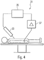
- Fig. 4 shows a schematic representation of a surgical device 31 according to the present invention.
- the surgical device 31 comprises a surgical instrument, in particular an electrosurgical instrument 33, a control device 35 and a biosensor 37.
- the biosensor 37 is designed to detect the blood properties of a patient during, before or at the beginning of a surgical procedure.
- the biosensor preferably comprises a piezoelectric element or the like to determine which blood clotting properties a patient has.
- the recorded data of the biosensor can then be forwarded to a control unit 35, which makes appropriate settings of the RF generator or other surgical aids for performing the surgical procedure.
- the recorded data of the biosensor are preferably used to make a corresponding adjustment and in particular a corresponding combination of the thermal haemostatic component and the biochemical haemostatic component and of the other haemostatic components.
- the biosensor can analyze the patient's blood count or blood clotting properties.
- a "quartz crystal microbalance" sensor (Gehring et al.) Can be used to determine the prothrombin time (PT time), which provides information about the patient's individual blood coagulation property.
- PT time prothrombin time
- an optimal setting of the surgical device from biochemical and thermal haemostasis and the addition of adducts, ie the blood coagulation influencing agents, as well as the supply of a noble gas can be determined and provided by the surgical device.
- the individual components can be applied with different weights simultaneously or consecutively, whereby not every component must be used compulsorily during each surgical intervention.
- tissue sensor 17 By means of the tissue sensor 17, an actually occurring tissue effect is detected during an operation. This can be done, for example, via the qualitative or quantitative detection of the flue gas produced during the application or the detection of the electromagnetic radiation of an application of a medical plasma application. From the biochemical sensor signal and the tissue effect signal, a real-time optimization of the surgical application can take place via the control unit. Ultimately, this results in a patient-specific ability to perform hemostasis with minimal damage to the surrounding tissue.
Landscapes
- Health & Medical Sciences (AREA)
- Life Sciences & Earth Sciences (AREA)
- Engineering & Computer Science (AREA)
- Surgery (AREA)
- Physics & Mathematics (AREA)
- Plasma & Fusion (AREA)
- Heart & Thoracic Surgery (AREA)
- Animal Behavior & Ethology (AREA)
- General Health & Medical Sciences (AREA)
- Public Health (AREA)
- Veterinary Medicine (AREA)
- Biomedical Technology (AREA)
- Molecular Biology (AREA)
- Medical Informatics (AREA)
- Otolaryngology (AREA)
- Nuclear Medicine, Radiotherapy & Molecular Imaging (AREA)
- Pathology (AREA)
- Biophysics (AREA)
- Spectroscopy & Molecular Physics (AREA)
- Anesthesiology (AREA)
- Vascular Medicine (AREA)
- Hematology (AREA)
- Surgical Instruments (AREA)
- Electrotherapy Devices (AREA)
- Laser Surgery Devices (AREA)
Description
- Die Erfindung betrifft ein Blutstillungsinstrument gemäß dem Oberbegriff des Anspruchs 1 sowie eine Chirurgieeinrichtung zur Stillung von Blutungen gemäß dem Oberbegriff des Anspruchs 9.
- Viele chirurgische Operationen, insbesondere offene, laparoskopische oder endoskopische Operationen erfordern eine aktive Stillung von auftretenden Blutungen. Treten Blutungen auf, so müssen diese gestoppt werden, da sonst der Operations-Situs schnell mit Blut bedeckt ist und eine effektive Fortführung der Operation oft unmöglich ist. Bei schweren Blutungen besteht darüber hinaus eine akute Verblutungsgefahr des Patienten, wenn die Blutung nicht innerhalb kurzer Zeit zum Stillstand gebracht wird. Im Falle schwerer Blutungen werden oft Blutkonserven benötigt, die prinzipiell Mangelware sind und außerdem von Patienten häufig aus religiösen oder anderen Gründen abgelehnt werden. Darüber hinaus besteht bei der Verwendung von Blutkonserven immer ein gewisses Restrisiko einer Infektion. Herkömmliche Verfahren und Vorrichtungen zur Blutstillung bedienen sich mechanischer, elektrochirurgischer oder laserchirurgischer Mittel. Weiterhin besteht die Möglichkeit, Fibrinkleber oder Kollagenauflagen zur Blutstillung zu verwenden. Mechanisch kann eine Naht oder ein Hämoclip zur Blutstillung eingesetzt werden. Die mechanische Blutstillung ist gegenüber der elektrochirurgischen und der laserchirurgischen Blutstillung mit einem erheblich größeren Zeitaufwand und schwierigerer Handhabung verbunden. Ebenfalls können Naht- und Hämoclip-Unverträglichkeiten durch Fremdmaterial, insbesondere durch Fadenmaterial und metallische Werkstoffe auftreten. Hämoclips können wandern, sich öffnen, und im schlimmsten Fall Organe perforieren oder Nachbarstrukturen fälschlicherweise mit "verschließen". Nähte können Adhäsionen bzw. Verwachsungen und Abszesse verursachen. Insbesondere endoskopisch aber auch laparoskopisch sind nicht alle Blutungen aufgrund der Erreichbarkeit der Blutung mit den mechanischen Mitteln möglich. Für diffuse Blutungen sind mechanische Methoden der Blutstillung in der Regel nicht geeignet.
- Unter die sogenannte "thermische Blutstillung" fallen alle gängigen Verfahren der Elektrochirurgie und der Laserchirurgie. Zu den elektrochirurgischen Verfahren zählen Verfahren aus dem Bereich der Hochfrequenzchirurgie, der KontaktKlemmen-Koagulation und der kontaktlosen Plasma-Koagulation, wie beispielsweise die Argon-Plasma-Koagulation sowie blutungsarme Hochfrequenzschneidemethoden. Die Blutstillung mittels des Lasers und mittels der Elektrochirurgie zielen auf eine Erwärmung der blutenden Gewebestelle ab, wobei bei der Laserchirurgie eine exogene Erwärmung und bei der Elektrochirurgie eine endogene Erwärmung des Gewebes zur Blutstillung führt. Nachteilig ist hierbei das Auftreten hoher lokaler Temperaturen von bis zu 300°C und ein damit einhergehendes Risiko der thermischen Schädigung von angrenzendem Gewebe. Darüber hinaus heilen die Wunden, welche durch Laser und Elektrochirurgie erzeugt werden im Vergleich zu einer mechanischen Schädigung des Gewebes durch ein Skalpell nur verzögert. In vielen Fällen konnte gezeigt werden, dass auch thermisch induzierte Wunden Verwachsungen von Organen untereinander oder Verwachsungen mit der Bauchwand verursachen können. Besonders problematisch ist dieser Befund bei fast allen Operationen im Bereich der Gynäkologie, aber auch bei anderen operativen Fachdisziplinen. Hiervon sind besonders viele junge Frauen betroffen, da die typischen Operationsindikationen Frauen im gebärfähigen Alter betreffen (Ovarialzysten-Entfernung, Myom-Entfernung, Tubenchirurgie). Wenn Adhäsionen postoperativ auftreten, können als Folge chronische Schmerzzustände, Fertilitätsstörungen und mechanisch bedingte Darmverschlüsse auftreten. Adhäsionen und die sich hieraus ergebende Symptomatik bzw. Folgeoperationen stellen einen erheblichen Kostenfaktor für das Gesundheitswesen dar. Der Laser ist als Blutstillungsverfahren nicht sehr weit verbreitet, da das Verfahren apparativ sehr aufwändig und teuer ist. Im Wesentlichen ergeben sich dieselben Nachteile wie bei der elektrochirurgischen Blutstillung.
- Bei der elektrochirurgischen Blutstillung ist wie gesagt immer mit einer thermischen Schädigung des um eine Blutung herum befindlichen Gewebes zu rechnen. Bei thermisch sensiblen Strukturen können hierdurch erhebliche Nebenwirkungen eintreten.
- Der wichtigste Nachteil bei allen elektrochirurgischen Verfahren ist das Problem der optimalen Dosierung der elektrischen Energie. Der Chirurg ist auf allgemeine Einstellungsempfehlungen der Hersteller für einen bestimmten chirurgischen Eingriff angewiesen. Hierbei kann die individuelle Situation und der Patient selbst nicht berücksichtigt werden. Hinzu kommt, dass viele Chirurgen eine subjektive Dosierung der elektrischen Energie vornehmen, was oftmals durch evidenzbasierte Erkenntnisse nicht begründbar ist und oftmals zu unnötig großen thermischen Schädigungen bis hin zu schlimmsten Verbrennungen führt. Die Wahl der geeigneten Parameter für eine bestimmte chirurgische Anwendung wie Strom, Stromform, Spannung, Leistung, Pulsation etc., die letztendlich einen elektrochirurgischen Modus charakterisieren, wurde oftmals durch den Systemhersteller empirisch bestimmt und ist häufig weit von einer optimalen, auf die jeweilige Situation abgestimmte und patienten-individuelle Parametrisierung entfernt. Hinzu kommt die enorme Vielzahl an unterschiedlichen Moden, die dem Chirurgen für seine Tätigkeit zur Verfügung stehen. Beispielsweise bietet das Erbe VIO-System zehn verschiedene Moden zur Durchführung einer Blutstillung an. Durch die große Anzahl an Einstellungsmöglichkeiten ist der Chirurg oft damit überfordert, für die zu bewältigende Aufgabe den optimalen Modus herauszusuchen.
- Zu der Gruppe der sogenannten "nicht-thermischen Blutstillung" gehören Verfahren, welche einen biochemischen Eingriff an den blutenden Stellen vornehmen. Dazu zählt der Fibrinkleber, der eine Art Gewebeklebstoff ist und der als physiologischer Zweikomponentenklebstoff die Wundränder verklebt, anstelle sie durch klassische Nähte zu verschließen. Ein Vorteil des Fibrinklebers ist eine Schonung besonders empfindlichen Gewebes. Bei den Ausgangsstoffen Fibrin, Thrombin, Faktor XIII und Aprotinin handelt es sich um biologische Substanzen, bei denen eine Infektion mit Krankheitserregern nicht ganz ausgeschlossen werden kann. Die biologischen Komponenten sind sehr teuer. Weiterhin können insbesondere für großflächige Blutungen Kollagenauflagen verwendet werden. Kollagenauflagen sind poröse schwammartige Wundauflagen, die durch Gefriertrocknung aus einer Kollagendispersion gewonnen werden. Sie können Wundsekret aufnehmen und wirken blutstillend. Sie fördern die Bildung und Organisation des körpereigenen Kollagens. Nach Blutkontakt aggregieren die Thrombozyten an den Kollagenfasern und lösen die Gerinnungsreaktion aus. Nachteilig ist, dass diese Auflagen bei akuten Blutungen nicht die erforderliche Effizienz aufweisen und eher für chronische Blutungen geeignet sind.
-
DE 10 2009 002 278 A1 offenbart beispielsweise eine Vorrichtung, die eine Wundbehandlung durch ein nicht-thermisches Plasma in Kombination mit Ultraschallimpulsen ermöglicht. Dazu ist eine Möglichkeit vorgesehen, Wasser einzuspritzen, um die Einkopplung der Ultraschallwellen in das Gewebe zu verbessern. -
US 2006/0084158 A1 befasst sich ebenfalls mit der Wundbehandlung durch kaltes Plasma und weist dazu eine Elektrode auf, die von einem Dielektrikum ummantelt ist. - Bei allen der oben aufgeführten Methoden zur Blutstillung beeinflussen Antikoagulationsmittel, Blutverdünnungsmittel wie Aspirin® oder Marcumar® sowie der Blutdruck die Effizienz einer Blutstillungsmethode erheblich. Bei Patienten, welche eine erblich bedingte Störung in der Blutgerinnung aufweisen, beispielsweise Bluter oder Patienten mit Hämophilie, ist eine Anwendung von Standardmethoden zur Blutstillung oft kontraindiziert.
- Auf natürlichem Wege geschieht die Blutstillung nach einer Verletzung durch eine Folge komplexer biochemischer Reaktionen, welche zusammengefasst als Blutgerinnungskaskade bezeichnet werden. Die Blutgerinnungskaskade besteht aus einem intravaskulären und einem extravaskulären System. Im Rahmen der vorliegenden Erfindung ist nur der extravaskuläre Weg entscheidend, so dass dieser im Folgenden kurz dargestellt werden soll: Das extravaskuläre System wird durch eine Verletzung in Gang gesetzt, die den Gewebefaktor (tissue factor) freisetzt. Der aus Gewebefaktor und Faktor VII gebildete Komplex setzt eine Kaskade in Gang, welche die Aktivierung von Thrombin veranlasst. Aktiviertes Thrombin katalysiert die Reaktion von Fibrinogen zu Fibrin (letzter Schritt der Blutgerinnungskaskade). Fibrin liegt zunächst in Form von Fibrinmonomeren vor, welche schließlich zu einem Fibrinnetzwerk vernetzen. Es bildet sich somit ein Fibringerinnsel aus, welches den Blutfluss zum Erliegen bringt.
- Durch eine sogenannte "stille elektrische Entladung" ("dielectric barrier discharge") kann auf die oben erläuterte Blutgerinnungskaskade Einfluss genommen werden. Hierbei werden die vier Fibrinopeptide des Fibrinogen-Moleküls abgespalten und es entsteht ein Fibrinmonomer. Dieser Abspaltungsprozess, der natürlicherweise von Thrombin katalysiert wird, bildet die Grundvoraussetzung für eine Vernetzung der Fibrinmonomere zu einem Fibringerinnsel. Eine stille elektrische Entladung ist eine elektrische Entladung zwischen zwei Elektroden mit ausreichend hoher Potentialdifferenz, die durch eine isolierende dielektrische Barriere (Isolator, Dielektrikum) getrennt sind. Als Dielektrikum wird eine schwach oder nicht leitende Substanz bezeichnet, deren Ladungsträger im Allgemeinen nicht frei beweglich sind. Ein Dielektrikum kann ein Gas, eine Flüssigkeit oder ein Feststoff sein. Isolatoren werden dann Dielektrika genannt, wenn diese mit magnetischen oder elektrischen Feldern beaufschlagt werden. Im Gegensatz zu einer Funkenentladung verhindert das Dielektrikum bei der stillen elektrischen Entladung die Entwicklung eines Funkens oder Bogens und die Entladung findet entweder in Form einer sogenannten Mikroentladung (FSD = Filamentary Silent Discharge) oder als homogene Entladung (GSD = Glow Silent Discharge) statt. Diese Entladungsformen sind sehr kurz und stromschwach und deshalb relativ lautlos. Da bei der stillen Entladung meist nur Elektronen transferiert werden und die sehr energiereichen Elektronen beim Zusammenstoß mit Gasatomen aufgrund des Masseunterschieds nur sehr wenig Energie auf die entstehenden Ionen übertragen können, ist die Gastemperatur relativ niedrig (kaltes Plasma). Die Abstände zwischen den Elektrodenplatten betragen bei stillen elektrischen Entladungen üblicherweise 0,1 Millimeter bis mehrere Zentimeter. Der Abstand der aktiven Elektrode zum Gewebe beträgt bei der chirurgischen Einrichtung 0,1 mm bis 1 cm. Für die stille elektrische Entladung ist in der Regel eine hohe Wechselspannung im Radiofrequenz- oder Mikrowellenfrequenzbereich erforderlich, die üblicherweise im Bereich von 20 kV liegt. Die Erzeugung einer derart hohen Spannung erfordert besondere Generatoren mit besonders dicht gewickelten Windungen. Diese Geräte sind teuer und werden in elektrochirurgischen Systemen nicht verwendet. Herkömmliche elektrochirurgische Systeme liefern im Übrigen Spannungen von maximal 6 kV.
- Eine Vorrichtung zur stillen elektrischen Entladung ist beispielsweise aus der
WO 2006/116252 A2 bekannt. Dort wird eine stille Entladung bei relativ hohen Spannungen im Bereich von 20 kV durchgeführt. Eine entsprechende Vorrichtung ist auch aus derWO 2010/009103 A2 bekannt. Dort wird eine Vorrichtung zur Erzeugung einer stillen elektrischen Entladung zur Behandlung von Blutungen der Schleimhaut im Gastrointestinaltrakt beansprucht. Eine tragbare Vorrichtung zur Erzeugung eines Plasmas mit relativ niedriger Temperatur wird in derUS 2009/0206062 A1 beansprucht. Die Vorrichtung dient zur Sterilisation und zur Blut-Koagulation. Es handelt sich folglich um eine Blutstillung durch thermische Gewebe-Koagulation ähnlich der Argon-Plasma-Koagulation. - Eine Aufgabe der vorliegenden Erfindung ist es daher, das Auftreten von Blutungen, die durch ein chirurgisches Trauma ausgelöst sein können, durch ein Blutstillungsinstrument so minimal invasiv wie möglich bei gleichzeitig größtmöglicher Effektivität und Patientensicherheit zu stillen.
- Zur Lösung dieser Aufgabe wird ein Blutstillungsinstrument zur aktiven Stillung von Blutungen mit den Merkmalen des Anspruchs 1 vorgeschlagen. Es kommt insbesondere nach einer offenen, laparoskopischen oder endoskopischen Operation an einem Patienten zum Einsatz und weist mehrere Blutstillungskomponenten auf, wobei wenigstens die folgenden Blutstillungskomponenten bei dem Blutstillungsinstrument vorgesehen sind:
- Eine thermische Blutstillungseinrichtung zur Gewebekoagulation zum Erzeugen einer Temperatur oberhalb der Koagualationstemperatur von biologischem Gewebe mittels einer Koagulationselektrode;
- eine biochemische Blutstillungseinrichtung zur Erzeugung einer stillen elektrischen Entladung mittels wenigstens einer Entladungselektrode und einer Isoliereinrichtung, wobei die Isoliereinrichtung zwischen der Entladungselektrode und dem zu behandelnden Gewebe angeordnet ist;
- eine Zufuhreinrichtung zum Zuführen von die Blutgerinnung beeinflussenden Stoffen an das zu behandelnde Gewebe, und
- eine Zufuhreinrichtung zum Zuführen von Edelgas an das zu behandelnde Gewebe.
- Ein wesentlicher Punkt der Erfindung liegt also darin, dass mehrere Blutstillungskomponenten in einem einzigen Instrument vereint werden, so dass eine individuell auf den Patienten abgestimmte Blutungsstillung erfolgen kann. Dadurch kann eine Blutstillung besonders minimalinvasiv erfolgen, d.h. das behandelte Gewebe wird so schonend wie möglich koaguliert. Hierzu verfügt das erfindungsgemäße Blutstillungsinstrument über die Möglichkeit der biochemischen Blutstillung mittels einer stillen elektrischen Entladung, auch als "dielectric barrier discharge" (dielektrische Barrierenentladung) bekannt, sowie über eine thermische Blutstillung durch exogene oder endogene Erwärmung des biologischen Gewebes. Weiterhin erlaubt das Blutstillungsinstrument eine Zuführung von Edelgas sowie von die Blutgerinnung beeinflussenden Stoffen an das zu behandelnde Gewebe, wie beispielsweise die Zuführung von Fibrinogen, Thrombin, Aprotinin, Gerinnungsfaktoren oder anderen Stoffen, welche die Blutgerinnung beeinflussen und damit die Blutstillung beschleunigen können. Auf diese Weise kann mit ein und demselben Instrument eine schonende Blutstillung durchgeführt werden und zwar in Abhängigkeit vom Zustand des individuellen Patienten. Je nach Art der zu stillenden Blutung kann folglich entweder die thermische Blutstillung und/oder die biochemische Blutstillung zum Einsatz kommen und zusätzlich die Blutgerinnung beeinflussende Stoffen an das zu behandelnde Gewebe geleitet werden. Die Zufuhr eines Edelgases sorgt zum einen dafür, dass eine Plasma-Koagulation durchgeführt werden kann und zum anderen ermöglicht sie die Erzeugung einer stillen elektrischen Entladung unter Verwendung eines herkömmlichen elektrochirurgischen Systems mit einer Spannung von maximal 6 kV. Bei der stillen elektrischen Entladung erhitzt sich das behandelte biologische Gewebe nicht nennenswert, so dass also die Koagulationstemperatur (60°C) von biologischem Gewebe nicht erreicht wird und folglich eine thermische Schädigung von Gewebe durch Koagulation oder gar Karbonisierung oder Vaporisation ausgeschlossen ist. Im Gegensatz zu herkömmlichen Verfahren der Blutstillung wird das Gewebe bei der Blutstillung durch eine stille elektrische Entladung nicht erhitzt, so dass keine Denaturierung der Proteine stattfindet und das Gewebe biochemisch intakt bleibt.
Die vorliegende Erfindung ermöglicht folglich mit einem einzigen Instrument eine optimale Mischung aus thermischer, biochemischer Blutstillung unter Zugabe eventuell erforderlicher gerinnungsfördernder Substanzen. - Das Blutstillungsinstrument gemäß der Erfindung erlaubt wie gesagt die Erzeugung einer stillen elektrischen Entladung in einer Edelgasatmosphäre, z.B. durch die Verwendung von Argon, Helium oder Neon mit deren Hilfe es möglich wird, die benötigten Spannungen zur Erzeugung eines kalten Plasmas (stille elektrische Entladung) unterhalb von 4 kV zu halten. Die stille elektrische Entladung kann kontinuierlich oder pulsierend erfolgen. Das Blutstillungsinstrument bietet beide Möglichkeiten. Die Isoliereinrichtung der vorliegenden Erfindung kann ein Dielektrikum aus Glas, insbesondere aus Quarzglas oder ein anderes geeignetes Glas, Keramik, Glimmer oder ähnliche Materialien verwenden.
- Biochemische Blutstillungen haben den Nachteil, dass die Zeitdauer zur Erreichung einer effizienten Blutstillung bei ca. 20 Sekunden liegt. Um die Zeit für die Blutgerinnung weiter verkürzen zu können, bietet die chirurgische Einrichtung gemäß der vorliegenden Erfindung die Möglichkeit einer endogenen oder exogenen Aufheizung des Blutes auf eine Temperatur unterhalb der Koagulationstemperatur von biologischem Gewebe, d.h. auf eine Temperatur unterhalb 60°C. Hierzu kann das Gewebe mittels des erfindungsgemäßen Blutstillungsinstruments durch ein konventionelles Gasplasma schonend vorgewärmt werden, ohne dass es zu einer thermischen Schädigung des Gewebes kommt. Da die Fibrinopeptide durch β-Faltblattstrukturen über Wasserstoffbrückenverbindungen stabilisiert sind, führt die Zuführung der oben beschriebenen thermischen Energie zu einem Aufbrechen dieser Wasserstoffbrückenverbindungen und damit zu einer Destabilisierung der Fibrinopeptide. Auf diese Weise können die Fibrinopeptide durch die Anwendung der stillen elektrischen Entladung leichter abgespalten werden und die Fibrinbildung kann somit schneller einsetzen.
- Wie gesagt verfügt das Blutstillungsinstrument gemäß der vorliegenden Erfindung nicht nur über die Möglichkeit einer biochemischen Blutstillung sondern gleichzeitig über die Möglichkeit einer konventionellen thermischen Blutstillung durch Kontakt-Koagulation, Argon-Plasma-Koagulation oder über die Möglichkeit eines blutarmen Hochfrequenzschnitts. Bei der thermischen Blutstillung werden Gewebetemperaturen oberhalb der Koagulationstemperatur erreicht, d.h. Temperaturen oberhalb von 60°C mit den damit einhergehenden allgemein bekannten Gewebeeffekten. Durch die Anwendung von supraphysiologischen Temperaturen oberhalb der Koagulations-temperatur des biologischen Gewebes werden Proteine und Zellstrukturen verändert, was zu Zelltod und nachfolgender Gewebenekrose führen kann. Ein erster Schritt dieser thermisch bedingten Veränderungen ist die Denaturierung von Proteinen sowie RNA, DNA und Zellmembrankomponenten. Wird das Gewebe oberhalb der Koagulationstemperatur weiter erhitzt, werden Gewebemoleküle in kleinere Moleküle, d.h. in Abbauprodukte durch Spaltung kovalenter Bindungen überführt. Diese Moleküle können weiter untereinander reagieren und es entstehen neue Substanzen, die auf der Gewebeoberfläche vernetzen. Der Gesamtprozess führt letztendlich zu einer Stillung der Blutung.
- Bei Patienten mit schlechten Blutgerinnungseigenschaften werden die thermischen und biochemischen Maßnahmen zur Blutstillung nicht zum gewünschten Ergebnis führen. Aus diesem Grund weist das Blutgerinnungsinstrument gemäß der vorliegenden Erfindung ein integriertes Zuführungs- und Dosierungssystem für Fibrinogen, einer Fibrinogenzubereitung oder einer anderen Substanz, die Einfluss auf das Blutgerinnungsverhalten nimmt, auf. Hierdurch ist gewährleistet, dass die Bildung eines Fibrinnetzwerks durch die Zuführung einer entsprechenden Hilfssubstanz unterstützt wird.
- Das Blutstillungsinstrument gemäß der vorliegenden Erfindung umfasst vorzugsweise eine weitere Blutstillungskomponente in Form einer Erwärmungseinrichtung zur endogenen oder exogenen Erwärmung des Blutes während einer stillen elektrischen Entladung auf eine Temperatur unterhalb der Koagulationstemperatur von biologischem Gewebe, d.h. unterhalb von 60°C. Weiterhin ist vorzugsweise die thermische Blutstillungseinrichtung zur Kontakt-Koagulation oder zur Plasma-Koagulation, insbesondere zur Argon-Plasma-Koagulation ausgebildet. Besonders bevorzugt wird ein Blutstillungsinstrument, bei dem die Koagulationselektrode der thermischen Blutstillungseinrichtung gleichzeitig die Entladungselektrode der biochemischen Blutstillungseinrichtung bildet. Die Entladungselektrode der biochemischen Blutstillungseinrichtung kann dabei als Ringelektrode ausgebildet und koaxial zu der Koagulationselektrode der thermischen Blutstillungseinrichtung angeordnet sein. Weiterhin kann das Blutstillungsinstrument mindestens einen Gewebesensor aufweisen, der während des Einsatzes ein oder mehrerer Blutstillungskomponenten entstehende Gewebeeffekte erfasst. Der Gewebesensor kann beispielsweise in das chirurgische Instrument integriert sein. Denkbar ist es jedoch auch den Gewebesensor in das Chirurgiegerät zu integrieren oder den mindestens einen Gewebesensor als externe separate Einheit auszubilden. Weiterhin kann das Blutstillungsinstrument wenigstens einen Biosensor aufweisen, der Patienteninformationen erfasst, beispielsweise durch die Analyse einer Körperflüssigkeit des Patienten, so dass eine optimierte Einstellung und/oder Kombination der einzelnen Blutstillungskomponenten in Abhängigkeit von situations- und patientenbedingten Einflüssen erfolgt. Der wenigstens eine Biosensor ist vorzugsweise als Schwingquarzsensor bzw. als quartz crystal microbalance (QCM) Sensor ausgebildet, der aus einer Quarzscheibe mit beidseitig aufgedampften Goldelektroden besteht. Durch Anlegen einer elektrischen Wechselspannung an die beiden Goldelektroden entsteht innerhalb des Quarzes eine stehende akustische Transversalwelle. Dabei ist die Schwingungsfrequenz abhängig von der Massenanlagerung an der oberen Elektrode.
- Zur Lösung der oben genannten Aufgabe wird auch eine Chirurgieeinrichtung zur Stillung von Blutungen mit den Merkmalen des Anspruchs 9 vorgeschlagen. Die Chirurgieeinrichtung dient insbesondere zur Stillung von Blutungen nach einer offenen, laparoskopischen oder endoskopischen Operation an einem Patienten und weist vorzugsweise folgende Komponenten auf:
- wenigstens einen Biosensor zur Erfassung von Bluteigenschaften eines Patienten während, vor oder zu Beginn eines operativen Eingriffs;
- ein Blutstillungsinstrument gemäß der oben genannten Beschreibung,
- wobei eine optimierte Einstellung und/oder Kombination der einzelnen Blutstillungskomponenten jeweils in Abhängigkeit von den durch den Biosensor erfassten Bluteigenschaften des Patienten erfolgt.
- Durch die Chirurgieeinrichtung gemäß der vorliegenden Erfindung ergibt sich der Vorteil, dass die Stillung von Blutungen durch die Verwendung eines Biosensors jeweils in Abhängigkeit von den patienteneigenen Bluteigenschaften erfolgt, so dass eine Blutstillung noch effektiver erfolgen kann. Ansonsten ergeben sich darüber hinaus die Vorteile des Blutstillungsinstruments gemäß der vorliegenden Erfindung.
- Die thermische Blutstillungskomponente ist vorzugsweise zur Gewebekoagulation durch Erzeugen einer Temperatur oberhalb der Koagulationstemperatur von biologischem Gewebe mittels einer Koagulationselektrode ausgebildet, wobei die Koagulationselektrode mit einem hochfrequenten Strom gespeist wird. Die thermische Blutstillungskomponente kann weiterhin zur Kontakt-Koagulation oder zur Plasma-Koagulation, insbesondere zur Argon-Plasma-Koagulation ausgebildet sein. Die biochemische Blutstillungskomponente ist hingegen vorzugsweise zur Erzeugung einer stillen elektrischen Entladung mittels wenigstens einer Entladungselektrode und einer Isoliereinrichtung ausgebildet, wobei die Isoliereinrichtung zwischen der Entladungselektrode und dem zu behandelnden Gewebe angeordnet ist und die Entladungselektrode mit einem HF-Strom gespeist wird. Darüber hinaus ist vorzugsweise eine weitere Blutstillungskomponente in Form einer Zufuhreinrichtung zum Zuführen von die Blutgerinnung beeinflussenden Stoffen an das zu behandelnde Gewebe vorgesehen. Eine weitere Blutstillungskomponente ist vorzugsweise in Form einer Zufuhreinrichtung zum Zuführen von Edelgas, insbesondere Argon, an das zu behandelnde Gewebe vorgesehen. Darüber hinaus kann noch eine weitere Blutstillungskomponente in Form einer Erwärmungseinrichtung zu endogenen oder exogenen Erwärmung des Blutes während einer stillen elektrischen Entladung auf eine Temperatur unterhalb der Koagulationstemperatur von biologischem Gewebe vorgesehen sein. Darüber hinaus kann die Chirurgieeinrichtung einen Gewebesensor aufweisen, der entstehende Gewebeeffekte während des Einsatzes ein oder mehrere Blutstillungskomponenten umfasst und der vorzugsweise im chirurgischen Instrument angeordnet ist. Eine Optimierung der Einstellung und/oder Kombination der einzelnen Blutstillungskomponenten erfolgt vorzugsweise auf der Grundlage der erfassten Werte des Gewebesensors und des Biosensors durch eine in der Chirurgieeinrichtung vorgesehene Steuereinheit "in Echtzeit".
- Die Erfindung wird im Folgenden anhand der Zeichnung näher erläutert. Es zeigen:
- Fig. 1A
- eine schematische Darstellung einer ersten Ausführungsform eines Blutstillungsinstruments zur aktiven Stillung von Blutungen gemäß der vorliegenden Erfindung in einem ersten Betriebszustand;
- Fig. 1B
- eine schematische Darstellung des Blutstillungsinstruments gemäß
Fig. 1A in einem zweiten Betriebszustand; - Fig. 1C
- eine schematische Darstellung des Blutstillungsinstruments gemäß
Fig. 1A in einem dritten Betriebszustand; - Fig. 2A
- eine schematische Darstellung einer zweiten Ausführungsform eines Blutstillungsinstruments gemäß der vorliegenden Erfindung in einem ersten Betriebszustand;
- Fig. 2B
- eine schematische Darstellung des Blutstillungsinstruments gemäß
Fig. 2A in einem zweiten Betriebszustand; - Fig. 2C
- eine schematische Darstellung des Blutstillungsinstruments gemäß
Fig. 2A in einem dritten Betriebszustand; - Fig. 3A
- eine schematische Darstellung einer weiteren Ausführungsform des Blutstillungsinstruments gemäß der vorliegenden Erfindung in einem ersten Betriebszustand;
- Fig. 3B
- das Blutstillungsinstrument gemäß
Fig. 3A in einem zweiten Betriebszustand; - Fig. 3C
- eine schematische Darstellung des Blutstillungsinstruments gemäß
Fig. 3A in einem dritten Betriebszustand, und - Fig. 4
- eine schematische Darstellung einer Chirurgieeinrichtung gemäß der vorliegenden Erfindung.
-
Fig. 1A zeigt eine schematische Darstellung einer ersten Ausführungsform des Blutstillungsinstruments 1 zur aktiven Stillung von Blutungen gemäß der vorliegenden Erfindung. Das Blutstillungsinstrument 1 umfasst ein Rohr oder einen Schlauch 3, der ein Lumen 5 einschließt. In dem Lumen ist ein Zufuhr- und Führungskanal 7 vorgesehen, der in ein distales Endstück 9 des Blutstillungsinstruments 1 hineinragt, wobei das distale Endstück 9 über das distale Ende 11 des Blutstillungsinstruments 1 hinausragt. - Sowohl der Zufuhr- und Führungskanal 7 als auch das distale Endstück 9 sind vorzugsweise zentrisch in dem Lumen 5 des Schlauchs 3 angeordnet. Das distale Endstück 9 ist mittels hier nur angedeuteter Verbindungselemente 13 mit dem Schlauch 3 verbunden. Parallel zu dem Lumen 5 und angrenzend an den Schlauch 3 ist ein Abzugskanal 15 vorgesehen, an dessen distalen Ende ein Gewebesensor 17 vorgesehen ist, der einen Gewebeeffekt, beispielsweise während der Operation entstehendes Rauchgas, erfassen kann. Das Emissionsgas wird durch den Abzugskanal 15 aus dem Operationsbereich entfernt.
- In dem Zufuhr- und Führungskanal 7 ist eine Elektrode 19 vorgesehen, die in den Zufuhr- und Führungskanal 7 beweglich gelagert ist und eine am distalen Ende 21 des distalen Endstücks 9 befindliche Isoliereinrichtung 21 durchstoßen kann. Die Isoliereinrichtung 21 ist hierzu vorzugsweise elastisch ausgebildet und kehrt in seine Ausgangsposition zurück, sobald die Elektrode 19 vollständig in dem Zufuhr- und Führungskanal 7 angeordnet ist (s.
Fig. 1C ). Damit sich die Isoliereinrichtung 21 bei einer Krafteinwirkung der Elektrode 19 öffnet, weist sie vorzugsweise einen Schlitz oder dergleichen Öffnung auf. - Die Elektrode 19 ist in den vorliegenden Ausführungsbeispielen als Hohlnadel ausgebildet, mit der eine Blutprobe aus dem zu behandelnden Gewebe gewonnen wird, wie in
Fig. 1A dargestellt ist. Mit der Hohlnadel kann auch ein Hochfrequenzschnitt am Gewebe durchgeführt werden. Die Elektrode 19 steht mit einem nicht dargestellten HF-Generator in Verbindung, der einen hochfrequenten Strom an die Elektrode liefert, sobald ein Hochfrequenzschnitt, eine Plasma-Koagulation oder eine andere elektrochirurgische Anwendung am Gewebe durchgeführt werden soll. - In
Fig. 1B ist das Blutstillungsinstrument 1 in einem zweiten Betriebszustand dargestellt, bei dem die Spitze der Hohlnadel, d.h. der Elektrode 19 nur minimal durch die Isoliereinrichtung 21 über das distale Ende des distalen Endstücks 9 hinausragt, während der Elektrode 19 ein hochfrequenter Strom zugeführt wird. Gleichzeitig wird über das Lumen 5 bzw. über den Schlauch 3 ein Inertgas, insbesondere Argon, an das Gewebe geleitet, wodurch es zur Erzeugung eines Argon-Plasmas kommt. Sofern die Elektrode 19 also die Isoliereinrichtung 21 durchstößt und über das distale Ende des distalen Endstücks 9 in Richtung des Gewebes übersteht, wird eine thermische Blutstillungseinrichtung zur Gewebekoagulation durch Erzeugung einer Temperatur oberhalb der Koagulationstemperatur von biologischem Gewebe, d.h. oberhalb von 60°C mittels der Elektrode 19 aktiviert, wobei die Elektrode in diesem Betriebszustand als Koagulationselektrode wirkt. Sofern die an die Elektrode 19 zugeführte Energie gering ist, kann die thermische Blutstillungseinrichtung auch dazu dienen, ein Gas-Plasma zu erzeugen, welches das Gewebe lediglich schonend vorwärmt. Sie bildet dann gleichzeitig eine thermische Erwärmungseinrichtung. -
Fig. 1C zeigt das Blutstillungsinstrument 1 in einem dritten Betriebszustand, in dem die Elektrode 19 vollständig in dem Zufuhr- und Führungskanal 7 angeordnet ist und somit die Isoliereinrichtung 21 den distalen Auslass des distalen Endstücks 9 vollständig verschließt, so dass die Elektrode 19 gegenüber dem Gewebe 23 isoliert ist. Die Isoliereinrichtung 21 ist in diesem Betriebszustand des Blutstillungsinstruments 1 zwischen der (Entladungs-)Elektrode 19 und dem zu behandelnden Gewebe angeordnet. Wird der Elektrode 19 in dieser Position ein HF-Strom zugeführt, wirkt sie als Entladungselektrode für die biochemische Blutstillung zur Erzeugung einer stillen elektrischen Entladung. Im Übrigen ist vorzugsweise auch das Endstück 9 isolierend ausgebildet. Denkbar ist es auch das Endstück einstückig mit der Isoliereinrichtung 21 auszubilden. - Während der in
Fig. 1C gezeigten Blutungsstillung mittels der stillen elektrischen Entladung kann ein die Blutgerinnung beeinflussendes Fluid über das Lumen 5 oder über den Zufuhr- und Führungskanal 7 an das Gewebe geleitet werden. - In der in
Fig. 1C gezeigten Position der Elektrode 19 wird folglich eine biochemische Blutstillungseinrichtung zur Erzeugung einer stillen elektrischen Entladung realisiert. Die Isoliereinrichtung 21 dient hierbei als Dielektrikum in einer Edelgasatmosphäre, wobei das Edelgas wiederum über das Lumen 5 in dem Zufuhrkanal 3 an das Gewebe 23 geleitet wird, um die benötigte Spannung zur Erzeugung der stillen elektrischen Entladung unterhalb von 4 kV zu halten. - Es zeigt sich somit, dass das Blutstillungsinstrument 1 gemäß der vorliegenden Ausführungsform sowohl eine thermische Blutstillungseinrichtung zur Gewebe-koagulation in Form der beweglich gelagerten Elektrode 19 aufweist und gleichzeitig eine biochemische Blutstillungseinrichtung zur Erzeugung einer stillen elektrischen Entladung mittels der Entladungselektrode 19, die hier gleichzeitig die Koagulationselektrode bildet sowie eine Isoliereinrichtung 21, wobei die Isoliereinrichtung zwischen der Entladungselektrode, hier also zwischen der Elektrode 19 und dem zu behandelnden Gewebe 23 angeordnet ist. Weiterhin ist eine Zufuhreinrichtung zum Zuführen von Edelgas und zum Zuführen von die Blutgerinnung beeinflussenden Stoffen an das zu behandelnde Gewebe in Form von dem Zufuhr- und Führungskanal 7 und das distale Endstück umgebende Lumen in dem Rohr 3 vorgesehen. Ein "Umschalten" zwischen den beiden Blutstillungseinrichtung erfolgt bei dem vorliegenden Ausführungsbeispiel durch eine Verlagerung der Elektrode 19 von einer ersten Position innerhalb des Zufuhr-und Führungskanals 7 in eine zweite Position außerhalb des Zufuhr- und Führungskanals 7.
- Eine weitere Ausführungsform eines Blutstillungsinstruments ist in den
Fig. 2A bis 2C gezeigt. Das Blutstillungsinstrument 1' gemäß denFig. 2A bis 2C unterscheidet sich von dem Blutstillungsinstrument 1 gemäß denFig. 1A bis 1C insbesondere dadurch, dass kein distales Endstück in dem Blutstillungsinstrument 1' vorgesehen ist, sondern vielmehr der Zufuhr- und Führungskanal 7 mit der darin gelagerten Elektrode 19, die wiederum als Hohlnadel insbesondere zur Entnahme einer Blutprobe ausgebildet ist, mittels Verbindungselementen 13' an dem Schlauch 3 befestigt ist. Auch bei dieser Ausführungsform der Erfindung ist die Elektrode 19 gleichzeitig als Hohlnadel ausgebildet und beweglich in dem Führungskanal 7 gelagert. - Darüber hinaus unterscheidet sich das Blutstillungsinstrument 1' gemäß den
Fig. 2A bis 2C von der Ausführungsform in denFig. 1A bis 1C dadurch, dass die Isoliereinrichtung 21' als ringförmiger Körper ausgebildet ist, der am distalen Ende eines das Lumen 5 bzw. das Rohr 3 koaxial umgebenden Ringraums bzw. Kanals 25 angeordnet ist. Am proximalen Ende der Isoliereinrichtung 21' ist eine ringförmige Entladungselektrode 27 vorgesehen, die flächig an der Isoliereinrichtung 21 anliegt und folglich ebenfalls koaxial den Schlauch 3 in dem Kanal 25 umgibt. Die Entladungselektrode 27 ist über Zuleitungen 29 mit einer entsprechenden Spannungs- bzw. Stromquelle, insbesondere mit dem HF-Generator eines Chirurgiegeräts verbunden. - Insgesamt zeigt sich, dass bei der vorliegenden Ausführungsform die Koagulationselektrode, d.h. die Elektrode 19 und die Entladungselektrode 27 der biochemischen Blutstillungseinrichtung als getrennte Elemente ausgebildet sind.
-
Fig. 2B zeigt das Blutstillungsinstrument 1' in einem Betriebszustand, in dem die thermische Blutstillungseinrichtung zur Gewebekoagulation durch Erzeugung einer Temperatur oberhalb der Koagulationstemperatur von biologischem Gewebe mittels der Koagulationselektrode 19 stattfinden kann. In diesem Fall ist die als Elektrode 19 fungierende Hohlnadel wiederum in den Zufuhr- und Führungskanal 7 eingezogen, während die Elektrode 19 (inFig. 2B nicht sichtbar) mit einem hochfrequenten Strom beaufschlagt wird. Gleichzeitig wird durch das Lumen 5 ein Inertgas, insbesondere Argon, an das zu behandelnde Gewebe 23 geführt, so dass ein Argon-Plasma zwischen dem Blutstillungsinstrument 1' und dem Gewebe 23 entsteht. -
Fig. 2C zeigt das Blutstillungsinstrument 1 gemäß der vorliegenden Ausführungsform in einem aktivierten Betriebszustand der biochemischen Blutstillungseinrichtung. In diesem Betriebszustand wird durch das Lumen 5 wiederum ein Inertgas, insbesondere ein Edelgas in Form von Argon an das Gewebe 23 herangeführt, während gleichzeitig die Entladungselektrode 27 über die Zuleitung 29 mit einem HF-Strom beaufschlagt wird, so dass sich eine stille elektrische Entladung, d.h. ein kaltes Plasma zwischen der Entladungselektrode 27 und dem als zweite Elektrode fungierenden Gewebe 23 ausbildet. Über das Lumen 5 des Schlauchs 3 kann gleichzeitig als Zufuhreinrichtung zum Zuführen von die Blutgerinnung beeinflussenden Stoffen an das zu behandelnde Gewebe ausgebildet sein. - Das Blutstillungsinstrument 1' gemäß den
Fig. 2A bis 2C ermöglicht ein Hochfrequenzschneiden von biologischem Gewebe mit zentrisch angeordneter ausfahrbarer HF-Elektrode 19, die insbesondere die Form einer Hohlnadel aufweist. Insgesamt kann durch das Blutstillungsinstrument 1' sowohl eine thermische Blutstillung mittels der Koagulationselektrode 19 als auch eine biochemische Blutstillung mittels der Entladungselektrode 27 realisiert werden. Das Blutstillungsinstruments 1' gemäß der vorliegenden Erfindung ist darüber hinaus zum Zuführen von Flüssigkeiten wie z.B. Addukten geeignet, welche die nicht-thermische, d.h. die biochemische Blutstillung unterstützen sollen, insbesondere zum Zuführung von Fibrinogen, Thrombin, Gerinnungsfaktoren oder Ähnlichem. Darüber hinaus ist das Instrument zum Zuführen eines Fluids zum Freispülen des OP-Situs nach einer Blutung geeignet. - Die
Fig. 3A bis 3C zeigen eine weitere Ausführungsform eines Blutstillungsinstruments gemäß der Erfindung. Im Gegensatz zu dem Blutstillungsinstrument 1' gemäß denFig. 2A bis 2C ist der Zufuhr- und Führungskanal 7 vorliegend als Elektrode 19' ausgebildet, umfasst also keine darin gelagerte Hohlnadel. Der Zufuhr- und Führungskanal 7 kann bei dieser Ausführungsform gleichzeitig als Zuleitung für eine Flüssigkeit, insbesondere unter Erzeugung eines Kegelstrahls dienen, durch die insbesondere die Zuführung von die Blutgerinnung beeinflussenden Stoffen an das zu behandelnde Gewebe 23 gewährleistet ist. Ansonsten entspricht die Ausführungsform und ihre Funktionsweise der Ausführungsform nach denFig. 2A bis 2C , auf die hiermit verwiesen wird. - Aus
Fig. 3B ist noch erkennbar, dass bei einer stillen elektrischen Entladung, d.h. bei einer aktiven biochemischen Blutstillungseinrichtung die Zufuhreinrichtung zum Zuführen von die Blutgerinnung beeinflussenden Stoffen an das zu behandelnde Gewebe 23 über den Zufuhrkanal 7 aktiviert sein kann. Eine Zufuhr von Edelgas kann wiederum über das Lumen 5 erfolgen. Es versteht sich, dass die Zufuhreinrichtung zum Zuführen von die Blutgerinnung beeinflussenden Stoffen auch unabhängig vom jeweiligen Blutungsstillungsmodus aktiviert sein kann. -
Fig. 4 zeigt noch eine schematische Darstellung einer Chirurgieeinrichtung 31 gemäß der vorliegenden Erfindung. Die Chirurgieeinrichtung 31 umfasst ein chirurgisches Instrument, insbesondere ein elektrochirurgisches Instrument 33, eine Steuereinrichtung 35 sowie einen Biosensor 37. Der Biosensor 37 ist dazu ausgelegt, die Bluteigenschaften eines Patienten während, vor oder zu Beginn eines operativen Eingriffs zu erfassen. Hierzu umfasst der Biosensor vorzugsweise ein piezoelektrisches Element oder dergleichen, um festzustellen, welche Blutgerinnungseigenschaften ein Patient aufweist. Die erfassten Daten des Biosensors können dann an eine Steuereinheit 35 weitergeleitet werden, die entsprechende Einstellungen des HF-Generators bzw. anderen operativen Hilfsmitteln zur Durchführung des operativen Eingriffs vornimmt. - Die erfassten Daten des Biosensors werden vorzugsweise dazu herangezogen, eine entsprechende Einstellung und insbesondere eine entsprechende Kombination der thermischen Blutstillungskomponente und der biochemischen Blutstillungskomponente sowie der anderen Blutstillungskomponenten vorzunehmen. Durch den Biosensor kann beispielsweise das Blutbild oder die Blutgerinnungseigenschaften des Patienten analysiert werden. Beispielsweise kann ein "quartz crystal microbalance"-Sensor (Gehring et al.) verwendet werden, um die Prothrombin-Zeit (PT-Zeit) zu ermitteln, welche eine Aussage über die patientenindividuelle Blutgerinnungseigenschaft ermöglicht. Auf diese Weise kann eine optimale Einstellung der Chirurgieeinrichtung aus biochemischer und thermischer Blutstillung sowie die Zuführung von Addukten, also die Blutgerinnung beeinflussenden Mitteln, sowie die Zufuhr eines Edelgases ermittelt und von der Chirurgieeinrichtung bereitgestellt werden. Dies bedeutet, dass die einzelnen Komponenten je nach Gerinnungssignal des Biosensors in unterschiedlicher Gewichtung gleichzeitig oder hintereinander angewandt werden können, wobei nicht jede Komponente bei jedem operativen Eingriff zwingend verwendet werden muss.
- Mittels des Gewebesensors 17 wird ein tatsächlich entstehender Gewebeeffekt während einer Operation detektiert. Dies kann beispielsweise über die qualitative oder quantitative Erfassung des während der Anwendung entstehenden Rauchgases oder der Detektion der elektromagnetischen Strahlung einer Anwendung einer medizinischen Plasmaanwendung geschehen. Über die Steuereinheit kann aus dem biochemischen Sensorsignal und dem Gewebeeffektsignal eine Echtzeitoptimierung der chirurgischen Anwendung erfolgen. Letztendlich ergibt sich daraus eine patientenspezifische Möglichkeit, Blutstillungen bei minimaler Schädigung des umliegenden Gewebes durchzuführen.
-
- 1
- Blutstillungsinstrument
- 1'
- Blutstillungsinstrument
- 1"
- Blutstillungsinstrument
- 3
- Schlauch
- 5
- Lumen
- 7
- Zufuhr- und Führungskanal
- 9
- Distales Endstück
- 11
- Distales Ende
- 13
- Verbindungselement
- 13'
- Verbindungselement
- 15
- Abzugskanal
- 17
- Gewebesensor
- 19
- Elektrode
- 19'
- Elektrode
- 21
- Isoliereinrichtung
- 21'
- Isoliereinrichtung
- 23
- Gewebe
- 25
- Kanal
- 27
- Entladungselektrode
- 29
- Zuleitung
- 31
- Chirurgieeinrichtung
- 33
- Chirurgisches Instrument
- 35
- Steuereinheit
- 37
- Biosensor
Claims (12)
- Blutstillungsinstrument (1, 1', 1") zur aktiven Stillung von Blutungen, insbesondere nach einer offenen, laparoskopischen oder endoskopischen Operation an einem Patienten, aufweisend mehrere Blutstillungskomponenten, wobei wenigstens die folgenden Blutstillungskomponenten vorgesehen sind:a. eine thermische Blutstillungseinrichtung zur Gewebekoagulation durch Erzeugen einer Temperatur oberhalb der Koagulations-temperatur von biologischem Gewebe mittels einer Koagulationselektrode (19);b. eine biochemische Blutstillungseinrichtung zur Erzeugung einer stillen elektrischen Entladung mittels wenigstens einer Entladungselektrode (19, 27) und einer Isoliereinrichtung (21, 21'), wobei die Isoliereinrichtung zwischen der Entladungselektrode und dem zu behandelnden Gewebe (23) angeordnet ist;c. ein integriertes Zuführungs- und Dosierungssystem für Fibrinogen oder eine Fibrinogenzubereitung, undd. eine Zufuhreinrichtung (5) zum Zuführen von Edelgas an das zu behandelnde Gewebe.
- Blutstillungsinstrument nach Anspruch 1,
dadurch gekennzeichnet, dass
das Blutstillungsinstrument eine weitere Blutstillungskomponente in Form einer Erwärmungseinrichtung zur endogenen oder exogenen Erwärmung des Blutes während einer stillen elektrischen Entladung auf eine Temperatur unterhalb der Koagulationstemperatur von biologischem Gewebe aufweist. - Blutstillungsinstrument nach Anspruch 1 oder 2,
dadurch gekennzeichnet, dass
die thermische Blutstillungseinrichtung zur Kontaktkoagulation oder zur Plasma-Koagulation, insbesondere zur Argon-Plasma-Koagulation ausgebildet ist. - Blutstillungsinstrument nach einem der vorhergehenden Ansprüche,
dadurch gekennzeichnet, dass
die Koagulationselektrode (19) der thermischen Blutstillungseinrichtung gleichzeitig die Entladungselektrode der biochemischen Blutstillungseinrichtung bildet. - Blutstillungsinstrument nach einem der Ansprüche 1 bis 3,
dadurch gekennzeichnet, dass
die Entladungselektrode (27) der biochemischen Blutstillungseinrichtung als Ringelektrode ausgebildet und koaxial zu der Koagulationselektrode (19) der thermischen Blutstillungseinrichtung angeordnet ist. - Blutstillungsinstrument nach einem der vorhergehenden Ansprüche,
dadurch gekennzeichnet, dass
es mindestens einen Gewebesensor (17) aufweist, der während des Einsatzes ein oder mehrerer Blutstillungskomponenten entstehende Gewebeeffekte erfasst. - Blutstillungsinstrument nach einem der vorhergehenden Ansprüche,
dadurch gekennzeichnet, dass
eine optimale Einstellung und/oder Kombination der einzelnen Blutstillungskomponenten in Abhängigkeit von situations- und patientenbedingten Einflüssen erfolgt. - Blutstillungsinstrument nach Anspruch 7,
dadurch gekennzeichnet, dass
die patientenbedingten Einflüsse in Form von durch wenigstens einen Biosensor (37) erfasste Patienteninformationen vorliegen. - Chirurgieeinrichtung zur Stillung von Blutungen, insbesondere nach einer offenen, laparoskopischen oder endoskopischen Operation an einem Patienten, aufweisend:a. wenigstens einen Biosensor zur Erfassung von Bluteigenschaften eines Patienten während, vor oder zu Beginn eines operativen Eingriffs;b. ein Blutstillungsinstrument nach einem der vorhergehenden Ansprüche, undc. wobei die Chirurgieeinrichtung derart ausgebildet ist, dass eine optimale Einstellung und/oder Kombination der einzelnen Blutstillungskomponenten jeweils in Abhängigkeit von den durch den Biosensor erfassten Bluteigenschaften des Patienten erfolgt.
- Chirurgieeinrichtung nach Anspruch 9,
dadurch gekennzeichnet, dass die Koagulationselektrode mit einem HF-Strom gespeist wird. - Chirurgieeinrichtung nach Anspruch 9 bis 10, dadurch gekennzeichnet, dass die Entladungselektrode mit einem HF-Strom gespeist wird.
- Chirurgieeinrichtung nach den Ansprüchen 9 und 6, dadurch gekennzeichnet, dass die Chirurgieeinrichtung derart ausgebildet ist, dass eine Optimierung der Einstellung und/oder Kombination der einzelnen Blutstillungskomponenten auf der Grundlage der erfassten Werte des Gewebesensors und des Biosensors durch eine in der Chirurgieeinrichtung vorgesehene Steuereinheit in "Echtzeit" erfolgt.
Priority Applications (1)
| Application Number | Priority Date | Filing Date | Title |
|---|---|---|---|
| PL11773008T PL2632359T3 (pl) | 2010-10-26 | 2011-10-24 | Urządzenie do hemostazy |
Applications Claiming Priority (5)
| Application Number | Priority Date | Filing Date | Title |
|---|---|---|---|
| DE102010060165 | 2010-10-26 | ||
| DE102010060163 | 2010-10-26 | ||
| DE102010061058 | 2010-12-07 | ||
| DE201010061059 DE102010061059B4 (de) | 2010-10-26 | 2010-12-07 | Chirurgieeinrichtung zur Stillung von Blutungen |
| PCT/EP2011/068542 WO2012055816A1 (de) | 2010-10-26 | 2011-10-24 | Blutstillungsinstrument |
Publications (2)
| Publication Number | Publication Date |
|---|---|
| EP2632359A1 EP2632359A1 (de) | 2013-09-04 |
| EP2632359B1 true EP2632359B1 (de) | 2019-07-03 |
Family
ID=45063102
Family Applications (1)
| Application Number | Title | Priority Date | Filing Date |
|---|---|---|---|
| EP11773008.5A Active EP2632359B1 (de) | 2010-10-26 | 2011-10-24 | Blutstillungsinstrument |
Country Status (8)
| Country | Link |
|---|---|
| US (1) | US10335223B2 (de) |
| EP (1) | EP2632359B1 (de) |
| JP (1) | JP5999778B2 (de) |
| KR (1) | KR20130103751A (de) |
| CN (1) | CN103189008B (de) |
| BR (1) | BR112013009795B1 (de) |
| PL (1) | PL2632359T3 (de) |
| WO (1) | WO2012055816A1 (de) |
Families Citing this family (20)
| Publication number | Priority date | Publication date | Assignee | Title |
|---|---|---|---|---|
| DE102012025079B4 (de) * | 2012-08-31 | 2016-09-08 | NorthCo Ventures GmbH & Co. KG | Vorrichtung und Verfahren zur Behandlung von biologischem Gewebe mit einem Niederdruckplasma |
| US9437401B2 (en) | 2013-12-20 | 2016-09-06 | Plasmology4, Inc. | System and method for plasma treatment using directional dielectric barrier discharge energy system |
| CN103892909A (zh) * | 2014-03-24 | 2014-07-02 | 上海大学 | 带光纤束镊子 |
| WO2015167623A2 (en) * | 2014-04-29 | 2015-11-05 | William Dean Wallace | Treatment methods and portable surgical devices for treating neoplastic and hyperplastic cells in the cervix and other dermatologically or surface related disorders |
| CN104146764A (zh) * | 2014-08-29 | 2014-11-19 | 超微(上海)医院投资管理有限公司 | 可变换角度的腔镜止血双极分离镊子 |
| GB2547941A (en) | 2016-03-04 | 2017-09-06 | Creo Medical Ltd | Electrosurgical instrument |
| CN106361427A (zh) * | 2016-09-14 | 2017-02-01 | 苏州超等医疗科技有限公司 | 一种无痛非麻醉性等离子伤口治疗装置 |
| WO2018193997A1 (ja) | 2017-04-19 | 2018-10-25 | 日本特殊陶業株式会社 | プラズマ照射装置、ハンドピース、及び手術用装置 |
| CN111465355B (zh) * | 2017-09-25 | 2023-06-09 | 保罗·韦伯 | 组织微创解剖设备和系统 |
| CN107874829A (zh) * | 2017-11-29 | 2018-04-06 | 成都美创医疗科技股份有限公司 | 一种切割等离子电极及切割方法 |
| CN108433806B (zh) | 2018-03-29 | 2021-03-16 | 江南大学 | 一种体内处理肿瘤的等离子体喷枪装置 |
| CN113748749B (zh) * | 2019-04-03 | 2025-07-15 | 瑞德科技有限公司 | 等离子体处理装置及其使用方法 |
| PL3831291T3 (pl) * | 2019-12-05 | 2025-04-28 | Erbe Elektromedizin Gmbh | Instrument do leczenia elektrochirurgicznego |
| US20230125841A1 (en) * | 2020-03-19 | 2023-04-27 | Caps Medical Ltd. | Plasma system with directional features |
| ES2995243T3 (en) | 2020-03-19 | 2025-02-07 | Caps Medical Ltd | Treatment of internal spaces using plasma generating device |
| CN111295032B (zh) * | 2020-03-27 | 2024-11-08 | 南京苏曼生物医学科技有限公司 | 一种具有电磁生物学效应的等离子体发生装置 |
| IT202000019642A1 (it) | 2020-08-07 | 2022-02-07 | Gaetano Mauro | Erogatore di polvere emostatica ad aria compressa, dotato di cannula biocompatibile, da usare nel primo trattamento di ferite da arma da fuoco o da taglio |
| CN114451945B (zh) * | 2022-02-25 | 2024-07-30 | 河南郑大心医助医学工程有限公司 | 一种腹腔手术用伤口夹闭器 |
| CN115778526B (zh) * | 2022-07-11 | 2023-08-29 | 南京康友医疗科技有限公司 | 一种微波、射频和测温一体式消融针 |
| CN115444487B (zh) * | 2022-09-21 | 2025-10-28 | 上海微创医疗机器人(集团)股份有限公司 | 止血器械末端及通用止血器械及腔镜手术系统 |
Citations (2)
| Publication number | Priority date | Publication date | Assignee | Title |
|---|---|---|---|---|
| US20060084158A1 (en) * | 2003-06-03 | 2006-04-20 | Wolfgang Viol | Treatment of biological material containing living cells using a plasma generated by a gas discharge |
| DE102009002278A1 (de) * | 2009-04-08 | 2010-10-14 | Robert Bosch Gmbh | Vorrichtung zum Erzeugen von nichtthermischem Plasma und Ultraschall-Pulsen |
Family Cites Families (31)
| Publication number | Priority date | Publication date | Assignee | Title |
|---|---|---|---|---|
| JP2544880B2 (ja) * | 1992-10-16 | 1996-10-16 | オリンパス光学工業株式会社 | 気腹装置の煙除去システム |
| US4781175A (en) * | 1986-04-08 | 1988-11-01 | C. R. Bard, Inc. | Electrosurgical conductive gas stream technique of achieving improved eschar for coagulation |
| DE3642077C2 (de) | 1986-12-10 | 1996-09-12 | Storz Karl Gmbh & Co | Vorrichtung, mit einem Generator und einer damit verbundenen Applikationssonde |
| US5449356A (en) | 1991-10-18 | 1995-09-12 | Birtcher Medical Systems, Inc. | Multifunctional probe for minimally invasive surgery |
| US6210402B1 (en) * | 1995-11-22 | 2001-04-03 | Arthrocare Corporation | Methods for electrosurgical dermatological treatment |
| US5554172A (en) | 1995-05-09 | 1996-09-10 | The Larren Corporation | Directed energy surgical method and assembly |
| DE19537897A1 (de) | 1995-09-19 | 1997-03-20 | Erbe Elektromedizin | Multifunktionales chirurgisches Instrument |
| EP0915682B1 (de) * | 1996-07-04 | 2003-10-01 | Erbe Elektromedizin GmbH | Gasunterstützte, axial verschiebbare chirurgische elektrode |
| US5759179A (en) * | 1996-12-31 | 1998-06-02 | Johnson & Johnson Medical, Inc. | Needle and valve assembly for use with a catheter |
| US5769826A (en) * | 1997-03-14 | 1998-06-23 | Johnson; Kenneth H. | Spring loaded retractable cannula blood drawing device |
| US5792053A (en) | 1997-03-17 | 1998-08-11 | Polartechnics, Limited | Hybrid probe for tissue type recognition |
| DE19858375B4 (de) | 1997-07-24 | 2008-07-03 | Erbe Elektromedizin Gmbh | Einrichtung zur HF-Koagulation biologischer Gewebe mittels flexibler Endoskopie |
| JP2002301088A (ja) * | 2001-04-05 | 2002-10-15 | Olympus Optical Co Ltd | 内視鏡用治療装置 |
| DE10328181A1 (de) | 2003-01-26 | 2004-08-05 | Eberhard-Karls-Universität Tübingen | Verfahren zum Separieren und Nachweisen von zumindest einem Analyten in biologischen Matrizes |
| DE10327237A1 (de) * | 2003-06-17 | 2005-01-13 | Trumpf Medizin Systeme Gmbh + Co. Kg | Elektrochirurgisches Instrument für ein Endoskop |
| DE102004009978A1 (de) | 2003-07-29 | 2005-04-28 | Hannover Laser Zentrum | Verfahren und Vorrichtung zur berührungslosen und zeitaufgelösten In-Vivo-Bestimmung von biologischem Gewebe |
| US7291145B2 (en) | 2003-10-14 | 2007-11-06 | Arnold Steven Seid | Oxygen sensing during a surgical procedure |
| US7182762B2 (en) * | 2003-12-30 | 2007-02-27 | Smith & Nephew, Inc. | Electrosurgical device |
| AU2006239843B8 (en) * | 2005-04-25 | 2011-06-30 | Drexel University | Methods for non-thermal application of gas plasma to living tissue |
| US8388618B2 (en) | 2005-04-25 | 2013-03-05 | Drexel University | Control of mucus membrane bleeding with cold plasma |
| DE102005021304A1 (de) | 2005-05-09 | 2006-11-23 | Erbe Elektromedizin Gmbh | Endoskopische Chirurgieeinrichtung für eine Argon-Plasma-Koagulation (APC) |
| US8123744B2 (en) * | 2006-08-29 | 2012-02-28 | Covidien Ag | Wound mediating device |
| DE102007003836A1 (de) | 2007-01-25 | 2008-08-07 | Erbe Elektromedizin Gmbh | Bipolares Instrument und Verfahren zur elektrochirurgischen Behandlung von Gewebe |
| DE102007019333A1 (de) | 2007-04-24 | 2008-11-06 | Siemens Ag | Medizinisches Instrument zur Durchführung eines medizinischen Eingriffs |
| CN101801311A (zh) * | 2007-05-01 | 2010-08-11 | 布赖汉姆妇女医院有限公司 | 创伤愈合装置 |
| US20090076505A1 (en) * | 2007-09-13 | 2009-03-19 | Arts Gene H | Electrosurgical instrument |
| FR2923151B1 (fr) * | 2007-11-02 | 2010-09-03 | Commissariat Energie Atomique | Dispositif de prelevement sanguin comportant au moins un filtre . |
| US7777151B2 (en) | 2008-02-14 | 2010-08-17 | Adventix Technologies Inc. | Portable plasma sterilizer |
| KR101158800B1 (ko) * | 2008-11-14 | 2012-06-26 | 주식회사 피에스엠 | 의료용 플라즈마 건 |
| DE102010015899B4 (de) | 2010-02-04 | 2022-07-28 | Erbe Elektromedizin Gmbh | Elektrochirurgische Anordnung und elektrochirurgisches Instrument |
| US20110319887A1 (en) * | 2010-06-23 | 2011-12-29 | Tyco Healthcare Group Lp | Electrosurgical Electrodes and Materials |
-
2011
- 2011-10-24 WO PCT/EP2011/068542 patent/WO2012055816A1/de not_active Ceased
- 2011-10-24 BR BR112013009795-7A patent/BR112013009795B1/pt not_active IP Right Cessation
- 2011-10-24 KR KR1020137011436A patent/KR20130103751A/ko not_active Ceased
- 2011-10-24 EP EP11773008.5A patent/EP2632359B1/de active Active
- 2011-10-24 CN CN201180051550.0A patent/CN103189008B/zh not_active Expired - Fee Related
- 2011-10-24 JP JP2013535388A patent/JP5999778B2/ja not_active Expired - Fee Related
- 2011-10-24 US US13/881,269 patent/US10335223B2/en active Active
- 2011-10-24 PL PL11773008T patent/PL2632359T3/pl unknown
Patent Citations (2)
| Publication number | Priority date | Publication date | Assignee | Title |
|---|---|---|---|---|
| US20060084158A1 (en) * | 2003-06-03 | 2006-04-20 | Wolfgang Viol | Treatment of biological material containing living cells using a plasma generated by a gas discharge |
| DE102009002278A1 (de) * | 2009-04-08 | 2010-10-14 | Robert Bosch Gmbh | Vorrichtung zum Erzeugen von nichtthermischem Plasma und Ultraschall-Pulsen |
Also Published As
| Publication number | Publication date |
|---|---|
| KR20130103751A (ko) | 2013-09-24 |
| JP5999778B2 (ja) | 2016-09-28 |
| US20130218069A1 (en) | 2013-08-22 |
| CN103189008B (zh) | 2016-10-12 |
| US10335223B2 (en) | 2019-07-02 |
| JP2013544122A (ja) | 2013-12-12 |
| WO2012055816A1 (de) | 2012-05-03 |
| EP2632359A1 (de) | 2013-09-04 |
| BR112013009795B1 (pt) | 2022-11-29 |
| CN103189008A (zh) | 2013-07-03 |
| PL2632359T3 (pl) | 2019-12-31 |
| BR112013009795A2 (pt) | 2016-07-26 |
Similar Documents
| Publication | Publication Date | Title |
|---|---|---|
| EP2632359B1 (de) | Blutstillungsinstrument | |
| DE102010061059B4 (de) | Chirurgieeinrichtung zur Stillung von Blutungen | |
| DE102009041167B4 (de) | Multifunktionsinstrument und Verfahren zur Verhinderung der Karbonisierung von Gewebe mittels eines Multifunktionsinstrument | |
| DE69834596T2 (de) | Elektrochirurgische, blutstillende Klammervorrichtung | |
| DE102014000609B4 (de) | Systeme und Verfahren zur Reduktion der Nasenmuschel | |
| EP0555549B1 (de) | Verfahren und Vorrichtung zum Hochdruckflüssigkeitsschneiden | |
| KR101296325B1 (ko) | 단백질성 조직의 해리 및 제거를 위한 시스템 | |
| DE60023849T2 (de) | Chirurgischer generator mit doppelfrequenz | |
| US20140052216A1 (en) | Methods for promoting wound healing | |
| DE112009005425T5 (de) | Einzelapertur-Elektrodenanordnung | |
| DE9290062U1 (de) | Elektrochirurgische und ultraschallchirurgische Vorrichtung | |
| DE102012002532A1 (de) | Elektrochirurgische Vorrichtung zur Feindissektion | |
| WO2011063798A2 (de) | Vorrichtung und verfahren zur erzeugung eines gepulsten anisothermen atmosphärendruck-plasmas | |
| US20140121656A1 (en) | Nerve and soft tissue cutting device | |
| EP1151724B1 (de) | Ultraschallzertrümmerer zum Zerkleinern oder Entfernen von Gewebe | |
| MacDonald et al. | Comparison of the effects of surgical dissection devices on the rabbit liver | |
| DE102014226903A1 (de) | HF-Hilfsstoff, medizinisches System und Verfahren zum selektiven Behandeln von Krebsgewebe | |
| DE102014003645B4 (de) | Elektrochirurgische Vorrichtung zur Feindissektion | |
| EP3071135B1 (de) | Hochfrequenz-chirurgiegerät und verfahren zum betreiben eines solchen | |
| EP3860316A1 (de) | Plasmen, medien, spezies, systeme, verfahren | |
| EP2394693A1 (de) | Einrichtung zur Elektroimpulsstimulation der Wunderheilung | |
| DE102013203999A1 (de) | Gewebefusionsmittel | |
| RU2517052C1 (ru) | Способ инертно-газо-усиленной плазменной коагуляции с использованием криптона в режиме "спрей" | |
| MOULYARAJ | A Comparative Study of Skin Incisions Made by Electrocautery Versus Stainless Steel Scalpel in Elective Surgical Cases | |
| EP1747763A1 (de) | Injektionsspritze mit einer Injektionsnadel |
Legal Events
| Date | Code | Title | Description |
|---|---|---|---|
| PUAI | Public reference made under article 153(3) epc to a published international application that has entered the european phase |
Free format text: ORIGINAL CODE: 0009012 |
|
| 17P | Request for examination filed |
Effective date: 20130516 |
|
| AK | Designated contracting states |
Kind code of ref document: A1 Designated state(s): AL AT BE BG CH CY CZ DE DK EE ES FI FR GB GR HR HU IE IS IT LI LT LU LV MC MK MT NL NO PL PT RO RS SE SI SK SM TR |
|
| DAX | Request for extension of the european patent (deleted) | ||
| RAP1 | Party data changed (applicant data changed or rights of an application transferred) |
Owner name: ERBE ELEKTROMEDIZIN GMBH |
|
| STAA | Information on the status of an ep patent application or granted ep patent |
Free format text: STATUS: EXAMINATION IS IN PROGRESS |
|
| 17Q | First examination report despatched |
Effective date: 20170620 |
|
| GRAP | Despatch of communication of intention to grant a patent |
Free format text: ORIGINAL CODE: EPIDOSNIGR1 |
|
| STAA | Information on the status of an ep patent application or granted ep patent |
Free format text: STATUS: GRANT OF PATENT IS INTENDED |
|
| INTG | Intention to grant announced |
Effective date: 20190107 |
|
| GRAJ | Information related to disapproval of communication of intention to grant by the applicant or resumption of examination proceedings by the epo deleted |
Free format text: ORIGINAL CODE: EPIDOSDIGR1 |
|
| STAA | Information on the status of an ep patent application or granted ep patent |
Free format text: STATUS: EXAMINATION IS IN PROGRESS |
|
| GRAR | Information related to intention to grant a patent recorded |
Free format text: ORIGINAL CODE: EPIDOSNIGR71 |
|
| GRAS | Grant fee paid |
Free format text: ORIGINAL CODE: EPIDOSNIGR3 |
|
| STAA | Information on the status of an ep patent application or granted ep patent |
Free format text: STATUS: GRANT OF PATENT IS INTENDED |
|
| GRAA | (expected) grant |
Free format text: ORIGINAL CODE: 0009210 |
|
| STAA | Information on the status of an ep patent application or granted ep patent |
Free format text: STATUS: THE PATENT HAS BEEN GRANTED |
|
| INTC | Intention to grant announced (deleted) | ||
| AK | Designated contracting states |
Kind code of ref document: B1 Designated state(s): AL AT BE BG CH CY CZ DE DK EE ES FI FR GB GR HR HU IE IS IT LI LT LU LV MC MK MT NL NO PL PT RO RS SE SI SK SM TR |
|
| INTG | Intention to grant announced |
Effective date: 20190528 |
|
| REG | Reference to a national code |
Ref country code: GB Ref legal event code: FG4D Free format text: NOT ENGLISH |
|
| REG | Reference to a national code |
Ref country code: CH Ref legal event code: EP Ref country code: AT Ref legal event code: REF Ref document number: 1150036 Country of ref document: AT Kind code of ref document: T Effective date: 20190715 |
|
| REG | Reference to a national code |
Ref country code: DE Ref legal event code: R096 Ref document number: 502011015871 Country of ref document: DE |
|
| REG | Reference to a national code |
Ref country code: IE Ref legal event code: FG4D Free format text: LANGUAGE OF EP DOCUMENT: GERMAN |
|
| REG | Reference to a national code |
Ref country code: NL Ref legal event code: MP Effective date: 20190703 |
|
| REG | Reference to a national code |
Ref country code: LT Ref legal event code: MG4D |
|
| PG25 | Lapsed in a contracting state [announced via postgrant information from national office to epo] |
Ref country code: NO Free format text: LAPSE BECAUSE OF FAILURE TO SUBMIT A TRANSLATION OF THE DESCRIPTION OR TO PAY THE FEE WITHIN THE PRESCRIBED TIME-LIMIT Effective date: 20191003 Ref country code: SE Free format text: LAPSE BECAUSE OF FAILURE TO SUBMIT A TRANSLATION OF THE DESCRIPTION OR TO PAY THE FEE WITHIN THE PRESCRIBED TIME-LIMIT Effective date: 20190703 Ref country code: LT Free format text: LAPSE BECAUSE OF FAILURE TO SUBMIT A TRANSLATION OF THE DESCRIPTION OR TO PAY THE FEE WITHIN THE PRESCRIBED TIME-LIMIT Effective date: 20190703 Ref country code: FI Free format text: LAPSE BECAUSE OF FAILURE TO SUBMIT A TRANSLATION OF THE DESCRIPTION OR TO PAY THE FEE WITHIN THE PRESCRIBED TIME-LIMIT Effective date: 20190703 Ref country code: HR Free format text: LAPSE BECAUSE OF FAILURE TO SUBMIT A TRANSLATION OF THE DESCRIPTION OR TO PAY THE FEE WITHIN THE PRESCRIBED TIME-LIMIT Effective date: 20190703 Ref country code: BG Free format text: LAPSE BECAUSE OF FAILURE TO SUBMIT A TRANSLATION OF THE DESCRIPTION OR TO PAY THE FEE WITHIN THE PRESCRIBED TIME-LIMIT Effective date: 20191003 Ref country code: NL Free format text: LAPSE BECAUSE OF FAILURE TO SUBMIT A TRANSLATION OF THE DESCRIPTION OR TO PAY THE FEE WITHIN THE PRESCRIBED TIME-LIMIT Effective date: 20190703 Ref country code: CZ Free format text: LAPSE BECAUSE OF FAILURE TO SUBMIT A TRANSLATION OF THE DESCRIPTION OR TO PAY THE FEE WITHIN THE PRESCRIBED TIME-LIMIT Effective date: 20190703 Ref country code: PT Free format text: LAPSE BECAUSE OF FAILURE TO SUBMIT A TRANSLATION OF THE DESCRIPTION OR TO PAY THE FEE WITHIN THE PRESCRIBED TIME-LIMIT Effective date: 20191104 |
|
| PG25 | Lapsed in a contracting state [announced via postgrant information from national office to epo] |
Ref country code: ES Free format text: LAPSE BECAUSE OF FAILURE TO SUBMIT A TRANSLATION OF THE DESCRIPTION OR TO PAY THE FEE WITHIN THE PRESCRIBED TIME-LIMIT Effective date: 20190703 Ref country code: AL Free format text: LAPSE BECAUSE OF FAILURE TO SUBMIT A TRANSLATION OF THE DESCRIPTION OR TO PAY THE FEE WITHIN THE PRESCRIBED TIME-LIMIT Effective date: 20190703 Ref country code: LV Free format text: LAPSE BECAUSE OF FAILURE TO SUBMIT A TRANSLATION OF THE DESCRIPTION OR TO PAY THE FEE WITHIN THE PRESCRIBED TIME-LIMIT Effective date: 20190703 Ref country code: RS Free format text: LAPSE BECAUSE OF FAILURE TO SUBMIT A TRANSLATION OF THE DESCRIPTION OR TO PAY THE FEE WITHIN THE PRESCRIBED TIME-LIMIT Effective date: 20190703 Ref country code: GR Free format text: LAPSE BECAUSE OF FAILURE TO SUBMIT A TRANSLATION OF THE DESCRIPTION OR TO PAY THE FEE WITHIN THE PRESCRIBED TIME-LIMIT Effective date: 20191004 Ref country code: IS Free format text: LAPSE BECAUSE OF FAILURE TO SUBMIT A TRANSLATION OF THE DESCRIPTION OR TO PAY THE FEE WITHIN THE PRESCRIBED TIME-LIMIT Effective date: 20191103 |
|
| PG25 | Lapsed in a contracting state [announced via postgrant information from national office to epo] |
Ref country code: TR Free format text: LAPSE BECAUSE OF FAILURE TO SUBMIT A TRANSLATION OF THE DESCRIPTION OR TO PAY THE FEE WITHIN THE PRESCRIBED TIME-LIMIT Effective date: 20190703 |
|
| PG25 | Lapsed in a contracting state [announced via postgrant information from national office to epo] |
Ref country code: RO Free format text: LAPSE BECAUSE OF FAILURE TO SUBMIT A TRANSLATION OF THE DESCRIPTION OR TO PAY THE FEE WITHIN THE PRESCRIBED TIME-LIMIT Effective date: 20190703 Ref country code: EE Free format text: LAPSE BECAUSE OF FAILURE TO SUBMIT A TRANSLATION OF THE DESCRIPTION OR TO PAY THE FEE WITHIN THE PRESCRIBED TIME-LIMIT Effective date: 20190703 Ref country code: DK Free format text: LAPSE BECAUSE OF FAILURE TO SUBMIT A TRANSLATION OF THE DESCRIPTION OR TO PAY THE FEE WITHIN THE PRESCRIBED TIME-LIMIT Effective date: 20190703 |
|
| PG25 | Lapsed in a contracting state [announced via postgrant information from national office to epo] |
Ref country code: MC Free format text: LAPSE BECAUSE OF FAILURE TO SUBMIT A TRANSLATION OF THE DESCRIPTION OR TO PAY THE FEE WITHIN THE PRESCRIBED TIME-LIMIT Effective date: 20190703 Ref country code: SM Free format text: LAPSE BECAUSE OF FAILURE TO SUBMIT A TRANSLATION OF THE DESCRIPTION OR TO PAY THE FEE WITHIN THE PRESCRIBED TIME-LIMIT Effective date: 20190703 Ref country code: IS Free format text: LAPSE BECAUSE OF FAILURE TO SUBMIT A TRANSLATION OF THE DESCRIPTION OR TO PAY THE FEE WITHIN THE PRESCRIBED TIME-LIMIT Effective date: 20200224 Ref country code: SK Free format text: LAPSE BECAUSE OF FAILURE TO SUBMIT A TRANSLATION OF THE DESCRIPTION OR TO PAY THE FEE WITHIN THE PRESCRIBED TIME-LIMIT Effective date: 20190703 |
|
| REG | Reference to a national code |
Ref country code: CH Ref legal event code: PL |
|
| REG | Reference to a national code |
Ref country code: DE Ref legal event code: R097 Ref document number: 502011015871 Country of ref document: DE |
|
| PLBE | No opposition filed within time limit |
Free format text: ORIGINAL CODE: 0009261 |
|
| STAA | Information on the status of an ep patent application or granted ep patent |
Free format text: STATUS: NO OPPOSITION FILED WITHIN TIME LIMIT |
|
| PG2D | Information on lapse in contracting state deleted |
Ref country code: IS |
|
| PG25 | Lapsed in a contracting state [announced via postgrant information from national office to epo] |
Ref country code: LI Free format text: LAPSE BECAUSE OF NON-PAYMENT OF DUE FEES Effective date: 20191031 Ref country code: LU Free format text: LAPSE BECAUSE OF NON-PAYMENT OF DUE FEES Effective date: 20191024 Ref country code: CH Free format text: LAPSE BECAUSE OF NON-PAYMENT OF DUE FEES Effective date: 20191031 |
|
| 26N | No opposition filed |
Effective date: 20200603 |
|
| REG | Reference to a national code |
Ref country code: BE Ref legal event code: MM Effective date: 20191031 |
|
| PG25 | Lapsed in a contracting state [announced via postgrant information from national office to epo] |
Ref country code: BE Free format text: LAPSE BECAUSE OF NON-PAYMENT OF DUE FEES Effective date: 20191031 Ref country code: SI Free format text: LAPSE BECAUSE OF FAILURE TO SUBMIT A TRANSLATION OF THE DESCRIPTION OR TO PAY THE FEE WITHIN THE PRESCRIBED TIME-LIMIT Effective date: 20190703 |
|
| PG25 | Lapsed in a contracting state [announced via postgrant information from national office to epo] |
Ref country code: IE Free format text: LAPSE BECAUSE OF NON-PAYMENT OF DUE FEES Effective date: 20191024 |
|
| REG | Reference to a national code |
Ref country code: AT Ref legal event code: MM01 Ref document number: 1150036 Country of ref document: AT Kind code of ref document: T Effective date: 20191024 |
|
| PG25 | Lapsed in a contracting state [announced via postgrant information from national office to epo] |
Ref country code: AT Free format text: LAPSE BECAUSE OF NON-PAYMENT OF DUE FEES Effective date: 20191024 |
|
| PG25 | Lapsed in a contracting state [announced via postgrant information from national office to epo] |
Ref country code: CY Free format text: LAPSE BECAUSE OF FAILURE TO SUBMIT A TRANSLATION OF THE DESCRIPTION OR TO PAY THE FEE WITHIN THE PRESCRIBED TIME-LIMIT Effective date: 20190703 |
|
| PG25 | Lapsed in a contracting state [announced via postgrant information from national office to epo] |
Ref country code: MT Free format text: LAPSE BECAUSE OF FAILURE TO SUBMIT A TRANSLATION OF THE DESCRIPTION OR TO PAY THE FEE WITHIN THE PRESCRIBED TIME-LIMIT Effective date: 20190703 Ref country code: HU Free format text: LAPSE BECAUSE OF FAILURE TO SUBMIT A TRANSLATION OF THE DESCRIPTION OR TO PAY THE FEE WITHIN THE PRESCRIBED TIME-LIMIT; INVALID AB INITIO Effective date: 20111024 |
|
| PG25 | Lapsed in a contracting state [announced via postgrant information from national office to epo] |
Ref country code: MK Free format text: LAPSE BECAUSE OF FAILURE TO SUBMIT A TRANSLATION OF THE DESCRIPTION OR TO PAY THE FEE WITHIN THE PRESCRIBED TIME-LIMIT Effective date: 20190703 |
|
| PGFP | Annual fee paid to national office [announced via postgrant information from national office to epo] |
Ref country code: PL Payment date: 20230929 Year of fee payment: 13 |
|
| PGFP | Annual fee paid to national office [announced via postgrant information from national office to epo] |
Ref country code: GB Payment date: 20231024 Year of fee payment: 13 |
|
| PGFP | Annual fee paid to national office [announced via postgrant information from national office to epo] |
Ref country code: IT Payment date: 20231024 Year of fee payment: 13 Ref country code: FR Payment date: 20231026 Year of fee payment: 13 Ref country code: DE Payment date: 20231027 Year of fee payment: 13 |
|
| REG | Reference to a national code |
Ref country code: DE Ref legal event code: R119 Ref document number: 502011015871 Country of ref document: DE |
|
| GBPC | Gb: european patent ceased through non-payment of renewal fee |
Effective date: 20241024 |
|
| PG25 | Lapsed in a contracting state [announced via postgrant information from national office to epo] |
Ref country code: DE Free format text: LAPSE BECAUSE OF NON-PAYMENT OF DUE FEES Effective date: 20250501 |
|
| PG25 | Lapsed in a contracting state [announced via postgrant information from national office to epo] |
Ref country code: GB Free format text: LAPSE BECAUSE OF NON-PAYMENT OF DUE FEES Effective date: 20241024 |
|
| PG25 | Lapsed in a contracting state [announced via postgrant information from national office to epo] |
Ref country code: FR Free format text: LAPSE BECAUSE OF NON-PAYMENT OF DUE FEES Effective date: 20241031 |
|
| PG25 | Lapsed in a contracting state [announced via postgrant information from national office to epo] |
Ref country code: IT Free format text: LAPSE BECAUSE OF NON-PAYMENT OF DUE FEES Effective date: 20241024 |