EP2682746B1 - Procédés pour le diagnostic d'états de santé associés au cancer de l'ovaire et du risque d'états de santé associés au cancer des ovaires - Google Patents
Procédés pour le diagnostic d'états de santé associés au cancer de l'ovaire et du risque d'états de santé associés au cancer des ovaires Download PDFInfo
- Publication number
- EP2682746B1 EP2682746B1 EP13176355.9A EP13176355A EP2682746B1 EP 2682746 B1 EP2682746 B1 EP 2682746B1 EP 13176355 A EP13176355 A EP 13176355A EP 2682746 B1 EP2682746 B1 EP 2682746B1
- Authority
- EP
- European Patent Office
- Prior art keywords
- metabolite
- ovarian cancer
- molecular formula
- daughter ions
- fragment pattern
- Prior art date
- Legal status (The legal status is an assumption and is not a legal conclusion. Google has not performed a legal analysis and makes no representation as to the accuracy of the status listed.)
- Not-in-force
Links
- 0 CC(CCC=C(C)CCCC(C)=CCCC(C)(CC1)*=C2C1=CC=C(C)C2C)C(OC)=N Chemical compound CC(CCC=C(C)CCCC(C)=CCCC(C)(CC1)*=C2C1=CC=C(C)C2C)C(OC)=N 0.000 description 10
- KRZAQIRXTBFSBU-UHFFFAOYSA-N CC(C(C)C(C(C1)C=C)[O-])C1O Chemical compound CC(C(C)C(C(C1)C=C)[O-])C1O KRZAQIRXTBFSBU-UHFFFAOYSA-N 0.000 description 1
- QWSWGBORJFCHTO-GQCTYLIASA-N CC(C/C=C/C(C)(C)O)C(O)=N Chemical compound CC(C/C=C/C(C)(C)O)C(O)=N QWSWGBORJFCHTO-GQCTYLIASA-N 0.000 description 1
- SEOVGMDCUYEDPX-UHFFFAOYSA-N CCC(C(C(C)C1C)O)=C=C1O Chemical compound CCC(C(C(C)C1C)O)=C=C1O SEOVGMDCUYEDPX-UHFFFAOYSA-N 0.000 description 1
- HNWMIFTUZYDFBT-UHFFFAOYSA-N CCCCCCCCC(C(C)=C1C)(C(CC)=CC1O)O Chemical compound CCCCCCCCC(C(C)=C1C)(C(CC)=CC1O)O HNWMIFTUZYDFBT-UHFFFAOYSA-N 0.000 description 1
Images




Classifications
-
- G—PHYSICS
- G01—MEASURING; TESTING
- G01N—INVESTIGATING OR ANALYSING MATERIALS BY DETERMINING THEIR CHEMICAL OR PHYSICAL PROPERTIES
- G01N33/00—Investigating or analysing materials by specific methods not covered by groups G01N1/00 - G01N31/00
- G01N33/48—Biological material, e.g. blood, urine; Haemocytometers
- G01N33/50—Chemical analysis of biological material, e.g. blood, urine; Testing involving biospecific ligand binding methods; Immunological testing
- G01N33/53—Immunoassay; Biospecific binding assay; Materials therefor
- G01N33/575—Immunoassay; Biospecific binding assay; Materials therefor for cancer
- G01N33/57545—Immunoassay; Biospecific binding assay; Materials therefor for cancer of the ovaries
-
- G—PHYSICS
- G01—MEASURING; TESTING
- G01N—INVESTIGATING OR ANALYSING MATERIALS BY DETERMINING THEIR CHEMICAL OR PHYSICAL PROPERTIES
- G01N33/00—Investigating or analysing materials by specific methods not covered by groups G01N1/00 - G01N31/00
- G01N33/48—Biological material, e.g. blood, urine; Haemocytometers
- G01N33/483—Physical analysis of biological material
- G01N33/487—Physical analysis of biological material of liquid biological material
- G01N33/48785—Electrical and electronic details of measuring devices for physical analysis of liquid biological material not specific to a particular test method, e.g. user interface or power supply
- G01N33/48792—Data management, e.g. communication with processing unit
-
- G—PHYSICS
- G01—MEASURING; TESTING
- G01N—INVESTIGATING OR ANALYSING MATERIALS BY DETERMINING THEIR CHEMICAL OR PHYSICAL PROPERTIES
- G01N33/00—Investigating or analysing materials by specific methods not covered by groups G01N1/00 - G01N31/00
- G01N33/48—Biological material, e.g. blood, urine; Haemocytometers
- G01N33/483—Physical analysis of biological material
- G01N33/487—Physical analysis of biological material of liquid biological material
- G01N33/49—Blood
-
- H—ELECTRICITY
- H01—ELECTRIC ELEMENTS
- H01J—ELECTRIC DISCHARGE TUBES OR DISCHARGE LAMPS
- H01J49/00—Particle spectrometers or separator tubes
- H01J49/0027—Methods for using particle spectrometers
- H01J49/0036—Step by step routines describing the handling of the data generated during a measurement
-
- G—PHYSICS
- G01—MEASURING; TESTING
- G01N—INVESTIGATING OR ANALYSING MATERIALS BY DETERMINING THEIR CHEMICAL OR PHYSICAL PROPERTIES
- G01N2560/00—Chemical aspects of mass spectrometric analysis of biological material
-
- G—PHYSICS
- G01—MEASURING; TESTING
- G01N—INVESTIGATING OR ANALYSING MATERIALS BY DETERMINING THEIR CHEMICAL OR PHYSICAL PROPERTIES
- G01N2800/00—Detection or diagnosis of diseases
- G01N2800/50—Determining the risk of developing a disease
-
- G—PHYSICS
- G01—MEASURING; TESTING
- G01N—INVESTIGATING OR ANALYSING MATERIALS BY DETERMINING THEIR CHEMICAL OR PHYSICAL PROPERTIES
- G01N2800/00—Detection or diagnosis of diseases
- G01N2800/56—Staging of a disease; Further complications associated with the disease
-
- G—PHYSICS
- G01—MEASURING; TESTING
- G01N—INVESTIGATING OR ANALYSING MATERIALS BY DETERMINING THEIR CHEMICAL OR PHYSICAL PROPERTIES
- G01N2800/00—Detection or diagnosis of diseases
- G01N2800/70—Mechanisms involved in disease identification
- G01N2800/7023—(Hyper)proliferation
- G01N2800/7028—Cancer
Definitions
- the present invention relates to methods for diagnosing ovarian cancer, or the risk of developing ovarian cancer.
- Ovarian cancer is the fifth leading cause of cancer death among women (1). It has been estimated that over 22,000 new cases of ovarian cancer will be diagnosed this year, with 16,210 deaths predicted in the United States alone (2). Ovarian cancer is typically not identified until the patient has reached stage III or IV, which is associated with a poor prognosis; the five-year survival rate is estimated at around 25- 30% (3).
- the current screening procedures for ovarian cancer involve the combination of bimanual pelvic examination, transvaginal ultrasonography, and serum screening for elevated cancer antigen-125 (CA125), a protein cancer antigen (2).
- CA125 cancer antigen-125
- CA125 screening for ovarian cancer is currently of unknown benefit, as there is a lack of evidence that the screen reduces mortality rates, and it is under scrutiny due to the risks associated with false positive results (1, 4).
- CA125 measurement and transvaginal ultrasonography are not reliable screening or diagnostic tests for ovarian cancer, and that the only current method available to make a definite diagnosis is by surgery (http://www.cancer.org).
- CA125 is a high molecular weight mucin that has been found to be elevated in most ovarian cancer cells as compared to normal cells (2).
- a CA125 test result that is higher than 30-35U/ml is typically accepted as being at an elevated level (2).
- There have been difficulties in establishing the accuracy, sensitivity, and specificity of the CA125 screen for ovarian cancer due to the different thresholds used to define elevated CA125, varying sizes of patient groups tested, and broad ranges in the age and ethnicity of patients (1).
- the CA125 test According to the Johns Hopkins University pathology website, the CA125 test only returns a true positive result for ovarian cancer in roughly 50% of stage I patients and about 80% in stage II, III and IV patients (http://pathology2.jhu.edu).
- CA125 is not an effective general screening test for ovarian cancer. They report that only about three out of 100 healthy women with elevated CA125 levels are actually found to have ovarian cancer, and about 20% of ovarian cancer diagnosed patients actually have elevated CA125 levels (http://www.nlm.nih.gov/medlineplus/ency/article/007217.htm).
- Zhang et al., Gynecologic Oncology, 2006, 102(1): 61-66 describes a proteomics-based method for identifying protein markers of ovarian cancer.
- Kozak et al., Proteomics 2005, 5:44589-4596 describes a study using micro-LC-MS/MS to identify four protein markers of ovarian cancer, i.e., transthyretin, beta-hemoglobin, apolipoprotein AI, and transferrin, from a group of previously reported SELDI-TOF-MS peaks.
- LC-MS/MS analysis identified four peptide fragments corresponding to apolipoprotein D, Sp-alpha, alpha-enolase, transketolase, and M2-PK.
- WO2007030928 discloses the use of a metabolite with the formula C28H46O4 as a marker for ovarian cancer.
- Denkert et al, Cancer Research 2006; 66: (22), November 15, 2006 relates to the use of mass spectrometry-based metabolic profiling in ovarian carcinomas.
- the present invention provides a method for diagnosing ovarian cancer or the risk of developing ovarian cancer in a test subject of unknown ovarian cancer status, comprising the steps of:
- the methods measure the intensities of specific small molecules, also referred to as metabolites, in the sample from the patient, and compare these intensities to the intensities observed in a population of healthy individuals.
- the sample obtained from the human is a blood sample.
- the present invention may significantly improve the ability to detect ovarian cancer or the risk of developing ovarian cancer, and may therefore save lives.
- the statistical performance of a test based on these samples suggests that the test will outperform the CA125 test, the only other serum-based diagnostic test for ovarian cancer.
- a combination of the test described herein and the CA125 test may improve the overall diagnostic performance of each test.
- a method for diagnosing a patient for the presence of an ovarian cancer, or the risk of developing ovarian cancer comprising the steps of: screening a sample from said patient for the presence or absence of one or more metabolic markers selected from the group consisting of metabolites with an accurate mass of, or substantially equivalent to masses to 474.3736, 478.4022, 484.3764, 490.3658, 492.3815, 494.3971, 496.4128, 502.4022, 504.4179, 512.4077, 518.3971, 520.4128, 522.8284, 530.43351, 532.44916, 540.4389, 550.4597, 558.4648, 574.4597, 576.4754, 578.4910, 596.5016, and 598.5172, where a +/- 5 ppm difference would indicate the same metabolite; wherein the absence or significant reduction of one or more of said metabolic markers indicates the presence of an ovarian cancer, or the risk of developing
- the molecular formulas of the metabolites with these masses are C30H50O4, C30H5404, C28H52O6, C30H5005, C30H52O5, C30H5405, C30H5605, C32H5404, C32H56O4, C30H56O6, C32H5405, C32H5605, C32H6005, C34H5804, C34H60O4, C32H6006, C34H6205, C36H62O4, C36H6205, C36H6405, C36H66O5, C36H68O6, and C36H7006, respectively and the proposed structures are as shown below: respectively.
- a method for diagnosing the presence or absence of ovarian cancer in a test subject of unknown ovarian cancer status comprising: analyzing a blood sample from a test subject to obtain quantifying data on molecules selected from the group comprised of molecules identified by the neutral accurate masses 474.3736, 478.4022, 484.3764, 490.3658, 492.3815, 494.3971, 496.4128, 502.4022, 504.4179, 512.4077, 518.3971, 520.4128, 522.8284, 530.43351, 532.44916, 540.4389, 550.4597, 558.4648, 574.4597, 576.4754, 578.4910, 596.5016, and 598.5172, where a +/- 5 ppm difference would indicate the same metabolite, or molecules having masses substantially equal to these molecules or fragments of derivatives thereof; comparing the quantifying data obtained on said molecules in said test subject with quantifying data obtained from said molecules from a
- the molecular formulas of the metabolites with these masses are C30H5004, C30H54O4, C28H52O6, C30H50O5, C30H52O5, C30H54O5, C30H56O5, C32H54O4, C32H56O4, C30H56O6, C32H54O5, C32H56O5, C32H60O5, C34H58O4, C34H60O4, C32H60O6, C34H62O5, C36H62O4, C36H62O5, C36H64O5, C36H66O5, C36H68O6, and C36H70O6, respectively and the proposed structures are as shown below: respectively.
- the identification of ovarian cancer biomarkers with improved diagnostic accuracy in human serum therefore, would be extremely beneficial, as the test would be non-invasive and could possibly be used to monitor individual susceptibility to disease prior to, or in combination with, conventional methods.
- a serum test is minimally invasive and would be accepted across the general population.
- the present invention relates to a method of diagnosing ovarian cancer, or the risk of developing ovarian cancer, by measuring the levels of specific small molecules present in human serum and comparing them to "normal" reference level s.
- any one or combination of the metabolites identified in the present invention can be used to indicate the presence of ovarian cancer.
- a diagnostic assay based on small molecules, or metabolites, in serum fulfills the above criteria for an ideal screening test, as development of assays capable of detecting specific metabolites is relatively simple and cost effective per assay. Translation of the method into a clinical assay compatible with current clinical chemistry laboratory hardware would be commercially acceptable and effective, and would result in a rapid deployment worldwide. Furthermore, the requirement for highly trained personnel to perform and interpret the test would be eliminated.
- the selected metabolites, identified according to the present invention, were further characterized by molecular formulae and structure. This additional information for the metabolites is shown in Table 35.
- the present invention relates to the diagnosis of ovarian cancer (OC), or the risk of developing OC,.
- the present invention describes the relationship between endogenous small molecules and OC.
- the present invention relates to the diagnosis of OC, or the risk of developing OC, through the measurement of vitamin E isoforms and related metabolites. More specifically, the present invention relates to the relationship between vitamin E-related metabolites in human serum and the implications thereof in OC.
- the present invention discloses for the first time clear and unambiguous biochemical changes specifically associated with OC. These findings also imply that the measurement of these biomarkers may provide a universal means of measuring the effectiveness of OC therapies. This would dramatically decrease the cost of performing clinical trials as a simple biochemical test can be used to assess the viability of new therapeutics. Furthermore, one would not have to wait until the tumor progresses or until the patient dies to determine whether the therapy provided any benefit. The use of such a test would enable researchers to determine in months, rather than years, the effectiveness of dose, formulation, and chemical structure modifications of OC therapies.
- the present invention relates to a method of diagnosing OC by measuring the levels of specific small molecules present in human serum and comparing them to "normal" reference levels.
- a novel method for the early detection and diagnosis of OC and the monitoring the effects of OC therapy is described.
- One method of the present invention uses accurate masses in an FTMS based method.
- the accurate masses that can be used according to this invention include the claimed masses shown in Table 1, or a subset thereof.
- a further method involves the use of a high-throughput screening (HTS) assay developed from a subset of claimed metabolites selected from Table 1 for the diagnosis of one or more diseases or particular health-states.
- HTS high-throughput screening
- vitamin E collectively refers to eight naturally occurring isoforms, four tocopherols (alpha, beta, gamma, and delta) and four tocotrienols (alpha, beta, gamma, and delta).
- the predominant form found in western diets is gamma-tocopherol whereas the predominant form found in human serum/plasma is alpha-tocopherol.
- Tocotrienols are also present in the diet, but are more concentrated in cereal grains and certain vegetable oils such as palm and rice bran oil. Interestingly, it is suggested that tocotrienols may be more potent than tocopherols in preventing cardiovascular disease and cancer (5).
- Plasma concentrations of the tocopherols are believed to be tightly regulated by the hepatic tocopherol binding protein. This protein has been shown to preferentially bind to alpha-tocopherol (10). Large increases in alpha-tocopherol consumption result in only small increases in plasma concentrations (11). Similar observations hold true for tocotrienols, where high dose supplementation has been shown to result in maximal plasma concentrations of approximately only 1 to 3 micromolar (12).
- Birringer et al (8) showed that although upwards of 50% of ingested gamma-tocopherol is metabolized by human hepatoma HepG2 cells by omega-oxidation to various alcohols and carboxylic acids, less than 3% of alpha-tocopherol is metabolized by this pathway. This system appears to be responsible for the increased turnover of gamma-tocopherol. In this paper, they showed that the creation of the omega COOH from gamma-tocopherol occured at a rate of >50X than the creation of the analogous omega COOH from alpha-tocopherol. Birringer also showed that the trienols are metabolized via a similar, but more complex omega carboxylation pathway requiring auxiliary enzymes (8).
- Birringer et al (8) propose that the purpose of the gamma-tocopherol-specific P450 omega hydroxylase is the preferential elimination of gamma-tocopherol/trienol as 2,7,8-trimethyl-2-(beta-carboxy-3'-carboxyethyl)-6-hydroxychroman (gamma-CEHC).
- gamma-CEHC 2,7,8-trimethyl-2-(beta-carboxy-3'-carboxyethyl)-6-hydroxychroman
- the alpha and gamma tocotrienols were also not detected in the serum of patients used in the studies reported in this work, suggesting that the primary purpose of the gamma-tocopherol/trineol-specific P450 omega hydroxylase is the formation of the omega COOH and not gamma-CEHC.
- the various gamma-tocopherol/tocotrienol omega COOH metabolites disclosed in the present application are novel bioactive agents and that they perform specific and necessary biological functions for the maintenance of normal health and for the prevention of disease.
- alpha tocopherol has been reported to have biological functions separate and distinct from alpha-tocopherol.
- key differences between alpha tocopherol and alpha tocotrienol include the ability of alpha tocotrienol to specifically prevent neurodegeneration by regulating specific mediators of cell death (17), the ability of trienols to lower cholesterol (18), the ability to reduce oxidative protein damage and extend life span of C. elegans (19), and the ability to suppress the growth of breast cancer cells (20, 21).
- tocopherol Key differences between the gamma and alpha forms of tocopherol include the ability of gamma to decrease proinflammatory eicosanoids in inflammation damage in rats (22) and inhibition of cyclooxygenase (COX-2) activity (23).
- Jiang et al (23) it was reported that it took 8-24 hours for gamma-tocopherol to be effective and that arachadonic acid competitively inhibits the suppression activity of gamma-tocopherol.
- the omega COOH metabolites of gamma-tocopherol may be the primary bioactive species responsible for its anti-inflammation activity.
- the conversion of arachadonic acid into eicosanoids is a critical step in inflammation. It is more conceivable that omega COOH forms of gamma-tocopherol, due to their structural similarities to arachadonic acid, are more potent competitive inhibitors of this formation than native gamma-tocopherol.
- novel metabolites disclosed herein are indicators of vitamin E activity and that the decrease of such metabolites is indicative of one of the following situations:
- the decreased levels of vitamin E-like metabolites are not the result of a simple dietary deficiency, but rather impairment in the colonic epithelial uptake of vitamin E and related molecules. This therefore represents a rate-limiting step for the sufficient provision of anti-oxidant capacity to epithelial cells under an oxidative stress load.
- the dietary effects of increased iron consumption through red meats, high saturated fat, and decreased fiber results in the previously mentioned Fenton-induced free radical propagation, of which sufficient scavenging is dependent upon adequate epithelial levels of vitamin E.
- Clinical Samples In order to determine whether there are biochemical markers of a given health-state in a particular population, a group of patients representative of the health-state (i.e. a particular disease) and a group of "normal" counterparts are required. Biological samples taken from the patients in a particular health-state category can then be compared to equivalent samples taken from the normal population with the objective of identifying differences between the two groups, by extracting and analyzing the samples using various analytical platforms including, but not limited to, FTMS and LC-MS. The biological samples originate from blood (serum/plasma).
- serum samples were obtained from representative populations of healthy ovarian cancer-negative individuals and professionally diagnosed ovarian cancer-positive patients.
- serum will be used, but it will be obvious to those skilled in the art that plasma or whole blood or a sub-fraction of whole blood may also be used in the method.
- the biochemical markers of ovarian cancer described in the invention were derived from the analysis of 20 serum samples from ovarian cancer positive patients and 25 serum samples from healthy controls. In subsequent validation tests, 539 control samples (not diagnosed with ovarian cancer; 289 subjects using the C28 HTS panel, and another 250 using the 31 molecule HTS panel) and 241 ovarian cancer samples were assessed.
- Non-Targeted Metabolomic Strategies Multiple non-targeted metabolomics strategies have been described in the scientific literature including NMR (28), GC-MS (29-31), LC-MS , and FTMS strategies (28, 32-34).
- the metabolic profiling strategy employed for the discovery of differentially expressed metabolites in this application was the non-targeted FTMS strategy invented by Phenomenome Discoveries Inc. (30, 34-37).
- Non-targeted analysis involves the measurement of as many molecules in a sample as possible, without any prior knowledge or selection of components prior to the analysis. Therefore, the potential for non-targeted analysis to discover novel metabolite biomarkers is high versus targeted methods, which detect a predefined list of molecules.
- the present invention uses a non-targeted method to identify metabolite components that differ between ovarian cancer-positive and healthy individuals, followed by the development of a high-throughput targeted assay for a subset of the metabolites identified from the non-targeted analysis.
- metabolite profiling strategies could potentially be used to discover some or all of the differentially regulated metabolites disclosed in this application, and that the metabolites described herein, however discovered or measured, represent unique chemical entities that are independent of the analytical technology that may be used to detect and measure them.
- sample Processing When a blood sample is drawn from a patient there are several ways in which the sample can be processed. The range of processing can be as little as none (i.e. frozen whole blood) or as complex as the isolation of a particular cell type. The most common and routine procedures involve the preparation of either serum or plasma from whole blood. All blood sample processing methods, including spotting of blood samples onto solid-phase supports, such as filter paper or other immobile materials, are also contemplated by the invention.
- the processed blood sample described above is then further processed to make it compatible with the analytical technique to be employed in the detection and measurement of the biochemicals contained within the processed blood sample (in our case, a serum sample).
- the types of processing can range from as little as no further processing to as complex as differential extraction and chemical derivatization.
- Extraction methods may include, but are not limited to, sonication, soxhlet extraction, microwave assisted extraction (MAE), supercritical fluid extraction (SFE), accelerated solvent extraction (ASE), pressurized liquid extraction (PLE), pressurized hot water extraction (PHWE), and/or surfactant assisted extraction (PHWE) in common solvents such as methanol, ethanol, mixtures of alcohols and water, or organic solvents such as ethyl acetate or hexane.
- the preferred method of extracting metabolites for FTMS non-targeted analysis is to perform a liquid/liquid extraction whereby non-polar metabolites dissolve in an organic solvent and polar metabolites dissolve in an aqueous solvent.
- the metabolites contained within the serum samples used in this application were separated into polar and non-polar extracts through sonication and vigorous mixing (vortex mixing).
- Mass spectrometry analysis of extracts is comprised of a source, which ionizes molecules within the sample, and a detector for detecting the ionized particles.
- Typical mass spectrometers are comprised of a source, which ionizes molecules within the sample, and a detector for detecting the ionized particles.
- sources include electron impact, electrospray ionization (ESI), atmospheric pressure chemical ionization (APCI), matrix assisted laser desorption ionization (MALDI), surface enhanced laser desorption ionization (SELDI), and derivations thereof.
- Common ion detectors can include quadrupole-based systems, time-of-flight (TOF), magnetic sector, ion cyclotron, and derivations thereof.
- the invention described herein involved the analysis of serum extracts from 45 individuals (20 with ovarian cancer, 25 healthy controls) by direct injection into a FTMS and ionization by either ESI or APCI in both positive and negative modes.
- the advantage of FTMS over other MS-based platforms is the high resolving capability that allows for the separation of metabolites differing by only hundredths of a Dalton, many which would be missed by lower resolution instruments.
- Sample extracts were diluted either three or six-fold in ethanol:0.1% (v/v) ammonium hydroxide (50:50, v/v) for negative ionization modes, or in methanol:0.1% (v/v) formic acid (50:50, v/v) for positive ionization modes.
- sample extracts were directly injected without diluting. All analyses were performed on a Bruker Daltonics APEX III FTMS equipped with a 7.0 T actively shielded superconducting magnet (Bruker Daltonics, Billerica, MA). Samples were directly injected using electrospray ionization (ESI) and atmospheric pressure chemical ionization (APCI) at a flow rate of 600 ⁇ L per hour. Ion transfer/detection parameters were optimized using a standard mix of serine, tetra-alanine, reserpine, Hewlett-Packard tuning mix, and the adrenocorticotrophic hormone fragment 4-10.
- ESI electrospray ionization
- APCI atmospheric pressure chemical ionization
- the instrument conditions were tuned to optimize ion intensity and broad-band accumulation over the mass range of 100-1000 amu according to the instrument manufacturer's recommendations.
- a mixture of the abovementioned standards was used to internally calibrate each sample spectrum for mass accuracy over the acquisition range of 100-1000 amu.
- Mass Spectrometry Data Processing Using a linear least-squares regression line, mass axis values were calibrated such that each internal standard mass peak had a mass error of ⁇ 1 ppm compared with its theoretical mass. Using XMASS software from Bruker Daltonics Inc., data file sizes of 1 megaword were acquired and zero-filled to 2 megawords. A sinm data transformation was performed prior to Fourier transform and magnitude calculations. The mass spectra from each analysis were integrated, creating a peak list that contained the accurate mass and absolute intensity of each peak. Compounds in the range of 100-2000 m/z were analyzed.
- the data were further processed, visualized and interpreted, and putative chemical identities were assigned. Each of the spectra were then peak picked to obtain the mass and intensity of all metabolites detected. These data from all of the modes were then merged to create one data file per sample. The data from all 45 samples were then merged and aligned to create a two-dimensional metabolite array in which each sample is represented by a column and each unique metabolite is represented by a single row. In the cell corresponding to a given metabolite sample combination, the intensity of the metabolite in that sample is displayed. When the data is represented in this format, metabolites showing differences between groups of samples (i.e., normal and cancer) can be determined.
- each mass alone could be individually used to determine whether the health state of a person is "normal” or "ovarian” in nature.
- this diagnosis could be performed by determining optimal cut-off points for each of the masses in Table 1, and by comparing the relative intensity of the biomarker in an unknown sample to the levels of the marker in the normal and ovarian population, a likelihood ratio for either being ovarian-positive or normal calculated for the unknown sample.
- This approach could be used individually for any or all of the masses listed in Table 1. Alternatively, this approach could be used on each mass, and then a combined average likelihood score based upon all the masses used.
- Similar approaches to the above example would include any methods that use each or all of the masses to generate an averaged or standardized value representing all measure biomarker intensities for ovarian cancer.
- the intensity of each mass would be measured, and then either used directly or following a normalization method (such as mean normalization, log normalization, Z-score transformation, min-max scaling, etc) to generate a summed or averaged score.
- a normalization method such as mean normalization, log normalization, Z-score transformation, min-max scaling, etc
- cutoff scores themselves, whether for individual masses or for averages or standardized averages of all the masses in Table 1, can be selected using standard operator-receiver characteristic calculations.
- a third example in which all masses listed in Table 1 could be used to provide a diagnostic output would be through the use of either a multivariate supervised or unsupervised classification or clustering algorithms. Similar to those listed below for optimal feature set selection, multivariate classification methods such as principal component analysis (PCA) and hierarchical clustering (HCA) (both unsupervised, ie, the algorithm does not know which samples belong to which disease variable), and supervised methods such as supervised PCA, partial least squared discriminant analysis (PLSDA), logistic regression, artificial neural networks (ANNs), support vector machine (SVMs), Bayesian methods and others (see 38 for review), perform optimally with more features.
- PCA principal component analysis
- HCA hierarchical clustering
- PLSDA partial least squared discriminant analysis
- ANNs artificial neural networks
- SVMs support vector machine
- Bayesian methods Bayesian methods and others
- ANNs artificial neural networks
- SVMs support vector machines
- PLSDA partial least squares discriminant analysis
- sub-linear association methods Bayesian inference methods
- supervised principal component analysis shrunken centroids, or others (see (38) for review).
- Example 2 Discovery of metabolites associated with ovarian cancer using a FTMS non-targeted metabolomic approach.
- Example 1 The identification of metabolites that can distinguish ovarian cancer patient serum from healthy control serum began with the generation of comprehensive metabolomic profiles of 20 ovarian cancer patients and 25 controls, as described in Example 1.
- the full dataset comprised 1,244 sample-specific masses, of which 424 showed p-values of less than 0.05 when the data was log(2) transformed and a student's t-test between the ovarian cancer samples and controls performed (Table 1).
- Each of these masses is statistically significant in discriminating between the ovarian cancer and control cohorts, and therefore has potential diagnostic utility.
- any subset of the 424-metabolite markers has potential diagnostic utility.
- Table 1 shows these masses ordered according to the p-value (with the lowest p-values at the beginning of the table).
- PCA principal component analysis
- the PCA plot indicates that there is a strong metabolic signature present that is capable of discriminating the ovarian cancer samples from the controls.
- a student's t-test was performed, resulting in 424 metabolites with p-values less than 0.05.
- the PCA plot in Figure 1B was generated using these 424 metabolites, which shows more tightly clustered groups, particularly for the control cohort (black). This further shows that the 424 masses not only retain, but improve upon the ability to discriminate between the two groups.
- the list of 37 metabolites are shown in Table 2, and include masses 440.3532, 446.3413, 448.3565, 450.3735, 464.3531, 466.3659, 468.3848, 474.3736, 478.405, 484.3793, 490.3678, 492.3841, 494.3973, 502.4055, 504.4195, 510.3943, 512.4083, 518.3974, 520.4131, 522.4323, 530.437, 532.4507, 534.3913, 538.427, 540.4393, 548.4442, 550.4609, 558.4653, 566.4554, 574.4597, 576.4762, 578.493, 590.4597, 592.4728, 594.4857, 596.5015, 598.5121, where a +/- 5 ppm difference would indicate the same metabolite.
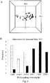
- FIG. 2A A PCA plot based solely on these masses, is shown in Figure 2A , which indicates a high degree of separation between the ovarian cancer and the control samples along the PC1 axis. Since the PC1 axis of this dataset is capturing 80% of the overall variance, the PC1 position of every sample could be used as a diagnostic score for each patient.
- Figure 2B A distribution of the PC1 scores of every sample for each cohort is shown in Figure 2B , which shows the number of ovarian cancer samples and controls that have PC1 scores falling within six binned ranges. If the origin of the PCA plot in Figure 2A is used as a cutoff point, one can see that two of the ovarian cancer patients cluster with the control side of the distribution, while three controls cluster with the ovarian cancer side. This suggests an approximate sensitivity of 90% and specificity of 88%.
- the PCA plot does not adequately allow one to visualize the actual intensities of the metabolites responsible for the separation of the clusters.
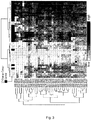
- a second statistical method was therefore used, called hierarchical clustering (HCA), to arrange the patient samples into groups based on a Euclidean distance measurements using the said 37 metabolites, which themselves were clustered using a Pearson correlation distance measurement.
- HCA hierarchical clustering
- the resulting metabolite array is shown in Figure 3 , and clearly reiterates the results observed with the PCA analysis, that is, the ovarian cancer and control cohorts are clearly discernable, with two ovarian cancer patients clustering within the control cohort, and three controls clustering within the ovarian cancer cohort.
- the array itself is comprised of cells representing the log(2) intensity from the FTMS, where white indicates metabolites with zero intensity, and increasing shades of grey indicate metabolites with increasing intensity values, respectively. It is clear that the 37 metabolites are all absent or relatively lower in intensity in the ovarian cancer cohort relative to the controls.
- the graph in Figure 4 further illustrates this point by plotting the average log(2) intensity (subsequently scaled between zero and one), of the 37 metabolites ( ⁇ 1 s.d.).
- Example 1 The metabolites and their associations with the clinical variables described in Example 1 are further confirmed using an independent mass spectrometry system.
- Representative sample extracts from each variable group are re-analyzed by LC-MS using an HP 1050 high-performance liquid chromatography (HPLC), or equivalent, interfaced to an ABI Q-Star (Applied Biosystems Inc., Foster City, CA), or equivalent, mass spectrometer to obtain mass and intensity information for the purpose of identifying metabolites that differ in intensity between the clinical variables under investigation.
- HPLC high-performance liquid chromatography
- ABI Q-Star Applied Biosystems Inc., Foster City, CA
- mass spectrometer mass spectrometer
- the retention times of the 29 metabolites shown in Figure 6 ranged between approximately 15 to 18 minutes under the chromatographic conditions.
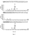
- averaged extracted mass spectra between 15 and 20 minutes for the controls, the ovarian cancers, and the net difference between the two cohorts were generated as shown in Figure 7 .
- the top panel (controls) to the middle panel (ovarian cancer)
- the peaks are at equal heights in both samples until approximately mass 400 is reached, at which point peaks are clearly detectable in the control group (upper panel), but not in the ovarian cancer subjects (middle panel).
- the bottom panel illustrates the net difference, which includes the 29 masses that overlap with the 3 7 identified in the FTMS data.
- the following example describes the tandem mass spectrometry analysis of a subset of the ovarian markers.
- the general principle is based upon the selection and fragmentation of each of the parent ions into a pattern of daughter ions.
- the fragmentation occurs within the mass spectrometer through a process called collision-induced dissociation, wherein an inert gas (such as argon) is allowed to collide with the parent ion resulting in its fragmentation into smaller components.
- the charge will then travel with one of the corresponding fragments.
- the pattern of resulting fragment or "daughter ions" represents a specific "fingerprint" for each molecule.
- Differently structured molecules (including those with the same formulas) will produce different fragmentation patterns, and therefore represents a very specific way of identifying the molecule.
- MSMS analysis was carried out on a subset of 31 ovarian markers (from Tables 2 and 3).
- the resulting fragment ions for each of the selected parent ions are listed in Tables 4 through 34.
- the parent ion is listed at the top of each table (as its neutral mass), and the subsequent fragments shown as negatively charged ions [M-H].
- the intensity is shown in the middle and right columns, respectively.
- the specific retention time is shown at the top of the middle column.
- the ovarian markers all had retention times under the chromatographic conditions used (see methods below) between 16 and 18 minutes.
- the metabolite markers are structurally related to the gamma-tocopherol form of vitamin E, in that they comprise a chroman ring-like moiety and phytyl side-chain. However, these molecules possess several important differences from gamma tocopherol:
- HPLC analysis were carried out with a high performance liquid chromatograph equipped with quaternary pump, automatic injector, degasser, and a Hypersil ODS column (5 ⁇ m particle size silica, 4.6 i.d x 200 mm) and semi-prep column (5 ⁇ m particle size silica, 9.1 i.d x 200 mm), with an inline filter.
- Mobile phase linear gradient H 2 O-MeOH to 100% MeOH in a 52 min period at a flow rate 1.0 ml/min.
- Eluate from the HPLC was analyzed using an ABI QSTAR® XL mass spectrometer fitted with an atmospheric pressure chemical ionization (APCI) source in negative mode.
- the scan type in full scan mode was time-of-flight (TOF) with an accumulation time of 1.0000 seconds, mass range between 50 and 1500 Da, and duration time of 55 min.
- Source parameters were as follows: Ion source gas 1 (GS1) 80; Ion source gas 2 (GS2) 10; Curtain gas (CUR) 30; Nebulizer Current (NC) -3.0; Temperature 400°C; Declustering Potential (DP) -60; Focusing Potential (FP) -265; Declustering Potential 2 (DP2) -15.
- MS/MS mode scan type was product ion, accumulation time was 1.0000 seconds, scan range between 50 and 650 Da and duration time 55 min.
- CE collision energy
- CAD collision gas
- Example 5 Targeted triple-quadrupole assay for selected ovarian markers
- the following example describes the development of a high-throughput screening (HTS) assay based upon triple-quadrupole mass spectrometry for a subset of the ovarian markers.
- the preliminary method was initially established to determine the ratio of six of the ovarian 28-carbon containing metabolites to an internal standard molecule added during the extraction procedure. This is similar to the HTS method reported in applicant's co-pending CRC/Ovarian PCT application published on March 22, 2007 ( WO 2007/030928 ).
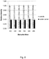
- the ability of this method to differentiate between ovarian cancer patients and subjects without ovarian cancer is shown in Figure 8 , where the 20 ovarian cancer subjects used to make the initial discovery are compared to 289 disease-free subjects.
- the six C28 carbon molecules (neutral masses 450 (C28H5004), 446 (C28H46O4), 468 (C28H52O5), 448 (C28H48O4), 464 (C28H48O5) and 466 (C28H50O5) were validated to be significantly lower in the serum of the ovarian patients versus the controls.
- the p-values for each of the molecules are shown in Table 66.
- This expanded triple-quadrupole method measures a comprehensive panel of the gamma Tocoenoic acids, and includes the metabolites listed in Table 67. The method measures the daughter fragment ion of each parent, as well an internal standard molecule (see methods below). The biomarker peak areas are then normalized by dividing by the internal standard peak areas.
- the method was then used to validate the reduction of gamma tocoenoic acids in a subsequent independent population of controls and ovarian cancer positive subjects.
- the graph in Figure 9 shows the average difference in signal intensity for each of the gamma tocoenoic acids in ovarian cancer patients relative to controls.
- the cohorts comprised 250 controls (i.e. not diagnosed with ovarian cancer at the time samples were taken, grey bars), and 241 ovarian cancer subjects (black bars).
- the averages of the original 20 ovarian cancer discovery samples (white bars) are also shown for this method.
- the results confirm that serum from ovarian cancer patients has low levels of gamma-tocoenoic acids relative to disease-free controls.
- the p-values for each metabolite 250 controls versus 241 ovarian cancers) are shown for each marker in Table 67 as well as in Figure 9 .
- Serum samples are extracted as described for non-targeted FTMS analysis.
- the ethyl acetate organic fraction is used for the analysis of each sample.
- 15uL of internal standard is added (1ng/mL of (24- 13 C)-Cholic Acid in methanol) to each sample aliquot of 120uL ethyl acetate fraction for a total volume of 135uL.
- the autosampler injects 100uL of the sample by flow-inj ection analysis into the 4000QTRAP.
- the carrier solvent is 90%methanol: 10%ethyl acetate, with a flow rate of 3 60uL/min into the APCI source.
- the MS/MS HTS method was developed on a quadrupole linear ion trap ABI 4000QTrap mass spectrometer equipped with a TurboVTM source with an APCI probe.
- the source gas parameters were as follows: CUR: 10.0, CAD: 6, NC: -3.0, TEM: 400, GS1: 15, interface heater on.
- "Compound" settings were as follows: entrance potential (EP): -10, and collision cell exit potential (CXP): -20.0.
- the method is based on the multiple reaction monitoring (MRM) of one parent ion transition for each metabolite and a single transition for the internal standard. Each of the transitions is monitored for 250 ms for a total cycle time of 2.3 seconds. The total acquisition time per sample is approximately 1 min.
- MRM multiple reaction monitoring
- the method is similar to that described in the PCT case referred to above ( WO 2007/030928 ), but was expanded to include a larger subset of the molecules as shown in Table 67.
- Table 4 446.4 CE: -35 V 16.4 min m / z (amu) intensity (counts) % intensity 401.3402 10.3333 100 445.3398 8.1667 79.0323 427.3226 4.5 43.5484 83.0509 2.8333 27.4194 223.1752 2.5 24.1935 222.1558 2.1667 20.9677 205.1506 1.8333 17.7419 383.3338 1.8333 17.7419 59.0097 1.6667 16.129 97.0644 1 9.6774 81.0348 0.6667 6.4516 109.0709 0.6667 6.4516 203.1555 0.6667 6.4516 221.1443 0.6667 6.4516 409.2901 0.6667 6.4516 123.0814 0.5 4.8387
- Table 36 MS/MS fragmentation of ovarian cancer biomarker 446.3544 m/z Formula Molecular fragment Fragment loss 445 C 28 H 45 O 4 -H + 427 C 28 H 43 O 3 -H 2 O 401 C 27 H 45 O 2 -CO 2 383 C 27 H 43 O -(CO 2 + H 2 O) 223 C 14 H 23 O 2 205 C 14 H 21 O 177 C 12 H 17 O (g) - C 2 H 4 162 C 11 H 114 O Table 37: MS/MS fragmentation of ovarian cancer biomarker 448.3715 m/z Formula Molecular fragment Fragment loss 447 C 28 H 47 O 4 -H + 429 C 28 H 45 O 3 -H 2 O 403 C 27 H 47 O 2 -CO 2 385 C 27 H 45 O - (CO 2+ H 2 O) 279 C 19 H 35 O Ring opening of 429 at O1-C2 and loss of 151 187 C 10 H 19 O 3 151 C 10 H 15 O Ring opening of 4
Landscapes
- Health & Medical Sciences (AREA)
- Life Sciences & Earth Sciences (AREA)
- Engineering & Computer Science (AREA)
- Biomedical Technology (AREA)
- Physics & Mathematics (AREA)
- Chemical & Material Sciences (AREA)
- Hematology (AREA)
- Immunology (AREA)
- Biophysics (AREA)
- Analytical Chemistry (AREA)
- Molecular Biology (AREA)
- Urology & Nephrology (AREA)
- General Health & Medical Sciences (AREA)
- Pathology (AREA)
- Food Science & Technology (AREA)
- Medicinal Chemistry (AREA)
- Biochemistry (AREA)
- General Physics & Mathematics (AREA)
- Ecology (AREA)
- Databases & Information Systems (AREA)
- Medical Informatics (AREA)
- Optics & Photonics (AREA)
- Human Computer Interaction (AREA)
- Cell Biology (AREA)
- Biotechnology (AREA)
- Microbiology (AREA)
- Other Investigation Or Analysis Of Materials By Electrical Means (AREA)
- Investigating Or Analysing Biological Materials (AREA)
Claims (9)
- Méthode de diagnostic d'un cancer de l'ovaire ou du risque de développement d'un cancer de l'ovaire chez un sujet d'essai de statut inconnu vis-à-vis du cancer de l'ovaire, comprenant les étapes suivantes:analyse d'un échantillon sanguin provenant d'un sujet d'essai pour obtenir des données quantitatives quant aux molécules choisies parmi les métabolites identifiés par les masses exactes 474.3736, 478.4022, 484.3764, 490.3658, 492.3815, 494.3971, 496.4128, 502.4022, 504.4179, 512.4077, 518.3971, 520.4128, 522.8284, 530.43351, 532.44916, 540.4389, 550.4597, 558.4648, 574.4597, 576.4754, 578.4910, 596.5016 et 598.5172 où une différence de ± 5 ppm indique le même métabolite ;où les formules moléculaires des métabolites correspondant à ces masses sont respectivement C30H50O4, C30H54O4, C28H52O6, C30H50O5, C30H52O5, C30H54O5, C30H56O5, C32H54O4, C32H56O4, C30H56O6, C32H54O5, C32H56O5, C32H60O5, C34H58O4, C34H60O4, C32H60O6, C34H62O5, C36H62O4, C36H62O5, C35H54O5, C36H66O5, C36H68O6 et C36H70O6;où les métabolites présentent des diagrammes de fragmentation LC-MS/MS analysés sous ionisation négative comme suit:le métabolite répondant à la formule moléculaire C30H50O4 présente un diagramme de fragmentation LC-MS/MS comprenant les ions dérivés de m/z: 473, 455, 429, 411, 223, 113 ;le métabolite répondant à la formule moléculaire C30H54O4 présente un diagramme de fragmentation LC-MS/MS comprenant les ions dérivés de m/z: 477, 460, 433, 415, 281 ;le métabolite répondant à la formule moléculaire C28H52O6 présente un diagramme de fragmentation LC-MS/MS comprenant les ions dérivés de m/z: 483, 465, 447, 439, 421, 315, 313, 297, 279, 241, 201, 171, 101 ;le métabolite répondant à la formule moléculaire C30H50O5 présente un diagramme de fragmentation LC-MS/MS comprenant les ions dérivés de m/z: 489, 471, 445, 427, 373, 345, 319, 267, 249, 223 ;le métabolite répondant à la formule moléculaire C30H52O5 présente un diagramme de fragmentation LC-MS/MS comprenant les ions dérivés de m/z 491, 473, 445, 427, 319, 249, 241, 223, 213 ;le métabolite répondant à la formule moléculaire C30H54O5 présente un diagramme de fragmentation LC-MS/MS comprenant les ions dérivés de m/z: 493, 475, 449, 431, 415, 307, 297, 279, 267, 241, 235, 223, 215, 197, 167, 151, 141, 113 ;le métabolite répondant à la formule moléculaire C30H56O5 présente un diagramme de fragmentation LC-MS/MS comprenant les ions dérivés de m/z: 495, 477, 451, 433, 297, 279, 241, 223, 215, 197, 179, 169 ;le métabolite répondant à la formule moléculaire C32H54O4 présente un diagramme de fragmentation LC-MS/MS comprenant les ions dérivés de m/z: 501, 483, 465, 457, 439, 279 ;le métabolite répondant à la formule moléculaire C32H56O4 présente un diagramme de fragmentation LC-MS/MS comprenant les ions dérivés de m/z: 503, 485, 467, 459, 441, 279, 263, 223, 169 ;le métabolite répondant à la formule moléculaire C30H56O6 présente un diagramme de fragmentation LC-MS/MS comprenant les ions dérivés de m/z: 511, 493, 467, 315, 297, 279, 259, 251, 151 ;le métabolite répondant à la formule moléculaire C32H54O5 présente un diagramme de fragmentation LC-MS/MS comprenant les ions dérivés de m/z: 517, 499, 481, 473, 455, 445, 437, 389, 279, 223 ;le métabolite répondant à la formule moléculaire C32H56O5 présente un diagramme de fragmentation LC-MS/MS comprenant les ions dérivés de m/z: 519, 501, 483, 475, 459, 457, 447, 297, 279, 241, 223, 195, 115 ;le métabolite répondant à la formule moléculaire C32H60O5 présente un diagramme de fragmentation LC-MS/MS comprenant les ions dérivés de m/z: 521, 503, 485, 477, 459, 441, 297, 279, 269, 241, 115;le métabolite répondant à la formule moléculaire C34H58O4 présente un diagramme de fragmentation LC-MS/MS comprenant les ions dérivés de m/z: 529, 511, 485, 467, 251, 205 ;le métabolite répondant à la formule moléculaire C34H60O4 présente un diagramme de fragmentation LC-MS/MS comprenant les ions dérivés de m/z: 531, 513, 495, 485, 469, 251, 181 ;le métabolite répondant à la formule moléculaire C32H60O6 présente un diagramme de fragmentation LC-MS/MS comprenant les ions dérivés de m/z: 539, 521, 495, 477, 315, 313, 297, 279, 259, 243, 241, 225, 223, 179, 171 ;le métabolite répondant à la formule moléculaire C34H62O5 présente un diagramme de fragmentation LC-MS/MS comprenant les ions dérivés de m/z: 549, 531, 513, 505, 487, 469, 297, 279, 269, 253, 125;le métabolite répondant à la formule moléculaire C36H62O4 présente un diagramme de fragmentation LC-MS/MS comprenant les ions dérivés de m/z: 557, 539, 513, 495, 279, 155 ;le métabolite répondant à la formule moléculaire C36H62O5 présente un diagramme de fragmentation LC-MS/MS comprenant les ions dérivés de m/z: 573, 555, 537, 529, 511, 493, 401, 295, 279, 223 ;le métabolite répondant à la formule moléculaire C36H64O5 présente un diagramme de fragmentation LC-MS/MS comprenant les ions dérivés de m/z: 575, 557, 539, 531, 513, 495, 403, 297, 279, 251, 183;le métabolite répondant à la formule moléculaire C36H66O5 présente un diagramme de fragmentation LC-MS/MS comprenant les ions dérivés de m/z: 577, 559, 541, 533, 515, 497, 373, 297, 281, 279 ;le métabolite répondant à la formule moléculaire C36H68O6 présente un diagramme de fragmentation LC-MS/MS comprenant les ions dérivés de m/z: 595, 577, 559, 551, 515, 315, 297, 281, 279, 171, 155, 141, 127 ;le métabolite répondant à la formule moléculaire C36H70O6 présente un diagramme de fragmentation LC-MS/MS comprenant les ions dérivés de m/z: 597, 579, 561, 517, 315, 297, 279 ; respectivementoù les molécules sont analysées par une méthode de chromatographie liquide-spectrométrie de masse (LC-MS) ou par une méthode de spectrométrie de masse à triple quadripôle et injection directe ;en comparant les données quantitatives obtenues sur des molécules issues dudit sujet d'essai à des données quantitatives obtenues à partir des petites molécules issues d'une multitude d'humains négatifs vis-à-vis du cancer de l'ovaire ; où l'absence ou la réduction significative d'un ou de plusieurs desdits métabolites indique la présence d'un cancer de l'ovaire ou le risque de développer un cancer de l'ovaire.
- Méthode selon la revendication 1, où ladite méthode est une méthode basée sur la spectrométrie de masse par résonance cyclotron des ions à transformée de Fourier.
- Méthode selon la revendication 1 ou 2, où une extraction liquide/liquide, par laquelle les métabolites non polaires sont dissous dans un solvant organique et les métabolites polaires sont dissous dans un solvant aqueux, est mise en oeuvre sur l'échantillon.
- Méthode selon l'une quelconque des revendications 1 à 3, où ladite méthode inclut l'utilisation d'un spectromètre de masse constitué d'une source qui ionise les molécules et d'un détecteur pour la détection des particules ionisées.
- Méthode selon la revendication 4, où le détecteur inclut des systèmes à base de quadripôle, à temps de vol, à secteur magnétique, à résonance cyclotron des ions, et leurs dérivés.
- Méthode selon la revendication 4 ou 5, où la source est choisie parmi l'ionisation par électrospray et l'ionisation chimique à pression atmosphérique.
- Méthode selon l'une quelconque des revendications 1 à 6, où le spectromètre de masse est équipé d'un système de séparation chromatographique.
- Méthode selon la revendication 1, où la transition d'intensité de chacune desdites molécules et la transition d'intensité d'un étalon interne sont mesurées.
- Méthode selon l'une quelconque des revendications 1 à 8, où l'échantillon sanguin est un échantillon de sang total, une sous-fraction de sang total, un échantillon de sérum sanguin ou un échantillon de plasma.
Applications Claiming Priority (2)
| Application Number | Priority Date | Filing Date | Title |
|---|---|---|---|
| US88769307P | 2007-02-01 | 2007-02-01 | |
| EP08714593A EP2115458A4 (fr) | 2007-02-01 | 2008-02-01 | Procedes de diagnostic d'etats de sante associes au cancer des ovaires, et de risque de tels etats |
Related Parent Applications (2)
| Application Number | Title | Priority Date | Filing Date |
|---|---|---|---|
| EP08714593A Division EP2115458A4 (fr) | 2007-02-01 | 2008-02-01 | Procedes de diagnostic d'etats de sante associes au cancer des ovaires, et de risque de tels etats |
| EP08714593.4 Division | 2008-02-01 |
Publications (3)
| Publication Number | Publication Date |
|---|---|
| EP2682746A2 EP2682746A2 (fr) | 2014-01-08 |
| EP2682746A3 EP2682746A3 (fr) | 2014-04-23 |
| EP2682746B1 true EP2682746B1 (fr) | 2017-05-24 |
Family
ID=39673619
Family Applications (2)
| Application Number | Title | Priority Date | Filing Date |
|---|---|---|---|
| EP13176355.9A Not-in-force EP2682746B1 (fr) | 2007-02-01 | 2008-02-01 | Procédés pour le diagnostic d'états de santé associés au cancer de l'ovaire et du risque d'états de santé associés au cancer des ovaires |
| EP08714593A Ceased EP2115458A4 (fr) | 2007-02-01 | 2008-02-01 | Procedes de diagnostic d'etats de sante associes au cancer des ovaires, et de risque de tels etats |
Family Applications After (1)
| Application Number | Title | Priority Date | Filing Date |
|---|---|---|---|
| EP08714593A Ceased EP2115458A4 (fr) | 2007-02-01 | 2008-02-01 | Procedes de diagnostic d'etats de sante associes au cancer des ovaires, et de risque de tels etats |
Country Status (8)
| Country | Link |
|---|---|
| US (3) | US20100086960A1 (fr) |
| EP (2) | EP2682746B1 (fr) |
| JP (2) | JP5221566B2 (fr) |
| CN (1) | CN101932934A (fr) |
| AU (2) | AU2008210207B2 (fr) |
| CA (1) | CA2676109C (fr) |
| MY (1) | MY184498A (fr) |
| WO (1) | WO2008092280A1 (fr) |
Families Citing this family (12)
| Publication number | Priority date | Publication date | Assignee | Title |
|---|---|---|---|---|
| CN101568838A (zh) * | 2006-12-21 | 2009-10-28 | 味之素株式会社 | 评估癌症状态的方法、癌症评估装置、癌症评估方法、癌症评估系统、癌症评估程序和记录介质 |
| JPWO2009110517A1 (ja) * | 2008-03-04 | 2011-07-14 | 味の素株式会社 | 癌種の評価方法 |
| CN104316701B (zh) * | 2008-06-20 | 2018-05-15 | 味之素株式会社 | 女性生殖器癌症的评价方法 |
| EP3124980A1 (fr) | 2009-10-01 | 2017-02-01 | Phenomenome Discoveries Inc. | Méthode de masse spectrophotométrique pour diagnostiquer le cancer pancréatique |
| WO2011156618A2 (fr) * | 2010-06-11 | 2011-12-15 | Board Of Regents, The University Of Texas System | Procédés d'identification de patientes avec des néoplasies épithéliales ovariennes basés sur la spectrométrie de masse haute résolution |
| WO2012099881A2 (fr) * | 2011-01-17 | 2012-07-26 | The John Hopkins University | Protéines mutantes en tant que biomarqueurs spécifiques du cancer |
| WO2013016700A1 (fr) * | 2011-07-27 | 2013-01-31 | The Research Foundation Of State University Of New York | Procédés de création de modèles prédictifs du cancer épithélial de l'ovaire et procédés d'identification du cancer épithélial de l'ovaire |
| BR112014004761A2 (pt) * | 2011-09-14 | 2017-03-21 | Basf Se | método para diagnosticar toxicidade gonadal, para determinar se um composto é capaz de induzir toxicidade gonadal em um indivíduo, para identificação de uma subtância para tratar toxicidade gonadal, uso de pelo menos um biomarcador, dispositivo para diagnosticar toxicidade gonadal em uma amostra de um indivíduo com suspeita de sofrer da mesma e kit para diagnosticar toxicidade gonadal |
| US20170097355A1 (en) * | 2015-10-06 | 2017-04-06 | University Of Washington | Biomarkers and methods to distinguish ovarian cancer from benign tumors |
| US9727697B1 (en) * | 2016-04-19 | 2017-08-08 | Honeywell International Inc. | System and approach for integration of parameters from wearable cloud connected access control devices |
| US10006925B2 (en) | 2016-05-30 | 2018-06-26 | Universal Diagnostics, S. L. | Methods and systems for metabolite and/or lipid-based detection of colorectal cancer and/or adenomatous polyps |
| BR112021003477A2 (pt) * | 2018-06-14 | 2021-05-18 | Metabolomycs, Inc. | assinaturas metabolômicas para prever, diagnosticar e prognosticar várias doenças incluindo câncer |
Family Cites Families (14)
| Publication number | Priority date | Publication date | Assignee | Title |
|---|---|---|---|---|
| CA2298181C (fr) * | 2000-02-02 | 2006-09-19 | Dayan Burke Goodnough | Analyse d'echantillons complexes non-cibles |
| US20030092638A1 (en) * | 2001-09-21 | 2003-05-15 | Dong Huang | Protopanaxadiol and protopanaxatriol and their use as anti-cancer agents |
| AU2002361812B2 (en) * | 2001-12-19 | 2007-11-08 | Research Development Foundation | Liposomal delivery of vitamin E based compounds |
| US20050214760A1 (en) * | 2002-01-07 | 2005-09-29 | Johns Hopkins University | Biomarkers for detecting ovarian cancer |
| AU2003202159A1 (en) * | 2002-04-08 | 2003-10-27 | Ginseng Science Inc. | Extract of processed panax genus plant, the preparation method thereof, and compositions containing the same |
| NZ562192A (en) * | 2002-08-06 | 2009-07-31 | Ciphergen Biosystems Inc | Use of biomarkers for detecting ovarian cancer |
| EP1649281A4 (fr) * | 2003-08-01 | 2007-11-07 | Correlogic Systems Inc | Multiples caracteristiques proteomiques de serums haute resolution pour la detection du cancer de l'ovaire |
| CA2535805A1 (fr) * | 2003-08-15 | 2005-02-24 | University Of Pittsburgh Of The Commnonwealth System Of Higher Education | Analyse multifactorielle pour la detection du cancer |
| WO2005098446A2 (fr) * | 2004-03-31 | 2005-10-20 | The Johns Hopkins University | Biomarqueurs du cancer des ovaires |
| CA2579034A1 (fr) * | 2004-07-30 | 2006-05-04 | Mount Sinai School Of Medicine Of New York University | Formes alternatives d'epissure de klf6 et polymorphisme d'adn de klf6 de cellules germinales associes a un risque accru de cancer |
| US8465929B2 (en) * | 2005-06-24 | 2013-06-18 | Vermillion, Inc. | Biomarkers for ovarian cancer |
| JP5038311B2 (ja) | 2005-09-12 | 2012-10-03 | フェノメノーム ディスカバリーズ インク | ビタミンe関連代謝産物の測定による、結腸直腸癌及び卵巣癌の診断方法 |
| US20080255764A1 (en) * | 2005-09-12 | 2008-10-16 | Phenomenome Discoveries Inc. | Methods for the Diagnosis of Colorectal Cancer and Ovarian Cancer by the Measurement of Vitamin E-Related Metabolites |
| US20080020472A1 (en) * | 2005-11-22 | 2008-01-24 | Frantz Biomarkers, Llc | Method for detecting an inflammatory disease or cancer |
-
2008
- 2008-02-01 EP EP13176355.9A patent/EP2682746B1/fr not_active Not-in-force
- 2008-02-01 CA CA2676109A patent/CA2676109C/fr not_active Expired - Fee Related
- 2008-02-01 MY MYPI20093031A patent/MY184498A/en unknown
- 2008-02-01 AU AU2008210207A patent/AU2008210207B2/en not_active Ceased
- 2008-02-01 CN CN2008800099823A patent/CN101932934A/zh active Pending
- 2008-02-01 JP JP2009547508A patent/JP5221566B2/ja not_active Expired - Fee Related
- 2008-02-01 WO PCT/CA2008/000270 patent/WO2008092280A1/fr not_active Ceased
- 2008-02-01 EP EP08714593A patent/EP2115458A4/fr not_active Ceased
- 2008-02-01 US US12/524,641 patent/US20100086960A1/en not_active Abandoned
-
2013
- 2013-01-24 JP JP2013010685A patent/JP5757964B2/ja not_active Expired - Fee Related
-
2014
- 2014-08-13 AU AU2014213513A patent/AU2014213513A1/en not_active Abandoned
- 2014-09-10 US US14/482,158 patent/US20160305928A1/en not_active Abandoned
-
2016
- 2016-06-24 US US15/192,578 patent/US20160377623A1/en not_active Abandoned
Non-Patent Citations (1)
| Title |
|---|
| C. DENKERT ET AL: "Mass Spectrometry-Based Metabolic Profiling Reveals Different Metabolite Patterns in Invasive Ovarian Carcinomas and Ovarian Borderline Tumors", CANCER RESEARCH, vol. 66, no. 22, 15 November 2006 (2006-11-15), pages 10795 - 10804, XP055176842, ISSN: 0008-5472, DOI: 10.1158/0008-5472.CAN-06-0755 * |
Also Published As
| Publication number | Publication date |
|---|---|
| CA2676109A1 (fr) | 2008-08-07 |
| MY184498A (en) | 2021-04-01 |
| JP5757964B2 (ja) | 2015-08-05 |
| EP2682746A2 (fr) | 2014-01-08 |
| EP2115458A1 (fr) | 2009-11-11 |
| AU2014213513A1 (en) | 2014-09-04 |
| JP2013101141A (ja) | 2013-05-23 |
| JP2010518363A (ja) | 2010-05-27 |
| CA2676109C (fr) | 2018-03-20 |
| US20100086960A1 (en) | 2010-04-08 |
| AU2008210207B2 (en) | 2014-05-15 |
| CN101932934A (zh) | 2010-12-29 |
| JP5221566B2 (ja) | 2013-06-26 |
| US20160305928A1 (en) | 2016-10-20 |
| US20160377623A1 (en) | 2016-12-29 |
| EP2115458A4 (fr) | 2011-06-08 |
| AU2008210207A1 (en) | 2008-08-07 |
| WO2008092280A1 (fr) | 2008-08-07 |
| EP2682746A3 (fr) | 2014-04-23 |
Similar Documents
| Publication | Publication Date | Title |
|---|---|---|
| AU2008210207B2 (en) | Methods for the diagnosis of ovarian cancer health states and risk of ovarian cancer health states | |
| US10697969B2 (en) | Methods for diagnosing a colorectal cancer (CRC) health state or change in CRC health state, or for diagnosing risk of developing CRC or the presence of CRC in a subject | |
| EP2483697B2 (fr) | Méthode pour diagnostiquer le cancer du pancréas | |
| EP2674435A2 (fr) | Biomarqueurs utiles pour diagnostiquer un cancer de la prostate et procédés associés | |
| EP3314264A1 (fr) | Moyens et procédés pour diagnostiquer un cancer du pancréas chez un sujet sur la base d'un panneau de biomarqueur | |
| US20080255764A1 (en) | Methods for the Diagnosis of Colorectal Cancer and Ovarian Cancer by the Measurement of Vitamin E-Related Metabolites | |
| US20170097355A1 (en) | Biomarkers and methods to distinguish ovarian cancer from benign tumors | |
| Nizioł et al. | Untargeted metabolomics of bladder tissue using liquid chromatography and quadrupole time-of-flight mass spectrometry for cancer biomarker detection | |
| HK1188824B (en) | Methods for the diagnosis of ovarian cancer health states and risk of ovarian cancer health states | |
| Kumar et al. | α-tocopherol deficiency in follicular ovarian cyst (FOCs) follicular fluid (FF) elevates oxidative stress and impairs oocyte maturation | |
| HK1188824A (en) | Methods for the diagnosis of ovarian cancer health states and risk of ovarian cancer health states | |
| HK1167182B (en) | Method to diagnose pancreatic cancer |
Legal Events
| Date | Code | Title | Description |
|---|---|---|---|
| PUAI | Public reference made under article 153(3) epc to a published international application that has entered the european phase |
Free format text: ORIGINAL CODE: 0009012 |
|
| AC | Divisional application: reference to earlier application |
Ref document number: 2115458 Country of ref document: EP Kind code of ref document: P |
|
| AK | Designated contracting states |
Kind code of ref document: A2 Designated state(s): AT BE BG CH CY CZ DE DK EE ES FI FR GB GR HR HU IE IS IT LI LT LU LV MC MT NL NO PL PT RO SE SI SK TR |
|
| PUAL | Search report despatched |
Free format text: ORIGINAL CODE: 0009013 |
|
| RIN1 | Information on inventor provided before grant (corrected) |
Inventor name: BINGHAM, ERIN Inventor name: RITCHIE, SHAWN |
|
| AK | Designated contracting states |
Kind code of ref document: A3 Designated state(s): AT BE BG CH CY CZ DE DK EE ES FI FR GB GR HR HU IE IS IT LI LT LU LV MC MT NL NO PL PT RO SE SI SK TR |
|
| RIC1 | Information provided on ipc code assigned before grant |
Ipc: H01J 49/26 20060101ALI20140317BHEP Ipc: G01N 27/00 20060101ALI20140317BHEP Ipc: G01N 33/49 20060101AFI20140317BHEP |
|
| REG | Reference to a national code |
Ref country code: HK Ref legal event code: DE Ref document number: 1188824 Country of ref document: HK |
|
| 17P | Request for examination filed |
Effective date: 20141022 |
|
| RBV | Designated contracting states (corrected) |
Designated state(s): AT BE BG CH CY CZ DE DK EE ES FI FR GB GR HR HU IE IS IT LI LT LU LV MC MT NL NO PL PT RO SE SI SK TR |
|
| 17Q | First examination report despatched |
Effective date: 20150319 |
|
| GRAP | Despatch of communication of intention to grant a patent |
Free format text: ORIGINAL CODE: EPIDOSNIGR1 |
|
| STAA | Information on the status of an ep patent application or granted ep patent |
Free format text: STATUS: GRANT OF PATENT IS INTENDED |
|
| INTG | Intention to grant announced |
Effective date: 20170222 |
|
| GRAS | Grant fee paid |
Free format text: ORIGINAL CODE: EPIDOSNIGR3 |
|
| GRAA | (expected) grant |
Free format text: ORIGINAL CODE: 0009210 |
|
| STAA | Information on the status of an ep patent application or granted ep patent |
Free format text: STATUS: THE PATENT HAS BEEN GRANTED |
|
| AC | Divisional application: reference to earlier application |
Ref document number: 2115458 Country of ref document: EP Kind code of ref document: P |
|
| AK | Designated contracting states |
Kind code of ref document: B1 Designated state(s): AT BE BG CH CY CZ DE DK EE ES FI FR GB GR HR HU IE IS IT LI LT LU LV MC MT NL NO PL PT RO SE SI SK TR |
|
| REG | Reference to a national code |
Ref country code: GB Ref legal event code: FG4D |
|
| REG | Reference to a national code |
Ref country code: CH Ref legal event code: EP |
|
| REG | Reference to a national code |
Ref country code: IE Ref legal event code: FG4D |
|
| REG | Reference to a national code |
Ref country code: AT Ref legal event code: REF Ref document number: 896265 Country of ref document: AT Kind code of ref document: T Effective date: 20170615 |
|
| REG | Reference to a national code |
Ref country code: DE Ref legal event code: R096 Ref document number: 602008050441 Country of ref document: DE |
|
| REG | Reference to a national code |
Ref country code: NL Ref legal event code: FP |
|
| REG | Reference to a national code |
Ref country code: SE Ref legal event code: TRGR |
|
| REG | Reference to a national code |
Ref country code: LT Ref legal event code: MG4D |
|
| REG | Reference to a national code |
Ref country code: AT Ref legal event code: MK05 Ref document number: 896265 Country of ref document: AT Kind code of ref document: T Effective date: 20170524 |
|
| PG25 | Lapsed in a contracting state [announced via postgrant information from national office to epo] |
Ref country code: FI Free format text: LAPSE BECAUSE OF FAILURE TO SUBMIT A TRANSLATION OF THE DESCRIPTION OR TO PAY THE FEE WITHIN THE PRESCRIBED TIME-LIMIT Effective date: 20170524 Ref country code: GR Free format text: LAPSE BECAUSE OF FAILURE TO SUBMIT A TRANSLATION OF THE DESCRIPTION OR TO PAY THE FEE WITHIN THE PRESCRIBED TIME-LIMIT Effective date: 20170825 Ref country code: LT Free format text: LAPSE BECAUSE OF FAILURE TO SUBMIT A TRANSLATION OF THE DESCRIPTION OR TO PAY THE FEE WITHIN THE PRESCRIBED TIME-LIMIT Effective date: 20170524 Ref country code: AT Free format text: LAPSE BECAUSE OF FAILURE TO SUBMIT A TRANSLATION OF THE DESCRIPTION OR TO PAY THE FEE WITHIN THE PRESCRIBED TIME-LIMIT Effective date: 20170524 Ref country code: ES Free format text: LAPSE BECAUSE OF FAILURE TO SUBMIT A TRANSLATION OF THE DESCRIPTION OR TO PAY THE FEE WITHIN THE PRESCRIBED TIME-LIMIT Effective date: 20170524 Ref country code: HR Free format text: LAPSE BECAUSE OF FAILURE TO SUBMIT A TRANSLATION OF THE DESCRIPTION OR TO PAY THE FEE WITHIN THE PRESCRIBED TIME-LIMIT Effective date: 20170524 Ref country code: NO Free format text: LAPSE BECAUSE OF FAILURE TO SUBMIT A TRANSLATION OF THE DESCRIPTION OR TO PAY THE FEE WITHIN THE PRESCRIBED TIME-LIMIT Effective date: 20170824 |
|
| PG25 | Lapsed in a contracting state [announced via postgrant information from national office to epo] |
Ref country code: IS Free format text: LAPSE BECAUSE OF FAILURE TO SUBMIT A TRANSLATION OF THE DESCRIPTION OR TO PAY THE FEE WITHIN THE PRESCRIBED TIME-LIMIT Effective date: 20170924 Ref country code: BG Free format text: LAPSE BECAUSE OF FAILURE TO SUBMIT A TRANSLATION OF THE DESCRIPTION OR TO PAY THE FEE WITHIN THE PRESCRIBED TIME-LIMIT Effective date: 20170824 Ref country code: LV Free format text: LAPSE BECAUSE OF FAILURE TO SUBMIT A TRANSLATION OF THE DESCRIPTION OR TO PAY THE FEE WITHIN THE PRESCRIBED TIME-LIMIT Effective date: 20170524 |
|
| PG25 | Lapsed in a contracting state [announced via postgrant information from national office to epo] |
Ref country code: EE Free format text: LAPSE BECAUSE OF FAILURE TO SUBMIT A TRANSLATION OF THE DESCRIPTION OR TO PAY THE FEE WITHIN THE PRESCRIBED TIME-LIMIT Effective date: 20170524 Ref country code: DK Free format text: LAPSE BECAUSE OF FAILURE TO SUBMIT A TRANSLATION OF THE DESCRIPTION OR TO PAY THE FEE WITHIN THE PRESCRIBED TIME-LIMIT Effective date: 20170524 Ref country code: SK Free format text: LAPSE BECAUSE OF FAILURE TO SUBMIT A TRANSLATION OF THE DESCRIPTION OR TO PAY THE FEE WITHIN THE PRESCRIBED TIME-LIMIT Effective date: 20170524 Ref country code: CZ Free format text: LAPSE BECAUSE OF FAILURE TO SUBMIT A TRANSLATION OF THE DESCRIPTION OR TO PAY THE FEE WITHIN THE PRESCRIBED TIME-LIMIT Effective date: 20170524 Ref country code: RO Free format text: LAPSE BECAUSE OF FAILURE TO SUBMIT A TRANSLATION OF THE DESCRIPTION OR TO PAY THE FEE WITHIN THE PRESCRIBED TIME-LIMIT Effective date: 20170524 |
|
| REG | Reference to a national code |
Ref country code: FR Ref legal event code: PLFP Year of fee payment: 11 |
|
| REG | Reference to a national code |
Ref country code: DE Ref legal event code: R097 Ref document number: 602008050441 Country of ref document: DE |
|
| PG25 | Lapsed in a contracting state [announced via postgrant information from national office to epo] |
Ref country code: IT Free format text: LAPSE BECAUSE OF FAILURE TO SUBMIT A TRANSLATION OF THE DESCRIPTION OR TO PAY THE FEE WITHIN THE PRESCRIBED TIME-LIMIT Effective date: 20170524 Ref country code: PL Free format text: LAPSE BECAUSE OF FAILURE TO SUBMIT A TRANSLATION OF THE DESCRIPTION OR TO PAY THE FEE WITHIN THE PRESCRIBED TIME-LIMIT Effective date: 20170524 |
|
| PGFP | Annual fee paid to national office [announced via postgrant information from national office to epo] |
Ref country code: NL Payment date: 20180214 Year of fee payment: 11 |
|
| PLBE | No opposition filed within time limit |
Free format text: ORIGINAL CODE: 0009261 |
|
| STAA | Information on the status of an ep patent application or granted ep patent |
Free format text: STATUS: NO OPPOSITION FILED WITHIN TIME LIMIT |
|
| REG | Reference to a national code |
Ref country code: HK Ref legal event code: GR Ref document number: 1188824 Country of ref document: HK |
|
| PGFP | Annual fee paid to national office [announced via postgrant information from national office to epo] |
Ref country code: CH Payment date: 20180213 Year of fee payment: 11 Ref country code: DE Payment date: 20180206 Year of fee payment: 11 Ref country code: GB Payment date: 20180207 Year of fee payment: 11 |
|
| 26N | No opposition filed |
Effective date: 20180227 |
|
| PG25 | Lapsed in a contracting state [announced via postgrant information from national office to epo] |
Ref country code: SI Free format text: LAPSE BECAUSE OF FAILURE TO SUBMIT A TRANSLATION OF THE DESCRIPTION OR TO PAY THE FEE WITHIN THE PRESCRIBED TIME-LIMIT Effective date: 20170524 |
|
| PGFP | Annual fee paid to national office [announced via postgrant information from national office to epo] |
Ref country code: SE Payment date: 20180213 Year of fee payment: 11 Ref country code: FR Payment date: 20180223 Year of fee payment: 11 |
|
| REG | Reference to a national code |
Ref country code: DE Ref legal event code: R082 Ref document number: 602008050441 Country of ref document: DE Representative=s name: D YOUNG & CO LLP, DE Ref country code: DE Ref legal event code: R081 Ref document number: 602008050441 Country of ref document: DE Owner name: MED-LIFE DISCOVERIES LP, SASKATOON, CA Free format text: FORMER OWNER: PHENOMENOME DISCOVERIES INC., SASKATOON, SASKATCHEWAN, CA |
|
| PG25 | Lapsed in a contracting state [announced via postgrant information from national office to epo] |
Ref country code: MC Free format text: LAPSE BECAUSE OF FAILURE TO SUBMIT A TRANSLATION OF THE DESCRIPTION OR TO PAY THE FEE WITHIN THE PRESCRIBED TIME-LIMIT Effective date: 20170524 |
|
| REG | Reference to a national code |
Ref country code: CH Ref legal event code: PUE Owner name: MED-LIFE DISCOVERIES LP, CA Free format text: FORMER OWNER: PHENOMENOME DISCOVERIES INC., CA |
|
| REG | Reference to a national code |
Ref country code: IE Ref legal event code: MM4A |
|
| REG | Reference to a national code |
Ref country code: BE Ref legal event code: MM Effective date: 20180228 |
|
| PG25 | Lapsed in a contracting state [announced via postgrant information from national office to epo] |
Ref country code: LU Free format text: LAPSE BECAUSE OF NON-PAYMENT OF DUE FEES Effective date: 20180201 |
|
| REG | Reference to a national code |
Ref country code: NL Ref legal event code: PD Owner name: MED-LIFE DISCOVERIES LP; CA Free format text: DETAILS ASSIGNMENT: CHANGE OF OWNER(S), ASSIGNMENT; FORMER OWNER NAME: PHENOMENOME DISCOVERIES INC. Effective date: 20181112 |
|
| PG25 | Lapsed in a contracting state [announced via postgrant information from national office to epo] |
Ref country code: IE Free format text: LAPSE BECAUSE OF NON-PAYMENT OF DUE FEES Effective date: 20180201 |
|
| PG25 | Lapsed in a contracting state [announced via postgrant information from national office to epo] |
Ref country code: BE Free format text: LAPSE BECAUSE OF NON-PAYMENT OF DUE FEES Effective date: 20180228 |
|
| REG | Reference to a national code |
Ref country code: DE Ref legal event code: R119 Ref document number: 602008050441 Country of ref document: DE |
|
| REG | Reference to a national code |
Ref country code: CH Ref legal event code: PL |
|
| REG | Reference to a national code |
Ref country code: SE Ref legal event code: EUG |
|
| REG | Reference to a national code |
Ref country code: NL Ref legal event code: MM Effective date: 20190301 |
|
| GBPC | Gb: european patent ceased through non-payment of renewal fee |
Effective date: 20190201 |
|
| PG25 | Lapsed in a contracting state [announced via postgrant information from national office to epo] |
Ref country code: SE Free format text: LAPSE BECAUSE OF NON-PAYMENT OF DUE FEES Effective date: 20190202 |
|
| PG25 | Lapsed in a contracting state [announced via postgrant information from national office to epo] |
Ref country code: LI Free format text: LAPSE BECAUSE OF NON-PAYMENT OF DUE FEES Effective date: 20190228 Ref country code: CH Free format text: LAPSE BECAUSE OF NON-PAYMENT OF DUE FEES Effective date: 20190228 |
|
| PG25 | Lapsed in a contracting state [announced via postgrant information from national office to epo] |
Ref country code: GB Free format text: LAPSE BECAUSE OF NON-PAYMENT OF DUE FEES Effective date: 20190201 Ref country code: MT Free format text: LAPSE BECAUSE OF NON-PAYMENT OF DUE FEES Effective date: 20180201 Ref country code: NL Free format text: LAPSE BECAUSE OF NON-PAYMENT OF DUE FEES Effective date: 20190301 Ref country code: DE Free format text: LAPSE BECAUSE OF NON-PAYMENT OF DUE FEES Effective date: 20190903 |
|
| PG25 | Lapsed in a contracting state [announced via postgrant information from national office to epo] |
Ref country code: FR Free format text: LAPSE BECAUSE OF NON-PAYMENT OF DUE FEES Effective date: 20190228 |
|
| PG25 | Lapsed in a contracting state [announced via postgrant information from national office to epo] |
Ref country code: TR Free format text: LAPSE BECAUSE OF FAILURE TO SUBMIT A TRANSLATION OF THE DESCRIPTION OR TO PAY THE FEE WITHIN THE PRESCRIBED TIME-LIMIT Effective date: 20170524 |
|
| PG25 | Lapsed in a contracting state [announced via postgrant information from national office to epo] |
Ref country code: PT Free format text: LAPSE BECAUSE OF FAILURE TO SUBMIT A TRANSLATION OF THE DESCRIPTION OR TO PAY THE FEE WITHIN THE PRESCRIBED TIME-LIMIT Effective date: 20170524 Ref country code: HU Free format text: LAPSE BECAUSE OF FAILURE TO SUBMIT A TRANSLATION OF THE DESCRIPTION OR TO PAY THE FEE WITHIN THE PRESCRIBED TIME-LIMIT; INVALID AB INITIO Effective date: 20080201 |
|
| PG25 | Lapsed in a contracting state [announced via postgrant information from national office to epo] |
Ref country code: CY Free format text: LAPSE BECAUSE OF FAILURE TO SUBMIT A TRANSLATION OF THE DESCRIPTION OR TO PAY THE FEE WITHIN THE PRESCRIBED TIME-LIMIT Effective date: 20170524 |