EP3124980A1 - Méthode de masse spectrophotométrique pour diagnostiquer le cancer pancréatique - Google Patents
Méthode de masse spectrophotométrique pour diagnostiquer le cancer pancréatique Download PDFInfo
- Publication number
- EP3124980A1 EP3124980A1 EP16177004.5A EP16177004A EP3124980A1 EP 3124980 A1 EP3124980 A1 EP 3124980A1 EP 16177004 A EP16177004 A EP 16177004A EP 3124980 A1 EP3124980 A1 EP 3124980A1
- Authority
- EP
- European Patent Office
- Prior art keywords
- lysopc
- pancreatic cancer
- sample
- accurate mass
- mass
- Prior art date
- Legal status (The legal status is an assumption and is not a legal conclusion. Google has not performed a legal analysis and makes no representation as to the accuracy of the status listed.)
- Withdrawn
Links
- 0 C[N+](C)(C)CCOP([O-])(OCC(*)C*)=O Chemical compound C[N+](C)(C)CCOP([O-])(OCC(*)C*)=O 0.000 description 4
Images





















Classifications
-
- G—PHYSICS
- G01—MEASURING; TESTING
- G01N—INVESTIGATING OR ANALYSING MATERIALS BY DETERMINING THEIR CHEMICAL OR PHYSICAL PROPERTIES
- G01N33/00—Investigating or analysing materials by specific methods not covered by groups G01N1/00 - G01N31/00
- G01N33/48—Biological material, e.g. blood, urine; Haemocytometers
- G01N33/50—Chemical analysis of biological material, e.g. blood, urine; Testing involving biospecific ligand binding methods; Immunological testing
- G01N33/53—Immunoassay; Biospecific binding assay; Materials therefor
- G01N33/575—Immunoassay; Biospecific binding assay; Materials therefor for cancer
- G01N33/57525—Immunoassay; Biospecific binding assay; Materials therefor for cancer of the liver or pancreas
-
- G—PHYSICS
- G01—MEASURING; TESTING
- G01N—INVESTIGATING OR ANALYSING MATERIALS BY DETERMINING THEIR CHEMICAL OR PHYSICAL PROPERTIES
- G01N33/00—Investigating or analysing materials by specific methods not covered by groups G01N1/00 - G01N31/00
- G01N33/48—Biological material, e.g. blood, urine; Haemocytometers
- G01N33/50—Chemical analysis of biological material, e.g. blood, urine; Testing involving biospecific ligand binding methods; Immunological testing
- G01N33/92—Chemical analysis of biological material, e.g. blood, urine; Testing involving biospecific ligand binding methods; Immunological testing involving lipids, e.g. cholesterol, lipoproteins, or their receptors
-
- G—PHYSICS
- G01—MEASURING; TESTING
- G01N—INVESTIGATING OR ANALYSING MATERIALS BY DETERMINING THEIR CHEMICAL OR PHYSICAL PROPERTIES
- G01N2405/00—Assays, e.g. immunoassays or enzyme assays, involving lipids
- G01N2405/04—Phospholipids, i.e. phosphoglycerides
-
- Y—GENERAL TAGGING OF NEW TECHNOLOGICAL DEVELOPMENTS; GENERAL TAGGING OF CROSS-SECTIONAL TECHNOLOGIES SPANNING OVER SEVERAL SECTIONS OF THE IPC; TECHNICAL SUBJECTS COVERED BY FORMER USPC CROSS-REFERENCE ART COLLECTIONS [XRACs] AND DIGESTS
- Y10—TECHNICAL SUBJECTS COVERED BY FORMER USPC
- Y10T—TECHNICAL SUBJECTS COVERED BY FORMER US CLASSIFICATION
- Y10T436/00—Chemistry: analytical and immunological testing
- Y10T436/16—Phosphorus containing
- Y10T436/163333—Organic [e.g., chemical warfare agents, insecticides, etc.]
Definitions
- the present invention relates to biomarkers and methods of detecting diseases and physiological conditions. More specifically, the invention relates to biomarkers of pancreatic cancer and methods using these compounds for detecting diseases and physiological conditions, especially pancreatic cancer.
- pancreatic cancer has increased during the past decades throughout the world, and ranks as the fourth and sixth leading causes of cancer in North America and the European Union respectively (1). This high rank is due to a very poor overall survival (OS) rate (less than 4%), which is illustrated by an annual incidence rate of pancreatic cancer almost identical to the mortality rate.
- OS overall survival
- 3800 new cases were expected to be diagnosed in 2008 with 3700 anticipated deaths from this cancer.
- pancreatic cancer displays a poor response to chemotherapy, radiation therapy, and surgery as conventionally used.
- the OS rate is less than 1% at five years, whereas for the rare patients diagnosed at an early stage, when surgery is possible, the after resection OS rate climbs to 20% (2).
- Cancer Antigen 19-9 (CA 19-9) is present primarily in pancreatic and biliary tract cancers, but also in patients with other malignancies (e.g. colorectal cancer) and benign conditions such as cirrhosis and pancreatitis.
- CA 19-9 is detected in most proteomics studies in pancreatic cancer serum samples (such as (7)), but its low specificity does not recommend it as a pancreatic cancer biomarker.
- Another glycosylation-related potential biomarker of pancreatic cancer is the core fusylation of biantennary glycans of RNase I, which displayed a 40% increase in the serum of two pancreatic cancer patients relative to two healthy controls (8).
- CEA carcinoembryonic antigen
- HIP/PAP-I and MIC-1 macrophage inhibitory cytokine I
- MIC-1 and CA19-9 seem the markers with the highest sensitivity and specificity, in the sense of specificity vs. chronic pancreatitis (and not vs. colon cancer for example), when compared to osteopontin, TIMP-1 and HIP/PAP-I (9).
- CA19-9 is now recommended in combination with other markers, such as the mutation status of pancreatic cancer -related oncogenes like K-ras (2).
- K-ras is reported to be mutated in 78% of pancreatic adenocarcinomas (11).
- Molecular events in pancreatic carcinogenesis have been extensively studied (12), and beside K-ras, p53, p21, p16, p27, SMAD4, and cyclin D1 are a few of these genes whose mutations or alterations in expression have been associated to pancreatic cancer (12).
- evidence regarding their application as prognostic indicators is conflicting. For instance, there is no consensus on the association between mutation inp53 and decreased survival (12).
- MicroRNA profiling has also been performed for pancreatic cancer, with the identification of some common microRNAs specifically altered (13-15).
- Protein markers show the advantage of simple screening through an ELISA (Enzyme-linked immunosorbent assay) method, and research in this field is therefore very intensive. Newer proteomics studies have identified additional protein markers, such as apolipoproteins A-I and A-II, and transthyretin (7), all decreased in serum of pancreatic cancer patients, as well as MMP-9, DJ-1 and A1BG, each of which is overexpressed in pancreatic juice from cancer patients (16).
- apolipoproteins The involvement of apolipoproteins is interesting since they participate in lipid metabolism (17) and other members of this family have been associated to cancer (18).
- fatty acid composition of lipids in plasma and bile from patients with pancreatic cancer has also been analyzed (19, 20), even though neither of these studies has detailed the chemical subfamilies of the altered lipids.
- Plasma from pancreatic patients showed significantly lower levels of phospholipids that contain the side chain 18:2( ⁇ 6), 20:5( ⁇ 3) or 22:5( ⁇ 3), without distinction of lipid classes (19).
- Bile from hepatopancreaticobiliary cancer patients was found to contain a much lower level of phosphatidylcholines without distinction of side chains (20).
- pancreatic cancer induced new-onset DM could be discriminated from type 2 DM with 77% sensitivity and 69% specificity (21).
- the methods described above are not ideally suited for large-scale population screening (either for low compliance or low sensitivity and specificity except in the case of a still-to-optimize multiple method combination), and most are capable of detecting pancreatic cancer after the formation of a tumor only. As a result, there still remains a need for accurate methods of detection, particularly for methods to detect early stages of the disease.
- the invention relates to methods and diagnostic markers for detecting or diagnosing cancer. Such methods and diagnostic markers are particularly useful for detecting pancreatic cancer.
- a method for diagnosing a subject's pancreatic cancer health state or change in health state, or for diagnosing pancreatic cancer or the risk of pancreatic cancer in a subject comprising steps of:
- the accurate mass intensity is measured at one or more of the following masses: 78.0516; 84.0575; 112.0974; 116.5696; 191.5055; 197.0896; 200.1389; 202.045; 203.1155; 214.1204; 214.1205; 232.1309; 233.1345; 240.0997; 243.0714; 244.0554; 254.1127; 255.1161; 256.2403; 260.0033; 262.0814; 268.1284; 270.0323; 270.0867; 276.0948; 280.2403; 280.2404; 281.2432; 281.2435; 282.2558; 282.2559; 283.2591; 283.2595; 284.9259; 300.1186; 300.2067; 302.0945; 302.222; 302.2457; 304.2375; 304.2407; 317.9613; 318.0931; 326.2048; 326.2458; 327.9902
- the accurate mass intensity is measured at an accurate mass of 519.3295, 523.3661, 541.3134,702.5709,724.5477,757.556, 779.5405, 783.569, 785.5913, 803.5373, 805.5549, 807.5734, 809.5796, 812.6774, 829.5516, 833.5864, 576.4751, 594.4863, 596.5017 or combinations thereof.
- a decrease in accurate mass intensity is generally identified in the comparing step (b).
- the accurate mass is measured at an accurate mass of 600.5117.
- an increase in accurate mass intensity is identified in the comparing step (b).
- the term “substantially equivalent” may in certain non-limiting embodiments refer to ⁇ 5 ppm of the hydrogen and electron adjusted accurate mass, or neutral accurate mass, and in further embodiments, ⁇ 1 ppm of the hydrogen and electron adjusted accurate mass, or neutral accurate mass.
- a method for diagnosing a patient's pancreatic cancer health state, or change in pancreatic cancer health state, or for diagnosing risk of developing pancreatic cancer or the presence of pancreatic cancer in a patient comprising the steps of:
- the one or more metabolite marker comprises one or more molecule having a molecular formula as follows: C 36 H 62 O 4 , C 36 H 62 O 5 , C 36 H 64 O 5 , C 36 H 66 O 5 , C 36 H 64 O 6 , C 36 H 66 O 6 , C 36 H 68 O 6 , C 22 H 46 NO 7 P, C 22 H 48 NO 7 P, C 24 H 50 NO 7 P, C 24 H 48 NO 7 P, C 24 H 46 NO 7 P, C 26 H 54 NO 7 P, C 26 11 52 NO 7 P, C 26 H 50 NO 7 P, C 26 H 48 NO 7 P, C 28 H 56 NO 7 P, C 28 H 54 NO 7 P, C 28 H 52 NO 7 P, C 28 H 50 NO 7 P, C 28 H 48 NO 7 P, C 28 H 46 NO 7 P, C 30 H 56 NO 7 P, C 30 H 54 NO 7 P, C 30 H 52 NO 7 P, C 30 H 52 NO 7 P, C 30 H 50 NO 7 P, C 32 H 58 NO 7 P
- the metabolite marker may be a diacylphosphatidylcholine, plasmanylphosphocholine or plasmenylphosphocholine as defined in Formula (I): including adducts or salts thereof, wherein
- the metabolite marker may be a 2-lysophosphatidylcholine as defined in Formula (II) or a 1-lysophosphatidylcholine as defined in Formula (III): including adducts or salts thereof, wherein R 1 is a 14:0, 14:1, 16:0, 16:1, 16:2, 18:0, 18:1, 18:2, 18:3, 20:1, 20:2, 20:3, 20:4, 20:5, 20:6, 22:3, 22:4, 22:5, 22:6, 24:4, 24:6, 30:1, 32:0, 32:1, 32:2 or 32:6 fatty acid moiety bonded to the glycerol backbone through an acyl linkage.
- the metabolite marker may be a sphingomyelin as defined in Formula (IV):
- R 2 of the sphingomyelin of Formula (IV) may be a C 11 alkyl group, a C 13 alkyl group, a C 15 alkyl group, a C 17 alkyl group, a C 17 alkenyl group with 3 double bonds, a C 19 alkyl group, a C 21 alkyl group, a C 23 alkenyl group with 1 double bond, a C 23 alkyl group, a C 24 alkyl group, a C 25 alkenyl group with 1 double bond, a C 25 alkyl group.
- the above described methods may further include steps of: analyzing a sample from the patient to obtain quantifying data for one or more than one internal standard molecule; and obtaining a ratio for each of the levels of the one or more than one metabolite marker to the level obtained for the one or more than one internal standard molecule; wherein the comparing step (b) comprises comparing each ratio to one or more corresponding ratios obtained for the one or more than one reference sample.
- the above-described methods can be carried out, at least in part, with the assistance of a computer.
- the computer may be integrated with the instrument used to perform the analysis, or it may be a separate computer adapted to receive data output from the instrument according to the knowledge and skill of those in the art.
- the analyzing step (a) will typically be carried out using the instrument, for example but not limited to a mass spectrometer, and the comparing step (b) carried out using the computer or other processing means programmed to receive the accurate mass intensity data or quantifying data from the instrument and perform the calculations required to identify an increase or decrease in the level of the one or more than one metabolite marker in the sample.
- This data from step (b) may be output for use by an individual trained to identify the noted increase or decrease and make the diagnosis of step (c), or alternatively the computer or processing means may be further programmed to generate an output of a diagnosis.
- the output may comprise a positive or negative diagnosis factor, and may optionally include additional details including but not limited to statistical data, threshold data, patient data and other details.
- the data may be output to a display, such as a monitor, to a printer for generating a copy of the details of diagnosis, to a data receiving centre or directly to a service provider, or in any other way as would be understood by one skilled in the art.
- the metabolite may be a lysophosphatidylcholine (LysoPC), including LysoPC 14:0, LysoPC 14:1, LysoPC 16:0, LysoPC 16:1, LysoPC 16:2, LysoPC 18:0, LysoPC 18:1, LysoPC 18:2, LysoPC 18:3, LysoPC 20:1, LysoPC 20:2, LysoPC 20:3, LysoPC 20:4, LysoPC 20:5, LysoPC 20:6, LysoPC 22:3, LysoPC 22:4, LysoPC 22:5, LysoPC 22:6, LysoPC 24:4, LysoPC 24:6, LysoPC 30:1, LysoPC 32:0, LysoPC 32:1, LysoPC 32:2, LysoPC 32:6, or combinations thereof.
- LysoPC lysophosphatidylcholine
- the metabolite may be a phosphatidylcholine, including phosphatidylcholine molecules having a molecular formula of C 42 H 78 NO 8 P, C 42 H 80 NO 8 P, C 42 H 82 NO 8 P, C 42 H 84 NO 8 P, C 44 H 78 NO 8 P, C 44 H 80 NO 8 P, C 44 H 82 NO 8 P, C 44 H 84 NO 8 P, C 44 H 86 NO 8 P, C 44 H 88 NO 8 P, C 46 H 78 NO 8 P, C 46 H 80 NO 8 P, C 46 H 82 NO 8 P, C 46 H 84 NO 8 P, C 48 H 80 NO 8 P, C 48 H 82 NO 8 P, C 48 H 84 NO 8 P, C 48 H 86 NO 8 P, or combinations thereof.
- the metabolite may be a plasmenylphosphocholine, including plasmenylphosphocholine molecules having a formula of C 42 H 80 NO 7 P, C 42 H 82 NO 7 P, C 42 H 84 NO 7 P, C 44 H 82 NO 7 P, C 44 H 84 NO 7 P, C 44 H 86 NO 7 P, C 44 H 88 NO 7 P, C 46 H 82 NO 7 P, C 46 H 84 NO 7 P, C 46 H 86 NO 7 P, C 48 H 84 NO 7 P, C 48 H 86 NO 7 P, or combinations thereof.
- the metabolite may be a sphingomyelin, including sphingomyelin molecules having a molecular formula of C 39 H 79 N 2 O 6 P (or C 39 H 80 N 2 O 6 P + ), C 41 H 81 N 2 O 6 P (or C 41 H 82 N 2 O 6 P + ), or C 41 H 83 N 2 O 6 P (or C 41 H 84 N 2 O 6 P + ), or C 47 H 93 N 2 O 6 P (or C 47 H 94 N 2 O 6 P + ), or C 47 H 95 N 2 O 6 P (or C 47 H 96 N 2 O 6 P + ), or combinations thereof.
- sphingomyelin molecules having a molecular formula of C 39 H 79 N 2 O 6 P (or C 39 H 80 N 2 O 6 P + ), C 41 H 81 N 2 O 6 P (or C 41 H 82 N 2 O 6 P + ), or C 41 H 83 N 2 O 6 P (or C 41 H 84 N 2
- alterations in the levels of the metabolite markers may be detected by MS/MS transition.
- a metabolite marker of molecular formula C 36 H 64 O 5 may be monitored for level fluctuations of organic extracts in negative ionization mode (such as atmospheric pressure chemical ionization (APCI)) at a MS/MS transition of 575.5 / 513.5, 575.5 / 557.5, 575.5 / 539.5, 575.5 / 531.5, 575.5 / 499.5, 575.5 / 495.5, 575.5 / 459.4, 575.5 / 417.4, 575.5 / 415.3, 575.5 / 413.3, 575.5 / 403.3, 575.5 / 295.2, 575.5 / 279.2, 575.5 / 260.2, 575.5 / 251.2, 575.5 / 197.9, 575.5 / 119.4, 575.5 / 113.1, and 575.5 / 97.0, or combinations thereof.
- negative ionization mode such as atmospheric pressure
- ESI Electrospray Ionization
- Other useful MS/MS transitions for aqueous extracts in positive ionization mode include: 520.3 / 184.2 for C 26 H 50 NO 7 P; 524.3 / 184.2 for C 26 H 54 NO 7 P; 542.3 / 184.2 for C 28 H 48 NO 7 P; 758.6 / 184.2 for C 42 H 80 NO 8 P; 784.6 / 184.2 for C 44 H 82 NO 8 P; 786.6 / 184.2 for C 44 H 84 NO 8 P; 788.6 / 184.2 for C 44 H 86 NO 8 P; 790.6 / 184.2 for C 44 H 88 NO 8 P; 806.6 / 184.2 for C 46 H 80 NO 8 P; 808.6 / 184.2 for C 46 H 82 NO 8 P; 810.6 / 184.2 for C 46 H 84 NO 8 P; 834.6 / 184.2 for C 48 H 84 NO 8 P; 836.6 / 184.2
- the step of comparing accurate mass intensity data to reference data to identify an increase or decrease in accurate mass intensity; or the step of comparing quantifying data for a metabolite marker to reference data to identify an increase or decrease in the level of the metabolite marker can in certain non-limiting embodiments comprise or otherwise relate to a step of determining the level of the specified markers, metabolites or molecules, either by determining a change in accurate mass intensity or by other analytical means.
- the invention further relates to an assay standard comprising a metabolite marker as described herein labeled with a detection agent.
- the standard will be useful for carrying out a diagnostic method as described herein, and may include one or more of the following non-limiting detection agents: a stable isotope, an enzyme, or a protein that enables detection in vitro.
- the assay standard may comprise as the metabolite marker a diacylphosphatidylcholine, plasmanylphosphocholine or plasmenylphosphocholine as defined in Formula (I):
- the assay standard may comprise as the metabolite marker a 2-lysophosphatidylcholine as defined in Formula (II) and a 1-lysophosphatidylcholine in Formula (III): including adducts or salts thereof, wherein R 1 is a 14:0, 14:1, 16:0, 16:1, 16:2, 18:0, 18:1, 18:2, 18:3, 20:1, 20:2, 20:3, 20:4, 20:5, 20:6, 22:3, 22:4, 22:5, 22:6, 24:4, 24:6, 30:1, 32:0, 32:1, 32:2 or 32:6 fatty acid moiety bonded to the glycerol backbone through an acyl linkage.
- R 1 is a 14:0, 14:1, 16:0, 16:1, 16:2, 18:0, 18:1, 18:2, 18:3, 20:1, 20:2, 20:3, 20:4, 20:5, 20:6, 22:3, 22:4, 22:5, 22:6, 24:4, 24:6, 30:
- the assay standard may comprise as the metabolite marker a sphingomyelin as defined in Formula (IV):
- R 2 of the sphingomyelin of Formula (IV) may be a C 11 alkyl group, a C 13 alkyl group, a C 15 alkyl group, a C 17 alkyl group, a C 17 alkenyl group with 3 double bonds, a C 19 alkyl group, a C 21 alkyl group, a C 23 alkenyl group with 1 double bond, a C 23 alkyl group, a C 24 alkyl group, a C 25 alkenyl group with 1 double bond, or a C 25 alkyl group.
- the assay standard may comprise as the metabolite marker a lysophosphatidylcholine (LysoPC, either 1-LysoPC or 2-LysoPC) including LysoPC 14:0, LysoPC 14:1, LysoPC 16:0, LysoPC 16:1, LysoPC 16:2, LysoPC 18:0, LysoPC 18:1, LysoPC 18:2, LysoPC 18:3, LysoPC 20:1, LysoPC 20:2, LysoPC 20:3, LysoPC 20:4, LysoPC 20:5, LysoPC 20:6, LysoPC 22:3, LysoPC 22:4, LysoPC 22:5, LysoPC 22:6, LysoPC 24:4, LysoPC 24:6, LysoPC 30:1, LysoPC 32:0, LysoPC 32:1, LysoPC 32:2, or LysoPC 32:6.
- LysoPC lysophosphatidylcholine
- the invention further relates to a kit or commercial package comprising the above-described standard and instructions for quantitating an analyte or performing a diagnostic test as described herein.
- the present inventors have identified cancer-specific biomarkers in human serum, and accordingly present herein a non-invasive cancer detection method that is useful for monitoring an individual's susceptibility to disease, and that may be used either alone or in combination with other known diagnostic methods.
- the methods described are particularly useful for detecting or diagnosing pancreatic cancer.
- a "non-targeted” approach was developed for the identification of biomarkers specific to pancreatic cancer.
- This discovery platform incorporated the use of Fourier transform ion cyclotron resonance mass spectrometry (FTICR-MS), which is capable of detecting ions with mass accuracy below 1 part per million (ppm).
- FTICR-MS Fourier transform ion cyclotron resonance mass spectrometry
- liquid sample extracts can be directly infused, for instance using electrospray ionization (ESI) and atmospheric pressure chemical ionization (APCI), without chromatographic separation. Ions with differing mass to charge (M/Z) ratios are then simultaneously resolved using a Fourier transformation.
- ESI electrospray ionization
- APCI atmospheric pressure chemical ionization
- M/Z mass to charge
- the inventors When analyzing the serum metabolomic profiles of pancreatic cancer patients and healthy asymptomatic subjects included in their study, the inventors identified specific biomarkers that had significantly altered serum levels in pancreatic cancer patients when compared to controls in a set of 90 samples. Structural characterization was performed by MS/MS technology, and some of the markers were found to be choline-related compounds. Alterations in the serum levels of these biomarkers were confirmed by targeted mass spectrometry using a targeted high-throughput triple-quadrupole MRM (TQ-MRM) method on the same samples.
- TQ-MRM targeted high-throughput triple-quadrupole MRM
- the inventors have accordingly developed methods to monitor levels of these biomarkers in a subject in a specific and sensitive manner, and to use this information as a useful tool for the early detection and screening of pancreatic cancer.
- the present invention accordingly relates to a method of diagnosing cancer by measuring the levels of specific biomarkers present in human serum and comparing them to "normal" reference levels.
- the described method may be used for the early detection and diagnosis of cancer as well as for monitoring the effects of treatment on cancer patients.
- the method also may be incorporated into a high-throughput screening method for testing large numbers of individuals, and further enables longitudinal screening throughout the lifetime of a subject to assess risk and detect disease early on.
- the method therefore has the potential to detect disease progression prior to that detectable by conventional methods, which is critical to positive treatment outcome.
- biological samples taken from one or more subjects of a particular health-state category are compared to the same samples taken from the normal population to identify differences in the levels of the described biomarkers.
- the samples are extracted and analyzed using various analytical platforms including, but not limited to, Fourier transform ion cyclotron resonance mass spectrometry (FTMS) and liquid chromatography mass spectrometry (LC-MS).
- FTMS Fourier transform ion cyclotron resonance mass spectrometry
- LC-MS liquid chromatography mass spectrometry
- the biological samples could originate from anywhere within the body, for example but not limited to, blood (serum/plasma), cerebral spinal fluid (CSF), bile, urine, stool, breath, saliva, or biopsy of any solid tissue including tumor, adjacent normal, smooth and skeletal muscle, adipose tissue, liver, skin, hair, brain, kidney, pancreas, lung, colon, stomach, or other.
- samples that are serum or CSF. While the term "serum" is used herein, those skilled in the art will recognize that plasma or whole blood or a sub-fraction of whole blood may be used.
- a blood sample When a blood sample is drawn from a patient there are several ways in which the sample can be processed.
- the range of processing can be as little as none (i.e. frozen whole blood) or as complex as the isolation of a particular cell type.
- the most common and routine procedures involve the preparation of either serum or plasma from whole blood. All blood sample processing methods, including spotting of blood samples onto solid-phase supports, such as filter paper or other immobile materials, are also contemplated by the invention.
- the processed blood or plasma sample described above may then be further processed to make it compatible with the methodical analysis technique to be employed in the detection and measurement of the metabolites contained within the processed blood sample.
- the types of processing can range from as little as no further processing to as complex as differential extraction and chemical derivatization.
- Extraction methods may include sonication, soxhlet extraction, microwave assisted extraction (MAE), supercritical fluid extraction (SFE), accelerated solvent extraction (ASE), pressurized liquid extraction (PLE), pressurized hot water extraction (PHWE) and/or surfactant assisted extraction (PHWE) in common solvents such as methanol, ethanol, mixtures of alcohols and water, or organic solvents such as ethyl acetate or hexane.
- a method of particular interest for extracting metabolites for FTMS non-targeted analysis and for flow injection LC-MS/MS analysis is to perform a liquid/liquid extraction whereby non-polar metabolites dissolve in an organic solvent and polar metabolites dissolve in an aqueous solvent.
- the extracted samples may be analyzed using any suitable method including those known in the art.
- extracts of biological samples are amenable to analysis on essentially any mass spectrometry platform, either by direct injection or following chromatographic separation.
- Typical mass spectrometers are comprised of a source that ionizes molecules within the sample, and a detector for detecting the ionized molecules or fragments of molecules.
- Non-limiting examples of common sources include electron impact, electrospray ionization (ESI), atmospheric pressure chemical ionization (APCI), atmospheric pressure photo ionization (APPI), matrix assisted laser desorption ionization (MALDI), surface enhanced laser desorption ionization (SELDI), and derivations thereof.
- Common mass separation and detection systems can include quadrupole, quadrupole ion trap, linear ion trap, time-of-flight (TOF), magnetic sector, ion cyclotron (FTMS), Orbitrap, and derivations and combinations thereof.
- TOF time-of-flight
- FTMS ion cyclotron
- Orbitrap derivations and combinations thereof.
- the advantage of FTMS over other MS-based platforms is its high resolving capability that allows for the separation of metabolites differing by only hundredths of a Dalton, many of which would be missed by lower resolution instruments.
- metabolic it is meant specific small molecules, the levels or intensities of which are measured in a sample, and that may be used as markers to diagnose a disease state. These small molecules may also be referred to herein as “metabolite marker”, “metabolite component”, “biomarker”, or “biochemical marker”.
- the metabolites are generally characterized by their accurate mass, as measured by mass spectrometry technique.
- the accurate mass may also be referred to as "accurate neutral mass” or “neutral mass”.
- the accurate mass of a metabolite is given herein in Daltons (Da), or a mass substantially equivalent thereto. By “substantially equivalent thereto”, it is meant that a +/- 5 ppm difference in the accurate mass would indicate the same metabolite.
- the accurate mass is given as the mass of the neutral metabolite.
- the metabolite will cause either a loss or gain of one or more hydrogen atoms and a loss or gain of an electron. This changes the accurate mass to the "ionized mass", which differs from the accurate mass by the mass of hydrogen atoms and electrons lost or gained during ionization.
- the accurate neutral mass will be referred to herein.
- the molecular formula of the neutral metabolite will be given.
- the molecular formula of the ionized metabolite will differ from the neutral molecular formula by the number of hydrogen atoms lost or gained during ionization or due to the addition of a non-hydrogen adduct ion.
- Data is collected during analysis and quantifying data for one or more than one metabolite is obtained.
- Quantifying data is obtained by measuring the levels or intensities of specific metabolites present in a sample.
- the quantifying data is compared to corresponding data from one or more than one reference sample.
- the "reference sample” is any suitable reference sample for the particular disease state.
- the reference sample may be a sample from a control individual, i.e., a person not suffering from cancer with or without a family history of cancer (also referred to herein as a " 'normal' counterpart"); the reference sample may also be a sample obtained from a patient clinically diagnosed with cancer.
- more than one reference sample may be used for comparison to the quantifying data.
- the one or more than one reference sample may be a first reference sample obtained from a non-cancer control individual.
- the reference sample may include a sample obtained at an earlier time period either pre-therapy or during therapy to compare the change in disease state as a result of therapy.
- an "internal control metabolite” refers to an endogenous metabolite naturally present in the patient. Any suitable endogenous metabolite that does not vary over the disease states can be used as the internal control metabolite.
- Non-targeted analysis involves the measurement of as many molecules in a sample as possible, without any prior knowledge or selection of the components prior to the analysis (see WO 01/57518, published August 9,2001 ). Therefore, the potential for non-targeted analysis to discover novel metabolite biomarkers is high versus targeted methods, which detect a predefined list of molecules.
- the present inventors used a non-targeted method to identify metabolite components that differ between cancer-positive and healthy individuals, followed by the development of a high-throughput targeted assay for a subset of the metabolites identified from the non-targeted analysis.
- HTS assay platform options There are multiple types of HTS assay platform options currently available depending on the molecules being detected. These include, but are not limited to, colorimetric chemical assays (UV, or other wavelength), antibody-based enzyme-linked immunosorbant assays (ELISAs), chip-based and polymerase-chain reaction for nucleic acid detection assays, bead-based nucleic-acid detection methods, dipstick chemical assays, image analysis such as MRI, petscan, CT scan, and various mass spectrometry-based systems.
- colorimetric chemical assays UV, or other wavelength
- ELISAs antibody-based enzyme-linked immunosorbant assays
- chip-based and polymerase-chain reaction for nucleic acid detection assays
- bead-based nucleic-acid detection methods dipstick chemical assays
- image analysis such as MRI, petscan, CT scan, and various mass spectrometry-based systems.
- the HTS assay is based upon conventional triple-quadrupole mass spectrometry technology.
- the HTS assay works by directly injecting a serum extract into the triple-quad mass spectrometer, which then individually isolates each of the parent molecules by single-ion monitoring (SIM). This is followed by the fragmentation of each molecule using an inert gas (called a collision gas, collectively referred to as collision-induced dissociation or CID). The intensity of a specific fragment from each parent biomarker is then measured and recorded, through a process called multiple-reaction monitoring (MRM).
- MRM multiple-reaction monitoring
- an internal standard molecule is also added to each sample and subjected to fragmentation as well.
- This internal standard fragment should have the same intensity in each sample if the method and instrumentation is operating correctly.
- biomarker fragment intensities, as well as the internal standard fragment intensities are collected, a ratio of the biomarker to IS fragment intensity is calculated, and the ratio log-transformed.
- the values for each patient sample are then compared to a previously determined distribution of disease-positive and controls, to determine the relative likelihood that the person is positive or negative for the disease.
- a commercial method for screening patients for cancer using the described assay methods is also envisioned.
- C36 markers One group of diagnostic biomarkers, referred to herein as the C36 markers (558.4, 574.5, 576.5, 578.5, 592.5, 594.5, 596.5), were determined to have the following molecular formulae, respectively: C 36 H 62 O 4 , C 36 H 62 O 5 , C 36 H 64 O 5 , C 36 H 66 O 5 , C 36 H 64 O 6 , C 36 H 66 O 6 , and C 36 H 68 O 6 .
- a second group of choline-related diagnostic biomarkers including lysophosphatidylcholines, phosphatidylcholines and sphingomyelins were also identified.
- the lysophosphatidylcholines include: LysoPC 14:0; LysoPC 14:1; LysoPC 16:0; LysoPC 16:1; LysoPC 16:2; LysoPC 18:0; LysoPC 18:1; LysoPC 18:2; LysoPC 18:3; LysoPC 20:1; LysoPC 20:2; LysoPC 20:3; LysoPC 20:4; LysoPC 20:5; LysoPC 20:6; LysoPC 22:3; LysoPC 22:4; LysoPC 22:5; LysoPC 22:6; LysoPC 24:4; LysoPC 24:6; LysoPC 30:1; LysoPC 32:0; LysoPC 32:
- the phosphatidylcholines (755.55; 757.56; 759.58; 761.59; 779.54; 781.56; 783.58; 785.59; 787.61; 803.54; 805.56; 807.58; 809.59; 829.55; 831.58; and 833.59) were determined to have the following molecular formulae, respectively: C 42 H 78 NO 8 P; C 42 H 80 NO 8 P; C 42 H 82 NO 8 P; C 42 H 84 NO 8 P; C 44 H 78 NO 8 P; C 44 H 80 NO 8 P; C 44 H 82 NO 8 P; C 44 H 84 NO 8 P; C 44 H 86 NO 8 P; C 46 H 78 NO 8 P; C 46 H 80 NO 8 P; C 46 H 82 NO 8 P; C 46 H 84 NO 8 P; C 48 H 80 NO 8 P; C 48 H 82 NO 8 P; and C 48 H 84 NO 8 P.
- the sphingomyelins 702.57 and 812.68 were determined to have the respective formulae C 39 H 72 N 2 O 6 P and C 47 H 93 N 2 O 6 P.
- the molecular weight, formulae and MS/MS transitions for each of these biomarkers are described in further detail below.
- CRT chemoradiation therapy
- Table 2 Clinical characteristics of the studied population.
- Stage I Stage II
- Stage III Stage IVa
- Stage IVb CRT 4 2 2 7 5 no CRT 0 2 3 9 6
- Serum samples were stored at -80°C until thawed for analysis, and were only thawed once. All extractions were performed on ice. Serum samples were prepared for FTICR-MS analysis by first sequentially extracting equal volumes of serum with 1% ammonium hydroxide and ethyl acetate (EtOAc) in the ratio of 1:1:5 respectively three times. Samples were centrifuged between extractions at 4°C for 10 min at 3500 rpm, and the organic layer removed and transferred to a new tube (extract A). After the third EtOAc extraction, 0.33 % formic acid was added, followed by two more EtOAc extractions.
- EtOAc ammonium hydroxide and ethyl acetate
- extract B protein removed by precipitation with 3:1 acetonitrile
- BuOH butanol
- Extracts were diluted either three or six-fold in methanol:0.1%(v/v) ammonium hydroxide (50:50, v/v) for negative ionization modes, or in methanol:0.1% (v/v) formic acid (50:50, v/v) for positive ionization modes.
- sample extracts were directly injected without diluting. All analyses were performed on a Bruker Daltonics APEX III Fourier transform ion cyclotron resonance mass spectrometer equipped with a 7.0 T actively shielded superconducting magnet (Bruker Daltonics, Billerica, MA).
- the scan type in full scan mode was time-of-flight (TOF-MS) with a scan time of 1.0000 second, mass range between 50 and 1500 Da, and duration time of 30 min.
- Source parameters were as follows: Ion source gas 1 (GS1) 80; Ion source gas 2 (GS2) 10; Curtain gas (CUR) 30; Nebulizer Current (NC) -3.0; Temperature 400°C; Declustering Potential (DP) -60; Focusing Potential (FP) -265; Declustering Potential 2 (DP2) -15.
- MS/MS mode scan type was Product Ion, scan time was 1.0000 second, scan range was 50 to 1500 Da and duration time was 30 min. All source parameters are the same as above, with collision energies (CE) of - 35 V and collision gas (CAD, nitrogen) of 5.
- CE collision energies
- CAD collision gas
- Solvent A H 2 O- MeOH-formic acid (94.9 : 5 : 0.1, v/v/v) and solvent B: MeOH-formic acid (99.9 : 0.1, v/v) were used as the mobile phase; the gradient solvent program was applied starting from 100% of A to 80% of B and 20% of A at 11 min, then held up to 20 min, then to 100% of B at 30 min, then held up to 45 min. Eluate from the HPLC was analyzed in negative and positive modes, using an Applied Biosystem (AB) QSTAR ® XL mass spectrometer fitted with an ESI source.
- AB Applied Biosystem
- the scan type in full scan mode was time-of-flight (TOF-MS) with a scan time of 1.0000 second, mass range between 50 and 1500 Da, and duration time of 60 min.
- Source parameters are as follows: Ion source gas 1 (GS1), 65; Ion source gas 2 (GS2), 75; Curtain gas (CUR), 30; Temperature 425°C; for negative mode: Ion Spray (IS), - 4200V; Declustering Potential (DP), -60; Focusing Potential (FP), -265; Declustering Potential 2 (DP2), -15; and for positive mode: Ion Spray (IS), 5500V; Declustering Potential (DP), 60; Focusing Potential (FP), 265; Declustering Potential 2 (DP2), 15.
- the scan type was Product Ion
- scan time was set as 1.0000 second
- scan range was 50 to 1500 Da
- duration time was 60 min. All source parameters are the same as above, with collision energy (CE) of -30 V and +30V, respectively, and collision gas (CAD, nitrogen) of 5.
- CE collision energy
- CAD collision gas
- Sample was prepared by adding 15 ⁇ L of internal standard (0.1 ⁇ g/mL of (24- 13 C)-Cholic Acid (Cambridge Isotope Laboratories, Andover, MA) in methanol) to 120 ⁇ L ethyl acetate fraction of each sample. 100 ⁇ L of sample was injected by flow injection analysis (FIA), and monitored under negative Atmospheric Pressure Chemical Ionization (APCI) mode. The method was based on multiple reaction monitoring (MRM) of one parent/fragment transition for each metabolite and (24- 13 C)-Cholic Acid (Table 3). Table 3.
- MRM multiple reaction monitoring
- the negative ESI mode transitions for phosphatidylcholines have been selected as follows: formate adduct and qualifier (both common to same mass phosphatidylcholines), and sn-2 fatty acid (specific to individual phosphatidylcholines).
- FTICR-MS accurate mass array alignments were performed using DlSCOVAmetrics TM (Phenomenome Discoveries Inc., Saskatoon). Initial statistical analysis and graphs of FTICR-MS data were carried out using Microsoft Office Excel 2007. Two-tailed unpaired Student's t -tests were used for determination of significant difference between pancreatic cancer and controls. P-values of less than 0.05 were considered significant. ROC curves were generated from logistic regression analysis using SAS Enterprise Guide 4.2.
- Serum metabolites were captured through a liquid extraction process (see methods) and extracts were directly infused by electrospray ionization (ESI) or atmospheric pressure chemical ionization (APCI) on an FTICR mass spectrometer.
- ESI electrospray ionization
- APCI atmospheric pressure chemical ionization
- the resulting spectral data of all the subjects was aligned within 1 ppm mass accuracy, background peaks were subtracted, and a two-dimensional array table comprising the intensities of each of the sample-specific spectral peaks was created using custom informatics software DISCOVAmetrics TM.
- Ratio pancreatic cancer / control AVG controls AVG pancreatic cancer 786.593 1101 5.24E-14 0.30 15.59 4.61 595.4897 1202 7.48E-14 0.36 5.30 1.88 594.4863 1202 9.91E-14 0.31 14.02 4.40 785.5913 1101 1.39E-13 0.27 33.03 8.93 808.5783 1101 1.63E-13 0.30 15.62 4.75 702.5709 1101 2.39E-13 0.47 9.60 4.48 780.5452 1101 3.57E-13 0.30 22.48 6.68 807.5734 1101 5.49E-13 0.28 34.47 9.70 576.4751 1202 5.61E-13 0.40 4.85 1.93 541.3134 1101 6.66E-13 0.37 6.27 2.30 804.5422 1101 2.04E-12 0.34 6.56 2.26 779.5405 1101 2.66E-12 0.26 53.60 13.93 812.6774 1101 3.81E-12 0.54 5.11 2.77 758.5626 1101 1.17E-11 0.
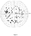
- Figure 2 illustrates the separation resulting from this unsupervised classification between pancreatic cancer (with individual samples in grey) and controls (in black).
- 13 C isotopic peaks were identified, such as the first two markers, 786.593 and 595.4897, which are the isotopic peaks of the fourth and third markers respectively, 785.5913 and 594.4863.
- Table 6 lists the 20 best biomarkers without 13 C isotopic peaks. All of these markers except 600.5117 have decreased levels in the pancreatic cancer cohort relative to controls. Table 6. List of the 20 best FTICR biomarkers of pancreatic cancer, sorted by mass within their analysis mode.
- Figure 3 illustrates (a) the separation resulting from this unsupervised classification between pancreatic cancer (with individual samples in grey) and controls (in black), as well as (b) the relative intensities of these 20 biomarkers in both populations.
- FIG. 4 illustrates each ROC along with the distribution of sample values for the first six best biomarkers ( p -value ⁇ E-12).
- Tandem mass spectrometric fragmentation fingerprints were generated for the markers mentioned above.
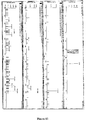
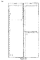
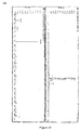
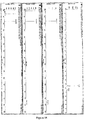
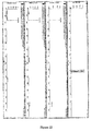
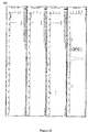
- the compound with a mass of 519.3 is confirmed to be a lysophosphatidylcholine with a fatty acid moiety of C18:2, and the two different retention times correspond to two different subspecies: the lower time shows the fragmentation pattern of the 1-linoleoyl- sn -glycero-3-phosphocholine ( Figure 14a ) whereas the higher shows the fragmentation pattern of the 2-linoleoyl- sn -glycero-3-phosphocholine ( Figure 14b ).
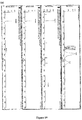
- the compound with a mass of 523.3 is confirmed to be a lysophosphatidylcholine with a fatty acid moiety of C18:0, and different retention times correspond to two different subspecies: the lower time shows the fragmentation pattern of the 2-stearoyl- sn -glycero-3-phosphocholine ( Figure 15a ) whereas the higher shows the fragmentation pattern of the 1-stearoyl- sn -glycero-3-phosphocholine ( Figure 15b ).
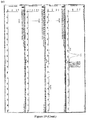
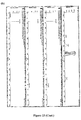
- the compounds with a mass of 541.3 seem to be a mixture of the lysophosphatidylcholines with a fatty acid moiety of C20:5 and of the sodium adduct of the compounds with a mass of 519.3 above mentioned ( Figure 16 ).
- the lowest retention time shows indeed two fragmentation patterns corresponding to 1-eicosapentaenoyl- sn -glycero-3-phosphocholine ( Figure 16a ) and 2 - ⁇ -eicosapentaenoyl- sn -glycero-3-phosphocholine ( Figure 16b ).
- the two higher retention times observed reflect the two retention times observed for 519.3, with the lower corresponding to the fragmentation pattern of the sodium adduct of the 1-linoleoyl- sn -glycero-3-phosphocholine ( Figure 16c ), and the higher corresponding to the fragmentation pattern of the sodium adduct of the 2-linoleoyl- sn -glycero-3-phosphocholine ( Figure 16d ).
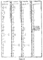
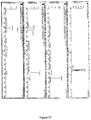
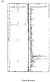
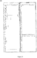
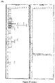
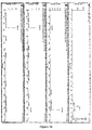
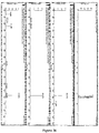
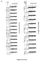
- Fragmentation pattern of all compounds seems restricted to one main fragment (m/z 184) for all masses, which likely corresponds to choline phosphate ( Figures 17 to 20 , 22 to 26 ), except for 803.5 ( Figure 21 ).
- the fragmentation pattern of 803.5 rather suggests the majority of the compounds at this mass to be the sodium adducts of 781.5566.
- Side chain combinations may be unique, such as in 757.6, corresponding to both PtdCho 16:0/18:2 and PtdCho 18:2/16:0 ( Figure 27 ), or multiple, such as in 807.6, corresponding to PtdCho 18:0/20:5, PtdCho 16:0/22:5 and PtdCho 18:1/20:4, all with the same chemical formula C 46 H 82 NO 8 P ( Figure 32 ).
- Confirmed side chains for all PtdCho biomarkers are reported in Table 15. Table 15.
- the fragmentation pattern of the putative sphingomyelins confirmed the presence of a choline phosphate fragment as the major peak for 702.6 and 812.7, suggesting that these two compounds respectively are the common sphingomyelins SM(d18:1/16:0) and SM(d18:1/24:1(15Z)) with the sphingosine (18:1) as the sphingoid base ( Figures 33 and 34 ).
- the fragmentation pattern of 724.5 suggests that the compound is the sodium adduct of 702.6 above mentioned ( Figure 35 ).
- the sphingomyelin identity of these two compounds was confirmed by a further analysis in aqueous negative ESI mode, through the comparison between the serum compounds with a mass of 702.6 and 812.7 and the commercially available sphingomyelins SM(d18:1/16:0) and SM(d18:1/24:1(15Z)).
- the fragmentation pattern of the serum compound with a mass of 702.6 detected as a formic acid adduct in negative ESI mode ( Figure 36 ) is indeed identical to the fragmentation pattern of the synthetic SM(d18:1/16:0) ( Figure 37 ).
- 600.5117 compound in 1203 analysis mode was further analyzed by tandem mass spectrometry mass fragmentation.
- the fragmentation pattern dominated by peaks at 545.5, 527.5 and 263.3, confirms that a compound with the molecular formula indicated in table 6 is present and can be classified as 1-alkenyl-2-acylglycerol with 18:2 at both side chains ( Figure 40 ).
- MRM multiple-reaction monitoring
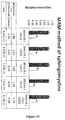
- Figure 44 reports the confirmation that the levels in the seven C36 markers tested are significantly decreased in pancreatic cancer patients relative to controls.
- the best putative C36 marker among all FTICR biomarkers which is also the best biomarker of pancreatic cancer, "594", is also the best biomarker among all C36 tested by MRM, with a p -value of 1.42E-11. Again, it is interesting to note that as a whole family, the C36 markers seem down-regulated in pancreatic cancer serum.
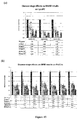
- Lysophosphatidylcholines 18:2, 18:3 and 20:5 show the strongest decrease of all LysoPC tested. All 27 PtdCho tested (with nine included in the top list of Table 6) show significantly decreased levels in pancreatic cancer patients relative to controls ( Figure 42a , b ). Most of the 10 PtdCho in Table 8 are predicted or shown to have 18:2,20:5 or 22:5 as one of the two side chains, as seen in Table 15. In summary, phosphatidylcholines and lysophosphatidylcholines that contain 18:2, 18:3, 20:5 and in a lesser extent, 22:5, show the strongest decrease.
- sphingomyelins among the best biomarkers is extremely interesting.
- sphingomyelin addition to pancreatic cancer cell lines has been shown to drastically enhance chemosensitivity to anticancer agents, presumably by redirecting the cell to enter the apoptotic pathway (29).
- pancreatic cancer we have identified a metabolic dysregulation specific to pancreatic cancer.
- the described diagnostic methods when conducted in conjunction with therapeutic optimization steps, may also be used to design more efficacious drug therapies for the disease.
Landscapes
- Health & Medical Sciences (AREA)
- Life Sciences & Earth Sciences (AREA)
- Engineering & Computer Science (AREA)
- Molecular Biology (AREA)
- Immunology (AREA)
- Urology & Nephrology (AREA)
- Biomedical Technology (AREA)
- Hematology (AREA)
- Chemical & Material Sciences (AREA)
- Medicinal Chemistry (AREA)
- Biochemistry (AREA)
- Microbiology (AREA)
- Pathology (AREA)
- Cell Biology (AREA)
- Food Science & Technology (AREA)
- Biotechnology (AREA)
- Physics & Mathematics (AREA)
- Analytical Chemistry (AREA)
- General Physics & Mathematics (AREA)
- General Health & Medical Sciences (AREA)
- Endocrinology (AREA)
- Biophysics (AREA)
- Other Investigation Or Analysis Of Materials By Electrical Means (AREA)
- Investigating Or Analysing Biological Materials (AREA)
- Gastroenterology & Hepatology (AREA)
- Measuring Or Testing Involving Enzymes Or Micro-Organisms (AREA)
Applications Claiming Priority (3)
| Application Number | Priority Date | Filing Date | Title |
|---|---|---|---|
| US24782809P | 2009-10-01 | 2009-10-01 | |
| EP14164872.5A EP2770328B1 (fr) | 2009-10-01 | 2010-10-01 | Procédé pour diagnostiquer le cancer du pancréas |
| EP10819788.0A EP2483697B2 (fr) | 2009-10-01 | 2010-10-01 | Méthode pour diagnostiquer le cancer du pancréas |
Related Parent Applications (4)
| Application Number | Title | Priority Date | Filing Date |
|---|---|---|---|
| EP10819788.0A Division EP2483697B2 (fr) | 2009-10-01 | 2010-10-01 | Méthode pour diagnostiquer le cancer du pancréas |
| EP10819788.0A Division-Into EP2483697B2 (fr) | 2009-10-01 | 2010-10-01 | Méthode pour diagnostiquer le cancer du pancréas |
| EP14164872.5A Division EP2770328B1 (fr) | 2009-10-01 | 2010-10-01 | Procédé pour diagnostiquer le cancer du pancréas |
| EP14164872.5A Division-Into EP2770328B1 (fr) | 2009-10-01 | 2010-10-01 | Procédé pour diagnostiquer le cancer du pancréas |
Publications (1)
| Publication Number | Publication Date |
|---|---|
| EP3124980A1 true EP3124980A1 (fr) | 2017-02-01 |
Family
ID=43825467
Family Applications (3)
| Application Number | Title | Priority Date | Filing Date |
|---|---|---|---|
| EP16177004.5A Withdrawn EP3124980A1 (fr) | 2009-10-01 | 2010-10-01 | Méthode de masse spectrophotométrique pour diagnostiquer le cancer pancréatique |
| EP14164872.5A Revoked EP2770328B1 (fr) | 2009-10-01 | 2010-10-01 | Procédé pour diagnostiquer le cancer du pancréas |
| EP10819788.0A Active EP2483697B2 (fr) | 2009-10-01 | 2010-10-01 | Méthode pour diagnostiquer le cancer du pancréas |
Family Applications After (2)
| Application Number | Title | Priority Date | Filing Date |
|---|---|---|---|
| EP14164872.5A Revoked EP2770328B1 (fr) | 2009-10-01 | 2010-10-01 | Procédé pour diagnostiquer le cancer du pancréas |
| EP10819788.0A Active EP2483697B2 (fr) | 2009-10-01 | 2010-10-01 | Méthode pour diagnostiquer le cancer du pancréas |
Country Status (7)
| Country | Link |
|---|---|
| US (3) | US10024857B2 (fr) |
| EP (3) | EP3124980A1 (fr) |
| JP (2) | JP5696152B2 (fr) |
| AU (2) | AU2010302909A1 (fr) |
| CA (3) | CA2797960A1 (fr) |
| SG (3) | SG179122A1 (fr) |
| WO (1) | WO2011038509A1 (fr) |
Cited By (1)
| Publication number | Priority date | Publication date | Assignee | Title |
|---|---|---|---|---|
| CN111679018A (zh) * | 2020-08-14 | 2020-09-18 | 宝枫生物科技(北京)有限公司 | 用于诊断认知障碍的生物标记物及其应用 |
Families Citing this family (23)
| Publication number | Priority date | Publication date | Assignee | Title |
|---|---|---|---|---|
| EP3124980A1 (fr) | 2009-10-01 | 2017-02-01 | Phenomenome Discoveries Inc. | Méthode de masse spectrophotométrique pour diagnostiquer le cancer pancréatique |
| AU2011260390B2 (en) * | 2010-06-01 | 2016-11-24 | Ernst-Moritz-Arndt Universitat Greifswald | Means and methods for diagnosing pancreatic cancer in a subject |
| EP2603622A4 (fr) * | 2010-08-13 | 2015-04-22 | Somalogic Inc | Biomarqueurs du cancer du pancréas et leurs utilisations |
| CA2857401A1 (fr) * | 2011-11-30 | 2013-06-06 | Metanomics Health Gmbh | Dispositif et procedes pour diagnostiquer le cancer du pancreas |
| WO2013176740A1 (fr) * | 2012-05-25 | 2013-11-28 | Health Diagnostic Laboratory, Inc. | Analyse rapide et à haut débit des stérols/stanols ou de leurs dérivés |
| AU2014368412A1 (en) * | 2013-12-20 | 2016-07-07 | Metanomics Health Gmbh | Means and methods for diagnosing pancreatic cancer in a subject based on a metabolite panel |
| CA2955204A1 (fr) | 2014-07-30 | 2016-02-04 | Metabolon, Inc. | Procedes, compositions et kits pour l'analyse de lipides complexes de structures diverses |
| US10006925B2 (en) | 2016-05-30 | 2018-06-26 | Universal Diagnostics, S. L. | Methods and systems for metabolite and/or lipid-based detection of colorectal cancer and/or adenomatous polyps |
| US11073522B2 (en) | 2016-10-03 | 2021-07-27 | Lincoln Memorial University | Structural validation of very long chain dicarboxylic acids |
| KR102549063B1 (ko) * | 2016-12-15 | 2023-06-28 | 더 보드 오브 리젠츠 오브 더 유니버시티 오브 텍사스 시스템 | 췌관 선암종의 검출 및 치료를 위한 방법 |
| US20180246112A1 (en) * | 2017-02-28 | 2018-08-30 | University Of Kentucky Research Foundation | Biomarkers of Breast and Lung Cancer |
| MX2019015210A (es) * | 2017-06-15 | 2020-08-17 | Med Life Discoveries Lp | Dimeros de acidos grasos dicarboxilicos y sus derivados, a manera de patrones para cuantificar niveles en biomuestras. |
| SG11202006667VA (en) * | 2018-01-22 | 2020-08-28 | Univ Pardubice | A method of diagnosing cancer based on lipidomic analysis of a body fluid |
| EP3514545B1 (fr) * | 2018-01-22 | 2020-10-07 | Univerzita Pardubice | Procédé de diagnostic du cancer du pancréas basé sur l'analyse lipidomique d'un fluide corporel |
| WO2019163245A1 (fr) * | 2018-02-26 | 2019-08-29 | 株式会社島津製作所 | Procédé d'analyse de quinones et système sfe-sfc en ligne permettant la mise en œuvre dudit procédé |
| RU2679123C1 (ru) * | 2018-04-09 | 2019-02-06 | Олег Геннадьевич Сивков | Способ прогнозирования тяжелого течения острого некротизирующего панкреатита |
| BR112021003477A2 (pt) * | 2018-06-14 | 2021-05-18 | Metabolomycs, Inc. | assinaturas metabolômicas para prever, diagnosticar e prognosticar várias doenças incluindo câncer |
| CN113939737A (zh) * | 2019-04-01 | 2022-01-14 | 创新生物有限公司 | 实体癌诊断装置和实体癌诊断信息提供方法 |
| WO2022140380A1 (fr) * | 2020-12-21 | 2022-06-30 | Pancreomics Llc | Signatures métabolomiques pour prédire, diagnostiquer et pronostiquer la pancréatite chronique |
| AU2022244454A1 (en) * | 2021-03-23 | 2023-02-09 | Kashiv Biosciences, Llc | Method for size based evaluation of pancreatic protein mixture |
| WO2023028835A1 (fr) * | 2021-08-31 | 2023-03-09 | 深圳迈瑞动物医疗科技股份有限公司 | Appareil d'analyse d'échantillon et procédé d'analyse d'échantillon |
| KR102421466B1 (ko) * | 2021-10-06 | 2022-07-18 | (주)이노베이션바이오 | 암 진단용 바이오마커 및 이의 용도 |
| WO2025111761A1 (fr) * | 2023-11-27 | 2025-06-05 | Precogify Pharmaceutical China Co., Ltd. | Biomarqueurs, systèmes et procédés de détection d'un adénocarcinome canalaire pancréatique réséquable |
Citations (1)
| Publication number | Priority date | Publication date | Assignee | Title |
|---|---|---|---|---|
| WO2001057518A2 (fr) | 2000-02-02 | 2001-08-09 | Yol Bolsum Canada Inc. | Methode d'analyse non ciblee d'echantillons complexes |
Family Cites Families (23)
| Publication number | Priority date | Publication date | Assignee | Title |
|---|---|---|---|---|
| JP2002510386A (ja) * | 1997-03-21 | 2002-04-02 | アタイアジン テクノロジーズ,インコーポレーテッド | リゾリン脂質の濃度変化に関連する癌の検出方法 |
| AU2004239418A1 (en) * | 2003-05-15 | 2004-11-25 | Europroteome Ag | Biomarkers for the differential diagnosis of pancreatitis and pancreatic cancer |
| WO2006044680A1 (fr) | 2004-10-14 | 2006-04-27 | The Cleveland Clinic Foundation | Procedes de detection de cancer colorectal |
| EP1852503A4 (fr) * | 2005-02-16 | 2010-04-07 | Takeda Pharmaceutical | Inhibiteur de production d il-13 |
| EP1870711B1 (fr) | 2005-03-14 | 2010-03-24 | Japan Health Sciences Foundation | Protéine marqueur utilisée lors du diagnostic du cancer du pancréas |
| JP5119405B2 (ja) * | 2005-06-17 | 2013-01-16 | 財団法人ヒューマンサイエンス振興財団 | 液体クロマトグラフィーのデータ補正方法 |
| JP5038311B2 (ja) * | 2005-09-12 | 2012-10-03 | フェノメノーム ディスカバリーズ インク | ビタミンe関連代謝産物の測定による、結腸直腸癌及び卵巣癌の診断方法 |
| JP5303273B2 (ja) | 2005-09-15 | 2013-10-02 | フェノメノーム ディスカバリーズ インク | フーリエ変換イオンサイクロトロン共鳴質量分析法についての方法及び装置 |
| US20080020472A1 (en) * | 2005-11-22 | 2008-01-24 | Frantz Biomarkers, Llc | Method for detecting an inflammatory disease or cancer |
| WO2007062142A2 (fr) | 2005-11-23 | 2007-05-31 | President And Fellows Of Harvard College | Procede d'identification de biomarqueurs associes au cancer |
| EP2322531A3 (fr) | 2006-02-28 | 2011-09-07 | Phenomenome Discoveries Inc. | Méthodes permettant de diagnostiquer la démence et autres troubles neurologiques |
| EP2674435A3 (fr) | 2006-03-24 | 2014-04-30 | Phenomenome Discoveries Inc. | Biomarqueurs utiles pour diagnostiquer un cancer de la prostate et procédés associés |
| CA2835964C (fr) | 2006-05-26 | 2016-07-19 | Phenomenome Discoveries Inc. | Biomarqueurs et methodes pour diagnostiquer la sclerose en plaques |
| EP2682746B1 (fr) | 2007-02-01 | 2017-05-24 | Phenomenome Discoveries Inc. | Procédés pour le diagnostic d'états de santé associés au cancer de l'ovaire et du risque d'états de santé associés au cancer des ovaires |
| EP2620147B1 (fr) | 2007-02-08 | 2020-06-03 | Med-Life Discoveries LP | Composés pour abaisser le cholestérol |
| CA2680748C (fr) | 2007-04-13 | 2012-08-28 | Phenomenome Discoveries Inc. | Procedes pour le diagnostic et l'evaluation de risque de maladies dues au vieillissement induites par la deficience en plasmalogene |
| JP5306345B2 (ja) | 2007-07-26 | 2013-10-02 | フェノメノーム ディスカバリーズ インク | 自閉症スペクトラム障害の診断、リスク評価、及びモニタリングを行うための方法 |
| CA2705044A1 (fr) † | 2007-11-07 | 2009-05-14 | Lian Shan | Procedes pour detecter ou surveiller un cancer a l'aide de lpc en tant que marqueur |
| WO2009088022A1 (fr) | 2008-01-07 | 2009-07-16 | Kagoshima University | Nouveau marqueur de cancer et diagnostic l'utilisant |
| WO2009151967A1 (fr) † | 2008-05-28 | 2009-12-17 | Georgia Tech Research Corporation | Biomarqueurs métaboliques pour le cancer des ovaires et procédés pour leur utilisation |
| TWI475989B (zh) | 2008-12-22 | 2015-03-11 | Phenomenome Discoveries Inc | 縮醛磷脂類化合物,含彼之醫藥組成物以及治療老化疾病的方法 |
| KR20120046754A (ko) | 2009-07-29 | 2012-05-10 | 페노미넘 디스커버리스 인코포레이티드 | 히드록시 지방산 화합물 및 질병 치료 및 진단을 위한 그의 용도 |
| EP3124980A1 (fr) | 2009-10-01 | 2017-02-01 | Phenomenome Discoveries Inc. | Méthode de masse spectrophotométrique pour diagnostiquer le cancer pancréatique |
-
2010
- 2010-10-01 EP EP16177004.5A patent/EP3124980A1/fr not_active Withdrawn
- 2010-10-01 EP EP14164872.5A patent/EP2770328B1/fr not_active Revoked
- 2010-10-01 AU AU2010302909A patent/AU2010302909A1/en not_active Abandoned
- 2010-10-01 CA CA2797960A patent/CA2797960A1/fr not_active Abandoned
- 2010-10-01 JP JP2012531198A patent/JP5696152B2/ja not_active Expired - Fee Related
- 2010-10-01 WO PCT/CA2010/001565 patent/WO2011038509A1/fr not_active Ceased
- 2010-10-01 US US13/499,369 patent/US10024857B2/en active Active
- 2010-10-01 EP EP10819788.0A patent/EP2483697B2/fr active Active
- 2010-10-01 SG SG2012017786A patent/SG179122A1/en unknown
- 2010-10-01 CA CA2797941A patent/CA2797941C/fr active Active
- 2010-10-01 SG SG10201405975VA patent/SG10201405975VA/en unknown
- 2010-10-01 CA CA2774869A patent/CA2774869C/fr active Active
- 2010-10-01 SG SG10201405974SA patent/SG10201405974SA/en unknown
-
2014
- 2014-08-18 JP JP2014165663A patent/JP6173274B2/ja not_active Expired - Fee Related
-
2016
- 2016-06-16 AU AU2016204043A patent/AU2016204043B2/en not_active Ceased
-
2018
- 2018-07-05 US US16/027,958 patent/US10656155B2/en active Active
-
2020
- 2020-03-25 US US16/830,038 patent/US11079385B2/en not_active Expired - Fee Related
Patent Citations (1)
| Publication number | Priority date | Publication date | Assignee | Title |
|---|---|---|---|---|
| WO2001057518A2 (fr) | 2000-02-02 | 2001-08-09 | Yol Bolsum Canada Inc. | Methode d'analyse non ciblee d'echantillons complexes |
Non-Patent Citations (34)
| Title |
|---|
| AHARONI, A.; C. H. RIC DE VOS; H. A. VERHOEVEN; C. A. MALIEPAARD; G. KRUPPA; R. BINO; D. B. GOODENOWE: "Nontargeted metabolome analysis by use of Fourier Transform Ion Cyclotron Mass Spectrometry", OMICS, vol. 6, 2002, pages 217 - 234, XP009037503, DOI: doi:10.1089/15362310260256882 |
| BARRABES, S.; L. PAGES-PONS; C. M. RADCLIFFE; G. TABARES; E. FORT; L. ROYLE; D. J. HARVEY; M. MOENNER; R. A. DWEK; P. M. RUDD: "Glycosylation of serum ribonuclease 1 indicates a major endothelial origin and reveals an increase in core fucosylation in pancreatic cancer", GLYCOBIOLOGY, vol. 17, 2008, pages 388 - 400, XP002638711, DOI: doi:10.1093/GLYCOB/CWM002 |
| BERNSTEIN, H.; C. BERNSTEIN; C. M. PAYNE; K. DVORAK: "Bile acids as endogenous etiologic agents in gastrointestinal cancer", WORLD J GASTROENTEROL, vol. 15, 2009, pages 3329 - 3340 |
| BILLICH, A.; T. BAUMRUKER.: "Sphingolipid metabolizing enzymes as novel therapeutic targets.", SUBCELL BIOCHEM, vol. 49, 2008, pages 487 - 522 |
| BLOOMSTON, M.; W. L. FRANKEL; F. PETROCCA; S. VOLINIA; H. ALDER; J. P. HAGAN; C. G. LIU; D. BHATT; C. TACCIOLI; C. M. CROCE: "MicroRNA expression patterns to differentiate pancreatic adenocarcinoma from normal pancreas and chronic pancreatitis", JAMA, vol. 297, 2007, pages 1901 - 1908, XP055048225, DOI: doi:10.1001/jama.297.17.1901. Text Size: A A A |
| BOYLE, P.; J. FERLAY.: "Cancer incidence and mortality in Europe, 2004", ANN ONCOL, vol. 16, 2005, pages 481 - 488 |
| CLARIA, J.: "Regulation of cell proliferation and apoptosis by bioactive lipid mediators.", RECENT PAT ANTICANCER DRUG DISCOV, vol. 1, 2006, pages 369 - 382 |
| D.E. MISEK ET AL.: "Early Detection and Biomarkers in Pancreatic cancer", JOURNAL OF THE NATIONAL COMPREHENSIVE CANCER NETWORK, vol. 5, no. 10, 1 November 2007 (2007-11-01), Fort Washington, PA , USA, pages 1034 - 1041, XP055053031 * |
| DABRITZ, J.; R. PRESTON; J. HAULIER; H. OETTLE: "Follow-up study of K-ras mutations in the plasma of patients with pancreatic cancer: correlation with clinical features and carbohydrate antigen 19-9", PANCREAS, vol. 38, 2009, pages 534 - 541 |
| DYATLOVITSKAYA, E. V.; A. G. KANDYBA: "Role of biologically active sphingolipids in tumor growth", BIOCHEMISTRY (MO,SC), vol. 71, 2006, pages 10 - 17, XP019294685 |
| EHMANN, M.; K. FELIX; D. HARTMANN; M. SCHNDLZER; M. NEES; S. VORDERWIILBECKE; R. BOGUMIL; M. W. BIICHLER; H. FRIESS: "Identification of potential markers for the detection of pancreatic cancer through comparative serum protein expression profiling", PANCREAS, vol. 34, 2007, pages 205 - 214, XP008183746, DOI: doi:10.1097/01.mpa.0000250128.57026.b2 |
| GARCEA, G.; C. P. NEAL; C. J. PATTENDEN; W. P. STEWARD; D. P. BERRY: "Molecular prognostic markers in pancreatic cancer: a systematic review", EUR J CANCER, vol. 41, 2005, pages 2213 - 2236, XP025298123, DOI: doi:10.1016/j.ejca.2005.04.044 |
| GEMMEL, C.; A. EICKHOFF; L. HELMSTADTER; J. F. RIEMANN: "Pancreatic cancer screening: state of the art", EXPERT REV GASTROENTEROL HEPATOL., vol. 3, 2009, pages 89 - 96 |
| GOODENOWE, D. B.; L. L. COOK; J. LIU; Y. LU; D. A. JAYASINGHE; P. W. AHIAHONU; D. HEATH; Y. YAMAZAKI; J. FLAX; K. F. KRENITSKY: "Peripheral ethanolamine plasmalogen deficiency: a logical causative factor in Alzheimer's disease and dementia", JOURNAL OF LIPID RESEARCH, vol. 48, 2007, pages 2485 - 2498, XP008122068, DOI: doi:10.1194/JLR.P700023-JLR200 |
| GRAY, G. R.; D. HEATH: "A global reorganization of the metabolome in Arabidopsis during cold acclimation is revealed by metabolic fingerprinting", PHYSIOLOGIA PLANTARUM, vol. 124, 2005, pages 236 - 248, XP002475400, DOI: doi:10.1111/j.1399-3054.2005.00507.x |
| HELMSTAEDTER, L.; J. F. RIEMANN: "Pancreatic cancer--EUS and early diagnosis", LANGENBECKS ARCH SURG, vol. 393, 2008, pages 923 - 927, XP019657904, DOI: doi:10.1007/s00423-007-0275-1 |
| HSU F F ET AL: "Electrospray ionization with low-energy collisionally activated dissociation tandem mass spectrometry of glycerophospholipids: Mechanisms of fragmentation and structural characterization", JOURNAL OF CHROMATOGRAPHY B: BIOMEDICAL SCIENCES & APPLICATIONS, ELSEVIER, AMSTERDAM, NL, vol. 877, no. 26, 15 September 2009 (2009-09-15), pages 2673 - 2695, XP026422743, ISSN: 1570-0232, [retrieved on 20090221] * |
| JANARDHAN, S.; P. SRIVANI; G. N. SASTRY: "Choline kinase: an important target for cancer.", CURR MED CHEM, vol. 13, 2006, pages 1169 - 1186, XP002517956, DOI: doi:10.2174/092986706776360923 |
| KHAN, S. A.; I. J. COX; A. V. THILLAINAYAGAM; D. S. BANSI; H, C. THOMAS; S. D. TAYLOR-ROBINSON: "Proton and phosphorus-31 nuclear magnetic resonance spectroscopy of human bile in hepatopancreaticobiliary cancer", EUR J GASTROENTEROL HEPATOL, vol. 17, 2005, pages 733 - 738 |
| KLAPMAN, J.; M. P. MALAFA: "Early detection of pancreatic cancer: why, who, and how to screen", CANCER CONTROL., vol. 15, 2008, pages 280 - 287 |
| KOLB, A.; S. RIEDER; D. BORN; N. A. GIESE; T. GIESE; G. RUDOFSKY; J. WERNER; M. W. BUCHLER; H. FRIESS; I. ESPOSITO: "Glucagon/insulin ratio as a potential biomarker for pancreatic cancer in patients with new-onset diabetes mellitus", CANCER BIOL THER, 2009, pages 8 |
| KOOPMANN, J.; C. N. WHITE ROSENZWEIG; Z. ZHANG; M. I. CANTO; D. A. BROWN; M. HUNTER, C. YEO; D. W. CHAN; S. N. BREIT; M. GOGGINS: "Serum Markers in Patients with Resectable Pancreatic Adenocarcinoma: Macrophage Inhibitory Cytokine 1 versus CA19-9", CLINICAL CANCER RESEARCH, vol. 12, 2006, pages 442 - 446 |
| LEE, E. J.; Y. GUSEV; J. JIANG; G. J. NUOVO; M. R. LERNER; W. L. FRANKEL; D. L. MORGAN; R. G. POSTIER; D. J. BRACKETT; T. D. SCHMI: "Expression profiling identifies microRNA signature in pancreatic cancer", INT J CANCER, vol. 120, 2007, pages 1046 - 1054, XP002569593, DOI: doi:10.1002/IJC.22394 |
| MODRAK, D. E.; D. V. GOLD; D. M. GOLDENBERG: "Sphingolipid targets in cancer therapy.", MOL CANCER THER, vol. 5, 2006, pages 200 - 208, XP002378561, DOI: doi:10.1158/1535-7163.MCT-05-0420 |
| MODRAK, D. E.; E. LEON; D. M. GOLDENBERG; D. V. GOLD: "Ceramide regulates gemcitabine-induced senescence and apoptosis in human pancreatic cancer cell lines", MOL CANCER RES, vol. 7, 2009, pages 890 - 896 |
| PERACAULA, R.; S. BARRABES; A. SARRATS; P. M. RUDD; R. DE LLORENS: "Altered glycosylation in tumours focused to cancer diagnosis.", DIS MARKERS, vol. 25, 2008, pages 207 - 218 |
| RAMIREZ DE MOLINA, A.; D. GALLEGO-ORTEGA; J. SARMENTERO-ESTRADA; D. LAGARES; T. GOMEZ DEL PULGAR; E. BANDRES; J. GARCIA-FONCILLAS;: "Choline kinase as a link connecting phospholipid metabolism and cell cycle regulation: implications in cancer therapy", THE INTERNATIONAL JOURNAL OF BIOCHEMISTRY & CELL BIOLOGY, vol. 40, 2008, pages 1753 - 1763, XP022734731, DOI: doi:10.1016/j.biocel.2008.01.013 |
| ROSTY, C.; L. CHRISTA; S. KUZDZAL, B. W.M.; M. L. ZAHURAK; F. CARNOT; D. W. CHAN; M. CANTO; K. D. LILLEMOE; J. L. CAMERON; C. J. Y: "Identification of hepatocarcinoma-intestine-pancreas/pancreatitis-associated protein I as a biomarker for pancreatic ductal adenocarcinoma by protein biochip technology.", CANCER RESEARCH, vol. 62, 2002, pages 1868 - 1875, XP001179968 |
| S A RITCHIE ET AL: "Metabolic system alterations in pancreatic cancer patient serum: potential for early detection", BMC CANCER (BIOMED CENTRAL), vol. 13, no. 1, 12 September 2013 (2013-09-12), London UK, pages 416, XP021159052, ISSN: 1471-2407, DOI: 10.1186/1471-2407-13-416 * |
| SZAFRANSKA, A. E.; T. S. DAVISON; J. JOHN; T. CANNON; B. SIPOS; A. MAGHNOUJ; E. LABOURIER; S. A. HAHN: "MicroRNA expression alterations are linked to tumorigenesis and non-neoplastic processes in pancreatic ductal adenocarcinoma", ONCOGENE, vol. 26, 2007, pages 4442 - 4452 |
| TIAN, M.; Y. Z. CUI; G. H. SONG; M. J. ZONG; X. Y. ZHOU; Y. R. CHEN; J. X. HAN.: "Proteomic analysis identifies MMP-9, DJ-1 and A1BG as overexpressed proteins in pancreatic juice from pancreatic ductal adenocarcinoma patients", BMC CANCER, vol. 8, 2008, pages 241, XP021042880, DOI: doi:10.1186/1471-2407-8-241 |
| TROUGAKOS, I. P.; E. S. GONOS: "Clusterin/apolipoprotein J in human aging and cancer", THE INTERNATIONAL JOURNAL OF BIOCHEMISTRY & CELL BIOLOGY, vol. 34, 2002, pages 1430 - 1448, XP002348133, DOI: doi:10.1016/S1357-2725(02)00041-9 |
| WARDEN, C. H.; A. DALUISKI; X. BU; D. A. PURCELL-HUYNH; C. DE MEESTER; B. H. SHIEH; D. L. PUPPIONE; R. M. GRAY; G. M. REAVEN; Y. D: "Evidence for linkage of the apolipoprotein A-II locus to plasma apolipoprotein A-II and free fatty acid levels in mice and humans", PROCEEDINGS OF THE NATIONAL ACADEMY OF SCIENCES OF THE UNITED STATES OF AMERICA, vol. 90, 1993, pages 10886 - 10890 |
| ZUIJDGEEST-VAN LEEUWEN, S. D.; M. S. VAN DER HEIJDEN; T. RIETVELD; J. W. VAN DEN BERG; H. W. TILANUS; J. A. BURGERS; J. H. WILSON;: "Fatty acid composition of plasma lipids in patients with pancreatic, lung and oesophageal cancer in comparison with healthy subjects", CLINICAL NUTRITION, vol. 21, 2002, pages 225 - 230 |
Cited By (1)
| Publication number | Priority date | Publication date | Assignee | Title |
|---|---|---|---|---|
| CN111679018A (zh) * | 2020-08-14 | 2020-09-18 | 宝枫生物科技(北京)有限公司 | 用于诊断认知障碍的生物标记物及其应用 |
Also Published As
| Publication number | Publication date |
|---|---|
| JP2014240841A (ja) | 2014-12-25 |
| WO2011038509A1 (fr) | 2011-04-07 |
| EP2483697A1 (fr) | 2012-08-08 |
| US20210033614A1 (en) | 2021-02-04 |
| EP2770328A2 (fr) | 2014-08-27 |
| EP2483697B1 (fr) | 2014-06-04 |
| CA2797941A1 (fr) | 2011-04-07 |
| SG10201405974SA (en) | 2014-10-30 |
| US20120202188A1 (en) | 2012-08-09 |
| AU2016204043A1 (en) | 2016-07-07 |
| EP2483697B2 (fr) | 2017-07-05 |
| US11079385B2 (en) | 2021-08-03 |
| AU2010302909A1 (en) | 2012-04-05 |
| AU2016204043B2 (en) | 2018-05-10 |
| CA2797960A1 (fr) | 2011-04-07 |
| EP2483697A4 (fr) | 2013-03-27 |
| HK1167182A1 (en) | 2012-11-23 |
| CA2774869C (fr) | 2017-10-24 |
| CA2774869A1 (fr) | 2011-04-07 |
| JP2013506142A (ja) | 2013-02-21 |
| US10024857B2 (en) | 2018-07-17 |
| HK1201322A1 (en) | 2015-08-28 |
| US10656155B2 (en) | 2020-05-19 |
| SG10201405975VA (en) | 2014-11-27 |
| CA2797941C (fr) | 2015-01-27 |
| EP2770328A3 (fr) | 2015-05-13 |
| JP5696152B2 (ja) | 2015-04-08 |
| SG179122A1 (en) | 2012-05-30 |
| US20190004049A1 (en) | 2019-01-03 |
| JP6173274B2 (ja) | 2017-08-02 |
| EP2770328B1 (fr) | 2017-12-27 |
Similar Documents
| Publication | Publication Date | Title |
|---|---|---|
| US11079385B2 (en) | Serum-based biomarkers of pancreatic cancer and uses thereof for disease detection and diagnosis | |
| US8409794B2 (en) | Biomarkers useful for diagnosing prostate cancer, and methods thereof | |
| Ros-Mazurczyk et al. | Serum lipid profile discriminates patients with early lung cancer from healthy controls | |
| CN105785003B (zh) | 用于在受试者中诊断胰腺癌的工具和方法 | |
| US20200363419A1 (en) | A method of diagnosing cancer based on lipidomic analysis of a body fluid | |
| AU2006291988B2 (en) | Method for the diagnosis of colorectal cancer and ovarian cancer by the measurement of vitamin E-related metabolites | |
| Zhang et al. | Altered phosphatidylcholines expression in sputum for diagnosis of non-small cell lung cancer | |
| HK1201322B (en) | Method to diagnose pancreatic cancer | |
| HK1167182B (en) | Method to diagnose pancreatic cancer | |
| EP4372382A1 (fr) | Procédés de diagnostic et de pronostic du cancer colorectal | |
| Zhang et al. | Altered levels of sphingomyelins, ceramides, glycerophospholipids, and diacylglycerols in thymoma tissues | |
| Zhu et al. | Serum Metabolomic Profiles for Human Gastric Adenocarcinoma Discrimination | |
| AU2013201305A1 (en) | Method for the diagnosis of colorectal cancer and ovarian cancer by the measurement of vitamin E-related metabolites |
Legal Events
| Date | Code | Title | Description |
|---|---|---|---|
| PUAI | Public reference made under article 153(3) epc to a published international application that has entered the european phase |
Free format text: ORIGINAL CODE: 0009012 |
|
| STAA | Information on the status of an ep patent application or granted ep patent |
Free format text: STATUS: THE APPLICATION HAS BEEN PUBLISHED |
|
| AC | Divisional application: reference to earlier application |
Ref document number: 2770328 Country of ref document: EP Kind code of ref document: P Ref document number: 2483697 Country of ref document: EP Kind code of ref document: P |
|
| AK | Designated contracting states |
Kind code of ref document: A1 Designated state(s): AL AT BE BG CH CY CZ DE DK EE ES FI FR GB GR HR HU IE IS IT LI LT LU LV MC MK MT NL NO PL PT RO RS SE SI SK SM TR |
|
| STAA | Information on the status of an ep patent application or granted ep patent |
Free format text: STATUS: THE APPLICATION IS DEEMED TO BE WITHDRAWN |
|
| 18D | Application deemed to be withdrawn |
Effective date: 20170802 |