EP2769701A1 - Intravaskuläres implantat-kit und verfahren zur herstellung einer implantatvorrichtung - Google Patents
Intravaskuläres implantat-kit und verfahren zur herstellung einer implantatvorrichtung Download PDFInfo
- Publication number
- EP2769701A1 EP2769701A1 EP20110874217 EP11874217A EP2769701A1 EP 2769701 A1 EP2769701 A1 EP 2769701A1 EP 20110874217 EP20110874217 EP 20110874217 EP 11874217 A EP11874217 A EP 11874217A EP 2769701 A1 EP2769701 A1 EP 2769701A1
- Authority
- EP
- European Patent Office
- Prior art keywords
- stent
- derivatives
- implant device
- aneurysm
- film material
- Prior art date
- Legal status (The legal status is an assumption and is not a legal conclusion. Google has not performed a legal analysis and makes no representation as to the accuracy of the status listed.)
- Withdrawn
Links
- 239000007943 implant Substances 0.000 title claims abstract description 25
- 238000002360 preparation method Methods 0.000 title claims abstract description 15
- 206010002329 Aneurysm Diseases 0.000 claims abstract description 66
- 239000000463 material Substances 0.000 claims abstract description 52
- -1 alkyl cyanoacrylate Chemical compound 0.000 claims abstract description 14
- 229920001651 Cyanoacrylate Polymers 0.000 claims abstract description 10
- 102000008186 Collagen Human genes 0.000 claims description 11
- 108010035532 Collagen Proteins 0.000 claims description 11
- 229920001436 collagen Polymers 0.000 claims description 11
- 229920006237 degradable polymer Polymers 0.000 claims description 10
- 239000002872 contrast media Substances 0.000 claims description 8
- 238000006116 polymerization reaction Methods 0.000 claims description 8
- 229920000642 polymer Polymers 0.000 claims description 7
- OUYCCCASQSFEME-QMMMGPOBSA-N L-tyrosine Chemical compound OC(=O)[C@@H](N)CC1=CC=C(O)C=C1 OUYCCCASQSFEME-QMMMGPOBSA-N 0.000 claims description 6
- 239000003112 inhibitor Substances 0.000 claims description 5
- 239000004014 plasticizer Substances 0.000 claims description 5
- 229920000747 poly(lactic acid) Polymers 0.000 claims description 4
- 239000004626 polylactic acid Substances 0.000 claims description 4
- 231100000216 vascular lesion Toxicity 0.000 claims description 4
- KIUKXJAPPMFGSW-DNGZLQJQSA-N (2S,3S,4S,5R,6R)-6-[(2S,3R,4R,5S,6R)-3-Acetamido-2-[(2S,3S,4R,5R,6R)-6-[(2R,3R,4R,5S,6R)-3-acetamido-2,5-dihydroxy-6-(hydroxymethyl)oxan-4-yl]oxy-2-carboxy-4,5-dihydroxyoxan-3-yl]oxy-5-hydroxy-6-(hydroxymethyl)oxan-4-yl]oxy-3,4,5-trihydroxyoxane-2-carboxylic acid Chemical compound CC(=O)N[C@H]1[C@H](O)O[C@H](CO)[C@@H](O)[C@@H]1O[C@H]1[C@H](O)[C@@H](O)[C@H](O[C@H]2[C@@H]([C@@H](O[C@H]3[C@@H]([C@@H](O)[C@H](O)[C@H](O3)C(O)=O)O)[C@H](O)[C@@H](CO)O2)NC(C)=O)[C@@H](C(O)=O)O1 KIUKXJAPPMFGSW-DNGZLQJQSA-N 0.000 claims description 3
- SQDAZGGFXASXDW-UHFFFAOYSA-N 5-bromo-2-(trifluoromethoxy)pyridine Chemical compound FC(F)(F)OC1=CC=C(Br)C=N1 SQDAZGGFXASXDW-UHFFFAOYSA-N 0.000 claims description 3
- 229920001817 Agar Polymers 0.000 claims description 3
- 229920002101 Chitin Polymers 0.000 claims description 3
- 229920001661 Chitosan Polymers 0.000 claims description 3
- 229920001287 Chondroitin sulfate Polymers 0.000 claims description 3
- 102000009123 Fibrin Human genes 0.000 claims description 3
- 108010073385 Fibrin Proteins 0.000 claims description 3
- BWGVNKXGVNDBDI-UHFFFAOYSA-N Fibrin monomer Chemical compound CNC(=O)CNC(=O)CN BWGVNKXGVNDBDI-UHFFFAOYSA-N 0.000 claims description 3
- 108010022355 Fibroins Proteins 0.000 claims description 3
- 108010010803 Gelatin Proteins 0.000 claims description 3
- AEMRFAOFKBGASW-UHFFFAOYSA-N Glycolic acid Polymers OCC(O)=O AEMRFAOFKBGASW-UHFFFAOYSA-N 0.000 claims description 3
- 229920002732 Polyanhydride Polymers 0.000 claims description 3
- 229920000954 Polyglycolide Polymers 0.000 claims description 3
- 229920001710 Polyorthoester Polymers 0.000 claims description 3
- 239000008272 agar Substances 0.000 claims description 3
- 229940024606 amino acid Drugs 0.000 claims description 3
- 150000001413 amino acids Chemical class 0.000 claims description 3
- 229940059329 chondroitin sulfate Drugs 0.000 claims description 3
- 229950003499 fibrin Drugs 0.000 claims description 3
- 239000008273 gelatin Substances 0.000 claims description 3
- 229920000159 gelatin Polymers 0.000 claims description 3
- 235000019322 gelatine Nutrition 0.000 claims description 3
- 235000011852 gelatine desserts Nutrition 0.000 claims description 3
- 229920002674 hyaluronan Polymers 0.000 claims description 3
- 229960003160 hyaluronic acid Drugs 0.000 claims description 3
- 239000007769 metal material Substances 0.000 claims description 3
- 229920002627 poly(phosphazenes) Polymers 0.000 claims description 3
- 229920001610 polycaprolactone Polymers 0.000 claims description 3
- 239000004632 polycaprolactone Substances 0.000 claims description 3
- 229960004441 tyrosine Drugs 0.000 claims description 3
- 238000009941 weaving Methods 0.000 claims description 3
- 210000004204 blood vessel Anatomy 0.000 abstract description 10
- 201000008450 Intracranial aneurysm Diseases 0.000 abstract description 9
- 230000000694 effects Effects 0.000 abstract description 5
- 208000019553 vascular disease Diseases 0.000 abstract 1
- 239000010408 film Substances 0.000 description 40
- 230000003073 embolic effect Effects 0.000 description 14
- 238000000034 method Methods 0.000 description 13
- 230000002792 vascular Effects 0.000 description 13
- 230000017531 blood circulation Effects 0.000 description 12
- 210000004369 blood Anatomy 0.000 description 8
- 239000008280 blood Substances 0.000 description 8
- 210000001367 artery Anatomy 0.000 description 7
- 230000000903 blocking effect Effects 0.000 description 6
- 239000007788 liquid Substances 0.000 description 6
- GUVRBAGPIYLISA-UHFFFAOYSA-N tantalum atom Chemical compound [Ta] GUVRBAGPIYLISA-UHFFFAOYSA-N 0.000 description 6
- 230000003902 lesion Effects 0.000 description 5
- 238000002560 therapeutic procedure Methods 0.000 description 5
- 208000032843 Hemorrhage Diseases 0.000 description 4
- TZCXTZWJZNENPQ-UHFFFAOYSA-L barium sulfate Chemical compound [Ba+2].[O-]S([O-])(=O)=O TZCXTZWJZNENPQ-UHFFFAOYSA-L 0.000 description 4
- 230000010102 embolization Effects 0.000 description 4
- 229910052751 metal Inorganic materials 0.000 description 4
- 239000002184 metal Substances 0.000 description 4
- 239000000203 mixture Substances 0.000 description 4
- RPQUGMLCZLGZTG-UHFFFAOYSA-N octyl cyanoacrylate Chemical compound CCCCCCCCOC(=O)C(=C)C#N RPQUGMLCZLGZTG-UHFFFAOYSA-N 0.000 description 4
- 208000007536 Thrombosis Diseases 0.000 description 3
- 230000015572 biosynthetic process Effects 0.000 description 3
- 238000007917 intracranial administration Methods 0.000 description 3
- 239000002245 particle Substances 0.000 description 3
- 229920002635 polyurethane Polymers 0.000 description 3
- 239000004814 polyurethane Substances 0.000 description 3
- 239000007787 solid Substances 0.000 description 3
- 238000001356 surgical procedure Methods 0.000 description 3
- ASHGTJPOSUFTGB-UHFFFAOYSA-N 3-methoxyphenol Chemical compound COC1=CC=CC(O)=C1 ASHGTJPOSUFTGB-UHFFFAOYSA-N 0.000 description 2
- IRIAEXORFWYRCZ-UHFFFAOYSA-N Butylbenzyl phthalate Chemical compound CCCCOC(=O)C1=CC=CC=C1C(=O)OCC1=CC=CC=C1 IRIAEXORFWYRCZ-UHFFFAOYSA-N 0.000 description 2
- NIQCNGHVCWTJSM-UHFFFAOYSA-N Dimethyl phthalate Chemical compound COC(=O)C1=CC=CC=C1C(=O)OC NIQCNGHVCWTJSM-UHFFFAOYSA-N 0.000 description 2
- QIGBRXMKCJKVMJ-UHFFFAOYSA-N Hydroquinone Chemical compound OC1=CC=C(O)C=C1 QIGBRXMKCJKVMJ-UHFFFAOYSA-N 0.000 description 2
- 206010020772 Hypertension Diseases 0.000 description 2
- NBIIXXVUZAFLBC-UHFFFAOYSA-N Phosphoric acid Chemical compound OP(O)(O)=O NBIIXXVUZAFLBC-UHFFFAOYSA-N 0.000 description 2
- RAHZWNYVWXNFOC-UHFFFAOYSA-N Sulphur dioxide Chemical compound O=S=O RAHZWNYVWXNFOC-UHFFFAOYSA-N 0.000 description 2
- 150000001450 anions Chemical class 0.000 description 2
- 230000000740 bleeding effect Effects 0.000 description 2
- 230000002490 cerebral effect Effects 0.000 description 2
- 230000006378 damage Effects 0.000 description 2
- 230000007547 defect Effects 0.000 description 2
- 238000000151 deposition Methods 0.000 description 2
- 238000013461 design Methods 0.000 description 2
- DOIRQSBPFJWKBE-UHFFFAOYSA-N dibutyl phthalate Chemical compound CCCCOC(=O)C1=CC=CC=C1C(=O)OCCCC DOIRQSBPFJWKBE-UHFFFAOYSA-N 0.000 description 2
- FLKPEMZONWLCSK-UHFFFAOYSA-N diethyl phthalate Chemical compound CCOC(=O)C1=CC=CC=C1C(=O)OCC FLKPEMZONWLCSK-UHFFFAOYSA-N 0.000 description 2
- 208000037265 diseases, disorders, signs and symptoms Diseases 0.000 description 2
- 238000009472 formulation Methods 0.000 description 2
- PCHJSUWPFVWCPO-UHFFFAOYSA-N gold Chemical compound [Au] PCHJSUWPFVWCPO-UHFFFAOYSA-N 0.000 description 2
- 230000000004 hemodynamic effect Effects 0.000 description 2
- 238000001727 in vivo Methods 0.000 description 2
- BDAGIHXWWSANSR-UHFFFAOYSA-N methanoic acid Natural products OC=O BDAGIHXWWSANSR-UHFFFAOYSA-N 0.000 description 2
- 238000012986 modification Methods 0.000 description 2
- 230000004048 modification Effects 0.000 description 2
- 239000000178 monomer Substances 0.000 description 2
- 239000000843 powder Substances 0.000 description 2
- IIZPXYDJLKNOIY-JXPKJXOSSA-N 1-palmitoyl-2-arachidonoyl-sn-glycero-3-phosphocholine Chemical compound CCCCCCCCCCCCCCCC(=O)OC[C@H](COP([O-])(=O)OCC[N+](C)(C)C)OC(=O)CCC\C=C/C\C=C/C\C=C/C\C=C/CCCCC IIZPXYDJLKNOIY-JXPKJXOSSA-N 0.000 description 1
- OSWFIVFLDKOXQC-UHFFFAOYSA-N 4-(3-methoxyphenyl)aniline Chemical compound COC1=CC=CC(C=2C=CC(N)=CC=2)=C1 OSWFIVFLDKOXQC-UHFFFAOYSA-N 0.000 description 1
- QZCLKYGREBVARF-UHFFFAOYSA-N Acetyl tributyl citrate Chemical compound CCCCOC(=O)CC(C(=O)OCCCC)(OC(C)=O)CC(=O)OCCCC QZCLKYGREBVARF-UHFFFAOYSA-N 0.000 description 1
- 206010003210 Arteriosclerosis Diseases 0.000 description 1
- 241000894006 Bacteria Species 0.000 description 1
- 108010041308 Endothelial Growth Factors Proteins 0.000 description 1
- DGAQECJNVWCQMB-PUAWFVPOSA-M Ilexoside XXIX Chemical compound C[C@@H]1CC[C@@]2(CC[C@@]3(C(=CC[C@H]4[C@]3(CC[C@@H]5[C@@]4(CC[C@@H](C5(C)C)OS(=O)(=O)[O-])C)C)[C@@H]2[C@]1(C)O)C)C(=O)O[C@H]6[C@@H]([C@H]([C@@H]([C@H](O6)CO)O)O)O.[Na+] DGAQECJNVWCQMB-PUAWFVPOSA-M 0.000 description 1
- OIRFJRBSRORBCM-UHFFFAOYSA-N Iopanoic acid Chemical compound CCC(C(O)=O)CC1=C(I)C=C(I)C(N)=C1I OIRFJRBSRORBCM-UHFFFAOYSA-N 0.000 description 1
- UXIGWFXRQKWHHA-UHFFFAOYSA-N Iotalamic acid Chemical compound CNC(=O)C1=C(I)C(NC(C)=O)=C(I)C(C(O)=O)=C1I UXIGWFXRQKWHHA-UHFFFAOYSA-N 0.000 description 1
- XUHXFSYUBXNTHU-UHFFFAOYSA-N Iotrolan Chemical compound IC=1C(C(=O)NC(CO)C(O)CO)=C(I)C(C(=O)NC(CO)C(O)CO)=C(I)C=1N(C)C(=O)CC(=O)N(C)C1=C(I)C(C(=O)NC(CO)C(O)CO)=C(I)C(C(=O)NC(CO)C(O)CO)=C1I XUHXFSYUBXNTHU-UHFFFAOYSA-N 0.000 description 1
- 208000004717 Ruptured Aneurysm Diseases 0.000 description 1
- YSMRWXYRXBRSND-UHFFFAOYSA-N TOTP Chemical compound CC1=CC=CC=C1OP(=O)(OC=1C(=CC=CC=1)C)OC1=CC=CC=C1C YSMRWXYRXBRSND-UHFFFAOYSA-N 0.000 description 1
- RTAQQCXQSZGOHL-UHFFFAOYSA-N Titanium Chemical compound [Ti] RTAQQCXQSZGOHL-UHFFFAOYSA-N 0.000 description 1
- 208000009443 Vascular Malformations Diseases 0.000 description 1
- 208000002223 abdominal aortic aneurysm Diseases 0.000 description 1
- 230000002159 abnormal effect Effects 0.000 description 1
- 229910000147 aluminium phosphate Inorganic materials 0.000 description 1
- 238000002583 angiography Methods 0.000 description 1
- 238000010171 animal model Methods 0.000 description 1
- 208000007474 aortic aneurysm Diseases 0.000 description 1
- 239000012300 argon atmosphere Substances 0.000 description 1
- 208000011775 arteriosclerosis disease Diseases 0.000 description 1
- WPYMKLBDIGXBTP-UHFFFAOYSA-N benzoic acid Chemical compound OC(=O)C1=CC=CC=C1 WPYMKLBDIGXBTP-UHFFFAOYSA-N 0.000 description 1
- QEPMPXAUMUWNNO-UHFFFAOYSA-N bis(methylsulfanyl)methyl-trimethylsilane Chemical compound CSC(SC)[Si](C)(C)C QEPMPXAUMUWNNO-UHFFFAOYSA-N 0.000 description 1
- 229910052797 bismuth Inorganic materials 0.000 description 1
- JCXGWMGPZLAOME-UHFFFAOYSA-N bismuth atom Chemical compound [Bi] JCXGWMGPZLAOME-UHFFFAOYSA-N 0.000 description 1
- WMWLMWRWZQELOS-UHFFFAOYSA-N bismuth(III) oxide Inorganic materials O=[Bi]O[Bi]=O WMWLMWRWZQELOS-UHFFFAOYSA-N 0.000 description 1
- 210000004556 brain Anatomy 0.000 description 1
- 210000005013 brain tissue Anatomy 0.000 description 1
- 229910052799 carbon Inorganic materials 0.000 description 1
- 125000003178 carboxy group Chemical group [H]OC(*)=O 0.000 description 1
- 230000004663 cell proliferation Effects 0.000 description 1
- 206010008118 cerebral infarction Diseases 0.000 description 1
- 208000026106 cerebrovascular disease Diseases 0.000 description 1
- 230000008859 change Effects 0.000 description 1
- 239000003795 chemical substances by application Substances 0.000 description 1
- 239000011248 coating agent Substances 0.000 description 1
- 238000000576 coating method Methods 0.000 description 1
- 150000001875 compounds Chemical class 0.000 description 1
- 239000013039 cover film Substances 0.000 description 1
- 125000004093 cyano group Chemical group *C#N 0.000 description 1
- 230000008021 deposition Effects 0.000 description 1
- 238000011161 development Methods 0.000 description 1
- 230000010339 dilation Effects 0.000 description 1
- FBSAITBEAPNWJG-UHFFFAOYSA-N dimethyl phthalate Natural products CC(=O)OC1=CC=CC=C1OC(C)=O FBSAITBEAPNWJG-UHFFFAOYSA-N 0.000 description 1
- 229960001826 dimethylphthalate Drugs 0.000 description 1
- YWEUIGNSBFLMFL-UHFFFAOYSA-N diphosphonate Chemical compound O=P(=O)OP(=O)=O YWEUIGNSBFLMFL-UHFFFAOYSA-N 0.000 description 1
- 201000010099 disease Diseases 0.000 description 1
- JJJFUHOGVZWXNQ-UHFFFAOYSA-N enbucrilate Chemical compound CCCCOC(=O)C(=C)C#N JJJFUHOGVZWXNQ-UHFFFAOYSA-N 0.000 description 1
- 230000007613 environmental effect Effects 0.000 description 1
- 239000012530 fluid Substances 0.000 description 1
- 235000013305 food Nutrition 0.000 description 1
- 235000019253 formic acid Nutrition 0.000 description 1
- 239000003292 glue Substances 0.000 description 1
- 238000005469 granulation Methods 0.000 description 1
- 230000003179 granulation Effects 0.000 description 1
- 230000035876 healing Effects 0.000 description 1
- XMBWDFGMSWQBCA-UHFFFAOYSA-N hydrogen iodide Chemical group I XMBWDFGMSWQBCA-UHFFFAOYSA-N 0.000 description 1
- 238000003384 imaging method Methods 0.000 description 1
- 238000002513 implantation Methods 0.000 description 1
- 230000006698 induction Effects 0.000 description 1
- 208000014674 injury Diseases 0.000 description 1
- 238000002697 interventional radiology Methods 0.000 description 1
- 238000011835 investigation Methods 0.000 description 1
- 229960004901 iodamide Drugs 0.000 description 1
- VVDGWALACJEJKG-UHFFFAOYSA-N iodamide Chemical compound CC(=O)NCC1=C(I)C(NC(C)=O)=C(I)C(C(O)=O)=C1I VVDGWALACJEJKG-UHFFFAOYSA-N 0.000 description 1
- 150000004694 iodide salts Chemical class 0.000 description 1
- LAYLQVBQIBQVLL-UHFFFAOYSA-N iofendylate Chemical compound CCOC(=O)CCCCCCCCC(C)C1=CC=C(I)C=C1 LAYLQVBQIBQVLL-UHFFFAOYSA-N 0.000 description 1
- 229960001025 iohexol Drugs 0.000 description 1
- NTHXOOBQLCIOLC-UHFFFAOYSA-N iohexol Chemical compound OCC(O)CN(C(=O)C)C1=C(I)C(C(=O)NCC(O)CO)=C(I)C(C(=O)NCC(O)CO)=C1I NTHXOOBQLCIOLC-UHFFFAOYSA-N 0.000 description 1
- 229960002979 iopanoic acid Drugs 0.000 description 1
- 229960000929 iotalamic acid Drugs 0.000 description 1
- 229960003182 iotrolan Drugs 0.000 description 1
- 229960001707 ioxaglic acid Drugs 0.000 description 1
- TYYBFXNZMFNZJT-UHFFFAOYSA-N ioxaglic acid Chemical compound CNC(=O)C1=C(I)C(N(C)C(C)=O)=C(I)C(C(=O)NCC(=O)NC=2C(=C(C(=O)NCCO)C(I)=C(C(O)=O)C=2I)I)=C1I TYYBFXNZMFNZJT-UHFFFAOYSA-N 0.000 description 1
- 239000000787 lecithin Substances 0.000 description 1
- 229940067606 lecithin Drugs 0.000 description 1
- 235000010445 lecithin Nutrition 0.000 description 1
- 230000004807 localization Effects 0.000 description 1
- 239000011553 magnetic fluid Substances 0.000 description 1
- 238000012423 maintenance Methods 0.000 description 1
- 229960004712 metrizoic acid Drugs 0.000 description 1
- 239000011858 nanopowder Substances 0.000 description 1
- 230000007658 neurological function Effects 0.000 description 1
- 239000003960 organic solvent Substances 0.000 description 1
- 230000001575 pathological effect Effects 0.000 description 1
- DLYUQMMRRRQYAE-UHFFFAOYSA-N phosphorus pentoxide Inorganic materials O1P(O2)(=O)OP3(=O)OP1(=O)OP2(=O)O3 DLYUQMMRRRQYAE-UHFFFAOYSA-N 0.000 description 1
- BASFCYQUMIYNBI-UHFFFAOYSA-N platinum Chemical compound [Pt] BASFCYQUMIYNBI-UHFFFAOYSA-N 0.000 description 1
- 238000012545 processing Methods 0.000 description 1
- 102000004169 proteins and genes Human genes 0.000 description 1
- 108090000623 proteins and genes Proteins 0.000 description 1
- 238000002601 radiography Methods 0.000 description 1
- 238000012827 research and development Methods 0.000 description 1
- 230000035945 sensitivity Effects 0.000 description 1
- 239000011734 sodium Substances 0.000 description 1
- 229910052708 sodium Inorganic materials 0.000 description 1
- 239000007921 spray Substances 0.000 description 1
- 238000003756 stirring Methods 0.000 description 1
- 208000024891 symptom Diseases 0.000 description 1
- 229910052715 tantalum Inorganic materials 0.000 description 1
- 210000001519 tissue Anatomy 0.000 description 1
- 231100000331 toxic Toxicity 0.000 description 1
- 230000002588 toxic effect Effects 0.000 description 1
- 230000008733 trauma Effects 0.000 description 1
- WFKWXMTUELFFGS-UHFFFAOYSA-N tungsten Chemical compound [W] WFKWXMTUELFFGS-UHFFFAOYSA-N 0.000 description 1
- 210000004509 vascular smooth muscle cell Anatomy 0.000 description 1
- 210000002073 venous valve Anatomy 0.000 description 1
- 230000009385 viral infection Effects 0.000 description 1
- XLYOFNOQVPJJNP-UHFFFAOYSA-N water Substances O XLYOFNOQVPJJNP-UHFFFAOYSA-N 0.000 description 1
Images

Classifications
-
- A—HUMAN NECESSITIES
- A61—MEDICAL OR VETERINARY SCIENCE; HYGIENE
- A61F—FILTERS IMPLANTABLE INTO BLOOD VESSELS; PROSTHESES; DEVICES PROVIDING PATENCY TO, OR PREVENTING COLLAPSING OF, TUBULAR STRUCTURES OF THE BODY, e.g. STENTS; ORTHOPAEDIC, NURSING OR CONTRACEPTIVE DEVICES; FOMENTATION; TREATMENT OR PROTECTION OF EYES OR EARS; BANDAGES, DRESSINGS OR ABSORBENT PADS; FIRST-AID KITS
- A61F2/00—Filters implantable into blood vessels; Prostheses, i.e. artificial substitutes or replacements for parts of the body; Appliances for connecting them with the body; Devices providing patency to, or preventing collapsing of, tubular structures of the body, e.g. stents
- A61F2/02—Prostheses implantable into the body
- A61F2/04—Hollow or tubular parts of organs, e.g. bladders, tracheae, bronchi or bile ducts
- A61F2/06—Blood vessels
- A61F2/07—Stent-grafts
-
- A—HUMAN NECESSITIES
- A61—MEDICAL OR VETERINARY SCIENCE; HYGIENE
- A61F—FILTERS IMPLANTABLE INTO BLOOD VESSELS; PROSTHESES; DEVICES PROVIDING PATENCY TO, OR PREVENTING COLLAPSING OF, TUBULAR STRUCTURES OF THE BODY, e.g. STENTS; ORTHOPAEDIC, NURSING OR CONTRACEPTIVE DEVICES; FOMENTATION; TREATMENT OR PROTECTION OF EYES OR EARS; BANDAGES, DRESSINGS OR ABSORBENT PADS; FIRST-AID KITS
- A61F2/00—Filters implantable into blood vessels; Prostheses, i.e. artificial substitutes or replacements for parts of the body; Appliances for connecting them with the body; Devices providing patency to, or preventing collapsing of, tubular structures of the body, e.g. stents
- A61F2/82—Devices providing patency to, or preventing collapsing of, tubular structures of the body, e.g. stents
- A61F2/86—Stents in a form characterised by the wire-like elements; Stents in the form characterised by a net-like or mesh-like structure
- A61F2/90—Stents in a form characterised by the wire-like elements; Stents in the form characterised by a net-like or mesh-like structure characterised by a net-like or mesh-like structure
-
- A—HUMAN NECESSITIES
- A61—MEDICAL OR VETERINARY SCIENCE; HYGIENE
- A61F—FILTERS IMPLANTABLE INTO BLOOD VESSELS; PROSTHESES; DEVICES PROVIDING PATENCY TO, OR PREVENTING COLLAPSING OF, TUBULAR STRUCTURES OF THE BODY, e.g. STENTS; ORTHOPAEDIC, NURSING OR CONTRACEPTIVE DEVICES; FOMENTATION; TREATMENT OR PROTECTION OF EYES OR EARS; BANDAGES, DRESSINGS OR ABSORBENT PADS; FIRST-AID KITS
- A61F2/00—Filters implantable into blood vessels; Prostheses, i.e. artificial substitutes or replacements for parts of the body; Appliances for connecting them with the body; Devices providing patency to, or preventing collapsing of, tubular structures of the body, e.g. stents
- A61F2/82—Devices providing patency to, or preventing collapsing of, tubular structures of the body, e.g. stents
- A61F2/94—Stents retaining their form, i.e. not being deformable, after placement in the predetermined place
- A61F2/945—Stents retaining their form, i.e. not being deformable, after placement in the predetermined place hardenable, e.g. stents formed in situ
-
- A—HUMAN NECESSITIES
- A61—MEDICAL OR VETERINARY SCIENCE; HYGIENE
- A61F—FILTERS IMPLANTABLE INTO BLOOD VESSELS; PROSTHESES; DEVICES PROVIDING PATENCY TO, OR PREVENTING COLLAPSING OF, TUBULAR STRUCTURES OF THE BODY, e.g. STENTS; ORTHOPAEDIC, NURSING OR CONTRACEPTIVE DEVICES; FOMENTATION; TREATMENT OR PROTECTION OF EYES OR EARS; BANDAGES, DRESSINGS OR ABSORBENT PADS; FIRST-AID KITS
- A61F2/00—Filters implantable into blood vessels; Prostheses, i.e. artificial substitutes or replacements for parts of the body; Appliances for connecting them with the body; Devices providing patency to, or preventing collapsing of, tubular structures of the body, e.g. stents
- A61F2/02—Prostheses implantable into the body
- A61F2/04—Hollow or tubular parts of organs, e.g. bladders, tracheae, bronchi or bile ducts
- A61F2/06—Blood vessels
- A61F2/07—Stent-grafts
- A61F2002/072—Encapsulated stents, e.g. wire or whole stent embedded in lining
Definitions
- the present invention relates to medical instrument field. More particularly, the present invention relates to an intravascular implant device kit and a preparation method for implant device. This device can be implanted into blood vessel to treat vascular malformation diseases such as aneurysm, especially intracranial aneurysm.
- aneurysm vascular wall, in particular an arterial wall, will show a local abnormal dilation or bulge, which is called aneurysm.
- Aneurysms can happen at different body parts and abdominal aortic aneurysm and intracranial aneurysm are the most common.
- Intracranial aneurysm as a cystic dilatation of the intracranial vascular wall can grow continuously under the impact of blood flow and oppress the surrounding brain tissues to cause a symptom.
- Intracranial arteries lack surrounding support and are characterized by thin vessel walls, circuitous and multi-bended vessels that are highly branched, sensitivity to stimuli and the like.
- Intracranial aneurysm is often found at a branched, bifurcated or bended site, primarily due to the hemodynamic factors at this site, that is, the impact of the axial blood flow causes damages in the vascular elastic layer, forming a cystic bulge which then grows into an aneurysm.
- Methods for treating an aneurysm typically include the two of surgical operation and vascular intervention therapy.
- Surgical operation is to block an already-formed aneurysm by means of snipping and ligating the parent artery of the aneurysm, and closing the neck of the aneurysm with a clip so as to shield the aneurysm from the impact of the blood flow.
- this method is time consuming and is associated with traumas, high operational risk and high incidence of postoperational complications.
- intravascular intervention therapy is gradually becoming the primary method for treating aneurysms with the development in the field of vascular imaging.
- This method can be classified into two kinds according to the implementation manner thereof.
- the first kind of intravascular intervention therapy uses embolic materials to embolize the aneurysm.
- embolic agents can be delivered to the cavity of an aneurysm by a microcatheter to embolize the aneurysm so as to achieve the treating effect.
- Useful embolic materials comprise detachable coils, micro-wire loop, detachable balloon and curable liquid such as NBCA, Glubran and Onyx glue etc.
- NBCA detachable coils
- Glubran and Onyx glue etc.
- solid embolic materials such as coils for embolizing an aneurysm often provides lower embolization rate and tends to induce formation of thrombus in a large scale, leading to cerebral infarction.
- liquid embolic materials in the prior art are not ideal tools because they are either causing serious clingy catheter problem in which case a bleeding can easily occur when the catheter is withdrawn, or are using toxic organic solvent.
- Other disadvantages include complex handling, lengthy surgical operation, and high relapse rate.
- the second kind of intravascular intervention therapy is stent implantation.
- Covered stents have been used in intravascular intervention therapy for intracranial aneurysms.
- a closed duct can be formed in the pathological vessel, protecting the aneurysm against the impact of the blood flow and completely insulating the blood flow at the lesion site from normal blood circulation, thereby achieving the purpose of healing the aneurysm.
- intracranial arteries are highly branched, a corresponding neurological function will be lost if a covered stent covers an important branch of the vessel. It is also reported that dense mesh stent - flow-diverter can be used to treat aneurysm.
- the dense mesh stent works in such a way that the stent is fully expanded at the lesion site, the mesh of the stent being small enough to alter the hemodynamics at the neck of the aneurysm to allow the formation of thrombus in the aneurysm, and at the same time, the branch vessel of the vessel equipped with the dense mesh stent being left unblocked due to the presence of a difference in liquid pressure.
- the application of dense mesh stents is extended.
- due to the contradiction between blocking the aneurysm and keeping the branch unblocked it is difficult to accomplish both.
- bi-stent or multi-stent techniques are also employed by some people to get a better blocking effect so as to treat an intracranial aneurysm.
- Such techniques increase the mesh density at the neck of the aneurysm by overlapping the stents in a staggered manner to relatively reduce the size of the stent mesh at the neck of the aneurysm.
- this method has a risk of blocking the branch vessel.
- the defects of dense mesh stents are mainly that the design and processing thereof are limited, the mesh size is relatively large and the mesh density can hardly be improved.
- Jianmin LIU et al. disclose a vascular reconstructive stent.
- the vascular reconstructive stent is a mesh tubular structure having a plurality of meshes formed by interlacing a plurality of wire chains, wherein each wire chain is continuous, and nodes of the mesh are movable.
- the vascular reconstructive stent of the invention has high softness and flexibility, can pass through curve and fine brain blood vessels to reach a target lesion, and conforms to the curve blood vessels so as to maintain the blood vessel inner cavity.
- This stent has high-density mesh, and can obviously change the blood circulation in the aneurysm so that the blood can be detained to form blood clots and organic solids.
- the stent is a vascular stent partly covered with a polyurethane patch for blocking the aneurysm neck and thereby preventing the blood from flowing into the aneurysm.
- the results show that the proximal end is completely healed but the distal end is partially closed, and further investigation about the aneurysm treatment would be required.
- this locally-covered stent has the defects of inaccurate localization, posing difficulty for the microcatheter to rotate three dimensionally therein, and the like.
- Xia TIAN et al. discloses a vascular embolic formulation prepared from n-octyl alpha-cyanoacrylate, tantalum powder and iodized oil (myodil) at the ratio of volume percentages of 80-85: 1-5: 10-15, preferably 83.75: 2.5: 13.75, wherein the single component has a good stability with convenience of use; and the preparation method thereof.
- the formulation is a permanent vascular embolic material which aggregates rapidly in vivo accompanying with low exotherm, resulting in a soft aggregate which cannot be absorbed by blood or tissue fluid, or degraded, so as to lose its efficacy, and can be developed in vivo under the irradiation of X-ray.
- Chinese patent application No. 201110172325.4 also discloses a novel collagen-based liquid embolic material, wherein the alkyl cyanoacrylate and the collagen contained therein self-assemble into a multiple-junction polymer during embolization, to thereby produce an embolic material system.
- the alkyl cyanoacrylate as a typical liquid embolic material, has a good embolic effect; and, in another aspect, the collagen can absorb water and expand rapidly, and can induce the growth of endothelial growth factor upon embolization, which provides an embolic body with a low density and a good flexibility, as well as a permanent embolization with no recurrence.
- the disclosure of this application is incorporated herein by reference.
- Kerber et al. discloses a method for treating a blood vessel. Said method comprises providing at least one manipulable tool in a blood vessel, depositing a non-solid polymerizable material into a deposition area of the vessel, wherein the polymerizable liquid hardens over time upon contact with blood in the blood vessel, and altering the shape of the polymerizable material while it hardens by manipulating the tool.
- This implant device kit useful for preparing a locally covered intravascular stent comprises two components, one of which is a film material for coating and covering that can be delivered by a microcatheter to be within the aneurysm to quickly solidify in blood and meanwhile has certain adhesiveness allowing the adhesion and formation of a dense film at any position of a stent so as to prevent blood from flowing into the aneurysm; and the other one of which is a stent having a sufficient mesh density as the backbone of the locally covering film.
- the stent can pass through the curve and fine cerebral vessels to arrive at the target lesion and can also conform to the circuitous vessels to maintain the blood vessel inner cavity.
- the two components coordinate with each other leading to both the precise and rapid blocking of the aneurysm neck, and the maintenance of the patency of a branch vessel. Furthermore, the space occupying effect can be remarkably improved and thereby a real cure of the aneurysm can be achieved.
- the present invention provides a method for preparing an implant device locally covered with a film, comprising the following steps: placing in the cavity of an aneurysm a microcatheter for delivering a film material, then implanting a stent to the vascular lesion site through a delivery system, releasing the stent, injecting the film material, and the film material forming a film on the surface of the stent at the position of the aneurysm neck.
- the present invention further provides another method for preparing an implant device locally covered with a film, comprising the following steps: delivering a stent to a vascular lesion site, releasing the stent, then delivering a film material into the cavity of the aneurysm by a microcatheter passing through the mesh of the stent, and the film material forming a film on the surface of the stent at the position of the aneurysm neck.
- the major component of the film material is an alkyl cyanoacrylate having the structure as shown in the following formula (1): wherein 1 ⁇ n ⁇ 12, preferably 4 ⁇ n ⁇ 10, most preferably 5 ⁇ n ⁇ 8.
- This kind of alkyl cyanoacrylates contain cyano and carboxyl groups and have a double electron-withdrawing effect which makes the ⁇ -carbon prone to be catalyzed by an environmental anion to quickly polymerize and cure. Blood is rich in anions and thus, it is extremely easy to induce the polymerization of the film material during a surgery to realize on-site film covering.
- said film material further comprises a contrast agent, a polymerization inhibitor and/or a plasticizer.
- Said polymerization inhibitor which allows stable storage of the film material can be selected from m-methoxyphenol, hydroquinone, phosphoric acid, phosphorus pentoxide, sulfur dioxide, or the like.
- Said plasticizer which guarantees the softness and flexibility of the film and makes film covering convenient can be selected from dimethyl phthalate, diethyl phthalate, dibutyl phthalate, butyl benzyl phthalate, acetyl tributyl citrate, benzoate, tricresyl phosphate or the like.
- Contrast agent for angiography is blended in the film material to adapt to the fluorescence X-radiography used during and after the surgery, allowing the film covering details being observable with naked eyes.
- Said contrast agent comprises iodides such as 6-triiodo formic acid, sodium 6-triiodobenzoate, iothalamic acid, metrizoic acid, iodamide, ioxaglic acid, iopanoic acid, iohexol, iotrolan and the like; bismuth-containing compounds such as bismuth trioxide; barium sulfate; metal powders such as tantalum powder, gold powder and the like; and any mixtures thereof with other common contrast agents.
- said contrast agent is an iodide, gold powder, platinum powder, tantalum powder, titanium powder, tungsten powder or barium sulfate.
- the amounts of said polymerization inhibitor, plasticizer and contrast agent to be added are not particularly limited, with preference to the amounts well known in the art.
- said film material is blended with a degradable polymer for use in forming the film.
- Said degradable polymer is selected from one or more of polylactic acid, polyglycolide, polycaprolactone, polyanhydrides, poly(1,2-propylene fumarate), polyphosphazenes, L-tyrosine-derived polymers, polyorthoesters, polymers of amino acids, chitin and derivatives thereof, chitosan and derivatives thereof, hyaluronic acid and derivatives thereof, chondroitin sulfate and derivatives thereof, collagen and derivatives thereof, gelatin and derivatives thereof, agar and derivatives thereof, fibrin and derivatives thereof, and fibroin and derivatives thereof.
- said degradable polymer is collagen or polylactic acid.
- said stent is a mesh tubular stent processed by laser, or a woven stent formed by weaving a continuous wire chain.
- the mesh density of said stent changes asymmetrically along the axial direction.
- the material of said stent is selected from biocompatible metallic materials and/or polymeric materials, or the above degradable polymers.
- the implant device of the present invention has the following features:
- Figure 1 shows a stent with an uneven mesh structure, wherein the part of the stent covering the neck of the aneurysm on the parent artery has higher mesh density (i.e., larger metal coverage rate), and other parts of the stent have lower mesh density (i.e., smaller metal coverage rate).
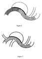
- Figure 2 shows a stent with an uneven mesh structure, wherein part of the neck of the aneurysm is covered with denser mesh and the branch vessel is covered with less dense mesh.
- Figure 3 shows a stent with an even mesh structure.
- a microcatheter for delivering a film material In the cavity of an aneurysm is placed a microcatheter for delivering a film material, followed by implanting a stent to the aneurysm lesion site through the delivery system, releasing the stent, and injecting the film material.
- the film material cures and adheres to the stent forming a dense film at the position of the aneurysm neck; thereby the stent is covered locally with the film and is accurately located.
- the microcatheter for delivering the film material can also arrive at the cavity of the aneurysm by passing through the mesh of the stent after the stent is released, then the film can be formed at the neck of the aneurysm.
- the implant device of the present invention is consisted of a stent and a film (1-0) delivered with a microcatheter and being coated at the scene of the surgery (see Figure 1 ).
- Said film (1-0) is formed via polymerization and curing of the film material comprising n-octyl cyanoacrylate and tantalum powder.
- Said film (1-0) can further be formed via polymerization and curing of a mixture of collagen with the film material comprising n-octyl cyanoacrylate and tantalum powder.
- collagen particles having a particle size of about 50mesh are produced by granulating 0.5mol of collagen on a fluidized bed (see the article " Study on Optimal Preparation of Protein Powder with Lecithin Using Spray Granulation” by Jiang Zhao et al., published on Food Research and Development, 2007 ); then the collagen particles are added into 3mol of n-octyl cyanoacrylate monomers and are ultrasonically dispersed homogenously in the monomers; 30g of tantalum nanopowders are added at 37-50°C in an argon atmosphere with stirring to produce a evenly dispersed magnetic fluid of n-octyl cyanoacrylate.
- the stent can be one with an uneven mesh structure, i.e., having different metal coverage rates at different positions.
- the portion of the stent with a higher mesh density (1-2) covers the surface on the neck of the aneurysm (1-6) on the parent artery (1-1), to alter the blood flow in the aneurysm to a maximum extent.
- Other portions of the stent are structures having lower mesh density (1-3) which provide support for the stent to conform to the normal vascular wall (1-4) so as to maintain the patency of the lumen and reduces as much as possible the covering of the branch vessel (1-5).
- the present invention can also achieve the treatment of the aneurysm (see Figure 2 ).
- a stent with an even mesh structure can also be used as long as the blood flow in the branch vessel is kept patent, then similarly, the treatment of the aneurysm can totally be accomplished (see Figure 3 ).
- the above device can accurately and rapidly block the aneurysm neck, protecting the aneurysm against the impact of the blood flow. Meanwhile, the blood flow in a normal artery branch can be maintained, remarkably improving the space occupying effect and thereby a real cure of the aneurysm is achieved.
Landscapes
- Health & Medical Sciences (AREA)
- Engineering & Computer Science (AREA)
- Biomedical Technology (AREA)
- Cardiology (AREA)
- Oral & Maxillofacial Surgery (AREA)
- Transplantation (AREA)
- Heart & Thoracic Surgery (AREA)
- Vascular Medicine (AREA)
- Life Sciences & Earth Sciences (AREA)
- Animal Behavior & Ethology (AREA)
- General Health & Medical Sciences (AREA)
- Public Health (AREA)
- Veterinary Medicine (AREA)
- Gastroenterology & Hepatology (AREA)
- Pulmonology (AREA)
- Prostheses (AREA)
- Materials For Medical Uses (AREA)
Applications Claiming Priority (2)
| Application Number | Priority Date | Filing Date | Title |
|---|---|---|---|
| CN201110318052XA CN102499732A (zh) | 2011-10-19 | 2011-10-19 | 一种血管内植入装置包和植入装置的制备方法 |
| PCT/CN2011/084143 WO2013056497A1 (zh) | 2011-10-19 | 2011-12-16 | 一种血管内植入装置包和植入装置的制备方法 |
Publications (2)
| Publication Number | Publication Date |
|---|---|
| EP2769701A1 true EP2769701A1 (de) | 2014-08-27 |
| EP2769701A4 EP2769701A4 (de) | 2015-06-03 |
Family
ID=46211912
Family Applications (1)
| Application Number | Title | Priority Date | Filing Date |
|---|---|---|---|
| EP11874217.0A Withdrawn EP2769701A4 (de) | 2011-10-19 | 2011-12-16 | Intravaskuläres implantat-kit und verfahren zur herstellung einer implantatvorrichtung |
Country Status (3)
| Country | Link |
|---|---|
| EP (1) | EP2769701A4 (de) |
| CN (1) | CN102499732A (de) |
| WO (1) | WO2013056497A1 (de) |
Cited By (1)
| Publication number | Priority date | Publication date | Assignee | Title |
|---|---|---|---|---|
| US10888414B2 (en) | 2019-03-20 | 2021-01-12 | inQB8 Medical Technologies, LLC | Aortic dissection implant |
Families Citing this family (13)
| Publication number | Priority date | Publication date | Assignee | Title |
|---|---|---|---|---|
| CN103198202B (zh) * | 2012-12-19 | 2017-05-17 | 首都医科大学 | 颅内动脉瘤介入治疗支架植入图像仿真方法 |
| CN118304067A (zh) * | 2016-10-04 | 2024-07-09 | 微仙美国有限公司 | 用于支架输送的方法及装置 |
| CN108614306B (zh) * | 2018-03-20 | 2020-03-27 | 迈克医疗电子有限公司 | 一种凝块检测的方法、装置、终端设备和介质 |
| CN110731832B (zh) * | 2018-07-20 | 2022-04-12 | 上海微创医疗器械(集团)有限公司 | 下腔静脉滤器 |
| CN112754729B (zh) * | 2019-11-05 | 2025-08-01 | 微创神通医疗科技(上海)有限公司 | 覆膜装置及其装载方法、覆膜系统和覆膜支架系统 |
| CN110893114A (zh) * | 2019-12-26 | 2020-03-20 | 上海加奇生物科技苏州有限公司 | 直接治疗动脉瘤且不阻断动脉瘤周边血管的支架 |
| CN114052981B (zh) * | 2020-07-29 | 2024-08-16 | 上海苏畅医疗科技有限公司 | 一种覆膜支架及覆膜方法 |
| DE102020128124A1 (de) * | 2020-10-26 | 2022-04-28 | Acandis Gmbh | Medizinisches Implantat zur Behandlung von Aneurysmen |
| CN113398320B (zh) * | 2021-02-04 | 2022-05-13 | 艾柯医疗器械(北京)有限公司 | 悬浮微粒造影液体栓塞剂及其制备方法 |
| CN115068164A (zh) * | 2022-06-14 | 2022-09-20 | 成都百瑞恒通医疗科技有限公司 | 血管支架及其制作方法 |
| CN115670541A (zh) * | 2022-12-02 | 2023-02-03 | 徐州亚太科技有限公司 | 封堵器及其制作方法 |
| CN119424059B (zh) * | 2024-12-23 | 2025-12-30 | 上海以心医疗器械有限公司 | 介入式支架转换组件、介入式止血支架及组装方法 |
| CN120053864A (zh) * | 2025-03-06 | 2025-05-30 | 西南医科大学 | 一种可降解药物涂层覆膜球囊导管及其使用方法 |
Family Cites Families (17)
| Publication number | Priority date | Publication date | Assignee | Title |
|---|---|---|---|---|
| CN1015229B (zh) | 1986-10-13 | 1992-01-01 | 西安化工研究所 | 血管栓塞剂的制备方法 |
| US5213580A (en) * | 1988-08-24 | 1993-05-25 | Endoluminal Therapeutics, Inc. | Biodegradable polymeric endoluminal sealing process |
| CN1164183A (zh) * | 1994-11-23 | 1997-11-05 | 艾罗魁普公司 | 管腔内的斯滕特氏移植物及将其植入血管的方法 |
| US5637113A (en) * | 1994-12-13 | 1997-06-10 | Advanced Cardiovascular Systems, Inc. | Polymer film for wrapping a stent structure |
| IT1289728B1 (it) * | 1996-12-10 | 1998-10-16 | Sorin Biomedica Cardio Spa | Dispositivo di impianto e corredo che lo comprende |
| CN1270780C (zh) * | 2002-04-09 | 2006-08-23 | 株式会社林原生物化学研究所 | 栓塞材料 |
| US20040236410A1 (en) * | 2003-05-22 | 2004-11-25 | Atrium Medical Corp. | Polymeric body formation |
| US20090011116A1 (en) * | 2004-09-28 | 2009-01-08 | Atrium Medical Corporation | Reducing template with coating receptacle containing a medical device to be coated |
| US7238828B2 (en) * | 2005-03-24 | 2007-07-03 | Ethicon, Inc. | Absorbable α-cyanoacrylate compositions |
| CN101631514A (zh) * | 2007-01-11 | 2010-01-20 | R·L·小比约克 | 用于经皮冠状动脉介入的多药物洗脱冠状动脉支架 |
| US20090036965A1 (en) * | 2007-07-30 | 2009-02-05 | Robert Glenmore Walsh | Conjunctive stent therapy |
| CN101917927A (zh) * | 2007-11-12 | 2010-12-15 | 维勒医学有限公司 | 医用级氰基丙烯酸酯的单瓶制剂 |
| US20090137981A1 (en) | 2007-11-26 | 2009-05-28 | Valor Medical | Methods of treating a blood vessel |
| CN101991477B (zh) | 2009-08-27 | 2014-03-26 | 上海微创医疗器械(集团)有限公司 | 一种血管重构支架 |
| US8845682B2 (en) * | 2009-10-13 | 2014-09-30 | E-Pacing, Inc. | Vasculature closure devices and methods |
| CN102100929B (zh) * | 2010-08-27 | 2013-10-02 | 上海微创医疗器械(集团)有限公司 | 一种栓塞材料及其制备方法 |
| CN102048602B (zh) * | 2011-01-11 | 2012-11-07 | 同济大学 | 一种网状可降解血管支架的制备方法 |
-
2011
- 2011-10-19 CN CN201110318052XA patent/CN102499732A/zh active Pending
- 2011-12-16 WO PCT/CN2011/084143 patent/WO2013056497A1/zh not_active Ceased
- 2011-12-16 EP EP11874217.0A patent/EP2769701A4/de not_active Withdrawn
Cited By (1)
| Publication number | Priority date | Publication date | Assignee | Title |
|---|---|---|---|---|
| US10888414B2 (en) | 2019-03-20 | 2021-01-12 | inQB8 Medical Technologies, LLC | Aortic dissection implant |
Also Published As
| Publication number | Publication date |
|---|---|
| WO2013056497A1 (zh) | 2013-04-25 |
| EP2769701A4 (de) | 2015-06-03 |
| CN102499732A (zh) | 2012-06-20 |
Similar Documents
| Publication | Publication Date | Title |
|---|---|---|
| EP2769701A1 (de) | Intravaskuläres implantat-kit und verfahren zur herstellung einer implantatvorrichtung | |
| CN111278367B (zh) | 阻塞装置 | |
| US9750504B2 (en) | Embolization device and a method of using the same | |
| JP2022525316A (ja) | 血管障害の治療のためのフィラメント状デバイス | |
| EP1163012B1 (de) | Kit zur inhibierung von endolecken assoziert mit der endovaskulären ausbesserung von bauch-aortenaneurysmen | |
| JP2022525317A (ja) | 血管障害の治療のための可撓性継合部を有するフィラメント状デバイス | |
| US6585755B2 (en) | Polymeric stent suitable for imaging by MRI and fluoroscopy | |
| JP4440953B2 (ja) | 動脈瘤治療デバイス | |
| JP5456974B2 (ja) | 血管の障害を治療するための組成物、システムおよび方法 | |
| JP2022525745A (ja) | 血管障害の治療のためのフィラメント状デバイス | |
| EP1649829A1 (de) | Einführsystem mit einem Ballon für einen selbstexpandierbaren Stent | |
| WO2007146655A1 (en) | Implantable stent with degradable portions | |
| JP4751661B2 (ja) | 細胞に制御された炎症性応答を起こさせるための血管内装置 | |
| JP2007521041A (ja) | 重合性の再拘束可能で再配置可能で取外し可能な経皮的血管内ステントグラフト | |
| US20120158122A1 (en) | Removable stent and method of production | |
| CN107427612A (zh) | 按需可降解的医疗装置 | |
| CN108245216A (zh) | 一种动脉瘤治疗系统 | |
| CA2820405C (en) | Removable stent and method of production | |
| EP1295615A1 (de) | Strahlungsundurchlässiger Stent | |
| US20160346423A1 (en) | Cyanoacrylate compositions including non-agglomerating radiopaque nanoparticles | |
| CN212592565U (zh) | 一种防i型内漏支架装置 | |
| Hwang et al. | Fabrication of bioabsorbable polylactic-co-glycolic acid/polycaprolactone nanofiber coated stent and investigation of biodegradability in porcine animal model | |
| CN111904657A (zh) | 一种防i型内漏支架装置 | |
| WO2014036809A1 (en) | Endoluminal drug delivery devices with applications in blood vessels | |
| EP1691721B1 (de) | Unter einem darstellungsstrahl sichtbare vorrichtung |
Legal Events
| Date | Code | Title | Description |
|---|---|---|---|
| PUAI | Public reference made under article 153(3) epc to a published international application that has entered the european phase |
Free format text: ORIGINAL CODE: 0009012 |
|
| 17P | Request for examination filed |
Effective date: 20140515 |
|
| AK | Designated contracting states |
Kind code of ref document: A1 Designated state(s): AL AT BE BG CH CY CZ DE DK EE ES FI FR GB GR HR HU IE IS IT LI LT LU LV MC MK MT NL NO PL PT RO RS SE SI SK SM TR |
|
| DAX | Request for extension of the european patent (deleted) | ||
| RA4 | Supplementary search report drawn up and despatched (corrected) |
Effective date: 20150508 |
|
| RIC1 | Information provided on ipc code assigned before grant |
Ipc: A61F 2/86 20130101ALI20150430BHEP Ipc: A61F 2/06 20130101ALI20150430BHEP Ipc: A61F 2/92 20130101ALI20150430BHEP Ipc: A61F 2/82 20130101AFI20150430BHEP |
|
| STAA | Information on the status of an ep patent application or granted ep patent |
Free format text: STATUS: THE APPLICATION IS DEEMED TO BE WITHDRAWN |
|
| 18D | Application deemed to be withdrawn |
Effective date: 20151208 |