EP2783211B1 - Procédé de détection de cellules cancéreuses dans un échantillon de cellules - Google Patents
Procédé de détection de cellules cancéreuses dans un échantillon de cellules Download PDFInfo
- Publication number
- EP2783211B1 EP2783211B1 EP12788531.7A EP12788531A EP2783211B1 EP 2783211 B1 EP2783211 B1 EP 2783211B1 EP 12788531 A EP12788531 A EP 12788531A EP 2783211 B1 EP2783211 B1 EP 2783211B1
- Authority
- EP
- European Patent Office
- Prior art keywords
- sample
- cell
- cells
- data
- biomarker
- Prior art date
- Legal status (The legal status is an assumption and is not a legal conclusion. Google has not performed a legal analysis and makes no representation as to the accuracy of the status listed.)
- Not-in-force
Links
Images




Classifications
-
- G—PHYSICS
- G01—MEASURING; TESTING
- G01N—INVESTIGATING OR ANALYSING MATERIALS BY DETERMINING THEIR CHEMICAL OR PHYSICAL PROPERTIES
- G01N33/00—Investigating or analysing materials by specific methods not covered by groups G01N1/00 - G01N31/00
- G01N33/48—Biological material, e.g. blood, urine; Haemocytometers
- G01N33/483—Physical analysis of biological material
- G01N33/487—Physical analysis of biological material of liquid biological material
- G01N33/4875—Details of handling test elements, e.g. dispensing or storage, not specific to a particular test method
- G01N33/48778—Containers specially adapted therefor, e.g. for dry storage
-
- A—HUMAN NECESSITIES
- A61—MEDICAL OR VETERINARY SCIENCE; HYGIENE
- A61B—DIAGNOSIS; SURGERY; IDENTIFICATION
- A61B10/00—Instruments for taking body samples for diagnostic purposes; Other methods or instruments for diagnosis, e.g. for vaccination diagnosis, sex determination or ovulation-period determination; Throat striking implements
- A61B10/02—Instruments for taking cell samples or for biopsy
- A61B10/0291—Instruments for taking cell samples or for biopsy for uterus
-
- B—PERFORMING OPERATIONS; TRANSPORTING
- B01—PHYSICAL OR CHEMICAL PROCESSES OR APPARATUS IN GENERAL
- B01L—CHEMICAL OR PHYSICAL LABORATORY APPARATUS FOR GENERAL USE
- B01L3/00—Containers or dishes for laboratory use, e.g. laboratory glassware; Droppers
- B01L3/50—Containers for the purpose of retaining a material to be analysed, e.g. test tubes
- B01L3/502—Containers for the purpose of retaining a material to be analysed, e.g. test tubes with fluid transport, e.g. in multi-compartment structures
-
- B—PERFORMING OPERATIONS; TRANSPORTING
- B01—PHYSICAL OR CHEMICAL PROCESSES OR APPARATUS IN GENERAL
- B01L—CHEMICAL OR PHYSICAL LABORATORY APPARATUS FOR GENERAL USE
- B01L3/00—Containers or dishes for laboratory use, e.g. laboratory glassware; Droppers
- B01L3/50—Containers for the purpose of retaining a material to be analysed, e.g. test tubes
- B01L3/508—Rigid containers without fluid transport within
- B01L3/5082—Test tubes per se
-
- G—PHYSICS
- G01—MEASURING; TESTING
- G01N—INVESTIGATING OR ANALYSING MATERIALS BY DETERMINING THEIR CHEMICAL OR PHYSICAL PROPERTIES
- G01N1/00—Sampling; Preparing specimens for investigation
- G01N1/28—Preparing specimens for investigation including physical details of (bio-)chemical methods covered elsewhere, e.g. G01N33/50, C12Q
- G01N1/40—Concentrating samples
- G01N1/405—Concentrating samples by adsorption or absorption
-
- G—PHYSICS
- G01—MEASURING; TESTING
- G01N—INVESTIGATING OR ANALYSING MATERIALS BY DETERMINING THEIR CHEMICAL OR PHYSICAL PROPERTIES
- G01N21/00—Investigating or analysing materials by the use of optical means, i.e. using sub-millimetre waves, infrared, visible or ultraviolet light
- G01N21/01—Arrangements or apparatus for facilitating the optical investigation
- G01N21/03—Cuvette constructions
-
- G—PHYSICS
- G01—MEASURING; TESTING
- G01N—INVESTIGATING OR ANALYSING MATERIALS BY DETERMINING THEIR CHEMICAL OR PHYSICAL PROPERTIES
- G01N33/00—Investigating or analysing materials by specific methods not covered by groups G01N1/00 - G01N31/00
- G01N33/48—Biological material, e.g. blood, urine; Haemocytometers
- G01N33/50—Chemical analysis of biological material, e.g. blood, urine; Testing involving biospecific ligand binding methods; Immunological testing
- G01N33/53—Immunoassay; Biospecific binding assay; Materials therefor
- G01N33/575—Immunoassay; Biospecific binding assay; Materials therefor for cancer
- G01N33/5755—Immunoassay; Biospecific binding assay; Materials therefor for cancer of the uterine cervix, uterine corpus or endometrium
-
- A—HUMAN NECESSITIES
- A61—MEDICAL OR VETERINARY SCIENCE; HYGIENE
- A61B—DIAGNOSIS; SURGERY; IDENTIFICATION
- A61B10/00—Instruments for taking body samples for diagnostic purposes; Other methods or instruments for diagnosis, e.g. for vaccination diagnosis, sex determination or ovulation-period determination; Throat striking implements
- A61B10/02—Instruments for taking cell samples or for biopsy
- A61B2010/0216—Sampling brushes
-
- B—PERFORMING OPERATIONS; TRANSPORTING
- B01—PHYSICAL OR CHEMICAL PROCESSES OR APPARATUS IN GENERAL
- B01L—CHEMICAL OR PHYSICAL LABORATORY APPARATUS FOR GENERAL USE
- B01L2200/00—Solutions for specific problems relating to chemical or physical laboratory apparatus
- B01L2200/06—Fluid handling related problems
- B01L2200/0647—Handling flowable solids, e.g. microscopic beads, cells, particles
-
- B—PERFORMING OPERATIONS; TRANSPORTING
- B01—PHYSICAL OR CHEMICAL PROCESSES OR APPARATUS IN GENERAL
- B01L—CHEMICAL OR PHYSICAL LABORATORY APPARATUS FOR GENERAL USE
- B01L2200/00—Solutions for specific problems relating to chemical or physical laboratory apparatus
- B01L2200/06—Fluid handling related problems
- B01L2200/0647—Handling flowable solids, e.g. microscopic beads, cells, particles
- B01L2200/0652—Sorting or classification of particles or molecules
-
- B—PERFORMING OPERATIONS; TRANSPORTING
- B01—PHYSICAL OR CHEMICAL PROCESSES OR APPARATUS IN GENERAL
- B01L—CHEMICAL OR PHYSICAL LABORATORY APPARATUS FOR GENERAL USE
- B01L2300/00—Additional constructional details
- B01L2300/02—Identification, exchange or storage of information
- B01L2300/023—Sending and receiving of information, e.g. using Bluetooth®
-
- B—PERFORMING OPERATIONS; TRANSPORTING
- B01—PHYSICAL OR CHEMICAL PROCESSES OR APPARATUS IN GENERAL
- B01L—CHEMICAL OR PHYSICAL LABORATORY APPARATUS FOR GENERAL USE
- B01L2300/00—Additional constructional details
- B01L2300/04—Closures and closing means
- B01L2300/041—Connecting closures to device or container
- B01L2300/042—Caps; Plugs
-
- B—PERFORMING OPERATIONS; TRANSPORTING
- B01—PHYSICAL OR CHEMICAL PROCESSES OR APPARATUS IN GENERAL
- B01L—CHEMICAL OR PHYSICAL LABORATORY APPARATUS FOR GENERAL USE
- B01L2300/00—Additional constructional details
- B01L2300/06—Auxiliary integrated devices, integrated components
- B01L2300/0609—Holders integrated in container to position an object
-
- B—PERFORMING OPERATIONS; TRANSPORTING
- B01—PHYSICAL OR CHEMICAL PROCESSES OR APPARATUS IN GENERAL
- B01L—CHEMICAL OR PHYSICAL LABORATORY APPARATUS FOR GENERAL USE
- B01L2300/00—Additional constructional details
- B01L2300/06—Auxiliary integrated devices, integrated components
- B01L2300/0681—Filter
-
- B—PERFORMING OPERATIONS; TRANSPORTING
- B01—PHYSICAL OR CHEMICAL PROCESSES OR APPARATUS IN GENERAL
- B01L—CHEMICAL OR PHYSICAL LABORATORY APPARATUS FOR GENERAL USE
- B01L2300/00—Additional constructional details
- B01L2300/08—Geometry, shape and general structure
- B01L2300/0803—Disc shape
-
- B—PERFORMING OPERATIONS; TRANSPORTING
- B01—PHYSICAL OR CHEMICAL PROCESSES OR APPARATUS IN GENERAL
- B01L—CHEMICAL OR PHYSICAL LABORATORY APPARATUS FOR GENERAL USE
- B01L2300/00—Additional constructional details
- B01L2300/08—Geometry, shape and general structure
- B01L2300/0832—Geometry, shape and general structure cylindrical, tube shaped
-
- B—PERFORMING OPERATIONS; TRANSPORTING
- B01—PHYSICAL OR CHEMICAL PROCESSES OR APPARATUS IN GENERAL
- B01L—CHEMICAL OR PHYSICAL LABORATORY APPARATUS FOR GENERAL USE
- B01L2300/00—Additional constructional details
- B01L2300/08—Geometry, shape and general structure
- B01L2300/0848—Specific forms of parts of containers
- B01L2300/0851—Bottom walls
-
- B—PERFORMING OPERATIONS; TRANSPORTING
- B01—PHYSICAL OR CHEMICAL PROCESSES OR APPARATUS IN GENERAL
- B01L—CHEMICAL OR PHYSICAL LABORATORY APPARATUS FOR GENERAL USE
- B01L2300/00—Additional constructional details
- B01L2300/08—Geometry, shape and general structure
- B01L2300/0848—Specific forms of parts of containers
- B01L2300/0858—Side walls
-
- B—PERFORMING OPERATIONS; TRANSPORTING
- B01—PHYSICAL OR CHEMICAL PROCESSES OR APPARATUS IN GENERAL
- B01L—CHEMICAL OR PHYSICAL LABORATORY APPARATUS FOR GENERAL USE
- B01L2400/00—Moving or stopping fluids
- B01L2400/08—Regulating or influencing the flow resistance
- B01L2400/084—Passive control of flow resistance
- B01L2400/086—Passive control of flow resistance using baffles or other fixed flow obstructions
-
- G—PHYSICS
- G03—PHOTOGRAPHY; CINEMATOGRAPHY; ANALOGOUS TECHNIQUES USING WAVES OTHER THAN OPTICAL WAVES; ELECTROGRAPHY; HOLOGRAPHY
- G03H—HOLOGRAPHIC PROCESSES OR APPARATUS
- G03H1/00—Holographic processes or apparatus using light, infrared or ultraviolet waves for obtaining holograms or for obtaining an image from them; Details peculiar thereto
- G03H1/04—Processes or apparatus for producing holograms
- G03H1/0443—Digital holography, i.e. recording holograms with digital recording means
-
- G—PHYSICS
- G03—PHOTOGRAPHY; CINEMATOGRAPHY; ANALOGOUS TECHNIQUES USING WAVES OTHER THAN OPTICAL WAVES; ELECTROGRAPHY; HOLOGRAPHY
- G03H—HOLOGRAPHIC PROCESSES OR APPARATUS
- G03H1/00—Holographic processes or apparatus using light, infrared or ultraviolet waves for obtaining holograms or for obtaining an image from them; Details peculiar thereto
- G03H1/0005—Adaptation of holography to specific applications
- G03H2001/005—Adaptation of holography to specific applications in microscopy, e.g. digital holographic microscope [DHM]
Definitions
- the invention pertains to the technical field of the early diagnosis of carcinomas as well as their preliminary stages, particularly carcinomas of the cervix and uterus.
- cells need to be sampled from a patient and a thorough analysis of the cell sample is required in order to evaluate whether abnormal or aberrant cells are present.
- a pathologist or other skilled medical personnel will base the diagnosis on specific characteristics of the cells in the sample, such as cell morphology, the presence of certain types of cells or proteins and more.
- These cytological tests are based on a two-dimensional presentation of the cells present in the sample and mostly require the fixation of cells on a substratum and the use of dyes or stainings to visualize specific features of the cells. This is a time consuming and cumbersome work, and requires well-trained specialists.
- the Papanicolaou (PAP) smear has been the cornerstone of cervical cancer screening since 1949.
- the PAP smear is a stain performed on cells smeared on a slide and visualized by microscopy.
- LBC liquid-based cervical cytology
- cells from the cervix were obtained using a brush, suspended in a fixative solution, and then applied to a slide prior to staining.
- Highly trained cytotechnologists and cytopathologists review the stained slides looking for evidence of abnormal cells based on morphological appreances (as indicated in Table 1).
- the PAP smear and HPV testing are used together though they are very disparate technologies.
- the PAP smear has relatively low sensitivity (50%) and relatively high specificity (90%) for high grade cervical lesions (pre-cervical cancer and cervical cancer).
- HPV DNA testing has high sensitivity (>90%) but low specificity (30%) for high grade cervical lesions (pre-cervical cancer and cervical cancer.
- Peter D. Beitsch et al. "Detection of Carcinoma Cells in the Blood of Breast Cancer Patients", The American Journal of Surgery, Vol. 180, December 200, pp. 446-449 relates to liquid cell samples, in particular blood samples and cell samples in a liquid medium. More particularly, the method of this article involves separating epithelial cells in a first step. Then, in a second step, the epithelial cells are resuspended in a solution. Finally, the data is obtained by a FACScan flow cytometer.
- International application WO 2011/099925 A1 relates to use of a digital holographic microscopy and imaging setup and a method of digital holographic microscopy and imaging for detecting molecules or structures stained or labelled to at least one cell or conjugated to antibodies which are bound either directly to said at least one cell or indirectly via another or several antibodies in a chain bound to said at least one cell.
- the field of cancer diagnosis is in need for methods and devices that analyses cell samples in a non-destructive, non-detrimental and objective manner.
- a diagnosis is performed on the 'raw' sample whereby information is provided on the status of the sample and the cells present without the need for further processing of the cell sample (e.g. centrifuging, cell lysis, etc.).
- the diagnosis is based on both morphological data and protein expression profiles of said cells.
- the current invention provides a method of predicting the presence of cancerous cells in a liquid cell sample obtained from a subject. More in particular, the current invention provides a method according to claim 1.
- the method allows a fast and accurate prediction, without the need of cumbersome and laborious handling steps. Prediction occurs on the basis of morphological and biomarker data of the raw sample, whereby both data are simultaneously obtained from the raw sample.
- DHM Digital Holographic Microscopy
- the present document discloses a liquid medium for use in the method according to any of the claims, comprising
- sample vial comprising a liquid medium for use in the method according to any of the claims, characterized in that said sample vial comprises at least two compartments in fluid connection with one another, said compartments comprising at least one pair of screening surfaces, said screening surfaces are essentially flat; and characterized in that the distance between the pair of screening surfaces of the second compartment is smaller than the distance between the pair of screening surfaces of the first compartment.
- the present document also discloses a kit comprising a cell sample collecting device and a sample vial as above, for use in the method according to any of the claims.
- the current invention provides methods of predicting whether in a cell sample obtained from a subject, preferably a human subject, cancerous cells are present. Aspects of the methods include obtaining morphological as well as biomarker data and/or non-specific cell data from a cell sample by assaying the sample in suspension, directly after obtainment of the cell sample, without any prior sample handling or processing such as centrifugation, re-suspending, hybridization, etc. Prediction is based on two different types of data in order to eventually come to a very reliable and trustworthy diagnosis. Also provided are consumables specifically developed and optimised to be used in conjunction with the method of the current invention and a system that finds use in practicing the method.
- the value to which the modifier "about” refers is itself also specifically disclosed.
- % by weight refers to the relative weight of the respective component based on the overall weight of the formulation.
- embodiments of the invention are directed to methods of predicting or diagnosing the presence of cancerous or pre-cancerous cells in a sample obtained from a subject. For instance, the presence of a cervical intraepithelial neoplasia (CIN) lesion or cervical cancer may be predicted.
- CIN cervical intraepithelial neoplasia
- sample refers to any specimen obtained from a subject, preferably a biological organism, preferably a living organism, which comprises cells from said biological organism.
- the term relates also to specimen obtained from non-living, i.e. dead biological organisms, in particular recently deceased organisms.
- a cell sample may be derived from an animal, preferably from a mammal, e.g.
- said cell sample is a tissue sample, a biopsy sample, a blood sample, a serum sample, a brushing or scraping sample from oral cavities, nipple secretions, skin lesions, and eye brushings, a fine-needle-aspiration sample, a smear sample, a mucoid specimens taken from respiratory and gastrointestinal tracts and body fluids such as serous effusions or urinary or cerebrospinal fluids.
- said sample is a smear sample.
- said smear sample is a cervical sample.
- CIN lesion also referred to in the art as cervical dysplasia
- CIN lesions may be histologically graded as CIN1, CIN2/3, CIN2 and CIN3.
- CIN1 lesions are those lesions that are confined to the basal 1/3 of the epithelium, and have the least risk of developing into a cancerous lesion, relative to the other categories of lesions.
- CIN2 lesions are characterized by moderate dysplasia confined to the basal 2/3 of the epithelium.
- CIN3 lesions (sometimes referred to by those of skill in the art as cervical carcinoma in situ) are categorized by the presence of severe dysplasia that traverses more than 2/3 of the epithelium.
- the CIN2/3 category (i.e., CIN2+) collectively refers to both CIN2 and CIN3 lesions.
- Embodiments of the invention are characterized by predicting the presence of a CIN lesion in a subject with a high degree of sensitivity and specificity. By predicting is meant to prognosticate or foresee the presence of a CIN lesion in the subject without actually taking a biopsy of the cervix of the subject.
- sensitivity is a measure of the proportion of actual positives (as opposed to false positives) that are correctly identified while specificity is a measure of negatives which are correctly identified.
- Embodiments of the invention may predict the presence or absence of any type of cancerous cell in a cell sample obtained from a subject, independent of the origin of the cell sample. More in particular, embodiments may predict the presence of a CIN lesion. Embodiments of the invention may also predict the type of CIN lesion, e.g., whether the CIN lesion is a CIN1, CIN2+, CIN2 or CIN3 lesion.
- Embodiments of the methods make this prediction, e.g., whether a lesion is present, what type of lesion is present (such as whether a CIN2+ lesion is present) with a high degree of sensitivity and specificity.
- sensitivity is 85% or more, such as 90% or more, including 95% or more.
- specificity is 85% or more, such as 87% or more, including 90% or more.
- aspects of the methods include obtaining both: (1) morphological data and (2) biomarker data from a labelled liquid sample of cells in suspension from the subject.
- a sample comprising cells is collected from the subject by any convenient protocol, whereby the sample is solubilised or suspended and preserved in a sample vial comprising a liquid medium.
- said liquid medium comprises means for labelling cells or epitope(s) on or in said cells.
- the solubilisation of the obtained sample occurs immediately after obtainment of the sample from said subject.
- the sample can be obtained from the cervix of a patient by a practitioner using a cell collector such as a swab or cervical brush, after which the practitioner (e.g. the gynaecologist) immediately will transfer the collected cells to a sample vial for solubilising and preservation of the sample.
- a labelled liquid sample according to aspects of the methods may be a biomarker labelled liquid sample, a non-specific cell labelled liquid sample, or a biomarker and non-specific cell labelled liquid sample.
- the liquid medium is thus developed for use in the methods and systems according the current invention and assures preservation of mammalian cells in suspension at ambient temperature.
- the solution enhances maintenance of the nuclear structure of the cells, in that it maintains cell membranes intact.
- the solution also effectively destroys microbial pathogens in a sample, and inhibits retroviral activity.
- the liquid medium usable in the current invention preferably comprises:
- the liquid medium may furthermore comprise anti-bacterial agents such as antibiotics.
- the liquid medium may comprise a permeabilizing reagent.
- Permeabilizing reagents of interest are reagents that allow the labelled biomarker probes, e.g., as described in greater detail below, to access to the intracellular environment. Any convenient permeabilizing reagent may be employed, where suitable reagents include, but are not limited to: mild detergents, such as Triton X-100, Tween, NP-40, Igepal CA-630, N-Octyl-Gluoosid, saponin, etc.; methanol, and the like. It may also be desirable to label cells with a positive heavy metal control, e.g.
- the liquid medium may furthermore also comprise a viability dye to stain the cells in said sample, e.g. ethidium bromide, propidium iodide, DAPI, RhCI3, etc.
- the means for labelling cells or epitope(s) of the liquid medium according to the current invention comprise a labelled biomarker probe.
- Said labelled biomarker probe include nucleic acid analytes such as RNA or DNA probes, a protein or a protein analyte, such as an antibody or an antibody fragment, or any custom-made or engineered protein able to bound to or detect an epitope.
- the sample that is assayed to obtain morphological and biomarker data is a biomarker labelled sample.
- the sample is one that has been labelled for one or more biomarkers of interest.
- biomarker labelled sample is meant a sample which has been contacted with a labelled biomarker probe (e.g., as described in greater detail below) that specifically binds to a biomarker of interest if the biomarker is present in the cellular sample.
- Biomarkers of interest include, but are not limited to, cervical cancer biomarkers.
- Cervical cancer biomarkers are a distinctive biological or biologically derived indicator, for example nucleic acids or proteins, whose presence is associated or linked with either the propensity of a subject to develop cervical cancer or the presence of cervical cancer in a subject. Accordingly, biomarkers of interest include nucleic acid and protein analytes whose presence and/or amount in a cell can be used to make a prediction of at least the propensity of a subject to suffer from cervical cancer.
- Biomarkers of interest include, but are not limited to: HPV expression products of HPV genes, such as HPV genes L1 , L2, E2, E4, E5, E6 or E7; cyclin-dependent kinase inhibitors, e.g., p14Arf, p15INK4b, p16 (i.e., p16INK4a as described in Serrano, M., et al., Nature, 1993 Dec.
- HPV expression products of HPV genes such as HPV genes L1 , L2, E2, E4, E5, E6 or E7
- cyclin-dependent kinase inhibitors e.g., p14Arf, p15INK4b, p16 (i.e., p16INK4a as described in Serrano, M., et al., Nature, 1993 Dec.
- p18INK4c p19INK4d
- p21WAF1/CIP1 p27KIP1
- cell cycle regulatory proteins e.g., p14ARF
- annexins such as annexin A1, A2 or A5
- specific microRNAs or chromosome alterations 3q- associated with cervical cancer etc.
- the liquid medium may be combined with a single labelled biomarker probe or two or more distinct labelled biomarker probes that bind to different biomarkers of different molecular composition, where the number of such distinct labelled biomarker probes may be two or more, e.g., three or more, four or more, five or more, etc; e.g., where the assay is a multiplex assay for two or more biomarkers.
- the sample is combined with one or more labelled biomarker probes to produce a reaction mixture.
- Labelled biomarker probes of interest include a specific binding domain and a label domain.
- the specific binding domain comprises a capture ligand that specifically binds to the biomarker of interest.
- the biomarker of interest may be a variety of different types of molecules, including but not limited to: proteins, polypeptides, proteoglycans, glycoproteins and the respective fragments of these molecules; nucleic acids, e.g., DNA and RNA, such as mRNA, etc.
- the capture ligand is therefore a ligand that binds to the biomarker molecule of interest, wherein this capture ligand may of course vary depending on the specific type of biomarker molecule to be detected, e.g., antibody for protein biomarker, oligonucleotide for mRNA biomarker.
- the affinity between a capture ligand and the biomarker molecule to which it specifically binds when they are specifically bound to each other in a binding complex is characterized by a Kd (dissociation constant) of 10 -6 M or less, 10 -7 M or less, 10 -8 M or less, 10 -9 M or less, 10 -10 M or less, 10 -11 M or less, 10 -12 M or less, 10 -13 M or less, 10 -14 M or less, including 10 -15 M or less.
- Said Kd value can be measured be any conventional method known in the art, such as, but not limiting to, surface plasmon resonance.
- antibody binding agent includes polyclonal or monoclonal antibodies or fragments that are sufficient to bind to an analyte of interest.
- the antibody fragments can be, for example, monomeric Fab fragments, monomeric Fab' fragments, or dimeric F(ab)'2 fragments.
- antibody binding agent molecules produced by antibody engineering, such as single- chain antibody molecules (scFv) or humanized or chimeric antibodies produced from monoclonal antibodies by replacement of the constant regions of the heavy and light chains to produce chimeric antibodies or replacement of both the constant regions and the framework portions of the variable regions to produce humanized antibodies.
- said antibody binding agent might be conjugated to nanoparticles, such as for instance magnetic, silver or gold nanoparticles, as described in EP 1 658 497 or EP 1 740 951 .
- Nucleic acid binding agents of interest are nucleic acids that specifically bind to biomarker nucleic acids in a cell.
- the length of these nucleic acids may vary, so long as it is sufficient for the oligonucleotide to serve as a specific binding agent, and in some instances ranges from 13 to 100 nt, such as 14 to 50 nt, e.g., 15 to 25 nt.
- the oligonucleotides that make up these nucleic acid binding agents may be DNA or RNA, or a synthetic analogue thereof, as desired.
- the labelled biomarker probes further include a detectable label. Said detectable label might be directly bound on the biomarker probe (primary label) or indirectly bound, e.g.
- biomarker probe comprises an antibody
- secondary antibody directed against said biomarker probe (secondary label).
- detectable labels are fluorescent dyes. Fluorescent dyes (fluorophores) can be selected from any of the many dyes suitable for use in imaging applications. A large number of dyes are commercially available from a variety of sources, such as, for example, Molecular Probes (Eugene, OR) and Exciton (Dayton, OH).
- fluorophores of interest include, but are not limited to, 4- acetamido-4'-isothiocyanatostilbene-2,2'disulfonic acid; acridine and derivatives such as acridine, acridine orange, acrindine yellow, acridine red, and acridine isothiocyanate; 5-(2'-aminoethyl)aminonaphthalene-1-sulfonic acid (EDANS); 4-amino-N-[3-vinylsulfonyl)phenyl]naphthalimide-3,5 disulfonate (Lucifer Yellow VS); N-(4-anilino-1-naphthyl)maleimide; anthranilamide; Brilliant Yellow; coumarin and derivatives such as coumarin, 7-amino-4-methylcoumarin (AMC, Coumarin 120), 7-amino-4- trifluoromethylcouluarin (Couma
- fluorophores or combinations thereof known to those skilled in the art may also be used, for example those available from Molecular Probes (Eugene, OR) and Exciton (Dayton, OH).
- Other detectable labels of interest are metal-based labels, such as polymer-coated gold or silver coated nanoparticles. Examples of such labels are given in EP1740951 and EP165849 .
- the label of each distinct probe may be chosen to provide a distinguishable signal.
- first and second distinct labelled biomarker probes are employed, e.g. fluorescent labels
- the label in the second probe is a fluorescent label which produces a fluorescent signal that is distinguishable from the first fluorescent signal of the label of the first probe.
- the first and second fluorescent signals produced upon excitation of the first and second fluorescent labels are distinguishable from each other, meaning that both can be detected at the same time and that the signal from one does not modify or change the signal from the other.
- Each distinct label may produce signals that are distinguishable from any other label.
- the cells may be stained with a third fluorescent label which produces a third fluorescent signal that is distinguishable from the first and second fluorescent signals.
- said labelled biomarker probe is directed to a biomarker which is known to be upregulated in cancer cells, or which presence is connected to the malignant status of a cell or gives a predisposition to possible malignancy.
- the labelled biomarker probe that is employed can be directed to p16INK4a, Ki-67, annexin A5 or HPV E6, E7 labelled probe.
- p16INK4a, Ki-67 or annexin A5 expression are correlated to malignancy in cervical cells (e.g. Li et al., 2012, Mol Med. Report, Sep. 12 2012 ; Cavalcante et al., 2012, Indian J Pathol Microbiol, Jul; 55(3):339-45-2 ).
- Expression of HPV viral proteins E6 and E7 are equally correlated with a predisposition to dysplasia.
- Exemplary probe sequences specific for HPV E6/E7 protein include for instance those described in US 2007 016 669 9 , WO 2006 085 822 or US 2012 014 150 2 .
- Non-limiting examples of components to which cancer or several hereditary conditions are associated with are provided in the following list and, one or more of which are detectable using the method of the invention.
- a diagnosis may require detection of one of more of the listed molecules comprising of BRCA1, TP53, CFTR, APP, APOE, BRCA2, HBB, APC, MYC, HD, BCL2, ABL1, BAX, DMD, p16INK4A, ATM, TNF, RB1, VEGF, ERBB2, FGG, HPRT1, MAPT, MDM2, RUNX1, SOD1, CDKN1A (cyclin-dependnet kinase inhibitor 1A), PAX6, NF1, fibronectin 1, caspase 3, phenylalanine hydroxylase, glyceraldehyde-3-phosphate dehydrogenase, phosphatase and tensin homolog (mutated in multiple advanced cancers 1), hemochromatosis, FGFR3 fibroblast growth factor receptor 3, EGFR epidermal growth factor receptor, MLH1 mutL homolog 1, colon cancer, nonpolyposis type 2, PABPC1 poly(A) binding protein, cytoplasmic 1,
- AKT2 v-akt murine thymoma viral oncogene homolog 2 PLAT plasminogen activator, tissue, CHRNA7 cholinergic receptor, nicotinic, alpha polypeptide 7, TIMP1 tissue inhibitorof metalloproteinase 1 (erythrold potentiating activity, collagenase inhibitor), NFKB1 nuclear factor of kappa light polypeptide gene enhancer in B-cells 1 (p105), STAT3 signal transducer and activator of transcription 3 (acute-phase response factor), CDC42 cell division cycle 42 (GTP binding protein, 25kD), VDR vitamin D (1,25- dihydroxyvitamin D3) receptor, NTRK1 neurotrophic tyrosine kinase, receptor, type 1, VIM vimentin, TGFBR2 transforming growth factor, beta receptor II, DHFR dihydrofolate reductase, CYP2A6 cytochrome P450, subfamily IIA, HSPCA heat shock 90kD protein 1, al
- the means for labeling cells or epitope(s) are provided in a concentration suitable to detect a biomarker present in a sample at a concentration of 500 pg or more. More preferably said biomarker present in a sample is able to detect a biomarker in a sample at a concentration of 250 pg or more, even more preferably of 150 pg or more, more preferably of 100 pg or more, most preferably of 50 pg or more.
- the means for labeling cells or epitope(s) on or in said cells are preferably provided in the liquid medium in a concentration between 0.01 ⁇ g/ml and 50 ⁇ g/ml, more preferably in a concentration between 0.04 ⁇ g/ml and 20 ⁇ g/ml, more preferably between 0.04 ⁇ g/ml and 10 ⁇ g/ml, more preferably between 0.04 ⁇ g/ml and 5 ⁇ g/ml, more preferably between 0.04 ⁇ g/ml and 2 ⁇ g/ml, more preferably between 0.04 ⁇ g/ml and 1 ⁇ g/ml, more preferably between 0.08 ⁇ g/ml and 0.5 ⁇ g/ml, most preferably between 0.2 ⁇ g/ml and 0.4 ⁇ g/ml.
- the sample with the labelled biomarker probe(s) present in the liquid medium is performed under incubation conditions that provide for binding of probes to their respective biomarkers, if present, in the sample.
- the probes and samples are contacted and combined at a temperature ranging from 15 to 50, such as from 20 to about 40°C.
- Contact may be performed with mixing or agitation, e.g., with vortexing etc., to provide for sufficient combination of the reaction components and the sample.
- the resultant reaction mixture may then be maintained or incubated for a period of time prior to assay for morphological and biomarker data, e.g., via digital holographic microscopy (e.g., as described in greater detail below).
- the reaction mixture is incubated at a temperature ranging from 15 to 50, such as from 20 to about 40°C for a period of time ranging from about 30 minutes to 72 hours, such as 1 hour to 24 hours, including 1 hour to 3 hours.
- the sample may be assayed immediately or stored for assay at a later time. If stored, in some embodiments the sample is stored at a reduced temperature; e.g at 4°C or below, between -10°C and -40°C.
- the liquid medium comprises a non-specific cell stain, in addition to a biomarker label. The cells may be stained with a non-specific stain using any convenient protocol.
- Dyes and stains that are specific for DNA (or preferentially bind double stranded polynucleotides in contrast to single-stranded polynucleotides) and therefore may be employed as non-specific stains include, but are not limited to: Hoechst 33342 (2'-[4-ethoxyphenyl]-5-[4-methyl-1-piperazinyl]-2,5'-bi-1H-benzimidazole) and Hoechst 33258 (2'-[4-ethoxyphenyl]-5-[4-methyl-1-piperazinyl]-2,5'-bi-1H-benzimidazole) and others of the Hoechst series; SYTO 40, SYTO 1 1 , 12, 13, 14, 15, 16, 20, 21 , 22, 23, 24, 25 (green); SYTO 17, 59 (red), DAPI, DRAQ5(TM) (an anthraquinone dye with high affinity for double strand
- the stain may serve in quantitation of biomarker, as an indication of cell cycle, etc.
- the sample is assayed to obtain both morphological as well as biomarker and/or non-specific cell data.
- analysis occurs in the same aliquot of sample, i.e., the same physical quantity of sample, is assayed to obtain both the morphological and biomarker/non-specific cell data.
- Obtainment of the data occurs simultaneously, meaning that both the morphological data and the biomarker data are obtained from one sample, with the same device and at the same time-point.
- these embodiments are distinguished from protocols in the prior art, in which a first aliquot of a sample is assayed using one protocol, e.g., slide based protocol, for morphological data and a second aliquot of the sample is assayed using another protocol.
- one protocol e.g., slide based protocol
- second aliquot of the sample is assayed using another protocol.
- Morphological data refers to any type of data from which cell morphology information, i.e., information about the size, shape and/or structure of the cells, may be derived.
- Morphological data or parameters of interest include, but are not limited to: nuclear area, perimeter, texture or spatial frequency content, centroid position, shape (i.e., round, elliptical, barbell-shaped, etc.), volume, optical nuclear height, cell nucleus diameter, chromatin texture, cell size, percent nucleation, cell form, cell quantity, nuclear size, nuclear volume, nuclear size variability, nuclear volume variability, chromatin texture, cell size, nuclear optical density, cytoplasm optical density, background optical density, and ratios of any of these parameters.
- the obtained morphological date may be for cells as a whole or for subparts thereof, e.g., the cytoplasm of cells.
- the morphological data may include an actual designation of whether a cell is normal or abnormal, including the type of abnormal cell.
- morphological data may, in some instances, include a designation that a given cell is abnormal, for example that the cell is: an atypical squamous cell, e.g., atypical squamous cell of undetermined significance (ASC-US), atypical squamous cell - cannot exclude HSIL (ASC-H); low grade squamous intraepithelial lesion (LGSIL or LSIL); high grade squamous intraepithelial lesion (HGSIL or HSIL); squamous cell carcinoma; atypical glandular cell not otherwise specified (AGC-NOS); atypical clandular cell, suspicious for AIS or cancer (AGC- neoplastic); and adenocarcinoma in situ (AIS).
- ASC-US atypical squamous cell of undetermined significance
- ASC-H atypical squamous cell of
- Biomarker data refers to any type of data from which biomarker information for the cell may be derived.
- biomarker data is data that includes a signal provided by the label of the labelled biomarker probe that is employed in the assay.
- the biomarker data may be in the form of the presence and amplitude of emitted light, the number of discrete positions in a cell or other object from which the light signal(s) originate(s), the relative placement of the signal sources, and the colour (wavelength or waveband) of the light emitted at each position in the cell.
- the biomarker data may take the form of qualitative, semi-quantitative or quantitative data. Qualitative data is simply the presence or absence of the biomarker.
- Semi-quantitative or quantitative data is data that provides some indication of the amount, e.g., concentration, etc., of the biomarker in the cell.
- semi-quantitative data may take the form of an indication that a biomarker of interest is above a certain threshold number.
- Quantitative data provides an indication of an absolute value, e.g., amount, etc., of the biomarker in the cell.
- Semi-quantitative and quantitative data may be collectively referred to as biomarker quantitation data.
- the biomarker of interest is a protein that is present when a subject is infected with a high risk HPV strain. Proteins of interest include but are not limiting to HPV E6, E7, L1, L2.
- the biomarker of interest is a protein upregulated in cervical cancer or dysplasia.
- proteins include but are not limiting to for instance p16, Ki-67 or annexin A5.
- detection of biomarkers of both groups occurs. Said biomarker data will be retrieved by omitting any background signal due to unbound biomarker probe or aspecific bound biomarker probe. As one of the preferred embodiments of the current invention does not include any washing step or removal of the unbound biomarker probe, deduction of this aspecific signal is indispensable to retrieve reliable information. Deduction of aspecific signal occurs preferably by use of software implementation.
- threshold information on the background signal and other aspecific signals is standardly stored in said software as a predefined value.
- Said threshold information can be obtained from a control sample in the liquid medium supplemented with biomarker probe according to the current invention but negative for the presence of the used biomarker.
- the obtained signal will account as negative background signal and can be used for deduction when predicting the state of subject samples. Samples showing a certain threshold signal after deduction of the background signal can be accounted as positive.
- the background signal will be determined each time a round of analysis starts. Again, a sample known to be negative for the assessed biomarker will be used and the retrieved signal will account as background signal.
- cells are isolated from the background (segmented) based on their optical properties and specifically the phase information extracted from the hologram.
- the morphological data is isolated from the background originating from unbound biomarker probe, based on phase information retrieved by the digital holographic microscope.
- This phase information is a quantitative measure of the thickness of the objects present in the image.
- the background can be defined as an object that has an optical thickness below a given threshold.
- the biomarker signal analysis can hence be limited to the regions of the image where objects (cells) are present and the aspecific signals originating from unbound or aspecifically bound biomarker is thereby eliminated.
- Non-specific cell data is any type of data from which non-specific (i.e., non-biomarker specific) cell information for the cells in the sample may be derived.
- nonspecific cell data is data that includes a signal emitted by a non-specific cell stain that is employed in the assay.
- the non-specific cell data may be in the form of the presence and amplitude of emitted light, the number of discrete positions in a cell or other object from which the light signal(s) originate(s), the relative placement of the signal sources, and the color (wavelength or waveband) of the light emitted at each position in the cell.
- the nonspecific cell data may take the form of qualitative, semi-quantitative or quantitative data.
- non-specific cell data may include data on the DNA content of a cell obtained using a DNA stain (e.g., as described above).
- Other quantitative data includes electronic volume (EV).
- non-specific cell data may provide an indication of an absolute or relative value, e.g., copy number, amount, etc., of the nonspecific cell component in the cell.
- Semi-quantitative and quantitative data may be collectively referred to as non-specific cell quantitation data.
- Qualitative non-specific cell data can provide information with respect to the presence of absence of a particular feature of a cell. For example, in cells stained with a DNA stain, the presence or absence of a cell nucleus can be determined (e.g., the number of nucleated cells in a sample, also referred to as percent nucleation).
- the labels and stains employed may be chosen to provide a distinguishable signal (as described above for using multiple biomarker probes in a single sample, above).
- the label for the biomarker probe is a fluorescent label which produces a fluorescent signal that is distinguishable from the fluorescent signal of the DNA stain.
- the fluorescent signals produced upon excitation of the biomarker probe label and the DNA stain are distinguishable from each other, meaning that both can be detected at the same time and that the signal from one does not modify or change the signal from the other.
- Each distinct label may produce signals that are distinguishable from any other label.
- the cells may be stained with three fluorescent labels which produce three distinct fluorescent signals that are distinguishable from each other upon excitation.
- the above types of data e.g., the morphological, biomarker, and non-specific cell data, is simultaneously obtained from the same aliquot of sample. In a preferred embodiment of the current invention, above mentioned types of data is obtained by Digital Holographic Microscopy.
- DHM Digital Holographic Microscopy
- a holographic representation is recorded by a digital camera such as a CCD- or a CMOS-camera, which can subsequently be stored or processed on a computer.
- a highly coherent or a partially coherent light source such as laser-light
- the light form the source is split into two beams, an object beam and a reference beam.
- the object beam is sent via an optical system to the sample and interacts with it, thereby altering the phase and amplitude of the light depending on the object's optical properties and 3D shape.
- the object beam which has been reflected on or transmitted through the sample is then made (e.g. by set of mirrors and/or beam splitters) to interfere with the reference beam, resulting in an interference pattern which is digitally recorded.
- an absorptive element can be introduced in the reference beam which decreases its amplitude to the level of the object beam, but does not alter the phase of the reference beam or at most changes the phase globally, i.e. not dependent on where and how the reference beam passes through the absorptive element.
- the recorded interference pattern contains information on the phase and amplitude changes which depend on the object's optical properties and 3D shape.
- In-line DHM is similar to the more traditional DHM, but does not split the beam, at least not by a beam splitter or other external optical element.
- In-line DHM is most preferably used to look at a not-too-dense solution of particles, e.g. cells, in a fluid. Thereby some part of the at least partially coherent light will pass through the sample without interacting with the particles (reference beam) and interfere with light that has interacted with the particles (object beam), giving rise to an interference pattern which is recorded digitally and processed.
- In-line DHM is used in transmission mode, it needs light with a relatively large coherence length, and cannot be used if the samples are too thick or dense.
- DHM differential DHM
- DHM differential digital holographic microscope
- DHM differential digital holographic microscope
- DHM digital tomography
- Transparent objects like living biological cells, are traditionally viewed in a phase contrast microscope or in a differential interference contrast microscope. These methods visualize phase shifting transparent objects by distorting the bright field image with phase shift information. Instead of distorting the bright field image, transmission DHM creates a separate phase shift image showing the optical thickness of the object. Digital holographic microscopy thus makes it possible to visualize and quantify transparent objects and is therefore also referred to as quantitative phase contrast microscopy. More so, DHM allows imaging subcellular structures in three dimensions.
- a sample image is calculated at a given focal distance.
- the recorded hologram contains all the necessary object wave front information, it is possible to calculate the object at any focal plane by changing the focal distance parameter in the reconstruction algorithm.
- the hologram contains all the information needed to calculate a complete image stack.
- DHM system where the object wave front is recorded from multiple angles, it is possible to fully characterize the optical characteristics of the object and create tomography images of the object.
- DHM systems do not have an image forming lens
- traditional optical aberrations do not apply to DHM.
- Optical aberrations are "corrected" by design of the reconstruction algorithm. A reconstruction algorithm that truly models the optical setup will not suffer from optical aberrations.
- DHM allows obtaining both two-dimensional and three-dimensional morphological data of the cell sample, as well as biomarker data, preferably based on fluorescent technology.
- a digital report is created comprising all obtained data linked to a sample, as well as identification data related to said sample and ID of the subject.
- Said digital report will comprise morphological data, comprising both detailed two-dimensional and three-dimensional images of the sample and cells within sample, as well as quantitative information on cell parameters as described above.
- Said digital report will furthermore comprise biomarker data, such as biomarker signal measured, information on background signal and threshold values defining presence or absence of for instance HPV infection.
- morphological and biomarker/non-specific cell data obtained from the same aliquot of cervical cellular sample is employed to predict whether the cell sample of the subject comprises cancerous or pre-cancerous cells.
- at least one cellular parameter derived from obtained holographic information comprises the optical nuclear height or optical height delta.
- the optical nuclear height or optical height delta can be defined as the difference between Nucleus top height minus Cytoplasm average height and is preferably derived from the obtained holographic information. It has been found by the inventors that the latter is a highly reliable parameter for detecting (pre-) cancerous cell and that it can be correlated to the status of malignancy of said cells.
- N/C ratio analysis e.g., to identify abnormal cells
- DAPI staining and fluorescence signal of for instance E6, E7 or p16 presence
- N/C ratio analysis coupled with cell cycle analysis as determined by p16 staining and green fluorescence of the E6, E7 mRNA hybridization signal d) N/C ratio analysis coupled with cell cycle analysis as determined by DAPI (or other DNA stain) staining, etc.
- N/C ratios include N/C ratios that are 0.25 or higher, 0.30 or higher, 0.4 or higher, 0.5 or higher, 0.6 or higher, 0.7 or higher, 0.8 or higher, 0.9 or higher, 0.95 or higher, etc.
- nuclear area (NA) assessment may be used as morphological data. Cells in a cervical cell sample having an increased nuclear area, e.g., as compared to nuclei in normal intermediate squamous cells, can be identified cells as abnormal cells.
- Abnormal cells may have a nuclear area-to-nuclear area ratio (nuclear area of cell of interest/nuclear area of normal intermediate squamous cell) of 1 .25 or more, 1 .75 or more, 2.0 or more, 2.25 or more, 2.75 or more, 3.0 or more, 3.25 or more, 3.5 or more, etc. It is noted here that using standard microscopy observations, the accuracy of estimating nuclear area is low (see, e.g., Schmidt et al., 2008, "Visual estimates of nucleus- to-nucleus ratios: can we trust our eyes to use the Bethesda ASCUS and LSIL size criteria?" Cancer 1 14(5):287-93 .
- the methods include determining that the assayed cellular sample includes cancerous cells.
- the methods include identifying the presence of one or more cancerous cells in the sample, where the identification is made based on morphological and biomarker data, e.g., as described above.
- Such embodiments may or may not include predicting the presence of CIN in a subject, since the methods of these embodiments identify the presence of actual cancerous cells in the sample.
- the method preferably includes a step whereby the obtained data is compared and correlated to a threshold database comprising a set of thresholds related to known cellular parameters in order to classify or predict the health status of said cells.
- threshold database refers to any suitable collection of reference information or reference parameters related to a sample.
- Said threshold database comprises at least one of the above mentioned morphological parameters and may include data on cell size, cell morphology, number of cells in a defined area, optical density of the nucleus of cell, optical height of nucleus, optical height of the cytoplasm, ratio between optical height nucleus and cytoplasm, ratio between cytoplasm and nucleus of a cell, colour of a cell, colour of a nucleus, colour of a cell wall, number and form of internal cellular structures like the number and form of vacuoles, the number and form of mitochondria, division related structures like chromosomal structures, form, size, morphology of the nucleus and/or the location of the nucleus within the cell, association of cells, the degree of independence of cells, volume of a cell, proportion of the length of the cell wall to the cell size, number of identical or similar cells in an image, or number of ruptures, fissures,
- said threshold database includes information on biomarker thresholds in cells.
- said threshold database might comprise a threshold value regarding the presence or absence of HPV infection in said cells, based on the detected signal of the biomarker. Above a certain detected threshold, one can with high certainty state that infection is present in the cells.
- said threshold database also comprises information on the background signal of unbound biomarker probe. Said information on background signal will depend on the used concentration range of the biomarker probe. Hence, the effective biomarker data will obtained by omitting or retracting the background signal resulting from unbound biomarker probe from the overall detected biomarker signal.
- the reference parameters or the reference information on morphological parameters and biomarker information may be stored in the form of predefined threshold values, which allow a fast and reliable comparison of measured values with predefined default values.
- a prediction or diagnosis is appointed to the entire sample or constituents of said sample, based upon the comparison of the obtained data with said data stored in the threshold database. Said prediction or diagnosis is appointed by use of queries.
- said threshold database can be stored locally on an internal server, for instance directly accessible by the practitioner analysing said cell sample. This way the practitioner can consult his own version of the database stored on his computer or internal server.
- said threshold database is stored on an external server, which requires sending the obtained holographic information to said external server.
- Said prediction is achieved by use of queries on said internal or external server.
- said database and queries are applicable for cloud computing and being stored and/or computed in the cloud.
- a practitioner presented with the analysis and prediction of the sample can indicate whether in his opinion, the prediction appointed to said sample, cells or cell types in sample matches with his diagnosis, equally based on retrieved data by DHM but independent from the queries and threshold database information. This opinion will be resend to said external server where the opinion of the practitioner can be compared to the opinion provided by the server.
- the latter will serve as a constant quality control of the threshold database and the algorithms used for analysis and provides for a dynamic system as the threshold database and the algorithms will be constantly adapted and updated, based on the findings of said practitioners.
- the method according to the current invention will allow the concept of "collaborative diagnostics".
- the term "collaborative diagnostics” is to be understood as a diagnostic method, whereby the diagnosis of a sample, preferably a cell sample, is retrieved by a collaboration of professionals in a relevant field of interest (e.g. pathologists, medical doctors, scientists, etc.), whereby each said professional is able to give an opinion or state a diagnosis related to the sample, based on the data retrieved by the DHM.
- Said professional can be independent and does not have to be professionally linked to the patient, cell sample or practitioner that obtained the cell sample.
- Said professional can retrieve the data from a remote location (a collaborative diagnostic platform) and provide an independent opinion/diagnosis on the status of the sample. Said diagnosis is then communicated to the practitioner in charge of the final diagnosis of the sample and/or to other professionals, member of the collaborative diagnostic platform.
- the final diagnosis may be based on both the opinion/diagnosis of the practitioner directly related to patient and sample, and on the opinion/diagnosis of the external professionals.
- said threshold database will be an intelligent, self-sustaining database, based on the input from the practitioner and from professionals providing an opinion/diagnosis on said sample.
- the methods further include performing further analysis of a subject if the methods result in a prediction of the presence of cancerous cells in the subject.
- the methods may then further include providing a recommendation to a subject that further action be taken, e.g., in the form of further diagnostic procedures, such as biopsy.
- further diagnostic action may include a colposcopy, in which a magnified visual inspection of the cervix is performed to identify abnormal cells on the surface of the cervix. If the biopsy indicates that cancer or pre-cancerous lesions may be present, further diagnostic and treatment procedures may be taken, such as loop electrical excision procedure (LEEP) and conization, in which the inner lining of the cervix is removed to be examined pathologically.
- LEEP loop electrical excision procedure
- the sample vial according to the current invention preferable comprises at least two compartments in fluid connection with one another, said compartments comprising at least one pair of screening surfaces, said screening surfaces are essentially flat; whereby the distance between the pair of screening surfaces of the second compartment is smaller than the distance between the pair of screening surfaces of the first compartment.
- the term 'screening surfaces' as used in current invention is to be understood of the specific area of the vial which is at the inner side of the vial in contact with the sample and whereby said area is suitable for passage of a light beam in order to create a hologram of the sample.
- the distance between the pair of screening surfaces is to be understood as the inner-distance measured at the inside of said sample vial between two opposite points of said pair of screening surfaces.
- said opposite points of pair of screening surfaces are, when vial is filled with a sample, in contact with said sample.
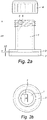
- said distance is the shortest distance that can be measured between two opposite points of said pair of screening surfaces (see Figure 2a ).
- the distance of the screening surfaces of the first compartment By making the distance of the screening surfaces of the first compartment larger than the distance of the screening surfaces of the second compartment, one can establish different cell densities within the same vial. Obtaining an optimal cell density is from crucial importance when wanting to analyse cells, especially from a suspension, when cells are mostly free-floating cells in a liquid cell sample. When said sample vial is immobile, allowing cells to settle by gravitation, typically more dense concentrations of cells will be found at the centre of the bottom of the first compartment compared to the cell density in the second compartment, whereby the nature of the cell density is determined by the distance between the pair of screening surfaces of each compartment.
- the density obtained in the second compartment will closely mimic the cell density that is obtained with thin cell layer techniques, whereby cells originating from a liquid cell sample are deposited on an analytical carrier such as a microscope slide by for instance centrifugation forces or gravity force.
- the current sample vial omits all handling steps which precede the acquirement of such a thin cell layer, hence gaining time and money.
- the cell density may be higher at the periphery of the first compartment and in the second compartment of the vial.
- the sample vial according the current invention provides for at least two different cell densities within the same sample vial.
- each pair of screening surfaces of each compartment will vary according to the type of cell sample that is to be analysed and is linked to the minimal cell density known to be required for analysing such a cell sample.
- the nature of the cell sample as well as the purpose of the analysis will define which pair of screening surfaces is to be used for the analysis by DHM. For instance, when a cell sample is composed of various sorts of cells, whereby only a specific subset of cells is important to the analysis, one can deduct in which compartment the latter are enriched, hence only focusing on that specific compartment.
- screening can be performed through the pair of screening surfaces of said compartment with the highest distance between the latter, typically at the centre of the bottom of the first compartment.
- Focusing on the compartment with the highest cell density will furthermore also limit the amount of images or fields of view required for coming to an adequate and significant analysis.
- screening can be performed through the pair of screening surfaces of the second compartment, in the situation where the sample vial is immobile. Again, the latter will depend on the nature of the cell sample, the cell quantity required for an optimal cell analysis and the purpose of the analysis.
- the distant-ratio's as disclosed herein cells will become immobilized in the second compartment of the vial in a very short amount of time, usually within seconds. This is of an enormous importance, since the current invention deals with cells which are freely moving within a solution.
- said ratio of the distance between said pair of screening surfaces of said first compartment and said pair of screening surfaces of second compartment comprises between 200:1 and 20:1, preferably 80:1, more preferably 40:1.
- the latter are optimized for the screening of various types of cell samples by DHM and for acquiring optimal cell densities in the compartments of the sample vial. The exact chosen ratio of the distances within these margins will entirely depend on the nature of the cell sample, the cells present in said cell sample which are to be analysed and the purpose of the analysis. For each of these requirements, an optimal ratio is determined.
- said the ratio of the surface area of a screening surface of the first compartment and a screening surface of the second compartment comprises between 1:1000 and 50:1, preferably between 1:100 and 10:1, more preferably 1:10, most preferably 1:3.
- the latter ratios are equally optimized for acquiring optimal cell densities in the compartments of the vial and for a representative scanning by DHM. Again, the exact chosen ratio will depend on the nature of the cell sample.
- said vial comprises two compartments.
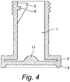
- said compartments of the vial form respectively a hollow platform as the base of said vial, and a column, preferably vertically placed on said platform.
- Said base of vial may comprise any chosen outline, preferably a round, ellipse, rectangular or square outline.
- Said platform may be cylindrical, cuboid, conical, parallelepiped, or frustroconical.
- Said column of the vial may be cylindrical, conical, frustro-conical, parallelepiped, or a cuboid.
- said vial comprises at the base of said first compartment, preferably at the centre of said base, a raised area. Said raised area enhances the flow of the cells from the first to the second compartment, hence ensuring an optimal, high cell density in the latter. Especially for cell sample whereby the analysis with said DHM requires a high cell density or a high number of cells, the latter has been proven to be beneficial.
- Said raised area may be conical, frustro-conical or hemispherical.
- Cells may migrate from the first to the second compartment under the influence of gravity alone or a vial comprising a cell sample may be rotated in order to help the cells migrating from the first to the second compartment. Therefore, in a preferred embodiment, the vial is easily rotatable around at least one axis. In a more preferred embodiment, the vial comprises at least one axis of rotational symmetry around which the vial can be easily rotated.
- said raised area has a shape which is capable of magnifying the image of objects located in said first compartment. In a more preferred embodiment, said raised area comprises a lens-shaped form.
- Said raised area has an inner surface which preferably bulges inward in the first compartment, and preferably comprises a form whose width near the base of said first compartment is wider than its width further away from said base. More preferably, said raised area comprises an outer surface which is flat, which bulges inward into said first compartment, and/or bulges outward from said compartment, said outer surface preferably comprising a shape suitable for magnifying the image of objects located in said first compartment.
- said first compartment comprises a liquid medium with a pre-determined medium refraction index and said lens-shaped form comprises at least one curved surface capable of magnifying the image of objects located in said first compartment with a magnification factor depending on said medium refraction index and said curved surface, whereby preferably said magnification factor is pre-fixed and said medium refraction index and/or said cured surface are adapted to result in said pre-fixed magnification factor.
- said second compartment has a stepwise upper surface area, whereby said smallest distance between the two screening surfaces of the second compartment lie at the outer rim of said compartment. This configuration allows obtaining the perfect amount of cells per field of view.
- said screening surfaces comprise an optically transparent material, whereby said optically transparent material is to be understood as being transparent to light with wavelengths equal to or in the range of the wavelengths of the illumination means of said DHM.
- optically transparent material can be, but is not limited to glass, plastic, polycarbonate, certain polymers such as polymethylmethacrylaat (PMMA), polystyrene crystals, polycrystalline materials.
- PMMA polymethylmethacrylaat
- polystyrene crystals polycrystalline materials.
- said material is further treated by for instance a positively charged surface coating such as poly-L-lysine, or by exposing the surface to a plasma treatment, or treated with anti-reflective substances.
- said vial is obtained by extrusion or co-moulding.
- said sample comprises supporting means at said base of vial.
- Said supporting means support vial when placed on a surface and prevent said screening surfaces of vial from coming into contact with said surface. The latter prevents said screening surfaces from being scratched or stained by the surface which can cause aberrant or divergent scanning from deviation of the beams of the DHM.
- Said supporting means may comprise, but are not limited to an upstanding rim at the circumference of the base of said vial or supporting feet at distinct positions of said circumference.
- Said supporting means may be produced of any material known to artisan skilled in the art, such as plastic, glass or rubber.
- said second compartment can comprise means for evacuating air bubbles.
- said second vial should be filled entirely with fluid sample.
- said means for discharging air bubbles can comprise grooves, provided at said bottom of said second compartment. Said grooves aid to evacuate any air bubbles present in said second compartment, preferably in the direction of the first compartment and/or the outer environment.
- said vial comprises an opening, preferably said opening is located at the first compartment, in a most preferred embodiment said first compartment being the column. In a preferred embodiment, said opening is capable of engaging with a lid in order to make said vial liquid-tight.
- the term 'liquid-tight' is to be understood as not allowing any passage or spillage of sample fluid from the inner side of the sample to the outer environment.
- Said lid preferably comprises a snap-on cap, a friction-fit or a threaded screw-cap.
- said lid comprises an optically transparent material, whereby said material is only optically transparent for light with wave lengths equal to these from the illumination means of DHM and partially or not optically transparent for light with other wavelengths. The latter allows transmission of said illumination beam of DHM and illumination of said sample inside liquid-tight vial.
- said lid is entirely produced of an optically transparent material.
- said lid is only partially produced from an optically transparent material.
- said only one specific side or section, such as for instance a window, of said lid is produced from an optically transparent material.
- said opening of first compartment provides for an entrance for receiving a cell collecting device.
- Said cell collecting device is used for collecting a cell sample from an organism, preferably a human, or for transferring a part of a cell sample stored in a first vial, to a sample vial of the current invention.
- the cell collecting device may comprise a brush, a spatula, a cotton swab, a needle, a scraper, a pipette or a Pasteur pipette.
- said cell collection device comprises means for collecting a sample, such as a brush, a spatula or a cotton ball, engaged to a longitudinal handle. More preferably, said means for collecting cells is attachable to said longitudinal handle by a snap-release joint as described in EP 2 263 552 . The latter allows said means for collecting cells to disengage after collecting the cells from the handle with a simple exertion of pressure.
- the means for collecting cells are positioned against a surface inside a sample vial, preferably one of the walls of the first compartment of said vial. This has for effect that the cell collection device can be transferred to a fixation fluid, prior to the disconnection of the sampling head from the longitudinal handle.
- said vial comprises means for aiding the disengagement of means for collecting cells from said cell collection device.
- said means for aiding the disengagement are provided to the wall of a compartment, preferably said compartment is the first compartment.
- Said means for aiding the disengagement may comprise one or more wall protrusions such as for instance pins, and/or one or more wall indentations.
- said fluid connection between said compartments prevents said cell collecting device, more specifically said means for collecting cells, from entering the second compartment of said vial.
- said vial comprises a sub-compartment for receiving means for collecting cells.
- Said sub-compartment is preferably present in the first compartment of said sample vial.
- the sub-compartment is specifically designed to receive means for collecting cells and retaining said means therein. This prevents said means from freely floating in the vial, thereby possibly obstructing the illumination beams of the DHM when screening through the pair of screening surfaces, hence causing distortion in the analysis.
- said sub-compartment encompasses said means for aiding the disengagement of means for collecting cells.
- Said sub-compartment is in liquid connection with the other compartments of said vial.
- said liquid connection is provided by an opening, connecting said sub-compartment with other compartments, or more preferably by a filter membrane, delineating one or more sides of said sub-compartments.
- said filter membrane is designed to ensure passage of both liquid and cells, simultaneously preventing passage of cell debris, tissue clumps and/or mucus.
- said filter membrane comprises a mesh which pore size is optimized for the latter.
- said lid is only partially produced of an optically transparent material, preferably at one side or one section of said lid, such as a window.
- said side or section produced of an optically transparent material is located opposite from said sub-sub-compartment for receiving means for collecting cells, when said lid is engaged in a liquid-tight manner to said vial.
- said vial is internally provided with a filter membrane, whereby said filter membrane is located at either the first compartment or at the fluid connection of said first and second compartment.
- said filter membrane will span an entire section of said first compartment or section determining fluid connection between said first and second compartment. Said filter membrane ensure passage of cells and liquid, but prevent passage of mucus, cell debris and tissue particles, which are undesirable during screening by DHM.
- said sample vial comprises identifying indicia
- said indicia may be fixed indicia and/or programmable indicia.
- Said indicia can correlate to type of sample, information on the origin of sample such as patient identity, general patient ID info (e.g. age, gender, residential area, ...), sample information (e.g. place and time when the sample was taken), owner ID info (e.g. the name of the user/institution from which the info comes from), DHM-ID info (e.g. a serial number of the DHM with which the DHM-obtained object properties are measured or observed), etc. or any combination thereof.
- Said identifying indicia are preferably machine-readable.
- said indicia comprise a bar code label, which corresponds to and uniquely identifies the vial and the sample contained therein.
- said indicia comprise an RFID tag.
- the indicia are read by identifying means, such as a laser scanner bar code reader in the case of the indicia being a bar code, or an RFID reader when indicia being an RFID tag.
- identifying means such as a laser scanner bar code reader in the case of the indicia being a bar code, or an RFID reader when indicia being an RFID tag.
- information related to the date and time of the obtained holographic information can be added, in addition to the initial sample indicia.
- the name or other identifier of the cytological laboratory analysing the sample with the system may be linked to the identification information as well.
- said all of the above-mentioned data is obtained by screening the sample in the first and/or second compartment of the vial via a scanning-pattern.
- Said scanning pattern may comprise a random line-pattern, a non-periodically line-pattern, a periodically line-pattern, a continue or discontinue pattern, a zigzag pattern, a line-pattern with phase and amplitude etc.
- Said screening the sample via a line-pattern ensures that the DHM will scan only unique sections of said sample, hence preventing that one section is screened multiple times. The latter ensures that significantly reliable and adequate information is obtained from said cell sample.
- Systems of interest include a Digital Holographic Microscope configured to assay a liquid sample for both morphological and biomarker data, e.g., as described above.
- Said system comprises a digital holographic microscope and at least one sample vial according to the current invention, characterized in that system comprises means for screening said sample via a scanning pattern.
- said means may comprise a movable platform or sample vial holder, whereby said platform or holder will move said sample vial during analysis of said sample.
- said movement involves a rotational movement.
- said means may comprise a movable lens, movable illumination means of said DHM or means that can alter the pathway of said illumination means such as for instance a mirror.
- aspects of the document further include systems, e.g., computer based systems, which are configured to predict the presence of a CIN lesion in a subject, e.g., as described above.
- a "computer-based system” refers to the hardware means, software means, and data storage means used to analyse the information of the present invention, e.g. the threshold database and used queries according to the current invention.
- the minimum hardware of the computer-based systems of the present invention comprises a central processing unit (CPU), input means, output means, and data storage means.
- the data storage means may comprise any manufacture comprising a recording of the present information as described above, or a memory access means that can access such a manufacture.
- record data programming or other information on a computer readable medium refers to a process for storing information, using any such methods as known in the art. Any convenient data storage structure may be chosen, based on the means used to access the stored information.
- data processor programs and formats can be used for storage, e.g. word processing text file, database format, etc.
- a "processor” references any hardware and/or software combination that will perform the functions required of it.
- any processor herein may be a programmable digital microprocessor such as available in the form of an electronic controller, mainframe, server or personal computer (desktop or portable).
- suitable programming can be communicated from a remote location to the processor, or previously saved in a computer program product (such as a portable or fixed computer readable storage medium, whether magnetic, optical or solid state device based).
- a computer program product such as a portable or fixed computer readable storage medium, whether magnetic, optical or solid state device based.
- a magnetic medium or optical disk may carry the programming, and can be read by a suitable reader communicating with each processor at its corresponding station.
- Embodiments of the subject systems include the following components: (a) a communications module for facilitating information transfer between the system and one or more users, e.g., via a user computer, as described below; and (b) a processing module for performing one or more tasks involved in the quantitative analysis methods of the invention.
- a computer program product comprising a computer usable medium having control logic (computer software program, including program code) stored therein.
- the control logic when executed by the processor of the computer, causes the processor to perform functions described herein.
- some functions are implemented primarily in hardware using, for example, a hardware state machine. Implementation of the hardware state machine so as to perform the functions described herein may be accomplished using any convenient method and techniques.
- systems of the document may include a number of additional components, such as data output devices, e.g., monitors and/or speakers, data input devices, e.g., interface ports, keyboards, etc., fluid handling components, power sources, etc.
- the subject methods and systems find use in a variety of different applications where detection prediction of the presence of (pre)cancerous cells in a subject is desired.
- Such applications include both research and diagnostic applications, e.g., applications where a subject is diagnosed with respect to the presence of or propensity to develop cervical cancer, e.g., as described above.
- the clinical utility of the methods and systems described herein provide powerful tools to detect and screen for the presence of cancerous cells and cancer development, both early and late stages, thus allowing therapeutic intervention to prevent disease progression as well as a chance to provide early treatment.
- the subject methods and systems finds use in the prognosis or risk assessment of an HPV-related disease or condition, for monitoring of the evolution of an HPV-related disease or condition, and for monitoring the efficiency of an anti-HPV drug or treatment, such as e.g., an anti-HPV vaccine or an anti-HPV vaccine candidate.
- an anti-HPV drug or treatment such as e.g., an anti-HPV vaccine or an anti-HPV vaccine candidate.
- the present document provides kits for practicing the subject methods, e.g., as described above.
- the subject kits may include a separate set of labelled biomarker probes en liquid medium, e.g., as described above, to be combined prior to sample solubilisation.
- said kit comprises the liquid medium already supplemented with biomarker probes.
- kits may furthermore comprise a sample vial according to the current invention, empty or pre-filled with the liquid medium (either with or without biomarker probes) according to the current invention.
- Kits may further include sample obtainment devices, e.g., cervical brooms, as described above.
- the above components may be present in separate containers or one or more components may be combined into a single container, e.g., a glass or plastic vial.
- the subject kits will further include instructions for practicing the subject methods. These instructions may be present in the subject kits in a variety of forms, one or more of which may be present in the kit.
- a suitable medium or substrate e.g., a piece or pieces of paper on which the information is printed, in the packaging of the kit, in a package insert, etc.
- a computer readable medium e.g., diskette, CD, etc.
- a website address which may be used via the internet to access the information at a removed site. Any convenient means may be present in the kits.
- FIGs 1 to 7 and 9 depict preferred embodiments of the sample vial according to the current invention.
- Said sample vial comprises a first (1) and second (2) compartment, whereby said first (1) and second (2) compartment are in liquid connection with one other and whereby each said compartment comprises a pair of screening surfaces.
- the shading pattern in Figure 1b defines further the meaning of said first (1) and second compartment (2).
- said sample vial may be T-shaped, comprised of cylindrical compartments as shown in Figure 1 , or comprised of cuboids ( Fig. 5 ) or comprised of a combination of cylindrical and frustro-conical compartments (as shown in Fig. 7 ).
- said vial may be L-shaped, as shown in Figure 6 .
- Figure 9 shows an embodiment of the vial with a stairwise formed second compartment.
- the vial may be provided with a lid (4), making said vial liquid-tight when said lid is engaged to said vial.
- said lid (4) is a screw cap.
- Said vial may comprise an optically transparent material, whereby said optically transparent material may be restricted to for instance only a section of said lid.
- the lid (4) of the sample vial as depicted in Figure 3 comprises a window (12), comprised of optically transparent material. Said window allows screening of the sample by DHM through the lid (4).
- the sample vial is preferably foreseen of supporting means (3) at the base of the vial, for providing support when placed on a surface.
- Said supporting means (3) prevent the screening surfaces at the base of the vial from being scratched and/or stained.
- the vial may be equipped with a sub-compartment (5) and/or means for aiding the disengagement (6) of means for collecting cells (10) from a cell collection device (8) and the handle (9) of the cell collection device (8).
- the means for aiding the disengagement may be, as shown in Figures 2 to 7 , protrusions (6) extending from the wall of the first compartment (1).
- protrusions will help a practitioner with the disengagement of a sample brush (10) from a longitudinal handle and is preferably encompassed by a sub-compartment (5).
- This sub-compartment is designed to receive the sample brush (10) or any other means for collecting cells.
- the cells may be provided by a stirring movement of the device (8) as shown in Figure 8a .
- the means for collecting cells (10) in the current example a brush, will be disconnected from the handle (9), leaving the brush at the bottom of the first compartment of the vial (see Figure 8b ). The fluid connection between the first and second compartment will prevent the brush from entering the second compartment.
- FIG. 8c Another option is shown in Figure 8c , whereby the sample vial is provided in the first compartment with a sub-compartment (5), for receiving and retaining the brush (10), after the latter is released from the handle (9).
- the sub-compartment (5) and brush (10) remains in fluid connection with the first compartment, allowing the collected cells to migrate from the sub-compartment to the first and second compartment.
- this connection is ensured by a filter membrane delineating the sub-compartment (5) or an opening in said sub-compartment (5).
- the base of the first compartment (1) of the vial may at the inner side be accommodated with a raised area (11).
- the raised area (11) enhances the flow of the cells from the first to the second compartment, hence ensuring an optimal, high cell density in the latter.
- a sample vial as shown in Figure 1 or Figure 2 suitable for the screening of a cervical cell sample by DHM, may comprise the following dimensions:
- DHM enables a quantitative multifocal phase contrast imaging that has been found suitable for quantitative and qualitative inspection, and for 3-dimensional cell imaging. Both biomarker data and morphological data was obtained.
- 188 cells were identified and measured in an automated way. Cells were in a first instance analyzed based on the presence of HPV specific proteins. In a second instance, information regarding the Nucleus/Cell Ratio (NCR) and Optical Height Delta (OHD) was extracted from the 3D holographic image. The Optical Height Delta is the difference between Nucleus top height minus Cytoplasm average height. NCR and OHD were separately determined in 2 groups: CIN1 or CIN 2/3 patients.
- Figures 11A and 11B Graphical overview of the obtained results are shown in Figures 11A and 11B .
- Cell population analysis by this objective tool reveals an increased NCR and OHD in the dysplastic cells.
- a correlation can be observed between the OHD and the diagnosis.
- This correlation allows the application of an automatic scoring algorithm in a smear in a liquid cell sample without the need for additional expensive preparation to make a cytological diagnosis.
- Figure 10A-C show examples of morphological data obtained from one of the analyzed sample, whereby the morphological data comprise two-dimensional and three-dimensional images from cells present within sample.
- cervical samples of 15 patients were obtained and solubilized in a liquid medium of the current invention, comprising a fluorescently labelled bio-marker against p16 (antibody).
- p16 antibody
- the method of the current invention using combined morphological and biomarker data (e.g. E6, E7, p16, p16INK4a, Ki-67 or annexin A5 overexpression) on a cell-by-cell basis performed on cells in suspension increased the sensitivity and specificity for detection of (pre)cancerous cells (e.g. a CIN2 an CIN3 lesion to more than 90%, compared to currently known single tests such as PAP smear and HPV DNA.
- precancerous cells e.g. a CIN2 an CIN3 lesion to more than 90%
Landscapes
- Health & Medical Sciences (AREA)
- Life Sciences & Earth Sciences (AREA)
- Chemical & Material Sciences (AREA)
- Engineering & Computer Science (AREA)
- Biomedical Technology (AREA)
- General Health & Medical Sciences (AREA)
- Physics & Mathematics (AREA)
- Analytical Chemistry (AREA)
- Pathology (AREA)
- Immunology (AREA)
- Hematology (AREA)
- General Physics & Mathematics (AREA)
- Biochemistry (AREA)
- Molecular Biology (AREA)
- Chemical Kinetics & Catalysis (AREA)
- Clinical Laboratory Science (AREA)
- Urology & Nephrology (AREA)
- Medicinal Chemistry (AREA)
- Food Science & Technology (AREA)
- Optics & Photonics (AREA)
- Heart & Thoracic Surgery (AREA)
- Biophysics (AREA)
- Animal Behavior & Ethology (AREA)
- Surgery (AREA)
- Gynecology & Obstetrics (AREA)
- Medical Informatics (AREA)
- Public Health (AREA)
- Veterinary Medicine (AREA)
- Reproductive Health (AREA)
- Microbiology (AREA)
- Cell Biology (AREA)
- Biotechnology (AREA)
- Investigating Or Analysing Biological Materials (AREA)
- Measuring Or Testing Involving Enzymes Or Micro-Organisms (AREA)
- Micro-Organisms Or Cultivation Processes Thereof (AREA)
- Sampling And Sample Adjustment (AREA)
Claims (11)
- Procédé pour détecter la présence de cellules cancéreuses ou précancéreuses dans un échantillon de cellules liquide , comprenant les étapes de :- mise en suspension et préservation d'un échantillon de cellules prélevé sur un sujet dans un flacon d'échantillon comprenant un milieu liquide, ledit milieu liquide comprenant un moyen pour le marquage des cellules ou d'un ou plusieurs épitope(s) sur ou dans lesdites cellules, ledit moyen comprenant une sonde de biomarqueur marquée ;- obtention de données à partir dudit échantillon de cellules liquide marqué ; et- détection de la présence desdites cellules cancéreuses ou précancéreuses sur la base desdites données obtenues ; dans lequel lesdites données comprennent des données morphologiques et des données de biomarqueur,caractérisé en ce que lesdites données sont obtenues à partir dudit échantillon de cellules liquide marqué de cellules en suspension dans le flacon d'échantillon par microscopie holographique numérique.
- Procédé selon la revendication 1, dans lequel lesdites données morphologiques et données de biomarqueur sont obtenues simultanément.
- Procédé selon la revendication 1 ou 2, dans lequel ledit échantillon est solubilisé immédiatement après obtention.
- Procédé selon l'une quelconque des revendications 1 à 3, dans lequel lesdites données morphologiques comprennent une hauteur nucléaire optique, un diamètre de noyau de cellule, une texture de chromatine, une taille de cellule, un pourcentage de nucléation, une forme de cellule, une quantité de cellules, une taille nucléaire, un volume nucléaire, une variabilité de taille nucléaire, une variabilité de volume nucléaire, ou une combinaison quelconque de ceux-ci.
- Procédé selon l'une quelconque des revendications 1 à 4, dans lequel ledit échantillon de cellules liquide comprend des cellules de col de l'utérus.
- Procédé selon l'une quelconque des revendications 1 à 5, dans lequel ladite sonde de biomarqueur est dirigée contre au moins un des marqueurs choisis dans le groupe de p14Arf, p15INK4b, p16INK4a, p18INKc, p19INK4d, p21WAF1/CIP1, p27Kip1, Ki-67, annexine A5, protéines virales de HPV E1, E2, E3, E4, E5, E6, E7, L1, L2 ; plus préférablement E6 ou E7 ; ou des combinaisons quelconques de ceux-ci.
- Procédé selon l'une quelconque des revendications 1 à 6, dans lequel lesdites données morphologiques obtenues sont comparées et corrélées à une base de données de référence.
- Procédé selon la revendication 7, dans lequel lesdites données morphologiques obtenues sont stockées dans ladite base de données de référence.
- Procédé selon l'une quelconque des revendications 1 à 8, dans lequel lesdites données morphologiques sont isolées de l'arrière-plan provenant de sonde de biomarqueur non liée sur la base d'informations de phase extraites par le microscope holographique numérique.
- Procédé selon l'une quelconque des revendications précédentes, dans lequel ledit milieu liquide comprend un fixateur, de préférence un alcool ou un fixateur à base de zinc ; un agent tampon qui maintient le pH dudit milieu liquide entre 2 et 7 ; un agent antiagglomérant pour éviter les agrégats de cellules dans ledit milieu liquide, de préférence la N-acétyl ou N-méthyl-cystéine, EDTA ou EDTA étant le sel disodique.
- Procédé selon la revendication 1 à 10, dans lequel ladite sonde de biomarqueur est marquée par fluorescence.
Priority Applications (1)
| Application Number | Priority Date | Filing Date | Title |
|---|---|---|---|
| EP12788531.7A EP2783211B1 (fr) | 2011-11-21 | 2012-11-20 | Procédé de détection de cellules cancéreuses dans un échantillon de cellules |
Applications Claiming Priority (3)
| Application Number | Priority Date | Filing Date | Title |
|---|---|---|---|
| EP11189986.0A EP2594334A1 (fr) | 2011-11-21 | 2011-11-21 | Fiole à échantillon pour analyse holographique numérique d'un échantillon de cellules liquides |
| PCT/EP2012/073124 WO2013076089A2 (fr) | 2011-11-21 | 2012-11-20 | Flacon pour milieu liquide et échantillon destiné à un procédé de détection de cellules cancéreuses dans un échantillon de cellules. |
| EP12788531.7A EP2783211B1 (fr) | 2011-11-21 | 2012-11-20 | Procédé de détection de cellules cancéreuses dans un échantillon de cellules |
Publications (2)
| Publication Number | Publication Date |
|---|---|
| EP2783211A2 EP2783211A2 (fr) | 2014-10-01 |
| EP2783211B1 true EP2783211B1 (fr) | 2018-08-15 |
Family
ID=47216271
Family Applications (3)
| Application Number | Title | Priority Date | Filing Date |
|---|---|---|---|
| EP11189986.0A Withdrawn EP2594334A1 (fr) | 2011-07-19 | 2011-11-21 | Fiole à échantillon pour analyse holographique numérique d'un échantillon de cellules liquides |
| EP12790540.4A Withdrawn EP2782675A1 (fr) | 2011-11-21 | 2012-11-20 | Flacon à échantillon pour analyse holographique numérique d'un échantillon liquide de cellules |
| EP12788531.7A Not-in-force EP2783211B1 (fr) | 2011-11-21 | 2012-11-20 | Procédé de détection de cellules cancéreuses dans un échantillon de cellules |
Family Applications Before (2)
| Application Number | Title | Priority Date | Filing Date |
|---|---|---|---|
| EP11189986.0A Withdrawn EP2594334A1 (fr) | 2011-07-19 | 2011-11-21 | Fiole à échantillon pour analyse holographique numérique d'un échantillon de cellules liquides |
| EP12790540.4A Withdrawn EP2782675A1 (fr) | 2011-11-21 | 2012-11-20 | Flacon à échantillon pour analyse holographique numérique d'un échantillon liquide de cellules |
Country Status (3)
| Country | Link |
|---|---|
| US (2) | US10060905B2 (fr) |
| EP (3) | EP2594334A1 (fr) |
| WO (2) | WO2013076089A2 (fr) |
Families Citing this family (25)
| Publication number | Priority date | Publication date | Assignee | Title |
|---|---|---|---|---|
| EP2594334A1 (fr) | 2011-11-21 | 2013-05-22 | Drive O2 | Fiole à échantillon pour analyse holographique numérique d'un échantillon de cellules liquides |
| EP2626686A1 (fr) | 2012-02-13 | 2013-08-14 | Ovizio Imaging Systems NV/SA | Cytomètre de flux avec microscope holographique numérique |
| WO2013168767A1 (fr) * | 2012-05-10 | 2013-11-14 | コニカミノルタ株式会社 | Procédé d'élimination des globules rouges ainsi que tube de centrifugation pour prélèvement sanguin |
| WO2014044823A1 (fr) | 2012-09-20 | 2014-03-27 | Ovizio Imaging Systems NV/SA | Microscope holographique numérique ayant des systèmes de fluide |
| DE102013014901A1 (de) * | 2013-09-06 | 2015-03-12 | Testo Ag | Verfahren zur Untersuchung einer Probe auf Mikroorganismen und/oder Viren und korrespondierender Tupfer |
| DE102014200911A1 (de) * | 2013-10-09 | 2015-04-09 | Siemens Aktiengesellschaft | In-Vitro-Verfahren zum markierungsfreien Bestimmen eines Zelltyps einer Zelle |
| WO2016044508A1 (fr) * | 2014-09-17 | 2016-03-24 | Hologic, Inc. | Dispositif de collecte de spécimen séparable |
| EP3026424A1 (fr) * | 2014-11-27 | 2016-06-01 | Hach Lange GmbH | Turbidimètre néphélométrique utilisant une cuvette avec étiquette |
| EP3196631A1 (fr) | 2016-01-19 | 2017-07-26 | Ovizio Imaging Systems NV/SA | Microscope holographique numérique avec système électrofluidique, ledit système électrofluidique et procédés d'utilisation |
| MX2019006793A (es) | 2016-12-09 | 2019-11-18 | Zanetta Malanowska Stega | Dispositivo, kit y metodos de cepillo para biopsia. |
| GB201706947D0 (en) | 2017-05-02 | 2017-06-14 | Cytosight Ltd | Fluid sample enrichment system |
| WO2018237239A1 (fr) | 2017-06-23 | 2018-12-27 | Texas Tech University System | Procédé et dispositif pour le criblage holographique sans étiquette et à haut rendement et l'énumération de cellules tumorales dans le sang |
| JP6966272B2 (ja) * | 2017-09-20 | 2021-11-10 | シスメックス株式会社 | 細胞分析方法、細胞情報提供装置、細胞情報提供システム、制御プログラム、記録媒体 |
| BE1026111B1 (nl) * | 2018-03-15 | 2019-10-16 | Ovizio Imaging Systems Nv | Digitale holografische microscopie voor het bepalen van de status van een virale infectie |
| WO2019175386A1 (fr) * | 2018-03-15 | 2019-09-19 | Ovizio Imaging Systems NV/SA | Microscopie holographique numérique pour déterminer un état d'infection virale |
| CA3099275A1 (fr) * | 2018-05-09 | 2019-11-14 | Dermtech, Inc. | Nouveaux classificateurs de genes et leurs utilisations dans des maladies auto-immunes |
| CN111220599B (zh) * | 2018-11-24 | 2022-04-29 | 朱燕超 | 一种用于水质检测的取样装置及取样方法 |
| WO2020247770A1 (fr) * | 2019-06-07 | 2020-12-10 | The Board Of Trustees Of The University Of Illinois | Dispositif de cristallisation microfluidique à écoulement continu, bien mélangé, pour le criblage de polymorphes, de morphologies et cinétique de cristallisation à une sursaturation contrôlée |
| EP4022637A2 (fr) | 2019-08-30 | 2022-07-06 | Juno Therapeutics, Inc. | Procédés d'apprentissage automatique pour la classification de cellules |
| CN111172270B (zh) * | 2019-12-30 | 2022-12-13 | 青岛人和医学检验有限公司 | 用于子宫颈鳞状上皮内病变分级的全血转录基因标志物及其应用 |
| CN112951410A (zh) * | 2021-02-08 | 2021-06-11 | 青岛大学附属医院 | 超声引导下的细针穿刺病理涂片快速现场评估系统 |
| CN113514455B (zh) * | 2021-06-29 | 2024-06-21 | 广州朗坤生物科技有限公司 | 检测上皮组织脱落细胞表面叶酸受体表达的方法及试剂盒 |
| CN113917149B (zh) * | 2021-09-30 | 2024-05-24 | 江苏扬新生物医药有限公司 | 凝溶胶蛋白检测物在制备子宫癌评估检测试剂中的应用 |
| CN121100267A (zh) | 2022-12-09 | 2025-12-09 | 朱诺治疗学股份有限公司 | 使用全息成像预测细胞表型的机器学习方法 |
| CN117169091B (zh) * | 2023-07-28 | 2024-09-13 | 恩大细胞基因工程有限公司 | 一种细胞活性检测系统 |
Citations (2)
| Publication number | Priority date | Publication date | Assignee | Title |
|---|---|---|---|---|
| US7009700B2 (en) * | 2001-06-29 | 2006-03-07 | Universite Libre De Bruxelles | Method and device for obtaining a sample with three-dimensional microscopy |
| US20080317325A1 (en) * | 1999-01-25 | 2008-12-25 | Amnis Corporation | Detection of circulating tumor cells using imaging flow cytometry |
Family Cites Families (78)
| Publication number | Priority date | Publication date | Assignee | Title |
|---|---|---|---|---|
| FR2565233B1 (fr) | 1984-05-30 | 1986-09-12 | Rhone Poulenc Rech | Copolymere greffe de silicone et de polylactone lie par une liaison urethanne |
| US5089416A (en) | 1984-12-24 | 1992-02-18 | Caribbean Microparticles Corporation | Method of use of non-fluorescent particles to determine fluorescence threshold of a flow cytometer relative to the autofluorescence of samples |
| US4786594A (en) * | 1986-05-14 | 1988-11-22 | Syntex (U.S.A.) Inc. | Enzyme immunoassay |
| US5740270A (en) | 1988-04-08 | 1998-04-14 | Neuromedical Systems, Inc. | Automated cytological specimen classification system and method |
| FR2642522B1 (fr) | 1989-01-31 | 1991-05-10 | Elf Aquitaine | Appareil de mesure de densite optique in situ pour fermenteur |
| US5427959A (en) * | 1990-10-01 | 1995-06-27 | Canon Kabushiki Kaisha | Apparatus and method for measuring specimen |
| US5256571A (en) | 1991-05-01 | 1993-10-26 | Cytyc Corporation | Cell preservative solution |
| US5495333A (en) | 1992-07-24 | 1996-02-27 | Sumitomo Electric Industries, Ltd. | Method and apparatus of detecting impurities in fluid |
| US6924094B1 (en) | 1996-02-08 | 2005-08-02 | Affymetrix, Inc. | Chip-based species identification and phenotypic characterization of microorganisms |
| AU7831798A (en) | 1997-06-09 | 1998-12-30 | Guava Technologies, Inc. | Method and apparatus for detecting microparticles in fluid samples |
| GB2330516A (en) | 1997-10-22 | 1999-04-28 | Elizabeth Acton | Cryopreservation of cell suspensions |
| US6235536B1 (en) * | 1998-03-07 | 2001-05-22 | Robert A. Levine | Analysis of quiescent anticoagulated whole blood samples |
| DE19826470C2 (de) | 1998-06-13 | 2001-10-18 | Eppendorf Ag | Küvettensystem und Küvette |
| US6574401B2 (en) | 1999-03-30 | 2003-06-03 | Ceramoptec Industries, Inc. | Optical fiber-handpiece combination for medical laser treatments |
| US6651008B1 (en) | 1999-05-14 | 2003-11-18 | Cytokinetics, Inc. | Database system including computer code for predictive cellular bioinformatics |
| DE10016838B4 (de) | 2000-04-05 | 2006-10-19 | Jan-Gerd Dipl.-Ing. Frerichs | In-situ Mikroskopvorrichtung für Reaktoren |
| US6394966B1 (en) * | 2000-09-08 | 2002-05-28 | Diagnostic Cytology Laboratories, Inc. | Apparatus and method for removing cells from an endocervical brush in a liquid-based pap test system |
| US20020164825A1 (en) | 2000-09-09 | 2002-11-07 | Wen-Tien Chen | Cell separation matrix |
| US7027633B2 (en) | 2000-11-30 | 2006-04-11 | Foran David J | Collaborative diagnostic systems |
| US7050620B2 (en) | 2001-03-30 | 2006-05-23 | Heckman Carol A | Method of assaying shape and structural features in cells |
| US6965793B2 (en) | 2001-06-28 | 2005-11-15 | Chemimage Corporation | Method for Raman chemical imaging of endogenous chemicals to reveal tissue lesion boundaries in tissue |
| WO2003048868A1 (fr) | 2001-12-04 | 2003-06-12 | Ecole Polytechnique Federale De Lausanne (Epfl) | Appareil et procede d'imagerie holographique numerique |
| US20030113832A1 (en) * | 2001-12-14 | 2003-06-19 | Lauf Robert J. | Apparatus and method for assaying electrophysiological effects |
| US20030199649A1 (en) | 2002-03-26 | 2003-10-23 | Orbison David Robert | Method and apparatus for the controlled dilution of organometallic compounds |
| DE10237082B4 (de) | 2002-08-09 | 2014-12-31 | Sartorius Stedim Biotech Gmbh | Verfahren und Vorrichtung zur biotechnologischen Herstellung von Wertstoffen |
| US20040123234A1 (en) | 2002-12-18 | 2004-06-24 | Schemalogic, Inc. | Method and system for change control management of shema definition objects |
| EP1524491A1 (fr) | 2003-10-16 | 2005-04-20 | Universite Libre De Bruxelles | Appareil associant un interféromètre et un microscope |
| EP1631788B1 (fr) | 2003-05-16 | 2007-03-14 | Universite Libre De Bruxelles | Microscope holographique numerique pour imagerie 3d et procede l'utilisant |
| US20070065810A1 (en) * | 2003-07-18 | 2007-03-22 | Georgetown University | Diagnosis and treatment of cervical cancer |
| US7286222B2 (en) | 2003-07-18 | 2007-10-23 | Chemimage Corporation | Sample container and system for a handheld spectrometer and method for using therefor |
| DE60325107D1 (de) | 2003-08-25 | 2009-01-15 | Marc Ramael | Verfahren und kit zum quantitativen und/oder qualitativen nachweis von komponenten in einer probe |
| US20070166699A1 (en) | 2003-09-17 | 2007-07-19 | Amynon Biotech Gmbh | Combination of anti-hpv-16 and 18 antibodies and uses thereof |
| EP1740951B1 (fr) | 2004-04-29 | 2008-03-05 | Marc Ramael | Procede et kit pour detecter des composants dans un echantillon |
| US20060088814A1 (en) | 2004-10-26 | 2006-04-27 | Cytyc Corporation | Enhanced cell preservative solution and methods for using same |
| WO2006047252A1 (fr) | 2004-10-26 | 2006-05-04 | Cytyc Corporation | Solution de conservation de cellules amelioree, et methodes d'utilisation associees |
| US20070037180A1 (en) | 2005-02-10 | 2007-02-15 | Olle Nilsson | Site-specific immunization in order to establish antibodies with specificity for the E7 oncoprotein of high-risk HPV's |
| US7275682B2 (en) | 2005-03-24 | 2007-10-02 | Varian, Inc. | Sample identification utilizing RFID tags |
| EP1731099A1 (fr) | 2005-06-06 | 2006-12-13 | Paul Scherrer Institut | Interféromètre pour l'imagerie et la tomographie à contraste de phase avec une source de rayons X incohérente et polychromatique |
| WO2007002898A2 (fr) | 2005-06-29 | 2007-01-04 | University Of South Florida | Balayage tomographique variable avec holographie interferentielle numerique a balayage en longueur d'onde |
| US7948632B2 (en) | 2005-12-22 | 2011-05-24 | Phase Holographic Imaging Phi Ab | Method and apparatus for analysis of a sample of cells |
| US7651869B2 (en) | 2006-03-14 | 2010-01-26 | Research International, Inc. | Optical assay apparatus and methods |
| US7616320B2 (en) | 2006-03-15 | 2009-11-10 | Bahram Javidi | Method and apparatus for recognition of microorganisms using holographic microscopy |
| EP2027252A4 (fr) | 2006-04-21 | 2012-10-03 | Bayer Corp | Appareil, système et procédé de mesures in situ |
| US8428331B2 (en) | 2006-08-07 | 2013-04-23 | Northeastern University | Phase subtraction cell counting method |
| US8859218B2 (en) * | 2008-06-13 | 2014-10-14 | Oncohealth Corp. | In situ detection of early stages and late stages HPV infection |
| CA2964611C (fr) | 2007-03-28 | 2021-06-01 | Bionano Genomics, Inc. | Procedes d'analyse macromoleculaire utilisant des reseaux de nanocanaux |
| GB0707776D0 (en) | 2007-04-23 | 2007-05-30 | Robio Systems Ltd | A Container for culturing and/or transporting embryos or oocytes. an insert for the container and a method of transporting same |
| US8785180B2 (en) | 2007-06-18 | 2014-07-22 | Shanghai Guoqiang Bioengineering Equipment Co., Ltd. | Biochemical reactor |
| DE502007006368D1 (de) * | 2007-06-25 | 2011-03-10 | Ibidi Gmbh | Probenkammer |
| US20090082637A1 (en) | 2007-09-21 | 2009-03-26 | Michael Galperin | Multi-modality fusion classifier with integrated non-imaging factors |
| US20090132464A1 (en) | 2007-10-16 | 2009-05-21 | Decript Inc. | Methods for processing generic chemical structure representations |
| US8049814B2 (en) | 2008-03-27 | 2011-11-01 | The Rockefeller University | Holographic microscope |
| CN102067046A (zh) | 2008-06-19 | 2011-05-18 | 相位全息成像技术公司 | 透明生物物体的分析 |
| JP2010057902A (ja) | 2008-08-06 | 2010-03-18 | Toshiba Corp | レポート作成支援装置、レポート作成支援システム、及び医用画像参照装置 |
| US20110212440A1 (en) | 2008-10-10 | 2011-09-01 | Cnrs-Dae | Cell sorting device |
| CN102498217A (zh) | 2009-04-20 | 2012-06-13 | 阿波维塔公司 | Hpv的e6蛋白的特异性抗体及其应用 |
| US8599383B2 (en) * | 2009-05-06 | 2013-12-03 | The Regents Of The University Of California | Optical cytometry |
| US8885035B2 (en) | 2009-06-16 | 2014-11-11 | Lester F. Ludwig | Electronic imaging flow-microscope for environmental remote sensing, bioreactor process monitoring, and optical microscopic tomography |
| US20100315502A1 (en) | 2009-06-16 | 2010-12-16 | Ikonisys, Inc. | System and method for remote control of a microscope |
| EP2263552A1 (fr) | 2009-06-17 | 2010-12-22 | Switch bvba | Dispositif de collecte de cellule à libération de fermeture |
| US8610085B2 (en) | 2009-08-20 | 2013-12-17 | Bio-Rad Laboratories, Inc. | High-speed cellular cross sectional imaging |
| KR101817110B1 (ko) | 2009-10-08 | 2018-02-21 | 유니베르시테 리브레 드 브룩크젤즈 | 비축 간섭계 |
| EP3136079B1 (fr) | 2009-10-20 | 2020-02-12 | The Regents of The University of California | Cellule holographique incohérente sans lentille et microscopie sur une puce |
| EP2507639B1 (fr) | 2009-12-04 | 2020-04-01 | Life Technologies Corporation | Appareils, systèmes, procédés et supports lisibles par ordinateur destinés à la cytométrie de flux acoustique |
| EP2534540B1 (fr) * | 2010-02-09 | 2019-05-22 | Phase Holographic Imaging PHI AB | Procédé de microscopie holographique numérique et de formation d'image sur des échantillons de cellules identifiées, et son utilisation |
| DE102010023099B3 (de) | 2010-06-09 | 2011-11-17 | Celltool Gmbh | Verfahren und Vorrichtung zum Charakterisieren von biologischen Objekten |
| US9569664B2 (en) | 2010-10-26 | 2017-02-14 | California Institute Of Technology | Methods for rapid distinction between debris and growing cells |
| US9434937B2 (en) | 2011-03-07 | 2016-09-06 | Accelerate Diagnostics, Inc. | Rapid cell purification systems |
| WO2013010595A1 (fr) | 2011-07-19 | 2013-01-24 | Ovizio Imaging Systems N.V. | Base de données orientée objet et procédé d'amélioration de base de données orientée objet |
| US9684281B2 (en) | 2011-07-19 | 2017-06-20 | Ovizio Imaging Systems NV/SA | Method and system for detecting and/or classifying cancerous cells in a cell sample |
| US20140195568A1 (en) | 2011-07-19 | 2014-07-10 | Ovizio Imaging Systems NV/SA | Object database and object database improving method |
| EP2594334A1 (fr) | 2011-11-21 | 2013-05-22 | Drive O2 | Fiole à échantillon pour analyse holographique numérique d'un échantillon de cellules liquides |
| WO2013018024A1 (fr) | 2011-07-29 | 2013-02-07 | Ecole Polytechnique Federale De Lausanne (Epfl) | Appareil et procédé pour la tomographie de phase quantitative au moyen de balayage linéaire avec détection cohérente et non cohérente |
| EP2626686A1 (fr) | 2012-02-13 | 2013-08-14 | Ovizio Imaging Systems NV/SA | Cytomètre de flux avec microscope holographique numérique |
| CN202808799U (zh) | 2012-09-14 | 2013-03-20 | 江苏嘉语生物医药技术有限公司 | 一种可控氧环境的微生物培养检测装置 |
| WO2014044823A1 (fr) | 2012-09-20 | 2014-03-27 | Ovizio Imaging Systems NV/SA | Microscope holographique numérique ayant des systèmes de fluide |
| WO2015014858A1 (fr) | 2013-07-31 | 2015-02-05 | Ovizio Imaging Systems NV/SA | Couvercle amélioré pour surveiller des objets en suspension |
| EP3196631A1 (fr) | 2016-01-19 | 2017-07-26 | Ovizio Imaging Systems NV/SA | Microscope holographique numérique avec système électrofluidique, ledit système électrofluidique et procédés d'utilisation |
-
2011
- 2011-11-21 EP EP11189986.0A patent/EP2594334A1/fr not_active Withdrawn
-
2012
- 2012-11-20 WO PCT/EP2012/073124 patent/WO2013076089A2/fr not_active Ceased
- 2012-11-20 US US14/359,553 patent/US10060905B2/en active Active
- 2012-11-20 EP EP12790540.4A patent/EP2782675A1/fr not_active Withdrawn
- 2012-11-20 EP EP12788531.7A patent/EP2783211B1/fr not_active Not-in-force
- 2012-11-20 US US14/359,556 patent/US9846151B2/en active Active
- 2012-11-20 WO PCT/EP2012/073116 patent/WO2013076082A1/fr not_active Ceased
Patent Citations (2)
| Publication number | Priority date | Publication date | Assignee | Title |
|---|---|---|---|---|
| US20080317325A1 (en) * | 1999-01-25 | 2008-12-25 | Amnis Corporation | Detection of circulating tumor cells using imaging flow cytometry |
| US7009700B2 (en) * | 2001-06-29 | 2006-03-07 | Universite Libre De Bruxelles | Method and device for obtaining a sample with three-dimensional microscopy |
Non-Patent Citations (4)
| Title |
|---|
| CHRISTOPHER J. MANN ET AL: "Dual modality live cell imaging with multiple-wavelength digital holography and epi-fluorescence", 3D RESEARCH, vol. 2, no. 1, 1 March 2011 (2011-03-01), XP055053318, DOI: 10.1007/3DRes.01(2011)5 * |
| KOSMEIER S ET AL: "Determination of the integral refractive index of cells in suspension by digital holographic phase contrast microscopy", BIOPHOTONICS: PHOTONIC SOLUTIONS FOR BETTER HEALTH, 2008, DOI: 10.1117/12.781125 * |
| NICOLAS PAVILLON ET AL: "Cell morphology and intracellular ionic homeostasis explored with a multimodal approach combining epifluorescence and digital holographic microscopy", JOURNAL OF BIOPHOTONICS, vol. 3, no. 7, 1 July 2010 (2010-07-01), pages 432 - 436, XP055015192, ISSN: 1864-063X, DOI: 10.1002/jbio.201000018 * |
| WANG P ET AL: "Nanoscale nuclear architecture for cancer diagnosis beyond pathology via spatial-domain low-coherence quantitative phase microscopy", JOURNAL OF BIOMEDICAL OPTICS, vol. 15, no. 6, 23 December 2010 (2010-12-23), XP055353960, DOI: 10.1117/1.3523618 * |
Also Published As
| Publication number | Publication date |
|---|---|
| EP2782675A1 (fr) | 2014-10-01 |
| US9846151B2 (en) | 2017-12-19 |
| WO2013076082A1 (fr) | 2013-05-30 |
| WO2013076089A2 (fr) | 2013-05-30 |
| US20140329231A1 (en) | 2014-11-06 |
| EP2783211A2 (fr) | 2014-10-01 |
| WO2013076089A3 (fr) | 2013-07-25 |
| US20140349336A1 (en) | 2014-11-27 |
| EP2594334A1 (fr) | 2013-05-22 |
| US10060905B2 (en) | 2018-08-28 |
Similar Documents
| Publication | Publication Date | Title |
|---|---|---|
| US10060905B2 (en) | Liquid medium and sample vial for use in a method for detecting cancerous cells in a cell sample | |
| CA2300862C (fr) | Lame porte-objet | |
| JP6505657B2 (ja) | 子宮頸部細胞の浮遊試料から、対象が子宮頸部上皮内腫瘍(cin)病変を有するかどうかを予測するための方法およびシステム | |
| JP5872655B2 (ja) | 異形成の進行を評価する方法および疾患段階の決定に用いるキット | |
| US6567214B2 (en) | Microscope slide having culture media and method for use | |
| Fu et al. | Liquid biopsy technologies for hematological diseases | |
| JP2021501321A (ja) | 組織および細胞染色のためのデバイスおよび方法 | |
| JP7199362B2 (ja) | シグナル発生型デジタルアッセイにおけるノイズを低減する方法 | |
| US6239906B1 (en) | Flexible microscope slide | |
| Gormley et al. | A simplified molecular method to detect high‐risk HPV using the Aptima HPV assay on head and neck FNA smears | |
| O'Neill et al. | Use of an optical biosensor to measure prostate-specific antigen in whole blood | |
| JP2023534375A (ja) | 細胞病理学的サンプルに対する多重染色切片調製法 | |
| CN119290545B (zh) | 一种胸腺组织免疫微环境b细胞及高内皮小静脉的检测试剂盒 | |
| Ho et al. | Comparison of the CMV brite turbo assay and the digene hybrid capture CMV DNA (Version 2.0) assay for quantitation of cytomegalovirus in renal transplant recipients | |
| He et al. | Cytology Techniques | |
| WO2026020032A1 (fr) | Immuno-profilage multiplexé de vésicules extracellulaires individuelles | |
| JP6526946B2 (ja) | 生物学的単位のサブグループの選択的解放 | |
| Singh et al. | DIAGNOSTIC AIDS IN VIRAL DETECTION: A REVIEW | |
| Cuaycal et al. | GeoMx Digital Spatial Profiler Immuno-oncology (IO) Proteome Atlas (IPA) Assay for Human Pancreas | |
| JP2022509821A (ja) | 光学バイオセンサを用いて細胞外小胞を分析するシステムおよび方法 | |
| HK1188628A (en) | Methods and systems for predicting whether a subject has a cervical intraepithelial neoplasia (cin) lesion from a suspension sample of cervical cells | |
| HK1188628B (en) | Methods and systems for predicting whether a subject has a cervical intraepithelial neoplasia (cin) lesion from a suspension sample of cervical cells |
Legal Events
| Date | Code | Title | Description |
|---|---|---|---|
| PUAI | Public reference made under article 153(3) epc to a published international application that has entered the european phase |
Free format text: ORIGINAL CODE: 0009012 |
|
| 17P | Request for examination filed |
Effective date: 20140623 |
|
| AK | Designated contracting states |
Kind code of ref document: A2 Designated state(s): AL AT BE BG CH CY CZ DE DK EE ES FI FR GB GR HR HU IE IS IT LI LT LU LV MC MK MT NL NO PL PT RO RS SE SI SK SM TR |
|
| DAX | Request for extension of the european patent (deleted) | ||
| RAP1 | Party data changed (applicant data changed or rights of an application transferred) |
Owner name: OVIZIO IMAGING SYSTEMS NV |
|
| 17Q | First examination report despatched |
Effective date: 20150317 |
|
| TPAC | Observations filed by third parties |
Free format text: ORIGINAL CODE: EPIDOSNTIPA |
|
| RAP1 | Party data changed (applicant data changed or rights of an application transferred) |
Owner name: OVIZIO IMAGING SYSTEMS NV |
|
| STAA | Information on the status of an ep patent application or granted ep patent |
Free format text: STATUS: EXAMINATION IS IN PROGRESS |
|
| TPAB | Information related to observations by third parties deleted |
Free format text: ORIGINAL CODE: EPIDOSDTIPA |
|
| TPAC | Observations filed by third parties |
Free format text: ORIGINAL CODE: EPIDOSNTIPA |
|
| REG | Reference to a national code |
Ref country code: DE Ref legal event code: R079 Ref document number: 602012049862 Country of ref document: DE Free format text: PREVIOUS MAIN CLASS: G01N0033500000 Ipc: G01N0033487000 |
|
| RIC1 | Information provided on ipc code assigned before grant |
Ipc: G01N 33/574 20060101ALI20171116BHEP Ipc: G01N 33/487 20060101AFI20171116BHEP |
|
| GRAP | Despatch of communication of intention to grant a patent |
Free format text: ORIGINAL CODE: EPIDOSNIGR1 |
|
| STAA | Information on the status of an ep patent application or granted ep patent |
Free format text: STATUS: GRANT OF PATENT IS INTENDED |
|
| TPAC | Observations filed by third parties |
Free format text: ORIGINAL CODE: EPIDOSNTIPA |
|
| INTG | Intention to grant announced |
Effective date: 20180130 |
|
| GRAS | Grant fee paid |
Free format text: ORIGINAL CODE: EPIDOSNIGR3 |
|
| GRAJ | Information related to disapproval of communication of intention to grant by the applicant or resumption of examination proceedings by the epo deleted |
Free format text: ORIGINAL CODE: EPIDOSDIGR1 |
|
| GRAL | Information related to payment of fee for publishing/printing deleted |
Free format text: ORIGINAL CODE: EPIDOSDIGR3 |
|
| STAA | Information on the status of an ep patent application or granted ep patent |
Free format text: STATUS: EXAMINATION IS IN PROGRESS |
|
| GRAR | Information related to intention to grant a patent recorded |
Free format text: ORIGINAL CODE: EPIDOSNIGR71 |
|
| STAA | Information on the status of an ep patent application or granted ep patent |
Free format text: STATUS: GRANT OF PATENT IS INTENDED |
|
| INTC | Intention to grant announced (deleted) | ||
| GRAA | (expected) grant |
Free format text: ORIGINAL CODE: 0009210 |
|
| STAA | Information on the status of an ep patent application or granted ep patent |
Free format text: STATUS: THE PATENT HAS BEEN GRANTED |
|
| AK | Designated contracting states |
Kind code of ref document: B1 Designated state(s): AL AT BE BG CH CY CZ DE DK EE ES FI FR GB GR HR HU IE IS IT LI LT LU LV MC MK MT NL NO PL PT RO RS SE SI SK SM TR |
|
| INTG | Intention to grant announced |
Effective date: 20180709 |
|
| REG | Reference to a national code |
Ref country code: CH Ref legal event code: EP Ref country code: GB Ref legal event code: FG4D Ref country code: AT Ref legal event code: REF Ref document number: 1030371 Country of ref document: AT Kind code of ref document: T Effective date: 20180815 |
|
| REG | Reference to a national code |
Ref country code: IE Ref legal event code: FG4D |
|
| REG | Reference to a national code |
Ref country code: DE Ref legal event code: R096 Ref document number: 602012049862 Country of ref document: DE |
|
| REG | Reference to a national code |
Ref country code: NL Ref legal event code: FP |
|
| REG | Reference to a national code |
Ref country code: SE Ref legal event code: TRGR |
|
| REG | Reference to a national code |
Ref country code: LT Ref legal event code: MG4D |
|
| REG | Reference to a national code |
Ref country code: AT Ref legal event code: MK05 Ref document number: 1030371 Country of ref document: AT Kind code of ref document: T Effective date: 20180815 |
|
| PG25 | Lapsed in a contracting state [announced via postgrant information from national office to epo] |
Ref country code: GR Free format text: LAPSE BECAUSE OF FAILURE TO SUBMIT A TRANSLATION OF THE DESCRIPTION OR TO PAY THE FEE WITHIN THE PRESCRIBED TIME-LIMIT Effective date: 20181116 Ref country code: NO Free format text: LAPSE BECAUSE OF FAILURE TO SUBMIT A TRANSLATION OF THE DESCRIPTION OR TO PAY THE FEE WITHIN THE PRESCRIBED TIME-LIMIT Effective date: 20181115 Ref country code: BG Free format text: LAPSE BECAUSE OF FAILURE TO SUBMIT A TRANSLATION OF THE DESCRIPTION OR TO PAY THE FEE WITHIN THE PRESCRIBED TIME-LIMIT Effective date: 20181115 Ref country code: AT Free format text: LAPSE BECAUSE OF FAILURE TO SUBMIT A TRANSLATION OF THE DESCRIPTION OR TO PAY THE FEE WITHIN THE PRESCRIBED TIME-LIMIT Effective date: 20180815 Ref country code: LT Free format text: LAPSE BECAUSE OF FAILURE TO SUBMIT A TRANSLATION OF THE DESCRIPTION OR TO PAY THE FEE WITHIN THE PRESCRIBED TIME-LIMIT Effective date: 20180815 Ref country code: IS Free format text: LAPSE BECAUSE OF FAILURE TO SUBMIT A TRANSLATION OF THE DESCRIPTION OR TO PAY THE FEE WITHIN THE PRESCRIBED TIME-LIMIT Effective date: 20181215 Ref country code: FI Free format text: LAPSE BECAUSE OF FAILURE TO SUBMIT A TRANSLATION OF THE DESCRIPTION OR TO PAY THE FEE WITHIN THE PRESCRIBED TIME-LIMIT Effective date: 20180815 Ref country code: RS Free format text: LAPSE BECAUSE OF FAILURE TO SUBMIT A TRANSLATION OF THE DESCRIPTION OR TO PAY THE FEE WITHIN THE PRESCRIBED TIME-LIMIT Effective date: 20180815 |
|
| PG25 | Lapsed in a contracting state [announced via postgrant information from national office to epo] |
Ref country code: AL Free format text: LAPSE BECAUSE OF FAILURE TO SUBMIT A TRANSLATION OF THE DESCRIPTION OR TO PAY THE FEE WITHIN THE PRESCRIBED TIME-LIMIT Effective date: 20180815 Ref country code: HR Free format text: LAPSE BECAUSE OF FAILURE TO SUBMIT A TRANSLATION OF THE DESCRIPTION OR TO PAY THE FEE WITHIN THE PRESCRIBED TIME-LIMIT Effective date: 20180815 Ref country code: LV Free format text: LAPSE BECAUSE OF FAILURE TO SUBMIT A TRANSLATION OF THE DESCRIPTION OR TO PAY THE FEE WITHIN THE PRESCRIBED TIME-LIMIT Effective date: 20180815 |
|
| PG25 | Lapsed in a contracting state [announced via postgrant information from national office to epo] |
Ref country code: EE Free format text: LAPSE BECAUSE OF FAILURE TO SUBMIT A TRANSLATION OF THE DESCRIPTION OR TO PAY THE FEE WITHIN THE PRESCRIBED TIME-LIMIT Effective date: 20180815 Ref country code: PL Free format text: LAPSE BECAUSE OF FAILURE TO SUBMIT A TRANSLATION OF THE DESCRIPTION OR TO PAY THE FEE WITHIN THE PRESCRIBED TIME-LIMIT Effective date: 20180815 Ref country code: CZ Free format text: LAPSE BECAUSE OF FAILURE TO SUBMIT A TRANSLATION OF THE DESCRIPTION OR TO PAY THE FEE WITHIN THE PRESCRIBED TIME-LIMIT Effective date: 20180815 Ref country code: IT Free format text: LAPSE BECAUSE OF FAILURE TO SUBMIT A TRANSLATION OF THE DESCRIPTION OR TO PAY THE FEE WITHIN THE PRESCRIBED TIME-LIMIT Effective date: 20180815 Ref country code: ES Free format text: LAPSE BECAUSE OF FAILURE TO SUBMIT A TRANSLATION OF THE DESCRIPTION OR TO PAY THE FEE WITHIN THE PRESCRIBED TIME-LIMIT Effective date: 20180815 Ref country code: RO Free format text: LAPSE BECAUSE OF FAILURE TO SUBMIT A TRANSLATION OF THE DESCRIPTION OR TO PAY THE FEE WITHIN THE PRESCRIBED TIME-LIMIT Effective date: 20180815 |
|
| REG | Reference to a national code |
Ref country code: DE Ref legal event code: R097 Ref document number: 602012049862 Country of ref document: DE |
|
| PG25 | Lapsed in a contracting state [announced via postgrant information from national office to epo] |
Ref country code: SM Free format text: LAPSE BECAUSE OF FAILURE TO SUBMIT A TRANSLATION OF THE DESCRIPTION OR TO PAY THE FEE WITHIN THE PRESCRIBED TIME-LIMIT Effective date: 20180815 Ref country code: DK Free format text: LAPSE BECAUSE OF FAILURE TO SUBMIT A TRANSLATION OF THE DESCRIPTION OR TO PAY THE FEE WITHIN THE PRESCRIBED TIME-LIMIT Effective date: 20180815 Ref country code: SK Free format text: LAPSE BECAUSE OF FAILURE TO SUBMIT A TRANSLATION OF THE DESCRIPTION OR TO PAY THE FEE WITHIN THE PRESCRIBED TIME-LIMIT Effective date: 20180815 |
|
| PLBE | No opposition filed within time limit |
Free format text: ORIGINAL CODE: 0009261 |
|
| STAA | Information on the status of an ep patent application or granted ep patent |
Free format text: STATUS: NO OPPOSITION FILED WITHIN TIME LIMIT |
|
| 26N | No opposition filed |
Effective date: 20190516 |
|
| PG25 | Lapsed in a contracting state [announced via postgrant information from national office to epo] |
Ref country code: LU Free format text: LAPSE BECAUSE OF NON-PAYMENT OF DUE FEES Effective date: 20181120 Ref country code: MC Free format text: LAPSE BECAUSE OF FAILURE TO SUBMIT A TRANSLATION OF THE DESCRIPTION OR TO PAY THE FEE WITHIN THE PRESCRIBED TIME-LIMIT Effective date: 20180815 |
|
| REG | Reference to a national code |
Ref country code: IE Ref legal event code: MM4A |
|
| PG25 | Lapsed in a contracting state [announced via postgrant information from national office to epo] |
Ref country code: SI Free format text: LAPSE BECAUSE OF FAILURE TO SUBMIT A TRANSLATION OF THE DESCRIPTION OR TO PAY THE FEE WITHIN THE PRESCRIBED TIME-LIMIT Effective date: 20180815 |
|
| PG25 | Lapsed in a contracting state [announced via postgrant information from national office to epo] |
Ref country code: IE Free format text: LAPSE BECAUSE OF NON-PAYMENT OF DUE FEES Effective date: 20181120 |
|
| PG25 | Lapsed in a contracting state [announced via postgrant information from national office to epo] |
Ref country code: MT Free format text: LAPSE BECAUSE OF NON-PAYMENT OF DUE FEES Effective date: 20181120 |
|
| PG25 | Lapsed in a contracting state [announced via postgrant information from national office to epo] |
Ref country code: TR Free format text: LAPSE BECAUSE OF FAILURE TO SUBMIT A TRANSLATION OF THE DESCRIPTION OR TO PAY THE FEE WITHIN THE PRESCRIBED TIME-LIMIT Effective date: 20180815 |
|
| PG25 | Lapsed in a contracting state [announced via postgrant information from national office to epo] |
Ref country code: PT Free format text: LAPSE BECAUSE OF FAILURE TO SUBMIT A TRANSLATION OF THE DESCRIPTION OR TO PAY THE FEE WITHIN THE PRESCRIBED TIME-LIMIT Effective date: 20180815 |
|
| PG25 | Lapsed in a contracting state [announced via postgrant information from national office to epo] |
Ref country code: CY Free format text: LAPSE BECAUSE OF FAILURE TO SUBMIT A TRANSLATION OF THE DESCRIPTION OR TO PAY THE FEE WITHIN THE PRESCRIBED TIME-LIMIT Effective date: 20180815 Ref country code: HU Free format text: LAPSE BECAUSE OF FAILURE TO SUBMIT A TRANSLATION OF THE DESCRIPTION OR TO PAY THE FEE WITHIN THE PRESCRIBED TIME-LIMIT; INVALID AB INITIO Effective date: 20121120 Ref country code: MK Free format text: LAPSE BECAUSE OF NON-PAYMENT OF DUE FEES Effective date: 20180815 |
|
| PGFP | Annual fee paid to national office [announced via postgrant information from national office to epo] |
Ref country code: NL Payment date: 20201125 Year of fee payment: 9 |
|
| PGFP | Annual fee paid to national office [announced via postgrant information from national office to epo] |
Ref country code: SE Payment date: 20201125 Year of fee payment: 9 Ref country code: CH Payment date: 20201118 Year of fee payment: 9 |
|
| PGFP | Annual fee paid to national office [announced via postgrant information from national office to epo] |
Ref country code: BE Payment date: 20201125 Year of fee payment: 9 |
|
| REG | Reference to a national code |
Ref country code: CH Ref legal event code: PL |
|
| REG | Reference to a national code |
Ref country code: NL Ref legal event code: MM Effective date: 20211201 |
|
| PG25 | Lapsed in a contracting state [announced via postgrant information from national office to epo] |
Ref country code: SE Free format text: LAPSE BECAUSE OF NON-PAYMENT OF DUE FEES Effective date: 20211121 Ref country code: BE Free format text: LAPSE BECAUSE OF NON-PAYMENT OF DUE FEES Effective date: 20211130 |
|
| REG | Reference to a national code |
Ref country code: BE Ref legal event code: MM Effective date: 20211130 |
|
| PG25 | Lapsed in a contracting state [announced via postgrant information from national office to epo] |
Ref country code: LI Free format text: LAPSE BECAUSE OF NON-PAYMENT OF DUE FEES Effective date: 20211130 Ref country code: CH Free format text: LAPSE BECAUSE OF NON-PAYMENT OF DUE FEES Effective date: 20211130 |
|
| PG25 | Lapsed in a contracting state [announced via postgrant information from national office to epo] |
Ref country code: NL Free format text: LAPSE BECAUSE OF NON-PAYMENT OF DUE FEES Effective date: 20211201 |
|
| PGFP | Annual fee paid to national office [announced via postgrant information from national office to epo] |
Ref country code: GB Payment date: 20221125 Year of fee payment: 11 Ref country code: FR Payment date: 20221129 Year of fee payment: 11 Ref country code: DE Payment date: 20221123 Year of fee payment: 11 |
|
| P01 | Opt-out of the competence of the unified patent court (upc) registered |
Effective date: 20230403 |
|
| REG | Reference to a national code |
Ref country code: DE Ref legal event code: R119 Ref document number: 602012049862 Country of ref document: DE |
|
| GBPC | Gb: european patent ceased through non-payment of renewal fee |
Effective date: 20231120 |
|
| PG25 | Lapsed in a contracting state [announced via postgrant information from national office to epo] |
Ref country code: DE Free format text: LAPSE BECAUSE OF NON-PAYMENT OF DUE FEES Effective date: 20240601 |
|
| PG25 | Lapsed in a contracting state [announced via postgrant information from national office to epo] |
Ref country code: GB Free format text: LAPSE BECAUSE OF NON-PAYMENT OF DUE FEES Effective date: 20231120 |
|
| PG25 | Lapsed in a contracting state [announced via postgrant information from national office to epo] |
Ref country code: FR Free format text: LAPSE BECAUSE OF NON-PAYMENT OF DUE FEES Effective date: 20231130 |
|
| PG25 | Lapsed in a contracting state [announced via postgrant information from national office to epo] |
Ref country code: GB Free format text: LAPSE BECAUSE OF NON-PAYMENT OF DUE FEES Effective date: 20231120 Ref country code: FR Free format text: LAPSE BECAUSE OF NON-PAYMENT OF DUE FEES Effective date: 20231130 Ref country code: DE Free format text: LAPSE BECAUSE OF NON-PAYMENT OF DUE FEES Effective date: 20240601 |