EP2922048A1 - Hybrider laparoskopischer simulator - Google Patents
Hybrider laparoskopischer simulator Download PDFInfo
- Publication number
- EP2922048A1 EP2922048A1 EP13855297.1A EP13855297A EP2922048A1 EP 2922048 A1 EP2922048 A1 EP 2922048A1 EP 13855297 A EP13855297 A EP 13855297A EP 2922048 A1 EP2922048 A1 EP 2922048A1
- Authority
- EP
- European Patent Office
- Prior art keywords
- imitator
- trocar
- imitators
- laparoscopic
- coupled
- Prior art date
- Legal status (The legal status is an assumption and is not a legal conclusion. Google has not performed a legal analysis and makes no representation as to the accuracy of the status listed.)
- Withdrawn
Links
- 238000006073 displacement reaction Methods 0.000 claims abstract description 47
- 238000012800 visualization Methods 0.000 claims abstract description 38
- 210000000683 abdominal cavity Anatomy 0.000 claims abstract description 17
- 238000010586 diagram Methods 0.000 description 49
- 238000004422 calculation algorithm Methods 0.000 description 47
- 230000033001 locomotion Effects 0.000 description 42
- 210000001519 tissue Anatomy 0.000 description 19
- 230000015271 coagulation Effects 0.000 description 18
- 238000005345 coagulation Methods 0.000 description 18
- 238000001514 detection method Methods 0.000 description 18
- 210000000056 organ Anatomy 0.000 description 17
- 238000002224 dissection Methods 0.000 description 15
- 238000012549 training Methods 0.000 description 14
- 230000009471 action Effects 0.000 description 13
- 231100000319 bleeding Toxicity 0.000 description 13
- 230000000740 bleeding effect Effects 0.000 description 13
- 208000032843 Hemorrhage Diseases 0.000 description 11
- 208000034158 bleeding Diseases 0.000 description 11
- 210000000232 gallbladder Anatomy 0.000 description 10
- 208000002193 Pain Diseases 0.000 description 9
- 230000008878 coupling Effects 0.000 description 9
- 238000010168 coupling process Methods 0.000 description 9
- 238000005859 coupling reaction Methods 0.000 description 9
- 238000005520 cutting process Methods 0.000 description 8
- 230000008859 change Effects 0.000 description 7
- 238000000034 method Methods 0.000 description 7
- 230000036407 pain Effects 0.000 description 7
- 238000003780 insertion Methods 0.000 description 6
- 230000037431 insertion Effects 0.000 description 6
- 210000003041 ligament Anatomy 0.000 description 6
- 239000000203 mixture Substances 0.000 description 6
- 238000009434 installation Methods 0.000 description 5
- 230000002262 irrigation Effects 0.000 description 5
- 238000003973 irrigation Methods 0.000 description 5
- 210000000563 mesosalpinx Anatomy 0.000 description 5
- 210000001367 artery Anatomy 0.000 description 4
- 238000002192 cholecystectomy Methods 0.000 description 4
- 229910052736 halogen Inorganic materials 0.000 description 4
- 150000002367 halogens Chemical class 0.000 description 4
- 230000000704 physical effect Effects 0.000 description 4
- 210000000626 ureter Anatomy 0.000 description 4
- 229910052724 xenon Inorganic materials 0.000 description 4
- FHNFHKCVQCLJFQ-UHFFFAOYSA-N xenon atom Chemical compound [Xe] FHNFHKCVQCLJFQ-UHFFFAOYSA-N 0.000 description 4
- 230000023597 hemostasis Effects 0.000 description 3
- 230000003902 lesion Effects 0.000 description 3
- 229920001296 polysiloxane Polymers 0.000 description 3
- 238000003825 pressing Methods 0.000 description 3
- 238000003860 storage Methods 0.000 description 3
- BPYKTIZUTYGOLE-IFADSCNNSA-N Bilirubin Chemical compound N1C(=O)C(C)=C(C=C)\C1=C\C1=C(C)C(CCC(O)=O)=C(CC2=C(C(C)=C(\C=C/3C(=C(C=C)C(=O)N\3)C)N2)CCC(O)=O)N1 BPYKTIZUTYGOLE-IFADSCNNSA-N 0.000 description 2
- 241000229175 Calotes Species 0.000 description 2
- 206010015866 Extravasation Diseases 0.000 description 2
- 206010028813 Nausea Diseases 0.000 description 2
- 230000003187 abdominal effect Effects 0.000 description 2
- 208000005298 acute pain Diseases 0.000 description 2
- 238000013459 approach Methods 0.000 description 2
- 230000015572 biosynthetic process Effects 0.000 description 2
- 239000008280 blood Substances 0.000 description 2
- 210000004369 blood Anatomy 0.000 description 2
- 238000004364 calculation method Methods 0.000 description 2
- 230000000747 cardiac effect Effects 0.000 description 2
- 238000006243 chemical reaction Methods 0.000 description 2
- 238000005094 computer simulation Methods 0.000 description 2
- 230000036251 extravasation Effects 0.000 description 2
- 238000005755 formation reaction Methods 0.000 description 2
- 230000003993 interaction Effects 0.000 description 2
- 210000004185 liver Anatomy 0.000 description 2
- 210000004379 membrane Anatomy 0.000 description 2
- 239000012528 membrane Substances 0.000 description 2
- 230000008693 nausea Effects 0.000 description 2
- 210000001672 ovary Anatomy 0.000 description 2
- 210000003101 oviduct Anatomy 0.000 description 2
- 239000002245 particle Substances 0.000 description 2
- 238000002360 preparation method Methods 0.000 description 2
- 230000008569 process Effects 0.000 description 2
- 238000004088 simulation Methods 0.000 description 2
- 239000000779 smoke Substances 0.000 description 2
- 238000002604 ultrasonography Methods 0.000 description 2
- 230000000007 visual effect Effects 0.000 description 2
- 230000003313 weakening effect Effects 0.000 description 2
- 208000004998 Abdominal Pain Diseases 0.000 description 1
- 206010000084 Abdominal pain lower Diseases 0.000 description 1
- 241000272814 Anser sp. Species 0.000 description 1
- 206010011224 Cough Diseases 0.000 description 1
- 206010037660 Pyrexia Diseases 0.000 description 1
- 206010047700 Vomiting Diseases 0.000 description 1
- 230000001154 acute effect Effects 0.000 description 1
- 210000000577 adipose tissue Anatomy 0.000 description 1
- 210000003152 adnexa uteri Anatomy 0.000 description 1
- 230000004075 alteration Effects 0.000 description 1
- XAGFODPZIPBFFR-UHFFFAOYSA-N aluminium Chemical compound [Al] XAGFODPZIPBFFR-UHFFFAOYSA-N 0.000 description 1
- 229910052782 aluminium Inorganic materials 0.000 description 1
- 230000003444 anaesthetic effect Effects 0.000 description 1
- 210000003484 anatomy Anatomy 0.000 description 1
- 210000000941 bile Anatomy 0.000 description 1
- 210000000013 bile duct Anatomy 0.000 description 1
- 230000023555 blood coagulation Effects 0.000 description 1
- 201000001352 cholecystitis Diseases 0.000 description 1
- 210000001096 cystic duct Anatomy 0.000 description 1
- 238000013461 design Methods 0.000 description 1
- 238000011161 development Methods 0.000 description 1
- 238000002845 discoloration Methods 0.000 description 1
- 210000003608 fece Anatomy 0.000 description 1
- 208000001130 gallstones Diseases 0.000 description 1
- 210000000936 intestine Anatomy 0.000 description 1
- 238000002357 laparoscopic surgery Methods 0.000 description 1
- 230000017074 necrotic cell death Effects 0.000 description 1
- 238000010606 normalization Methods 0.000 description 1
- 230000001314 paroxysmal effect Effects 0.000 description 1
- 238000011477 surgical intervention Methods 0.000 description 1
- 210000003932 urinary bladder Anatomy 0.000 description 1
- 210000002700 urine Anatomy 0.000 description 1
- 210000004291 uterus Anatomy 0.000 description 1
- 238000004383 yellowing Methods 0.000 description 1
Images









Classifications
-
- G—PHYSICS
- G09—EDUCATION; CRYPTOGRAPHY; DISPLAY; ADVERTISING; SEALS
- G09B—EDUCATIONAL OR DEMONSTRATION APPLIANCES; APPLIANCES FOR TEACHING, OR COMMUNICATING WITH, THE BLIND, DEAF OR MUTE; MODELS; PLANETARIA; GLOBES; MAPS; DIAGRAMS
- G09B23/00—Models for scientific, medical, or mathematical purposes, e.g. full-sized devices for demonstration purposes
- G09B23/28—Models for scientific, medical, or mathematical purposes, e.g. full-sized devices for demonstration purposes for medicine
- G09B23/30—Anatomical models
-
- G—PHYSICS
- G09—EDUCATION; CRYPTOGRAPHY; DISPLAY; ADVERTISING; SEALS
- G09B—EDUCATIONAL OR DEMONSTRATION APPLIANCES; APPLIANCES FOR TEACHING, OR COMMUNICATING WITH, THE BLIND, DEAF OR MUTE; MODELS; PLANETARIA; GLOBES; MAPS; DIAGRAMS
- G09B23/00—Models for scientific, medical, or mathematical purposes, e.g. full-sized devices for demonstration purposes
- G09B23/28—Models for scientific, medical, or mathematical purposes, e.g. full-sized devices for demonstration purposes for medicine
- G09B23/285—Models for scientific, medical, or mathematical purposes, e.g. full-sized devices for demonstration purposes for medicine for injections, endoscopy, bronchoscopy, sigmoidscopy, insertion of contraceptive devices or enemas
-
- A—HUMAN NECESSITIES
- A61—MEDICAL OR VETERINARY SCIENCE; HYGIENE
- A61B—DIAGNOSIS; SURGERY; IDENTIFICATION
- A61B1/00—Instruments for performing medical examinations of the interior of cavities or tubes of the body by visual or photographical inspection, e.g. endoscopes; Illuminating arrangements therefor
- A61B1/313—Instruments for performing medical examinations of the interior of cavities or tubes of the body by visual or photographical inspection, e.g. endoscopes; Illuminating arrangements therefor for introducing through surgical openings, e.g. laparoscopes
Definitions
- the invention relates to the medical educational means and is used, in particular, in the sphere of education and training of joint work of a surgical team, and of endosurgical interventions.
- the LapSim medical simulator (manufacturer - Surgical Science, Geteborg, Sweden) contains an ECM, three trocar imitators coupled to the ECM, three laparoscopic instrument imitators coupled to the trocar imitators, a visualization system coupled to the ECM, and pedals of a coagulator coupled to the ECM.
- the closest analogue solution to the proposed hybrid medical laparoscopic simulator is Computer simulator for development of manual skills of endosurgery and exercising of laparoscopic intervention methods ( LAP MENTOR, LAP MENTOR Haptic, LAP MENTOR Express models, POCC IL.
- MJI 13.B08453 certificate of conformity dated 21.07.2011, Simbionix, USA containing an ECM, three trocar imitators coupled to the ECM, three laparoscopic instrument imitators coupled to the trocar imitators, a simulator's body frame containing the ECM and coupled to the trocar imitators, a visualization system coupled to the ECM, and pedals of a coagulator coupled to the ECM.
- the above-described simulator does not provide the real actions and real position of a surgical team regarding a robot-patient and operational site; opportunities of different options of laparoscopic access execution in case of complicated clinical situations; complicated access (angle-wise, at a distance) according to the chosen position of the robot-patient (American, French, etc.); training of the team work of operating and assisting surgeons and scrub nurses by the different scenarios; complex training of the surgical team starting from the decision-making on the intervention on the base of the clinical record and current complaints of a virtual patient to the exercising of actions in emergency situations in the course of the intervention; and usage of more than three trocar imitators, change of the quantity of the trocar imitators, choose of the location of the trocar imitators, reality of the different instrument imitators, their insertion in the trocar imitators, and their change in the course of the intervention.
- a hybrid medical laparoscopic simulator including an ECM, laparoscopic instrument imitators coupled to the ECM, trocar imitators coupled to the ECM, a visualization system coupled to the ECM attained by the presence of a robot-patient, laparoscopic unit imitators coupled to the ECM, and operating room equipment, wherein each of the laparoscopic instrument imitators is executed in a form of a real instrument, contains a sensor unit, and is separated from at least four trocar imitators installed in an abdominal cavity of the robot-patient, each of the trocar imitators is coupled to a corresponding displacement node of the trocar imitator, and a position of the trocar imitator on an anterior wall of the abdominal cavity of the robot-patient is traced and defined.
- the laparoscopic unit imitators are executed in the form of a coagulator control unit, pedals of a coagulator, a pedal of an aspirator-irrigator, an endovideocamera control unit, an aspirator-irrigator control unit, and an insufflator control unit with an insufflator's pipe.
- the laparoscopic instrument imitators are executed in the form of a laparoscopic grasper imitator, an endoscope imitator, an aspirator-irrigator imitator, a coagulator imitator, a laparoscopic scissors imitator, a dissector imitator, a hook-imitator, and a laparoscopic clip-applier imitator.
- the operating room equipment is executed in the form of an operating table, a surgical stand, and a side table.
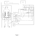
- a hybrid medical laparoscopic simulator represented on FIG. 1 includes: robot-patient 1, ECM (electronic computing machine) 2, laparoscopic grasper imitators 3, which are the laparoscopic instruments imitators, coupled to the ECM 2 through interface unit 4, endoscope imitator 5, which is the laparoscopic instrument imitator, coupled to the ECM 2 through endovideocamera control unit 6 and interface unit 4, aspirator-irrigator imitator 7, which is the laparoscopic instrument imitator, coupled to the ECM 2 through aspirator-irrigator control unit 8 and interface unit 4, insufflator pipe 9 coupled to insufflator control unit 10, trocar imitators 11, with their quantity of more than three (four in this execution), installed in the abdominal cavity of the robot-patient 1 and coupled to the ECM 2, visualization system 12 coupled to the ECM 2, interface unit 4 coupling the ECM 2 to the laparoscopic graspers imitators 3, coagulator control unit 13, which is one of the laparoscopic unit imitators, endovideocamera control unit 6, which is one of the lapar
- FIG. 1 represents the operating room equipment in the form of: operating table 14, surgical stand 15, and side table 16.
- FIG. 1 represents laparoscopic instrument imitators in the form of: laparoscopic grasper imitators 3, endoscope imitator 5, and aspirator-irrigator imitator 7.
- FIG. 1 represents the laparoscopic unit imitators in the form of endovideocamera control unit 6, aspirator-irrigator control unit 8, insufflator control unit 10 with insufflator pipe 9, coagulator control unit 13, pedals of coagulator 17, and a pedal of aspirator-irrigator 18.
- Trocar imitators 11 are located in the abdominal cavity of the robot-patient 1, robot-patient 1 is located on the operating table 14, visualization system 12 is coupled to the ECM 2, interface unit 4 is coupled to the ECM 2, laparoscopic graspers imitators 3 are coupled to the interface unit 4, endoscope imitator 5 is coupled to the endovideocamera control unit 6, aspirator-irrigator imitator 7 is coupled to the aspirator-irrigator control unit 8, and insufflator pipe 9 is coupled to the insufflator control unit 10. Laparoscopic grasper imitators 3, endoscope imitator 5, aspirator-irrigator imitator 7, and insufflator pipe 9 are located on the side table 16.
- Coagulator control unit 13 endovideocamera control unit 6, aspirator-irrigator control unit 8, and insufflator control unit 10 are coupled to the interface unit 4.
- Pedals of coagulator 17 are coupled to the coagulator control unit 13 and a pedal of aspirator-irrigator 18 is coupled to the aspirator-irrigator control unit 8.
- the circuit design of the ECM 2 could be implemented as IntelCorei7 processor with the frequency of 3500 MHz, guitarist RAM such as DDR3 with the storage space of 8 GB, NVIDIA Ge Force GTX560 video card with the storage space of 2 GB, Seagate hard disk drive with the storage space of 500 GB, and Microsoft Windows 7 Processional operating system.
- ECM 2 contains a pointing device allowing input the data to the program running in the processor of the ECM 2.
- Visualization system 12 can be realized on the model of TS 1716L-6(S/U) 17" LCD, Neovo X - 19AV White display, supplied by ELLIPS Partner LLC.
- Operating table 14 can be realized on the model of StarTech 3008C supplied by the Delrus Kazan LLC.
- Surgical stand 15 contains 5 shelves and can be realized on the model of Spya-03-05-KMT supplied by FinStar LLC.
- Side table 16 can be realized on the model of Goose table supplied by Belaya Mebel LLC.
- Pedals of coagulator 17 can be realized on the model of two-keyed pedal for ESHF A-001 supplied by ELLIPS Partner LLC.
- a pedal of aspirator-irrigator 18 can be realized on the model of one-keyed pedal of aspirator-irrigator supplied by ELLIPS Partner LLC.
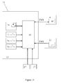
- Laparoscopic grasper imitator 3 which is a laparoscopic instrument imitator, represented on FIG. 2 drawing includes: branches 19, rod 20, sensor unit 21, handle 22, and instrument wing nut 23. Branches 19 are coupled to the rod 20, rod 20 is coupled to the sensor unit 21, handle 22 is coupled to the instrument wing nut 23, and instrument wing nut 23 is coupled to the sensor unit 21.
- Sensor unit 21 of the laparoscopic instrument imitator represented on FIG. 3 drawing includes: instrument wing nut 23, encoder 24, encoder pulley 25, sensor unit body frame 26, belt 27, pipe 28, bearing 29, handle pulley 30, lock ring 31, tie 32, stroke limitation cage 33, magnetic head 34, Hall sensor 35, infrared LED 36, and sensor unit microcontroller 37. Grooves for installation of encoder 24, Hall sensor 35, infrared LED 36, sensor unit microcontroller 37, handle pulley 30, encoder pulley 25, and bearing 29 are provided in the sensor unit body frame 26.
- Pipe 28 is coupled to the bearing 29, tie 32 is clenched with the lock ring 31, magnetic head 34 is fixed on the end of the tie 32, handle pulley 30 is set onto the pipe 28, encoder pulley 25 is set on the axis of the encoder 24, and encoder pulley 25 and handle pulley 30 are coupled with the belt 27.
- Hall sensor 35, encoder 24, infrared LED 36, and sensor unit microcontroller 37 are coupled to the sensor unit body frame 26.
- Encoder 24 can be realized on the model of LIR212A, produced by SKB IS, Saint-Petersburg.
- Trocar imitator 38 is coupled to the laparoscopic instrument receptacle 39, trocar imitator body frame 38 is coupled to the holding rollers 40, trocar imitator body frame 38 is coupled to the trocar imitator body frame shaft 41, trocar imitator body frame 38 is coupled to the instrument longitudinal motion detection sensor 42, the instrument longitudinal motion detection sensor 42 is coupled to the instrument longitudinal motion detection sensor shaft 43, shaft bearing 44 is coupled to the trocar imitator body frame shaft 41, shaft bearing 44 is coupled to the movable angle bar 45, Y-direction rotation sensor 46 is coupled to the trocar imitator body frame shaft 41, Y-direction rotation sensor 46 is coupled to the Y-direction rotation sensor catch 47, Y-direction rotation sensor catch 47 is coupled to the movable angle bar 45, movable angle bar 45 is coupled to the movable angle bar shaft 48, movable angle bar shaft 48 is coupled to the X-direction rotation sensor 49, movable angle bar shaft 48 is coupled to the movable angle bar bearing 50, holding body frame 51
- sensor unit microcontroller 37 which can be realized on the model of AtMega8and is located in the sensor unit 21 represented on FIG. 3 , encoder 24, which can be executed on the model of LIR212, Hall sensor 35, which can be executed on the model of SS49, infrared LED 36, and interface unit microcontroller 68.
- Sensor unit microcontroller 37 is coupled to the Hall sensor, the encoder 24, the interface unit microcontroller 68, and the infrared LED 36 respectively.
- the microcontroller of displacement node of the trocar imitator 66 1 is coupled to the instrument longitudinal motion detection sensor 42, the Y-direction rotation sensor 46, the X-direction rotation sensor 49, the guideway longitudinal motion sensor 55, the guideway rotation sensor 58, and the infrared detector 52.
- Every microcontroller of displacement node of the trocar imitator 66 2 -66 4 is realized analogously to the microcontroller of displacement node of the trocar imitator 66 1 and is coupled to the outputs of the instrument longitudinal motion detection sensor 42, the Y-direction rotation sensor 46, the X-direction rotation sensor 49, the guideway longitudinal motion sensor 55, the guideway rotation sensor 58, and the infrared detector 52 respectively (are not represented on FIG. 10 ).
- Coagulator control unit microcontroller 69 is coupled to the pedals of the coagulator 17, the coagulator's current power controller 74, the coagulation mode switch 75, the cutting mode switch 76, the blend mode switch 77, the interface unit microcontroller 68, and the sensor unit microcontroller 37.
- Coagulator's current power controller 74, coagulation mode switch 75, cutting mode switch 76, and blend mode switch 77 are located on the coagulator control unit body frame 78.
- Coagulator control unit microcontroller 69 is located inside the coagulator control unit body frame 78.
- Endoscope imitator 5 which is the laparoscopic instrument imitator, represented on FIG. 18 drawing includes: rod 20, sensor unit 21,and instrument wing nut 23.
- Rod 20 is coupled to the sensor unit 21 and instrument wing nut 23 is coupled to the sensor unit 21.
- Principal scheme of the aspirator-irrigator control unit 8 coupled to the interface unit 4 represented on FIG. 20 includes: aspirator-irrigator control unit 8, interface unit 4, aspirator-irrigator control unit microcontroller 71, a pedal of the aspirator-irrigator 18, aspirator-irrigator switch 87, aspiration mode switch 88, irrigation mode switch 89, opening and closing switch 90, interface unit microcontroller 68, and aspirator-irrigator control unit body frame 91.
- Aspirator-irrigator control unit microcontroller 71 is coupled to the pedal of the aspirator-irrigator 18, the aspirator-irrigator switch 87, the aspiration mode switch 88, the irrigation mode switch 89, the opening and closing switch 90, and the interface unit microcontroller 68.
- Principal scheme of the insufflator control unit 10 coupled to the interface unit 4 represented on FIG. 21 includes: insufflator control unit 10, interface unit 4, insufflator control unit microcontroller 72, interface unit microcontroller 68, insufflator switch 92, insufflation start button 93, pressure increase switch 94, pressure decrease switch 95, pressure memorization switch 96, flow increase switch 97, flow decrease switch 98, flow memorization switch 99, set pressure display 100, measured pressure display 101, set flow display 102, measured flow display 103, and insufflator control unit body frame 104.
- Guideway 54 allows the surgeon to set the required for the work position of the trocar imitators 11. The surgeon slackens the displacement node wing nut 67, chooses the rotation angle and the distance of the trocar imitators 11 form the node, then tightens the displacement node wing nut 67. Rotation of the guideway 54 is communicated through the guideway pulley 59 and sensor pulley 64 to the guideway rotation sensor 58. Guideway longitudinal motion sensor 55 defines longitudinal motion of the guideway 54. Thus the position of the trocar imitators 11 on the anterior wall of the abdominal cavity of the robot-patient 1 is defined.
- Trocar imitators 11 are installed into the robot-patient 1 and are located in its abdominal cavity. There are four movable trocar imitators 11 in this execution of the hybrid medical laparoscopic simulator. According to the type of the endosurgical intervention, different positions of the trocar imitators 11 are being set.
- Interface unit microcontroller 68 consequently requests the data from n-number of the sensor unit microcontrollers 37 (n is the number of the coupled laparoscopic instrument imitators), the endovideocamera control unit microcontroller 70, the aspirator-irrigator control unit 71, insufflator control unit microcontroller 72, and coagulator control unit microcontroller 69 through the TWI interface.
- Interface unit microcontroller 68 converts the received data to data bursts detected by the program on the ECM 2. The data are being communicated to the ECM 2 through RS232 COM-port.
- This execution of the hybrid medical laparoscopic simulator contains 4 movable trocar imitators 11 and 4 microcontrollers of displacement nodes of the trocar imitators 66 represented on FIG. 10 correspondingly.
- the main microcontroller of displacement node of the trocar imitators 66 4 (master) consequently requests the data via SPI interface from the other three displacement node of the trocar imitators 66 1 -66 3 , converts all the data received into one data burst recognized by the program of the ECM 2, and through the RS232 serial port communicates the data to the ECM 2.
- Data from the sensor unit microcontroller 37 located in the endoscope imitator 5 that is the laparoscopic instrument imitator are being communicated to the microcontroller of the endovideocamera control unit 70 through TWI interface.
- the microcontroller of the endovideocamera control unit 70 is communicating the collected data to the interface unit microcontroller 68 through TWI interface.
- the illuminance controller 81 On pushing the illuminance controller 81 the setting values are being displayed on the brightness display 85.
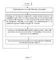
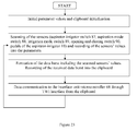
- the laparoscopic instrument imitator is being inserted in the trocar imitator 11 and the minimum and maximum value of each calibrated sensor is being fixed sequentially. For instance, first of all for the instrument longitudinal motion detection sensor 42 calibration, the laparoscopic instrument imitator is inserted in the trocar imitator 11 all the way in, an appropriate minimum position fixation mode of the sensor is chosen in the dialog box with the manipulator of the ECM 2, then, the laparoscopic instrument imitator is taken-out incompletely from the trocar imitator 11 and an appropriate maximum position fixation mode of the sensor is chosen in the dialog box. Values of the calibration factors are saved by the program to the data base of the EMC 2 automatically in accordance with the algorithm represented on FIG.
- Surgical team actions while working with the hybrid medical laparoscopic simulator are following: a nurse gives and takes instruments of a surgeon and his/her assistant at their request, connects the laparoscopic instrument imitators to the corresponding control units if necessary, aligns the equipment according to the surgeon's request, and provides support to the surgeon and his/her assistant.
- the surgeon guides the intervention process and accepts important decisions.
- the assistant holds the endoscope imitator 5 with one hand and the laparoscopic grasper 3 by the other hand as a rule, helping the surgeon to hold the virtual organs for comfortable execution of the intervention.
- Extra specialists' participation is possible on inserting in the simulator other types of operating room equipment, laparoscopic unit imitator, and other units. For instance, the participation of an anesthesiologist is necessary when an imitator of a stand type anesthetic unit is on.
- 3D models of the tissues and organs are simulated in Autodesk 3ds Max system, developed by Autodesk company, inserted in the data base of the ECM 2.
- the physics module is software developed with use of SDK PhysX of nVidia company.
- the graphic representation module is software developed with use of SDK DirectX of Microsoft company.
- the surgeon connects the insufflator pipe 9 to the trocar valve 53 on the insufflators control unit 10, and the nurse (or the surgeon) sets the pressure value with the pressure increase switch 94 and the pressure decrease switch 95 and presses the pressure memorization switch 96 over-watching the set pressure display 100.
- the endoscope imitator 5, which is a laparoscopic instrument imitator, is being inserted.
- the inserted endoscope imitator 5 through the infrared LED 36 sends the signal with the instrument code to the infrared detector 52 of the trocar imitator 11.
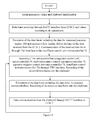
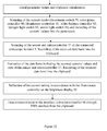
- the visualization system 12 displays the video data generated by the graphic representation module according to the position of the virtual endoscope by the algorithm represented on FIG. 12 block diagram.
- the nurse sets up the image quality on the endovideocamera control unit 6.
- the surgeon examines virtual organs and based on the received video data installs the other trocar imitators 11, gives the endoscope imitator 5 to the assistant, weakens the displacement node wing nut 67 for each trocar imitator 11, sets the necessary position of the trocar imitators 11, fixes the displacement node wing nut 67, and inserts the corresponding laparoscopic instrument imitator.
- the physics module simulates the physical properties of the laparoscopic instrument imitator on insertion of the each laparoscopic instrument imitator in the trocar imitator 11: firmness, volume, electrical discharges, extravasations, cutting surfaces, strains, space position, and interaction with the virtual organs and tissues; by the algorithm represented on FIG. 12 block diagram, the graphic representation module generates the image of each virtual laparoscopic instrument among the virtual organs and tissues according to the position of the virtual laparoscopic instruments.
- the physics module simulates the physical properties of the virtual organs analogous to the real ones and forms the anatomic variant including the size, form, color, location of a gall-bladder, bile ducts, arteries, etc.
- one of the variants of the 3D computer model of the organs is being loaded from the database of the ECM 2 including the description of the organ surface (the surface is set by the points in 3D space, point surface normals, and connections between the points), of the textures (visual reflection of the imitated organs superimposed onto the 3D surface), and of the properties (elasticity, fragility, etc).
- the conversion of the loaded data into the structures necessary for deformation and dissection simulation is performed.
- Cholecystectomy intervention training could be performed both stage-by-stage and completely.
- Stage-by-stage mode Separate training of a selected intervention stage (traction, Calot's triangle preparation, clipping and the transection of the cystic duct and artery, and gall-bladder mobilization).
- the physics module initializes in the database of the ECM 2 the physical models of organs in conditions relevant to the completed stages previous to the selected stage and sends a signal to the graphic representation module forming the 3D image of the virtual organs being sent to the visualization system 12.
- the physics module modifies the 3D surfaces simulating the organs (for example, the performed traction, dissection and clips applied) by the algorithm represented on FIG. 12 block diagram.
- the physics module initializes in the database of the ECM 2 the physical models of organs in initial conditions (as they are located and deformed in the body of a real patient being laid on the operation table) and the graphic representation module forms the 3D image being sent to the visualization system 12 considering the data received from the physics module and other modules represented on FIG. 12 block diagram.
- the logic module defines the attributes of the start and end of the next stage including the definition of the intervention stages failure. The exercise is over when all the intervention stages are completed.
- the surgeon or assistant should perform the traction - gallbladder virtual model displacement for provision of an access to the area being operated.
- the assistant inserts the laparoscopic grasper imitator 3 in the trocar imitator 11, performs manipulations with the laparoscopic grasper imitator 3 displayed in the visualization system 12 in the form of a virtual instrument, captures the gallbladder fundus, and displaces the virtual gallbladder with a laparoscopic grasper imitator 3, herein the physics module figures out the deformation of the 3D surface of the virtual gallbladder and its influence on the other virtual organs.
- the physics module estimates the current condition of the virtual organs surfaces a few times per second and the graphic representation module generates a signal incoming to the visualization system 12.
- the logic module defines the accuracy of the traction direction (motion pattern is arranged on grounds of the calculated coordinates of the laparoscopic instrument imitator, the pattern is compared to the sample variant stored in the database of the ECM 2, and permissible variations are considered), the correctness of the area being operated visualization (the location of the virtual gallbladder and virtual vessels is estimated on the base of the coordinates of the surface points of the virtual organs and is compared to the sample variant stored in the database of the ECM 2, and permissible variations are considered), and correctness of the virtual organs grasp (the area of the grasp with the laparoscopic instruments is being estimated, the correspondence of the grasping zone to the permissible limits, and the instrument code is being compared to the allowed instrument codes stored in the database of the ECM 2).
- the surgeon has to dissect the abdominal membrane and the adipose tissue to allocate (to visualize) the tubular formations (artery and duct) to be clipped and transected.
- the surgeon inserts one of the laparoscopic instruments imitator, for instance, a dissector imitator and a hook imitator which are the laparoscopic instruments imitators, and the nurse hooks up the connector of the instrument to the coagulator control unit 13 and sets up the necessary mode and rate of the current with the corresponding switches (coagulation mode switch 75, cutting mode switch 76, a blend mode switch, and coagulator's current power controller 74) according to the surgeon's request.
- switches coagulation mode switch 75, cutting mode switch 76, a blend mode switch, and coagulator's current power controller 74
- the values of the mode and rate of the currents are being communicated to the ECM 2.
- the surgeon grasps the virtual adipose tissue, pulls it back, then dissects by pressing the pedal of coagulator 17 (the signal of the pressing the pedal of coagulator 17 is received by the ECM 2, and the physics module generates the virtual current flow through the virtual tissues).
- the physics module figures out what areas of the tissue are in contact with the virtual branches according to the coordinates of the virtual branches of the virtual instrument and coordinates of the surface points of the virtual tissue, and if the contact condition is fulfilled then the current flow through these areas of the tissue will be imitated, in the visualization system 12 the smoke and particles of the dissected tissue moving randomly are reflected, for the period of 1-3 seconds the full dissection of the contiguous areas of the virtual tissue, at that they disappear smoothly on the image of the visualization system 12.
- the physics module figures out changes of the 3D surface of the virtual vessel as a result of the virtual incision (the graphic representation module reflects the changes in the visualization system 12).
- the logic module defines the correctness of the applied clip location (the application area is being compared to the sample variants of the allowed application area), the quantity of the clips, and the correctness of the places of tubular formations transection (the transaction area is being compared to the sample variant of the allowed transaction areas) by the algorithm represented on FIG. 12 block diagram and description of the actions mentioned below.
- the surgeon inserts the laparoscopic grasper imitator 3 in the second trocar imitator 11 (for the left hand), manipulates it, and grasps and pulls back the virtual gallbladder (the physics module figures out the corresponding surface deformations of the gallbladder and movement and deformation of other virtual organs depending on the movement of the virtual laparoscopic instruments).
- the surgeon manipulates the laparoscopic instrument imitators, brings the virtual hook to the adhesion place of the virtual gallbladder and the virtual liver, and presses the pedal of the coagulator 17.
- a 43-year-old female whose ultrasonography shows a tumorous cavitary lesion, is presented with acute pain in iliac region (clinical case of oothecoma torsion); and a 30-year-old female is admitted to the hospital with acute lower abdominal pain in the left inguinal region, diagnostic laparoscopy due to the left appendages torsion is in the past history, detorsion of the left appendages with their preserving is performed.
- the surgeon has to decide how to place the robot-patient 1 on the operating table 14 and location of the trocar imitators 11, for example, considering the cicatrization following the previous interventions.
- the robot-patient 1 is being located on the operating table 14.
- the position of the first trocar imitator 11 where the endoscope imitator 5, which is the laparoscopic instrument imitator, is to be inserted is being chosen by the weakening of the displacement node wing nut 67, the position of the trocar imitator 11 in the abdominal cavity of the robot patient 1 relevant to the position during the real intervention is being set, then the displacement node wing nut 67 is being tightened.
- the visualization system 12 displays the video data generated by the graphic representation module according to the position of the virtual endoscope by the algorithm represented on FIG. 12 block diagram.
- the nurse sets up the image quality on the endovideocamera control unit 6.
- the surgeon examines virtual organs and on the base of the received video data installs the other trocar imitators 11, gives the endoscope imitator 5 to the assistant, weakens the displacement node wing nut 67 for each trocar imitator 11, sets the necessary position of the trocar imitator 11, fixes the displacement node wing nut 67 and inserts the corresponding laparoscopic instrument imitator.
- the logic module defines the accuracy of the traction direction (motion pattern is arranged on grounds of the calculated coordinates of the laparoscopic instrument imitator, the pattern is compared to the sample variant stored in the database of the ECM 2, and permissible variations are considered), rough or excessive displacement of the virtual fallopian tube (the logic module figures out the displacement speed and distance on the basis of the change of the laparoscopic instrument imitator coordinates, being communicated few times per second (20-50), meanwhile the physics module figures out the 3D surface deformation that is reflected by the graphic representation module in the visualization system 12, calculates the stretch ratio, the logic module compares it the sample variant stored in the database of the ECM 2), incorrect overlapping of the laparoscopic grasper imitator 3 (the physics module figures out the overlapping area on the basis of the virtual laparoscopic grasper's branches coordinates, the logic module compares it the sample variant stored in the database of the ECM 2), incorrect instrument choice (the logic module compares the instrument code to the allowed instrument codes in the database of the ECM 2), damage of the abdominal
- the surgeon has to coagulate and dissect the virtual mesosalpinx, pulled back with the virtual laparoscopic grasper, overlapped to the tube, its edge, the lateral and bottom edge of the virtual ovary.
- the surgeon inserts the laparoscopic grasper imitator 3 to the one trocar imitator 11, and the coagulator imitator to the other one.
- the surgeon grasps and pulls back the virtual mesosalpinx with the laparoscopic grasper imitator 3, overlaps the forceps of the virtual coagulator, presses the pedal of the coagulator 17, and holds down for 1-2 seconds.
- the surgeon takes out the coagulator imitator and inserts the laparoscopic scissors imitator instead of it, approaches the branches 19 of the laparoscopic scissors imitator, the actions of which are coordinated with the virtual ones, to the coagulated area of the virtual tissue, and dissects it pressing the handle 22 that is reflected in the visualization system 12 (the physics module figures out the dissection area on the base of the coordinated of the virtual branches at the moment of dissection, the relevant alterations are made in the 3D surface, and data are saved into the database of the ECM 2.).
- the logic module figures out the incorrect lesion identification of the virtual fallopian tube, the utero-ovarian ligament, and the infundibulopelvic ligament (the logic module figures out the contact area and compares it to the sample variants stored in the database of the ECM 2when signals of coagulation or dissection of the virtual tissue areas arrive, if the contact areas do not coincide with the sample variants regarding the permissible deviations, the it will generate a text message coming through the graphic representation module to the visualization system 12 informing that the surgeon started the surgical intervention having identified the anatomical structures incorrectly), coagulation being performed too close to the pelvic walls (if the logic module identifies the crossing of the coagulation area with the surface area stored in the database of the ECM 2 as the virtual pelvic wall, or if the distance to this area is less than the minimum
- the surgeon examines all the area being operated with the virtual endoscope coordinated to the real endoscope imitator 5. If any damages are found, then the surgeon will performed the coagulation in a manner described above.
- the logic module analyzes the examination for bleeding (the track of the virtual endoscope movement and stop time for every area of the area being operated are saved to the database of the ECM 2, the logic module compares it to the sample variants in the database considering the permissible deviations) and the damage of the organs nearby.
Landscapes
- Engineering & Computer Science (AREA)
- General Physics & Mathematics (AREA)
- Physics & Mathematics (AREA)
- Health & Medical Sciences (AREA)
- Mathematical Analysis (AREA)
- Mathematical Optimization (AREA)
- Medical Informatics (AREA)
- Medicinal Chemistry (AREA)
- Chemical & Material Sciences (AREA)
- Algebra (AREA)
- Computational Mathematics (AREA)
- Theoretical Computer Science (AREA)
- Educational Technology (AREA)
- General Health & Medical Sciences (AREA)
- Mathematical Physics (AREA)
- Pure & Applied Mathematics (AREA)
- Business, Economics & Management (AREA)
- Educational Administration (AREA)
- Pulmonology (AREA)
- Radiology & Medical Imaging (AREA)
- Endoscopes (AREA)
- Surgical Instruments (AREA)
- Instructional Devices (AREA)
- Manipulator (AREA)
Applications Claiming Priority (2)
| Application Number | Priority Date | Filing Date | Title |
|---|---|---|---|
| RU2012148301 | 2012-11-13 | ||
| PCT/RU2013/000419 WO2014077732A1 (ru) | 2012-11-13 | 2013-05-21 | Гибридный медицинский тренажер лапароскопии |
Publications (2)
| Publication Number | Publication Date |
|---|---|
| EP2922048A1 true EP2922048A1 (de) | 2015-09-23 |
| EP2922048A4 EP2922048A4 (de) | 2015-10-07 |
Family
ID=50731519
Family Applications (1)
| Application Number | Title | Priority Date | Filing Date |
|---|---|---|---|
| EP13855297.1A Withdrawn EP2922048A4 (de) | 2012-11-13 | 2013-05-21 | Hybrider laparoskopischer simulator |
Country Status (5)
| Country | Link |
|---|---|
| US (1) | US20160098943A1 (de) |
| EP (1) | EP2922048A4 (de) |
| JP (1) | JP2016500157A (de) |
| EA (1) | EA027466B1 (de) |
| WO (1) | WO2014077732A1 (de) |
Cited By (1)
| Publication number | Priority date | Publication date | Assignee | Title |
|---|---|---|---|---|
| WO2019171339A1 (en) * | 2018-03-09 | 2019-09-12 | Laparo Sp. Z O.O. | Working tool and manipulation and measurement set of laparoscopic trainer |
Families Citing this family (15)
| Publication number | Priority date | Publication date | Assignee | Title |
|---|---|---|---|---|
| WO2016069660A1 (en) | 2014-10-27 | 2016-05-06 | Intuitive Surgical Operations, Inc. | System and method for monitoring control points during reactive motion |
| KR102655083B1 (ko) | 2014-10-27 | 2024-04-08 | 인튜어티브 서지컬 오퍼레이션즈 인코포레이티드 | 브레이크 해제가 능동적으로 제어되는 의료 장치 |
| EP3212150B1 (de) * | 2014-10-27 | 2021-08-11 | Intuitive Surgical Operations, Inc. | System zur registrierung an einem operationstisch |
| JP6676061B2 (ja) | 2014-10-27 | 2020-04-08 | インテュイティブ サージカル オペレーションズ, インコーポレイテッド | 統合された手術台運動のためのシステム及び方法 |
| JP6682512B2 (ja) | 2014-10-27 | 2020-04-15 | インテュイティブ サージカル オペレーションズ, インコーポレイテッド | 一体化された手術台のシステム及び方法 |
| CN110584789B (zh) | 2014-10-27 | 2022-09-20 | 直观外科手术操作公司 | 用于器械干扰补偿的系统和方法 |
| JPWO2017126313A1 (ja) * | 2016-01-19 | 2018-11-22 | 株式会社ファソテック | 生体質感臓器を用いる手術トレーニング及びシミュレーションシステム |
| US10255829B2 (en) * | 2016-10-10 | 2019-04-09 | Medtronic Holding Company Sàrl | In-situ training apparatus, method and system |
| WO2018118858A1 (en) | 2016-12-19 | 2018-06-28 | National Board Of Medical Examiners | Medical training and performance assessment instruments, methods, and systems |
| KR101887805B1 (ko) * | 2017-03-23 | 2018-08-10 | 최재용 | 증강현실 기반의 복강경 수술용 시뮬레이션 시스템 및 이를 이용한 방법 |
| WO2019051005A1 (en) * | 2017-09-08 | 2019-03-14 | Covidien Lp | HIGH PRECISION INSTRUMENT CONTROL MODE FOR ROBOTIC SURGICAL SYSTEMS |
| FR3073657B1 (fr) * | 2017-11-10 | 2023-05-05 | Virtualisurg | Systeme de simulation d'acte chirurgical |
| KR102143784B1 (ko) * | 2018-12-27 | 2020-08-12 | 가톨릭대학교 산학협력단 | 가상현실 기반 이비인후과 및 신경외과 시뮬레이터의 수술 평가 시스템 |
| WO2020257757A1 (en) * | 2019-06-21 | 2020-12-24 | The Board Of Supervisors Of Louisiana State University And Agricultural And Mechanical College | Surgical simulator and methods of use |
| JP2024528855A (ja) * | 2021-07-30 | 2024-08-01 | アプライド メディカル リソーシーズ コーポレイション | 外科訓練のための婦人科病理学手術シミュレーションモデル及びシステム |
Family Cites Families (22)
| Publication number | Priority date | Publication date | Assignee | Title |
|---|---|---|---|---|
| US5403191A (en) * | 1991-10-21 | 1995-04-04 | Tuason; Leo B. | Laparoscopic surgery simulator and method of use |
| US5352219A (en) * | 1992-09-30 | 1994-10-04 | Reddy Pratap K | Modular tools for laparoscopic surgery |
| US5791231A (en) * | 1993-05-17 | 1998-08-11 | Endorobotics Corporation | Surgical robotic system and hydraulic actuator therefor |
| US5830214A (en) * | 1994-11-08 | 1998-11-03 | Heartport, Inc. | Fluid-evacuating electrosurgical device |
| WO1996016389A1 (en) * | 1994-11-17 | 1996-05-30 | Staneff John E Jr | Medical procedure simulator |
| RU2096997C1 (ru) * | 1995-05-03 | 1997-11-27 | Джейкоб Фелдман | Устройство для эндохирургических манипуляций |
| US6113395A (en) * | 1998-08-18 | 2000-09-05 | Hon; David C. | Selectable instruments with homing devices for haptic virtual reality medical simulation |
| US6544041B1 (en) * | 1999-10-06 | 2003-04-08 | Fonar Corporation | Simulator for surgical procedures |
| US20050142525A1 (en) * | 2003-03-10 | 2005-06-30 | Stephane Cotin | Surgical training system for laparoscopic procedures |
| JP2004348095A (ja) * | 2003-03-26 | 2004-12-09 | National Institute Of Advanced Industrial & Technology | トレーニングシステム |
| US8403675B2 (en) * | 2004-03-08 | 2013-03-26 | The Johns Hopkins University | Device and method for medical training and evaluation |
| US7621926B2 (en) * | 2004-04-16 | 2009-11-24 | Applied Medical Resources Corporation | Multi-fire surgical clip applier |
| US20070275359A1 (en) * | 2004-06-22 | 2007-11-29 | Rotnes Jan S | Kit, operating element and haptic device for use in surgical simulation systems |
| RU60768U1 (ru) * | 2006-08-30 | 2007-01-27 | Константин Валерьевич Бражников | Лапароскопический тренажер |
| US20080085499A1 (en) * | 2006-10-05 | 2008-04-10 | Christopher Horvath | Surgical console operable to simulate surgical procedures |
| WO2009000939A1 (es) * | 2007-06-22 | 2008-12-31 | Gmv, S.A. | Simulador para cirugía laparoscópica |
| US9171484B2 (en) * | 2008-03-06 | 2015-10-27 | Immersion Corporation | Determining location and orientation of an object positioned on a surface |
| JP2009236963A (ja) * | 2008-03-25 | 2009-10-15 | Panasonic Electric Works Co Ltd | 内視鏡手術用トレーニング装置、内視鏡手術用技能評価方法 |
| US20090263775A1 (en) * | 2008-04-22 | 2009-10-22 | Immersion Medical | Systems and Methods for Surgical Simulation and Training |
| US20100167250A1 (en) * | 2008-12-31 | 2010-07-01 | Haptica Ltd. | Surgical training simulator having multiple tracking systems |
| JP5614980B2 (ja) * | 2009-12-25 | 2014-10-29 | 三菱プレシジョン株式会社 | トロカー位置設定用模擬術具位置設定装置 |
| CA2811235C (en) * | 2010-10-01 | 2020-03-10 | Applied Medical Resources Corporation | Portable laparoscopic trainer |
-
2013
- 2013-05-21 US US14/442,538 patent/US20160098943A1/en not_active Abandoned
- 2013-05-21 JP JP2015541738A patent/JP2016500157A/ja active Pending
- 2013-05-21 EA EA201590789A patent/EA027466B1/ru not_active IP Right Cessation
- 2013-05-21 WO PCT/RU2013/000419 patent/WO2014077732A1/ru not_active Ceased
- 2013-05-21 EP EP13855297.1A patent/EP2922048A4/de not_active Withdrawn
Cited By (5)
| Publication number | Priority date | Publication date | Assignee | Title |
|---|---|---|---|---|
| WO2019171339A1 (en) * | 2018-03-09 | 2019-09-12 | Laparo Sp. Z O.O. | Working tool and manipulation and measurement set of laparoscopic trainer |
| PL424841A1 (pl) * | 2018-03-09 | 2019-09-23 | Laparo Spółka Z Ograniczoną Odpowiedzialnością | Człon manipulacyjno-pomiarowy trenażera laparoskopowego |
| CN111819611A (zh) * | 2018-03-09 | 2020-10-23 | 拉帕罗有限公司 | 腹腔镜训练器的作业工具以及操纵和测量套件 |
| CN111819611B (zh) * | 2018-03-09 | 2022-12-27 | 拉帕罗有限公司 | 腹腔镜训练器的作业工具以及操纵和测量套件 |
| US11610511B2 (en) | 2018-03-09 | 2023-03-21 | Laparo Sp. Z.O.O | Working tool and manipulation and measurement set of laparoscopic trainer |
Also Published As
| Publication number | Publication date |
|---|---|
| US20160098943A1 (en) | 2016-04-07 |
| EA201590789A1 (ru) | 2015-07-30 |
| JP2016500157A (ja) | 2016-01-07 |
| WO2014077732A1 (ru) | 2014-05-22 |
| EP2922048A4 (de) | 2015-10-07 |
| EA027466B1 (ru) | 2017-07-31 |
Similar Documents
| Publication | Publication Date | Title |
|---|---|---|
| EP2922048A1 (de) | Hybrider laparoskopischer simulator | |
| RU128762U1 (ru) | Гибридный медицинский тренажер лапароскопии | |
| CN103886797B (zh) | 一种高度仿真腹腔镜手术模拟训练器 | |
| CN102341046B (zh) | 利用增强现实技术的手术机器人系统及其控制方法 | |
| JP7526835B2 (ja) | 全直腸間膜切除手術シミュレータ | |
| JPH11197159A (ja) | 手術支援システム | |
| JP2023090809A (ja) | 手術内容評価システム、手術内容評価方法及びコンピュータプログラム | |
| CN105448155A (zh) | 脊柱内镜虚拟训练系统 | |
| Yuh | The Bedside Assistant in Robotic Surgery-Keys to Success. | |
| JP7355514B2 (ja) | 医用画像処理装置、医用画像処理方法、及び医用画像処理プログラム | |
| JPWO2017126313A1 (ja) | 生体質感臓器を用いる手術トレーニング及びシミュレーションシステム | |
| Hoznek et al. | Laparoscopic and robotic surgical training in urology | |
| Den Boer et al. | Peroperative analysis of the surgical procedure | |
| Singapogu et al. | A perspective on the role and utility of haptic feedback in laparoscopic skills training | |
| CN205862668U (zh) | 一种真实腔镜微创手术仿真训练系统 | |
| Wang et al. | Modelling and application of laparoscopic simulation system for panhysterectomy | |
| RU2205599C2 (ru) | Способ определения параметров операционного доступа к желчному пузырю для лапароскопической холецистэктомии | |
| CN114424974B (zh) | 基于气腹仿真对腹腔镜操作孔的定位方法与系统 | |
| Klein et al. | LAP | |
| Feussner et al. | Hybrid surgery-the way towards notes the challenge for computer science | |
| Chekan et al. | Performance-Guided Surgery: Digital Solutions and the Senhance Surgical System | |
| Bachur et al. | Major Vascular Injuries in Laparoscopic Urological Surgeries | |
| Grimbergen | Karen T. den Boer, Dirk J. Gouma, Cornells A. Grimbergen, and Jenny Dankelman | |
| Shu et al. | Robotic-Assisted Segmentectomy | |
| Grimbergen et al. | of the Surgical Process |
Legal Events
| Date | Code | Title | Description |
|---|---|---|---|
| PUAI | Public reference made under article 153(3) epc to a published international application that has entered the european phase |
Free format text: ORIGINAL CODE: 0009012 |
|
| 17P | Request for examination filed |
Effective date: 20150602 |
|
| AK | Designated contracting states |
Kind code of ref document: A1 Designated state(s): AL AT BE BG CH CY CZ DE DK EE ES FI FR GB GR HR HU IE IS IT LI LT LU LV MC MK MT NL NO PL PT RO RS SE SI SK SM TR |
|
| AX | Request for extension of the european patent |
Extension state: BA ME |
|
| RA4 | Supplementary search report drawn up and despatched (corrected) |
Effective date: 20150904 |
|
| RIC1 | Information provided on ipc code assigned before grant |
Ipc: A61B 1/313 20060101ALI20150831BHEP Ipc: G09B 23/28 20060101ALI20150831BHEP Ipc: G09B 9/00 20060101AFI20150831BHEP |
|
| DAX | Request for extension of the european patent (deleted) | ||
| STAA | Information on the status of an ep patent application or granted ep patent |
Free format text: STATUS: EXAMINATION IS IN PROGRESS |
|
| 17Q | First examination report despatched |
Effective date: 20180213 |
|
| STAA | Information on the status of an ep patent application or granted ep patent |
Free format text: STATUS: THE APPLICATION HAS BEEN WITHDRAWN |
|
| 18W | Application withdrawn |
Effective date: 20180605 |