EP2946791B1 - Anti-CD277 antibodies and uses thereof - Google Patents
Anti-CD277 antibodies and uses thereof Download PDFInfo
- Publication number
- EP2946791B1 EP2946791B1 EP15151003.9A EP15151003A EP2946791B1 EP 2946791 B1 EP2946791 B1 EP 2946791B1 EP 15151003 A EP15151003 A EP 15151003A EP 2946791 B1 EP2946791 B1 EP 2946791B1
- Authority
- EP
- European Patent Office
- Prior art keywords
- cells
- antibody
- cancer
- cell
- mab
- Prior art date
- Legal status (The legal status is an assumption and is not a legal conclusion. Google has not performed a legal analysis and makes no representation as to the accuracy of the status listed.)
- Active
Links
Images



Classifications
-
- A—HUMAN NECESSITIES
- A61—MEDICAL OR VETERINARY SCIENCE; HYGIENE
- A61P—SPECIFIC THERAPEUTIC ACTIVITY OF CHEMICAL COMPOUNDS OR MEDICINAL PREPARATIONS
- A61P31/00—Antiinfectives, i.e. antibiotics, antiseptics, chemotherapeutics
-
- C—CHEMISTRY; METALLURGY
- C07—ORGANIC CHEMISTRY
- C07K—PEPTIDES
- C07K16/00—Immunoglobulins [IG], e.g. monoclonal or polyclonal antibodies
- C07K16/18—Immunoglobulins [IG], e.g. monoclonal or polyclonal antibodies against material from animals or humans
- C07K16/28—Immunoglobulins [IG], e.g. monoclonal or polyclonal antibodies against material from animals or humans against receptors, cell surface antigens or cell surface determinants
- C07K16/2896—Immunoglobulins [IG], e.g. monoclonal or polyclonal antibodies against material from animals or humans against receptors, cell surface antigens or cell surface determinants against molecules with a "CD"-designation, not provided for elsewhere
-
- A—HUMAN NECESSITIES
- A61—MEDICAL OR VETERINARY SCIENCE; HYGIENE
- A61P—SPECIFIC THERAPEUTIC ACTIVITY OF CHEMICAL COMPOUNDS OR MEDICINAL PREPARATIONS
- A61P35/00—Antineoplastic agents
-
- A—HUMAN NECESSITIES
- A61—MEDICAL OR VETERINARY SCIENCE; HYGIENE
- A61P—SPECIFIC THERAPEUTIC ACTIVITY OF CHEMICAL COMPOUNDS OR MEDICINAL PREPARATIONS
- A61P37/00—Drugs for immunological or allergic disorders
- A61P37/02—Immunomodulators
- A61P37/04—Immunostimulants
-
- A—HUMAN NECESSITIES
- A61—MEDICAL OR VETERINARY SCIENCE; HYGIENE
- A61P—SPECIFIC THERAPEUTIC ACTIVITY OF CHEMICAL COMPOUNDS OR MEDICINAL PREPARATIONS
- A61P37/00—Drugs for immunological or allergic disorders
- A61P37/02—Immunomodulators
- A61P37/06—Immunosuppressants, e.g. drugs for graft rejection
-
- C—CHEMISTRY; METALLURGY
- C07—ORGANIC CHEMISTRY
- C07K—PEPTIDES
- C07K16/00—Immunoglobulins [IG], e.g. monoclonal or polyclonal antibodies
- C07K16/18—Immunoglobulins [IG], e.g. monoclonal or polyclonal antibodies against material from animals or humans
- C07K16/28—Immunoglobulins [IG], e.g. monoclonal or polyclonal antibodies against material from animals or humans against receptors, cell surface antigens or cell surface determinants
- C07K16/2803—Immunoglobulins [IG], e.g. monoclonal or polyclonal antibodies against material from animals or humans against receptors, cell surface antigens or cell surface determinants against the immunoglobulin superfamily
- C07K16/2818—Immunoglobulins [IG], e.g. monoclonal or polyclonal antibodies against material from animals or humans against receptors, cell surface antigens or cell surface determinants against the immunoglobulin superfamily against CD28 or CD152
-
- C—CHEMISTRY; METALLURGY
- C07—ORGANIC CHEMISTRY
- C07K—PEPTIDES
- C07K16/00—Immunoglobulins [IG], e.g. monoclonal or polyclonal antibodies
- C07K16/18—Immunoglobulins [IG], e.g. monoclonal or polyclonal antibodies against material from animals or humans
- C07K16/28—Immunoglobulins [IG], e.g. monoclonal or polyclonal antibodies against material from animals or humans against receptors, cell surface antigens or cell surface determinants
- C07K16/2803—Immunoglobulins [IG], e.g. monoclonal or polyclonal antibodies against material from animals or humans against receptors, cell surface antigens or cell surface determinants against the immunoglobulin superfamily
- C07K16/2827—Immunoglobulins [IG], e.g. monoclonal or polyclonal antibodies against material from animals or humans against receptors, cell surface antigens or cell surface determinants against the immunoglobulin superfamily against B7 molecules, e.g. CD80, CD86
-
- C—CHEMISTRY; METALLURGY
- C07—ORGANIC CHEMISTRY
- C07K—PEPTIDES
- C07K2317/00—Immunoglobulins specific features
- C07K2317/50—Immunoglobulins specific features characterized by immunoglobulin fragments
- C07K2317/55—Fab or Fab'
-
- C—CHEMISTRY; METALLURGY
- C07—ORGANIC CHEMISTRY
- C07K—PEPTIDES
- C07K2317/00—Immunoglobulins specific features
- C07K2317/70—Immunoglobulins specific features characterized by effect upon binding to a cell or to an antigen
- C07K2317/73—Inducing cell death, e.g. apoptosis, necrosis or inhibition of cell proliferation
-
- C—CHEMISTRY; METALLURGY
- C07—ORGANIC CHEMISTRY
- C07K—PEPTIDES
- C07K2317/00—Immunoglobulins specific features
- C07K2317/70—Immunoglobulins specific features characterized by effect upon binding to a cell or to an antigen
- C07K2317/75—Agonist effect on antigen
Definitions
- the invention relates to anti-CD277 antibodies and uses thereof.
- White blood cells are cells of the immune system involved in defending the body against pathogens. Among these cells, lymphocytes, monocytes, and dendritic cells can be cited. Monocytes may migrate from the bloodstream to other tissues and differentiate into tissue resident macrophages or dendritic cells. Dendritic cells play a role as antigen presenting cells (APC) that activate lymphocytes. Among lymphocytes, T cells can be divided into ⁇ T cells and ⁇ T cells.
- V ⁇ 9/V ⁇ 2 T cells are important effectors of the immune defence. They lyse directly pathogen infected or abnormal cells. In addition, they regulate immune responses by inducing dendritic cell (DC) maturation as well as the isotypic switching and immunoglobulin production. This important cell platform of the immune system is strictly regulated by surface receptors, chemokines and cytokines.
- T-cell activation is the result of two synergistic events.
- the first is the interaction between the receptor of T cell (TCR) and the major histocompatibility complex (MHC) conjugated with processed antigen on the surface of the antigen presenting cells (APC).
- the second event is a co-stimulatory antigen-independent signal involving B7 molecules.
- the lack of co-stimulatory signal induces anergy, i.e. the inhibition of T cells proliferation, cytokines secretion and cytotoxic activities.
- the study of these pathways may provide insight about the triggering of pathologic events, such as autoimmune or lymphoproliferative disorders.
- the B7 family is an extended group of costimulatory molecules (Coyle et al., 2001).
- B7-1 CD80
- B7-2 CD86
- their receptors are CD28, which leads to T cell activation (Linsley and Ledbetter, 1993, June, et al., 1994, Lenschow et al., 1996)
- CTLA-4 CD152
- the critical role of CD152 as a negative regulator of T cell activation is demonstrated by the occurrence of lymphoproliferative disorders in CTLA-4 deficient mice (Waterhouse et al., 1995).
- CD152 is expressed following T-lymphocyte activation and inhibits the cytolytic functions of CTL clones obtained following PHA stimulation or Ag selection (Saverino et al., 1998).
- B7-H1 (PD-L1, CD274) and B7-DC (PD-L2, CD273), whose receptor is PD-1 (CD279), proved to inhibit T-cell proliferation and cytokine secretion (Freeman et al., 2000, Latchman et al., 2001). Otherwise, different studies showed that PD-L1 and PD-L2 engagement increase T cell proliferation and IL-10 or IFN- ⁇ production (Dong et al., 1999, Freeman et al., 2000, Latchman et al., 2001, Chapoval et al., 2001, Tseng et al., 2001).
- B7-H2 B7-H2
- B7-H3, B7-H4 B7-H3 whose roles are not fully understood (Hutloff et al., 1999, Sun et al., 2002).
- BT butyrophilin
- Bt2 and Bt3 coding for a new group of co-stimulatory molecules belonging to the Ig superfamily (IgSF) (Williams and Barclay, 1988) and related to B7 family by sequence similarity analysis: in particular, it shows similarity with the Ig-V like extracellular domain of CD80 and CD86 (Linsley et al., 1992).
- IgSF Ig superfamily
- BT3.1 is also called BTF5 (Rhodes et al., 2001), or BTN3A1 (Ruddy et al., 1997), or more recently CD277 (Bensussan and Olive, 2005);
- BT3.2 is also called BTF4 (Rhodes et al., 2001), or BTN3A2 (Ruddy et al., 1997);
- BT3.3 appears also as BTF3 (Rhodes et al., 2001) or BTN3A3 (Ruddy et al., 1997).
- BT3 has two Ig-like extracellular domains that characterize the IgSF.
- b7 genes and MHC class I and II genes may have a common ancestral gene and could encode for proteins involved in similar function, such as T cell activation (Rhodes et al., 2001).
- BT3 molecules have been found on immune cells, such as T, B and NK cells, monocytes and dendritic cells as well as hematopoietic precursors and some neoplastic cell lines (Compte et al., 2004).
- the present disclosure relates to an anti-CD277 antibody, which activates the cytolytic function, cytokine production and proliferation of V ⁇ 9/V ⁇ 2 T cells.
- the present disclosure also relates to an anti-CD277 antibody, which costimulates T cells together with CD3-TCR, and/or which costimulates T cells in addition to CD28-B7 costimulation.
- the present disclosure also relates to an anti-CD277 antibody, which increases the activity and/or survival of monocytes and dendritic cells.
- the antibody according to the disclosure is an anti-CD277 antibody, which:
- the present disclosure relates to an anti-CD277 antibody (chosen from mAbs 20.1, 7.2) which is obtainable from one of the hybridomas accessible under CNCM deposit number I-4401 and I-4402.
- the disclosure also relates to an anti-CD277 antibody which comprises the CDRs of mAb 20.1.
- the disclosure also relates to an anti-CD277 antibody which comprises the CDRs of mAb 7.2.
- the disclosure also relates to one of the mAbs 20.1, 7.2, or a derivative thereof, for the use in therapy.
- the disclosure relates to one of the mAbs 20.1 and 7.2, or a derivative thereof, for use for the treatment of a cancer or a chronic infection.
- the disclosure relates to a vaccine for the treatment of a cancer or a chronic infection comprising one of the mAbs 20.1 and 7.2, or a derivative thereof.
- the disclosure relates to a kit for the treatment of a cancer or a chronic infection comprising:
- the disclosure relates to an anti-CD277 antibody as defined above for the use in therapy.
- antibody or “immunoglobulin” have the same meaning, and will be used equally in the present disclosure.
- the term “antibody” as used herein refers to immunoglobulin molecules and immunologically active portions of immunoglobulin molecules, i.e., molecules that contain an antigen binding site that immunospecifically binds an antigen.
- the term antibody encompasses not only whole antibody molecules, but also antibody fragments or derivatives.
- Antibody fragments include but are not limited to Fv, Fab, F(ab')2, Fab', dsFv, scFv, Sc(Fv) 2 and diabodies.
- each heavy chain is linked to a light chain by a disulfide bond.
- Each chain contains distinct sequence domains.
- the light chain includes two domains, a variable domain (VL) and a constant domain (CL).
- the heavy chain includes four domains, a variable domain (VH) and three constant domains (CH1, CH2 and CH3, collectively referred to as CH).
- variable regions of both light (VL) and heavy (VH) chains determine binding recognition and specificity to the antigen.
- the constant region domains of the light (CL) and heavy (CH) chains confer important biological properties such as antibody chain association, secretion, trans-placental mobility, complement binding, and binding to Fc receptors (FcR).
- the Fv fragment is the N-terminal part of the Fab fragment of an immunoglobulin and consists of the variable portions of one light chain and one heavy chain.
- the specificity of the antibody resides in the structural complementarity between the antibody combining site and the antigenic determinant.
- Antibody combining sites are made up of residues that are primarily from the hypervariable or complementarity determining regions (CDRs).
- Complementarity Determining Regions or CDRs refer to amino acid sequences which together define the binding affinity and specificity of the natural Fv region of a native immunoglobulin binding site.
- the light and heavy chains of an immunoglobulin each have three CDRs, designated L-CDRI, L-CDR2, L-CDR3 and H-CDRI, H-CDR2, H-CDR3, respectively.
- An antigen-binding site therefore, includes six CDRs, comprising the CDR set from each of a heavy and a light chain V region.
- Framework Regions (FRs) refer to amino acid sequences interposed between CDRs.
- chimeric antibody refers to a genetically engineered fusion of parts of an animal antibody, typically a mouse antibody, with parts of a human antibody. Generally, chimeric antibodies contain approximately 33% mouse protein and 67% human protein. Developed to reduce the Human Anti-animal Antibodies response elicited by animal antibodies, they combine the specificity of the animal antibody with the efficient human immune system interaction of a human antibody.
- humanized antibody refers to an antibody having variable region framework and constant regions from a human antibody but retains the CDRs of the animal antibody.
- Fab denotes an antibody fragment having a molecular weight of about 50,000 and antigen binding activity, in which about a half of the N-terminal side of H chain and the entire L chain, among fragments obtained by treating IgG with a protease, papaine, are bound together through a disulfide bond.
- F(ab') 2 refers to an antibody fragment having a molecular weight of about 100,000 and antigen binding activity, which is slightly larger than the Fab bound via a disulfide bond of the hinge region, among fragments obtained by treating IgG with a protease, pepsin.
- Fab' refers to an antibody fragment having a molecular weight of about 50,000 and antigen binding activity, which is obtained by cutting a disulfide bond of the hinge region of the F(ab')2.
- a single chain Fv (“scFv”) polypeptide is a covalently linked VH::VL heterodimer which is usually expressed from a gene fusion including VH and VL encoding genes linked by a peptide-encoding linker.
- dsFv is a VH:: VL heterodimer stabilised by a disulfide bond.
- Divalent and multivalent antibody fragments can form either spontaneously by association of monovalent scFvs, or can be generated by coupling monovalent scFvs by a peptide linker, such as divalent sc(Fv) 2 .
- the term "diabodies” refers to small antibody fragments with two antigen-binding sites, which fragments comprise a heavy-chain variable domain (VH) connected to a light-chain variable domain (VL) in the same polypeptide chain (VH-VL).
- VH heavy-chain variable domain
- VL light-chain variable domain
- VH-VL light-chain variable domain
- purified and “isolated” it is meant, when referring to a polypeptide (i.e. an antibody according to the disclosure) or to a nucleotide sequence, that the indicated molecule is present in the substantial absence of other biological macromolecules of the same type.
- purified as used herein preferably means at least 75% by weight, more preferably at least 85% by weight, more preferably still at least 95% by weight, and most preferably at least 98% by weight, of biological macromolecules of the same type are present.
- nucleic acid molecule which encodes a particular polypeptide refers to a nucleic acid molecule which is substantially free of other nucleic acid molecules that do not encode the polypeptide; however, the molecule may include some additional bases or moieties which do not deleteriously affect the basic characteristics of the composition.
- treating means reversing, alleviating, inhibiting the progress of, or preventing the disorder or condition to which such term applies, or one or more symptoms of such disorder or condition.
- a “therapeutically effective amount” is intended for a minimal amount of active agent which is necessary to impart therapeutic benefit to a subject.
- a “therapeutically effective amount” to a mammal is such an amount which induces, ameliorates or otherwise causes an improvement in the pathological symptoms, disease progression or physiological conditions associated with or resistance to succumbing to a disorder.
- prevention refers to preventing the disease or condition from occurring in a subject which has not yet been diagnosed as having it.
- the term “subject” denotes a mammal, such as a rodent, a feline, a canine, and a primate.
- a subject according to the disclosure is a human.
- cancer refers to cells having the capacity for autonomous growth, i.e., an abnormal state or condition characterized by rapidly proliferating cell growth.
- hyperproliferative and neoplastic disease states may be categorized as pathologic, i.e., characterizing or constituting a disease state, or may be categorized as non-pathologic, i.e., a deviation from normal but not associated with a disease state.
- pathologic i.e., characterizing or constituting a disease state
- non-pathologic i.e., a deviation from normal but not associated with a disease state.
- the term is meant to include all types of cancerous growths or oncogenic processes, metastatic tissues or malignantly transformed cells, tissues, or organs, irrespective of histopathologic type or stage of invasiveness.
- cancer or “neoplasms” include malignancies of the various organ systems, such as affecting lung, breast, thyroid, lymphoid, gastrointestinal, and genito-urinary tract, as well as adenocarcinomas which include malignancies such as most colon cancers, renal-cell carcinoma, prostate cancer and/or testicular tumors, non-small cell carcinoma of the lung, cancer of the small intestine and cancer of the esophagus.
- the inventors have deposited a murine anti-CD277 antibody (mAb 20.1) producing hybridoma at the Collection Nationale de Cultures de Microorganismes (CNCM, Institut Pasteur, 25 rue du Dondel Roux, 75724 Paris Cedex 15, France), in accordance with the terms of Budapest Treaty, on November 24, 2010.
- the deposited hybridoma for mAb 20.1 has CNCM deposit number I-4402.
- mAb 20.1 refers to an isolated anti-CD277 antibody which is obtainable from the hybridoma accessible under CNCM deposit number I-4402.
- the expression “a derivative of mAb 20.1” refers to an anti-CD277 antibody which comprises the 6 CDRs of mAb 20.1.
- the inventors have deposited a murine anti-CD277 antibody (mAb 7.2) producing hybridoma at the Collection Nationale de Cultures de Microorganismes (CNCM, Institut Pasteur, 25 rue du Dondel Roux, 75724 Paris Cedex 15, France), in accordance with the terms of Budapest Treaty, on November 24, 2010.
- the deposited hybridoma for mAb 7.2 has CNCM deposit number I-4401.
- mAb 7.2 refers to an isolated anti-CD277 antibody which is obtainable from the hybridoma accessible under CNCM deposit number I-4401.
- the expression "a derivative of mAb 7.2” refers to an anti-CD277 antibody which comprises the 6 CDRs of mAb 7.2.
- Antibodies of the disclosure and nucleic acids encoding them are provided.
- the present disclosure relates to an isolated anti-CD277 antibody, which activates the cytolytic function, cytokine production and proliferation of V ⁇ 9/V ⁇ 2 T cells.
- activating the cytolytic function of V ⁇ 9/V ⁇ 2 T cells it is meant that a significant increase of the cytotoxicity of V ⁇ 9/V ⁇ 2 T cells, i.e. a significant increase of the specific lysis of the target cells by V ⁇ 9/V ⁇ 2 T cells, is observed.
- increasing the cytokine production by V ⁇ 9/V ⁇ 2 T cells it is meant that a significant increase of the cytokine production by V ⁇ 9/V ⁇ 2 T cells is observed, as compared to control V ⁇ 9/V ⁇ 2 T cells (i.e. non stimulated and non treated).
- V ⁇ 9/V ⁇ 2 T cells By increasing the proliferation of V ⁇ 9/V ⁇ 2 T cells, it is meant that a significant increase of the proliferation of V ⁇ 9/V ⁇ 2 T cells is observed, as compared to control V ⁇ 9/V ⁇ 2 T cells (i.e. non stimulated and non treated).
- the activation of the cytolytic function of V ⁇ 9/V ⁇ 2 T cells may be measured according to the method described in example 3 (i.e. "Analysis of V ⁇ 9V ⁇ 2 T cell responses by direct cytotoxicity assay", and for example the results "CD277 potentiate the anti-tumor cytolysis mediated by V ⁇ 9V ⁇ 2 T cells").
- isolated anti-CD277 antibodies which activate the cytolytic function, cytokine production and proliferation of V ⁇ 9/V ⁇ 2 T cells, are mAbs 20.1 or 7.2, or derivatives thereof.
- the present disclosure also relates to an anti-CD277 antibody, which costimulates T cells together with CD3-TCR.
- costimulating T cells together with CD3-TCR it is meant that the reaction cascade following the binding of CD3 with TCR is activated.
- costimulation of T cells together with CD3-TCR may be measured according to the method described in example 2 (particularly "CD277 costimulates CD3 signals").
- isolated anti-CD277 antibodies, which costimulate T cells together with CD3-TCR are mAbs 20.1 or 7.2, or derivatives thereof.
- the present disclosure also relates to an anti-CD277 antibody, which costimulates T cells in addition to CD28-B7 costimulation.
- costimulating T cells together with CD28-B7 it is meant that the reaction cascade following the binding of CD28 with B7 is activated.
- costimulation of T cells together with CD28-B7 may be measured according to the method described in example 2 (particularly "CD277 costimulates CD3 signals").
- Examples of isolated anti-CD277 antibodies, which costimulate T cells together with CD28-B7 are mAbs 20.1 or 7.2, or derivatives thereof.
- the present disclosure also relates to an anti-CD277 antibody, which increases the activity and/or survival of monocytes and dendritic cells.
- an anti-CD277 antibody increases the activity and/or survival of monocytes and dendritic cells.
- said anti-CD277 antibody increases costimulatory molecules expression (like CD86, CD80 and HLA-DR) on the surface of monocytes and dendritic cells, and increases the proinflammatory responses induced by TLR ligands in these cells.
- the increase of the activity and/or survival of monocytes and dendritic cells may be measured according to the method described in example 1 (particularly "Apoptosis detection” and "Cytokines production”).
- Examples of isolated anti-CD277 antibodies, which increase the activity and/or survival of monocytes and dendritic cells are mAb 20.1, or derivatives thereof.
- the present disclosure also relates to an isolated anti-CD277 antibody (mAb 20.1) which is obtainable from the hybridoma accessible under CNCM deposit number I-4402.
- the present disclosure relates to the hybridoma accessible under CNCM deposit number I-4402.
- the disclosure relates to an antibody which comprises the 6 CDRs of mAb 20.1.
- the disclosure relates to a derivative of mAb 20.1 which comprises the VL chains and the VH chains of mAb 20.1.
- the disclosure relates to a derivative of mAb 20.1 which is a chimeric antibody, which comprises the variable domains of mAb 20.1.
- the present disclosure also relates to an isolated anti-CD277 antibody (mAb 7.2) which is obtainable from the hybridoma accessible under CNCM deposit number I-4401.
- the present disclosure relates to the hybridoma accessible under CNCM deposit number I-4401.
- the disclosure relates to an antibody which comprises the 6 CDRs of mAb 7.2.
- the disclosure relates to a derivative of mAb 7.2 which comprises the VL chains and the VH chains of mAb 7.2.
- the disclosure relates to a derivative of mAb 7.2 which is a chimeric antibody, which comprises the variable domains of mAb 7.2.
- an antibody of the disclosure is a monoclonal antibody. In an embodiment, an antibody of the disclosure is a chimeric antibody. In an embodiment, an antibody of the disclosure is a humanized antibody.
- a further embodiment of the disclosure relates to a nucleic acid sequence encoding an antibody of the disclosure.
- the disclosure relates to a nucleic acid sequence encoding the VH domain or the VL domain of an antibody of the disclosure.
- said nucleic acid is a DNA or RNA molecule, which may be included in any suitable vector, such as a plasmid, cosmid, episome, artificial chromosome, phage or a viral vector.
- vector means the vehicle by which a DNA or RNA sequence (e.g. a foreign gene) can be introduced into a host cell, so as to transform the host and promote expression (e.g. transcription and translation) of the introduced sequence.
- a further object of the disclosure relates to a vector comprising a nucleic acid of the disclosure.
- Such vectors may comprise regulatory elements, such as a promoter, enhancer, terminator and the like, to cause or direct expression of said antibody upon administration to a subject.
- regulatory elements such as a promoter, enhancer, terminator and the like.
- promoters and enhancers used in the expression vector for animal cell include early promoter and enhancer of SV40, LTR promoter and enhancer of Moloney mouse leukemia virus, promoter and enhancer of immunoglobulin H chain and the like.
- Any expression vector for animal cell can be used, so long as a gene encoding the human antibody C region can be inserted and expressed.
- suitable vectors include pAGE107, pAGE103, pHSG274, pKCR, pSGI beta d2-4- and the like.
- plasmids include replicating plasmids comprising an origin of replication, or integrative plasmids, such as for instance pUC, pcDNA, pBR, and the like.
- viral vector include adenoviral, retroviral, herpes virus and AAV vectors. Such recombinant viruses may be produced by techniques known in the art, such as by transfecting packaging cells or by transient transfection with helper plasmids or viruses.
- virus packaging cells include PA317 cells, PsiCRIP cells, GPenv+ cells, 293 cells, etc.
- Detailed protocols for producing such replication-defective recombinant viruses may be found for instance in WO 95/14785 , WO 96/22378 , US 5,882,877 , US 6,013,516 , US 4,861,719 , US 5,278,056 and WO 94/19478 .
- a further object of the present disclosure relates to a cell which has been transfected, infected or transformed by a nucleic acid and/or a vector according to the disclosure.
- transformation means the introduction of a "foreign” (i.e. extrinsic or extracellular) gene, DNA or RNA sequence to a host cell, so that the host cell will express the introduced gene or sequence to produce a desired substance, typically a protein or enzyme coded by the introduced gene or sequence.
- a host cell that receives and expresses introduced DNA or RNA bas been "transformed".
- the nucleic acids of the disclosure may be used to produce an antibody of the disclosure in a suitable expression system.
- expression system means a host cell and compatible vector under suitable conditions, e.g.
- E. coli host cells and plasmid vectors for the expression of a protein coded for by foreign DNA carried by the vector and introduced to the host cell.
- Common expression systems include E. coli host cells and plasmid vectors, insect host cells and Baculovirus vectors, and mammalian host cells and vectors.
- host cells include, without limitation, prokaryotic cells (such as bacteria) and eukaryotic cells (such as yeast cells, mammalian cells, insect cells, plant cells, etc.). Specific examples include E.
- mammalian cell lines e.g., Vero cells, CHO cells, 3T3 cells, COS cells, etc.
- primary or established mammalian cell cultures e.g., produced from lymphoblasts, fibroblasts, embryonic cells, epithelial cells, nervous cells, adipocytes, etc.
- Examples also include mouse SP2/0-Agl4 cell (ATCC CRL1581), mouse P3X63-Ag8.653 cell (ATCC CRL1580), CHO cell in which a dihydrofolate reductase gene (hereinafter referred to as "DHFR gene") is defective, rat YB2/3HL.P2.G11.16Ag.2O cell (ATCC CRL1662, hereinafter referred to as "YB2/0 cell”), and the like.
- DHFR gene dihydrofolate reductase gene
- YB2/0 cell rat YB2/3HL.P2.G11.16Ag.2O cell
- the present disclosure also relates to a method of producing a recombinant host cell expressing an antibody according to the disclosure, said method comprising the steps of:
- Antibodies of the disclosure may be produced by any technique known in the art, such as, without limitation, any chemical, biological, genetic or enzymatic technique, either alone or in combination.
- antibodies of the disclosure can be synthesized by recombinant DNA techniques well-known in the art.
- antibodies can be obtained as DNA expression products after incorporation of DNA sequences encoding the antibodies into expression vectors and introduction of such vectors into suitable eukaryotic or prokaryotic hosts that will express the desired antibodies, from which they can be later isolated using well-known techniques.
- the disclosure further relates to a method of producing an antibody of the disclosure, which method comprises the steps consisting of:
- the method comprises the steps of:
- Antibodies of the disclosure are suitably separated from the culture medium by conventional immunoglobulin purification procedures such as, for example, protein A-Sepharose, hydroxylapatite chromatography, gel electrophoresis, dialysis, or affinity chromatography.
- the human chimeric antibody of the present disclosure can be produced by obtaining nucleic sequences encoding VL and VH domains as previously described, constructing a human chimeric antibody expression vector by inserting them into an expression vector for animal cell having genes encoding human antibody CH and human antibody CL, and expressing the coding sequence by introducing the expression vector into an animal cell.
- CH domain of a human chimeric antibody it may be any region which belongs to human immunoglobulin, but those of IgG class are suitable and any one of subclasses belonging to IgG class, such as IgGI, IgG2, IgG3 and IgG4, can also be used.
- CL of a human chimeric antibody it may be any region which belongs to Ig, and those of kappa class or lambda class can be used.
- the humanized antibody of the present disclosure may be produced by obtaining nucleic acid sequences encoding CDR domains, as previously described, constructing a humanized antibody expression vector by inserting them into an expression vector for animal cell having genes encoding (i) a heavy chain constant region identical to that of a human antibody and (ii) a light chain constant region identical to that of a human antibody, and expressing the genes by introducing the expression vector into an animal cell.
- the humanized antibody expression vector may be either of a type in which a gene encoding an antibody heavy chain and a gene encoding an antibody light chain exists on separate vectors or of a type in which both genes exist on the same vector (tandem type).
- tandem type humanized antibody expression vector examples include pKANTEX93 ( WO 97/10354 ), pEE18 and the like.
- Antibodies can be humanized using a variety of techniques known in the art including, for example, CDR-grafting ( EP 239,400 ; PCT publication WO91/09967 ; U.S. Pat. Nos. 5,225,539 ; 5,530,101 ; and 5,585,089 ), veneering or resurfacing ( EP 592,106 ; EP 519,596 ), and chain shuffling ( U.S. Pat. No.5,565 , 332 ).
- CDR-grafting EP 239,400 ; PCT publication WO91/09967 ; U.S. Pat. Nos. 5,225,539 ; 5,530,101 ; and 5,585,089
- veneering or resurfacing EP 592,106 ; EP 519,596
- chain shuffling U.S. Pat. No.5,565 , 332 .
- the general recombinant DNA technology for preparation of such antibodies is also known (see European Patent Application EP 125023 and International Patent Application WO
- the Fab of the present disclosure can be obtained by treating an antibody which specifically reacts with CD277 with a protease, papaine. Also, the Fab can be produced by inserting DNA encoding Fab of the antibody into a vector for prokaryotic expression system, or for eukaryotic expression system, and introducing the vector into a procaryote or eucaryote (as appropriate) to express the Fab.
- the F(ab')2 of the present disclosure can be obtained treating an antibody which specifically reacts with CD277 with a protease, pepsin. Also, the F(ab')2 can be produced by binding Fab' described below via a thioether bond or a disulfide bond.
- the Fab' of the present disclosure can be obtained by treating F(ab')2 which specifically reacts with human CD277 with a reducing agent, dithiothreitol. Also, the Fab' can be produced by inserting DNA encoding Fab' fragment of the antibody into an expression vector for prokaryote, or an expression vector for eukaryote, and introducing the vector into a prokaryote or eukaryote (as appropriate) to perform its expression.
- the scFv of the present disclosure can be produced by obtaining cDNA encoding the VH and VL domains as previously described, constructing DNA encoding scFv, inserting the DNA into an expression vector for prokaryote, or an expression vector for eukaryote, and then introducing the expression vector into a prokaryote or eukaryote (as appropriate) to express the scFv.
- CDR grafting involves selecting the complementary determining regions (CDRs) from a donor scFv fragment, and grafting them onto a human scFv fragment framework of known three dimensional structure (see, e. g., WO98/45322 ; WO 87/02671 ; US5,859,205 ; US5,585,089 ; US4,816,567 ; EP0173494 ).
- Amino acid sequence modification(s) of the antibodies described herein are contemplated. For example, it may be desirable to improve the binding affinity and/or other biological properties of the antibody. It is known that when a humanized antibody is produced by simply grafting only CDRs in VH and VL of an antibody derived from a non-human animal in FRs of the VH and VL of a human antibody, the antigen binding activity is reduced in comparison with that of the original antibody derived from a non-human animal. It is considered that several amino acid residues of the VH and VL of the non-human antibody, not only in CDRs but also in FRs, are directly or indirectly associated with the antigen binding activity. Hence, substitution of these amino acid residues with different amino acid residues derived from FRs of the VH and VL of the human antibody would reduce of the binding activity.
- hydropathic index of amino acids may be considered.
- the importance of the hydropathic amino acid index in conferring interactive biologic function on a protein is generally understood in the art. It is accepted that the relative hydropathic character of the amino acid contributes to the secondary structure of the resultant protein, which in turn defines the interaction of the protein with other molecules, for example, enzymes, substrates, receptors, DNA, antibodies, antigens, and the like.
- Each amino acid has been assigned a hydropathic index on the basis of their hydrophobicity and charge characteristics these are: isoleucine (+4.5); valine (+4.2); leucine (+3.8) ; phenylalanine (+2.8); cysteine/cystine (+2.5); methionine (+1.9); alanine (+1.8); glycine (-0.4); threonine (-0.7); serine (-0.8); tryptophane (-0.9); tyrosine (-1.3); proline (-1.6); histidine (-3.2); glutamate (-3.5); glutamine (-3.5); aspartate (-3.5); asparagine (-3.5); lysine (-3.9); and arginine (-4.5).
- a further embodiment of the present disclosure also encompasses function-conservative variants of the antibodies of the present disclosure.
- “Function-conservative variants” are those in which a given amino acid residue in a protein or enzyme has been changed without altering the overall conformation and function of the polypeptide, including, but not limited to, replacement of an amino acid with one having similar properties (such as, for example, polarity, hydrogen bonding potential, acidic, basic, hydrophobic, aromatic, and the like).
- Amino acids other than those indicated as conserved may differ in a protein so that the percent protein or amino acid sequence similarity between any two proteins of similar function may vary and may be, for example, from 70 % to 99 % as determined according to an alignment scheme such as by the Cluster Method, wherein similarity is based on the MEGALIGN algorithm.
- a “function-conservative variant” also includes a polypeptide which has at least 60 % amino acid identity as determined by BLAST or FASTA algorithms, preferably at least 75 %, more preferably at least 85%, still preferably at least 90 %, and even more preferably at least 95%, and which has the same or substantially similar properties or functions as the native or parent protein to which it is compared.
- Two amino acid sequences are "substantially homologous" or “substantially similar” when greater than 80 %, preferably greater than 85 %, preferably greater than 90 % of the amino acids are identical, or greater than about 90 %, preferably grater than 95 %, are similar (functionally identical) over the whole length of the shorter sequence.
- the similar or homologous sequences are identified by alignment using, for example, the GCG (Genetics Computer Group, Program Manual for the GCG Package, Version 7, Madison, Wisconsin) pileup program, or any of sequence comparison algorithms such as BLAST, FASTA, etc.
- GCG Genetics Computer Group, Program Manual for the GCG Package, Version 7, Madison, Wisconsin
- sequence comparison algorithms such as BLAST, FASTA, etc.
- amino acids may be substituted by other amino acids in a protein structure without appreciable loss of activity. Since the interactive capacity and nature of a protein define the protein's biological functional activity, certain amino acid substitutions can be made in a protein sequence, and, of course, in its DNA encoding sequence, while nevertheless obtaining a protein with like properties. It is thus contemplated that various changes may be made in the antibodies sequences of the disclosure, or corresponding DNA sequences which encode said antibodies, without appreciable loss of their biological activity.
- amino acids may be substituted by other amino acids having a similar hydropathic index or score and still result in a protein with similar biological activity, i.e. still obtain a biological functionally equivalent protein.
- amino acid substitutions are generally therefore based on the relative similarity of the amino acid side-chain substituents, for example, their hydrophobicity, hydrophilicity, charge, size, and the like.
- Exemplary substitutions which take various of the foregoing characteristics into consideration are well known to those of skill in the art and include: arginine and lysine; glutamate and aspartate; serine and threonine; glutamine and asparagine; and valine, leucine and isoleucine.
- Another type of amino acid modification of the antibody of the disclosure may be useful for altering the original glycosylation pattern of the antibody.
- altering is meant deleting one or more carbohydrate moieties found in the antibody, and/or adding one or more glycosylation sites that are not present in the antibody.
- N-linked refers to the attachment of the carbohydrate moiety to the side chain of an asparagine residue.
- the tripeptide sequences asparagine-X-serine and asparagines-X-threonine, where X is any amino acid except proline, are the recognition sequences for enzymatic attachment of the carbohydrate moiety to the asparagine side chain.
- X is any amino acid except proline
- the sugar(s) may be attached to (a) arginine and histidine, (b) free carboxyl groups, (c) free sulfhydryl groups such as thoseof cysteine, (d) free hydroxyl groups such as those of serine, threonine, orhydroxyproline, (e) aromatic residues such as those of phenylalanine, tyrosine, or tryptophan, or (f) the amide group of glutamine.
- Removal of any carbohydrate moieties present on the antibody may be accomplished chemically or enzymatically.
- Chemical deglycosylation requires exposure of the antibody to the compound trifluoromethanesulfonic acid, or an equivalent compound. This treatment results in the cleavage of most or all sugars except the linking sugar (N-acetylglucosamine or N-acetylgalactosamine), while leaving the antibody intact.
- Enzymatic cleavage of carbohydrate moieties on antibodies can be achieved by the use of a variety of endo-and exo-glycosidases.
- Another type of covalent modification of the antibody comprises linking the antibody to one of a variety of non proteinaceous polymers, eg., polyethylene glycol, polypropylene glycol, or polyoxyalkylenes, in the manner set forth in US Patent Nos. 4,640, 835 ; 4,496, 689 ; 4,301, 144 ; 4,670, 417 ; 4,791, 192 or 4,179,337 . It may be also desirable to modify the antibody of the invention with respect to effector function, e.g. so as to enhance antigen-dependent cell-mediated cytotoxicity (ADCC) and/or complement dependent cytotoxicity (CDC) of the antibody. This may be achieved by introducing one or more amino acid substitutions in an Fc region of the antibody.
- ADCC antigen-dependent cell-mediated cytotoxicity
- CDC complement dependent cytotoxicity
- cysteine residue(s) may be introduced in the Fc region, thereby allowing inter-chain disulfide bond formation in this region.
- the homodimeric antibody thus generated may have improved internalization capability and/or increased complement-mediated cell killing and/or antibody- dependent cellular cytotoxicity (ADCC) ( Caron PC. et al. J Exp Med. 1992 Oct 1;176(4):1191-5 and Shopes B. J Immunol. 1992 May I;148(9):2918-22 ).
- ADCC antibody- dependent cellular cytotoxicity
- Some antibodies like mAbs 20.1 and 7.2, activate the cytolytic function, cytokine production and proliferation of V ⁇ 9/V ⁇ 2 T cells, and thereby may be used to overcome the immunosuppressive mechanisms observed in cancer patients and during chronic infections.
- Some antibodies costimulate T cells together with CD3-TCR, or costimulate T cells in addition to CD28-B7 costimulation; said antibodies may be used in the same therapeutic applications.
- Some antibodies increase the survival of monocytes and dendritic cells; said antibodies may be used in the same therapeutic applications.
- the disclosure relates to mAbs 20.1 or 7.2 or a derivative thereof for the use in therapy.
- the disclosure relates to mAbs 20.1 or 7.2 or a derivative thereof for use for treating a cancer or a chronic infection.
- the disclosure also relates to a method for treating a cancer or a chronic infection, wherein said method comprises the step of administering to a subject in need thereof a therapeutically effective amount of mAbs 20.1 or 7.2 or of a derivative thereof.
- cancers include, but are not limited to, hematological malignancies such as B-cell lymphoid neoplasm, T-cell lymphoid neoplasm, non-Hodgkin lymphoma (NHL), B-NHL, T-NHL, chronic lymphocytic leukemia (CLL), small lymphocytic lymphoma (SLL), mantle cell lymphoma (MCL), NK-cell lymphoid neoplasm and myeloid cell lineage neoplasm.
- hematological malignancies such as B-cell lymphoid neoplasm, T-cell lymphoid neoplasm, non-Hodgkin lymphoma (NHL), B-NHL, T-NHL, chronic lymphocytic leukemia (CLL), small lymphocytic lymphoma (SLL), mantle cell lymphoma (MCL), NK-cell lymphoid neoplasm and myeloid cell lineage
- non-hematological cancers include, but are not limited to, colon cancer, breast cancer, lung cancer, brain cancer, prostate cancer, head and neck cancer, pancreatic cancer, bladder cancer, colorectal cancer, bone cancer, cervical cancer, liver cancer, oral cancer, esophageal cancer, thyroid cancer, kidney cancer, stomach cancer, testicular cancer and skin cancer.
- Examples of chronic infections include, but are not limited to, viral, bacterial, parasitic or fungal infections such as chronic hepatitis, lung infections, lower respiratory tract infections, bronchitis, influenza, pneumoniae and sexually transmitted diseases.
- viral infections include include, but are not limited to, hepatitis (HAV, HBV, HCV), herpes simplex (HSV), herpes zoster, HPV, influenza (Flu), AIDS and AIDS related complex, chickenpox (varicella), common cold, cytomegalovirus (CMV) infection, smallpox (variola), Colorado tick fever, dengue fever, ebola hemorrhagic fever, foot and mouth disease, lassa fever, measles, marburg hemorrhagic fever, infectious mononucleosis, mumps, norovirus, poliomyelitis, progressive multifocal leukencephalopathy (PML), rabies, rubella, SARS, viral encephalitis, viral gastroenteritis
- bacterial infections include include, but are not limited to, pneumonia, bacterial meningitis, cholera, diphtheria, tuberculosis,anthrax, botulism, brucellosis, campylobacteriosis, typhus, gonorrhea, listeriosis, lyme disease, rheumatic fever, pertussis (Whooping Cough), plague, salmonellosis, scarlet fever, shigellosis, syphilis, tetanus, trachoma, tularemia, typhoid fever, and urinary tract infections.
- parasitic infections include include, but are not limited to, malaria, leishmaniasis, trypanosomiasis, chagas disease, cryptosporidiosis, fascioliasis, filariasis, amebic infections, giardiasis, pinworm infection, schistosomiasis, taeniasis, toxoplasmosis, trichinellosis, and trypanosomiasis.
- fungal infections include include, but are not limited to, candidiasis, aspergillosis, coccidioidomycosis, cryptococcosis, histoplasmosis and tinea pedis.
- mAbs 20.1 or 7.2 or a derivative thereof may be used as a vaccine adjuvant for the treatment of a cancer or a chronic infection.
- the disclosure relates to a vaccine for the treatment of a cancer or a chronic infection comprising mAbs 20.1 or 7.2 or a derivative thereof.
- the disclosure relates to a kit for the treatment of a cancer or a chronic infection comprising:
- the two elements of the kit may be administered concomitantly or sequentially over time.
- Examples of vaccine for the treatment of a cancer or a chronic infection include, but are not limited to vaccines against viral, bacterial, parasitic or fungal infections such as HIV and HBV and vaccines against viral associated cancers (for instance HPV or HBV) or anti cancer vaccines for instance used to treat patients with melanoma, leukemia, breast cancers, lung cancers.
- the disclosure also relates to pharmaceutical composition comprising an antibody of the invention.
- an antibody of the disclosure may be combined with pharmaceutically acceptable excipients, and optionally sustained-release matrices, such as biodegradable polymers, to form therapeutic compositions.
- pharmaceutically acceptable excipients such as a carboxylate, aminoethylcellulose, aminoethylcellulose, aminoethylcellulose, aminoethylcellulose, aminoethylcellulose, aqueous graftyl, adiluent, encapsulating material or formulation auxiliary of any type.
- compositions for example, the route of administration, the dosage and the regimen naturally depend upon the condition to be treated, the severity of the illness, the age, weight, and sex of the patient, etc.
- compositions of the disclosure can be formulated for a topical, oral, parenteral, intranasal, intravenous, intramuscular, subcutaneous or intraocular administration and the like.
- the pharmaceutical compositions contain vehicles which are pharmaceutically acceptable for a formulation capable of being injected.
- vehicles which are pharmaceutically acceptable for a formulation capable of being injected.
- These may be in particular isotonic, sterile, saline solutions (monosodium or disodium phosphate, sodium, potassium, calcium or magnesium chloride and the like or mixtures of such salts), or dry, especially freeze-dried compositions which upon addition, depending on the case, of sterilized water or physiological saline, permit the constitution of injectable solutions.
- the doses used for the administration can be adapted as a function of various parameters, and in particular as a function of the mode of administration used, of the relevant pathology, or alternatively of the desired duration of treatment.
- an effective amount of the antibody may be dissolved or dispersed in a pharmaceutically acceptable carrier or aqueous medium.
- the pharmaceutical forms suitable for injectable use include sterile aqueous solutions or dispersions; formulations including sesame oil, peanut oil or aqueous propylene glycol; and sterile powders for the extemporaneous preparation of sterile injectable solutions or dispersions. In all cases, the form must be sterile and must be fluid to the extent that easy syringability exists. It must be stable under the conditions of manufacture and storage and must be preserved against the contaminating action of microorganisms, such as bacteria and fungi.
- Solutions of the active compounds as free base or pharmacologically acceptable salts can be prepared in water suitably mixed with a surfactant, such as hydroxypropylcellulose. Dispersions can also be prepared in glycerol, liquid polyethylene glycols, and mixtures thereof and in oils. Under ordinary conditions of storage and use, these preparations contain a preservative to prevent the growth of microorganisms.
- An antibody of the disclosure can be formulated into a composition in a neutral or salt form.
- Pharmaceutically acceptable salts include the acid addition salts (formed with the free amino groups of the protein) and which are formed with inorganic acids such as, for example, hydrochloric or phosphoric acids, or such organic acids as acetic, oxalic, tartaric, mandelic, and the like. Salts formed with the free carboxyl groups can also be derived from inorganic bases such as, for example, sodium, potassium, ammonium, calcium, or ferric hydroxides, and such organic bases as isopropylamine, trimethylamine, histidine, procaine and the like.
- inorganic acids such as, for example, hydrochloric or phosphoric acids, or such organic acids as acetic, oxalic, tartaric, mandelic, and the like.
- Salts formed with the free carboxyl groups can also be derived from inorganic bases such as, for example, sodium, potassium, ammonium, calcium, or ferric hydroxides, and such organic bases as isopropylamine, trimethylamine,
- the carrier can also be a solvent or dispersion medium containing, for example, water, ethanol, polyol (for example, glycerol, propylene glycol, and liquid polyethylene glycol, and the like), suitable mixtures thereof, and vegetables oils.
- a solvent or dispersion medium containing, for example, water, ethanol, polyol (for example, glycerol, propylene glycol, and liquid polyethylene glycol, and the like), suitable mixtures thereof, and vegetables oils.
- the proper fluidity can be maintained, for example, by the use of a coating, such as lecithin, by the maintenance of the required particle size in the case of dispersion and by the use of surfactants.
- microorganisms can be brought about by various antibacterial and antifungal agents, for example, parabens, chlorobutanol, phenol, sorbic acid, thimerosal, and the like.
- antibacterial and antifungal agents for example, parabens, chlorobutanol, phenol, sorbic acid, thimerosal, and the like.
- isotonic agents for example, sugars or sodium chloride.
- Prolonged absorption of the injectable compositions can be brought about by the use in the compositions of agents delaying absorption, for example, aluminium monostearate and gelatin.
- Sterile injectable solutions are prepared by incorporating the active compounds in the required amount in the appropriate solvent with various of the other ingredients enumerated above, as required, followed by filtered sterilization.
- dispersions are prepared by incorporating the various sterilized active ingredients into a sterile vehicle which contains the basic dispersion medium and the required other ingredients from those enumerated above.
- the preferred methods of preparation are vacuum-drying and freeze-drying techniques which yield a powder of the active ingredient plus any additional desired ingredient from a previously sterile- filtered solution thereof.
- solutions Upon formulation, solutions will be administered in a manner compatible with the dosage formulation and in such amount as is therapeutically effective.
- the formulations are easily administered in a variety of dosage forms, such as the type of injectable solutions described above, but drug release capsules and the like can also be employed.
- the solution should be suitably buffered if necessary and the liquid diluent first rendered isotonic with sufficient saline or glucose.
- aqueous solutions are especially suitable for intravenous, intramuscular, subcutaneous and intraperitoneal administration.
- sterile aqueous media which can be employed will be known to those of skill in the art in light of the present disclosure.
- one dosage could be dissolved in 1 ml of isotonic NaCl solution and either added to 1000 ml of hypodermoclysis fluid or injected at the proposed site of infusion, (see for example, " Remington's Pharmaceutical Sciences” 15th Edition, pages 1035-1038 and 1570-1580 ).
- Some variation in dosage will necessarily occur depending on the condition of the subject being treated. The person responsible for administration will, in any event, determine the appropriate dose for the individual subject.
- the antibodies of the disclosure may be formulated within a therapeutic mixture to comprise about 0.0001 to 1.0 milligrams, or about 0.001 to 0.1 milligrams, or about 0.1 to 1.0 or even about 10 milligrams per dose or so. Multiple doses can also be administered.
- other pharmaceutically acceptable forms include, e.g. tablets or other solids for oral administration; time release capsules; and any other form currently used.
- liposomes and/or nanoparticles are contemplated for the introduction of antibodies into host cells.
- the formation and use of liposomes and/or nanoparticles are known to those of skill in the art.
- Nanocapsules can generally entrap compounds in a stable and reproducible way.
- ultrafine particles sized around 0.1 ⁇ m
- Biodegradable polyalkyl-cyanoacrylate nanoparticles that meet these requirements are contemplated for use in the present disclosure, and such particles may be are easily made.
- Liposomes are formed from phospholipids that are dispersed in an aqueous medium and spontaneously form multilamellar concentric bilayer vesicles (also termed multilamellar vesicles (MLVs)).
- MLVs generally have diameters of from 25 nm to 4 ⁇ m. Sonication of MLVs results in the formation of small unilamellar vesicles (SUVs) with diameters in the range of 200 to 500 A, containing an aqueous solution in the core.
- SUVs small unilamellar vesicles
- the physical characteristics of liposomes depend on pH, ionic strength and the presence of divalent cations.
- iDC immature dendritic cells
- monocytes and iDC were cultured with 10 ng/ml of LPS, ligand for TLR4, 30 ⁇ g/ml of R848 (Sigma-Aldrich, Milano, Italy), ligand for TLR7/8 and with 2.5 mg/ml of poly (I:C) (Sigma-Aldrich), ligand for TLR3, and were harvested after 48h.
- the cells were cultured in RPMI 1640 supplemented with 10% heat-inactivated FCS, 5mM L-glutamine and 50 IU/ml penicillin-streptomycin (from here on referred to as complete medium).
- Monocytes and iDCs before and after stimulation were analysed by imunofluorescence flow cytometry (FACScalibur Becton Dickinson, Milano, Italy) to verify their activation state.
- mAb specific for CD80, CD86, HLA-DR, CD1a, CD14 (BD Becton Dickinson), BT3 (clone 20.1, IgG1) and BT3 Fab 2 were previously selected, purified and characterised (Bensussan and Olive, 2005, Compte et al., 2004) or irrelevant molecules (anti-CD19, Becton Dickinson, and anti-CD31, clone Moon-1, provided by F. Malavasi, both IgG1) were used.
- Monocytes and iDC were cultured on plastic coated with anti-BT3 mAb (clone 20.1) or with isotype-matched irrelevant mAb.
- the cells were stimulated with 20 ng/ml recombinant human GM-CSF (Schering-Plough Research Institute). After three days, cells were harvested, stained with Annexin V-FITC (Bender MedSystems, Wien, Austria) and analysed by flow cytometry.
- qRT-PCR analysis was performed with the Applied Biosystems 7900HT Fast Real-Time PCR system using Taqman detection. Briefly, total RNA was isolated from THP-1 cells using the standard TRIzol reagent protocol (Invitrogen Life Technologies). 2 ⁇ g of the obtained RNA was reverse transcribed using oligo(dT). Each PCR assay was performed in a 25 ⁇ l reaction containing 2x TaqMan universal Mix (Applied Biosystems) reagent, 20X primers and 2 ⁇ l cDNA (equivalent to 40 ng of total RNA). Thermal cycle conditions were 95 °C for 15 s and 60 °C for 60 s (40 cycles).
- BT3.1, BT3.2, BT3.3 and GAPDH TaqMan Gene Expression assays were purchased from Applied Biosystems.
- BT3 receptors are constitutively expressed on the surface of monocytes and iDCs.
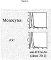
- BT3 expression was evaluated on freshly isolated monocytes and iDC using clone 20.1 anti-BT3 mAb ( Figure 1A).
- Figure 1A shows BT3 expression on freshly isolated monocytes and iDC using clone 20.1 anti-BT3 mAb.
- the differentiation of monocytes in DC was verified by expression of cell surface markers: CD14 was down-regulated and remained low in both immature and mature DC, whereas CD1a and HLA-DR was up-regulated (data not shown).
- the experiment shown is representative of five on different donors.
- the level of expression of BT3 was slightly higher on monocytes.
- BT3 receptors are constitutively expressed on the surface of monocytes and iDC, independently on their activation state.
- BT3 ligation provides a survival signal for monocytes and iDC in culture.
- BT3 is a stably expressed receptor family
- BT3 engagement was used to modify the life span of ex vivo monocytes and iDC cultured in the absence of survival factors (i.e. completed medium with serum) for 72 hours.
- Cell stimulation by plastic-coated mAb was used to engage BT3 on the surface of monocytes and iDC.
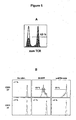
- Approximately 77.98% of monocytes treated with the mAb specific for BT3 survived in the absence of endogenous survival factors for 3 days (range 51.13-77.98%, p 0.0040) ( Figure 1B , upper row, shows a representative experiment of five).
- BT3 receptors are able to promote monocytes and iDC survival and to extend the duration of cells responses by attenuating apoptosis.
- survival differences following anti-BT3 versus control isotype-matched mAb was statistically significant (as for GM-CSF treatment).
- a dose response effect of anti-BT3 mAb on survival of both monocytes and iDC is apparent (data not shown).
- the lowest anti-BT3 mAb dilution able to promote a good survival capability was 10 ⁇ g/ml.
- BT3 engagement increases costimulatory molecules expression on the surface of monocytes and iDC.
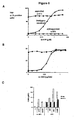
- BT3 To investigate the role of BT3 in primary inflammatory responses, freshly isolated blood monocytes and monocyte-derived dendritic cells were stimulated by mAb, coated on tissue culture plates, or by TLR ligands known to stimulate monocytes and dendritic cells (LPS). After 24h stimulation, coated anti-BT3 mAb was able to trigger the activation of monocytes, as shown by up-regulation of the cell surface costimulatory molecule CD86 ( Figure 1C ). The isotype-matched control mAb (whose antigen is not expressed on these cells), had no significant effect. Thus, the activation observed using anti-BT3.1 mAb was specific and not due to the engagement of FcR.
- PBMCs Peripheral blood mononuclear cells
- mice anti-human programmed death-1 (PD-1) mAB and three different clones of mouse anti-human CD277 were purified from ascites in our laboratory: Anti-PD-1 (clone PD1 3.1 with an IgG1 isotype) ( ghiotto et al. Int Immunol., 2010 ), Anti-CD277 (clone 103.2 with an IgG2a isotype, clones 20.1 and 7.2 both with an IgG1 isotype) (compte et al.).
- the anti-CD277 (clone 20.1) mAb was labelled with Alexa Fluor 647 using a commercial kit (Invitrogen, Eugene, OR).
- ECD-conjugated anti-CD3, PC5-conjugated anti-CD14, PC5-conjugated anti-CD19 (to select CD3 + CD14 - CD19 - cells) all from Beckman Coulter, Marseilles, France
- Pacific Blue-conjugated anti-CD4, Alexa700-conjugated anti-CD8 all from BD Pharmingen (San Diego, USA)
- APC-Alexa750-conjugated anti-CD27 from Caltag, Invitrogen, USA
- PC7-conjugated anti-CD45RA from BD Biosciences
- Alexa647-conjugated anti-CD277 clone 20.1, homemade.
- APC-conjugated IgG1 (Beckman Coulter) was used as control and LIVE/DEAD Fixable Dead Cell Stain Kit was used for viability.
- Cells were incubated 20 minutes at 4°C then washed twice in phosphate-buffered saline (PBS, Lonza) fixed with 2% paraformaldehyde, and analyzed on a FACSAria flow cytometer (BD Biosciences). Data were analyzed using FlowJo Software (TreeStar, Ashland, OR).
- Purified CD4 + T cells (200 X 10 3 cells/well) from thawed human PBMCs were cultured during 96h in RPMI 1640 10% FBS in flat bottom 96-well plates (MicrotestTM 96, Becton Dickinson) with previously plate-immobilized mouse anti-human CD3/CD28 or not (unstimulated).
- Anti-CD3 (clone OKT3)
- anti-CD28 (clone CD28.2, home-made) were used at 0.3 ⁇ g/ml and 10 ⁇ g/ml respectively. Cells were placed into an atmosphere of 5% CO 2 at 37°C in a humidified incubator.
- TF H Folliculat T Helper cells
- NK cells Live dead ⁇ negative cells
- NK cells were selected based on the expression of CD56+CD3- from healthy human PBMCs after 20 minutes incubation at 4°C with the anti-CD277-Alexa647 (clone 20.1).
- Cells were further washed twice in PBS (PBS, Lonza), then fixed with 2% formaldehyde and analyzed on a FACSAria ⁇ flow cytometer (BD Biosciences). Data were analyzed using FlowJo Software (TreeStar, Ashland).
- Purified CD4 + T cells 200 X 10 3 cells/well) from thawed human PBMCs were cultured in RPMI 1640 10% FBS in flat bottom 96-well plates (MicrotestTM 96, BD) with previously plate-immobilized mouse anti-human CD3 (clone OKT3)/CD28 (clone CD28.2) or anti-CD3/anti-CD277 (clone 20.1) or anti-CD3/isotypic control (IgG1). Purified anti-CD3 was used at 0, 3 ⁇ g/ml. Anti-CD28, anti-CD277 and isotypic control were used at 10 ⁇ g/ml. Cells were placed into an atmosphere of 5% CO 2 at 37°C in a humidified incubator.
- cytokines production (Interleukine-2, IL-2 and Interferon gamma, IFN- ⁇ ) was measured by ELISA assay according to the manufacturer's protocol (OptEIA, human IFN- ⁇ or IL-2 Set, BD Pharmingen). After 5 days, cells were stained with 3 ⁇ l of PE-conjugated anti-CD25 (BD Biosciences), and 5 ⁇ l of 7-AAD for 30 minutes at 4°C then washed twice in PBS, fixed with 2% paraformaldehyde and analyzed on a BD FACS Canto (BD Bioscience). Data were analyzed using FlowJo Software (TreeStar, Ashland, OR).
- CD4 + T cells were purified by negative selection from PBMCs using magnetic beads (Miltenyi Biotec) according to the manufacturer's protocol. CD4+ T cells were routinely more than 97% pure.
- CD4 + T cells were labelled with 0.5 ⁇ M CFSE (Invitrogen) for 10 min at 37°C, washed and stimulated (1.5x10 5 cells/well) with aAPC at a ratio of 1:1 (cells to beads) comprised of magnetic beads in triplicate in 96-well round-bottom plates (Falcon; BD Biosciences). Cultures were incubated at 37°C, 5% CO2 for 5 days and then proliferation of CFSE labelled CD4 + T cells were measured by flow cytometry (FACS Canto, Beckman Coulter).
- NK cells were sorted with Easy Sep® negative selection kit and incubated over night in medium completed with sub-optimal concentrations of IL-2 (100 U/ml) and IL-15 (10ng/ml). NK cell receptors functions were tested in re-directed cytolytic experiments against the FcyR positive P815 mastocytoma murine cell line.
- effector cells were incubated with P815 cells pre-coated for 30 minutes with the mAb of interest (irrelevant mouse IgG1: 11 ⁇ g/ml, anti-NKp46: 1 ⁇ g/ml, anti-DNAM: 5 ⁇ g/ml, anti-CD277 20.1: 10 ⁇ g/ml) according to a 1:1 Effector:Target (E/T) ratio.
- the mAb of interest irrelevant mouse IgG1: 11 ⁇ g/ml
- anti-NKp46 1 ⁇ g/ml
- anti-DNAM 5 ⁇ g/ml
- anti-CD277 20.1 10 ⁇ g/ml
- Cytotoxic tests were performed in 4-hours assays in the presence of GolgiStop® and soluble CD107 (a&b)-FITC (both from BD Biosciences), afterward cells were stained for surface markers (CD56-PeCy7 (Beckman Coulter, Immunotech), fixed and permeabilized (Cytofix/Cytoperm®) then stained with intracellular mAb (IFN- ⁇ (Beckman Coulter, Immunotech)). Cells were finally re-suspended in PBS 2% paraformaldehyde and extemporaneously analyzed on a BD FACS Canto® (BD Biosciences, San Jose, CA). The degree of activation of NK cells was measured based on the percentage of cells positive for CD107a and CD107b (degranulation) and/or the production of inflammatory cytokine (IFN- ⁇ ).
- Magnetic beads (Dynabeads M-450 Epoxy, Dynal Biotech) were coated with the following mAbs as described in Serriari et al. (serriari, ji 2010) : anti-CD3 (OKT3), anti-human CD28 (CD28.2), and/or various concentrations of anti-human CD277 (CD277, 20.1) or IgG1 or anti-MHC class I (MHC I) (YJ4) or IgG1.
- aAPCs were coated with suboptimal anti-CD3 Ab (5%), suboptimal levels of anti-CD28 Ab (10%), and either IgG1 Ab (CD3/CD28/IgG1), anti-CD277 Ab (CD3/CD28/CD277 + IgG1) or anti-MHC class I (CD3/28/ anti-MHC class I + IgG1), constituting the remaining 85% of protein added to the bead.
- the amount of protein was kept constant at 20 mg/ml by the addition of control IgG1.
- cytokines To determine the production of cytokines, cell-free supernatants were collected at 48 h and assayed for IL-2, IL-10 and IFN- ⁇ by ELISA using OptEIA kits (BD Pharmingen) according to the manufacturer's instructions.
- CD277 immunostainings were performed on total frozen sections of reactive lymph nodes as previously described (25). The final dilution for CD277.20.1 mAbs was 1/800. Negative control samples were prepared by omitting the primary mAb.
- CD8 and CD4 data were retrieved from the public GEO datasets (Sharp et al.) (http://www.ncbi.nlm.nih.gov/gds), while NK and gamma delta sets were personal.
- RMA Robust Multichip Average
- RMA was applied to the raw data collected from the various series. Quantile normalization and Loess' correction were done in R using Bioconductor and associated packages.
- the probe set corresponding to the three isoforms of BTN3A were retrieved from the normalized data sets and the corresponding log values were linearized for graphical representation.
- Human CD4 + T cells were purified by negative selection from PBMCs using magnetic beads (Miltenyi Biotec) according to the manufacturer's protocol. CD4 T cells were routinely more than 97% pure. Cells were incubated 24 hours in RPMI 1640 10% FBS at 37°C. CD4 + T cells were washed and stimulated at different times (2, 5, 10 and 30 minutes) with aAPC at a ratio of 1:3 (cells to beads) comprised of magnetic beads (Dynabeads M-450 Epoxy, Dynal Biotech) were coated with the following mAbs: anti-CD3 (OKT3), anti-human CD28 (CD28.2), and/or various concentrations of anti-human CD277 (CD277.
- mAbs anti-CD3 (OKT3), anti-human CD28 (CD28.2), and/or various concentrations of anti-human CD277 (CD277.
- IgG1 isotype control IgG1.
- aAPCs were coated with suboptimal anti-CD3 Ab (5%), suboptimal levels of anti-CD28 Ab (10%), and either IgG1 Ab (CD3/CD28/IgG1), anti-CD277.20.1 Ab (CD3/CD28/CD277.20.1 + IgG1), constituting the remaining 85% of protein added to the bead.
- the amount of protein was kept constant at 20 mg/ml by the addition of control IgG1.
- TCR T Cell Receptor
- T F H Follicular T helper cells
- T F H Follicular T helper cells
- ICSO chemokine receptor
- PD-1 PD-1 and BTLA
- CD277 expression could be differentially regulated in lymph nodes.
- Figure 4 Total frozen sections of reactive lymph nodes were also immuno stained for CD277. The results of immunohistochemical analysis showed a strong positivity on both inter-follicular T-cells area and mantle zone B-cells indicating that they were positive on T cells as well as B cells.
- CD277 is expressed on all subtypes of T lymphocytes in the peripheral blood as well as in lymph nodes and NK cells, but its expression is not modulated under stimulation as it is often the case for the molecules of the B7/CD28 family, or any other molecules involved in lymphocytes regulation.
- AKT and ERK phosphorylation is augmented after CD277 engagement on CD4 + T cells
- CD277 triggering was able to induce the phosphorylation of the two most important kinases of the lymphocyte signaling pathway: ERK from the mitogen-activated protein kinase and the serine threonine kinase AKT. It is known that Akt and ERK signaling plays a central role in T cell functions including proliferation, protein synthesis and regulation of apoptosis.
- CD277 20.1 induced the phosphorylation of AKT and ERK ( Figure 2 ), demonstrating that CD277 stimulation is involved in the regulation of T cell activation (purified CD4+ T cells were stimulated with antibody-coated Epoxy dynabeads with mAb anti-CD3 plus mAb anti-CD277.20.1)
- CD277 as a co-modulator of the TCR signaling pathway, we stimulated purified CD4+ T cells with various concentrations of mAb to CD277.20.1 clone or isotype control IgG1, together with anti-CD3 plus anti-CD28 at different times (2, 5, 10 and 30 minutes). We observed that the cross-linking of CD277 with mAb CD277.20.1 clone strongly up regulated the phosphorylation of AKT and ERK induced by CD3 +CD28 stimulation. This effect was dose and time dependent (data not shown).
- CD277 costimulates CD3 signals
- CD277 engagement was investigated by CD3 mediated signals.
- Purified CD4 + T cells from at least 4 healthy donors were cultured during 24 to 72 hours with anti-CD3/anti-CD28 or anti-CD3/anti-CD277 (clone 20.1) or anti-CD3/IgG1 (control condition).
- IL-2 and IFN- ⁇ production by CD4+ T cells were measured by ELISA.
- CD277 is a co-stimulatory molecule of T lymphocytes activation signal.
- CD277 further enhances CD3-CD28 costimulation and can act as a third signal to enhance T cell proliferation and cytokine production
- CD277 cosignals on T cell proliferation and cytokine production induced by CD3+CD28 signals We stimulated purified CD4 + T cells with with various concentrations of mAb to CD277, together with anti-CD3 plus anti-CD28. We kept the amount of antibody on the beads constant by adding isotype control IgG1 and anti-MHC class I (MHC I). We demonstrated that the cross-linking of CD277 on CD4 + T cells strongly activated CD4 + T cell proliferation mediated by anti-CD3 plus anti-CD28 in a dose-dependent way. Indeed, we measured the proliferation of CD4 cells by measuring the dilution of cytosolic dye CFSE (data not shown).
- NK cells we investigated whether a similar co-stimulatory effect was obtained in NK cells.
- CD277 alone did not have any effect on NK cell stimulation.
- CD277 has three isoforms btn3a1, btn3a2 and btn3a3, with ( btn3a1 and btn3a3), or without ( btn3a2 ) the B30.2 domain
- btn3a1 is the main form expressed by T lymphocytes whereas the decoy form ( btn3a2 ) is mostly expressed on NK cells (data not shown). This result was validated by quantitative PCR on 4 healthy donors. We thus emitted the hypothesis that the absence of co-stimulation in response to CD277 stimulation of NK cells might be attributed to this form of BTN3A.
- Anti-CD277 anti-BT3-20.1 and 103.2 mAb were generated and validated as previously described [10]. Fab fragments of anti-BT3-20.1 were generated and purified with the Immunopure Fab Preparation Kit following the manufacturer's recommendation (Pierce). Protein purity was assessed by nonreducing SDS-PAGE.
- FIGENIX FIGENIX Annotation Platform: [http://figenix2.up.univmrs.fr/Figenix/index.jsp]) to retrieve sequences and alignments and perform phylogenetic reconstruction.
- the pipeline used applied three different methods of phylogenetic tree reconstruction, i.e. Maximum Parsimony [38], Maximum likelihood [39] and Neighbour Joining [40], and a midpoint rooted consensus tree was built. Bootstrapping was carried out with 1000 replications. Bootstrap values are reported for each method (for a detailed description of the pipelines and models used, see [41].
- P815 mouse mastocytoma cell line
- K562 chronic myeloid leukaemia cell line
- Raji and Daudi Burkitt lymphoma cell lines
- RPMI 1640 medium Invitrogen
- FCS foetal calf serum
- the PBMC from healthy donors were distributed at 106/ml in 24-well culture plates at 37°C in 5% CO2 in RPMI 1640 medium and 10% FCS.
- Polyclonal V ⁇ 9V ⁇ 2 T cells were specifically expanded with 3 ⁇ mol/l of Phosphostim (BrHPP molecule, Innate Pharma, Marseille, France) and 100 U/ml IL-2 (Chiron, Basel, Switzerland) for 12 days.
- Phosphostim was added once at the onset of the culture. Every 2 days, one-half of the culture medium volume was replaced with fresh medium containing 100 U/ml IL-2. The last day, the percentage of ⁇ T cells was evaluated using anti Vd2 FITC and anti-CD3-Cy7. Only cells cultures that reached more than 90% of ⁇ T Cells, were selected to be used in functional tests.
- ⁇ T cells and target cells were co-incubated at 37°C with anti-CD107a FITC and anti-CD107b FITC in presence of monensin (10 ⁇ M, GoligiStop, BD Bioscience). Cells were collected and washed with PBS, 4 hours after incubation. ⁇ T cells were labelled with anti-pan ⁇ TCR PE and anti-CD3 PECy7 mAbs (BD Pharmingen). All samples were measured on FACSCanto or FACSAria flow cytometers (BD Biosciences) using FACSDiva software. Analyses were performed with FlowJo software (Tree Star).
- Intracellular staining was performed according to the recommended by BD Pharmingen Fix and Perm Kit (BD Biosciences).100 ⁇ l of ⁇ T cells at 2.106cells/ml were plated on 96-well plates. They were incubated in presence or not of BrHPP at 3 ⁇ M and with anti-CD277 or control isotypes at 10 ⁇ g/ml, for 30 min at 4°C. Cells were stimulated at different times at 37°C. Stimulation were stopped adding 100 ⁇ l Cytofix/Cytoper solution at 37°C for 10min.
- Intracellular phosphorylated proteins were stained with purified monoclonal rabbit antibodies: anti-pZap 70, anti-pAKT and anti-pErk from Cell Signaling Technology (Danvers, USA) ; labelled with Biotin-SP-conjugated F(ab')2 Donkey anti-Rabbit (Jackson) ; and revealed with streptavindin-PE (Beckman Coulter).
- PBMC peripheral blood mononuclear cells
- ⁇ T cells were plated and stimulated as described in the paragraph: cell lines and expansion of ⁇ T cells, except cells were stimulated with anti-CD277 mAbs (10 ⁇ g/ml); or control isotypes (10 ⁇ g/ml); and with or without BrHPP at different concentrations. Cultures were stopped 9 days after. Percentages of V ⁇ 9V ⁇ 2 were measured the first day and the last day of culture as described above. Expanded ⁇ T cells effectors were stimulated with or without different doses of BrHPP added in anti-CD277 mAbs at 10 ⁇ g/ml; or with OKT3 (4ng/ml) associated with anti-BT3 19.5 mAb in different concentrations. Activation of degranulation was measured by CD107 labelling assay as described above.
- P815 mastocytoma mouse cells were incubated 30 min, with mouse controls isotypes or anti-CD277 mAbs (10 ⁇ g/ml) or/and with anti-CD3 (OKT3, 4ng/ml). After washing, P815 were incubated 4h at 37°C, with expanded V ⁇ 9V ⁇ 2 T effectors cells at the same concentration.
- a Flow-based CD107a degranulation assay was performed as described above.
- StatXact software (version 8 PC) produced by the Cytel, was used for all statistical analyses. Significance values for comparisons between groups were determined by the nonparametric Mann and Whitney analysis. Assuming an unequal variance with 95% confidence levels, and p values ⁇ 0.05 were considered significant.
- the first stop codon is located immediately downstream to the signal peptide, at the begining of the IgV sequence and the second downstream of the first transmembrane domain [7]. It is unlikely to be due to sequencing errors because we find the very same mutations in Pongo abelii DNA sequence. These data indicated that Skint genes were not necessary for primate species and suggested that their function could be performed by other genes. In a reciprocal way, CD277 gene was absent in Ratus norvegicus and in Mus musculus DNA sequence using NCBI ntBlast. Indeed, within the BTN families only BTN2 sequence was present in these two species. These results indicated that CD277 really disappeared in rodents and consequently were not necessary for survival of these species.
- CD277 function has been assumed by other gene(s). Both functional redundancies in rodents and primates common ancestors could be on one hand at the origin of disappearance of CD277 in rodents and on the other hand of Skint-1 in primates.
- both molecules have similarities in their extracellular region within both IgV and IgC domains, but differ by the number of transmembrane domains and above all by their intracytoplasmic region (data not shown).
- CD277 has only one transmembrane domain instead of three for Skint-1.
- CD277 has a B30.2 domain. This domain is absent in Skint-1.
- CD277 is expressed in V ⁇ 9V ⁇ 2 T cells and not modulated by their differentiation
- CD277 was expressed on conventional T lymphocytes suggesting a role of CD277 in the regulation of T cell response.
- Naives cells CD45RA+/CD27+
- Central Memory cells CD45RA-/CD27+
- Effectors Memory CD45RA-/CD27-/CCR7-
- Effectors Memory RA+ CD45RA+/CD27-/CCR7-
- CD45RA+/CD27-/CCR7- Effectors Memory RA+
- V ⁇ 9V ⁇ 2 T cells express CD277 with higher mean fluorescence intensity than ⁇ T cells suggesting an important role in homeostasis of ⁇ T cells.
- every cells express CD277. Its level of expression is approximately the same in the analysed ⁇ T cell subpopulations. The only slight increase was found in the naive CD45RA+/CD27+/CD28+/CCR7+, but this variation was not significant in the series if healthy volunteers tested.
- Anti-CD277mAb 20.1 induces proliferation of V ⁇ 9V ⁇ 2 T cells whereas 103.2 inhibits the stimulation mediated by optimal TCR stimulation by phosphoantigens.
- CD277 modulate the degranulation in V ⁇ 9V ⁇ 2 T cells mediated by the TcR complex
- CD277 modulates the ability of ⁇ T cells to express CD107 and hence to degranulate following phosphoantigen or anti-CD3 stimulation.
- CD277 effect is biphasic: enhancing at low stimulation levels and decreasing at higher levels of TCR crosslinking the ⁇ T cell degranulation activity.
- Crosslinking of CD277 triggers degranulation together with INF ⁇ and TNF ⁇ release.
- CD277 potentiate the anti-tumor cytolysis mediated by V ⁇ 9V ⁇ 2 T cells.
- CD277 mAbs potentiate or inhibit the anti-tumor cytolysis mediated by responding V ⁇ 9V ⁇ 2 T cells.
Landscapes
- Health & Medical Sciences (AREA)
- Immunology (AREA)
- Chemical & Material Sciences (AREA)
- Organic Chemistry (AREA)
- General Health & Medical Sciences (AREA)
- Medicinal Chemistry (AREA)
- Life Sciences & Earth Sciences (AREA)
- Proteomics, Peptides & Aminoacids (AREA)
- Molecular Biology (AREA)
- Genetics & Genomics (AREA)
- Biophysics (AREA)
- Biochemistry (AREA)
- Veterinary Medicine (AREA)
- Public Health (AREA)
- Animal Behavior & Ethology (AREA)
- Pharmacology & Pharmacy (AREA)
- Nuclear Medicine, Radiotherapy & Molecular Imaging (AREA)
- General Chemical & Material Sciences (AREA)
- Chemical Kinetics & Catalysis (AREA)
- Engineering & Computer Science (AREA)
- Bioinformatics & Cheminformatics (AREA)
- Communicable Diseases (AREA)
- Oncology (AREA)
- Transplantation (AREA)
- Peptides Or Proteins (AREA)
- Medicines Containing Antibodies Or Antigens For Use As Internal Diagnostic Agents (AREA)
- Micro-Organisms Or Cultivation Processes Thereof (AREA)
- Preparation Of Compounds By Using Micro-Organisms (AREA)
- Medicines Containing Material From Animals Or Micro-Organisms (AREA)
- Fats And Perfumes (AREA)
Priority Applications (6)
| Application Number | Priority Date | Filing Date | Title |
|---|---|---|---|
| EP21182452.9A EP3915582A3 (en) | 2010-12-15 | 2011-12-14 | Anti-cd277 antibodies and uses thereof |
| HRP20211823TT HRP20211823T1 (hr) | 2010-12-15 | 2011-12-14 | Anti-cd277 antitijela i njihove uporabe |
| PL15151003T PL2946791T3 (pl) | 2010-12-15 | 2011-12-14 | Przeciwciała anty-CD277 i ich zastosowania |
| SM20210676T SMT202100676T1 (it) | 2010-12-15 | 2011-12-14 | Anticorpi anti-cd277 e loro usi |
| RS20211469A RS62700B1 (sr) | 2010-12-15 | 2011-12-14 | Anti-cd277 antitela i njihove upotrebe |
| SI201132018T SI2946791T1 (sl) | 2010-12-15 | 2011-12-14 | Protitelesa proti CD277 in uporabe le-teh |
Applications Claiming Priority (3)
| Application Number | Priority Date | Filing Date | Title |
|---|---|---|---|
| PCT/IB2010/003417 WO2012080769A1 (en) | 2010-12-15 | 2010-12-15 | Anti-cd277 antibodies and uses thereof |
| PCT/EP2011/072787 WO2012080351A1 (en) | 2010-12-15 | 2011-12-14 | Anti-cd277 antibodies and uses thereof |
| EP11802346.4A EP2651441A1 (en) | 2010-12-15 | 2011-12-14 | Anti-cd277 antibodies and uses thereof |
Related Parent Applications (2)
| Application Number | Title | Priority Date | Filing Date |
|---|---|---|---|
| EP11802346.4A Division EP2651441A1 (en) | 2010-12-15 | 2011-12-14 | Anti-cd277 antibodies and uses thereof |
| PCT/EP2011/072787 Previously-Filed-Application WO2012080351A1 (en) | 2010-12-15 | 2011-12-14 | Anti-cd277 antibodies and uses thereof |
Related Child Applications (2)
| Application Number | Title | Priority Date | Filing Date |
|---|---|---|---|
| EP21182452.9A Division EP3915582A3 (en) | 2010-12-15 | 2011-12-14 | Anti-cd277 antibodies and uses thereof |
| EP21182452.9A Division-Into EP3915582A3 (en) | 2010-12-15 | 2011-12-14 | Anti-cd277 antibodies and uses thereof |
Publications (2)
| Publication Number | Publication Date |
|---|---|
| EP2946791A1 EP2946791A1 (en) | 2015-11-25 |
| EP2946791B1 true EP2946791B1 (en) | 2021-09-08 |
Family
ID=43828073
Family Applications (3)
| Application Number | Title | Priority Date | Filing Date |
|---|---|---|---|
| EP15151003.9A Active EP2946791B1 (en) | 2010-12-15 | 2011-12-14 | Anti-CD277 antibodies and uses thereof |
| EP21182452.9A Pending EP3915582A3 (en) | 2010-12-15 | 2011-12-14 | Anti-cd277 antibodies and uses thereof |
| EP11802346.4A Withdrawn EP2651441A1 (en) | 2010-12-15 | 2011-12-14 | Anti-cd277 antibodies and uses thereof |
Family Applications After (2)
| Application Number | Title | Priority Date | Filing Date |
|---|---|---|---|
| EP21182452.9A Pending EP3915582A3 (en) | 2010-12-15 | 2011-12-14 | Anti-cd277 antibodies and uses thereof |
| EP11802346.4A Withdrawn EP2651441A1 (en) | 2010-12-15 | 2011-12-14 | Anti-cd277 antibodies and uses thereof |
Country Status (14)
| Country | Link |
|---|---|
| US (1) | US20140322235A1 (sr) |
| EP (3) | EP2946791B1 (sr) |
| JP (2) | JP6138693B2 (sr) |
| CY (1) | CY1125057T1 (sr) |
| ES (1) | ES2899733T3 (sr) |
| HR (1) | HRP20211823T1 (sr) |
| HU (1) | HUE057004T2 (sr) |
| LT (1) | LT2946791T (sr) |
| PL (1) | PL2946791T3 (sr) |
| PT (1) | PT2946791T (sr) |
| RS (1) | RS62700B1 (sr) |
| SI (1) | SI2946791T1 (sr) |
| SM (1) | SMT202100676T1 (sr) |
| WO (2) | WO2012080769A1 (sr) |
Families Citing this family (28)
| Publication number | Priority date | Publication date | Assignee | Title |
|---|---|---|---|---|
| EP0557306B1 (en) | 1990-09-25 | 1998-12-23 | Innovasive Devices, Inc. | Bone fastener |
| US20150353643A1 (en) * | 2013-09-24 | 2015-12-10 | Universite De La Mediterranee - Aix-Marseille Ii | Anti-cd277 antibodies and uses thereof |
| US20180028566A1 (en) * | 2016-07-29 | 2018-02-01 | New York University | Gamma delta t cells as a target for treatment of solid tumors |
| WO2018082590A1 (zh) * | 2016-11-02 | 2018-05-11 | 北京蛋白质组研究中心 | 肿瘤免疫治疗靶标及其应用 |
| WO2018201130A1 (en) * | 2017-04-28 | 2018-11-01 | The Brigham And Women's Hospital, Inc. | Targeting gamma-delta t cells in obesity and cachexia |
| KR102764182B1 (ko) | 2017-09-21 | 2025-02-07 | 임체크 테라퓨틱스 | Btn2에 대해 특이성을 갖는 항체 및 이의 용도 |
| EP3743081A4 (en) | 2018-01-23 | 2021-12-01 | New York University | SPECIFIC ANTIBODIES OF THE DELTA 1 CHAIN OF THE T-LYMPHOCYTE RECEPTOR |
| WO2019211370A1 (en) | 2018-05-03 | 2019-11-07 | INSERM (Institut National de la Santé et de la Recherche Médicale) | Methods and pharmaceutical compositions for treating cancer |
| WO2019211369A1 (en) * | 2018-05-03 | 2019-11-07 | INSERM (Institut National de la Santé et de la Recherche Médicale) | Methods and pharmaceutical compositions for treating cancer |
| MX2021001268A (es) * | 2018-08-01 | 2021-09-08 | Imcheck Therapeutics Sas | Anticuerpos anti-btn3a y su uso en el tratamiento del cáncer o trastornos infecciosos. |
| WO2020033925A2 (en) * | 2018-08-09 | 2020-02-13 | Compass Therapeutics Llc | Antibodies that bind cd277 and uses thereof |
| WO2020033923A1 (en) * | 2018-08-09 | 2020-02-13 | Compass Therapeutics Llc | Antigen binding agents that bind cd277 and uses thereof |
| EP3833442A2 (en) * | 2018-08-09 | 2021-06-16 | Compass Therapeutics LLC | Antibodies that bind cd277 and uses thereof |
| WO2020131764A1 (en) * | 2018-12-18 | 2020-06-25 | Merck Sharp & Dohme Corp. | Methods of using butyrophilin antibodies for treating hiv infection |
| JP7649744B2 (ja) | 2018-12-26 | 2025-03-21 | イムチェック セラピューティクス エスエーエス | Btn3a結合タンパク質及びその使用 |
| US12084500B2 (en) | 2019-01-23 | 2024-09-10 | New York University | Antibodies specific to delta 1 chain of T cell receptor |
| JP7652705B2 (ja) | 2019-03-20 | 2025-03-27 | イムチェック セラピューティクス エスエーエス | Btn2に対する特異性を有する抗体及びその使用 |
| EP4288074A4 (en) * | 2021-02-08 | 2025-04-30 | The Regents of University of California | Regulation of butvrophilin subfamily 3 member a1 (btn3a1, cd277) |
| TW202313695A (zh) | 2021-09-15 | 2023-04-01 | 法商感應檢查療法公司 | 抗btn3a抗體在製備用於治療腫瘤的藥物的用途 |
| WO2023052541A1 (en) | 2021-09-30 | 2023-04-06 | Imcheck Therapeutics | Combination of an anti-btn3a activating antibody and an il-2 agonist for use in therapy |
| CA3234598A1 (en) | 2021-10-27 | 2023-05-04 | Daniel Olive | Butyrophilin (btn) 3a activating antibodies for use in methods for treating infectious disorders |
| EP4482859A1 (en) | 2022-02-27 | 2025-01-01 | Boehringer Ingelheim International GmbH | Bispecific antibodies against cd277 and a tumor-antigen |
| WO2023217743A1 (en) | 2022-05-10 | 2023-11-16 | Imcheck Therapeutics | Anti-btn3a antibodies for use in methods of treating gastro-intestinal inflammatory disorders |
| WO2024074498A1 (en) | 2022-10-04 | 2024-04-11 | Imcheck Therapeutics | Combination of a btn3a activating antibody, a bcl2 inhibitor and hypomethylating agent for use in treating cancer |
| IL322506A (en) | 2023-02-23 | 2025-10-01 | Imcheck Therapeutics | Combinations of BTN3A activating antibody and immune checkpoint inhibitors |
| TW202506731A (zh) | 2023-04-27 | 2025-02-16 | 法商感應檢查療法公司 | 治療多重抗藥性細菌感染之方法 |
| EP4731681A1 (en) | 2023-06-23 | 2026-04-29 | Imcheck Therapeutics | Bispecific antibodies targeting btn3a and the pd-1/pd-l1 inhibitory axis |
| CN119193482B (zh) * | 2024-11-26 | 2026-03-17 | 北京国卫生物科技有限公司 | 一种γδT细胞体外诱导培养方法 |
Family Cites Families (30)
| Publication number | Priority date | Publication date | Assignee | Title |
|---|---|---|---|---|
| US4179337A (en) | 1973-07-20 | 1979-12-18 | Davis Frank F | Non-immunogenic polypeptides |
| JPS6023084B2 (ja) | 1979-07-11 | 1985-06-05 | 味の素株式会社 | 代用血液 |
| US4640835A (en) | 1981-10-30 | 1987-02-03 | Nippon Chemiphar Company, Ltd. | Plasminogen activator derivatives |
| US4816567A (en) | 1983-04-08 | 1989-03-28 | Genentech, Inc. | Recombinant immunoglobin preparations |
| US4496689A (en) | 1983-12-27 | 1985-01-29 | Miles Laboratories, Inc. | Covalently attached complex of alpha-1-proteinase inhibitor with a water soluble polymer |
| EP0173494A3 (en) | 1984-08-27 | 1987-11-25 | The Board Of Trustees Of The Leland Stanford Junior University | Chimeric receptors by dna splicing and expression |
| EP0206448B1 (en) | 1985-06-19 | 1990-11-14 | Ajinomoto Co., Inc. | Hemoglobin combined with a poly(alkylene oxide) |
| DE3689123T2 (de) | 1985-11-01 | 1994-03-03 | Xoma Corp | Modulare einheit von antikörpergenen, daraus hergestellte antikörper und verwendung. |
| WO1987005330A1 (en) | 1986-03-07 | 1987-09-11 | Michel Louis Eugene Bergh | Method for enhancing glycoprotein stability |
| US5225539A (en) | 1986-03-27 | 1993-07-06 | Medical Research Council | Recombinant altered antibodies and methods of making altered antibodies |
| GB8607679D0 (en) | 1986-03-27 | 1986-04-30 | Winter G P | Recombinant dna product |
| US4861719A (en) | 1986-04-25 | 1989-08-29 | Fred Hutchinson Cancer Research Center | DNA constructs for retrovirus packaging cell lines |
| US4791192A (en) | 1986-06-26 | 1988-12-13 | Takeda Chemical Industries, Ltd. | Chemically modified protein with polyethyleneglycol |
| US5202238A (en) | 1987-10-27 | 1993-04-13 | Oncogen | Production of chimeric antibodies by homologous recombination |
| US5204244A (en) | 1987-10-27 | 1993-04-20 | Oncogen | Production of chimeric antibodies by homologous recombination |
| US5278056A (en) | 1988-02-05 | 1994-01-11 | The Trustees Of Columbia University In The City Of New York | Retroviral packaging cell lines and process of using same |
| US5530101A (en) | 1988-12-28 | 1996-06-25 | Protein Design Labs, Inc. | Humanized immunoglobulins |
| US5859205A (en) | 1989-12-21 | 1999-01-12 | Celltech Limited | Humanised antibodies |
| GB8928874D0 (en) | 1989-12-21 | 1990-02-28 | Celltech Ltd | Humanised antibodies |
| US5670488A (en) | 1992-12-03 | 1997-09-23 | Genzyme Corporation | Adenovirus vector for gene therapy |
| EP0519596B1 (en) | 1991-05-17 | 2005-02-23 | Merck & Co. Inc. | A method for reducing the immunogenicity of antibody variable domains |
| US5565332A (en) | 1991-09-23 | 1996-10-15 | Medical Research Council | Production of chimeric antibodies - a combinatorial approach |
| US5639641A (en) | 1992-09-09 | 1997-06-17 | Immunogen Inc. | Resurfacing of rodent antibodies |
| DE69434860T2 (de) | 1993-02-22 | 2007-03-15 | The Rockefeller University | Herstellung von helfer-freien retroviren mit hohem titer mittels transienter transfektion |
| FR2712812B1 (fr) | 1993-11-23 | 1996-02-09 | Centre Nat Rech Scient | Composition pour la production de produits thérapeutiques in vivo. |
| DE69535243T2 (de) | 1994-07-13 | 2007-05-10 | Chugai Seiyaku K.K. | Gegen menschliches interleukin-8 gerichteter, rekonstituierter menschlicher antikörper |
| IL116816A (en) | 1995-01-20 | 2003-05-29 | Rhone Poulenc Rorer Sa | Cell for the production of a defective recombinant adenovirus or an adeno-associated virus and the various uses thereof |
| ES2233974T3 (es) | 1995-09-11 | 2005-06-16 | Kyowa Hakko Kogyo Co., Ltd. | Anticuerpo contra la cadena alfa del receptor de la interleucina 5 humana. |
| US6013516A (en) | 1995-10-06 | 2000-01-11 | The Salk Institute For Biological Studies | Vector and method of use for nucleic acid delivery to non-dividing cells |
| JP2001522241A (ja) | 1997-04-10 | 2001-11-13 | ロイヤル ネザーランズ アカデミー オブ アーツ アンド サイエンシズ | 診断方法および試薬 |
-
2010
- 2010-12-15 WO PCT/IB2010/003417 patent/WO2012080769A1/en not_active Ceased
-
2011
- 2011-12-14 JP JP2013543767A patent/JP6138693B2/ja not_active Expired - Fee Related
- 2011-12-14 HR HRP20211823TT patent/HRP20211823T1/hr unknown
- 2011-12-14 RS RS20211469A patent/RS62700B1/sr unknown
- 2011-12-14 LT LTEP15151003.9T patent/LT2946791T/lt unknown
- 2011-12-14 PL PL15151003T patent/PL2946791T3/pl unknown
- 2011-12-14 HU HUE15151003A patent/HUE057004T2/hu unknown
- 2011-12-14 EP EP15151003.9A patent/EP2946791B1/en active Active
- 2011-12-14 WO PCT/EP2011/072787 patent/WO2012080351A1/en not_active Ceased
- 2011-12-14 EP EP21182452.9A patent/EP3915582A3/en active Pending
- 2011-12-14 ES ES15151003T patent/ES2899733T3/es active Active
- 2011-12-14 SM SM20210676T patent/SMT202100676T1/it unknown
- 2011-12-14 US US13/994,277 patent/US20140322235A1/en not_active Abandoned
- 2011-12-14 SI SI201132018T patent/SI2946791T1/sl unknown
- 2011-12-14 EP EP11802346.4A patent/EP2651441A1/en not_active Withdrawn
- 2011-12-14 PT PT151510039T patent/PT2946791T/pt unknown
-
2016
- 2016-11-04 JP JP2016216470A patent/JP6408534B2/ja active Active
-
2021
- 2021-12-01 CY CY20211101047T patent/CY1125057T1/el unknown
Also Published As
| Publication number | Publication date |
|---|---|
| EP3915582A2 (en) | 2021-12-01 |
| RS62700B1 (sr) | 2022-01-31 |
| EP3915582A3 (en) | 2021-12-08 |
| EP2651441A1 (en) | 2013-10-23 |
| WO2012080351A1 (en) | 2012-06-21 |
| SI2946791T1 (sl) | 2022-02-28 |
| JP2014503522A (ja) | 2014-02-13 |
| HRP20211823T1 (hr) | 2022-03-04 |
| CY1125057T1 (el) | 2023-06-09 |
| EP2946791A1 (en) | 2015-11-25 |
| PT2946791T (pt) | 2021-12-07 |
| LT2946791T (lt) | 2022-01-10 |
| HUE057004T2 (hu) | 2022-04-28 |
| JP2017061502A (ja) | 2017-03-30 |
| ES2899733T3 (es) | 2022-03-14 |
| JP6138693B2 (ja) | 2017-05-31 |
| JP6408534B2 (ja) | 2018-10-17 |
| US20140322235A1 (en) | 2014-10-30 |
| WO2012080769A1 (en) | 2012-06-21 |
| PL2946791T3 (pl) | 2022-02-21 |
| SMT202100676T1 (it) | 2022-01-10 |
Similar Documents
| Publication | Publication Date | Title |
|---|---|---|
| EP2946791B1 (en) | Anti-CD277 antibodies and uses thereof | |
| US10716838B2 (en) | Anti-CD277 antibodies | |
| EP3002296B1 (en) | Btla antibodies and uses thereof | |
| US11541076B2 (en) | Chimeric antigen receptors targeting TIM-1 | |
| JP7358369B2 (ja) | Cd83結合キメラ抗原受容体 | |
| EP3663320A1 (en) | Nucleic acid sequence encoding a cs1-specific chimeric antigen receptor (car) polypeptide | |
| US20250084376A1 (en) | Ex vivo use of modified cells of leukemic origin for enhancing the efficacy of adoptive cell therapy | |
| CA3177488A1 (en) | Compositions and methods for tcr reprogramming using cd70 specific fusion proteins | |
| JP2025083396A (ja) | 骨髄性悪性腫瘍を処置するためのキメラ抗原受容体 | |
| US20230390391A1 (en) | Bi-specific chimeric antigen receptor t cells targeting cd83 and interleukin 6 receptor | |
| US11904004B2 (en) | Anti-CD277 antibodies | |
| US20230285527A1 (en) | Anti-cd277 antibodies and uses thereof | |
| Simone et al. | Ligation of the BT3 molecules, members of the B7 family, enhance the proinflammatory responses of human monocytes and monocyte-derived dendritic cells | |
| US20240197876A1 (en) | Methods to determine treatment efficacy with gamma-delta t cells | |
| Walker | ME (Linda) Joosse Iris Nederlof Lucy SK Walker Janneke N. Samsom |
Legal Events
| Date | Code | Title | Description |
|---|---|---|---|
| REG | Reference to a national code |
Ref country code: HR Ref legal event code: TUEP Ref document number: P20211823 Country of ref document: HR |
|
| PUAI | Public reference made under article 153(3) epc to a published international application that has entered the european phase |
Free format text: ORIGINAL CODE: 0009012 |
|
| AC | Divisional application: reference to earlier application |
Ref document number: 2651441 Country of ref document: EP Kind code of ref document: P |
|
| AK | Designated contracting states |
Kind code of ref document: A1 Designated state(s): AL AT BE BG CH CY CZ DE DK EE ES FI FR GB GR HR HU IE IS IT LI LT LU LV MC MK MT NL NO PL PT RO RS SE SI SK SM TR |
|
| RAP1 | Party data changed (applicant data changed or rights of an application transferred) |
Owner name: UNIVERSITE D'AIX-MARSEILLE Owner name: UNIVERSITE DE NANTES Owner name: CENTRE NATIONAL DE LA RECHERCHE SCIENTIFIQUE (CNRS Owner name: INSTITUT JEAN PAOLI & IRENE CALMETTES Owner name: INSERM (INSTITUT NATIONAL DE LA SANTE ET DE LA REC |
|
| 17P | Request for examination filed |
Effective date: 20160125 |
|
| RBV | Designated contracting states (corrected) |
Designated state(s): AL AT BE BG CH CY CZ DE DK EE ES FI FR GB GR HR HU IE IS IT LI LT LU LV MC MK MT NL NO PL PT RO RS SE SI SK SM TR |
|
| 17Q | First examination report despatched |
Effective date: 20160627 |
|
| STAA | Information on the status of an ep patent application or granted ep patent |
Free format text: STATUS: EXAMINATION IS IN PROGRESS |
|
| REG | Reference to a national code |
Ref country code: DE Ref legal event code: R079 Ref document number: 602011071763 Country of ref document: DE Free format text: PREVIOUS MAIN CLASS: A61K0039395000 Ipc: A61P0035000000 |
|
| GRAP | Despatch of communication of intention to grant a patent |
Free format text: ORIGINAL CODE: EPIDOSNIGR1 |
|
| STAA | Information on the status of an ep patent application or granted ep patent |
Free format text: STATUS: GRANT OF PATENT IS INTENDED |
|
| RIC1 | Information provided on ipc code assigned before grant |
Ipc: A61P 31/00 20060101ALI20210121BHEP Ipc: A61P 35/00 20060101AFI20210121BHEP Ipc: C07K 16/28 20060101ALI20210121BHEP Ipc: A61P 37/06 20060101ALI20210121BHEP Ipc: A61P 37/04 20060101ALI20210121BHEP |
|
| INTG | Intention to grant announced |
Effective date: 20210224 |
|
| GRAS | Grant fee paid |
Free format text: ORIGINAL CODE: EPIDOSNIGR3 |
|
| 111L | Licence recorded |
Designated state(s): AL AT BE BG CH CY CZ DE DK EE ES FI FR GB GR HR HU IE IS IT LT LU LV MC MK MT NL NO PL PT RO RS SE SI SK SM TR Name of requester: IMCHECK THERAPEUTICS, FR Effective date: 20210624 |
|
| GRAA | (expected) grant |
Free format text: ORIGINAL CODE: 0009210 |
|
| STAA | Information on the status of an ep patent application or granted ep patent |
Free format text: STATUS: THE PATENT HAS BEEN GRANTED |
|
| 111L | Licence recorded |
Designated state(s): AL AT BE BG CH CY CZ DE DK EE ES FI FR GB GR HR HU IE IS IT LT LU LV MC MK MT NL NO PL PT RO RS SE SI SK SM TR Name of requester: IMCHECK THERAPEUTICS, FR Effective date: 20210624 |
|
| AC | Divisional application: reference to earlier application |
Ref document number: 2651441 Country of ref document: EP Kind code of ref document: P |
|
| AK | Designated contracting states |
Kind code of ref document: B1 Designated state(s): AL AT BE BG CH CY CZ DE DK EE ES FI FR GB GR HR HU IE IS IT LI LT LU LV MC MK MT NL NO PL PT RO RS SE SI SK SM TR |
|
| REG | Reference to a national code |
Ref country code: GB Ref legal event code: FG4D |
|
| REG | Reference to a national code |
Ref country code: AT Ref legal event code: REF Ref document number: 1428030 Country of ref document: AT Kind code of ref document: T Effective date: 20210915 Ref country code: CH Ref legal event code: EP |
|
| REG | Reference to a national code |
Ref country code: IE Ref legal event code: FG4D |
|
| REG | Reference to a national code |
Ref country code: DE Ref legal event code: R096 Ref document number: 602011071763 Country of ref document: DE |
|
| REG | Reference to a national code |
Ref country code: FI Ref legal event code: FGE |
|
| REG | Reference to a national code |
Ref country code: DK Ref legal event code: T3 Effective date: 20211124 |
|
| REG | Reference to a national code |
Ref country code: PT Ref legal event code: SC4A Ref document number: 2946791 Country of ref document: PT Date of ref document: 20211207 Kind code of ref document: T Free format text: AVAILABILITY OF NATIONAL TRANSLATION Effective date: 20211129 |
|
| REG | Reference to a national code |
Ref country code: SE Ref legal event code: TRGR |
|
| REG | Reference to a national code |
Ref country code: NL Ref legal event code: FP |
|
| REG | Reference to a national code |
Ref country code: EE Ref legal event code: FG4A Ref document number: E021631 Country of ref document: EE Effective date: 20211201 |
|
| REG | Reference to a national code |
Ref country code: SK Ref legal event code: T3 Ref document number: E 38708 Country of ref document: SK |
|
| REG | Reference to a national code |
Ref country code: NO Ref legal event code: T2 Effective date: 20210908 |
|
| REG | Reference to a national code |
Ref country code: GR Ref legal event code: EP Ref document number: 20210403185 Country of ref document: GR Effective date: 20220113 |
|
| REG | Reference to a national code |
Ref country code: HR Ref legal event code: T1PR Ref document number: P20211823 Country of ref document: HR Ref country code: HR Ref legal event code: ODRP Ref document number: P20211823 Country of ref document: HR Payment date: 20220128 Year of fee payment: 11 |
|
| REG | Reference to a national code |
Ref country code: ES Ref legal event code: FG2A Ref document number: 2899733 Country of ref document: ES Kind code of ref document: T3 Effective date: 20220314 |
|
| REG | Reference to a national code |
Ref country code: HU Ref legal event code: AG4A Ref document number: E057004 Country of ref document: HU |
|
| REG | Reference to a national code |
Ref country code: DE Ref legal event code: R097 Ref document number: 602011071763 Country of ref document: DE |
|
| PLBE | No opposition filed within time limit |
Free format text: ORIGINAL CODE: 0009261 |
|
| STAA | Information on the status of an ep patent application or granted ep patent |
Free format text: STATUS: NO OPPOSITION FILED WITHIN TIME LIMIT |
|
| 26N | No opposition filed |
Effective date: 20220609 |
|
| REG | Reference to a national code |
Ref country code: HR Ref legal event code: ODRP Ref document number: P20211823 Country of ref document: HR Payment date: 20221213 Year of fee payment: 12 |
|
| REG | Reference to a national code |
Ref country code: AT Ref legal event code: UEP Ref document number: 1428030 Country of ref document: AT Kind code of ref document: T Effective date: 20210908 |
|
| P01 | Opt-out of the competence of the unified patent court (upc) registered |
Effective date: 20230519 |
|
| REG | Reference to a national code |
Ref country code: HR Ref legal event code: ODRP Ref document number: P20211823 Country of ref document: HR Payment date: 20231122 Year of fee payment: 13 |
|
| REG | Reference to a national code |
Ref country code: HR Ref legal event code: ODRP Ref document number: P20211823 Country of ref document: HR Payment date: 20241120 Year of fee payment: 14 |
|
| PGFP | Annual fee paid to national office [announced via postgrant information from national office to epo] |
Ref country code: PT Payment date: 20251120 Year of fee payment: 15 |
|
| PGFP | Annual fee paid to national office [announced via postgrant information from national office to epo] |
Ref country code: NL Payment date: 20251127 Year of fee payment: 15 Ref country code: LU Payment date: 20251127 Year of fee payment: 15 |
|
| REG | Reference to a national code |
Ref country code: HR Ref legal event code: ODRP Ref document number: P20211823 Country of ref document: HR Payment date: 20251124 Year of fee payment: 15 |
|
| REG | Reference to a national code |
Ref country code: CH Ref legal event code: U11 Free format text: ST27 STATUS EVENT CODE: U-0-0-U10-U11 (AS PROVIDED BY THE NATIONAL OFFICE) Effective date: 20260101 |
|
| PGFP | Annual fee paid to national office [announced via postgrant information from national office to epo] |
Ref country code: IS Payment date: 20251121 Year of fee payment: 15 |
|
| PGFP | Annual fee paid to national office [announced via postgrant information from national office to epo] |
Ref country code: GB Payment date: 20251229 Year of fee payment: 15 Ref country code: LT Payment date: 20251125 Year of fee payment: 15 |
|
| PGFP | Annual fee paid to national office [announced via postgrant information from national office to epo] |
Ref country code: NO Payment date: 20251127 Year of fee payment: 15 Ref country code: MC Payment date: 20251126 Year of fee payment: 15 |
|
| PGFP | Annual fee paid to national office [announced via postgrant information from national office to epo] |
Ref country code: AT Payment date: 20251124 Year of fee payment: 15 Ref country code: SM Payment date: 20251128 Year of fee payment: 15 Ref country code: MK Payment date: 20251120 Year of fee payment: 15 |
|
| PGFP | Annual fee paid to national office [announced via postgrant information from national office to epo] |
Ref country code: IT Payment date: 20251210 Year of fee payment: 15 Ref country code: FI Payment date: 20251124 Year of fee payment: 15 Ref country code: DK Payment date: 20251127 Year of fee payment: 15 |
|
| PGFP | Annual fee paid to national office [announced via postgrant information from national office to epo] |
Ref country code: HR Payment date: 20251124 Year of fee payment: 15 Ref country code: HU Payment date: 20251124 Year of fee payment: 15 Ref country code: FR Payment date: 20251121 Year of fee payment: 15 |
|
| PGFP | Annual fee paid to national office [announced via postgrant information from national office to epo] |
Ref country code: AL Payment date: 20251218 Year of fee payment: 15 Ref country code: TR Payment date: 20251205 Year of fee payment: 15 Ref country code: BE Payment date: 20251226 Year of fee payment: 15 Ref country code: GR Payment date: 20251124 Year of fee payment: 15 |
|
| PGFP | Annual fee paid to national office [announced via postgrant information from national office to epo] |
Ref country code: SE Payment date: 20251218 Year of fee payment: 15 |
|
| PGFP | Annual fee paid to national office [announced via postgrant information from national office to epo] |
Ref country code: IE Payment date: 20251126 Year of fee payment: 15 Ref country code: CZ Payment date: 20251212 Year of fee payment: 15 Ref country code: CY Payment date: 20251120 Year of fee payment: 15 |
|
| PGFP | Annual fee paid to national office [announced via postgrant information from national office to epo] |
Ref country code: MT Payment date: 20251124 Year of fee payment: 15 Ref country code: LV Payment date: 20251124 Year of fee payment: 15 |
|
| PGFP | Annual fee paid to national office [announced via postgrant information from national office to epo] |
Ref country code: BG Payment date: 20251127 Year of fee payment: 15 Ref country code: PL Payment date: 20251125 Year of fee payment: 15 |
|
| PGFP | Annual fee paid to national office [announced via postgrant information from national office to epo] |
Ref country code: EE Payment date: 20251124 Year of fee payment: 15 |
|
| PGFP | Annual fee paid to national office [announced via postgrant information from national office to epo] |
Ref country code: RO Payment date: 20251127 Year of fee payment: 15 Ref country code: SK Payment date: 20251124 Year of fee payment: 15 |
|
| PGFP | Annual fee paid to national office [announced via postgrant information from national office to epo] |
Ref country code: RS Payment date: 20251205 Year of fee payment: 15 |
|
| PGFP | Annual fee paid to national office [announced via postgrant information from national office to epo] |
Ref country code: SI Payment date: 20251125 Year of fee payment: 15 |
|
| PGFP | Annual fee paid to national office [announced via postgrant information from national office to epo] |
Ref country code: ES Payment date: 20260112 Year of fee payment: 15 |
|
| PGFP | Annual fee paid to national office [announced via postgrant information from national office to epo] |
Ref country code: DE Payment date: 20251231 Year of fee payment: 15 |
|
| PGFP | Annual fee paid to national office [announced via postgrant information from national office to epo] |
Ref country code: CH Payment date: 20260101 Year of fee payment: 15 |