EP2957299A1 - Composés à base de peptides et leurs utilisations pour l'imagerie et le ciblage de tumeurs - Google Patents
Composés à base de peptides et leurs utilisations pour l'imagerie et le ciblage de tumeurs Download PDFInfo
- Publication number
- EP2957299A1 EP2957299A1 EP14172984.8A EP14172984A EP2957299A1 EP 2957299 A1 EP2957299 A1 EP 2957299A1 EP 14172984 A EP14172984 A EP 14172984A EP 2957299 A1 EP2957299 A1 EP 2957299A1
- Authority
- EP
- European Patent Office
- Prior art keywords
- tumor
- peptide
- based compound
- tpp
- hsp70
- Prior art date
- Legal status (The legal status is an assumption and is not a legal conclusion. Google has not performed a legal analysis and makes no representation as to the accuracy of the status listed.)
- Ceased
Links
- 206010028980 Neoplasm Diseases 0.000 title claims abstract description 301
- 108090000765 processed proteins & peptides Proteins 0.000 title claims abstract description 145
- 150000001875 compounds Chemical class 0.000 title claims abstract description 109
- 230000008685 targeting Effects 0.000 title claims abstract description 36
- 238000003384 imaging method Methods 0.000 title claims abstract description 34
- 238000000034 method Methods 0.000 claims abstract description 43
- 206010027476 Metastases Diseases 0.000 claims abstract description 39
- 239000003814 drug Substances 0.000 claims abstract description 32
- 238000001727 in vivo Methods 0.000 claims abstract description 30
- 238000011282 treatment Methods 0.000 claims abstract description 29
- 229940079593 drug Drugs 0.000 claims abstract description 28
- 238000012216 screening Methods 0.000 claims abstract description 25
- 230000001225 therapeutic effect Effects 0.000 claims abstract description 19
- 201000011510 cancer Diseases 0.000 claims abstract description 14
- 238000000338 in vitro Methods 0.000 claims abstract description 14
- 238000012800 visualization Methods 0.000 claims abstract description 14
- 230000018732 detection of tumor cell Effects 0.000 claims abstract description 8
- 230000000259 anti-tumor effect Effects 0.000 claims abstract description 7
- 125000003275 alpha amino acid group Chemical group 0.000 claims abstract 4
- 101100507655 Canis lupus familiaris HSPA1 gene Proteins 0.000 claims description 66
- 210000004881 tumor cell Anatomy 0.000 claims description 59
- 201000009030 Carcinoma Diseases 0.000 claims description 29
- 210000004072 lung Anatomy 0.000 claims description 27
- 239000012528 membrane Substances 0.000 claims description 24
- 150000001413 amino acids Chemical class 0.000 claims description 23
- 238000012360 testing method Methods 0.000 claims description 17
- 208000032839 leukemia Diseases 0.000 claims description 16
- 238000013459 approach Methods 0.000 claims description 14
- 210000002950 fibroblast Anatomy 0.000 claims description 14
- 210000000496 pancreas Anatomy 0.000 claims description 14
- 238000001514 detection method Methods 0.000 claims description 13
- 238000003745 diagnosis Methods 0.000 claims description 13
- 210000002540 macrophage Anatomy 0.000 claims description 11
- 230000003211 malignant effect Effects 0.000 claims description 11
- 238000012544 monitoring process Methods 0.000 claims description 11
- 239000003053 toxin Substances 0.000 claims description 11
- 231100000765 toxin Toxicity 0.000 claims description 11
- 108700012359 toxins Proteins 0.000 claims description 11
- 208000014697 Acute lymphocytic leukaemia Diseases 0.000 claims description 10
- 201000003793 Myelodysplastic syndrome Diseases 0.000 claims description 10
- 208000015914 Non-Hodgkin lymphomas Diseases 0.000 claims description 10
- 239000003795 chemical substances by application Substances 0.000 claims description 10
- 102000018932 HSP70 Heat-Shock Proteins Human genes 0.000 claims description 9
- 108010027992 HSP70 Heat-Shock Proteins Proteins 0.000 claims description 9
- 230000000694 effects Effects 0.000 claims description 9
- 230000002269 spontaneous effect Effects 0.000 claims description 9
- 230000009870 specific binding Effects 0.000 claims description 8
- 208000026310 Breast neoplasm Diseases 0.000 claims description 7
- 230000001580 bacterial effect Effects 0.000 claims description 7
- 208000015181 infectious disease Diseases 0.000 claims description 7
- 230000000813 microbial effect Effects 0.000 claims description 7
- 238000011338 personalized therapy Methods 0.000 claims description 7
- 238000002560 therapeutic procedure Methods 0.000 claims description 7
- 230000004614 tumor growth Effects 0.000 claims description 7
- 230000003612 virological effect Effects 0.000 claims description 7
- 206010006187 Breast cancer Diseases 0.000 claims description 6
- 206010060862 Prostate cancer Diseases 0.000 claims description 6
- 208000000236 Prostatic Neoplasms Diseases 0.000 claims description 6
- 239000002872 contrast media Substances 0.000 claims description 6
- 201000010099 disease Diseases 0.000 claims description 6
- 208000037265 diseases, disorders, signs and symptoms Diseases 0.000 claims description 6
- 238000011156 evaluation Methods 0.000 claims description 6
- 201000007270 liver cancer Diseases 0.000 claims description 6
- 208000014018 liver neoplasm Diseases 0.000 claims description 6
- 238000010172 mouse model Methods 0.000 claims description 6
- 210000002784 stomach Anatomy 0.000 claims description 6
- BZTDTCNHAFUJOG-UHFFFAOYSA-N 6-carboxyfluorescein Chemical compound C12=CC=C(O)C=C2OC2=CC(O)=CC=C2C11OC(=O)C2=CC=C(C(=O)O)C=C21 BZTDTCNHAFUJOG-UHFFFAOYSA-N 0.000 claims description 5
- 208000007452 Plasmacytoma Diseases 0.000 claims description 5
- 206010039491 Sarcoma Diseases 0.000 claims description 5
- 230000001154 acute effect Effects 0.000 claims description 5
- 210000003169 central nervous system Anatomy 0.000 claims description 5
- 230000001684 chronic effect Effects 0.000 claims description 5
- 210000000867 larynx Anatomy 0.000 claims description 5
- 201000004792 malaria Diseases 0.000 claims description 5
- 230000002071 myeloproliferative effect Effects 0.000 claims description 5
- 238000006384 oligomerization reaction Methods 0.000 claims description 5
- 230000002093 peripheral effect Effects 0.000 claims description 5
- 210000001428 peripheral nervous system Anatomy 0.000 claims description 5
- 208000011580 syndromic disease Diseases 0.000 claims description 5
- AOJJSUZBOXZQNB-TZSSRYMLSA-N Doxorubicin Chemical compound O([C@H]1C[C@@](O)(CC=2C(O)=C3C(=O)C=4C=CC=C(C=4C(=O)C3=C(O)C=21)OC)C(=O)CO)[C@H]1C[C@H](N)[C@H](O)[C@H](C)O1 AOJJSUZBOXZQNB-TZSSRYMLSA-N 0.000 claims description 4
- 238000010171 animal model Methods 0.000 claims description 4
- 238000002512 chemotherapy Methods 0.000 claims description 4
- MHMNJMPURVTYEJ-UHFFFAOYSA-N fluorescein-5-isothiocyanate Chemical compound O1C(=O)C2=CC(N=C=S)=CC=C2C21C1=CC=C(O)C=C1OC1=CC(O)=CC=C21 MHMNJMPURVTYEJ-UHFFFAOYSA-N 0.000 claims description 4
- 238000001356 surgical procedure Methods 0.000 claims description 4
- -1 Cupper 64 (Cu64) Chemical compound 0.000 claims description 3
- 210000004899 c-terminal region Anatomy 0.000 claims description 3
- 238000009509 drug development Methods 0.000 claims description 3
- 230000004044 response Effects 0.000 claims description 3
- CMSMOCZEIVJLDB-UHFFFAOYSA-N Cyclophosphamide Chemical compound ClCCN(CCCl)P1(=O)NCCCO1 CMSMOCZEIVJLDB-UHFFFAOYSA-N 0.000 claims description 2
- GYHNNYVSQQEPJS-YPZZEJLDSA-N Gallium-68 Chemical compound [68Ga] GYHNNYVSQQEPJS-YPZZEJLDSA-N 0.000 claims description 2
- 102000001398 Granzyme Human genes 0.000 claims description 2
- 108060005986 Granzyme Proteins 0.000 claims description 2
- 229930012538 Paclitaxel Natural products 0.000 claims description 2
- GKLVYJBZJHMRIY-OUBTZVSYSA-N Technetium-99 Chemical compound [99Tc] GKLVYJBZJHMRIY-OUBTZVSYSA-N 0.000 claims description 2
- 230000021736 acetylation Effects 0.000 claims description 2
- 238000006640 acetylation reaction Methods 0.000 claims description 2
- 230000009435 amidation Effects 0.000 claims description 2
- 238000007112 amidation reaction Methods 0.000 claims description 2
- 238000004873 anchoring Methods 0.000 claims description 2
- RYYVLZVUVIJVGH-UHFFFAOYSA-N caffeine Chemical compound CN1C(=O)N(C)C(=O)C2=C1N=CN2C RYYVLZVUVIJVGH-UHFFFAOYSA-N 0.000 claims description 2
- 238000004113 cell culture Methods 0.000 claims description 2
- 229960004397 cyclophosphamide Drugs 0.000 claims description 2
- 229960004679 doxorubicin Drugs 0.000 claims description 2
- 238000007877 drug screening Methods 0.000 claims description 2
- 229960001101 ifosfamide Drugs 0.000 claims description 2
- HOMGKSMUEGBAAB-UHFFFAOYSA-N ifosfamide Chemical compound ClCCNP1(=O)OCCCN1CCCl HOMGKSMUEGBAAB-UHFFFAOYSA-N 0.000 claims description 2
- 229940055742 indium-111 Drugs 0.000 claims description 2
- APFVFJFRJDLVQX-AHCXROLUSA-N indium-111 Chemical compound [111In] APFVFJFRJDLVQX-AHCXROLUSA-N 0.000 claims description 2
- 229960001592 paclitaxel Drugs 0.000 claims description 2
- RCINICONZNJXQF-MZXODVADSA-N taxol Chemical compound O([C@@H]1[C@@]2(C[C@@H](C(C)=C(C2(C)C)[C@H](C([C@]2(C)[C@@H](O)C[C@H]3OC[C@]3([C@H]21)OC(C)=O)=O)OC(=O)C)OC(=O)[C@H](O)[C@@H](NC(=O)C=1C=CC=CC=1)C=1C=CC=CC=1)O)C(=O)C1=CC=CC=C1 RCINICONZNJXQF-MZXODVADSA-N 0.000 claims description 2
- 229940056501 technetium 99m Drugs 0.000 claims description 2
- 238000011191 terminal modification Methods 0.000 claims description 2
- QCWXUUIWCKQGHC-YPZZEJLDSA-N zirconium-89 Chemical compound [89Zr] QCWXUUIWCKQGHC-YPZZEJLDSA-N 0.000 claims description 2
- AYEKOFBPNLCAJY-UHFFFAOYSA-O thiamine pyrophosphate Chemical compound CC1=C(CCOP(O)(=O)OP(O)(O)=O)SC=[N+]1CC1=CN=C(C)N=C1N AYEKOFBPNLCAJY-UHFFFAOYSA-O 0.000 description 151
- 210000001519 tissue Anatomy 0.000 description 59
- 210000004027 cell Anatomy 0.000 description 49
- 241000699670 Mus sp. Species 0.000 description 33
- 238000010253 intravenous injection Methods 0.000 description 25
- 210000004379 membrane Anatomy 0.000 description 23
- 239000000523 sample Substances 0.000 description 23
- 238000010186 staining Methods 0.000 description 18
- 108010020950 Integrin beta3 Proteins 0.000 description 15
- 102000008607 Integrin beta3 Human genes 0.000 description 15
- 238000011503 in vivo imaging Methods 0.000 description 15
- 241000699666 Mus <mouse, genus> Species 0.000 description 14
- 210000003739 neck Anatomy 0.000 description 14
- 210000001072 colon Anatomy 0.000 description 13
- 238000002347 injection Methods 0.000 description 13
- 239000007924 injection Substances 0.000 description 13
- 102000004196 processed proteins & peptides Human genes 0.000 description 13
- 241001465754 Metazoa Species 0.000 description 12
- 241001529936 Murinae Species 0.000 description 12
- 210000004185 liver Anatomy 0.000 description 11
- 201000008129 pancreatic ductal adenocarcinoma Diseases 0.000 description 11
- 206010027458 Metastases to lung Diseases 0.000 description 10
- 238000002474 experimental method Methods 0.000 description 10
- 238000009825 accumulation Methods 0.000 description 9
- 210000003734 kidney Anatomy 0.000 description 9
- 238000007920 subcutaneous administration Methods 0.000 description 9
- 230000037396 body weight Effects 0.000 description 8
- 238000002073 fluorescence micrograph Methods 0.000 description 8
- 210000003128 head Anatomy 0.000 description 8
- 201000008275 breast carcinoma Diseases 0.000 description 7
- 210000000056 organ Anatomy 0.000 description 7
- 238000011002 quantification Methods 0.000 description 7
- 102100024748 E3 ubiquitin-protein ligase UHRF2 Human genes 0.000 description 6
- 101710131422 E3 ubiquitin-protein ligase UHRF2 Proteins 0.000 description 6
- 206010061902 Pancreatic neoplasm Diseases 0.000 description 6
- 210000000481 breast Anatomy 0.000 description 6
- 239000006285 cell suspension Substances 0.000 description 6
- 210000003679 cervix uteri Anatomy 0.000 description 6
- 238000000799 fluorescence microscopy Methods 0.000 description 6
- 231100000331 toxic Toxicity 0.000 description 6
- 230000002588 toxic effect Effects 0.000 description 6
- 101001015004 Homo sapiens Integrin beta-3 Proteins 0.000 description 5
- 102100032999 Integrin beta-3 Human genes 0.000 description 5
- 230000008901 benefit Effects 0.000 description 5
- 230000027455 binding Effects 0.000 description 5
- 210000000170 cell membrane Anatomy 0.000 description 5
- 206010009887 colitis Diseases 0.000 description 5
- 238000000684 flow cytometry Methods 0.000 description 5
- 210000002216 heart Anatomy 0.000 description 5
- 238000005259 measurement Methods 0.000 description 5
- 201000002528 pancreatic cancer Diseases 0.000 description 5
- 210000000952 spleen Anatomy 0.000 description 5
- 238000011725 BALB/c mouse Methods 0.000 description 4
- 102100031650 C-X-C chemokine receptor type 4 Human genes 0.000 description 4
- 206010009944 Colon cancer Diseases 0.000 description 4
- 101000922348 Homo sapiens C-X-C chemokine receptor type 4 Proteins 0.000 description 4
- 238000004458 analytical method Methods 0.000 description 4
- 230000001086 cytosolic effect Effects 0.000 description 4
- 238000011161 development Methods 0.000 description 4
- 230000018109 developmental process Effects 0.000 description 4
- 239000000975 dye Substances 0.000 description 4
- 230000003902 lesion Effects 0.000 description 4
- 208000002154 non-small cell lung carcinoma Diseases 0.000 description 4
- 208000029729 tumor suppressor gene on chromosome 11 Diseases 0.000 description 4
- 210000003462 vein Anatomy 0.000 description 4
- DGAKHGXRMXWHBX-ONEGZZNKSA-N Azoxymethane Chemical compound C\N=[N+](/C)[O-] DGAKHGXRMXWHBX-ONEGZZNKSA-N 0.000 description 3
- 238000002965 ELISA Methods 0.000 description 3
- 101001012157 Homo sapiens Receptor tyrosine-protein kinase erbB-2 Proteins 0.000 description 3
- 102100026547 Platelet-derived growth factor receptor beta Human genes 0.000 description 3
- 208000029742 colonic neoplasm Diseases 0.000 description 3
- 238000012377 drug delivery Methods 0.000 description 3
- 210000001198 duodenum Anatomy 0.000 description 3
- 208000021045 exocrine pancreatic carcinoma Diseases 0.000 description 3
- 210000001035 gastrointestinal tract Anatomy 0.000 description 3
- 238000005286 illumination Methods 0.000 description 3
- 238000003364 immunohistochemistry Methods 0.000 description 3
- 208000008443 pancreatic carcinoma Diseases 0.000 description 3
- 239000000843 powder Substances 0.000 description 3
- 150000003384 small molecules Chemical class 0.000 description 3
- 102000020313 Cell-Penetrating Peptides Human genes 0.000 description 2
- 108010051109 Cell-Penetrating Peptides Proteins 0.000 description 2
- WZUVPPKBWHMQCE-UHFFFAOYSA-N Haematoxylin Chemical compound C12=CC(O)=C(O)C=C2CC2(O)C1C1=CC=C(O)C(O)=C1OC2 WZUVPPKBWHMQCE-UHFFFAOYSA-N 0.000 description 2
- 241000282412 Homo Species 0.000 description 2
- 101000692455 Homo sapiens Platelet-derived growth factor receptor beta Proteins 0.000 description 2
- 101000738771 Homo sapiens Receptor-type tyrosine-protein phosphatase C Proteins 0.000 description 2
- 206010027457 Metastases to liver Diseases 0.000 description 2
- 108091092878 Microsatellite Proteins 0.000 description 2
- 102100030086 Receptor tyrosine-protein kinase erbB-2 Human genes 0.000 description 2
- 102100037422 Receptor-type tyrosine-protein phosphatase C Human genes 0.000 description 2
- 239000011543 agarose gel Substances 0.000 description 2
- 239000000427 antigen Substances 0.000 description 2
- 108091007433 antigens Proteins 0.000 description 2
- 102000036639 antigens Human genes 0.000 description 2
- 230000009286 beneficial effect Effects 0.000 description 2
- 208000019065 cervical carcinoma Diseases 0.000 description 2
- 239000003153 chemical reaction reagent Substances 0.000 description 2
- 201000011024 colonic benign neoplasm Diseases 0.000 description 2
- 229920003045 dextran sodium sulfate Polymers 0.000 description 2
- 230000001159 endocytotic effect Effects 0.000 description 2
- 230000005284 excitation Effects 0.000 description 2
- 201000003911 head and neck carcinoma Diseases 0.000 description 2
- 206010073071 hepatocellular carcinoma Diseases 0.000 description 2
- 231100000844 hepatocellular carcinoma Toxicity 0.000 description 2
- 230000005847 immunogenicity Effects 0.000 description 2
- 238000011532 immunohistochemical staining Methods 0.000 description 2
- 238000002513 implantation Methods 0.000 description 2
- 208000022760 infectious otitis media Diseases 0.000 description 2
- 102000006495 integrins Human genes 0.000 description 2
- 108010044426 integrins Proteins 0.000 description 2
- 238000012417 linear regression Methods 0.000 description 2
- 239000003550 marker Substances 0.000 description 2
- 239000000463 material Substances 0.000 description 2
- 239000008267 milk Substances 0.000 description 2
- 210000004080 milk Anatomy 0.000 description 2
- 235000013336 milk Nutrition 0.000 description 2
- 230000003278 mimic effect Effects 0.000 description 2
- 210000003205 muscle Anatomy 0.000 description 2
- 230000001613 neoplastic effect Effects 0.000 description 2
- 231100000915 pathological change Toxicity 0.000 description 2
- 230000036285 pathological change Effects 0.000 description 2
- 230000037361 pathway Effects 0.000 description 2
- 210000005259 peripheral blood Anatomy 0.000 description 2
- 239000011886 peripheral blood Substances 0.000 description 2
- 230000003595 spectral effect Effects 0.000 description 2
- 230000004083 survival effect Effects 0.000 description 2
- 210000004981 tumor-associated macrophage Anatomy 0.000 description 2
- 238000012447 xenograft mouse model Methods 0.000 description 2
- NHBKXEKEPDILRR-UHFFFAOYSA-N 2,3-bis(butanoylsulfanyl)propyl butanoate Chemical compound CCCC(=O)OCC(SC(=O)CCC)CSC(=O)CCC NHBKXEKEPDILRR-UHFFFAOYSA-N 0.000 description 1
- TVZRAEYQIKYCPH-UHFFFAOYSA-N 3-(trimethylsilyl)propane-1-sulfonic acid Chemical compound C[Si](C)(C)CCCS(O)(=O)=O TVZRAEYQIKYCPH-UHFFFAOYSA-N 0.000 description 1
- FWBHETKCLVMNFS-UHFFFAOYSA-N 4',6-Diamino-2-phenylindol Chemical compound C1=CC(C(=N)N)=CC=C1C1=CC2=CC=C(C(N)=N)C=C2N1 FWBHETKCLVMNFS-UHFFFAOYSA-N 0.000 description 1
- YXHLJMWYDTXDHS-IRFLANFNSA-N 7-aminoactinomycin D Chemical compound C[C@H]1OC(=O)[C@H](C(C)C)N(C)C(=O)CN(C)C(=O)[C@@H]2CCCN2C(=O)[C@@H](C(C)C)NC(=O)[C@H]1NC(=O)C1=C(N)C(=O)C(C)=C2OC(C(C)=C(N)C=C3C(=O)N[C@@H]4C(=O)N[C@@H](C(N5CCC[C@H]5C(=O)N(C)CC(=O)N(C)[C@@H](C(C)C)C(=O)O[C@@H]4C)=O)C(C)C)=C3N=C21 YXHLJMWYDTXDHS-IRFLANFNSA-N 0.000 description 1
- 108700012813 7-aminoactinomycin D Proteins 0.000 description 1
- 229920000936 Agarose Polymers 0.000 description 1
- 108010051479 Bombesin Proteins 0.000 description 1
- 102000013585 Bombesin Human genes 0.000 description 1
- 208000005623 Carcinogenesis Diseases 0.000 description 1
- 206010008342 Cervix carcinoma Diseases 0.000 description 1
- 101800001982 Cholecystokinin Proteins 0.000 description 1
- 102100025841 Cholecystokinin Human genes 0.000 description 1
- 102000029816 Collagenase Human genes 0.000 description 1
- 108060005980 Collagenase Proteins 0.000 description 1
- 208000001333 Colorectal Neoplasms Diseases 0.000 description 1
- 150000008574 D-amino acids Chemical class 0.000 description 1
- 238000000116 DAPI staining Methods 0.000 description 1
- 206010059866 Drug resistance Diseases 0.000 description 1
- 101150029707 ERBB2 gene Proteins 0.000 description 1
- 206010016654 Fibrosis Diseases 0.000 description 1
- 238000012413 Fluorescence activated cell sorting analysis Methods 0.000 description 1
- 102400000921 Gastrin Human genes 0.000 description 1
- 108010052343 Gastrins Proteins 0.000 description 1
- 102000002812 Heat-Shock Proteins Human genes 0.000 description 1
- 108010004889 Heat-Shock Proteins Proteins 0.000 description 1
- 208000002250 Hematologic Neoplasms Diseases 0.000 description 1
- 229940123038 Integrin antagonist Drugs 0.000 description 1
- 101100339694 Mus musculus Hspa1b gene Proteins 0.000 description 1
- 206010061309 Neoplasm progression Diseases 0.000 description 1
- 102400001103 Neurotensin Human genes 0.000 description 1
- 101800001814 Neurotensin Proteins 0.000 description 1
- 108700020796 Oncogene Proteins 0.000 description 1
- 102100024616 Platelet endothelial cell adhesion molecule Human genes 0.000 description 1
- 108010051742 Platelet-Derived Growth Factor beta Receptor Proteins 0.000 description 1
- 101100339690 Rattus norvegicus Hspa1a gene Proteins 0.000 description 1
- 206010040943 Skin Ulcer Diseases 0.000 description 1
- 206010041067 Small cell lung cancer Diseases 0.000 description 1
- 102220497176 Small vasohibin-binding protein_T47D_mutation Human genes 0.000 description 1
- 108010056088 Somatostatin Proteins 0.000 description 1
- 102000005157 Somatostatin Human genes 0.000 description 1
- 208000000102 Squamous Cell Carcinoma of Head and Neck Diseases 0.000 description 1
- 238000000692 Student's t-test Methods 0.000 description 1
- 101150080074 TP53 gene Proteins 0.000 description 1
- 108010040002 Tumor Suppressor Proteins Proteins 0.000 description 1
- 102000001742 Tumor Suppressor Proteins Human genes 0.000 description 1
- 108010003205 Vasoactive Intestinal Peptide Proteins 0.000 description 1
- 102400000015 Vasoactive intestinal peptide Human genes 0.000 description 1
- 210000000683 abdominal cavity Anatomy 0.000 description 1
- 230000003213 activating effect Effects 0.000 description 1
- 210000000577 adipose tissue Anatomy 0.000 description 1
- 230000002411 adverse Effects 0.000 description 1
- 230000003321 amplification Effects 0.000 description 1
- 230000003698 anagen phase Effects 0.000 description 1
- 238000003975 animal breeding Methods 0.000 description 1
- 239000003242 anti bacterial agent Substances 0.000 description 1
- 230000000840 anti-viral effect Effects 0.000 description 1
- 230000000890 antigenic effect Effects 0.000 description 1
- 239000003443 antiviral agent Substances 0.000 description 1
- BLFLLBZGZJTVJG-UHFFFAOYSA-N benzocaine Chemical compound CCOC(=O)C1=CC=C(N)C=C1 BLFLLBZGZJTVJG-UHFFFAOYSA-N 0.000 description 1
- 230000005540 biological transmission Effects 0.000 description 1
- 238000001574 biopsy Methods 0.000 description 1
- 210000004369 blood Anatomy 0.000 description 1
- 239000008280 blood Substances 0.000 description 1
- DNDCVAGJPBKION-DOPDSADYSA-N bombesin Chemical compound C([C@@H](C(=O)N[C@@H](CC(C)C)C(=O)N[C@@H](CCSC)C(N)=O)NC(=O)CNC(=O)[C@@H](NC(=O)[C@H](C)NC(=O)[C@H](CC=1NC2=CC=CC=C2C=1)NC(=O)[C@H](CCC(N)=O)NC(=O)[C@H](CC(N)=O)NC(=O)CNC(=O)[C@H](CC(C)C)NC(=O)[C@H](CCCNC(N)=N)NC(=O)[C@H](CCC(N)=O)NC(=O)[C@H]1NC(=O)CC1)C(C)C)C1=CN=CN1 DNDCVAGJPBKION-DOPDSADYSA-N 0.000 description 1
- 238000004364 calculation method Methods 0.000 description 1
- 230000036952 cancer formation Effects 0.000 description 1
- 231100000504 carcinogenesis Toxicity 0.000 description 1
- 230000001413 cellular effect Effects 0.000 description 1
- 230000030570 cellular localization Effects 0.000 description 1
- 238000005119 centrifugation Methods 0.000 description 1
- 230000008859 change Effects 0.000 description 1
- AOXOCDRNSPFDPE-UKEONUMOSA-N chembl413654 Chemical compound C([C@H](C(=O)NCC(=O)N[C@H](CC=1C2=CC=CC=C2NC=1)C(=O)N[C@H](CCSC)C(=O)N[C@H](CC(O)=O)C(=O)N[C@H](CC=1C=CC=CC=1)C(N)=O)NC(=O)[C@@H](C)NC(=O)[C@@H](CCC(O)=O)NC(=O)[C@@H](CCC(O)=O)NC(=O)[C@@H](CCC(O)=O)NC(=O)[C@H](CCC(O)=O)NC(=O)[C@H](CCC(O)=O)NC(=O)[C@H](CC(C)C)NC(=O)[C@H](CC=1C2=CC=CC=C2NC=1)NC(=O)[C@H]1N(CCC1)C(=O)CNC(=O)[C@@H](N)CCC(O)=O)C1=CC=C(O)C=C1 AOXOCDRNSPFDPE-UKEONUMOSA-N 0.000 description 1
- 238000006243 chemical reaction Methods 0.000 description 1
- 229940107137 cholecystokinin Drugs 0.000 description 1
- 239000007979 citrate buffer Substances 0.000 description 1
- 230000008045 co-localization Effects 0.000 description 1
- 229960002424 collagenase Drugs 0.000 description 1
- 201000010897 colon adenocarcinoma Diseases 0.000 description 1
- 239000002131 composite material Substances 0.000 description 1
- 238000012790 confirmation Methods 0.000 description 1
- 238000004624 confocal microscopy Methods 0.000 description 1
- 238000011443 conventional therapy Methods 0.000 description 1
- 230000008878 coupling Effects 0.000 description 1
- 238000010168 coupling process Methods 0.000 description 1
- 238000005859 coupling reaction Methods 0.000 description 1
- 229940121384 cxc chemokine receptor type 4 (cxcr4) antagonist Drugs 0.000 description 1
- 230000034994 death Effects 0.000 description 1
- 230000003247 decreasing effect Effects 0.000 description 1
- 238000012217 deletion Methods 0.000 description 1
- 230000037430 deletion Effects 0.000 description 1
- 238000002059 diagnostic imaging Methods 0.000 description 1
- 230000004069 differentiation Effects 0.000 description 1
- 230000029087 digestion Effects 0.000 description 1
- 239000000539 dimer Substances 0.000 description 1
- 108010007093 dispase Proteins 0.000 description 1
- 239000003651 drinking water Substances 0.000 description 1
- 235000020188 drinking water Nutrition 0.000 description 1
- 210000002889 endothelial cell Anatomy 0.000 description 1
- YQGOJNYOYNNSMM-UHFFFAOYSA-N eosin Chemical compound [Na+].OC(=O)C1=CC=CC=C1C1=C2C=C(Br)C(=O)C(Br)=C2OC2=C(Br)C(O)=C(Br)C=C21 YQGOJNYOYNNSMM-UHFFFAOYSA-N 0.000 description 1
- 230000007717 exclusion Effects 0.000 description 1
- 230000029142 excretion Effects 0.000 description 1
- 206010016256 fatigue Diseases 0.000 description 1
- 239000000835 fiber Substances 0.000 description 1
- 230000004761 fibrosis Effects 0.000 description 1
- 238000012921 fluorescence analysis Methods 0.000 description 1
- 239000000499 gel Substances 0.000 description 1
- 230000002068 genetic effect Effects 0.000 description 1
- 229910052736 halogen Inorganic materials 0.000 description 1
- 150000002367 halogens Chemical class 0.000 description 1
- 201000000459 head and neck squamous cell carcinoma Diseases 0.000 description 1
- 230000003862 health status Effects 0.000 description 1
- 210000003958 hematopoietic stem cell Anatomy 0.000 description 1
- 229940022353 herceptin Drugs 0.000 description 1
- 238000010562 histological examination Methods 0.000 description 1
- 210000005104 human peripheral blood lymphocyte Anatomy 0.000 description 1
- 238000011065 in-situ storage Methods 0.000 description 1
- 238000011534 incubation Methods 0.000 description 1
- 238000007689 inspection Methods 0.000 description 1
- 230000003834 intracellular effect Effects 0.000 description 1
- 238000001990 intravenous administration Methods 0.000 description 1
- 238000011835 investigation Methods 0.000 description 1
- VBUWHHLIZKOSMS-RIWXPGAOSA-N invicorp Chemical compound C([C@@H](C(=O)N[C@@H](CC(C)C)C(=O)N[C@@H](CC(N)=O)C(=O)N[C@@H](CO)C(=O)N[C@@H]([C@@H](C)CC)C(=O)N[C@@H](CC(C)C)C(=O)N[C@@H](CC(N)=O)C(O)=O)NC(=O)[C@H](CCCCN)NC(=O)[C@H](CCCCN)NC(=O)[C@@H](NC(=O)[C@H](C)NC(=O)[C@H](CCSC)NC(=O)[C@H](CCC(N)=O)NC(=O)[C@H](CCCCN)NC(=O)[C@H](CCCNC(N)=N)NC(=O)[C@H](CC(C)C)NC(=O)[C@H](CCCNC(N)=N)NC(=O)[C@@H](NC(=O)[C@H](CC=1C=CC(O)=CC=1)NC(=O)[C@H](CC(N)=O)NC(=O)[C@H](CC(O)=O)NC(=O)[C@@H](NC(=O)[C@H](CC=1C=CC=CC=1)NC(=O)[C@@H](NC(=O)[C@H](C)NC(=O)[C@H](CC(O)=O)NC(=O)[C@H](CO)NC(=O)[C@@H](N)CC=1NC=NC=1)C(C)C)[C@@H](C)O)[C@@H](C)O)C(C)C)C1=CC=C(O)C=C1 VBUWHHLIZKOSMS-RIWXPGAOSA-N 0.000 description 1
- 238000002372 labelling Methods 0.000 description 1
- 230000000670 limiting effect Effects 0.000 description 1
- 230000004807 localization Effects 0.000 description 1
- 208000020442 loss of weight Diseases 0.000 description 1
- 230000014759 maintenance of location Effects 0.000 description 1
- 230000036210 malignancy Effects 0.000 description 1
- 238000004519 manufacturing process Methods 0.000 description 1
- 230000035772 mutation Effects 0.000 description 1
- PCJGZPGTCUMMOT-ISULXFBGSA-N neurotensin Chemical compound C([C@@H](C(=O)N[C@@H]([C@@H](C)CC)C(=O)N[C@@H](CC(C)C)C(O)=O)NC(=O)[C@H]1N(CCC1)C(=O)[C@H](CCCN=C(N)N)NC(=O)[C@H](CCCN=C(N)N)NC(=O)[C@H]1N(CCC1)C(=O)[C@H](CCCCN)NC(=O)[C@H](CC(N)=O)NC(=O)[C@H](CCC(O)=O)NC(=O)[C@H](CC=1C=CC(O)=CC=1)NC(=O)[C@H](CC(C)C)NC(=O)[C@H]1NC(=O)CC1)C1=CC=C(O)C=C1 PCJGZPGTCUMMOT-ISULXFBGSA-N 0.000 description 1
- 238000003199 nucleic acid amplification method Methods 0.000 description 1
- 230000003287 optical effect Effects 0.000 description 1
- 230000000149 penetrating effect Effects 0.000 description 1
- LQRJAEQXMSMEDP-XCHBZYMASA-N peptide a Chemical compound N([C@@H](CC=1C2=CC=CC=C2NC=1)C(=O)N[C@@H](C)C(=O)NCCCC[C@@H](NC(=O)[C@H](C)NC(=O)[C@H](CC=1C2=CC=CC=C2NC=1)NC(=O)C(\NC(=O)[C@@H](CCCCN)NC(=O)CNC(C)=O)=C/C=1C=CC=CC=1)C(N)=O)C(=O)C(\NC(=O)[C@@H](CCCCN)NC(=O)CNC(C)=O)=C\C1=CC=CC=C1 LQRJAEQXMSMEDP-XCHBZYMASA-N 0.000 description 1
- 210000003200 peritoneal cavity Anatomy 0.000 description 1
- 210000004303 peritoneum Anatomy 0.000 description 1
- 230000004962 physiological condition Effects 0.000 description 1
- 238000004393 prognosis Methods 0.000 description 1
- 230000000750 progressive effect Effects 0.000 description 1
- 210000002307 prostate Anatomy 0.000 description 1
- 101150070243 ptf1a gene Proteins 0.000 description 1
- 238000000746 purification Methods 0.000 description 1
- 102000005962 receptors Human genes 0.000 description 1
- 108020003175 receptors Proteins 0.000 description 1
- 206010038038 rectal cancer Diseases 0.000 description 1
- 208000020615 rectal carcinoma Diseases 0.000 description 1
- IZTQOLKUZKXIRV-YRVFCXMDSA-N sincalide Chemical compound C([C@@H](C(=O)N[C@@H](CCSC)C(=O)NCC(=O)N[C@@H](CC=1C2=CC=CC=C2NC=1)C(=O)N[C@@H](CCSC)C(=O)N[C@@H](CC(O)=O)C(=O)N[C@@H](CC=1C=CC=CC=1)C(N)=O)NC(=O)[C@@H](N)CC(O)=O)C1=CC=C(OS(O)(=O)=O)C=C1 IZTQOLKUZKXIRV-YRVFCXMDSA-N 0.000 description 1
- 208000000587 small cell lung carcinoma Diseases 0.000 description 1
- NHXLMOGPVYXJNR-ATOGVRKGSA-N somatostatin Chemical compound C([C@H]1C(=O)N[C@H](C(N[C@@H](CO)C(=O)N[C@@H](CSSC[C@@H](C(=O)N[C@@H](CCCCN)C(=O)N[C@@H](CC(N)=O)C(=O)N[C@@H](CC=2C=CC=CC=2)C(=O)N[C@@H](CC=2C=CC=CC=2)C(=O)N[C@@H](CC=2C3=CC=CC=C3NC=2)C(=O)N[C@@H](CCCCN)C(=O)N[C@H](C(=O)N1)[C@@H](C)O)NC(=O)CNC(=O)[C@H](C)N)C(O)=O)=O)[C@H](O)C)C1=CC=CC=C1 NHXLMOGPVYXJNR-ATOGVRKGSA-N 0.000 description 1
- 229960000553 somatostatin Drugs 0.000 description 1
- 206010041823 squamous cell carcinoma Diseases 0.000 description 1
- 238000010561 standard procedure Methods 0.000 description 1
- 238000007619 statistical method Methods 0.000 description 1
- 238000013517 stratification Methods 0.000 description 1
- 238000011361 targeted radionuclide therapy Methods 0.000 description 1
- 238000002626 targeted therapy Methods 0.000 description 1
- 238000004448 titration Methods 0.000 description 1
- 231100000041 toxicology testing Toxicity 0.000 description 1
- 229960000575 trastuzumab Drugs 0.000 description 1
- 238000011269 treatment regimen Methods 0.000 description 1
- 230000005748 tumor development Effects 0.000 description 1
- 230000005751 tumor progression Effects 0.000 description 1
- 238000013414 tumor xenograft model Methods 0.000 description 1
- 230000007306 turnover Effects 0.000 description 1
- 238000012795 verification Methods 0.000 description 1
- 230000000007 visual effect Effects 0.000 description 1
- 238000005406 washing Methods 0.000 description 1
Images







Classifications
-
- A—HUMAN NECESSITIES
- A61—MEDICAL OR VETERINARY SCIENCE; HYGIENE
- A61K—PREPARATIONS FOR MEDICAL, DENTAL OR TOILETRY PURPOSES
- A61K49/00—Preparations for testing in vivo
- A61K49/001—Preparation for luminescence or biological staining
- A61K49/0013—Luminescence
- A61K49/0017—Fluorescence in vivo
- A61K49/005—Fluorescence in vivo characterised by the carrier molecule carrying the fluorescent agent
- A61K49/0056—Peptides, proteins, polyamino acids
-
- A—HUMAN NECESSITIES
- A61—MEDICAL OR VETERINARY SCIENCE; HYGIENE
- A61K—PREPARATIONS FOR MEDICAL, DENTAL OR TOILETRY PURPOSES
- A61K47/00—Medicinal preparations characterised by the non-active ingredients used, e.g. carriers or inert additives; Targeting or modifying agents chemically bound to the active ingredient
- A61K47/50—Medicinal preparations characterised by the non-active ingredients used, e.g. carriers or inert additives; Targeting or modifying agents chemically bound to the active ingredient the non-active ingredient being chemically bound to the active ingredient, e.g. polymer-drug conjugates
- A61K47/51—Medicinal preparations characterised by the non-active ingredients used, e.g. carriers or inert additives; Targeting or modifying agents chemically bound to the active ingredient the non-active ingredient being chemically bound to the active ingredient, e.g. polymer-drug conjugates the non-active ingredient being a modifying agent
- A61K47/62—Medicinal preparations characterised by the non-active ingredients used, e.g. carriers or inert additives; Targeting or modifying agents chemically bound to the active ingredient the non-active ingredient being chemically bound to the active ingredient, e.g. polymer-drug conjugates the non-active ingredient being a modifying agent the modifying agent being a protein, peptide or polyamino acid
- A61K47/64—Drug-peptide, drug-protein or drug-polyamino acid conjugates, i.e. the modifying agent being a peptide, protein or polyamino acid which is covalently bonded or complexed to a therapeutically active agent
- A61K47/6425—Drug-peptide, drug-protein or drug-polyamino acid conjugates, i.e. the modifying agent being a peptide, protein or polyamino acid which is covalently bonded or complexed to a therapeutically active agent the peptide or protein in the drug conjugate being a receptor, e.g. CD4, a cell surface antigen, i.e. not a peptide ligand targeting the antigen, or a cell surface determinant, i.e. a part of the surface of a cell
-
- A—HUMAN NECESSITIES
- A61—MEDICAL OR VETERINARY SCIENCE; HYGIENE
- A61K—PREPARATIONS FOR MEDICAL, DENTAL OR TOILETRY PURPOSES
- A61K49/00—Preparations for testing in vivo
- A61K49/001—Preparation for luminescence or biological staining
- A61K49/0013—Luminescence
- A61K49/0017—Fluorescence in vivo
- A61K49/0019—Fluorescence in vivo characterised by the fluorescent group, e.g. oligomeric, polymeric or dendritic molecules
- A61K49/0021—Fluorescence in vivo characterised by the fluorescent group, e.g. oligomeric, polymeric or dendritic molecules the fluorescent group being a small organic molecule
- A61K49/0032—Methine dyes, e.g. cyanine dyes
-
- A—HUMAN NECESSITIES
- A61—MEDICAL OR VETERINARY SCIENCE; HYGIENE
- A61K—PREPARATIONS FOR MEDICAL, DENTAL OR TOILETRY PURPOSES
- A61K49/00—Preparations for testing in vivo
- A61K49/001—Preparation for luminescence or biological staining
- A61K49/0013—Luminescence
- A61K49/0017—Fluorescence in vivo
- A61K49/0019—Fluorescence in vivo characterised by the fluorescent group, e.g. oligomeric, polymeric or dendritic molecules
- A61K49/0021—Fluorescence in vivo characterised by the fluorescent group, e.g. oligomeric, polymeric or dendritic molecules the fluorescent group being a small organic molecule
- A61K49/0041—Xanthene dyes, used in vivo, e.g. administered to a mice, e.g. rhodamines, rose Bengal
- A61K49/0043—Fluorescein, used in vivo
-
- G—PHYSICS
- G01—MEASURING; TESTING
- G01N—INVESTIGATING OR ANALYSING MATERIALS BY DETERMINING THEIR CHEMICAL OR PHYSICAL PROPERTIES
- G01N33/00—Investigating or analysing materials by specific methods not covered by groups G01N1/00 - G01N31/00
- G01N33/48—Biological material, e.g. blood, urine; Haemocytometers
- G01N33/50—Chemical analysis of biological material, e.g. blood, urine; Testing involving biospecific ligand binding methods; Immunological testing
- G01N33/5005—Chemical analysis of biological material, e.g. blood, urine; Testing involving biospecific ligand binding methods; Immunological testing involving human or animal cells
- G01N33/5008—Chemical analysis of biological material, e.g. blood, urine; Testing involving biospecific ligand binding methods; Immunological testing involving human or animal cells for testing or evaluating the effect of chemical or biological compounds, e.g. drugs, cosmetics
- G01N33/5011—Chemical analysis of biological material, e.g. blood, urine; Testing involving biospecific ligand binding methods; Immunological testing involving human or animal cells for testing or evaluating the effect of chemical or biological compounds, e.g. drugs, cosmetics for testing antineoplastic activity
-
- G—PHYSICS
- G01—MEASURING; TESTING
- G01N—INVESTIGATING OR ANALYSING MATERIALS BY DETERMINING THEIR CHEMICAL OR PHYSICAL PROPERTIES
- G01N33/00—Investigating or analysing materials by specific methods not covered by groups G01N1/00 - G01N31/00
- G01N33/48—Biological material, e.g. blood, urine; Haemocytometers
- G01N33/50—Chemical analysis of biological material, e.g. blood, urine; Testing involving biospecific ligand binding methods; Immunological testing
- G01N33/5005—Chemical analysis of biological material, e.g. blood, urine; Testing involving biospecific ligand binding methods; Immunological testing involving human or animal cells
- G01N33/5008—Chemical analysis of biological material, e.g. blood, urine; Testing involving biospecific ligand binding methods; Immunological testing involving human or animal cells for testing or evaluating the effect of chemical or biological compounds, e.g. drugs, cosmetics
- G01N33/5082—Supracellular entities, e.g. tissue, organisms
- G01N33/5088—Supracellular entities, e.g. tissue, organisms of vertebrates
-
- G—PHYSICS
- G01—MEASURING; TESTING
- G01N—INVESTIGATING OR ANALYSING MATERIALS BY DETERMINING THEIR CHEMICAL OR PHYSICAL PROPERTIES
- G01N2500/00—Screening for compounds of potential therapeutic value
- G01N2500/10—Screening for compounds of potential therapeutic value involving cells
Definitions
- the present invention relates to peptide-based compounds comprising (i) at least one peptide comprising the amino acid sequence of TKDNNLLGRFELXG wherein X is S or T and (ii) at least one label and/or drug.
- the present invention further relates to the use of said peptide-based compounds for tumor imaging and/or tumor targeting.
- the present invention further relates to the use of said peptide-based compounds as carrier of tumor therapeutic(s).
- the present invention further relates to methods for the in vitro and/or in vivo visualization, identification and/or detection of tumor cells and/or metastases as well as to methods for the treatment of cancer.
- the present invention further relates to a screening method for anti-tumor compounds.
- antibodies are their specificity and ability to target single molecules bearing the epitope.
- this can also be a disadvantage, as antibodies can only target those cancers which express the antigenic determinant. It is also the case that the identification of such targets does not always result in an effective treatment.
- Peptides such as somatostatin, bombesin, cholecystokinin/gastrin, neurotensin and vasoactive intestinal peptide are currently under investigation for their possible clinical applications in nuclear oncology (Okarvi 2008). Despite the potential of these molecules, it remains essential that more universally expressed molecules which can be used as recognition structures for imaging and targeting agents are identified.
- Tumor targeting peptides represent a promising new class of prognostic tools due to their advantageous biodistribution with fast body clearance and their capacity to effectively penetrate viable cells (Kondo et al., 2012). However, it is essential that such probes exhibit specific binding to tumor cells and exclude normal tissue. The identification of tumor-selective probes is therefore essential.
- this object is solved by the peptide-based compound of the present invention for use for tumor imaging and/or targeting.
- this object is solved by the peptide-based compound of the present invention for use as carrier or vehicle of tumor therapeutic(s).
- this object is solved by the peptide-based compound of the present invention for use for the diagnosis and/or treatment of bacterial and/or viral and/or microbial infection(s).
- this object is solved by a method for the in vitro and/or in vivo visualization, identification and/or detection of tumor cells and/or metastases, comprising the step of administering a diagnostically amount of at least one peptide-based compound of the present invention to a patient or subject.
- this object is solved by a method for the treatment of cancer, comprising the step of administering a therapeutically amount of at least one peptide-based compound of the present invention herein to a patient or subject in need thereof, wherein the peptide-based compound comprises tumor therapeutic(s).
- this object is solved by a screening method for anti-tumor compounds, comprising the steps of
- Peptide-based compounds for tumor imaging and targeting are Peptide-based compounds for tumor imaging and targeting
- peptide-based compounds comprising a peptide comprising the amino acid sequence of TKDNNLLGRFELXG (wherein X is S or T, SEQ ID NO. 1) or consisting of said amino acid sequence that specifically bind to membrane-bound Heat shock protein 70 (Hsp70) on tumors and metastases.
- Said peptide which comprises or consists of the amino acid sequence of SEQ ID NO. 1, specifically recognizes the oligomerization domain of Hsp70 that is present on the cell surface of a broad variety of mouse and human tumors and metastases, but not on normal tissues.
- Targeting membrane-bound Hsp70 on tumors offers unique advantages for detection, screening and staging of diseases_ ENREF _ 1 , as well as for drug development and treatment evaluation of a wide variety of neoplasms in theranostic approaches (Radons and Multhoff, 2005).
- Heat shock protein 70 (Hsp70-1, HspA1A, #3303), the major stress-inducible member of the 70kD heat shock proteins, has been found to be selectively localized on the plasma membrane of different human tumors, representing a universal tumor-specific target on viable tumor cells with intact plasma membrane while excluding normal, healthy body cells. Screening of tumor biopsies and corresponding normal tissues of over 1,000 cases has shown that carcinoma samples of primarily diagnosed patients, but none of the tested normal tissues, frequently exhibit an Hsp70 membrane-positive phenotype _ENREF_8 (Hantschel et al., 2000).
- Hsp70_ ENREF_9 cytosolic Hsp70_ ENREF_9
- an Hsp70 positive tumor phenotype has been shown to be associated with a significantly decreased overall survival in cancerous diseases such as leukemia, lung, breast, lower rectal carcinomas, prostate and liver cancer.
- TPP as a peptide-based membrane Hsp70 targeting probe (peptide-based compound), comprising or consisting of amino acid sequence TKDNNLLGRFELSG (SEQ ID NO. 2) which comprises the amino acid sequence aa 450-463 of the C-terminal oligomerization domain of Hsp70.
- TPP binds to tumor cells with a high affinity in vitro and in vivo.
- the inventors observed high tumor cell-penetrating capacity of TPP after binding to membrane Hsp70, as indicated by a rapid internalization of the peptide via an endocytotic pathway.
- TPP exhibits an improved in vivo biodistribution and an enhanced internalization capacity compared to an Hsp70-specific antibody termed cmHsp70.1 (see Stangl et al., 2011a; Stangl et al., 2011b).
- cmHsp70.1 an Hsp70-specific antibody termed cmHsp70.1
- the present invention provides a peptide-based compound comprising
- a "peptide-based compound” refers to a compound that comprises two components, namely at least one peptide and at least one label and/or drug.
- the peptide-based compound can comprise further component(s).
- the at least one peptide comprises or consists of the amino acid sequence of TKDNNLLGRFELSG [SEQ ID NO. 2].
- the at least one peptide comprises or consists of the amino acid sequence of TKDNNLLGRFELTG [SEQ ID NO. 3].
- the at least one label and/or drug is/are preferably covalently coupled to the peptide.
- the at least one drug is preferably selected from
- the peptide-based compound of the present invention comprises further component(s), which is/are preferably selected from
- the further component(s) is/are preferably covalently coupled to the peptide via a linker, an amino acid side chain, to the N- and/or C-terminus (such as to both termini, C- and N-terminus).
- the peptide has preferably a length of at least 14 amino acids.
- the peptide has preferably a length of 14 to about 45 amino acids, preferably 14 to about 40 amino acids or 14 to 30 amino acids, such as 14 to 25 amino acids.
- the peptide-based compound of the present invention comprises multiple copies of the amino acid sequence of SEQ ID NO. 1 (such as of SEQ ID NO. 2 and/or 3), such as dimers.
- amino acids of SEQ ID NO. 1 are L- and/or D-amino acids.
- the present invention provides the peptide-based compound of the present invention for use for tumor imaging and/or targeting.
- the present invention provides the peptide-based compound of the present invention for use as carrier or vehicle of tumor therapeutic(s) or for tumor specific drug delivery.
- the tumor cells and/or metastases that can be imaged and targeted by the peptide-based compound, express heat shock protein 70 (Hsp70).
- Hsp70 heat shock protein 70
- Hsp70 heat shock protein 70
- the peptide-based compound of the present invention comprises at least one drug which is a tumor therapeutic, which is specifically carried or delivered to the tumor and/or metastases.
- the peptide-based compound of the present invention comprises at least one label such that tumor cells and/or metastases can be specifically visualized etc.
- the use according to the present invention preferably comprises the visualization, identification and/or detection of tumor cells and/or metastases, preferably in vivo.
- the use according to the present invention comprises
- the use according to the present invention preferably comprises diagnosis, detection, screening and/or staging of tumors.
- the use according to the present invention preferably comprises therapy monitoring and/or treatment evaluation.
- the use according to the present invention preferably comprises real-time tumor imaging, such as the visualization of tumor boundaries during surgery.
- the use according to the present invention comprises drug development and/or screening for anti-tumor compounds.
- the use according to the present invention comprises screening of the at least one label and/or drug of the peptide-based compound (such as toxins, radionuclides) for personalized therapy (theranostic approaches).
- the peptide-based compound such as toxins, radionuclides
- the peptide is preferably labeled with a radionuclide.
- the peptide-based compound of the present invention is used in combination with further contrast agent(s) and/or drug(s).
- the peptide-based compound is administered intravenously, subcutaneously, intraperitoneallly, intratumorally.
- the present invention provides the peptide-based compound of the present invention for use for the diagnosis and/or treatment of viral and/or bacterial and/or microbial infection(s).
- plasmodium infections e.g. malaria
- malaria e.g. malaria
- the peptide-based agent is used as contrast agent, such as in CT or PET/CT.
- the peptide is preferably labeled with a radionuclide.
- the peptide-based compound of the present invention is used in combination with further contrast agent(s) and/or drug(s).
- the peptide-based compound is administered intravenously, subcutaneously, intraperitoneallly.
- the present invention provides a method for the in vitro and/or in vivo visualization, identification and/or detection of tumor cells and/or metastases.
- Said method comprises the step of administering a diagnostically amount of at least one peptide-based compound of the present invention to a patient or subject.
- a "diagnostically amount" of a peptide-based compound of the present invention is preferably from about 0.01 to about 2,000 mg per kg body weight, such as 0.01 to about 1,000 mg per kg body weight.
- the present invention provides a method for the treatment of cancer.
- Said treatment method comprises the step of administering a therapeutically amount of at least one peptide-based compound of the present invention to a patient or subject in need thereof, wherein the peptide-based compound comprises tumor therapeutic(s).
- a “therapeutically amount” of a peptide-based compound of the present invention is a similar amount as used in chemotherapies, such as preferably from about 0.01 to about 2,000 mg per kg body weight.
- the peptide-based compound is administered intravenously, subcutaneously, intraperitoneallly, intratumorally.
- the tumor cells and/or metastases express heat shock protein 70 (Hsp70), as discussed above.
- Hsp70 heat shock protein 70
- the tumors or cancers are selected from breast cancer, prostate cancer, liver cancer, carcinomas of lung, colorectum, pancreas, larynx, stomach, peripheral and central nervous system, other carcinomas, sarcomas, chronic myeloic leukemia (CML), acute myeloic leukemia (AML), acute lymphatic leukemia (ALL), non-Hodgkin Lymphoma (NHL), myeloproliferative syndrome (MPS), myelodysplastic syndrome (MDS), plasmocytoma, other leukemias, other malignant diseases, wherein Hsp70 is present on the surface of malignant or tumor cells.
- CML chronic myeloic leukemia
- AML acute myeloic leukemia
- ALL acute lymphatic leukemia
- NHL non-Hodgkin Lymphoma
- MDS myeloproliferative syndrome
- plasmocytoma other leukemias, other malignant diseases, wherein Hsp70 is present on the
- the methods of the present invention comprise preferably or are suitable for
- the methods of the present invention comprise preferably or are suitable for screening of the at least one label and/or drug of the peptide-based compound (such as toxins, radionuclides) for personalized therapy (theranostic approaches).
- the peptide-based compound such as toxins, radionuclides
- Theranostic approaches involve preferably
- a theranostic method is a method for the in vitro and/or in vivo visualization, identification and/or detection of tumor cells and/or metastases as well as a method of treatment of cancer.
- Said theranostic method according to the invention preferably comprises the steps of
- the "diagnostically amount” and the “therapeutically amount” of the peptide-based compound of the present invention are as defined above.
- the peptide-based compound is administered intravenously, subcutaneously, intraperitoneallly, intratumorally.
- the tumor cells and/or metastases express heat shock protein 70 (Hsp70), as discussed above.
- Hsp70 heat shock protein 70
- the tumors or cancers are selected from breast cancer, prostate cancer, liver cancer, carcinomas of lung, colorectum, pancreas, larynx, stomach, peripheral and central nervous system, other carcinomas, sarcomas, chronic myeloic leukemia (CML), acute myeloic leukemia (AML), acute lymphatic leukemia (ALL), non-Hodgkin Lymphoma (NHL), myeloproliferative syndrome (MPS), myelodysplastic syndrome (MDS), plasmocytoma, other leukemias, other malignant diseases, wherein Hsp70 is present on the surface of malignant or tumor cells.
- CML chronic myeloic leukemia
- AML acute myeloic leukemia
- ALL acute lymphatic leukemia
- NHL non-Hodgkin Lymphoma
- MDS myeloproliferative syndrome
- plasmocytoma other leukemias, other malignant diseases, wherein Hsp70 is present on the
- the present invention provides a method for the diagnosis and/or treatment of viral and/or bacterial and/or microbial infections.
- plasmodium infections e.g. malaria
- malaria e.g. malaria
- Said diagnosis method comprises the step of administering a diagnostically amount of at least one peptide-based compound of the present invention to a patient or subject.
- a "diagnostically amount" of a peptide-based compound of the present invention is preferably from about 0.01 to about 2,000 mg per kg body weight, such as 0.01 to about 1,000 mg per kg body weight.
- Said treatment method comprises the step of administering a therapeutically amount of at least one peptide-based compound of the present invention to a patient or subject in need thereof, wherein the peptide-based compound comprises anti-viral and/or anti-bacterial agent(s)/drug(s).
- a “therapeutically amount” of a peptide-based compound of the present invention is preferably from about 0.01 to about 2,000 mg per kg body weight.
- the peptide-based compound is administered intravenously, subcutaneously, intraperitoneallly.
- the methods of the present invention comprise preferably or are suitable for
- the present invention provides a screening method for anti-tumor compounds.
- Said screening method comprises the steps of
- steps (a) to (c) is not intended to be defined by choosing letters (a), (b) and (c). For example, their order can be:
- the peptide-based compound of the present invention comprises at least one label.
- the in vitro or in vivo tumor test system is an animal model, such as a syngeneic, spontaneous or xenograft animal model, e.g. a mouse model.
- animal model such as a syngeneic, spontaneous or xenograft animal model, e.g. a mouse model.
- the in vitro or in vivo tumor test system is a cell culture test system, such as tumor cell lines of different entities.
- Hsp70-tumor cell penetrating peptide TPP
- Hsp70-tumor cell penetrating peptide TPP
- Hsp70 membrane-bound Heat shock protein 70
- TPP TKDNNLLGRFELXG [SEQ ID NO. 1] wherein X is S or T, TKDNNLLGRFELSG [SEQ ID NO. 2], TKDNNLLGRFELTG [SEQ ID NO. 3].
- TPP recognizes the oligomerization domain of Hsp70 that is present on the cell surface of a broad variety of mouse and human tumors and metastases, but not on normal tissues.
- the rapid turn-over rate of membrane-bound Hsp70 causes an accumulation of the fluorescentlylabeled TPP but not of a scrambled control peptide in malignant cells.
- the tumor homing TPP enables a site-specific labeling of tumors and metastases in syngeneic, spontaneous and xenograft mouse models. Due to its remarkable specificity for many different tumors, its internalization capacity and its rapid wash-out from normal tissues, TPP selectively enriches in tumor cells.
- IntegriSenseTM A comparison with the commercially available non-peptide small molecule ⁇ v ⁇ 3 -integrin antagonist IntegriSenseTM revealed a significantly higher tumor-to-background contrast and a stronger tumor-specific signal intensity of TPP in all tested tumor models. Moreover, in contrast to IntegriSenseTM, TPP reliably differentiates between Hsp70 positive tumor cells and Hsp70 negative cells within the tumor microenvironment, such as tumor-associated macrophages and fibroblasts.
- TPP provides a useful tool for multimodal imaging of tumors and metastases that can help to improve the understanding of tumorigenesis and is highly suitable for the diagnosis and for therapeutic monitoring.
- the rapid and specific internalization of TPP into tumor cells makes this tool ideal for the development of Hsp70-based targeted therapies.
- Hsp70 TPP highly specific tumor cell penetrating peptide
- membrane-Hsp70 is present at a wide variety of human and murine tumors of different entities.
- Hsp70 with small molecules, such as peptides, benefits from the rapid turnover-rate of that molecule following the endocytotic pathway (Stangl et al., 2011a), which, in consequence, leads to an accumulation of TPP in the tumor cells aligned with effectively prevented wash-out of the tissue.
- the inventors demonstrate the comprehensive capacity of the novel Hsp70-targeting small molecule TTP at a variety of syngeneic and xenograft, as well as chemically induced and endogenous mouse tumor models.
- the use of a colitis-associated colon carcinoma model and an endogenous pancreatic tumor model best mimicked the human situation in terms of tumor morphology and tumor microenvironment, whereas xenograft carcinoma models of colon, mamma, pancreas, lung, head and neck and cervix were used to screen the performance of TPP in various human-derived carcinoma in vivo.
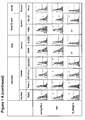
- TPP was able to specifically detect and penetrate each of the tested tumors at high yields in vivo (see Figures 3-5 , 11 ), as well as in vitro at single cell suspensions of the solid tumors (see Figure 1A and Table 1 below) with significant higher tumor to background ratios compared to a scrambled control peptide, and has further been used as the TPP molecule to be compared to the widely used tumor targeting probe IntegriSenseTM.
- ⁇ 3 -integrin was used for screening of the presence of the IntegriSenseTM targeting epitope.
- the IntegriSenseTM target epitope was absent at 6 of the 8 human tumor cell lines tested for ⁇ 3 -integrin (HCT-116, CX-2 (colon), MCF-7, MDA-MB231 (mamma), H-1339 (lung) and HeLa (cervix)), as well as at the spontaneous, colitis-associated murine colon carcinoma model.
- Table 1 Binding of TPP to different membrane-Hsp70 positive colon, mammary, pancreas, lung, head & neck and cervix mouse and human tumors.
- the proportion of membrane-Hsp70 positive cells was measured with cmHsp70.1 antibody and TPP peptide on single cell suspensions of in vivo -grown human and mouse tumors of different entities.
- the expression of CD61 ( ⁇ 3-integrin) was also determined on these tumor cells.
- a sample was determined as membrane positive if more than 20% of the cells showed a positive staining; n.t., not tested.
- Hsp70 and integrin positive tumors showed tumor cell specific uptake of both, TPP and IntegriSenseTM, and additional high uptake of IntegriSenseTM in tumor microenvironment, containing macrophages and fibroblasts (see Figures 3A-C ).
- Hsp70-positive and ⁇ 3 -integrin negative tumors showed comparable high enrichment of TPP in the tumor cells.
- IntegriSenseTM was present in the tumor microenvironment, while tumor cells remained unstained (see Figure 4 ).
- TPP is cleared out of the body via kidney while not accumulating in liver like other tumor targeting probes, as e.g. antibody like molecules. This makes TPP a potential targeting probe for hepatocellular carcinoma or liver metastases detection or their theranostic treatment (see Figure 2 ).
- CXCR4 Another recently discovered tumor targeting strategy involves the chemokine receptor CXCR4. Similar to Hsp70, CXCR4 offers the possibility to target different tumor entities. However, some carcinomas, like the non-small cell lung cancer (NSCLC), do not or only weakly express CXCR4 on their plasma membranes. In contrast, Hsp70 is present on all tested NSCLC tumors (xenograft, primary human tumors) (Hantschel et al., 2000). Furthermore, potential adverse effects of targeting CXCR4, such as the mobilization of hematopoietic progenitor cells by CXCR4 antagonists, remain to be elucidated.
- NSCLC non-small cell lung cancer
- TPP has any toxic side-effects, even if it was administered at 10-fold higher doses than those that were used for in vivo imaging.
- Ex vivo biodistribution analysis revealed the beneficial tumor-homing capacities of the TPP peptide.
- TPP was effectively washed out of the normal tissue 24 h after its administration, pronounced levels remained within the tumor tissue at this time point ( Figure 2 ).
- the small molecular weight of TPP ensures that it is cleared via the kidneys and does not accumulate in the liver like high molecular weight targeting probes such as antibodies. As a consequence, TPP did not result in non-specific background signals in the liver. Therefore, TPP is a useful imaging probe for hepatocellular carcinoma and liver metastases.
- TPP is a highly suitable peptide-based marker for diagnostic tumor targeting, as well as an efficient tool for drug delivery for a broad variety of human tumors.
- mice BALB/c, FvB and SHO- Prkdc scid Hr Hr (SHO) mice were obtained from an animal breeding colony (Charles River, Wilmington, MA, USA and Harlan Winkelmann, Borchen, Germany) and were maintained in pathogen-free, individually ventilated cages (Tecniplast, Hohenpeissenberg, Germany). All animal experiments were approved by the District Government of Upper Bavaria and performed in accordance with the German Animal Welfare and Ethical Guidelines of the Stammumpit der Isar, TUM, Kunststoff, Germany. Tumors were examined after the animals were sacrificed by cervical dislocation.
- CT26 mouse colon adenocarcinoma cells (5x10 5 ) or 4T1 (ATCC CRL-2539) mouse mammary carcinoma cells (2.5x10 5 ) (American Type Culture Collection, ATCC, authentication not applicable for mouse cell lines), both derived from BALB/c mouse strains, were harvested in the exponential growth phase and injected subcutaneously (s.c.) in the neck or orthotopically (o.t.) into the mammary fad pad of 8-week-old, female BALB/c mice, respectively. Primary tumor growth was followed by ultrasonic measurements (Logiq-5, GE Healthcare, Solingen, Germany). Intraoperative in vivo imaging was performed post mortem on exposed tumors when the CT26 tumors reached a size of approximately 0.4 cm 3 . 4T1-derived tumors and lung metastases were investigated after 25 to 30 days of tumor growth.
- mice were sacrificed and intraoperative in vivo imaging was performed on the exposed tumors when tumors had reached a volume of 0.2-0.4 cm 3 . If not stated otherwise, cell line authentication was performed by short tandem repeat profiles that are generated by simultaneous amplification of multiple short tandem repeat loci by ATCC and DSMZ. Cells were not cultured for longer than 5 months after testing.
- mice Spontaneous colonic tumors were induced in eight-week-old female FvB mice (Charles River) using an established approach. Briefly, mice were injected intraperitoneally (i.p.) with a single dose of azoxymethane (AOM; Sigma-Aldrich, Germany) at 10 mg/kg body weight. Following AOM treatment, the mice were subjected to 3 cycles of dextran sulfate sodium salt (DSS; MP Biomedicals Germany) administration in their drinking water (cycle 1: 3%, 5 days; cycle 2: 2%, 5 days; cycle 3: 1.5%, days). After 15-18 weeks of tumor development, the mice were sacrificed and the colon was resected for experiments. The presence of colorectal tumors was confirmed using standard histological techniques.
- AOM azoxymethane
- DSS dextran sulfate sodium salt
- TPP TPP-bearing mice
- s.c. tumors s.c. tumors, size 0.4 cm 3
- the health status and the general behavior of mice were inspected. Animals were sacrificed on day 5 and the heart, liver, spleen, lung and kidneys examined for pathological changes using standard histological techniques.
- the membrane-Hsp70 phenotype of tumors grown in vivo was determined by flow cytometric analysis of single cell suspensions that were generated following 30 min collagenase/dispase digestion using fluorescein-isothiocyanate (FITC)-conjugated cmHsp70.1 mAb (IgG1, multimmune GmbH, Kunststoff) or carboxyfluorescein-conjugated TPP (TPP[CF]).
- FITC fluorescein-isothiocyanate
- TPP[CF] carboxyfluorescein-conjugated TPP
- TPP Cy5.5 or DyLight750-conjugated Hsp70 specific 14-mer tumor penetrating peptide
- TPP Cy5.5 or DyLight750-conjugated Hsp70 specific 14-mer tumor penetrating peptide
- CP unspecific scrambled control peptide
- IntegriSenseTM 750 IS750, PerkinElmer, MA, USA
- the intra-operative imaging procedure which captured color and fluorescence images of the exposed tumors and its surrounding normal tissue was configured to simulate an intraoperative imaging situation.
- TPP[Cy5.5] or CP[DL750] 100 ⁇ g, equal to 45 nmol per animal
- IS750 at the recommended dose of 2 nmol per animal
- Concentrations above 2 nmol resulted in a higher staining of healthy tissues and thus negatively impacted the tumor-to-background ratio (data not shown).
- Signal specificity was determined by calculating the ratio of the mean signal intensity in the exposed tumor tissue and that of the surrounding normal tissue (tumor-to-background ratio, TBR) (Themelis et al., 2011).
- TBR tumor-to-background ratio
- Kinetic studies were performed 12, 24, 48 and 72 h after tail vein bolus injection of TPP[Cy5.5] and CP[DL750]. All other experiments were performed 24 h after i.v. injection of the respective probes.
- Fluorescent images were acquired by illuminating the specimens using 670 nm and 740 nm diode lasers and guiding the emitted fluorescence through appropriate emission filters before capturing it using a back illuminated EM-CCD camera (iXon DU888, Andor, Northern Ireland), as described previously (Stangl et al, 2011 a).
- EM-CCD camera iXon DU888, Andor, Northern Ireland
- the selection of the regions for tumor and background was done by two independent researchers in a blinded way. Within the field of view (1.2 x 1.2 cm 2 ) the complete tumor and normal tissue areas were gated macroscopically as separate regions of interest (ROI) in the true color image mode. ROI were then transferred one-to-one to the fluorescence images. Mean fluorescence intensities of the respective ROIs using unprocessed images were determined by using image J 1.48v software (NIH, USA) to calculate the tumor-to background ratios. A verification of the different tissue types (tumor and surrounding normal tissue) was done by a histological inspection of representative sections of both ROIs by a pathologist.
- ROI regions of interest
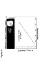
- TPP[Cy5.5] For biodistribution tumor-bearing mice were sacrificed 12, 24 or 48 h after i.v. injection of TPP[Cy5.5]. Fluorescent signal intensities of 0.25 cm 3 tissue cubes, taken from the tumor, spleen, pancreas, liver, lung, duodenum, kidney, heart, and 0.25 ml of peripheral blood were measured as described above. Agarose-gel cubes with standardized concentrations were used to calculate the probe concentrations in the investigated tissue samples. Detailed information concerning the quantification of TPP[Cy5.5] is provided in Figure 10 .
- TPP[Cy5.5] in the tumors and organs were assessed.
- standardized 0.25 cm 3 cubes of the tissues were cut using a 0.25 x 0.25cm x 0.25 cm form cutter with an open end and a scalpel to cut at this defined height.
- Biodistribution measurements were performed at time points of 12, 24 and 48 h of circulation in the body. Fluorescence intensities of the tissues were measured as described in the imaging procedures chapter. Quantification of the Cy5.5 contents in the defined tissue volumes was calculated using fluorescence signal intensities of standards with defined concentrations.
- TPP[Cy5.5] were dissolved in 1% agarose gel cubes, supplemented with 10% milk powder to mimic the optical properties of body tissue, and cut in the manner described above. Fluorescence signal intensity was measured using the same instrument settings than for the tissue specimen ( Figure 10 , upper panel). A control-cube containing 10% milk-powder in 1% agarose did not emit any fluorescence in the Cy5.5 channel. Linear regression equation of the standards was calculated with Microsoft Excel 2007 ( Figure 10 , lower panel). The concentration of TPP[Cy5.5] in organs was calculated using the regression equation.
- TPP[CF] 100 ⁇ g
- IS[750] IntegriSenseTM 750
- Tissue was collected and 8 ⁇ m cryosections counterstained with DAPI (DAKO, Hamburg, Germany) and examined microscopically using a Zeiss Observer Z1 (Carl Zeiss, Oberkochen, Germany) equipped with standard filters for GFP and Cy7 after 4 min of fixation with 1.8% w/v PFA.
- a cell population was defined to be membrane-positive if as more than 20% of the cells were stained.
- CD61 staining revealed a ⁇ 3 -integrin positivity on mouse colon (CT26) and mammary (4T1) carcinoma, on human mammary (MDA-MB231, T-47D) and head and neck (CAL-33) carcinoma, as well as on human and mouse pancreatic carcinoma (PDAC, Panc-1, MIA PaCa-2, COLO357).
- TPP only weakly stains (below 10%) tumor-associated fibroblasts and tumor-infiltrating macrophages; in contrast, ⁇ 3-integrin positivity was found on more than 60% of tumor-associated fibroblasts and tumor-infiltrating macrophages ( Figure 1B ).
- Hsp70 positivity was found on tumors.
- cryosections were stained with the Hsp70 specific antibody cmHsp70.1. Whereas tumor cells of the various tumor models were positive, cells of the tumor microenvironment remained negative for Hsp70.
- a representative staining of a PDAC tumor is shown in Figure 6B .
- TPP tumor-bearing BALB/c mice
- TPP tumor-bearing BALB/c mice
- Gross monitoring of the mice did not show any toxic reactions such as skin ulcerations or toxic death at any of the tested doses.
- any animal does not exhibit any loss of weight or change in mobility.
- An examination of the effects of TPP concentrations up to 500 ⁇ g in tumor-bearing mice also did not exert any side effects.
- TPP accumulates within the tumor with maximal specificity 24 h post i.v. injection.
- TPP[Cy5.5] TPP[Cy5.5] signal was detected within the tumor (7.6 ⁇ 0.7 ⁇ g/cm 3 ) 12 h after i.v. injection. This reached a maximum after 24 h (10.4 ⁇ 2.2 ⁇ g/cm 3 ) and dropped after 48 h.
- TPP accumulation was significantly lower at all investigated time points in all other tested organs, except the kidney.
- concentration of TPP[Cy5.5] reached its maximum in the kidney after 12 h at 13.7 ⁇ 0.6 ⁇ g/cm 3 , and this accumulation was followed by a sharp drop, 24 and 48 h post injection.
- TPP[Cy5.5] yielded maximal tumor-to-background ratio (TBR) in primary tumors 24 h after injection of the probe, whereas the peak TBR in metastasized lungs occurred after 48 h.
- the scrambled control peptide CP[DL750] did not show any specific accumulation in tumor tissue ( Figure 9A-C ).
- TPP and IS were further confirmed on cryo-sections of the lesions which were obtained 24 h after i.v. injection of TPP[CF] and IS[750] ( Figure 3D ). Both probes specifically enriched in the primary tumor and lung metastases, however, a background-staining of IS[750] remained in healthy lung tissue.
- TPP tumor-homing capacity
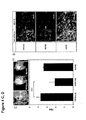
- FIG. 12 shows the uptake of TPP in three tumor (mamma carcinoma) cell lines which differ in their Hsp70 membrane expression (as shown in Figure 1 ), wherein TPP internalizes best in tumor cells with high Hsp70 membrane expression ( Figure 3A , right column) whereas scrambled control peptide (NGL) is not internalized ( Figure 3A , left column).
- scrambled control peptide (NGL) NGLTLKNDFSRLEG SEQ ID NO. 4
- TPP and IntegriSenseTM were also investigated using an endogenous pancreatic PDAC model which has an activating mutation in the Kras oncogene concomitant with a deletion of the tumor suppressor Tp53 (Ardito et al., 2012).
- Tumor cells of this model exhibited strong membrane positivity of both Hsp70 and ⁇ 3-integrin (seeTable 1). Imaging of the pancreatic tumors was performed after opening of the peritoneal cavity; the stomach, liver and gut were used to calculate the background fluorescence.
- Figure 4D shows fluorescence images of a cryo-sliced pancreatic tumors 24 h after simultaneous injection of TPP[CF] and IS[750]. Although both compounds stained tumor cells equally (left, white arrows), cells of the tumor microenvironment showed a weaker staining with TPP[CF] (right).
- TPP[Cy5.5] tumor homing capacities of TPP[Cy5.5] in human tumors
- imaging experiments were performed in 13 different human tumor cell lines derived from 6 entities (colon, mammary, pancreas, head and neck, cervix and lung; Figure 5 ).
- the TBRs for TPP[Cy5.5] in the xenograft tumors ranged from 5.6 ⁇ 1.6 (mammary carcinoma cell line, MDA-MB231) to 7.8 ⁇ 1.4 (squamous cell carcinoma cell line of the head and neck, Cal-33), all of which were significantly higher than those of CP[DL750]. This indicates a specific enrichment of TPP[Cy5.5] in all of the investigated xenograft tumors.
- T-47D human carcinoma cell lines which were determined as being ⁇ 3-integrin positive were T-47D, MDA-MB-231 (mammary carcinomas) and Cal-33 (squamous cell carcinoma of the head and neck; see Table 1).
- the TBR for IS[750] in all ⁇ 3-integrin negative tumors remained clearly below the TBRs for TPP[750].
Landscapes
- Health & Medical Sciences (AREA)
- Life Sciences & Earth Sciences (AREA)
- Engineering & Computer Science (AREA)
- Biomedical Technology (AREA)
- General Health & Medical Sciences (AREA)
- Immunology (AREA)
- Bioinformatics & Cheminformatics (AREA)
- Veterinary Medicine (AREA)
- Public Health (AREA)
- Epidemiology (AREA)
- Animal Behavior & Ethology (AREA)
- Chemical & Material Sciences (AREA)
- Molecular Biology (AREA)
- Proteomics, Peptides & Aminoacids (AREA)
- Cell Biology (AREA)
- Hematology (AREA)
- Urology & Nephrology (AREA)
- Medicinal Chemistry (AREA)
- Tropical Medicine & Parasitology (AREA)
- Biotechnology (AREA)
- Microbiology (AREA)
- Toxicology (AREA)
- Food Science & Technology (AREA)
- Physics & Mathematics (AREA)
- Analytical Chemistry (AREA)
- Biochemistry (AREA)
- General Physics & Mathematics (AREA)
- Pathology (AREA)
- Pharmacology & Pharmacy (AREA)
- Medicines Containing Antibodies Or Antigens For Use As Internal Diagnostic Agents (AREA)
Priority Applications (4)
| Application Number | Priority Date | Filing Date | Title |
|---|---|---|---|
| EP14172984.8A EP2957299A1 (fr) | 2014-06-18 | 2014-06-18 | Composés à base de peptides et leurs utilisations pour l'imagerie et le ciblage de tumeurs |
| PCT/EP2015/063697 WO2015193426A1 (fr) | 2014-06-18 | 2015-06-18 | Composés à base de peptide et leurs utilisations pour l'imagerie et le ciblage de tumeur |
| US15/319,759 US20170128596A1 (en) | 2014-06-18 | 2015-06-18 | Peptide-Based Compounds and Their Uses for Tumor Imaging and Targeting |
| EP15731284.4A EP3157570B1 (fr) | 2014-06-18 | 2015-06-18 | Composés à base de peptides et leurs utilisations pour l'imagerie et le ciblage de tumeurs |
Applications Claiming Priority (1)
| Application Number | Priority Date | Filing Date | Title |
|---|---|---|---|
| EP14172984.8A EP2957299A1 (fr) | 2014-06-18 | 2014-06-18 | Composés à base de peptides et leurs utilisations pour l'imagerie et le ciblage de tumeurs |
Publications (1)
| Publication Number | Publication Date |
|---|---|
| EP2957299A1 true EP2957299A1 (fr) | 2015-12-23 |
Family
ID=50976493
Family Applications (2)
| Application Number | Title | Priority Date | Filing Date |
|---|---|---|---|
| EP14172984.8A Ceased EP2957299A1 (fr) | 2014-06-18 | 2014-06-18 | Composés à base de peptides et leurs utilisations pour l'imagerie et le ciblage de tumeurs |
| EP15731284.4A Active EP3157570B1 (fr) | 2014-06-18 | 2015-06-18 | Composés à base de peptides et leurs utilisations pour l'imagerie et le ciblage de tumeurs |
Family Applications After (1)
| Application Number | Title | Priority Date | Filing Date |
|---|---|---|---|
| EP15731284.4A Active EP3157570B1 (fr) | 2014-06-18 | 2015-06-18 | Composés à base de peptides et leurs utilisations pour l'imagerie et le ciblage de tumeurs |
Country Status (3)
| Country | Link |
|---|---|
| US (1) | US20170128596A1 (fr) |
| EP (2) | EP2957299A1 (fr) |
| WO (1) | WO2015193426A1 (fr) |
Cited By (2)
| Publication number | Priority date | Publication date | Assignee | Title |
|---|---|---|---|---|
| CN106204609A (zh) * | 2016-07-20 | 2016-12-07 | 复旦大学附属中山医院 | 共聚焦激光显微内镜肺部影像的处理和分析系统及方法 |
| CN111225679A (zh) * | 2017-08-19 | 2020-06-02 | 俄亥俄州创新基金会 | 新型基于肽的癌症显像剂 |
Families Citing this family (2)
| Publication number | Priority date | Publication date | Assignee | Title |
|---|---|---|---|---|
| EP3687559B1 (fr) * | 2017-09-29 | 2025-11-12 | Sanford Burnham Prebys Medical Discovery Institute | Compositions ciblant des macrophages associés à une tumeur et leurs méthodes d'utilisation |
| WO2019154842A1 (fr) * | 2018-02-06 | 2019-08-15 | Klinikum Rechts Der Isar Der Technischen Universität München | Composé pour bioimagerie moléculaire peropératoire, son procédé de fabrication, son utilisation dans la bioimagerie moléculaire peropératoire et procédé chirurgical comprenant une bioimagerie moléculaire peropératoire |
Citations (1)
| Publication number | Priority date | Publication date | Assignee | Title |
|---|---|---|---|---|
| US20130156694A1 (en) * | 2011-12-15 | 2013-06-20 | Alternative Innovative Technologies Llc | Hsp70 fusion protein conjugates and uses thereof |
-
2014
- 2014-06-18 EP EP14172984.8A patent/EP2957299A1/fr not_active Ceased
-
2015
- 2015-06-18 WO PCT/EP2015/063697 patent/WO2015193426A1/fr not_active Ceased
- 2015-06-18 US US15/319,759 patent/US20170128596A1/en not_active Abandoned
- 2015-06-18 EP EP15731284.4A patent/EP3157570B1/fr active Active
Patent Citations (1)
| Publication number | Priority date | Publication date | Assignee | Title |
|---|---|---|---|---|
| US20130156694A1 (en) * | 2011-12-15 | 2013-06-20 | Alternative Innovative Technologies Llc | Hsp70 fusion protein conjugates and uses thereof |
Non-Patent Citations (25)
| Title |
|---|
| ARDITO, C. M. ET AL.: "EGF receptor is required for KRAS-induced pancreatic tumorigenesis", CANCER CELL, vol. 22, 2012, pages 304 - 317 |
| BIDWELL GL: "Peptides for cancer therapy: a drug-development opportunity and a drug-delivery challenge", THER DELIV., vol. 3, no. 5, May 2012 (2012-05-01), pages 609 - 21 |
| CARDOSO F; SAGHATCHIAN M; THOMPSON A; RUTGERS E: "Inconsistent criteria used in American Society of Clinical Oncology 2007 update of recommendations for the use of tumor markers in breast cancer", J CLIN ONCOL, vol. 26, 2008, pages 2058 - 9 |
| D. SCHILLING ET AL: "Binding of heat shock protein 70 to extracellular phosphatidylserine promotes killing of normoxic and hypoxic tumor cells", THE FASEB JOURNAL, vol. 23, no. 8, 16 March 2009 (2009-03-16), pages 2467 - 2477, XP055153541, ISSN: 0892-6638, DOI: 10.1096/fj.08-125229 * |
| DEARLING JL; BARNES JW; PANIGRAHY D; ZIMMERMAN RE; FAHEY F; TREVES ST; MORRISON MS; KIERAN MW; PACKARD AB: "Specific uptake of 99mTc-NC100692, an avp3-targeted imaging probe, in subcutaneous and orthotopic tumors", NUCL MED BIOL., vol. 40, no. 6, 2013, pages 788 - 94 |
| ECKMANN, L. ET AL.: "Opposing functions of IKKbeta during acute and chronic intestinal inflammation", PROCEEDINGS OF THE NATIONAL ACADEMY OF SCIENCES OF THE UNITED STATES OF AMERICA, vol. 105, 2008, pages 15058 - 15063, XP008156789, DOI: doi:10.1073/pnas.0808216105 |
| GEHRMANN, M. ET AL.: "Immunotherapeutic targeting of membrane Hsp70-expressing tumors using recombinant human granzyme B", PLOS ONE, vol. 7, 2012, pages E41341 |
| HANTSCHEL, M. ET AL.: "Hsp70 plasma membrane expression on primary tumor biopsy material and bone marrow of leukemic patients", CELL STRESS. CHAPERONES, vol. 5, 2000, pages 438 - 442, XP008033318, DOI: doi:10.1379/1466-1268(2000)005<0438:HPMEOP>2.0.CO;2 |
| HOSOTANI, R. ET AL.: "Expression of integrin alphaVbeta3 in pancreatic carcinoma: relation to MMP-2 activation and lymph node metastasis", PANCREAS, vol. 25, 2002, pages E30 - 35 |
| HUANG CW; LI Z; CONTI PS: "In vivo near-infrared fluorescence imaging of integrin ?2?1 in prostate cancer with cell-penetrating-peptide-conjugated DGEA probe", J NUCL MED., vol. 52, no. 12, 2011, pages 1979 - 86 |
| KAUMAYA PT; FOY KC: "Peptide vaccines and targeting HER and VEGF proteins may offer a potentially new paradigm in cancer immunotherapy", FUTURE ONCOL., vol. 8, no. 8, August 2012 (2012-08-01), pages 961 - 87, XP008173397, DOI: doi:10.2217/fon.12.95 |
| KONDO E; SAITO K; TASHIRO Y; KAMIDE K; UNO S; FURUYA T ET AL.: "Tumour lineage-homing cell-penetrating peptides as anticancer molecular delivery systems", NAT COM, vol. 3, 2012, pages 951 |
| MATHIAS GEHRMANN ET AL: "Tumor Imaging and Targeting Potential of an Hsp70-Derived 14-Mer Peptide", PLOS ONE, vol. 9, no. 8, 28 August 2014 (2014-08-28), pages e105344, XP055153404, DOI: 10.1371/journal.pone.0105344 * |
| MURPHY CG; MORRIS PG: "Recent advances in novel targeted therapies for HER2-positive breast cancer", ANTICANCER DRUGS, vol. 23, no. 8, September 2012 (2012-09-01), pages 765 - 76 |
| NYLANDSTED, J. ET AL.: "Heat shock protein 70 promotes cell survival by inhibiting lysosomal membrane permeabilization", THE JOURNAL OF EXPERIMENTAL MEDICINE, vol. 200, 2004, pages 425 - 435 |
| OKARVI SM: "Peptide-based radiopharmaceuticals and cytotoxic conjugates: potential tools against cancer", CANCER TREAT REV., vol. 34, no. 1, February 2008 (2008-02-01), pages 13 - 26, XP022448866, DOI: doi:10.1016/j.ctrv.2007.07.017 |
| S. STANGL ET AL: "Selective in vivo imaging of tumors with a tumor cell-specific Hsp70 peptide-based probe", CANCER RESEARCH, 9 October 2014 (2014-10-09), XP055153391, ISSN: 0008-5472, DOI: 10.1158/0008-5472.CAN-14-0413 * |
| SCHILLING D; BAYER C; LI W; MOLLS M; VAUPEL P; MULTHOFFG.: "Radiosensitization ofnormoxic and hypoxic h1339 lung tumor cells by heat shock protein 90 inhibition is independent of hypoxia inducible factor-la", PLOS ONE, vol. 7, no. 2, 7 February 2012 (2012-02-07), pages E31110 |
| SCHILSKY RL; TAUBE SE: "Tumor markers as clinical cancer tests - are we there yet?", SEM ONCOL, vol. 29, 2002, pages 211 - 2 |
| STANGL, S. ET AL.: "argeting membrane heat-shock protein 70 (Hsp70) on tumors by cmHsp70.1 antibody", PROCEEDINGS OF THE NATIONAL ACADEMY OF SCIENCES OF THE UNITED STATES OF AMERICA, vol. 108, 2011, pages 733 - 738, XP055261079, DOI: doi:10.1073/pnas.1016065108 |
| STANGL, S. ET AL.: "In vivo imaging of CT26 mouse tumours by using cmHsp70.1 monoclonal antibody", JOURNAL OF CELLULAR AND MOLECULAR MEDICINE, vol. 15, 2011, pages 874 - 887, XP055090343, DOI: doi:10.1111/j.1582-4934.2010.01067.x |
| STEFAN STANGL ET AL: "In vivo imaging of CT26 mouse tumours by using cmHsp70.1 monoclonal antibody", JOURNAL OF CELLULAR AND MOLECULAR MEDICINE, vol. 15, no. 4, 1 April 2011 (2011-04-01), pages 874 - 887, XP055090343, ISSN: 1582-1838, DOI: 10.1111/j.1582-4934.2010.01067.x * |
| THEMELIS G; HARLAAR NJ; KELDER W; BART J; SARANTOPOULOS A; VAN DAM GM ET AL.: "Enhancing surgical vision by using real-time imaging of alphaVbeta3-integrin targeted near-infrared fluorescent agent", ANNALS SURG ONCOL, vol. 18, 2011, pages 3506 - 13, XP019963745, DOI: doi:10.1245/s10434-011-1664-9 |
| TRAJKOVIC-ARSIC M; MOHAJERANI P; SARANTOPOULOS A; KALIDERIS E; STEIGER K; ESPOSITO I; MA X; THEMELIS G; BURTON N; MICHALSKI CW: "Multimodal molecular imaging of integrin avp3 for in vivo detection of pancreatic cancer", J NUCL MED., vol. 55, no. 3, 2014, pages 446 - 51 |
| XIN L; CAO J; CHENG H; ZENG F; HU X ET AL.: "Human monoclonal antibodies in cancer therapy: a review of recent developments", FRONT BIOSCI, vol. 18, 2013, pages 765 - 772 |
Cited By (4)
| Publication number | Priority date | Publication date | Assignee | Title |
|---|---|---|---|---|
| CN106204609A (zh) * | 2016-07-20 | 2016-12-07 | 复旦大学附属中山医院 | 共聚焦激光显微内镜肺部影像的处理和分析系统及方法 |
| CN106204609B (zh) * | 2016-07-20 | 2019-03-19 | 复旦大学附属中山医院 | 共聚焦激光显微内镜肺部影像的处理和分析系统及方法 |
| CN111225679A (zh) * | 2017-08-19 | 2020-06-02 | 俄亥俄州创新基金会 | 新型基于肽的癌症显像剂 |
| CN111225679B (zh) * | 2017-08-19 | 2024-03-29 | 俄亥俄州创新基金会 | 新型基于肽的癌症显像剂 |
Also Published As
| Publication number | Publication date |
|---|---|
| WO2015193426A1 (fr) | 2015-12-23 |
| EP3157570A1 (fr) | 2017-04-26 |
| EP3157570B1 (fr) | 2023-06-07 |
| EP3157570C0 (fr) | 2023-06-07 |
| US20170128596A1 (en) | 2017-05-11 |
Similar Documents
| Publication | Publication Date | Title |
|---|---|---|
| Xiang et al. | Superior performance of aptamer in tumor penetration over antibody: implication of aptamer-based theranostics in solid tumors | |
| Stangl et al. | Selective In Vivo Imaging of Syngeneic, Spontaneous, and Xenograft Tumors Using a Novel Tumor Cell–Specific Hsp70 Peptide-Based Probe | |
| US6919425B2 (en) | Isolation of a cell-specific internalizing peptide that infiltrates tumor tissue for targeted drug delivery | |
| US20200002401A1 (en) | Cd44 binding peptides | |
| US12458618B2 (en) | Compounds and methods for the detection of TRPV-6 cancers and drug delivery | |
| US20120093733A1 (en) | Multidomain biotags for cancer detection, diagnosis and therapy and methods of their use | |
| Gehrmann et al. | Tumor imaging and targeting potential of an Hsp70-derived 14-mer peptide | |
| TWI780082B (zh) | 新穎抗人類MUC1抗體Fab片段 | |
| Kobayashi et al. | A novel PET imaging using 64Cu‐labeled monoclonal antibody against mesothelin commonly expressed on cancer cells | |
| Stangl et al. | In vivo imaging of CT26 mouse tumours by using cmHsp70. 1 monoclonal antibody | |
| Pung et al. | Generation of peptides using phage display technology for cancer diagnosis and molecular imaging | |
| EP3157570B1 (fr) | Composés à base de peptides et leurs utilisations pour l'imagerie et le ciblage de tumeurs | |
| US11077212B2 (en) | Molecular imaging of cancer cells in vivo | |
| CN107868129A (zh) | 与肝癌标志物gpc3相关的亲和肽 | |
| Li et al. | Glucosamine-bound near-infrared fluorescent probes with lysosomal specificity for breast tumor imaging | |
| Conti et al. | Optical imaging detection of microscopic mammary cancer in ErbB‐2 transgenic mice through the DA364 probe binding αvβ3 integrins | |
| CN101773677A (zh) | 一种活体肿瘤显像靶分子及其特异性探针 | |
| JP2017510547A (ja) | 肝細胞ガンの処置のための方法及び医薬組成物 | |
| KR102557303B1 (ko) | 인간 피브로넥틴 도메인 ⅲ 기본 골격의 신규 칼레티큘린 특이적 결합 단백질 및 그의 용도 | |
| Zeng et al. | Identification of a novel peptide targeting TIGIT to evaluate immunomodulation of 125I seed brachytherapy in HCC by near-infrared fluorescence | |
| US20210138090A1 (en) | uPAR targeting peptide for use in peroperative optical imaging of invasive cancer | |
| WO2017086090A1 (fr) | Peptide présentant une accumulation spécifique au cancer du pancréas, et utilisation dudit peptide | |
| Liu et al. | PEGylated and non-PEGylated TCP-1 probes for imaging of colorectal cancer | |
| CN119638793A (zh) | 一种靶向cd105的多肽分子探针及其应用 | |
| Kampmeier | Site directed modification of recombinant antibody fragments for in vivo fluorescence imaging and targeted drug delivery |
Legal Events
| Date | Code | Title | Description |
|---|---|---|---|
| PUAI | Public reference made under article 153(3) epc to a published international application that has entered the european phase |
Free format text: ORIGINAL CODE: 0009012 |
|
| AK | Designated contracting states |
Kind code of ref document: A1 Designated state(s): AL AT BE BG CH CY CZ DE DK EE ES FI FR GB GR HR HU IE IS IT LI LT LU LV MC MK MT NL NO PL PT RO RS SE SI SK SM TR |
|
| AX | Request for extension of the european patent |
Extension state: BA ME |
|
| STAA | Information on the status of an ep patent application or granted ep patent |
Free format text: STATUS: THE APPLICATION HAS BEEN REFUSED |
|
| 18R | Application refused |
Effective date: 20160228 |