EP2982697A1 - Compositions et procédés pour la détection de la nitrosylation et de l'oxydation de protéines - Google Patents
Compositions et procédés pour la détection de la nitrosylation et de l'oxydation de protéines Download PDFInfo
- Publication number
- EP2982697A1 EP2982697A1 EP15179608.3A EP15179608A EP2982697A1 EP 2982697 A1 EP2982697 A1 EP 2982697A1 EP 15179608 A EP15179608 A EP 15179608A EP 2982697 A1 EP2982697 A1 EP 2982697A1
- Authority
- EP
- European Patent Office
- Prior art keywords
- antibody
- ptp1b
- pac
- protein
- nitrosylation
- Prior art date
- Legal status (The legal status is an assumption and is not a legal conclusion. Google has not performed a legal analysis and makes no representation as to the accuracy of the status listed.)
- Granted
Links
Images



Classifications
-
- C—CHEMISTRY; METALLURGY
- C07—ORGANIC CHEMISTRY
- C07K—PEPTIDES
- C07K16/00—Immunoglobulins [IG], e.g. monoclonal or polyclonal antibodies
- C07K16/44—Immunoglobulins [IG], e.g. monoclonal or polyclonal antibodies against material not provided for elsewhere, e.g. haptens, metals, DNA, RNA, amino acids
-
- G—PHYSICS
- G01—MEASURING; TESTING
- G01N—INVESTIGATING OR ANALYSING MATERIALS BY DETERMINING THEIR CHEMICAL OR PHYSICAL PROPERTIES
- G01N33/00—Investigating or analysing materials by specific methods not covered by groups G01N1/00 - G01N31/00
- G01N33/48—Biological material, e.g. blood, urine; Haemocytometers
- G01N33/50—Chemical analysis of biological material, e.g. blood, urine; Testing involving biospecific ligand binding methods; Immunological testing
- G01N33/68—Chemical analysis of biological material, e.g. blood, urine; Testing involving biospecific ligand binding methods; Immunological testing involving proteins, peptides or amino acids
- G01N33/6803—General methods of protein analysis not limited to specific proteins or families of proteins
- G01N33/6806—Determination of free amino acids
- G01N33/6812—Assays for specific amino acids
- G01N33/6815—Assays for specific amino acids containing sulfur, e.g. cysteine, cystine, methionine, homocysteine
-
- G—PHYSICS
- G01—MEASURING; TESTING
- G01N—INVESTIGATING OR ANALYSING MATERIALS BY DETERMINING THEIR CHEMICAL OR PHYSICAL PROPERTIES
- G01N33/00—Investigating or analysing materials by specific methods not covered by groups G01N1/00 - G01N31/00
- G01N33/48—Biological material, e.g. blood, urine; Haemocytometers
- G01N33/50—Chemical analysis of biological material, e.g. blood, urine; Testing involving biospecific ligand binding methods; Immunological testing
- G01N33/68—Chemical analysis of biological material, e.g. blood, urine; Testing involving biospecific ligand binding methods; Immunological testing involving proteins, peptides or amino acids
- G01N33/6803—General methods of protein analysis not limited to specific proteins or families of proteins
- G01N33/6842—Proteomic analysis of subsets of protein mixtures with reduced complexity, e.g. membrane proteins, phosphoproteins, organelle proteins
-
- G—PHYSICS
- G01—MEASURING; TESTING
- G01N—INVESTIGATING OR ANALYSING MATERIALS BY DETERMINING THEIR CHEMICAL OR PHYSICAL PROPERTIES
- G01N2440/00—Post-translational modifications [PTMs] in chemical analysis of biological material
- G01N2440/26—Post-translational modifications [PTMs] in chemical analysis of biological material nitrosylation
Definitions
- Nitric oxide which can be produced intracellularly by endogenous NO synthase (NOS), acts as an important second messenger.
- NOS endogenous NO synthase
- the known functions of NO include regulation of smooth muscle relaxation, platelet aggregation, neurotransmission, neurotoxicity, cell death and differentiation.
- NO targets the heme group located at the active-site of soluble guanylyl cyclase, resulting in elevation of its enzymatic activity, consequently leading to vasorelaxation.
- SNO S-nitrosothiol
- Insulin delivery to the skeletal muscle interstitium plays a key role in insulin-directed glucose uptake by skeletal muscle.
- This insulin signaling-dependent process is tightly controlled by the endothelial barrier function of the capillaries connected to skeletal muscle. It has been shown that intrinsic NO functions to promote insulin delivery across endothelial barrier.
- the mechanism underlying NO-dependent insulin responsiveness in endothelium remains uncharacterized due to technical limitations of current detection methods for protein S-nitrosylation.
- the present invention provides an isolated antibody as well as compositions and methods for detection of protein S -nitrosylation specifically in vivo and in vitro.
- the methods of the invention can eliminate false-positive detection of S-nitrosylated protein, and enable the monitoring of the subcellular localization of S -nitrosylated proteins in cells.
- the methods of the invention can facilitate the understanding of the molecular mechanism of S -nitrosylation's role in signal transduction pathways. This invention also provides the application of detection in both biochemical and cell imaging formats.
- the present invention relates to an isolated antibody that specifically binds to a moiety having the structure of formula (A):
- the antibody of the invention specifically targets N-phenyl-acetamide group, and does not cross react with the moieties of formulae (B), (C) and (D):
- the isolated antibody disclosed herein is useful for detecting protein nitrosylation and oxidation in a protein substrate, which includes, but is not limited to, cells and tissues.
- Another aspect of the invention provides a method for determining the presence or absence of protein S-nitrosylation and/or oxidation in vivo and in vitro.
- the inventive method for determining the presence or absence of protein S-nitrosylation or oxidation comprises the following steps: a) contacting a sample with one or more reducing agents, b) treating the sample with a thiol-reactive probe to form one or more N-phenyl-acetamidyl cysteines, and c) detecting the N-phenyl-acetamidyl cysteine or cysteines by an anti-N-phenyl-acetamidyl cysteine (PAC) antibody, thereby determining the presence or absence of protein S-nitrosylation or oxidation.
- PAC anti-N-phenyl-acetamidyl cysteine
- the method further comprises a step of blocking all sulfhydryl (SH) groups of reduced cysteine residues in the sample prior to step (a).
- the step of blocking SH groups can be performed by one or more sulfhydryl-reactive alkylating agents that selectively react with sulfhydryl (SH) groups.
- the sulfhydryl-reactive alkylating agents are haloacetyl reagents.
- alkylating agents are iodoacetyl or bromoacetyl reagents.
- the alkylating agents are iodoacetyl reagents.
- the alkylating agents comprise iodoacetamide (IAM), N-(4-acetylphenyl)-2-iodoacetamide (APIAM) and/or N-(2,3-dimethylphenyl)-2-iodoacetamide (DPIAM).
- IAM iodoacetamide
- APIAM N-(4-acetylphenyl)-2-iodoacetamide
- DPIAM N-(2,3-dimethylphenyl)-2-iodoacetamide
- the sample is treated with IAM and APIAM sequentially or simultaneously.
- sulfhydryl-reactive alkylating agents react with sulfhydryl (SH) groups of cysteine residues at physiologic to alkaline conditions (pH 7.2 to 9) to form stable thioether linkages.
- SH sulfhydryl
- step a) in the method is to convert S-nitrosothiol (SNO) moiety and/or disulfide moiety to sulfhydryl (SH) moiety by using one or more reducing agents.
- SNO S-nitrosothiol
- SH sulfhydryl
- the reducing agent is a disulfide reducing agent.
- the disulfide reducing agent includes, but not limited to, Tris-(2-carboxyethyl) phosphine (TCEP), Tris-(hydroxypropyl)phosphine (THP), Dithiothreitol (DTT), Tris(3-hydroxypropyl)phosphine (THPP), cysteine, glutathione or combinations thereof.
- the reducing agent is an ascorbate or ascorbic acid.
- Ascorbate is a physiological antioxidant that serves as a SNO-specific reducing agent. After ascorbate treatment, S-nitrosothiols release NO on decomposition.
- one or more nitrosothiols and/or one or more disulfides in the sample are converted to one or more sulfhydryls.
- the newly generated sulfhydryl can react with a thiol-reactive alkylating agent, 2-Iodo-N-phenylacetamide (IAN), to form stable N-phenyl-acetamidyl cysteine as described in step (b).
- a thiol-reactive alkylating agent 2-Iodo-N-phenylacetamide (IAN)
- N-phenyl-acetamidyl cysteine can be specifically targeted by the antibody of the invention, anti-N-phenyl-acetamidyl cysteine (PAC) antibody, thereby determining the presence or absence of protein S-nitrosylation or oxidation as described in step (c).
- PAC anti-N-phenyl-acetamidyl cysteine
- any detectable label may be used with the antibody of the invention, such as fluorescent labels, radiolabels, enzymatic labels, and others.
- the detectable label is an optically-detectable label, such as a fluorescent label.
- Exemplary fluorescent labels include Atto, cyanine, rhodamine, fluorescien, coumarin, BODIPY, alexa, and conjugated multi-dyes.
- the inventive method can both quantify and identify the nature of nitrosylated and/or oxidative proteins, and can be used for screening therapeutic drugs that potentially modulating protein nitrosylation or oxidation.
- this invention is directed to a kit suitable for performing an assay which determines the presence or absence of protein S-nitrosylation and/or oxidation.
- the kit of this invention comprises the components referred to in the methods described above.
- the kit of the invention comprises the antibody of the invention described herein.
- the kit comprises the antibody of the invention and 2-iodo-N-phenyl-acetamide (IAN).
- the kit further comprises sulfhydryl-reactive alkylating agents comprising IAM, APIAM and/or DPIAM.
- the kit may contain ascorbate and/or ascorbic acid.
- the kit further comprises one or more disulfide alkylating agents and/or at least one reducing agent selecting from the group consisting of TCEP, THP, DTT, THPP, cysteine, glutathione, and combinations thereof.
- a kit of this invention comprises all components described above, instructions setting forth a procedure according to any one of the methods described above, and a container for the contents of the kit.
- the present invention relates to visualization techniques for in situ imaging of nitrosylated and/or oxidized proteins within cells and/or tissues.
- the method for detecting a nitrosylated and/or oxidized protein in situ comprises the step of a) contacting a sample with one or more reducing agents, (b) reacting the sample with N-phenyl-acetamide to form N-phenyl-acetamidyl cysteines, and c) detecting N-phenyl-acetamidyl cysteines by an anti-N-phenyl-acetamidyl cysteine (PAC) antibody, thereby detecting the nitrosylated and/or oxidized protein.
- PAC anti-N-phenyl-acetamidyl cysteine
- This invention also provides an analytic platform derived from the method for screening for drugs that potentially modulate protein nitrosylation or oxidation. This analytic platform is useful for both in vitro and in vivo conditions.
- the invention provides a new antibody-based method to detect S-nitrosylated and/or oxidized proteins with high specificity and accuracy.
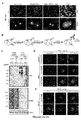
- the antibody-based strategy is illustrated in Figure 1a .
- the proteins that are recognized by the antibody of the invention can be those originally protected from post-fixing blocking (B) by a stimulus-dependent modification at the susceptible Cys, which can be reversed by reducing agents such as ascorbate then being alkylated (A), consistent with S-nitrosylation of the Cys.
- reducing agents such as ascorbate then being alkylated (A)
- S-nitrosoproteins As used herein, S-nitrosoproteins, S-nitrosothiols, and protein S-nitrosylation reactions are terms that refer to components with linkage through the thiol (-SH) group. These types of components play important roles in cell signaling processes such as neurotransmission, anion channel regulation, host defense and gene regulation.
- alkylating agent or “sulfhydryl-reactive alkylating agent” refers to an agent that forms alkylthiol groups when reacted under suitable conditions with free thiol groups.
- Alkylating agents contain straight or branched chain lower alkyl (C1-C6) groups that may be derivatized or functionalized.
- a blocking agent can be used to block free thiols in the test sample when carrying out the method of this invention. The blocking agent is preferably removed from the test sample prior to step (a) of the method of this invention.
- test samples any sample which may be suitably tested using the methods disclosed herein.
- Test samples can be e.g. in the form of any biological sample, for example, crude, purified or semipurified lysates of tissues that potentially comprise nitrosylated proteins, e.g. brain, peripheral nerve, muscle, blood vessels, blood cells, liver, etc.
- reducing agent refers to a compound such as ascorbate that reduces nitrosothiol bonds on a protein to form new free thiol groups.
- Agents such as Cu 2+ or Hg 2+ may also be used. Care must be taken to remove these, as these metals can interfere with antibody incubation and detection.
- the present invention provides an isolated antibody useful for in situ imaging of S- nitrosylated proteins in cells with high specificity and accuracy.
- the invention also provides an antibody-based method illustrated in Figure 1 to detect S-nitrosylated proteins with high specificity and accuracy.
- an antibody that "specifically binds to" or is “specific for” a particular polypeptide or an epitope on a particular polypeptide is one that binds to that particular polypeptide or epitope on a particular polypeptide without substantially binding to any other polypeptide or polypeptide epitope.
- the terms also mean that binding between two entities has an affinity as quantified by an association constant, K, of at least 10 6 M -1 , 10 7 M -1 , 10 8 M -1 , 10 9 M -1 , or 10 10 M -1 .
- the present invention provides an isolated antibody useful for in situ imaging of S- nitrosylated proteins in cells with high specificity and accuracy.
- the antibody described herein can be either monoclonal or polyclonal.
- a “monoclonal antibody” refers to a homogenous antibody population and a “polyclonal antibody” refers to a heterogenous antibody population.
- Antibodies described herein can be made by any method known in the art. See, for example, Harlow and Lane, (1988) Antibodies: A Laboratory Manual, Cold Spring Harbor Laboratory, New York .
- Polyclonal antibodies may be prepared by collecting blood from an immunized mammal examined for the increase of desired antibodies in the serum, and by separating serum from the blood using any conventional method.
- Polyclonal antibodies include serum containing the polyclonal antibodies, as well as a fraction containing the polyclonal antibodies that may be isolated from the serum.
- Polyclonal antibodies are generally raised in host animals (e.g., rabbit, mouse, guinea pig, horse, or goat) by multiple subcutaneous (sc) or intraperitoneal (ip) injections of a relevant antigen and an adjuvant.
- host animals e.g., rabbit, mouse, guinea pig, horse, or goat
- sc subcutaneous
- ip intraperitoneal
- a protein that is immunogenic in the species to be immunized e.g., keyhole limpet hemocyanin, serum albumin, bovine thyroglobulin, or soybean trypsin inhibitor
- a bifunctional or derivatizing agent for example, maleimidobenzoyl sulfosuccinimide ester (conjugation through cysteine residues), N-hydroxysuccinimide (through lysine residues), glutaraldehyde, succinic anhydride, SOCl 2 , etc.
- Any mammalian animal may be immunized with the antigen for producing the desired antibodies.
- animals of Rodentia, Lagomorpha, or Primates can be used.
- Animals of Rodentia include, for example, mouse, rat, and hamster.
- Animals of Lagomorpha include, for example, rabbit.
- Animals of Primates include, for example, a monkey of Catarrhini (old world monkey) such as Macaca fascicularis, rhesus monkey, baboon, and chimpanzees.
- antigens may be diluted and suspended in an appropriate amount of phosphate buffered saline (PBS), physiological saline, etc.
- PBS phosphate buffered saline
- the antigen suspension may be mixed with an appropriate amount of a standard adjuvant, such as Freund's complete adjuvant, made into emulsion, and then administered to mammalian animals.
- Animals can be immunized with the antigen, immunogenic conjugates, or derivatives by combining 1 mg or 1 ⁇ g of the antigen or conjugate (for rabbits or mice, respectively) with 3 volumes of Freund's incomplete adjuvant.
- Animals can be boosted until the titer plateaus by several administrations of antigen mixed with an appropriately amount of Freund's incomplete adjuvant every 4 to 21 days. Animals are boosted with 1/5 to 1/10 the original amount of antigen or conjugate in Freund's complete adjuvant by subcutaneous injection at multiple sites. Seven to 14 days later the animals are bled and the serum is assayed for antibody titer.
- An appropriate carrier may also be used for immunization. After immunization as above, serum is examined by a standard method for an increase in the amount of desired antibodies. Preferably, the animal is boosted with the conjugate of the same antigen, but conjugated to a different protein and/or through a different cross-linking reagent. Conjugates also can be made in recombinant cell culture as protein fusions. Also, aggregating agents such as alum are suitably used to enhance the immune response.
- antibodies can be made by the conventional hybridoma technology. Kohler et al., Nature, 256:495 (1975 ). In the hybridoma method, a mouse or other appropriate host animal, such as a hamster or rabbit, is immunized as hereinabove described to elicit lymphocytes that produce or are capable of producing antibodies that will specifically bind to the antigen used for immunization. Alternatively, lymphocytes may be immunized in vitro.
- immune cells are collected from the mammal immunized with the antigen and checked for the increased level of desired antibodies in the serum as described above, and are subjected to cell fusion.
- the immune cells used for cell fusion are preferably obtained from spleen.
- Other preferred parental cells to be fused with the above immunocyte include, for example, myeloma cells of mammalians, and more preferably myeloma cells having an acquired property for the selection of fused cells by drugs.
- Preferred myeloma cells are those that fuse efficiently, support stable high-level production of antibody by the selected antibody-producing cells, and are sensitive to a medium such as HAT medium.
- preferred myeloma cell lines are murine myeloma lines, such as those derived from MOPC-21 and MPC-11 mouse tumors available from the Salk Institute Cell Distribution Center, San Diego, Calif. USA, and SP-2 cells available from the American Type Culture Collection, Rockville, Md. USA.
- Human myeloma and mouse-human heteromyeloma cell lines also have been described for the production of human monoclonal antibodies ( Kozbor, J. Immunol., 133:3001 (1984 ); Brodeur et al., Monoclonal Antibody Production Techniques and Applications, pp. 51-63 (Marcel Dekker, Inc., New York, 1987 )).
- the above immunocyte and myeloma cells can be fused according to known methods, for example, the method of Milstein et al. ( Galfre et al., Methods Enzymol. 73:3-46, 1981 ). Lymphocytes are fused with myeloma cells using a suitable fusing agent, such as polyethylene glycol, to form a hybridoma cell ( Goding, Monoclonal Antibodies: Principles and Practice, pp.59-103 (Academic Press, 1986 )). Resulting hybridomas obtained by the cell fusion may be selected by cultivating them in a standard selection medium, such as HAT medium (hypoxanthine, aminopterin, and thymidine containing medium).
- HAT medium hyperxanthine, aminopterin, and thymidine containing medium.
- the cell culture is typically continued in the HAT medium for several days to several weeks, the time being sufficient to allow all the other cells, with the exception of the desired hybridoma, to die. Then, the standard limiting dilution is performed to screen and clone a hybridoma cell producing the desired antibody.
- the hybridoma cells thus prepared are seeded and grown in a suitable culture medium that preferably contains one or more substances that inhibit the growth or survival of the unfused, parental myeloma cells.
- a suitable culture medium that preferably contains one or more substances that inhibit the growth or survival of the unfused, parental myeloma cells.
- the culture medium for the hybridomas typically will include hypoxanthine, aminopterin, and thymidine (HAT medium), which substances prevent the growth of HGPRT-deficient cells.
- Culture medium in which hybridoma cells are growing is assayed for production of monoclonal antibodies directed against the antigen.
- the binding specificity of monoclonal antibodies produced by hybridoma cells is determined by immunoprecipitation or by an in vitro binding assay. Measurement of absorbance in enzyme-linked immunosorbent assay (ELISA), enzyme immunoassay (EIA), radioimmunoassay (RIA), and/or immunofluorescence may be used to measure the antigen binding activity of the antibody of the invention.
- ELISA enzyme-linked immunosorbent assay
- EIA enzyme immunoassay
- RIA radioimmunoassay
- immunofluorescence may be used to measure the antigen binding activity of the antibody of the invention.
- the antibody of the present invention is immobilized on a plate, protein of the invention is applied to the plate, and then a sample containing a desired antibody, such as culture supernatant of antibody producing cells or purified antibodies, is applied. Then, a secondary antibody that recognizes the primary antibody and is labeled with an enzyme, such as alkaline phosphatase, is applied, and the plate is incubated. Next, after washing, an enzyme substrate, such as p-nitrophenyl phosphate, is added to the plate, and the absorbance is measured to evaluate the antigen binding activity of the sample.
- a fragment of the protein, such as a C-terminal or N-terminal fragment may be used in this method.
- BIAcore (Pharmacia) may be used to evaluate the activity of the antibody according to the present invention.
- the binding affinity of the monoclonal antibody can, for example, be determined by the Scatchard analysis of Munson et al., Anal. Biochem., 107:220 (1980 ).
- hybridoma cells producing antibodies that bind to epitopes described herein can be identified and selected for further characterization.
- the corresponding clones may be subcloned by limiting dilution procedures and grown by standard methods ( Goding, Monoclonal Antibodies: Principles and Practice, pp.59-103 (Academic Press, 1986 )).
- Suitable culture media for this purpose include, for example, D-MEM or RPMI-1640 medium.
- the monoclonal antibodies secreted by the subclones are suitably separated from the culture medium, ascites fluid, or serum by conventional immunoglobulin purification procedures such as, for example, protein A-Sepharose, hydroxylapatite chromatography, gel electrophoresis, dialysis, or affinity chromatography.
- the hybridoma cells may be grown in vivo as ascites tumors in an animal.
- the obtained hybridomas can be subsequently transplanted into the abdominal cavity of a mouse and the ascites are harvested.
- the obtained monoclonal antibodies can be purified by, for example, ammonium sulfate precipitation, a protein A or protein G column, DEAE ion exchange chromatography, or an affinity column to which the protein of the present invention is coupled.
- the antibody of the present invention can be used not only for purification and detection of the protein of the present invention, but also as a candidate for agonists and antagonists of the protein of the present invention.
- this antibody can be applied to the antibody treatment for diseases related to the protein of the present invention.
- the invention provides a method for determining the presence or absence of S-nitrosylation or reversible Cys oxidation of protein substrates in vivo and in vitro.
- the method for assaying nitrosylation of protein substrates is described herein.
- This antibody together with the inventive method can also be used for assaying reversible Cys oxidation of protein substrates.
- the inventive method for determining the presence or absence of protein S-nitrosylation or oxidation comprises the following steps: a) contacting a sample with one or more reducing agents, b) treating the sample with a thiol-reactive probe to form N-phenyl-acetamidyl cysteine, and c) detecting N-phenyl-acetamidyl cysteine by an anti-N-phenyl-acetamidyl cysteine (PAC) antibody, thereby determining the presence or absence of protein S-nitrosylation or oxidation.
- PAC anti-N-phenyl-acetamidyl cysteine
- unmodified thiol moieties are present within the protein and/or proximate proteins near the protein suspected of being nitrosylated (or otherwise oxidized).
- the unmodified (i.e., non-nitrosylated) thiol moieties may be initially blocked or otherwise altered before the nitrosothiol moiety is converted into an alkylthio moiety, such that the unmodified thiol moieties are not able to react in the same fashion as the nitrosothiol moieties. Any suitable techniques for blocking unmodified thiol groups on a protein from reaction may be used.
- thiol moieties on the protein may first be converted to alkylthio moieties, prior to reaction of the nitrosothiol moieties with reducing agent and IAN to form alkylthio moieties.
- unmodified thiol moieties on a protein may be reacted with thiol-reactive alkylating agents that are haloacetyl reagents.
- alkylating agents are iodoacetyl or bromoacetyl reagents.
- the alkylating agents are iodoacetyl reagents.
- the alkylating agent includes an iodoacetamide moiety or an iodoacetate moiety, for example, as in 2-iodoacetamide or 2-iodoacetate.
- the alkylating agents comprise iodoacetamide (IAM), N-(4-acetylphenyl)-2-iodoacetamide (APIAM) and/or N-(2,3-dimethylphenyl)-2-iodoacetamide (DPIAM).
- IAM iodoacetamide
- APIAM N-(4-acetylphenyl)-2-iodoacetamide
- DPIAM N-(2,3-dimethylphenyl)-2-iodoacetamide
- a test sample comprising at least one protein substrate is treated with two alkylating reagents (e.g. IAM and APIAM) sequentially.
- a test sample comprising at least one protein substrate is treated with two alkylating reagents (e.g. IAM and APIAM) simultaneously.
- These sulfhydryl-reactive alkylating agents react with sulfhydryl (SH) groups of cysteine residue
- step a) in the method is to convert S-nitrosothiol (SNO) moiety and/or disulfide moiety to sulfhydryl (SH) moiety by one or more reducing agents.
- the reducing agent is an ascorbate or ascorbic acid.
- Disulfide reducing agents include , but not limited to, Tris-(2-carboxyethyl) phosphine (TCEP), Tris-(hydroxypropyl)phosphine (THP), Dithiothreitol (DTT), Tris(3-hydroxypropyl)phosphine (THPP), cysteine, glutathione, and combinations thereof.
- nitrosothiol bonds on the protein substrates are reduced by ascorbate to form free thiol groups.
- the reducing agent is an ascorbate or ascorbic acid.
- Ascorbate is a physiological antioxidant that serves as a SNO-specific reducing agent. After ascorbate treatment, S-nitrosothiols release NO on decomposition.
- N-phenyl-acetamidyl cysteine can be specifically targeted by the antibody of the invention, anti-N-phenyl-acetamidyl cysteine (PAC) antibody, thereby determining the presence or absence of protein S-nitrosylation or oxidation as described in step (c).
- PAC anti-N-phenyl-acetamidyl cysteine
- any detectable label may be used with the antibody of the invention, such as fluorescent labels, radiolabels, enzymatic labels, and others.
- the detectable label is an optically-detectable label, such as a fluorescent label.
- Exemplary fluorescent labels include Atto, cyanine, rhodamine, fluorescien, coumarin, BODIPY, alexa, and conjugated multi-dyes.
- Scheme 1 illustrates the steps (a) and (b) in the inventive method described above.
- Route (IA) shows the conversion of S-nitrosothiol (SNO) to sulfhydryl (SH) by reducing agents such as an ascorbate or ascorbic acid.
- Route (IB) shows the conversion of disulfide to sulfhydryl.
- Route (II) shows the formation of N-phenyl-acetamidyl cysteine.
- the invention in another aspect, may be used to determine a characteristic of a protein in vivo or in vitro.
- a protein may be detected in vitro or in isolation, e.g., within a protein assay, for example, within a 96-well plate or other microwell plate.
- an embodiment of the invention may be used to determine oxidized proteins such as nitrosylated proteins in a sample, e.g., a synthetically prepared sample, a sample from cell culture or tissue culture, a cell lysate, and/or a sample from a subject, such as a human, a non-human primate, a cow, a horse, a pig, a sheep, a goat, a dog, a cat or a rodent such as a mouse, a rat, a hamster, a guinea pig, etc.
- a "sample,” as used herein, is any cell, body tissue, or body fluid sample obtained from a subject.
- tissue and/or cells for use in the various methods described herein can be obtained through standard methods including, but not limited to, tissue biopsy, including punch biopsy and cell scraping, needle biopsy, collection of blood or other bodily fluids by aspiration, or other suitable methods.
- oxidized proteins such as nitrosylated proteins may be determined in an intact cell.
- the intact cell may be alive, or the intact cell may be fixed in some cases.
- Determination of the protein in the cell may include determining the presence or absence of the proteins within the cell, determining the concentration of the proteins within the cell, and/or determining the location of the proteins within the cell, e.g., within organelles within the cell, such as within the nucleus, within mitochondria, within lysosomes, etc.
- oxidized proteins such as nitrosylated proteins may be determined within tissue, for example, brain tissue, lung tissue, etc. In some embodiments, such determination of the nitrosylated and/or other oxidized proteins within the tissue allows the spatial locations and/or concentrations of the proteins within the tissues to be identified and/or measured, for example, quantitatively.
- the tissue may be alive, or fixed in some cases. Determination of the oxidized proteins may include determining the amount of protein present, and/or determining the spatial location of the oxidized proteins within the tissue, or within portions of the tissue (e.g., within certain structures comprising the tissue, within certain cells within the tissue, within certain regions of cells within the tissue, etc.).
- a reaction where nitrosylated proteins become fluorescent may be used, according to the invention, to resolve the location of nitrosylated proteins within a tissue sample, such as within lung tissue.
- the invention provides for the determination of oxidized proteins, such as nitrosylated proteins, within a subject.
- the application of this invention can eliminate false-positive results caused by free thiols.
- the inventors validated the strategy of the PAC-switch method by treating COS-7 cells with NO donor S-nitroso-N-acetylpenicillamine (SNAP). Following the protocol shown in Fig. 2b , the inventors subjected processed lysates to immunoblotting with anti-PAC antibody. In the absence of SNAP, the PAC signal was barely detected in total lysates, indicating successful blocking of free thiols by the alkylating reactions with IAM and APIAM. On the other hand, the treatment of SNAP led to significant increases of the PAC signal in a broad range of cellular proteins.
- biotin-avidin or biotin-streptavidin technique including the biotin-switch method.
- biotin-switch method The problems caused by endogenous biotin and biotinylated proteins suggest that many false-positive results have been generated by the biotin-switch method since its introduction 15 years ago. Therefore, it is proposed herein that published studies using biotin-avidin or biotin-streptavidin-based detection for protein S-nitrosylation should be critically reevaluated by the PAC-switch method, which significantly alleviates the chance of false-positive results.
- the inventive method can both quantify and identify the nature of nitrosylated and/or oxidative proteins, and can be used in screening for therapeutic drugs that modulate protein nitrosylation or oxidation.
- a test sample comprising at least one protein substrate is exposed to a drug of interest.
- the test sample is treated with one or more alkylating reagents; for examples, two alkylating reagents such as IAM and APIAM are used sequentially or simultaneously. This step blocks free thiol groups on the protein substrates or other irrelevant proteins present in the test sample.
- nitrosothiol bonds on the protein substrates can be reduced by ascorbate to form free thiol groups.
- reversibly oxidized Cys bonds or disulfide bonds on the protein substrates can be reduced by DTT or TCEP to form free thiol groups.
- Free thiol groups on the protein substrate can be reacted with alkylating reagent IAN to form a detectable tag phenylacetamidyl Cys.
- the detectable tag on the protein substrates can be detected by anti-PAC antibody, which recognizes phenylacetamidyl Cys specifically.
- Level of N-phenylacetamidyl Cys on the protein substrate of the test sample can be compared to a level in a control sample with identical treatment but is not exposed to the drug of interest.
- the drug with the potential to either increase or decrease protein nitrosylation or oxidation can be identified by the level of phenylacetamidyl Cys in the test sample relative to the control sample.
- kits suitable for performing an assay which detects the presence of protein S-nitrosylation Preferably, the kit provides the capability of detecting the nitrosylated and/or oxidative proteins and identifying the nitrosylated and/or oxidative proteins in a sample. Also, preferably, the kit provides the capability of quantifying the amount of nitrosylated and/or oxidative species in a biological sample using the kit.
- the kit of this invention comprises one or more of the components referred to in the methods described above.
- the kit of the invention comprises the antibody of the invention described herein.
- the kit comprises the antibody of the invention and 2-iodo-N-phenyl-acetamide (IAN).
- the kit further comprises sulfhydryl-reactive alkylating agents comprising IAM, APIAM and/or DPIAM.
- the kit may contain ascorbate and/or ascorbic acid.
- the kit further comprises one or more disulfide alkylating agents and/or at least one reducing agent selecting from TCEP, THP, DTT, THPP, cysteine, glutathione or combinations thereof.
- the kit of this invention comprises all components described above, instructions setting forth a procedure according to any one of the methods described above and a container for the contents of the kit.
- the kit includes instructional materials disclosing means of use of the components included in the kit.
- the instructional materials may be written, in an electronic form (such as a computer diskette or compact disk) or may be visual (such as video files).
- the kit may also include additional components to facilitate the particular application for which the kit is designed.
- the kit may additionally contain means of detecting a label (such as enzyme substrates for enzymatic labels, filter sets to detect fluorescent labels, appropriate secondary labels such as a secondary antibody, or the like).
- the kit may additionally include buffers and other reagents routinely used for the practice of a particular method. Such kits and appropriate contents are well known to those of skill in the art.
- a “label” or “reporter molecule” is chemical or biochemical moiety useful for labeling a nucleic acid or protein entity, e.g., amino acid or antibody. Examples include fluorescent agents, chemiluminescent agents, chromogenic agents, quenching agents, radioisotopes, enzymes, substrates, cofactors, inhibitors, magnetic particles, and other moieties known in the art. Labels or reporter molecules are capable of generating a measurable signal and may be covalently or noncovalently joined to a nucleic acid or protein.
- the term "contacting" and its variants when used in reference to any set of components, includes any process whereby the components to be contacted are mixed into same mixture (for example, are added into the same compartment or solution), and does not necessarily require actual physical contact between the recited components.
- the recited components can be contacted in any order or any combination (or subcombination), and can include situations where one or some of the recited components are subsequently removed from the mixture, optionally prior to addition of other recited components.
- contacting A with B and C includes any and all of the following situations: (i) A is mixed with C, then B is added to the mixture; (ii) A and B are mixed into a mixture; B is removed from the mixture, and then C is added to the mixture; and (iii) A is added to a mixture of B and C.
- "contacting a template with a reaction mixture” includes any or all of the following situations: (i) the template is contacted with a first component of the reaction mixture to create a mixture; then other components of the reaction mixture are added in any order or combination to the mixture; and (ii) the reaction mixture is fully formed prior to mixture with the template.
- determining means determining if a characteristic, trait, or feature is present or not. Assessing may be relative or absolute. Assessing the presence of a target includes determining the amount of the target present, as well as determining whether it is present or absent.
- peptide As used herein interchangeably to describe the arrangement of amino acid residues in a polymer.
- a peptide, polypeptide, or protein can be composed of the standard 20 naturally occurring amino acid, in addition to rare amino acids and synthetic amino acid analogs. They can be any chain of amino acids, regardless of length or post-translational modification (for example, glycosylation or phosphorylation).
- an “antigen” refers to a substance that elicits an immunological reaction or binds to the products of that reaction.
- epitopope refers to the region of an antigen to which an antibody or T cell binds.
- antibody is used in the broadest sense and specifically covers monoclonal antibodies (including full length monoclonal antibodies), polyclonal antibodies, multispecific antibodies (e.g., bispecific antibodies), and antibody fragments so long as they exhibit the desired biological activity.
- antibody fragments may comprise a portion of an intact antibody, generally including the antigen binding and/or variable region of the intact antibody and/or the Fc region of an antibody which retains FcR binding capability.
- antibody fragments include linear antibodies; single-chain antibody molecules; and multispecific antibodies formed from antibody fragments.
- the antibody fragments retain the entire constant region of an IgG heavy chain, and include an IgG light chain.
- the term "monoclonal antibody” as used herein refers to an antibody obtained from a population of substantially homogeneous antibodies, i.e., the individual antibodies comprising the population are identical except for possible naturally occurring mutations that may be present in minor amounts. Monoclonal antibodies are highly specific, being directed against a single antigenic site. Furthermore, in contrast to polyclonal antibody preparations that typically include different antibodies directed against different determinants (epitopes), each monoclonal antibody is directed against a single determinant on the antigen.
- the modifier "monoclonal” indicates the character of the antibody as being obtained from a substantially homogeneous population of antibodies, and is not to be construed as requiring production of the antibody by any particular method.
- the monoclonal antibodies to be used in accordance with the present invention may be made by the hybridoma method first described by Kohler and Milstein, Nature, 256, 495-497 (1975 ), which is incorporated herein by reference, or may be made by recombinant DNA methods (see, e.g., U.S. Patent No. 4,816,567 , which is incorporated herein by reference).
- the monoclonal antibodies may also be isolated from phage antibody libraries using the techniques described in Clackson et al., Nature, 352, 624-628 (1991 ) and Marks et al., J Mol Biol, 222, 581-597 (1991 ), for example, each of which is incorporated herein by reference.
- the monoclonal antibodies herein specifically include "chimeric" antibodies (immunoglobulins) in which a portion of the heavy and/or light chain is identical with or homologous to corresponding sequences in antibodies derived from a particular species or belonging to a particular antibody class or subclass, while the remainder of the chain(s) is identical with or homologous to corresponding sequences in antibodies derived from another species or belonging to another antibody class or subclass, as well as fragments of such antibodies, so long as they exhibit the desired biological activity (see U.S. Patent No.
- the present invention provided an isolated antibody useful for in situ imaging of S- nitrosylated proteins in cells with high specificity and accuracy.
- the inventors established a workflow that constructs two-step blocking and subsequent PAC modification at the time of ascorbate-induced reduction of nitroso-Cys, therefore termed the PAC-switch method, for detection of S-nitrosylated proteins ( Fig. 6 ).
- a synthetic alkylating reagent 2-iodo-phenylacetamide (iodoacetanilide, IAN; compound 1 in Fig. 1c ) was selected to introduce a high level of phenylacetamidylation on all 35 Cys residues of the recombinant bovine serum albumin (BSA), which had been processed to ensure that it was in its reduced and denatured state ( Fig. 1b ).
- BSA bovine serum albumin
- the treated BSA was subsequently used for immunization of guinea pig and generation of a strain of polyclonal antibody, named "anti-N- p henyl a cetamidyl C ys (PAC) antibody".
- the proteins recognized by the antibody would be those originally protected from post-fixing blocking (B) by a stimulus-dependent modification at the susceptible Cys, which was reversed by ascorbate and then being tagged by alkylation (A), consistent with S -nitrosylation of the Cys.
- the antibody specifically targeted the alkylated Cys after ascorbate-mediated reduction but did not react with the fraction of Cys residues being blocked or any endogenous biomolecules present in cells.
- EXAMPLE 2 Development of the PAC-switch method for visualization of cellular proteins susceptible to S -nitrosylation
- Permeabilized COS-7 cells pre-exposed to IAM or APIAM individually were subsequently incubated with IAN, followed by immunofluorescence staining with anti-PAC antibody.
- exposure of cells to either reagent markedly decreased, but did not eliminate, the signal detected by anti-PAC antibody.
- the PAC switch method was then used in the format of immunofluorescence staining to visualize S -nitrosylated proteins in COS-7 cells exposed to SNAP.
- Fig. 2d in the presence of ascorbate, which reduced S -nitrosylated Cys readily for IAN-mediated phenylacetamidylation, a robust signal registered by anti-PAC antibody throughout the entire cell was detected.
- proteins located at various subcellular compartments might be susceptible to S -nitrosylation as long as bioavailable NO is nearby.
- cells stained with anti-PAC antibody were co-stained with specific markers for subcellular organelles.
- fractions of proteins associated with endoplasmic reticulum (ER), Golgi and plasma membrane were likely S -nitrosylated.
- EXAMPLE 3 Endogenous PTP1B was susceptible to S -nitrosylation in endothelium stimulated with insulin
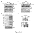
- Y1162/Y1163 motif was robustly phosphorylated at 5 min, peaked at 10 min and significantly dephosphorylated 30 min after insulin stimulation ( FIG. 3a ).
- insulin-induced phosphorylation of the Y1162/Y1163 motif was NO-dependent and this signaling event of IR was significantly diminished in eNOS-ablated MS-1 cells ( Fig. 3b ).
- activation of IR within the first 5 min might require NO-dependent S -nitrosylation and inactivation of PTP1B, thus allowing IR, which is a bona fide substrate of PTP1B ( Salmeen et al., Mol Cell 6, 1401-12 (2000 )), to be fully activated.
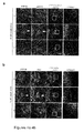
- EXAMPLE 5 Association between S -nitrosylated PTP1B and activated IR at the cell periphery in endothelium exposed to insulin
- This PTP1B-C215S might function as a dominant negative mutant to compete with endogenous PTP1B, which would be in the reduced and active form during eNOS knockdown, for the IR complex formation.
- PTP1B-C215S In attempting to understand the role of PTP1B-C215S in regulating insulin responsiveness without the influence of intrinsic NO, attention was drawn to the subcellular localization of activated IR in eNOS knockdown MS-1cells exposed to insulin for 5 min. As expected, without PTP1B-C215S, IR in its pYpY 1162/1163 form was barely detected in eNOS-ablated cells ( Fig. 6a ).
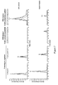
- EXAMPLE 7 Targeted mass spectrometric determination of IAN-tagged catalytic Cys463 of endogenous SHP-2 isolated from insulin-treated cells.
- the above-descibed PAC-switch method was used on another tyrosine phosphatase SHP-2.
- two samples, "IAN control,” and "Insulin-treated,” were analyzed using mass spec analysis.
- IAN control an aliquot of MS-1 cell lysates was exposed to IAN in vitro.
- insulin-treated sample MS-1 cells were exposed to insulin (10 nM) for 5 minutes, the cells then lyzed, and an aliquot of lysates was processed by the PAC-switch method.
- Endogenous SHP-2 protein was immunoprecipitated, followed by trypsin digestion for the subsequent analysis by the Q Exactive HF operated in a parallel-reaction monitoring mode.
- Two proteotypic peptides of SHP-2 as well as IAM- and IAN-labeled catalytic Cys (C463) containing peptides were selected as targets.
- S -nitrosylation is an essential mechanism preventing the immediate dephosphorylation of IR by PTP1B already present in the same complex ( Fig. 5c ).
- PTP1B since this pool of PTP1B is likely targeted by NO while traveling from the paranuclear region to the cell border, PTP1B may be in its S -nitrosylated and catalytically inactive form upon docking at its destination, the activated IR complex. This would explain why endothelial insulin responsiveness could be further enhanced for an extended period even after PTP1B was already associated with pIR ( Fig. 3a ).
- This pool of S -nitrosylated PTP1B may subsequently undergo Cys reduction, leading to rapid rebound of its phosphatase activity for down-regulating pIR in the same protein complex before dissociation of Nck/PTP1B complex from dephosphorylated IR.
- the PAC-switch method allows one to identify the new role of NO in promoting endothelial insulin signaling through S -nitrosylation of PTP1B.
- S -nitrosylation of PTP1B With the development of new reagent and methodology in detection of S -nitrosylated proteins in situ, the inventors have demonstrated that transient inactivation of PTP1B controlled by intrinsic NO plays an indispensable role in the process of endothelial insulin responsiveness.
- IAM Iodoacetamide
- SNAP N-(4-acetylphenyl)-2-iodoacetamide
- DPIAM N-(2,3-dimethylphenyl)-2-iodoacetamide
- IAM Iodoacetamide
- IAM 2-iodo-N-phenylacetamide
- TCEP Tris(2-carboxyethyl)phosphine
- the 21-nucleotide siRNA duplexes against mouse eNOS were purchased from Dharmacon Thermo Scientific. Two oligonucleotides, 5'-GAUCCUAACUUGCCCUGCAUU-3' (J-040956-07, SEQ ID No: 1) and 5'-GAAUGGAAGUGGUUCAGCUUU-3' (J-040956-08, SEQ ID No: 2), were chosen for knockdown of eNOS. Purified recombinant human PTP1B (C-terminally truncated 37 kDa) was described previously ( Chen et al. J Biol Chem 283, 35265-72 (2008 )).
- the cDNA construct of EGFP-tagged full-length PTP1B was established in a previous study ( Haj et al. PLoS One 7, e36633 (2012 )), and was used as a template to generate the EGFP-tagged C-terminally truncated human PTP1B.
- the HA-tagged C215S (C/S) mutant form of full-length PTP1B was constructed by site-direct mutagenesis using the WT form of human PTP1B as the template and then subcloned into pcDNA3.1 vector.
- the following antibodies were purchased from various vendors: GAPDH, insulin receptor beta-chain (InR ⁇ ), PTP1B for immunoprecipitation (sc-1718-R) and for immunoblotting (sc-1718-G) from Santa Cruz; PTP1B (clone 15 for immunofluorescence staining) from BD; tubulin from Sigma; phospho-specific pYpY 1162/1163 -InR ⁇ from Invitrogen; eNOS and phospho-specific pS 1177 -eNOS (for immunoblotting) from BD; pS 1177 -eNOS (for immunofluorescence staining) from Cell Signaling; HA from Millipore.
- Alexa 488-conjugated donkey anti-mouse IgG, Alexa 555-conjugated donkey anti-rabbit IgG, Alexa 555-conjugated goat anti-guinea pig IgG, and ProLong Gold antifade reagent were purchased from Invitrogen.
- Protein A magnetic Sepharose Xtra, protein A-Sepharose, and protein G-Sepharose beads were from GE Healthcare.
- Monkey kidney epithelial COS-7 cells were routinely maintained in high glucose Dulbecoo's modified Eagle's medium (DMEM) supplemented with 10% fetal bovine serum (FBS).
- DMEM Dulbecoo's modified Eagle's medium
- FBS fetal bovine serum
- Mouse endothelial MS-1 cells derived from capillary were maintained in high glucose DMEM supplemented with 5% FBS.
- MS-1 cells were mixed with siRNA (150 pmol with 7x10 5 cells) and plasmid DNA (1.5 ⁇ g with 7x10 5 cells), then electroporated by the NeonTM Transfection System (Invitrogen) according to the manufacturer's instructions. After electroporation, cells were maintained for additional 42 h followed by serum starvation for 6 h before insulin stimulation.
- Carrier protein bovine serum albumin (BSA, 3 mg/ml) was treated with 50 mM 1,4-dithioerythritol in buffer containing 20 mM Tris-HCl (pH 8.0), 5 mM EDTA, and 8 M urea at 37 °C for 1 h.
- An equal volume of 20% trichloroacetic acid was added into the solution with brief mixing.
- 10x volume of acetone After addition of 10x volume of acetone, the whole solution was vortexed and kept at-20 °C for overnight.
- Pellet was collected by centrifugation and washed with acetone. The pellet was then dissolved in 1 ml buffer containing 100 mM sodium bicarbonate (pH 9.4), 1% SDS, and 8 M urea.
- Iodoacetanilide (3 mg/0.1 ml) was prepared in DMSO and then added into the BSA solution followed by incubation at 37 °C for 2 h. After spinning centrifugation-dialysis using Concentrators (Pierce), the BSA protein with its Cys residues already phenylacetamidylated was re-suspended in PBS to form the antigen solution. Before injection, the antigen solution was thoroughly mixed with an equal volume of Freund's incomplete adjuvant. Approximately 200 ⁇ g of antigen BSA protein was injected subcutaneously into the back of a guinea pig during each biweekly immunization. Ten days after the fifth injection, the blood was withdrawn by heart puncture, and serum was stored at 4 °C.
- Total lysate proteins (harvested from COS-7 cells) were treated with 2 mM IAM, APIAM, DPIAM, or IAN, respectively. All labeling reactions were performed in the dark for 1 h at 37 °C in the buffer containing 25 mM ammonium bicarbonate (pH 8.0) and 5 mM TCEP. Then, total lysates (10 ⁇ g) were subjected to immunoblotting with anti-PAC antibody.
- Cells were harvested in lysis buffer containing 25 mM HEPES (pH 7.4), 50 mM NaCl, 100 mM EDTA, 1% NP-40, 0.5 mM PMSF, and protease inhibitors. An aliquot of total lysates (800 ⁇ g) was reacted with 50 mM IAM (the first-step blocking of free thiols) in HEN buffer with 2.5% SDS at room temperature for 30 min. Proteins were precipitated by cold acetone (100%) at -20 °C for 20 min and collected by centrifugation at 2000x g for 5 min, then washed with cold acetone (70%) 3 times.
- APIAM the second-step blocking of free thiols
- anti-PTP1B antibody (sc-1718-R, 8 ⁇ g each reaction) was first immobilized on beads in the mixture containing equal amounts of protein A- and G-Sepharose, then added into an aliquot of total lysate (0.6 mg) followed by incubation at 4 °C for overnight.
- Immunoprecipitated PTP1B was eluted by boiling beads in loading buffer containing 60 mM Tris-HCl (pH 6.5), 2% SDS, 0.05% bromophenol blue, 5% ⁇ -mercaptoethanol, and 10% glycerol for 5 min. Equal volumes of protein elutes were subjected to immunoblotting with anti-PAC and PTP1B (sc-1718-G) antibodies.
Landscapes
- Life Sciences & Earth Sciences (AREA)
- Health & Medical Sciences (AREA)
- Engineering & Computer Science (AREA)
- Molecular Biology (AREA)
- Chemical & Material Sciences (AREA)
- Immunology (AREA)
- Physics & Mathematics (AREA)
- Urology & Nephrology (AREA)
- Hematology (AREA)
- Biomedical Technology (AREA)
- Biophysics (AREA)
- General Health & Medical Sciences (AREA)
- Medicinal Chemistry (AREA)
- Biochemistry (AREA)
- Proteomics, Peptides & Aminoacids (AREA)
- Bioinformatics & Cheminformatics (AREA)
- Bioinformatics & Computational Biology (AREA)
- Organic Chemistry (AREA)
- Biotechnology (AREA)
- Cell Biology (AREA)
- Microbiology (AREA)
- Food Science & Technology (AREA)
- Analytical Chemistry (AREA)
- General Physics & Mathematics (AREA)
- Pathology (AREA)
- Genetics & Genomics (AREA)
- Peptides Or Proteins (AREA)
- Investigating Or Analysing Biological Materials (AREA)
Applications Claiming Priority (1)
| Application Number | Priority Date | Filing Date | Title |
|---|---|---|---|
| US201462032998P | 2014-08-04 | 2014-08-04 |
Publications (2)
| Publication Number | Publication Date |
|---|---|
| EP2982697A1 true EP2982697A1 (fr) | 2016-02-10 |
| EP2982697B1 EP2982697B1 (fr) | 2018-12-05 |
Family
ID=53776462
Family Applications (1)
| Application Number | Title | Priority Date | Filing Date |
|---|---|---|---|
| EP15179608.3A Not-in-force EP2982697B1 (fr) | 2014-08-04 | 2015-08-04 | Compositions et procédés pour la détection de la s-nitrosylation et de l'oxydation de protéines |
Country Status (3)
| Country | Link |
|---|---|
| US (1) | US10072097B2 (fr) |
| EP (1) | EP2982697B1 (fr) |
| TW (1) | TWI585107B (fr) |
Families Citing this family (1)
| Publication number | Priority date | Publication date | Assignee | Title |
|---|---|---|---|---|
| CN114409794B (zh) | 2021-12-24 | 2024-04-19 | 中国人民解放军海军军医大学 | 抗河豚毒素抗体y126c、制备方法及用途 |
Citations (4)
| Publication number | Priority date | Publication date | Assignee | Title |
|---|---|---|---|---|
| US4123431A (en) * | 1973-05-29 | 1978-10-31 | Syva Company | Non-oxo-carbonyl containing benzoyl ecgonine and cocaine compounds and derivatives thereof |
| US4816567A (en) | 1983-04-08 | 1989-03-28 | Genentech, Inc. | Recombinant immunoglobin preparations |
| EP1295606A1 (fr) * | 2000-06-20 | 2003-03-26 | Oleg Iliich Epshtein | Procede de traitement d'un syndrome pathologique et produit medicamenteux |
| EP1379877B1 (fr) | 2000-10-27 | 2006-04-05 | The Johns Hopkins University | Recherche de nytrosilation des proteines |
Family Cites Families (1)
| Publication number | Priority date | Publication date | Assignee | Title |
|---|---|---|---|---|
| US8053243B2 (en) | 2004-04-12 | 2011-11-08 | The University Of Vermont And State Agricultural College | Detection of nitrosylated proteins |
-
2015
- 2015-07-29 US US14/812,621 patent/US10072097B2/en active Active
- 2015-08-04 TW TW104125158A patent/TWI585107B/zh not_active IP Right Cessation
- 2015-08-04 EP EP15179608.3A patent/EP2982697B1/fr not_active Not-in-force
Patent Citations (4)
| Publication number | Priority date | Publication date | Assignee | Title |
|---|---|---|---|---|
| US4123431A (en) * | 1973-05-29 | 1978-10-31 | Syva Company | Non-oxo-carbonyl containing benzoyl ecgonine and cocaine compounds and derivatives thereof |
| US4816567A (en) | 1983-04-08 | 1989-03-28 | Genentech, Inc. | Recombinant immunoglobin preparations |
| EP1295606A1 (fr) * | 2000-06-20 | 2003-03-26 | Oleg Iliich Epshtein | Procede de traitement d'un syndrome pathologique et produit medicamenteux |
| EP1379877B1 (fr) | 2000-10-27 | 2006-04-05 | The Johns Hopkins University | Recherche de nytrosilation des proteines |
Non-Patent Citations (44)
| Title |
|---|
| ANDERIE ET AL., CELL SIGNALING, vol. 19, 2007, pages 582 - 592 |
| BARON, AM J PHYSIOL, vol. 269, 1995, pages E709 - 715 |
| BOBRICH ET AL., INT JOBES, vol. 35, 2011, pages 1385 - 94 |
| BOUTE ET AL., EMBO REP, vol. 4, 2003, pages 313 - 319 |
| BRODEUR ET AL.: "Monoclonal Antibody Production Techniques and Applications", 1987, MARCEL DEKKER, INC., pages: 51 - 63 |
| CHEN ET AL., J BIOL CHEM, vol. 283, 2008, pages 35265 - 72 |
| CLACKSON ET AL., NATURE, vol. 352, 1991, pages 624 - 628 |
| COOK, DIABETES, vol. 53, 2004, pages 2067 - 2072 |
| DUPLAIN, CIRCULATION, vol. 104, 2001, pages 342 - 345 |
| ELCHEBLY ET AL., SCIENCE, vol. 283, 1999, pages 1544 - 8 |
| FORRESTER M T ET AL: "Detection of protein S-nitrosylation with the biotin-switch technique", FREE RADICAL BIOLOGY AND MEDICINE, ELSEVIER INC, US, vol. 46, no. 2, 15 January 2009 (2009-01-15), pages 119 - 126, XP025845718, ISSN: 0891-5849, [retrieved on 20081017], DOI: 10.1016/J.FREERADBIOMED.2008.09.034 * |
| GALFRE ET AL., METHODS ENZYMOL., vol. 73, 1981, pages 3 - 46 |
| GODING: "Monoclonal Antibodies: Principles and Practice", 1986, ACADEMIC PRESS, pages: 59 - 103 |
| HAJ ET AL., PLOS ONE, vol. 7, 2012, pages E36633 |
| HAJ, PLOS ONE, vol. 7, 2012, pages E36633 |
| HARLOW; LANE: "Antibodies: A Laboratory Manual", 1988, COLD SPRING HARBOR LABORATORY |
| HART M N ET AL: "Phenacetin antibody cross-reactive with autoimmune erythrocyte antibody", AMERICAN JOURNAL OF CLINICAL PATHOLOGY, AMERICAN SOCIETY FOR CLINICAL PATHOLOGY, US, vol. 52, no. 6, 1 December 1969 (1969-12-01), pages 695 - 701, XP009187615, ISSN: 0002-9173 * |
| HOLLINSHEAD, J HISTOCHEM CYTOCHEM, vol. 45, 1997, pages 1053 - 1057 |
| IWAKIRI, PNAS, vol. 103, 2006, pages 19777 - 19782 |
| JEFFERY, NAT CELL BIOL, vol. 3, 2001, pages 193 - 197 |
| KLAMAN ET AL., MOL CELL BIOL, vol. 20, 2000, pages 5479 - 89 |
| KOHLER ET AL., NATURE, vol. 256, 1975, pages 495 |
| KOHLER; MILSTEIN, NATURE, vol. 256, 1975, pages 495 - 497 |
| KOZBOR, J. IMMUNOL., vol. 133, 1984, pages 3001 |
| KUBOTA ET AL., CELL METABOLISM, 2011 |
| MARKS ET AL., J MOL BIOL, vol. 222, 1991, pages 581 - 597 |
| MCKAY, J COMPARATIVE NEUROLOGY, vol. 473, 2004, pages 86 - 96 |
| MONTAGANANI ET AL., MOL ENDO, vol. 16, 2002, pages 1931 |
| MONTAGNANI ET AL., JBC, vol. 276, 2001, pages 30392 |
| MONTELEONE, PLOS ONE, vol. 7, 2012, pages E38948 |
| MORRISON ET AL., PROC NATL ACAD SCI USA, vol. 81, 1984, pages 6851 - 6855 |
| MUNSON ET AL., ANAL. BIOCHEM., vol. 107, 1980, pages 220 |
| NEUBERGER ET AL., NATURE, vol. 312, 1984, pages 604 - 608 |
| ROY, AM J PHYSIOL, vol. 274, 1998, pages E692 - 699 |
| SALMEEN ET AL., MOL CELL, vol. 6, 2000, pages 1401 - 12 |
| SALMON ET AL., CIRC RES, vol. 111, 2012, pages 685 - 96 |
| SHANKAR, DIABETES, vol. 49, 2000, pages 684 - 687 |
| SHI ET AL., J BIOCHEM, vol. 136, 2004, pages 89 - 96 |
| TAKEDA ET AL., NATURE, vol. 314, 1985, pages 452 - 454 |
| WANG ET AL., MOL ENDO, vol. 23, 2009, pages 1613 |
| WANG, CELL TISSUE RES, vol. 296, 1999, pages 511 - 516 |
| WU, BJ, vol. 439, 2011, pages 151 - 159 |
| Y.-Y. CHEN ET AL: "Cysteine S-Nitrosylation Protects Protein-tyrosine Phosphatase 1B against Oxidation-induced Permanent Inactivation", JOURNAL OF BIOLOGICAL CHEMISTRY, vol. 283, no. 50, 12 December 2008 (2008-12-12), US, pages 35265 - 35272, XP055234825, ISSN: 0021-9258, DOI: 10.1074/jbc.M805287200 * |
| YAMAZAKI ET AL., GENES CELLS, vol. 14, 2009, pages 425 - 34 |
Also Published As
| Publication number | Publication date |
|---|---|
| US20160033502A1 (en) | 2016-02-04 |
| US10072097B2 (en) | 2018-09-11 |
| EP2982697B1 (fr) | 2018-12-05 |
| TW201605906A (zh) | 2016-02-16 |
| TWI585107B (zh) | 2017-06-01 |
Similar Documents
| Publication | Publication Date | Title |
|---|---|---|
| Bitsikas et al. | Clathrin-independent pathways do not contribute significantly to endocytic flux | |
| Nguyen et al. | Antibodies against keratinocyte antigens other than desmogleins 1 and 3 can induce pemphigus vulgaris–like lesions | |
| US20090318354A1 (en) | Use of at least one isoform of progesterone receptor membrane component 1 (PGRMC1) | |
| Hsu et al. | S-nitrosylation of endogenous protein tyrosine phosphatases in endothelial insulin signaling | |
| US9347954B2 (en) | Antibody capable of binding to specific region of periostin, and method of measuring periostin using the same | |
| Sinensky et al. | An antibody which specifically recognizes prelamin A but not mature lamin A: application to detection of blocks in farnesylation-dependent protein processing | |
| Welsch et al. | Eps8 is recruited to lysosomes and subjected to chaperone-mediated autophagy in cancer cells | |
| EP1271152A1 (fr) | Technique d'investigation du cancer par epreuve biologique d'un autoanticorps contre mdm2 et reactif | |
| EP2982697B1 (fr) | Compositions et procédés pour la détection de la s-nitrosylation et de l'oxydation de protéines | |
| JP6875623B2 (ja) | 遺伝性疾患に関わるタンパク質の測定方法及び測定キット | |
| Yfanti et al. | Expression of metalloprotease insulin-degrading enzyme insulysin in normal and malignant human tissues | |
| US20140141017A1 (en) | Methods of diagnosing atherosclerosis by measuring apoci | |
| US20180284126A1 (en) | A method of detecting molecules in proximity to a target molecule in a sample | |
| Bogomolova et al. | Development of immunochemical systems for detection of human skeletal troponin i isoforms | |
| US11090310B2 (en) | Methods and compositions for treating hypertriglyceridemia | |
| McNulty et al. | Tissue distribution of GAP1IP4BP and GAP1m: Two inositol 1, 3, 4, 5-tetrakisphosphate-binding proteins | |
| US12005101B2 (en) | Modulation of proprotein convertase subtilisin/kexin 9 expression (PCSK9) with HSP 27 and/or HSP25 | |
| Warren et al. | Anti‐CD30 (Ber‐H2) epitope requires structural elements as shown by mass spectroscopy and dual‐site associated kinetics | |
| JP2019158606A (ja) | がんの判定方法 | |
| JP7403444B2 (ja) | 抗体の複写保護措置 | |
| US9551711B2 (en) | Compositions and methods for detecting nitration of PKG-1α and methods of use thereof | |
| JP7006599B2 (ja) | 試料に含まれるペリオスチンの測定試薬、ペリオスチン測定用前処理剤、ペリオスチン測定方法及びペリオスチン測定の感度の改善方法 | |
| Ren | Specific cellular labeling of nuclear and membrane proteins with antibody alternatives: Affimer and nanobody | |
| JP2026012139A (ja) | 抗体及びその応用 | |
| Lai | Cellular and Synaptic Mechanisms of Anti-NMDA Receptor Encephalitis |
Legal Events
| Date | Code | Title | Description |
|---|---|---|---|
| PUAI | Public reference made under article 153(3) epc to a published international application that has entered the european phase |
Free format text: ORIGINAL CODE: 0009012 |
|
| AK | Designated contracting states |
Kind code of ref document: A1 Designated state(s): AL AT BE BG CH CY CZ DE DK EE ES FI FR GB GR HR HU IE IS IT LI LT LU LV MC MK MT NL NO PL PT RO RS SE SI SK SM TR |
|
| AX | Request for extension of the european patent |
Extension state: BA ME |
|
| 17P | Request for examination filed |
Effective date: 20160723 |
|
| RBV | Designated contracting states (corrected) |
Designated state(s): AL AT BE BG CH CY CZ DE DK EE ES FI FR GB GR HR HU IE IS IT LI LT LU LV MC MK MT NL NO PL PT RO RS SE SI SK SM TR |
|
| STAA | Information on the status of an ep patent application or granted ep patent |
Free format text: STATUS: EXAMINATION IS IN PROGRESS |
|
| 17Q | First examination report despatched |
Effective date: 20161108 |
|
| GRAJ | Information related to disapproval of communication of intention to grant by the applicant or resumption of examination proceedings by the epo deleted |
Free format text: ORIGINAL CODE: EPIDOSDIGR1 |
|
| STAA | Information on the status of an ep patent application or granted ep patent |
Free format text: STATUS: GRANT OF PATENT IS INTENDED |
|
| GRAP | Despatch of communication of intention to grant a patent |
Free format text: ORIGINAL CODE: EPIDOSNIGR1 |
|
| INTG | Intention to grant announced |
Effective date: 20180704 |
|
| GRAS | Grant fee paid |
Free format text: ORIGINAL CODE: EPIDOSNIGR3 |
|
| GRAA | (expected) grant |
Free format text: ORIGINAL CODE: 0009210 |
|
| GRAA | (expected) grant |
Free format text: ORIGINAL CODE: 0009210 |
|
| STAA | Information on the status of an ep patent application or granted ep patent |
Free format text: STATUS: THE PATENT HAS BEEN GRANTED |
|
| AK | Designated contracting states |
Kind code of ref document: B1 Designated state(s): AL AT BE BG CH CY CZ DE DK EE ES FI FR GB GR HR HU IE IS IT LI LT LU LV MC MK MT NL NO PL PT RO RS SE SI SK SM TR |
|
| REG | Reference to a national code |
Ref country code: GB Ref legal event code: FG4D |
|
| REG | Reference to a national code |
Ref country code: CH Ref legal event code: EP |
|
| REG | Reference to a national code |
Ref country code: AT Ref legal event code: REF Ref document number: 1072929 Country of ref document: AT Kind code of ref document: T Effective date: 20181215 |
|
| REG | Reference to a national code |
Ref country code: IE Ref legal event code: FG4D |
|
| REG | Reference to a national code |
Ref country code: DE Ref legal event code: R096 Ref document number: 602015020743 Country of ref document: DE |
|
| REG | Reference to a national code |
Ref country code: NL Ref legal event code: MP Effective date: 20181205 |
|
| REG | Reference to a national code |
Ref country code: AT Ref legal event code: MK05 Ref document number: 1072929 Country of ref document: AT Kind code of ref document: T Effective date: 20181205 |
|
| REG | Reference to a national code |
Ref country code: LT Ref legal event code: MG4D |
|
| PG25 | Lapsed in a contracting state [announced via postgrant information from national office to epo] |
Ref country code: LV Free format text: LAPSE BECAUSE OF FAILURE TO SUBMIT A TRANSLATION OF THE DESCRIPTION OR TO PAY THE FEE WITHIN THE PRESCRIBED TIME-LIMIT Effective date: 20181205 Ref country code: FI Free format text: LAPSE BECAUSE OF FAILURE TO SUBMIT A TRANSLATION OF THE DESCRIPTION OR TO PAY THE FEE WITHIN THE PRESCRIBED TIME-LIMIT Effective date: 20181205 Ref country code: BG Free format text: LAPSE BECAUSE OF FAILURE TO SUBMIT A TRANSLATION OF THE DESCRIPTION OR TO PAY THE FEE WITHIN THE PRESCRIBED TIME-LIMIT Effective date: 20190305 Ref country code: NO Free format text: LAPSE BECAUSE OF FAILURE TO SUBMIT A TRANSLATION OF THE DESCRIPTION OR TO PAY THE FEE WITHIN THE PRESCRIBED TIME-LIMIT Effective date: 20190305 Ref country code: HR Free format text: LAPSE BECAUSE OF FAILURE TO SUBMIT A TRANSLATION OF THE DESCRIPTION OR TO PAY THE FEE WITHIN THE PRESCRIBED TIME-LIMIT Effective date: 20181205 Ref country code: AT Free format text: LAPSE BECAUSE OF FAILURE TO SUBMIT A TRANSLATION OF THE DESCRIPTION OR TO PAY THE FEE WITHIN THE PRESCRIBED TIME-LIMIT Effective date: 20181205 Ref country code: LT Free format text: LAPSE BECAUSE OF FAILURE TO SUBMIT A TRANSLATION OF THE DESCRIPTION OR TO PAY THE FEE WITHIN THE PRESCRIBED TIME-LIMIT Effective date: 20181205 Ref country code: ES Free format text: LAPSE BECAUSE OF FAILURE TO SUBMIT A TRANSLATION OF THE DESCRIPTION OR TO PAY THE FEE WITHIN THE PRESCRIBED TIME-LIMIT Effective date: 20181205 |
|
| PG25 | Lapsed in a contracting state [announced via postgrant information from national office to epo] |
Ref country code: SE Free format text: LAPSE BECAUSE OF FAILURE TO SUBMIT A TRANSLATION OF THE DESCRIPTION OR TO PAY THE FEE WITHIN THE PRESCRIBED TIME-LIMIT Effective date: 20181205 Ref country code: GR Free format text: LAPSE BECAUSE OF FAILURE TO SUBMIT A TRANSLATION OF THE DESCRIPTION OR TO PAY THE FEE WITHIN THE PRESCRIBED TIME-LIMIT Effective date: 20190306 Ref country code: AL Free format text: LAPSE BECAUSE OF FAILURE TO SUBMIT A TRANSLATION OF THE DESCRIPTION OR TO PAY THE FEE WITHIN THE PRESCRIBED TIME-LIMIT Effective date: 20181205 Ref country code: RS Free format text: LAPSE BECAUSE OF FAILURE TO SUBMIT A TRANSLATION OF THE DESCRIPTION OR TO PAY THE FEE WITHIN THE PRESCRIBED TIME-LIMIT Effective date: 20181205 |
|
| PG25 | Lapsed in a contracting state [announced via postgrant information from national office to epo] |
Ref country code: NL Free format text: LAPSE BECAUSE OF FAILURE TO SUBMIT A TRANSLATION OF THE DESCRIPTION OR TO PAY THE FEE WITHIN THE PRESCRIBED TIME-LIMIT Effective date: 20181205 |
|
| PG25 | Lapsed in a contracting state [announced via postgrant information from national office to epo] |
Ref country code: CZ Free format text: LAPSE BECAUSE OF FAILURE TO SUBMIT A TRANSLATION OF THE DESCRIPTION OR TO PAY THE FEE WITHIN THE PRESCRIBED TIME-LIMIT Effective date: 20181205 Ref country code: PT Free format text: LAPSE BECAUSE OF FAILURE TO SUBMIT A TRANSLATION OF THE DESCRIPTION OR TO PAY THE FEE WITHIN THE PRESCRIBED TIME-LIMIT Effective date: 20190405 Ref country code: PL Free format text: LAPSE BECAUSE OF FAILURE TO SUBMIT A TRANSLATION OF THE DESCRIPTION OR TO PAY THE FEE WITHIN THE PRESCRIBED TIME-LIMIT Effective date: 20181205 Ref country code: IT Free format text: LAPSE BECAUSE OF FAILURE TO SUBMIT A TRANSLATION OF THE DESCRIPTION OR TO PAY THE FEE WITHIN THE PRESCRIBED TIME-LIMIT Effective date: 20181205 |
|
| PG25 | Lapsed in a contracting state [announced via postgrant information from national office to epo] |
Ref country code: SK Free format text: LAPSE BECAUSE OF FAILURE TO SUBMIT A TRANSLATION OF THE DESCRIPTION OR TO PAY THE FEE WITHIN THE PRESCRIBED TIME-LIMIT Effective date: 20181205 Ref country code: RO Free format text: LAPSE BECAUSE OF FAILURE TO SUBMIT A TRANSLATION OF THE DESCRIPTION OR TO PAY THE FEE WITHIN THE PRESCRIBED TIME-LIMIT Effective date: 20181205 Ref country code: IS Free format text: LAPSE BECAUSE OF FAILURE TO SUBMIT A TRANSLATION OF THE DESCRIPTION OR TO PAY THE FEE WITHIN THE PRESCRIBED TIME-LIMIT Effective date: 20190405 Ref country code: SM Free format text: LAPSE BECAUSE OF FAILURE TO SUBMIT A TRANSLATION OF THE DESCRIPTION OR TO PAY THE FEE WITHIN THE PRESCRIBED TIME-LIMIT Effective date: 20181205 Ref country code: EE Free format text: LAPSE BECAUSE OF FAILURE TO SUBMIT A TRANSLATION OF THE DESCRIPTION OR TO PAY THE FEE WITHIN THE PRESCRIBED TIME-LIMIT Effective date: 20181205 |
|
| REG | Reference to a national code |
Ref country code: DE Ref legal event code: R097 Ref document number: 602015020743 Country of ref document: DE |
|
| PLBE | No opposition filed within time limit |
Free format text: ORIGINAL CODE: 0009261 |
|
| STAA | Information on the status of an ep patent application or granted ep patent |
Free format text: STATUS: NO OPPOSITION FILED WITHIN TIME LIMIT |
|
| PG25 | Lapsed in a contracting state [announced via postgrant information from national office to epo] |
Ref country code: SI Free format text: LAPSE BECAUSE OF FAILURE TO SUBMIT A TRANSLATION OF THE DESCRIPTION OR TO PAY THE FEE WITHIN THE PRESCRIBED TIME-LIMIT Effective date: 20181205 Ref country code: DK Free format text: LAPSE BECAUSE OF FAILURE TO SUBMIT A TRANSLATION OF THE DESCRIPTION OR TO PAY THE FEE WITHIN THE PRESCRIBED TIME-LIMIT Effective date: 20181205 |
|
| 26N | No opposition filed |
Effective date: 20190906 |
|
| PG25 | Lapsed in a contracting state [announced via postgrant information from national office to epo] |
Ref country code: TR Free format text: LAPSE BECAUSE OF FAILURE TO SUBMIT A TRANSLATION OF THE DESCRIPTION OR TO PAY THE FEE WITHIN THE PRESCRIBED TIME-LIMIT Effective date: 20181205 |
|
| PG25 | Lapsed in a contracting state [announced via postgrant information from national office to epo] |
Ref country code: CH Free format text: LAPSE BECAUSE OF NON-PAYMENT OF DUE FEES Effective date: 20190831 Ref country code: LU Free format text: LAPSE BECAUSE OF NON-PAYMENT OF DUE FEES Effective date: 20190804 Ref country code: LI Free format text: LAPSE BECAUSE OF NON-PAYMENT OF DUE FEES Effective date: 20190831 Ref country code: MC Free format text: LAPSE BECAUSE OF FAILURE TO SUBMIT A TRANSLATION OF THE DESCRIPTION OR TO PAY THE FEE WITHIN THE PRESCRIBED TIME-LIMIT Effective date: 20181205 |
|
| REG | Reference to a national code |
Ref country code: BE Ref legal event code: MM Effective date: 20190831 |
|
| PG25 | Lapsed in a contracting state [announced via postgrant information from national office to epo] |
Ref country code: IE Free format text: LAPSE BECAUSE OF NON-PAYMENT OF DUE FEES Effective date: 20190804 Ref country code: FR Free format text: LAPSE BECAUSE OF NON-PAYMENT OF DUE FEES Effective date: 20190831 |
|
| PG25 | Lapsed in a contracting state [announced via postgrant information from national office to epo] |
Ref country code: BE Free format text: LAPSE BECAUSE OF NON-PAYMENT OF DUE FEES Effective date: 20190831 |
|
| PG25 | Lapsed in a contracting state [announced via postgrant information from national office to epo] |
Ref country code: CY Free format text: LAPSE BECAUSE OF FAILURE TO SUBMIT A TRANSLATION OF THE DESCRIPTION OR TO PAY THE FEE WITHIN THE PRESCRIBED TIME-LIMIT Effective date: 20181205 |
|
| PG25 | Lapsed in a contracting state [announced via postgrant information from national office to epo] |
Ref country code: HU Free format text: LAPSE BECAUSE OF FAILURE TO SUBMIT A TRANSLATION OF THE DESCRIPTION OR TO PAY THE FEE WITHIN THE PRESCRIBED TIME-LIMIT; INVALID AB INITIO Effective date: 20150804 Ref country code: MT Free format text: LAPSE BECAUSE OF FAILURE TO SUBMIT A TRANSLATION OF THE DESCRIPTION OR TO PAY THE FEE WITHIN THE PRESCRIBED TIME-LIMIT Effective date: 20181205 |
|
| PG25 | Lapsed in a contracting state [announced via postgrant information from national office to epo] |
Ref country code: MK Free format text: LAPSE BECAUSE OF FAILURE TO SUBMIT A TRANSLATION OF THE DESCRIPTION OR TO PAY THE FEE WITHIN THE PRESCRIBED TIME-LIMIT Effective date: 20181205 |
|
| PGFP | Annual fee paid to national office [announced via postgrant information from national office to epo] |
Ref country code: GB Payment date: 20220829 Year of fee payment: 8 Ref country code: DE Payment date: 20220829 Year of fee payment: 8 |
|
| REG | Reference to a national code |
Ref country code: DE Ref legal event code: R119 Ref document number: 602015020743 Country of ref document: DE |
|
| GBPC | Gb: european patent ceased through non-payment of renewal fee |
Effective date: 20230804 |
|
| PG25 | Lapsed in a contracting state [announced via postgrant information from national office to epo] |
Ref country code: GB Free format text: LAPSE BECAUSE OF NON-PAYMENT OF DUE FEES Effective date: 20230804 |
|
| PG25 | Lapsed in a contracting state [announced via postgrant information from national office to epo] |
Ref country code: GB Free format text: LAPSE BECAUSE OF NON-PAYMENT OF DUE FEES Effective date: 20230804 Ref country code: DE Free format text: LAPSE BECAUSE OF NON-PAYMENT OF DUE FEES Effective date: 20240301 |