EP3071966B1 - Olfactomédine-4, neudésine et desmoplakine en tant que nouveaux biomarqueurs du cancer du sein - Google Patents
Olfactomédine-4, neudésine et desmoplakine en tant que nouveaux biomarqueurs du cancer du sein Download PDFInfo
- Publication number
- EP3071966B1 EP3071966B1 EP14820760.8A EP14820760A EP3071966B1 EP 3071966 B1 EP3071966 B1 EP 3071966B1 EP 14820760 A EP14820760 A EP 14820760A EP 3071966 B1 EP3071966 B1 EP 3071966B1
- Authority
- EP
- European Patent Office
- Prior art keywords
- expression level
- breast cancer
- biomarkers
- protein
- olfm4
- Prior art date
- Legal status (The legal status is an assumption and is not a legal conclusion. Google has not performed a legal analysis and makes no representation as to the accuracy of the status listed.)
- Not-in-force
Links
Images








Classifications
-
- G—PHYSICS
- G01—MEASURING; TESTING
- G01N—INVESTIGATING OR ANALYSING MATERIALS BY DETERMINING THEIR CHEMICAL OR PHYSICAL PROPERTIES
- G01N33/00—Investigating or analysing materials by specific methods not covered by groups G01N1/00 - G01N31/00
- G01N33/48—Biological material, e.g. blood, urine; Haemocytometers
- G01N33/50—Chemical analysis of biological material, e.g. blood, urine; Testing involving biospecific ligand binding methods; Immunological testing
- G01N33/53—Immunoassay; Biospecific binding assay; Materials therefor
- G01N33/575—Immunoassay; Biospecific binding assay; Materials therefor for cancer
- G01N33/57515—Immunoassay; Biospecific binding assay; Materials therefor for cancer of the breast
-
- G—PHYSICS
- G01—MEASURING; TESTING
- G01N—INVESTIGATING OR ANALYSING MATERIALS BY DETERMINING THEIR CHEMICAL OR PHYSICAL PROPERTIES
- G01N33/00—Investigating or analysing materials by specific methods not covered by groups G01N1/00 - G01N31/00
- G01N33/48—Biological material, e.g. blood, urine; Haemocytometers
- G01N33/50—Chemical analysis of biological material, e.g. blood, urine; Testing involving biospecific ligand binding methods; Immunological testing
- G01N33/5005—Chemical analysis of biological material, e.g. blood, urine; Testing involving biospecific ligand binding methods; Immunological testing involving human or animal cells
- G01N33/5008—Chemical analysis of biological material, e.g. blood, urine; Testing involving biospecific ligand binding methods; Immunological testing involving human or animal cells for testing or evaluating the effect of chemical or biological compounds, e.g. drugs, cosmetics
- G01N33/5011—Chemical analysis of biological material, e.g. blood, urine; Testing involving biospecific ligand binding methods; Immunological testing involving human or animal cells for testing or evaluating the effect of chemical or biological compounds, e.g. drugs, cosmetics for testing antineoplastic activity
-
- G—PHYSICS
- G01—MEASURING; TESTING
- G01N—INVESTIGATING OR ANALYSING MATERIALS BY DETERMINING THEIR CHEMICAL OR PHYSICAL PROPERTIES
- G01N2333/00—Assays involving biological materials from specific organisms or of a specific nature
- G01N2333/435—Assays involving biological materials from specific organisms or of a specific nature from animals; from humans
- G01N2333/46—Assays involving biological materials from specific organisms or of a specific nature from animals; from humans from vertebrates
- G01N2333/47—Assays involving proteins of known structure or function as defined in the subgroups
-
- G—PHYSICS
- G01—MEASURING; TESTING
- G01N—INVESTIGATING OR ANALYSING MATERIALS BY DETERMINING THEIR CHEMICAL OR PHYSICAL PROPERTIES
- G01N2333/00—Assays involving biological materials from specific organisms or of a specific nature
- G01N2333/435—Assays involving biological materials from specific organisms or of a specific nature from animals; from humans
- G01N2333/46—Assays involving biological materials from specific organisms or of a specific nature from animals; from humans from vertebrates
- G01N2333/47—Assays involving proteins of known structure or function as defined in the subgroups
- G01N2333/4701—Details
- G01N2333/4703—Regulators; Modulating activity
-
- G—PHYSICS
- G01—MEASURING; TESTING
- G01N—INVESTIGATING OR ANALYSING MATERIALS BY DETERMINING THEIR CHEMICAL OR PHYSICAL PROPERTIES
- G01N2333/00—Assays involving biological materials from specific organisms or of a specific nature
- G01N2333/435—Assays involving biological materials from specific organisms or of a specific nature from animals; from humans
- G01N2333/475—Assays involving growth factors
-
- G—PHYSICS
- G01—MEASURING; TESTING
- G01N—INVESTIGATING OR ANALYSING MATERIALS BY DETERMINING THEIR CHEMICAL OR PHYSICAL PROPERTIES
- G01N2333/00—Assays involving biological materials from specific organisms or of a specific nature
- G01N2333/435—Assays involving biological materials from specific organisms or of a specific nature from animals; from humans
- G01N2333/705—Assays involving receptors, cell surface antigens or cell surface determinants
- G01N2333/71—Assays involving receptors, cell surface antigens or cell surface determinants for growth factors; for growth regulators
-
- G—PHYSICS
- G01—MEASURING; TESTING
- G01N—INVESTIGATING OR ANALYSING MATERIALS BY DETERMINING THEIR CHEMICAL OR PHYSICAL PROPERTIES
- G01N2500/00—Screening for compounds of potential therapeutic value
- G01N2500/10—Screening for compounds of potential therapeutic value involving cells
-
- G—PHYSICS
- G01—MEASURING; TESTING
- G01N—INVESTIGATING OR ANALYSING MATERIALS BY DETERMINING THEIR CHEMICAL OR PHYSICAL PROPERTIES
- G01N2570/00—Omics, e.g. proteomics, glycomics or lipidomics; Methods of analysis focusing on the entire complement of classes of biological molecules or subsets thereof, i.e. focusing on proteomes, glycomes or lipidomes
-
- G—PHYSICS
- G01—MEASURING; TESTING
- G01N—INVESTIGATING OR ANALYSING MATERIALS BY DETERMINING THEIR CHEMICAL OR PHYSICAL PROPERTIES
- G01N2800/00—Detection or diagnosis of diseases
- G01N2800/52—Predicting or monitoring the response to treatment, e.g. for selection of therapy based on assay results in personalised medicine; Prognosis
Definitions
- the present invention is in the technical field of breast cancer management, and more particularly relates to the diagnosis of breast cancer.
- the invention is more particularly based on the finding that specific biomarkers are abberantly expressed in the blood of breast cancer patients.
- breast cancer With over 1.3 million cases of invasive breast cancers diagnosed annually, and more than 450,000 deaths reported per year, breast cancer is the most common malignancy diagnosed in women and one of the leading causes of cancer-related death in females.
- breast cancer screening campaigns are delivered through mammography and although there is no doubt of their efficacy, this approach does have limitations in terms of sensitivity in women who have very dense breast tissue and in young women considered "at risk" (family history or genetic predisposition) for whom the regular use of ionising radiation is not recommended.
- mammography screening leads to overdiagnosis in 19% of women. In other words, one in five diagnosis is said to be an overdiagnosis.
- Other imaging techniques such as sonography and nuclear magnetic resonance imaging are available, but they are not generally used for detection, being instead used as a further examination after mammography.
- recurrence also known as metastasis
- recurrence occurs in 10 to 20% of patients in the three to ten years following initial adjuvant treatment.
- recurrence is often either missed or identified as false positive by mammography, and unnecessary biopsies are performed on patients suspected of relapse.
- proteomic biomarkers are indeed particularly advantageous as they are more reflective of a tumour microenvironment and can undergo cancer specific posttranslational modifications.
- the inventors have demonstrated that dysregulation in protein expression level of Olfactomedin-4 (OLFM4), Neudesin (NENF) and/or Desmoplakin (DSP) correlates with breast cancer, and that such biomarkers are detectable in blood samples of patients. It has notably been discovered that the expression level of Olfactomedin-4 and Neudesin are higher in breast cancer patients thoughout progression of the disease, while the expression level of Desmoplakin is lower at an early stage and higher in the case of recurrence, by comparison to healthy subjects.
- OFM4 Olfactomedin-4
- NENF Neudesin
- DSP Desmoplakin
- Olfactomedin-4 is a secreted N-glycosylated protein belonging to the olfactomedin domain-containing protein family, which is characterized by a coil-coil domain N-terminal domain and a well-conserved C-terminal olfactomedin domain.
- the OFLM4 protein has been described in the literature as mediating cell adhesion through binding to cadherins and lectins (Liu et al., 2006), and as being involved in the regulation of cellular apoptosis and in the proliferation of cancer cells (Zhang et al., 2004; Kobayashi et al., 2007).
- OLFM4 has been described as a biomarker of colon cancer ( WO 2012/117267 ).
- gene expression of OLFM4 has been found to be increased in various cancers including breast and lung cancers (Koshida et al., 2007).
- Neudesin on the other hand is an extracellular heme-binding protein which has been described as displaying neurotrophic activity in neurons via the mitogen-activated protein kinase (MAPK) and phosphatidylinositol 3-kinase (PI3K) pathways (Kimura et al., 2013). Neudesin has been reported to be involved in breast tumorigenesis (Han et al., 2012).
- MAPK mitogen-activated protein kinase
- PI3K phosphatidylinositol 3-kinase
- Desmoplakin is a founding member of the plakin family, and is known as the principal plaque protein of desmosomes (Leung et al., 2002). It is therefore specialized in adhesion junctions found in various tissues and plays a critical role in the maintenance of epithelial tissue integrity. Recently, studies suggested that desmosomes participate in the regulation of cell motility, growth, differentiation and apoptosis (Allen et al., 1996; Wan et al., 2007; and Rickelt et al., 2009).
- Desmoplakin I 322 kDa
- Desmoplakin II (259 kDa)
- Desmoplakin proteins interact with plakoglobin (y-catenin), plakophilins and intermediate filaments, providing the intimate link between desmosomal cadherins and the cytoskeleton (Junkman et al., 2005; and Kowalczyk et al., 1997).
- biomarkers can be used herein to detect breast cancer, from a mere blood sample, to monitor disease progression, but also to develop and adapt a breast cancer treatment. They can also be used as therapeutic targets to design novel drugs.
- a reliable and easy to perform diagnostic method for breast cancer is provided, which is based on determination of the expression level of the above-mentioned biomarker(s).
- a screening method for identifying drugs is further provided. Kits and protein microarrays for carrying out the methods are also provided herein.
- the term “comprising” or “containing” means the inclusion of the referent and does not exclude the presence of any other element.
- the term “consisting of” means the sole inclusion of the referent and thus excludes the presence of any other element.
- subject or "patient” is used herein to describe any member of the animal kingdom, preferably a human being, more preferably a woman.
- diagnosis means assessing whether a subject suffers or not from a disease. As will be understood by those skilled in the art, such an assessment, although preferred to be, may usually not be correct for 100% of the investigated subjects. This term requires however that a statistically significant portion of subjects can be correctly assessed and, thus, diagnosed. Whether a portion is statistically significant can be easily determined by the person skilled in the art using various well-known statistic evaluation tools, such as determination of confidence intervals, p-value determination, Student's t-test, Mann-Whitney test, etc.. Details of such statistical methods can found in Dowdy and Wearden (1983). Statistical methods may notably allow the determination of the sensitivity and the specificity of a diagnostic test. The sensitivity of a diagnostic test can be defined as the proportion of subjects suffering from a disease who will have a positive result, while the specificity of a diagnostic test can be defined as the proportion of subjects without the disease who will have a negative result.
- breast cancer a cancer that forms in tissues of the breast, as defined by the National Cancer Institute.
- Types of breast cancer include, without limitation, ductal carcinoma, which begins in the lining of the milk ducts (thin tubes that carry milk from the lobules of the breast to the nipple); lobular carcinoma, which begins in the lobules (milk glands) of the breast; and invasive breast cancer (breast cancer that has spread from where it began in the breast ducts or lobules to surrounding normal tissue).
- breast cancer By “early breast cancer”, it is meant herein a breast cancer that has not spread beyond the breast or the axillary lymph nodes.
- TNM Tumor, Nodes, Metastasis
- pTis N0 M0 breast cancer refers to a breast cancer, wherein cancer cells can only be found inside the breast ducts or lobules (T0), without the tumour crossing the basal membrane.
- a stage I (T1 N0 M0) breast cancer refers to a breast cancer, wherein cancer cells have infiltrated the tissue surrounding the breast ducts and lobules, forming a tumour whose diameter is inferior or equal to 2 cm.
- the abbreviation N0 means that the cancer has not spread to lymph nodes, while the abbreviation M0 means that there is no distant metastasis.
- An early breast cancer is generally characterized by a 100% survival rate, within five years from the initial diagnosis.
- cancer recurrence By “cancer recurrence”, “recurring cancer”, “cancer relapse or “relapsing cancer”, it is meant herein, in the context of potential clinical outcomes of cancer and as defined by the National Cancer Institute, that the cancer has recurred (come back), usually after a period of time during which the cancer could not be detected.
- a recurring cancer may refer to a cancer that comes back to the same place as the original (primary) tumour or to another place in the body (also known as metastasis).
- a “biological fluid sample” as described herein can be any fluid sample that may be isolated from a subject, including, without limitation, blood or a fractional component thereof (serum, plasma, cellular extract), lymph, tumor interstitial fluid, saliva, mucus, sputum, sweat, or urine.
- circulating tumoral cells may be isolated from a biological fluid as defined above, preferably from blood, by techniques well-known in the art.
- An example of a technique allowing the isolation of circulating tumoral cells (CTCs) is Dean Flow Fractionation (DFF), as established by Hou et al. (2013).
- the biological fluid sample is preferably a blood sample, such as a serum or plasma sample, and even more preferably a serum sample.
- biomarker refers to a polypeptide or protein, fragment thereof, or epitope that is differentially present in a subject as compared to healthy subjects, including differentially modified (e.g. differentially glycosylated) and/or expressed biomarkers. It should be noted that the term “biomarker” includes soluble biomarkers, i.e. biomarkers which are differentially cleaved, secreted, released or shed from a tumor cell in a subject, and are thus detectable in a biological fluid as defined above.
- Biomarkers associated with breast cancer are listed in the following Table 1.
- Table 1 Biomarkers of breast cancer Symbol Full name Accession number UniprotKB/Swiss-Prot (SEQ ID number) OLM4 or OLFM4 Olfactomedin-4 (OLFM4)
- Alternative name(s) Antiapoptotic protein GW112 G-CSF-stimulated clone 1 protein (hGC-1) hOLfD Q6UX06 (SEQ ID NO:1)
- NENF Neudesin Alternative name(s): Cell immortalization-related protein 2 Neuron-derived neurotrophic factor Secreted protein of unknown function (SPUF protein) Q9UMX5 (SEQ ID NO:2) DSP or DP Desmoplakin : Isoforms 1 and 2 Alternative name(s): 250/210 kDA paraneoplastic pemphigus antigen P15924-1 (Isoform 1: SEQ ID NO: 3) P15924-2 (Isoform 2: SEQ ID NO: 4)
- expression level refers herein to the amount or level of a biomarker of interest expressed in a cell, tissue, biological fluid, or organ(s).
- level refers to an amount (e.g. relative amount or concentration) of a biomarker that is detectable or measurable in a sample.
- the level can be a concentration such as ⁇ g/L or a relative amount by comparison to a reference expression level.
- the act of actually "determining the expression level" of a biomarker in a biological sample refers to the act of actively detecting whether a biomarker is expressed in said sample or not, and notably allows to detect whether the biomarker expression is upregulated, downregulated or substantially unchanged when compared to a reference expression level.
- a "dysregulated expression level" of a given biomarker is, as described herein, a downregulated or upregulated expression level when compared to a reference expression level.
- reference expression level or "control expression level” of a biomarker, it is meant a predetermined expression level of said biomarker, which can be used as a reference in any method described herein.
- a reference expression level can be the expression level of a biomarker in a biological sample of a healthy subject, or the average or median, preferably median, expression level in a biological sample of a population of healthy subjects.
- OFM4 Olfactomedin-4
- NENF Neudesin
- DSP Desmoplakin
- an in vitro method for diagnosing a breast cancer in a subject comprising the steps of:
- the above method may optionally further comprise the step c) of determining whether said subject is suffering from breast cancer, based upon the comparison in step b).
- biomarkers are sufficient to perform a diagnosis as described herein. Nevertheless, the skilled person in the art will readily understand that the above biomarkers may be combined as a panel of biomarkers, each of which contributing to the final diagnosis.
- a protein expression level of said at least one biomarker dysregulated by comparison to a reference expression level of said biomarker obtained from a biological fluid sample of at least one healthy subject is indicative that said subject is suffering from breast cancer.
- a protein expression level of Olfactomedin-4 in step a) superior to the reference expression level of Olfactomedin-4 obtained from a biological fluid sample of at least one healthy subject, is indicative that the tested subject is suffering from breast cancer.
- a protein expression level of Neudesin in step a) superior to the reference expression level of Neudesin obtained from a biological fluid sample of at least one healthy subject, is indicative that the tested subject is suffering from breast cancer.
- a protein expression level of Desmoplakin in step a) inferior to the reference expression level of Desmoplakin obtained from a biological fluid sample of at least one healthy subject is indicative that the tested subject is suffering from breast cancer.
- step a) a protein expression level of Desmoplakin in step a) superior to the reference expression level of Desmoplakin obtained from a biological fluid sample of at least one healthy subject, is indicative that the tested subject is suffering from breast cancer.
- the protein expression level of any one of Olfactomedin-4, Neudesin and Desmoplakin, or of any combination thereof, may further indicate the stage of breast cancer.
- said expression level of Olfactomedin-4 superior to said reference expression level is indicative that the breast cancer is an early breast cancer.
- said expression level of Neudesin superior to said reference expression level is indicative that the breast cancer is an early breast cancer.
- said expression level of Desmoplakin inferior to said reference expression level is indicative that the breast cancer is an early breast cancer.
- Such diagnosis test of early breast cancer is particularly useful for patients at risk (e.g. having a family history of breast cancer), and for patients for which small breast tumours (e.g. tumour size below 1 cm) can not be accurately detected by conventional diagnostic methods such as ultrasound.
- said expression level of Desmoplakin superior to said reference expression level is indicative that the breast cancer is a recurring breast cancer.
- diagnosis test of a recurring breast cancer is particularly useful for the monitoring of a patient previously suffering from breast cancer. Notably if performed early on, such diagnostic test can help to improve the prognosis and survival of the patient.
- inferior to a reference expression level it is meant that the ratio between the expression level of said biomarker and the reference expression level is below 1.
- said expression level may be indicated as the concentration of biomarker in the tested biological fluid.
- said protein expression level of Olfactomedin-4 is preferably superior to 30, 31, 32, 34, 35, 36, 37, 38, 39, 40, 50, 60, 70, 80, 90, 100, 150, 200 ng/mL, more preferably superior to 30, 31, 32, 34, 35, 36, 37, 38, 39, 40 ng/mL, most preferably superior to 31 ng/mL, in the biological fluid sample of said subject suffering from breast cancer, preferably from early breast cancer.
- the inventors have demonstrated in a first cohort of 335 breast cancer patients and 65 healthy subjects that the sensitivity of a breast cancer diagnostic test based on Olfactomedin-4 is 67%, while the specificity of such test is 88%, for an Olfactomedin-4 sera concentration above 40 ng/mL. They further demonstrated in a second cohort of 766 breast cancer patients and 195 healthy subjects that the sensitivity of a breast cancer diagnostic test based on Olfactomedin-4 is ranging from 64 to 78%, with a specificity of about 80-90% for an Olfactomedin-4 sera concentration above 31 ng/mL. Similar values were observed in early breast cancer patients.
- said protein expression level of Neudesin is preferably superior to 15, 16, 17, 18, 19, 20, 25, 30, 35, 40 ng/mL, more preferably superior to 15, 16, 17, 18, 19, 20 ng/mL, most preferably superior to 16 ng/mL, in the biological fluid sample of said subject suffering from breast cancer, preferably early breast cancer.
- the inventors have demonstrated in a first cohort of 335 breast cancer patients and 65 healthy subjects, that the sensitivity of a breast cancer diagnostic test based on Neudesin is 47%, while the specificity of such test reached 91%, for a Neudesin sera concentration above 20 ng/mL.
- said protein expression level of Desmoplakin is preferably inferior to 600, 500, 400, 300 pg/mL, more preferably inferior to 600 pg/mL, in the biological fluid sample of said subject suffering from early breast cancer.
- said protein expression level of Desmoplakin is preferably superior to 1800, 1900, 2000 pg/mL, more preferably superior to 1800 pg/mL, in the biological fluid sample of said subject suffering from a recurring breast cancer.
- biomarkers can be combined as a panel of biomarkers, each of which contributing to the final diagnosis. Indeed, the inventors have demonstrated that such combination increases the sensivity and/or the specificity of the diagnosis test.
- the protein expression level of at least two, preferably three, of said biomarkers are determined in step a).
- the protein expression level of Olfactomedin-4 and Neudesin are determined in step a) of the above method.
- the combination of these biomarkers is particularly useful for diagnosing breast cancer, such as for diagnosing an early breast cancer. That is to say that, in a preferred embodiment, said protein expression level of:
- the inventors have demonstrated in a first cohort of 335 breast cancer patients and 65 healthy subjects that the sensitivity of a breast cancer diagnostic test based on the combination of Olfactomedin-4 and Neudesin is 74%, while the specificity of such test is 78%, for a sera concentration of those biomarkers above 44 ng/mL. They further demonstrated in a second cohort of 766 breast cancer patients and 195 healthy subjects that the sensitivity of a breast cancer diagnostic test based on the combination of Olfactomedin-4 and Neudesin is ranging from 75 to 85%, with a specificity reaching 87% for a sera concentration of those biomarkers above 38 ng/mL. Similar values were observed in early breast cancer patients.
- the combination of Olfactomedin-4 and Neudesin thus increases the sensitivity and/or specifity values of the diagnostic test of the invention, by comparison to a test based on only one of these biomarkers.
- the protein expression level of Olfactomedin-4 and Desmoplakin are preferably determined in step a) of the above method.
- the combination of these biomarkers is particularly useful for diagnosing breast cancer, and more particularily an early breast cancer. That is to say that, in an example, said protein expression level of:
- the inventors have demonstrated that the sensitivity of an early breast cancer diagnostic test based on the combination of Olfactomedin-4 and Desmoplakin is 87%, while the specificity of such test is 84%.
- the protein expression level of Neudesin and Desmoplakin are preferably determined in step a) of the above method.
- the combination of these biomarkers is particularly useful for diagnosing breast cancer, and more particularily an early breast cancer. That is to say that, in an example, said protein expression level of:
- the protein expression level of Olfactomedin-4, Neudesin and Desmoplakin are determined in step a) of the above method.
- the combination of these three biomarkers is particularly useful for diagnosing breast cancer, more particularily an early breast cancer. That is to say that, in a particular example, said protein expression level of:
- information obtained using the methods as described herein may be used in combination with other information, such as, but not limited to, expression levels of additional biomarkers which may be standard biomarkers, clinical chemical parameters, histopathological parameters, or age, gender and/or weight of the subject.
- additional biomarkers which may be standard biomarkers, clinical chemical parameters, histopathological parameters, or age, gender and/or weight of the subject.
- the in vitro diagnostic method of the invention further comprises the step of determining the protein expression level of at least one standard biomarker associated with breast cancer, such as estrogen receptor (ER), progesterone receptor (PR) or human epidermal growth factor receptor 2 (HER2).
- ER estrogen receptor
- PR progesterone receptor
- HER2 human epidermal growth factor receptor 2
- the expression level is measured at the protein level.
- Methods for measuring protein expression levels are well-known in the art and are notably reviewed by Reeves et al. (2000) and Schena (2005). Those methods generally involve contacting a biological sample of interest with one or more detectable reagents that is or are suitable for measuring protein expression level, such as an antibody, and subsequently determining protein expression level based on the level of detected reagent, preferably after normalization.
- detectable reagents that is or are suitable for measuring protein expression level, such as an antibody
- ELISA enzyme-linked immunosorbant assay
- ELISPOT enzyme-linked immunospot
- RIA radioimmunoassay
- FACS fluorescence activated cell sorting
- microscopy such as atomic force microscopy, flow cytometry, microcytometry, protein binding assay, ligand binding assay, microarray, polyacrylamide gel electrophoresis such as SDS-PAGE, surface plasmon resonance (SPR), Forster resonance energy transfer (FRET), Bioluminescence resonance energy transfer (BRET), chemiluminescence, fluorescent polarization, phosphorescence, mass spectrometry such as liquid chromatography mass spectrometry (LC-MS) or liquid chromatography/ mass spectrometry/ mass spectrometry (LC-MS-MS), matrix-assisted laser desorption/ionization time-of-flight (MALDI-TOF), surface-enhanced laser desorption/ionization time-of-flight (SELDI-TOF), and magnetic resonance imaging (MRI).
- FACS fluorescence activated cell sorting
- microscopy such as atomic force microscopy
- flow cytometry such as atomic force micro
- the step of determining the expression level of a biomarker of interest preferably further comprises a substep of normalizing the expression level of said biomarker.
- the method for normalizing expression level can be selected based upon the method used for measuring expression level. For example, if a Western-blot is performed, the expression level of a biomarker of interest in a biological sample may be normalized by assessing in parallel in said sample the expression level of a protein which is usually constitutively expressed in any cell of a living organism, preferably at the same expression level whether the cell is healthy or not (e.g. cancerous or not).
- constitutively expressed protein is a housekeeping protein, which may be selected, without limitation, among actin, beta-tubulin, and Glyceraldehyde 3-phosphate dehydrogenase (GAPDH), to name a few.
- GPDH Glyceraldehyde 3-phosphate dehydrogenase
- protein expression level can be normalized by total cell number.
- protein expression level can be normalized, for example, by loess-regression.
- an antibody directed against a specific biomarker may be prepared by any conventional method, e.g. by immunizing an animal, such as a mouse, with an immunogenic form of said biomarker which elicits an antibody response in said animal.
- the comparison of a determined or tested expression level with a reference expression level can be done by merely calculating the ratio between the expression level of a biomarker of interest in the tested biological sample and in at least one reference sample, preferably after normalization as described above. Accordingly, a ratio above 1 is indicative that the biomarker is overexpressed, while a ratio below 1 is indicative that the biomarker is underexpressed (i.e. downregulated).
- biomarkers disclosed herein might also be used to determine if a patient will respond or not to a cancer therapy. Associating a patient's response to treatment with such biomarker(s) can indeed elucidate new opportunities for treatment in non-responding patients or indicate one treatment over other treatment choices.
- an in vitro method for determining a drug-responding or non-responding phenotype in a subject suffering from breast cancer comprising the steps of:
- a "drug-responding phenotype” refers to a response state of a subject to the administration of a drug.
- a “response state” means that said subject responds to the treatment, i.e. that said treatment is efficacious in said subject.
- a responding phenotype is thus characterized by an improvement in clinical signs, i.e. in the present context, a responding phenotype is characterized for example by a regression or disappearance of breast cancer cells and metastases thereof, if any.
- a “drug-non responding phenotype” refers to the absence in said subject of a state response, meaning that said subject is refractory to the treatment.
- Protein expression level of the above-mentioned biomarkers in a subject suffering from breast cancer are as described above.
- biomarkers disclosed herein might also be used to design or adapt a breast cancer treatment.
- such treatment may be designed or adapted once a subject has been diagnosed as suffering from breast cancer, according to the method provided herein.
- a method for designing or adapting a treatment regimen for a subject suffering from breast cancer comprising the steps of:
- the present method is particularly useful for offering a therapy tailored to each patient affected by breast cancer.
- treatment regimen refers herein to a treatment plan that specifies the type of treatment (i.e. type of drug or combination of drugs, and mode of administration of said drug(s)), dosage, schedule and/or duration of a treatment provided to a subject in need thereof.
- a dosage, schedule and/or duration of treatment can vary, depending on the progression of disease and the selected type of treatment.
- therapeutic agents that may be used in the treatment regimen described herein include, without limitation, chemotherapeutic agents; hormone therapeutic agents such as tamoxifen or aromatase inhibitors (e.g.
- Raloxifene, Toremifene, Fulvestrant, Anastrozole, Exemestane, Letrozole human epidermal growth factor receptor 2 (HER2) inhibitors such as trastuzumab (Herceptin), pertuzumab, or lapatinib; vascular endothelial growth factor receptor (VEGFR) inhibitors such as bevacizumab; epidermal growth factor receptor (EGFR) inhibitors such as cetuximab and panitumumab; and any combination thereof.
- HER2 human epidermal growth factor receptor 2
- HER2 human epidermal growth factor receptor 2
- HER2 human epidermal growth factor receptor 2
- HER2 human epidermal growth factor receptor 2
- HER2 human epidermal growth factor receptor 2
- HER2 human epidermal growth factor receptor 2
- HER2 human epidermal growth factor receptor 2
- HER2 human epidermal growth factor receptor 2
- HER2 human epidermal growth factor receptor 2
- HER2 human epi
- Standard chemotherapeutic drugs for treating breast cancer include, without limitation, platinum-based agents such as oxaliplatin, cisplatin, carboplatin, spiroplatin, iproplatin, and satraplatin; alkylating agents such as cyclophosphamide, ifosfamide, chlorambucil, busulfan, melphalan, mechlorethamine, uramustine, thiotepa, and nitrosoureas; anti-metabolites such as 5-fluorouracil, azathioprine, 6-mercaptopurine, methotrexate, leucovorin, capecitabine, cytarabine, floxuridine, fludarabine, gemcitabine, pemetrexed, or raltitrexed; plant alkaloids such as vincristine, vinblastine, vinorelbine, vindesine, podophyllotoxin, or taxanes such as paclitaxel and docetaxel;
- the treatment regimen that is designed or adapted and optionally administered to the subject depends on the responding or non-responding phenotype.
- a treatment regimen may be selected for the first time, continued, adjusted or stopped based upon said phenotype.
- a treatment regimen may be adjusted by increasing the dose to be administered, or stopped and switched to an alternative treatment regimen, if the subject is non-responding.
- a treatment regimen may be selected for the first time or continued if a subject is responding.
- One skilled in the art would nevertheless easily design or adjust the type of treatment with the dosage, schedule and duration of treatment, depending upon the phenotype of the subject.
- the selected treatment regimen can be an aggressive one which is expected to result in the best clinical outcome (e.g., regression and/or disappearance of breast cancer) and which may be associated with some discomfort to the subject or adverse side effects (e.g., damage to healthy cells or tissue), or a more moderate one which may only slow the progression of the disease.
- An example of aggressive treatment regimen include a treatment regimen as described above combined with surgical intervention to remove tumoral cells, tissue or organs and/or an exposure to radiation therapy.
- An aggressive treatment regimen may also include a higher dosage of the therapeutic agent(s), a more frequent administration of said agent(s), and/or a longer duration of treatment.
- administering means that the drug(s) of interest is delivered or dispensed to a subject orally, or parenterally such as by subcutaneous, intravenous, intramuscular, intrathecal or intraperitoneal injection.
- biomarkers disclosed herein may be used for drug screening purposes.
- novel drug assays may be provided, which identify therapeutics efficiently interfering with the proliferation of breast cancer cells that aberrantly express those biomarkers.
- Current treatment of breast cancer mainly relies on chemotherapy and/or antiangiogenic drugs, which may be combined, if need be, with surgery.
- a screening method for identifying a drug or combination of drugs suitable for treating breast cancer comprising the steps of:
- drug or “agent” it is meant herein a compound such as chemical or a biological molecule that can be administered or tested.
- a chemical can be of any composition such as inorganic or organic.
- a biological molecule can be a molecule of any biological origin that can be found in or produced by, at least in part, a cell, such as, without limitation, peptides or proteins such as antibodies or affibodies, lipids, nucleic acids such as RNAi or aptamers, carbohydrates, and any combination thereof.
- drug suitable for treating breast cancer it is meant herein a drug that can slow or stop the growth of breast cancer cells and metastases thereof, if any, either by killing said cells, or by slowing or stopping their uncontrolled division.
- breast cancer cells or cell line as described herein, it is preferably meant breast cancer cells or cell line wherein the protein expression level of Olfactomedin-4, Neudesin, and/or Desmoplakin is dysregulated by comparison to a reference expression level of said biomarker(s) in the breast cells of at least one healthy subject.
- the cells or cell line used in the present screening method are breast cancer cells isolated from a subject diagnosed as suffering from breast cancer according to the method provided herein.
- the screening method described above is preferably an in vitro screening method.
- the cells or cell line used in the present method can be cultured in a three-dimensional (3D) culture system, so as to mimic a breast tumour micro-environment.
- said cells can be embedded in an extracellular matrix (ECM) as described by Weigelt et al. (2008), Kenny et al. (2007) and Li et al. (2010).
- ECM extracellular matrix
- said cells or cell line may, as an alternative or as a validation test, be grafted to a non-human animal, such as a mouse. Should such xenograft be carried out, the screening method described above preferably further comprises the step of killing said non-human animal.
- a protein expression level of Olfactomedin-4 in step b) inferior to the protein expression level of said biomarker in the absence of said drug or combination of drugs is indicative that said drug or combination of drugs is suitable for treating breast cancer.
- a protein expression level of Neudesin in step b) inferior to the protein expression level of said biomarker in the absence of said drug or combination of drugs is indicative that said drug or combination of drugs is suitable for treating breast cancer.
- a protein expression level Desmoplakin in step b) superior to the protein expression level of said biomarker in the absence of said drug or combination of drugs is indicative that said drug or combination of drugs is suitable for treating early breast cancer.
- a protein expression level of Desmoplakin in step b) inferior to the protein expression level of said biomarker in the absence of said drug or combination of drugs is indicative that said drug or combination of drugs is suitable for treating a recurring breast cancer.
- biomarkers may be combined to aid in the identification of a drug or combination of drugs. It is within the skill of the person in the art to select the appropriate biomarker to be combined.
- a protein expression level of Olfactomedin-4 and Neudesin in step b) inferior to the protein expression level of said biomarkers in the absence of said drug or combination of drugs is indicative that said drug or combination of drugs is suitable for treating breast cancer such as an early breast cancer.
- kits that can be employed in the methods described herein are provided.
- a kit for use in any method described above is provided herein, said kit comprising or consisting of:
- instructions refers to a publication, a recording, a diagram, or any other medium which can be used to communicate how to perform a method described herein.
- Said instructions can, for example, be affixed to a container which contains said kit.
- the instructions for using said kit include a reference expression level of said biomarker(s).
- reagent capable of specifically determining the protein expression level [of a given biomarker] designates a reagent or a set of reagents which specifically recognizes said biomarker and allows for the quantification of its protein expression level.
- reagents can be for example antibodies, aptamers or affibodies specifically recognizing a biomarker.
- such a reagent is said to be "specific” for its target (i.e. biomarker) or "recognizes specifically” its target if it 1) exhibits a threshold level of binding activity, and/or 2) does not significantly cross-react with target molecules known to be related to the biomarker of interest.
- the binding affinity of such reagent can be easily determined by one skilled in the art, for example, by Scatchard analysis. Cross-reactivity of a reagent can as well be easily determined by one skilled in the art, and thus need to be further detailed herein.
- the kit may further comprise: c) at least one reagent capable of specifically determining the protein expression level of at least one standard breast cancer biomarker, such as estrogen receptor (ER), progesterone receptor (PR) or human epidermal growth factor receptor 2 (HER2).
- ER estrogen receptor
- PR progesterone receptor
- HER2 human epidermal growth factor receptor 2
- the kit may also optionally comprise at least one reagent capable of specifically determining the protein expression level of a housekeeping protein, such as actin, beta-tubulin, or Glyceraldehyde 3-phosphate dehydrogenase (GAPDH).
- a housekeeping protein such as actin, beta-tubulin, or Glyceraldehyde 3-phosphate dehydrogenase (GAPDH).
- the methods described herein can be practiced using a microarray, so as to notably determine the expression level of biomarkers of interest.
- microarray refers herein to a spatially defined and separated collection of individual biological molecules which are immobilized on a solid surface, and to which one or several biomarkers of interest specifically bind(s). Those biological molecules allow for the determination of the expression level of said biomarker(s), and may be antibodies, affibodies or aptamers if the microarray is a protein microarray, which is a preferred type of microarray. Protein microarrays technologies are well-known to the skilled person, and are notably described in Mitchell (2002), Haab (2005), and Eckel-Passow et al. (2005), and in US patent Nos. 6,087,102 , 6,139,831 , and 6,087,103 .
- the protein of interest i.e. biomarker described herein, or target
- a specific marker e.g. a fluorescent or a radioisotope marker
- each biomarker is labeled with a distinct marker.
- the sample containing the biomarker of interest is hybridized to the microarray by specifically binding to an unlabeled reagent recognizing said biomarker, said reagent being conjugated to the surface of the protein microarray, and a secondary labeled reagent, specifically recognizing as well said biomarker, is then added.
- a secondary labeled reagent specifically recognizing as well said biomarker.
- the specificity and sensitivity of such indirect labeling method can further be enhanced by using a third labeled reagent, recognizing the secondary reagent (sandwich assay).
- each secondary or third reagent is labeled with a distinct marker.
- Label-free systems may also be used to determine the expression level of a biomarker on a protein microarray; in such system, detection of the biomarker, and hence of its expression level, may be done by surface plasmon resonance (SPR), microcantilever biosensing, SELDI-TOF-MS, or atomic force microscopy (Chandra et al., 2011).
- SPR surface plasmon resonance
- microcantilever biosensing SELDI-TOF-MS
- atomic force microscopy Chol et al., 2011.
- a protein microarray for use in any method described above comprising or consisting of:
- said protein microarray may further comprise: b) at least one reagent capable of specifically determining the protein expression level of at least one standard breast cancer biomarker, such as estrogen receptor (ER), progesterone receptor (PR) or human epidermal growth factor receptor 2 (HER2).
- ER estrogen receptor
- PR progesterone receptor
- HER2 human epidermal growth factor receptor 2
- the microarray may also optionally comprise at least one reagent capable of specifically determining the expression level of a housekeeping protein, such as actin, beta-tubulin, or Glyceraldehyde 3-phosphate dehydrogenase (GAPDH).
- a housekeeping protein such as actin, beta-tubulin, or Glyceraldehyde 3-phosphate dehydrogenase (GAPDH).
- the first population set consisted of 277 subjects (BC-1) among which 105 women had a small tumour ⁇ 1cm (T1ab-1); the second population set consisted of 171 individuals with 123 (BC-2) women with a small tumour ⁇ 1cm (T1ab-2); the third cohort consisted of 318 women (BC-3) among which 108 had a small tumor ⁇ 1cm (T1ab-3) (see their characteristics in Table 3 below). A total of 195 healthy controls from Etableau civil du Sang were also evaluated.
- MCF10A LXSN non-tumorigenic breast epithelial cell line, expressing a control empty vector
- MCF10A-RAS MCF10A KRASV12
- MCF10A LXSN and MCF10A KRASV12 cell lines were grown in DMEM/F12 (1:1) (Life Technologies) supplemented with 50 mM Hepes, 5% donor horse serum (DHS; Eurobio), 1% L-Glutamin, 20 ng/mL EGF (Peprotech), 10 ⁇ g/mL insulin, 0.5 ⁇ g/mL hydrocortisone, and 0.1 ⁇ g/mL cholera toxin. All supplements were purchased from Sigma-Aldrich unless otherwise noted. Cells were harvested using Trypsin EDTA, enzyme reaction were stopped with 2 volumes of supplemented DMEM/F12 medium, cells were washed twice with PBS and dried cell pellets were frozen.
- Detergent was removed from the lysates and the proteins were digested with trypsin according to the FASP protocol (Wi ⁇ niewski et al., 2009) using spin ultrafiltration units of nominal molecular weight cut of 30 000 Daltons. Briefly, the protein lysate was applied to an YM-30 microcon filter units (Cat No.
- MRCF0R030 Millipore spun down and washed three times with 200 ⁇ L of 8 M urea in 0.1 M Tris/HCI, pH 8.5. Then 6 ⁇ L of 200 mM MMTS in 8 M urea was added to the filters and the samples were incubated for 20 min. Filters were washed thrice with 200 ⁇ L of 8 M urea in 0.1 M Tris/HCI, pH 8.5, followed by six washes with 100 ⁇ L 0.5M TEAB. Finally, trypsin (AB sciex) was added in 100 ⁇ L 0.5M TEAB to each filter. The protein to enzyme ratio was 100:1.
- Frozen sections (12 ⁇ m thick) of breast tumors or normal breast area were cut on a cryostat (Bright Instrument Co Ltd, St Margarets Way, UK). Specific sections were stained with toluidine blue for visual reference. To take into account tumor heterogeneity, ten frozen sections per tumor of luminal A, Her-2 overexpressed and triple-negative breast tumors were lysed in a buffer consisting of 0.1 M Tris-HCI, pH 8.0, 0.1 M DTT, and 4% SDS at 95°C for 90 min.
- Detergent was removed from the lysates and the proteins were digested with trypsin according to the FASP protocol (Wisniewski et al., 2009) using spin ultrafiltration units of nominal molecular weight cut of 30 000 Daltons.
- FASP protocol Wired ewski et al., 2009
- YM-30 microcon filter units Cat No. MRCF0R030, Millipore
- 200 ⁇ L of 8 M urea in 0.1 M Tris/HCI, pH 8.5 (UA) was added and samples were centrifuged at 14 000 g at 20 C for 8 min. This step was performed thrice.
- 6 ⁇ L of 200 mM MMTS in 8 M urea was added to the filters and the samples were incubated for 20 min.
- iTRAQ labelling 100 ⁇ g of each peptide solution was labelled at room temperature for 2h with one iTRAQ reagent vial previously reconstituted with 70 ⁇ l of ethanol for 4plex iTRAQ reagent. Labelled peptides were subsequently mixed in a 1:1:1:1 ratio and dried completely using a Speed-Vac.
- the 3100 OFFGEL Fractionator (Agilent Technologies, Böblingen, Germany) was used with a 12 or 24-well set-up using the following protocol.
- samples were desalted onto a Sep-Pak C18 cartridge (Waters).
- peptide samples were diluted in the OFFGEL peptide sample solution to a final volume of 3.6 mL.
- the IPG gel strip of 24 cm-long (GE Healthcare, Ober, Germany) with a 3-10 linear pH range was rehydrated with the Peptide IPG Strip Rehydradation Solution, according to the protocol of the manufacturer, for 15 min. 150 ⁇ L of sample was loaded in each well.
- Electrofocusing of the peptides was performed at 20°C and 50 ⁇ A until the 50 kVh level was reached. After focusing, the 24 peptide fractions were withdrawn and the wells were washed with 200 ⁇ L of a solution of water/methanol/formic acid (49/50/1). After 15 min, each washing solution was pooled with its corresponding peptide fraction. All fractions were evaporated by centrifugation under vacuum and maintained at -20°C. For the 2D-OFFGEL approach, the peptides were first fractionated in 12 fractions in the pH range 3-10.
- fractions F1-F2, fraction F3 to F8 and fractions F9 to F12 were pooled and refractionated in 24 fractions in the pH range 3.5-4.5, 4-7 and 6-9, respectively. 72 fractions were obtained which were subsequently analysed by nanoLC-MS/MS.
- each fraction was resuspended in 20 ⁇ L of H 2 O with 0.1% (v/v) TFA.
- the samples were separated on an Ultimate 3,000 nano-LC system (Dionex, Sunnyvale, USA) using a C18 column (PepMap100, 3 ⁇ m, 100A, 75 ⁇ m id x 15cm, Dionex) at 300nL/min a flow rate.
- Buffer A was 2% ACN in water with 0.05% TFA and buffer B was 80% ACN in water with 0.04% TFA.
- Peptides were desalted for 3 min using only buffer A on the precolumn, followed by a separation for 105 min using the following gradient: 0 to 20% B in 10 min, 20% to 45% B in 85 min and 45% to 100% B in 10 min. Chromatograms were recorded at the wavelength of 214 nm. Peptide fractions were collected using a Probot microfraction collector (Dionex). CHCA (LaserBioLabs, Sophia-Antipolis, France) was used as MALDI matrix. The matrix (concentration of 2mg/mL in 70% ACN in water with 0.1% TFA) was continuously added to the column effluent via a micro "T" mixing piece at 1.2 ⁇ L/min flow rate. After 12 min run, a start signal was sent to the Probot to initiate fractionation. Fractions were collected for 10 sec and spotted on a MALDI sample plate (1,664 spots per plate, ABsciex, Foster City, CA.).
- MS and MS/MS analyses of off-line spotted peptide samples were performed using the 5800 MALDI-TOF/TOF Analyser (ABsciex) and 4000 Series Explorer software, version 4.0.
- the instrument was operated in positive ion mode and externally calibrated using a mass calibration standard kit (ABsciex).
- the laser power was set between 2800 and 3400 for MS and between 3600 and 4200 for MS/MS acquisition.
- the fragmentation of automatically-selected precursors was performed at a collision energy of 1kV using air as collision gas (pressure of ⁇ 2 x 10⁻⁶ Torr) with an accumulation of 3000 shots for each spectrum.
- MS spectra were acquired between m/z 1000 and 4000.
- the parent ion of Glu1-fibrinopeptide was used at m/z 1570.677 diluted in the matrix (30 femtomoles per spot) for internal calibration. Up to 12 of the most intense ion signals per spot position having a S/N > 20 were selected as precursors for MS/MS acquisition.
- the identification of peptides and proteins was performed by the ProteinPilotTM Software V 4.0 (AB Sciex) using the Paragon algorithm as the search engine (Shilov et al., 2007).
- Each MS/MS spectrum was searched for Homo sapiens species against the Uniprot/swissprot database (UniProtKB/Sprot 20110208 release 01, with 525997 sequence entries). The searches were run using the fixed modification of methylmethanethiosulfate labeled cysteine parameter enabled. Other parameters, such as tryptic cleavage specificity, precursor ion mass accuracy and fragment ion mass accuracy, were MALDI 5800 built-in functions of ProteinPilot software. The detected protein threshold (unused protscore (confidence)) in the software was set to 1.3 to achieve 95% confidence, and identified proteins were grouped by the ProGroup algorithm (ABsciex) to minimize redundancy. The bias correction option was executed.
- the FDR is defined as the percentage of decoy proteins identified against the total protein identification.
- the FDR was calculated by searching the spectral against the Uniprot Homo sapiens decoy database.
- ELISA kits from USCN Life Science Inc. or R&D were used to assay concentrations of OLFM4, NENF and DSP.
- the kits consisted of 96-well microtiter plates coated with antibody specific to each type of molecule, detection antibodies for identifying the antibody-protein in the plate by streptavidin-biotin labeling and TMB substrate which generated colored product.
- the sample was added and assay was conducted according to the manufacturer's instructions.
- the absorbance of the colored product developed at the end of the assay was quantified at wavelength 450 nm on ELISA reader (Tecan Magellan Sunrise).
- this approach was used to generate means, medians, the area under the ROC curve (AUC), and 95% confidence intervals (upper and lower) to test the hypothesis that the AUC was superior to 0.5 for each treatment-dependent change in protein expression by using peptide-level data for each component peptide.
- the extent of down-regulation was considered further if the higher limit of the credible interval had a value lower than 1. Conversely, for proteins whose iTRAQ ratios were increased, the extent of upregulation was considered further if the lower limit of the confidence interval had a value greater than 1.
- the width of these confidence intervals depended on the data available for a given protein.
- the iQuantitator software took into consideration all the peptides observed and the number of spectra used to quantify the change in expression for a given protein. In these conditions, it was possible to detect small but significant changes in up- or down-regulation when many peptides were available.
- the peptide selection criteria for relative quantification were performed as follows. Only peptides unique for a given protein were considered for relative quantification, excluding those common to other isoforms or proteins of the same family. Proteins were identified on the basis of having at least two peptides with an ion score above 95% confidence.
- the strategy used to identify new candidate biomarkers for breast tumours is summarised in Figure 1 .
- the inventors drew up a proteomic map of a transformed breast cell line using several breast tumours with different statuses in order to establish the most exhaustive list possible of the proteins that characterise breast tumours.
- the inventors mapped the proteome of the non-transformed breast cell line (MCF10A), as well as that of this same cell line transformed with the KRAS oncogene in order to mimic oncogenic activation and abnormal survival.
- MCF10A non-transformed breast cell line
- the RAS pathway is activated in this disease by overexpression of growth factor receptors signaling such as the ErbB2 receptor, which is activated in 30% of breast cancers.
- RAS-induced breast tumors are characterized by activation of mitogen-activated protein kinase signaling which is well known to be associated with early neoplasia and poor prognosis.
- 2152 proteins with at least two peptides were identified. Out of these proteins, 262 were found in the secreted proteins databases (data not shown).
- Dysregulated secreted proteins in triple-negative tumours Symbol Full name of the biomarker iTRAQ ratio APOH Beta-2-glycoprotein 1 0,602 CFH Complement factor 0,693 CMPK1 UMP-CMP kinase 1,332 ALDOA Fructose-bisphosphate aldolase A 1,37 COPA Coatomer subunit alpha 1,496 DDT D-dopachrome decarboxylase 1,504 CFL1 Cofilin-1 1,569 GSTO1 Glutathione S-transferase omega-1 1,586 ARF1 ADP-ribosylation factor 1 1,624 COTL1 Coactosin-like protein 1,703 FTL Ferritin light chain 1,743 DSTN Destrin 1,978 DSP Desmoplakin 2,119 ACTN1 Alpha-actinin-1 2,196 TNC Tenascin 1,616
- the inventors employed a systematic scoring system to segregate 5 candidates : DSP, NENF, OLFM4, TNC, VCAN.
- the concentration medians for TNC and VCAN cancer samples were not significantly (p>0.05) different from those of healthy controls.
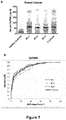
- the concentration medians for OLFM4 and NENF were 2.2 and 3.1-fold higher than healthy controls sera, respectively, with p-value ⁇ 0.005.
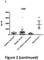
- the concentration median for DSP breast cancer samples was not significantly different from those of healthy controls but a significant difference (p-value ⁇ 0.032) between healthy controls samples and small tumors group (size ⁇ 2 cm, pT1) was identified ( Figure 2 ). In this case, DSP concentration was lower than in the control group.
- 3 overexpressed DSP samples were found which matched with 3 recurrent breast tumors.
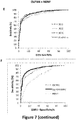
- ROC curves showed that the optimum diagnostic cutoff for OLFM4 was 29.8, 30.0 and 29.4 ng/ml for BC-1, BC-2 and BC-3 respectively.
- the optimum cutoff value for NENF was 13.8, 15.6 and 13.8 ng/ml for BC-1, BC-2 and BC-3, respectively.
- the cutoff values in this study were chosen to be 31 ng/ml for OLFM4 and 16 ng/ml for NENF (Table 7). With these cutoff values, the sensitivity was ranging from 64 to 78% for OLFM4 and from 52% to 60% for NENF.
- the combined markers OLFM4 and NENF were estimated by binary logistic regression and the values of this function was used as one marker and subjected to ROC analysis.
- the values corresponding to the addition of the Elisa concentrations of OLFM4 and NENF in the same serum were also tested and the ROC curves were as exhibited as equivalent (data not shown). So, when combining the OLFM4 and NENF Elisa concentrations, ROC curves showed that the optimum diagnostic cutoff was 38.3 or 38.4 ng/ml in the three independent cohorts or in the combined cohort with a specificity of 87% and a sensitivity ranging from 75 to 85% ( Figure 7E ).
- DSP is decreased in sera of patients with early tumors and increased in sera of patients with recurrent tumors
- OLFM4, NENF and DSP are biomarkers of breast cancer
- OLFM4, low-DSP and NENF are biomarkers for breast cancer in the early phase
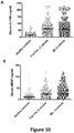
- the median concentration for OLFM4 and NENF were found to be significantly elevated (p ⁇ 0.0001) in T1a-T1b breast tumors sera as compared to healthy samples ( Figure 9A and 9C ). The values did not differ significantly between the three cohorts, and were similar with the BC cohorts.
- the OLFM4 median concentration was determined to be of 51.89 ng/ml (IQR 32.69-83.52) and the NENF median concentration of 17.40 ng/ml (IQR 7.56-30.99).
- the aim of the present studies was to use an innovative proteomic approach to identify novel breast cancer biomarkers. Starting from the fact that no serious candidate had been highlighted with the usual approaches, the present work was focused on secreted proteins that had never been described as potential biomarkers of breast cancer.
- the first step was to create a protein database from a transformed breast cell line, and from luminal A, Her2- overexpressed and triple negative breast tumors with a global proteomic approach.
- the identified proteins were compared with the HUPO Plasma project database and Mann's work to be sure these proteins could be detected in the blood of patients. Then, the identified proteins were compared with the Breast Cancer database and over a hundred secreted proteins which were never tested in breast pathology were identified.
- OLFM4 OLFM4, NENF and DSP as novel breast cancer biomarkers, easily detectable in sera from patients suffering from such cancer.
- the second study was carried out by the inventors on a larger cohort of patients with three independent sera cohorts, in order to further evaluate the breast cancer biomarkes NENF and OLFM4.
- NENF and OLFM4 When the diagnostic accuracy of the combination of both biomarkers was evaluated to distinguish breast cancer patients from healthy controls using a generalized ROC criterion, a very significant overall diagnostic accuracy was observed.
- an AUC of 91% with a sensitivity of 82% and a specificity of 87% were obtained.
- these values remained elevated in early stage breast cancer patients (tumor size inferior to 1cm), and the sensitivity and specificity differed slightly between the different sera cohorts.
- the diagnostic capabilities of serum OLFM4 and NENF were similar in all breast cancer and in early-stage breast cancer patient cohorts. Moreover, the association of the two markers was really beneficial for diagnosis prediction. In the breast cancer set, 421 patients (70%) were positive for OLFM4 alone and 342 patients (53%) were positive for NENF alone. When both markers were combined, 556 patients (85%) were detected. In the same way, for the early-stage breast cancer cohort, 78% of patients were positive for OLFM4 and 53% for NENF alone. This number rose to 82% when OLFM4 and NENF concentrations were combined.
- the present data therefore indicate that the serum biomarkers OLFM4, NENF and DSP, and more preferably OFLM4 and NENF, can be used to detect breast cancer, especially in the early-stage diagnosis.
- those proteins are secreted, their expression can be simply measured in a reliable manner, without the need for invasive techniques in order to detect breast cancer early on, either combined with mammogram to increase the rate of detected occult cancer, either by spacing the number of mammograms in the patient monitoring.
- Mammography has been shown to be the most effective screening tool for detecting breast cancer early and for saving lives.
- mammography has intrinsic limitations that may be difficult to overcome and its sensitivity ranges between 63 and 87%, depending on age, breast density, and tumor characteristics. Therefore, complementary tests are needed to detect women with breast cancer and to increase the diagnosis sensitivity of screening approaches.
- Serum biomarkers may be helpful to increase the positive predictive value of mammographic lesions, thereby decreasing the number of women who undergo unnecessary biopsies.
- biomarkers may also be used to select cases for more sensitive diagnostic techniques, such as magnetic resonance imaging. Another significant application will be the monitoring of young women "at risk”.
- Women at a high risk of developing breast cancer are essentially those carrying BRCA1 and BRCA2 gene mutations or with a high likelihood of a hereditary predisposition to breast cancer. Consequently, screening is reinforced in women carrying these mutated genes: they should undergo twice-yearly clinical examinations and imaging tests as soon as they reach the age of 30.
- Simple monitoring involving two yearly clinical examinations, breast MRI and a mammogram is an alternative associated with hardly any complications.
- the procedure does not reduce the risk of cancer, but aims to detect and treat precancer and cancer lesions as early as possible.
Landscapes
- Health & Medical Sciences (AREA)
- Life Sciences & Earth Sciences (AREA)
- Engineering & Computer Science (AREA)
- Immunology (AREA)
- Biomedical Technology (AREA)
- Molecular Biology (AREA)
- Chemical & Material Sciences (AREA)
- Hematology (AREA)
- Urology & Nephrology (AREA)
- Food Science & Technology (AREA)
- Biochemistry (AREA)
- Cell Biology (AREA)
- Biotechnology (AREA)
- Medicinal Chemistry (AREA)
- Physics & Mathematics (AREA)
- Analytical Chemistry (AREA)
- Microbiology (AREA)
- General Health & Medical Sciences (AREA)
- General Physics & Mathematics (AREA)
- Pathology (AREA)
- Bioinformatics & Cheminformatics (AREA)
- Toxicology (AREA)
- Tropical Medicine & Parasitology (AREA)
- Investigating Or Analysing Biological Materials (AREA)
- Measuring Or Testing Involving Enzymes Or Micro-Organisms (AREA)
Claims (13)
- Procédé in vitro pour diagnostiquer un cancer du sein chez un sujet, comprenant les étapes de :a) détermination à partir d'un échantillon de fluide biologique d'un sujet du niveau d'expression de protéine d'au moins deux biomarqueurs choisis dans le groupe consistant en l'Olfactomédine-4, la Neudésine et la Desmoplakine, dans lequel lesdits au moins deux biomarqueurs sont l'Olfactomédine-4 et la Neudésine ; etb) comparaison dudit niveau d'expression avec un niveau d'expression de référence dudit biomarqueur.
- Procédé selon la revendication 1, dans lequel un niveau d'expression de protéine desdits au moins deux biomarqueurs déréglés par comparaison audit niveau d'expression de référence obtenu à partir d'un échantillon de fluide biologique d'au moins un sujet sain, indique que ledit sujet souffre du cancer du sein.
- Procédé selon la revendication 2, dans lequel ledit niveau d'expression de :• l'Olfactomédine-4 est supérieure audit niveau d'expression de référence, et• la Neudésine est supérieure audit niveau d'expression de référence, et facultativement• la Desmoplakine est inférieure ou supérieure audit niveau d'expression de référence,dans l'échantillon de fluide biologique dudit sujet souffrant du cancer du sein, tel qu'un cancer du sein précoce.
- Procédé selon la revendication 3, dans lequel ledit niveau d'expression de la Desmoplakine inférieur audit niveau d'expression de référence indique que ledit cancer du sein est un cancer du sein précoce.
- Procédé selon la revendication 3, dans lequel ledit niveau d'expression de Desmoplakine supérieur audit niveau d'expression de référence indique que ledit cancer du sein est un cancer du sein récurrent.
- Procédé selon l'une quelconque des revendications 1 à 5, dans lequel le niveau d'expression de protéine d'au moins trois desdits biomarqueurs est déterminé à l'étape a).
- Procédé selon l'une quelconque des revendications 1 à 6, comprenant en outre l'étape de détermination du niveau d'expression de protéine d'au moins un biomarqueur standard associé au cancer du sein, ledit biomarqueur standard étant de préférence choisi dans le groupe consistant en un récepteur des oestrogènes (ER), un récepteur de la progestérone (PR), et le récepteur 2 de facteur de croissance épidermique humain (HER2).
- Procédé de criblage pour identifier un médicament ou une combinaison de médicaments convenant pour traiter le cancer du sein, comprenant les étapes de :a) mise en contact de cellules de cancer du sein ou d'une lignée cellulaire présentant un phénotype de cancer du sein isolée(s) avec un médicament candidat ou une combinaison de médicaments candidats ;b) détermination, à partir desdites cellules ou de ladite lignée cellulaire mise(s) en contact avec ledit médicament ou ladite combinaison de médicaments, du niveau d'expression de protéine desdits au moins deux biomarqueurs tels que définis dans la revendication 1 ; etc) comparaison du niveau d'expression de protéine dudit biomarqueur dans l'étape b) à son niveau d'expression en l'absence dudit médicament ou de ladite combinaison de médicaments.
- Procédé selon l'une quelconque des revendications 1 à 7, dans lequel ledit fluide biologique est choisi dans le groupe consistant en le sang, le sérum, le plasma, la lymphe, le liquide interstitiel tumoral, la salive, le mucus, l'expectoration, la sueur, et l'urine.
- Procédé selon la revendication 9, dans lequel ledit fluide biologique est le sérum.
- Procédé selon l'une quelconque des revendications 1 à 10, dans lequel le niveau d'expression de protéine est déterminé par un procédé choisi dans le groupe consistant en le transfert Western, l'immunotransfert, le dosage d'immunoabsorption enzymatique (ELISA), le dosage ELISPOT (« Enzyme-linked immunospot »), le radioimmunodosage (RIA), l'immunohistochimie, l'immunoprécipitation, le tri de cellules activé par fluorescence (FACS), la microscopie, la cytométrie en flux, la microcytométrie, le dosage de fixation de protéine, le dosage de fixation de ligand, un microréseau, l'électrophorèse sur gel de polyacrylamide tel que SDS-PAGE, la résonance plasmonique de surface (SPR), le transfert d'énergie par résonance de type Förster (FRET), le transfert d'énergie par résonance de bioluminescence (BRET), la chimioluminescence, la polarisation fluorescente, la phosphorescence, la spectrométrie de masse, l'imagerie par résonance magnétique (IRM), et toute combinaison de ceux-ci.
- Utilisation d'une trousse dans un procédé tel que défini dans l'une quelconque des revendications 1 à 11, ladite trousse comprenant :a) au moins un réactif capable de déterminer spécifiquement le niveau d'expression de protéine d'au moins lesdits deux biomarqueurs tels que définis dans la revendication 1 ; etb) des instructions pour réaliser ledit procédé.
- Utilisation d'un microréseau à protéines dans un procédé tel que défini dans l'une quelconque des revendications 1 à 11, ledit microréseau à protéines comprenant :a) au moins un réactif capable de déterminer spécifiquement le niveau d'expression de protéine d'au moins lesdits deux biomarqueurs tels que définis dans la revendication 1.
Priority Applications (2)
| Application Number | Priority Date | Filing Date | Title |
|---|---|---|---|
| EP14820760.8A EP3071966B1 (fr) | 2013-11-22 | 2014-11-24 | Olfactomédine-4, neudésine et desmoplakine en tant que nouveaux biomarqueurs du cancer du sein |
| EP19165351.8A EP3553519B1 (fr) | 2013-11-22 | 2014-11-24 | Olfactomédine-4, neudésine et desmoplakine en tant que nouveaux biomarqueurs du cancer du sein |
Applications Claiming Priority (3)
| Application Number | Priority Date | Filing Date | Title |
|---|---|---|---|
| EP13306604.3A EP2876442A1 (fr) | 2013-11-22 | 2013-11-22 | Olfactomedine 4, neudésine et desmoplakine en tant que biomarqueurs du cancer du sein |
| PCT/EP2014/075427 WO2015075242A1 (fr) | 2013-11-22 | 2014-11-24 | Olfactomédine-4, neudésine et desmoplakine en tant que nouveaux biomarqueurs du cancer du sein |
| EP14820760.8A EP3071966B1 (fr) | 2013-11-22 | 2014-11-24 | Olfactomédine-4, neudésine et desmoplakine en tant que nouveaux biomarqueurs du cancer du sein |
Related Child Applications (1)
| Application Number | Title | Priority Date | Filing Date |
|---|---|---|---|
| EP19165351.8A Division EP3553519B1 (fr) | 2013-11-22 | 2014-11-24 | Olfactomédine-4, neudésine et desmoplakine en tant que nouveaux biomarqueurs du cancer du sein |
Publications (2)
| Publication Number | Publication Date |
|---|---|
| EP3071966A1 EP3071966A1 (fr) | 2016-09-28 |
| EP3071966B1 true EP3071966B1 (fr) | 2019-03-27 |
Family
ID=49726674
Family Applications (3)
| Application Number | Title | Priority Date | Filing Date |
|---|---|---|---|
| EP13306604.3A Withdrawn EP2876442A1 (fr) | 2013-11-22 | 2013-11-22 | Olfactomedine 4, neudésine et desmoplakine en tant que biomarqueurs du cancer du sein |
| EP14820760.8A Not-in-force EP3071966B1 (fr) | 2013-11-22 | 2014-11-24 | Olfactomédine-4, neudésine et desmoplakine en tant que nouveaux biomarqueurs du cancer du sein |
| EP19165351.8A Active EP3553519B1 (fr) | 2013-11-22 | 2014-11-24 | Olfactomédine-4, neudésine et desmoplakine en tant que nouveaux biomarqueurs du cancer du sein |
Family Applications Before (1)
| Application Number | Title | Priority Date | Filing Date |
|---|---|---|---|
| EP13306604.3A Withdrawn EP2876442A1 (fr) | 2013-11-22 | 2013-11-22 | Olfactomedine 4, neudésine et desmoplakine en tant que biomarqueurs du cancer du sein |
Family Applications After (1)
| Application Number | Title | Priority Date | Filing Date |
|---|---|---|---|
| EP19165351.8A Active EP3553519B1 (fr) | 2013-11-22 | 2014-11-24 | Olfactomédine-4, neudésine et desmoplakine en tant que nouveaux biomarqueurs du cancer du sein |
Country Status (4)
| Country | Link |
|---|---|
| US (1) | US10436787B2 (fr) |
| EP (3) | EP2876442A1 (fr) |
| ES (2) | ES2739426T3 (fr) |
| WO (1) | WO2015075242A1 (fr) |
Families Citing this family (1)
| Publication number | Priority date | Publication date | Assignee | Title |
|---|---|---|---|---|
| CN113640092A (zh) * | 2020-04-23 | 2021-11-12 | 复旦大学 | 石蜡包埋组织样本的处理方法以及其质谱检测方法 |
Family Cites Families (18)
| Publication number | Priority date | Publication date | Assignee | Title |
|---|---|---|---|---|
| GB9704444D0 (en) * | 1997-03-04 | 1997-04-23 | Isis Innovation | Non-invasive prenatal diagnosis |
| US6087102A (en) | 1998-01-07 | 2000-07-11 | Clontech Laboratories, Inc. | Polymeric arrays and methods for their use in binding assays |
| US6087103A (en) | 1998-03-04 | 2000-07-11 | Lifespan Biosciences, Inc. | Tagged ligand arrays for identifying target-ligand interactions |
| US6139831A (en) | 1998-05-28 | 2000-10-31 | The Rockfeller University | Apparatus and method for immobilizing molecules onto a substrate |
| US6355623B2 (en) * | 1998-09-24 | 2002-03-12 | Hopital-Sainte-Justine | Method of treating IBD/Crohn's disease and related conditions wherein drug metabolite levels in host blood cells determine subsequent dosage |
| US7504222B2 (en) * | 2001-10-31 | 2009-03-17 | Millennium Pharmaceuticals, Inc. | Compositions, kits, and methods for identification, assessment, prevention, and therapy of breast cancer |
| EP1635178A3 (fr) * | 2004-09-14 | 2006-05-24 | ProteoSys AG | Proteine du cancer du sein |
| US7700299B2 (en) * | 2005-08-12 | 2010-04-20 | Hoffmann-La Roche Inc. | Method for predicting the response to a treatment |
| KR100873382B1 (ko) * | 2006-12-21 | 2008-12-10 | 김현기 | 인간 원암 유전자, 이에 의해 코딩되는 단백질, 이를포함하는 발현벡터, 및 상기 벡터로 형질전환된 세포 |
| US20110085982A1 (en) * | 2009-10-09 | 2011-04-14 | Duke University; The Regents of the University of California | Dkat cell line, a model for human triple-negative breast cancer |
| US20130059903A1 (en) * | 2010-03-12 | 2013-03-07 | The Johns Hopkins University | Compositions and Methods for Characterizing Breast Cancer |
| GB201004551D0 (en) * | 2010-03-19 | 2010-05-05 | Immatics Biotechnologies Gmbh | NOvel immunotherapy against several tumors including gastrointestinal and gastric cancer |
| WO2012031008A2 (fr) * | 2010-08-31 | 2012-03-08 | The General Hospital Corporation | Matières biologiques liées au cancer dans des microvésicules |
| ES2663843T3 (es) * | 2011-03-02 | 2018-04-17 | Institut de Cancérologie de l'Ouest (I.C.O) | Utilización de la proteína olfactomedina-4 (OLFM4) en el diagnóstico del cáncer colorrectal |
| EP2751570A4 (fr) * | 2011-08-31 | 2015-08-12 | Oncocyte Corp | Méthodes et compositions pour le traitement et le diagnostic du cancer |
| EP2963420A1 (fr) * | 2013-02-26 | 2016-01-06 | Wako Pure Chemical Industries, Ltd. | Procédé de détermination du cancer du sein |
| US20150005183A1 (en) * | 2013-07-01 | 2015-01-01 | Expression Pathology, Inc. | Protein biomarkers of late stage breast cancer |
| EP2876445A1 (fr) * | 2013-11-22 | 2015-05-27 | Institut de Cancérologie de l'Ouest | Procédé in vitro de diagnostic et de pronostic de récurrence de cancer du sein triple négatif |
-
2013
- 2013-11-22 EP EP13306604.3A patent/EP2876442A1/fr not_active Withdrawn
-
2014
- 2014-11-24 EP EP14820760.8A patent/EP3071966B1/fr not_active Not-in-force
- 2014-11-24 EP EP19165351.8A patent/EP3553519B1/fr active Active
- 2014-11-24 ES ES14820760T patent/ES2739426T3/es active Active
- 2014-11-24 ES ES19165351T patent/ES2897677T3/es active Active
- 2014-11-24 US US15/038,135 patent/US10436787B2/en not_active Expired - Fee Related
- 2014-11-24 WO PCT/EP2014/075427 patent/WO2015075242A1/fr not_active Ceased
Non-Patent Citations (1)
| Title |
|---|
| KYOU-HOON HAN ET AL: "The functional and structural characterization of a novel oncogene GIG47 involved in the breast tumorigenesis", BMC CANCER, BIOMED CENTRAL, LONDON, GB, vol. 12, no. 1, 2 July 2012 (2012-07-02), pages 274, XP021132675, ISSN: 1471-2407, DOI: 10.1186/1471-2407-12-274 * |
Also Published As
| Publication number | Publication date |
|---|---|
| EP3071966A1 (fr) | 2016-09-28 |
| US20160291022A1 (en) | 2016-10-06 |
| ES2897677T3 (es) | 2022-03-02 |
| WO2015075242A1 (fr) | 2015-05-28 |
| US10436787B2 (en) | 2019-10-08 |
| EP3553519A1 (fr) | 2019-10-16 |
| ES2739426T3 (es) | 2020-01-31 |
| EP3553519B1 (fr) | 2021-08-11 |
| EP2876442A1 (fr) | 2015-05-27 |
Similar Documents
| Publication | Publication Date | Title |
|---|---|---|
| Hudler et al. | Proteomic approaches in biomarker discovery: new perspectives in cancer diagnostics | |
| Chai et al. | Discovery of potential serum protein biomarkers for lymph node metastasis in oral cancer | |
| ES2703943T3 (es) | Método para el diagnóstico y pronóstico in vitro de recaída de cáncer de mama triple negativo | |
| KR20160057352A (ko) | 췌장암 진단용 조성물 및 이를 이용한 췌장암 진단방법 | |
| US10338076B2 (en) | Methods and compositions for the diagnosis of ovarian cancer | |
| CN102348983A (zh) | 含有作为活性成分的硫氧还蛋白-1的乳腺癌诊断标记物及使用所述硫氧还蛋白-1的乳腺癌诊断试剂盒 | |
| JP7325845B2 (ja) | 膵がん及び膵管内乳頭粘液性腫瘍のマーカー | |
| WO2011000573A1 (fr) | Méthode de diagnostic pour prédire le risque de récurrence d'un cancer sur la base d'isoformes de l'histone macroh2a | |
| JP2012526543A5 (fr) | ||
| AU2008325187A1 (en) | Methods for detecting or monitoring cancer using LPC as a marker | |
| EP3071966B1 (fr) | Olfactomédine-4, neudésine et desmoplakine en tant que nouveaux biomarqueurs du cancer du sein | |
| WO2006091861A2 (fr) | Compositions et methodes associees au lymphome du systeme nerveux central | |
| CN110291403B (zh) | 通过质谱法测定braf突变和野生型braf蛋白的方法 | |
| Sileikis et al. | Current role of proteomics in pancreatic cancer biomarkers research | |
| US20090239255A1 (en) | Odontogenic ameloblast-associated protein as a tumor biomarker | |
| US20120289431A1 (en) | Diagnosis of breast cancer based on expression level of thioredoxin-1 | |
| ES2896056T3 (es) | PD-ECGF como biomarcador de cáncer | |
| US20250224402A1 (en) | Mass spectrometry methods for cancer diagnosis | |
| Ebeed et al. | Diagnostic and prognostic value of circulating tumor cells in female breast cancer patients | |
| EP3722809A1 (fr) | Procédé de prédiction de gliome | |
| WO2016060382A1 (fr) | Composition pour le diagnostic de cancer du pancréas, et procédé pour le diagnostic de cancer du pancréas à l'aide de cette dernière | |
| López | Application of Nanotechnology for the Discovery of Circulating Proteins as Novel Biomarkers of Breast Cancer | |
| US20100291690A1 (en) | Methods for detecting or monitoring cancer using lpc as a marker | |
| HK40009490A (en) | A method to determine braf mutations and wild type braf protein by mass spectrometry | |
| Lee et al. | ANT2-Associated Mitochondrial HK2-VDAC1 Organization Underlies [18F] FDG PET Heterogeneity in Aggressive Cancers |
Legal Events
| Date | Code | Title | Description |
|---|---|---|---|
| PUAI | Public reference made under article 153(3) epc to a published international application that has entered the european phase |
Free format text: ORIGINAL CODE: 0009012 |
|
| 17P | Request for examination filed |
Effective date: 20160621 |
|
| AK | Designated contracting states |
Kind code of ref document: A1 Designated state(s): AL AT BE BG CH CY CZ DE DK EE ES FI FR GB GR HR HU IE IS IT LI LT LU LV MC MK MT NL NO PL PT RO RS SE SI SK SM TR |
|
| AX | Request for extension of the european patent |
Extension state: BA ME |
|
| DAX | Request for extension of the european patent (deleted) | ||
| STAA | Information on the status of an ep patent application or granted ep patent |
Free format text: STATUS: EXAMINATION IS IN PROGRESS |
|
| 17Q | First examination report despatched |
Effective date: 20170623 |
|
| GRAP | Despatch of communication of intention to grant a patent |
Free format text: ORIGINAL CODE: EPIDOSNIGR1 |
|
| STAA | Information on the status of an ep patent application or granted ep patent |
Free format text: STATUS: GRANT OF PATENT IS INTENDED |
|
| INTG | Intention to grant announced |
Effective date: 20180806 |
|
| GRAS | Grant fee paid |
Free format text: ORIGINAL CODE: EPIDOSNIGR3 |
|
| GRAJ | Information related to disapproval of communication of intention to grant by the applicant or resumption of examination proceedings by the epo deleted |
Free format text: ORIGINAL CODE: EPIDOSDIGR1 |
|
| GRAL | Information related to payment of fee for publishing/printing deleted |
Free format text: ORIGINAL CODE: EPIDOSDIGR3 |
|
| STAA | Information on the status of an ep patent application or granted ep patent |
Free format text: STATUS: EXAMINATION IS IN PROGRESS |
|
| GRAJ | Information related to disapproval of communication of intention to grant by the applicant or resumption of examination proceedings by the epo deleted |
Free format text: ORIGINAL CODE: EPIDOSDIGR1 |
|
| GRAP | Despatch of communication of intention to grant a patent |
Free format text: ORIGINAL CODE: EPIDOSNIGR1 |
|
| GRAP | Despatch of communication of intention to grant a patent |
Free format text: ORIGINAL CODE: EPIDOSNIGR1 |
|
| STAA | Information on the status of an ep patent application or granted ep patent |
Free format text: STATUS: GRANT OF PATENT IS INTENDED |
|
| INTC | Intention to grant announced (deleted) | ||
| INTG | Intention to grant announced |
Effective date: 20181204 |
|
| GRAA | (expected) grant |
Free format text: ORIGINAL CODE: 0009210 |
|
| STAA | Information on the status of an ep patent application or granted ep patent |
Free format text: STATUS: THE PATENT HAS BEEN GRANTED |
|
| AK | Designated contracting states |
Kind code of ref document: B1 Designated state(s): AL AT BE BG CH CY CZ DE DK EE ES FI FR GB GR HR HU IE IS IT LI LT LU LV MC MK MT NL NO PL PT RO RS SE SI SK SM TR |
|
| REG | Reference to a national code |
Ref country code: GB Ref legal event code: FG4D |
|
| REG | Reference to a national code |
Ref country code: CH Ref legal event code: EP |
|
| REG | Reference to a national code |
Ref country code: AT Ref legal event code: REF Ref document number: 1113720 Country of ref document: AT Kind code of ref document: T Effective date: 20190415 |
|
| REG | Reference to a national code |
Ref country code: IE Ref legal event code: FG4D |
|
| REG | Reference to a national code |
Ref country code: DE Ref legal event code: R096 Ref document number: 602014043752 Country of ref document: DE |
|
| PG25 | Lapsed in a contracting state [announced via postgrant information from national office to epo] |
Ref country code: NO Free format text: LAPSE BECAUSE OF FAILURE TO SUBMIT A TRANSLATION OF THE DESCRIPTION OR TO PAY THE FEE WITHIN THE PRESCRIBED TIME-LIMIT Effective date: 20190627 Ref country code: SE Free format text: LAPSE BECAUSE OF FAILURE TO SUBMIT A TRANSLATION OF THE DESCRIPTION OR TO PAY THE FEE WITHIN THE PRESCRIBED TIME-LIMIT Effective date: 20190327 Ref country code: FI Free format text: LAPSE BECAUSE OF FAILURE TO SUBMIT A TRANSLATION OF THE DESCRIPTION OR TO PAY THE FEE WITHIN THE PRESCRIBED TIME-LIMIT Effective date: 20190327 Ref country code: LT Free format text: LAPSE BECAUSE OF FAILURE TO SUBMIT A TRANSLATION OF THE DESCRIPTION OR TO PAY THE FEE WITHIN THE PRESCRIBED TIME-LIMIT Effective date: 20190327 |
|
| REG | Reference to a national code |
Ref country code: NL Ref legal event code: MP Effective date: 20190327 |
|
| REG | Reference to a national code |
Ref country code: CH Ref legal event code: NV Representative=s name: MICHELI AND CIE SA, CH |
|
| PG25 | Lapsed in a contracting state [announced via postgrant information from national office to epo] |
Ref country code: BG Free format text: LAPSE BECAUSE OF FAILURE TO SUBMIT A TRANSLATION OF THE DESCRIPTION OR TO PAY THE FEE WITHIN THE PRESCRIBED TIME-LIMIT Effective date: 20190627 Ref country code: GR Free format text: LAPSE BECAUSE OF FAILURE TO SUBMIT A TRANSLATION OF THE DESCRIPTION OR TO PAY THE FEE WITHIN THE PRESCRIBED TIME-LIMIT Effective date: 20190628 Ref country code: NL Free format text: LAPSE BECAUSE OF FAILURE TO SUBMIT A TRANSLATION OF THE DESCRIPTION OR TO PAY THE FEE WITHIN THE PRESCRIBED TIME-LIMIT Effective date: 20190327 Ref country code: LV Free format text: LAPSE BECAUSE OF FAILURE TO SUBMIT A TRANSLATION OF THE DESCRIPTION OR TO PAY THE FEE WITHIN THE PRESCRIBED TIME-LIMIT Effective date: 20190327 Ref country code: HR Free format text: LAPSE BECAUSE OF FAILURE TO SUBMIT A TRANSLATION OF THE DESCRIPTION OR TO PAY THE FEE WITHIN THE PRESCRIBED TIME-LIMIT Effective date: 20190327 Ref country code: RS Free format text: LAPSE BECAUSE OF FAILURE TO SUBMIT A TRANSLATION OF THE DESCRIPTION OR TO PAY THE FEE WITHIN THE PRESCRIBED TIME-LIMIT Effective date: 20190327 |
|
| REG | Reference to a national code |
Ref country code: AT Ref legal event code: MK05 Ref document number: 1113720 Country of ref document: AT Kind code of ref document: T Effective date: 20190327 |
|
| PG25 | Lapsed in a contracting state [announced via postgrant information from national office to epo] |
Ref country code: CZ Free format text: LAPSE BECAUSE OF FAILURE TO SUBMIT A TRANSLATION OF THE DESCRIPTION OR TO PAY THE FEE WITHIN THE PRESCRIBED TIME-LIMIT Effective date: 20190327 Ref country code: SK Free format text: LAPSE BECAUSE OF FAILURE TO SUBMIT A TRANSLATION OF THE DESCRIPTION OR TO PAY THE FEE WITHIN THE PRESCRIBED TIME-LIMIT Effective date: 20190327 Ref country code: RO Free format text: LAPSE BECAUSE OF FAILURE TO SUBMIT A TRANSLATION OF THE DESCRIPTION OR TO PAY THE FEE WITHIN THE PRESCRIBED TIME-LIMIT Effective date: 20190327 Ref country code: AL Free format text: LAPSE BECAUSE OF FAILURE TO SUBMIT A TRANSLATION OF THE DESCRIPTION OR TO PAY THE FEE WITHIN THE PRESCRIBED TIME-LIMIT Effective date: 20190327 Ref country code: EE Free format text: LAPSE BECAUSE OF FAILURE TO SUBMIT A TRANSLATION OF THE DESCRIPTION OR TO PAY THE FEE WITHIN THE PRESCRIBED TIME-LIMIT Effective date: 20190327 Ref country code: PT Free format text: LAPSE BECAUSE OF FAILURE TO SUBMIT A TRANSLATION OF THE DESCRIPTION OR TO PAY THE FEE WITHIN THE PRESCRIBED TIME-LIMIT Effective date: 20190727 |
|
| PG25 | Lapsed in a contracting state [announced via postgrant information from national office to epo] |
Ref country code: PL Free format text: LAPSE BECAUSE OF FAILURE TO SUBMIT A TRANSLATION OF THE DESCRIPTION OR TO PAY THE FEE WITHIN THE PRESCRIBED TIME-LIMIT Effective date: 20190327 Ref country code: SM Free format text: LAPSE BECAUSE OF FAILURE TO SUBMIT A TRANSLATION OF THE DESCRIPTION OR TO PAY THE FEE WITHIN THE PRESCRIBED TIME-LIMIT Effective date: 20190327 |
|
| PG25 | Lapsed in a contracting state [announced via postgrant information from national office to epo] |
Ref country code: IS Free format text: LAPSE BECAUSE OF FAILURE TO SUBMIT A TRANSLATION OF THE DESCRIPTION OR TO PAY THE FEE WITHIN THE PRESCRIBED TIME-LIMIT Effective date: 20190727 Ref country code: AT Free format text: LAPSE BECAUSE OF FAILURE TO SUBMIT A TRANSLATION OF THE DESCRIPTION OR TO PAY THE FEE WITHIN THE PRESCRIBED TIME-LIMIT Effective date: 20190327 |
|
| REG | Reference to a national code |
Ref country code: DE Ref legal event code: R097 Ref document number: 602014043752 Country of ref document: DE |
|
| PG25 | Lapsed in a contracting state [announced via postgrant information from national office to epo] |
Ref country code: DK Free format text: LAPSE BECAUSE OF FAILURE TO SUBMIT A TRANSLATION OF THE DESCRIPTION OR TO PAY THE FEE WITHIN THE PRESCRIBED TIME-LIMIT Effective date: 20190327 |
|
| REG | Reference to a national code |
Ref country code: ES Ref legal event code: FG2A Ref document number: 2739426 Country of ref document: ES Kind code of ref document: T3 Effective date: 20200131 |
|
| PLBE | No opposition filed within time limit |
Free format text: ORIGINAL CODE: 0009261 |
|
| STAA | Information on the status of an ep patent application or granted ep patent |
Free format text: STATUS: NO OPPOSITION FILED WITHIN TIME LIMIT |
|
| PG25 | Lapsed in a contracting state [announced via postgrant information from national office to epo] |
Ref country code: SI Free format text: LAPSE BECAUSE OF FAILURE TO SUBMIT A TRANSLATION OF THE DESCRIPTION OR TO PAY THE FEE WITHIN THE PRESCRIBED TIME-LIMIT Effective date: 20190327 |
|
| 26N | No opposition filed |
Effective date: 20200103 |
|
| PG25 | Lapsed in a contracting state [announced via postgrant information from national office to epo] |
Ref country code: TR Free format text: LAPSE BECAUSE OF FAILURE TO SUBMIT A TRANSLATION OF THE DESCRIPTION OR TO PAY THE FEE WITHIN THE PRESCRIBED TIME-LIMIT Effective date: 20190327 |
|
| PG25 | Lapsed in a contracting state [announced via postgrant information from national office to epo] |
Ref country code: LU Free format text: LAPSE BECAUSE OF NON-PAYMENT OF DUE FEES Effective date: 20191124 Ref country code: MC Free format text: LAPSE BECAUSE OF FAILURE TO SUBMIT A TRANSLATION OF THE DESCRIPTION OR TO PAY THE FEE WITHIN THE PRESCRIBED TIME-LIMIT Effective date: 20190327 |
|
| PG25 | Lapsed in a contracting state [announced via postgrant information from national office to epo] |
Ref country code: IE Free format text: LAPSE BECAUSE OF NON-PAYMENT OF DUE FEES Effective date: 20191124 |
|
| PG25 | Lapsed in a contracting state [announced via postgrant information from national office to epo] |
Ref country code: CY Free format text: LAPSE BECAUSE OF FAILURE TO SUBMIT A TRANSLATION OF THE DESCRIPTION OR TO PAY THE FEE WITHIN THE PRESCRIBED TIME-LIMIT Effective date: 20190327 |
|
| PG25 | Lapsed in a contracting state [announced via postgrant information from national office to epo] |
Ref country code: HU Free format text: LAPSE BECAUSE OF FAILURE TO SUBMIT A TRANSLATION OF THE DESCRIPTION OR TO PAY THE FEE WITHIN THE PRESCRIBED TIME-LIMIT; INVALID AB INITIO Effective date: 20141124 Ref country code: MT Free format text: LAPSE BECAUSE OF FAILURE TO SUBMIT A TRANSLATION OF THE DESCRIPTION OR TO PAY THE FEE WITHIN THE PRESCRIBED TIME-LIMIT Effective date: 20190327 |
|
| PGFP | Annual fee paid to national office [announced via postgrant information from national office to epo] |
Ref country code: FR Payment date: 20211130 Year of fee payment: 8 Ref country code: ES Payment date: 20211203 Year of fee payment: 8 Ref country code: GB Payment date: 20211122 Year of fee payment: 8 Ref country code: DE Payment date: 20211110 Year of fee payment: 8 |
|
| PGFP | Annual fee paid to national office [announced via postgrant information from national office to epo] |
Ref country code: IT Payment date: 20211112 Year of fee payment: 8 Ref country code: CH Payment date: 20211125 Year of fee payment: 8 Ref country code: BE Payment date: 20211130 Year of fee payment: 8 |
|
| PG25 | Lapsed in a contracting state [announced via postgrant information from national office to epo] |
Ref country code: MK Free format text: LAPSE BECAUSE OF FAILURE TO SUBMIT A TRANSLATION OF THE DESCRIPTION OR TO PAY THE FEE WITHIN THE PRESCRIBED TIME-LIMIT Effective date: 20190327 |
|
| REG | Reference to a national code |
Ref country code: DE Ref legal event code: R119 Ref document number: 602014043752 Country of ref document: DE |
|
| REG | Reference to a national code |
Ref country code: CH Ref legal event code: PL |
|
| GBPC | Gb: european patent ceased through non-payment of renewal fee |
Effective date: 20221124 |
|
| REG | Reference to a national code |
Ref country code: BE Ref legal event code: MM Effective date: 20221130 |
|
| PG25 | Lapsed in a contracting state [announced via postgrant information from national office to epo] |
Ref country code: LI Free format text: LAPSE BECAUSE OF NON-PAYMENT OF DUE FEES Effective date: 20221130 Ref country code: CH Free format text: LAPSE BECAUSE OF NON-PAYMENT OF DUE FEES Effective date: 20221130 |
|
| PG25 | Lapsed in a contracting state [announced via postgrant information from national office to epo] |
Ref country code: IT Free format text: LAPSE BECAUSE OF NON-PAYMENT OF DUE FEES Effective date: 20221124 Ref country code: GB Free format text: LAPSE BECAUSE OF NON-PAYMENT OF DUE FEES Effective date: 20221124 Ref country code: DE Free format text: LAPSE BECAUSE OF NON-PAYMENT OF DUE FEES Effective date: 20230601 |
|
| PG25 | Lapsed in a contracting state [announced via postgrant information from national office to epo] |
Ref country code: FR Free format text: LAPSE BECAUSE OF NON-PAYMENT OF DUE FEES Effective date: 20221130 Ref country code: BE Free format text: LAPSE BECAUSE OF NON-PAYMENT OF DUE FEES Effective date: 20221130 |
|
| REG | Reference to a national code |
Ref country code: ES Ref legal event code: FD2A Effective date: 20240103 |
|
| PG25 | Lapsed in a contracting state [announced via postgrant information from national office to epo] |
Ref country code: ES Free format text: LAPSE BECAUSE OF NON-PAYMENT OF DUE FEES Effective date: 20221125 |
|
| PG25 | Lapsed in a contracting state [announced via postgrant information from national office to epo] |
Ref country code: ES Free format text: LAPSE BECAUSE OF NON-PAYMENT OF DUE FEES Effective date: 20221125 |