EP3092940A1 - Endoscope - Google Patents
Endoscope Download PDFInfo
- Publication number
- EP3092940A1 EP3092940A1 EP15789296.9A EP15789296A EP3092940A1 EP 3092940 A1 EP3092940 A1 EP 3092940A1 EP 15789296 A EP15789296 A EP 15789296A EP 3092940 A1 EP3092940 A1 EP 3092940A1
- Authority
- EP
- European Patent Office
- Prior art keywords
- distal end
- optical system
- illumination
- inclined plane
- illuminating
- Prior art date
- Legal status (The legal status is an assumption and is not a legal conclusion. Google has not performed a legal analysis and makes no representation as to the accuracy of the status listed.)
- Withdrawn
Links
- 238000005286 illumination Methods 0.000 claims abstract description 85
- 230000003287 optical effect Effects 0.000 claims abstract description 74
- 230000002093 peripheral effect Effects 0.000 claims abstract description 37
- 238000003780 insertion Methods 0.000 claims abstract description 35
- 230000037431 insertion Effects 0.000 claims abstract description 35
- XLYOFNOQVPJJNP-UHFFFAOYSA-N water Substances O XLYOFNOQVPJJNP-UHFFFAOYSA-N 0.000 claims description 29
- 239000000853 adhesive Substances 0.000 claims description 20
- 230000001070 adhesive effect Effects 0.000 claims description 20
- 101000608734 Helianthus annuus 11 kDa late embryogenesis abundant protein Proteins 0.000 abstract description 5
- 239000008186 active pharmaceutical agent Substances 0.000 description 8
- 239000002184 metal Substances 0.000 description 7
- 239000013307 optical fiber Substances 0.000 description 6
- 238000005452 bending Methods 0.000 description 3
- 230000004075 alteration Effects 0.000 description 2
- 238000006243 chemical reaction Methods 0.000 description 2
- 230000007423 decrease Effects 0.000 description 2
- 238000010586 diagram Methods 0.000 description 2
- 230000000694 effects Effects 0.000 description 2
- 230000002349 favourable effect Effects 0.000 description 2
- 230000014509 gene expression Effects 0.000 description 2
- 239000011347 resin Substances 0.000 description 2
- 229920005989 resin Polymers 0.000 description 2
- 238000004140 cleaning Methods 0.000 description 1
- 239000000835 fiber Substances 0.000 description 1
- 230000004048 modification Effects 0.000 description 1
- 238000012986 modification Methods 0.000 description 1
- 238000000465 moulding Methods 0.000 description 1
- 229910001220 stainless steel Inorganic materials 0.000 description 1
- 239000010935 stainless steel Substances 0.000 description 1
Images




Classifications
-
- A—HUMAN NECESSITIES
- A61—MEDICAL OR VETERINARY SCIENCE; HYGIENE
- A61B—DIAGNOSIS; SURGERY; IDENTIFICATION
- A61B1/00—Instruments for performing medical examinations of the interior of cavities or tubes of the body by visual or photographical inspection, e.g. endoscopes; Illuminating arrangements therefor
- A61B1/06—Instruments for performing medical examinations of the interior of cavities or tubes of the body by visual or photographical inspection, e.g. endoscopes; Illuminating arrangements therefor with illuminating arrangements
- A61B1/0661—Endoscope light sources
- A61B1/0676—Endoscope light sources at distal tip of an endoscope
-
- A—HUMAN NECESSITIES
- A61—MEDICAL OR VETERINARY SCIENCE; HYGIENE
- A61B—DIAGNOSIS; SURGERY; IDENTIFICATION
- A61B1/00—Instruments for performing medical examinations of the interior of cavities or tubes of the body by visual or photographical inspection, e.g. endoscopes; Illuminating arrangements therefor
- A61B1/00064—Constructional details of the endoscope body
- A61B1/00071—Insertion part of the endoscope body
- A61B1/0008—Insertion part of the endoscope body characterised by distal tip features
- A61B1/00096—Optical elements
-
- A—HUMAN NECESSITIES
- A61—MEDICAL OR VETERINARY SCIENCE; HYGIENE
- A61B—DIAGNOSIS; SURGERY; IDENTIFICATION
- A61B1/00—Instruments for performing medical examinations of the interior of cavities or tubes of the body by visual or photographical inspection, e.g. endoscopes; Illuminating arrangements therefor
- A61B1/04—Instruments for performing medical examinations of the interior of cavities or tubes of the body by visual or photographical inspection, e.g. endoscopes; Illuminating arrangements therefor combined with photographic or television appliances
- A61B1/05—Instruments for performing medical examinations of the interior of cavities or tubes of the body by visual or photographical inspection, e.g. endoscopes; Illuminating arrangements therefor combined with photographic or television appliances characterised by the image sensor, e.g. camera, being in the distal end portion
-
- A—HUMAN NECESSITIES
- A61—MEDICAL OR VETERINARY SCIENCE; HYGIENE
- A61B—DIAGNOSIS; SURGERY; IDENTIFICATION
- A61B1/00—Instruments for performing medical examinations of the interior of cavities or tubes of the body by visual or photographical inspection, e.g. endoscopes; Illuminating arrangements therefor
- A61B1/06—Instruments for performing medical examinations of the interior of cavities or tubes of the body by visual or photographical inspection, e.g. endoscopes; Illuminating arrangements therefor with illuminating arrangements
- A61B1/0607—Instruments for performing medical examinations of the interior of cavities or tubes of the body by visual or photographical inspection, e.g. endoscopes; Illuminating arrangements therefor with illuminating arrangements for annular illumination
-
- A—HUMAN NECESSITIES
- A61—MEDICAL OR VETERINARY SCIENCE; HYGIENE
- A61B—DIAGNOSIS; SURGERY; IDENTIFICATION
- A61B1/00—Instruments for performing medical examinations of the interior of cavities or tubes of the body by visual or photographical inspection, e.g. endoscopes; Illuminating arrangements therefor
- A61B1/06—Instruments for performing medical examinations of the interior of cavities or tubes of the body by visual or photographical inspection, e.g. endoscopes; Illuminating arrangements therefor with illuminating arrangements
- A61B1/07—Instruments for performing medical examinations of the interior of cavities or tubes of the body by visual or photographical inspection, e.g. endoscopes; Illuminating arrangements therefor with illuminating arrangements using light-conductive means, e.g. optical fibres
-
- G—PHYSICS
- G02—OPTICS
- G02B—OPTICAL ELEMENTS, SYSTEMS OR APPARATUS
- G02B23/00—Telescopes, e.g. binoculars; Periscopes; Instruments for viewing the inside of hollow bodies; Viewfinders; Optical aiming or sighting devices
- G02B23/24—Instruments or systems for viewing the inside of hollow bodies, e.g. fibrescopes
- G02B23/2407—Optical details
- G02B23/2423—Optical details of the distal end
- G02B23/243—Objectives for endoscopes
-
- G—PHYSICS
- G02—OPTICS
- G02B—OPTICAL ELEMENTS, SYSTEMS OR APPARATUS
- G02B23/00—Telescopes, e.g. binoculars; Periscopes; Instruments for viewing the inside of hollow bodies; Viewfinders; Optical aiming or sighting devices
- G02B23/24—Instruments or systems for viewing the inside of hollow bodies, e.g. fibrescopes
- G02B23/2407—Optical details
- G02B23/2461—Illumination
- G02B23/2469—Illumination using optical fibres
-
- G—PHYSICS
- G02—OPTICS
- G02B—OPTICAL ELEMENTS, SYSTEMS OR APPARATUS
- G02B23/00—Telescopes, e.g. binoculars; Periscopes; Instruments for viewing the inside of hollow bodies; Viewfinders; Optical aiming or sighting devices
- G02B23/24—Instruments or systems for viewing the inside of hollow bodies, e.g. fibrescopes
- G02B23/2476—Non-optical details, e.g. housings, mountings, supports
- G02B23/2484—Arrangements in relation to a camera or imaging device
-
- G—PHYSICS
- G02—OPTICS
- G02B—OPTICAL ELEMENTS, SYSTEMS OR APPARATUS
- G02B23/00—Telescopes, e.g. binoculars; Periscopes; Instruments for viewing the inside of hollow bodies; Viewfinders; Optical aiming or sighting devices
- G02B23/24—Instruments or systems for viewing the inside of hollow bodies, e.g. fibrescopes
- G02B23/26—Instruments or systems for viewing the inside of hollow bodies, e.g. fibrescopes using light guides
Definitions
- the present invention relates to an endoscope, and particularly relates to an endoscope that emits illuminating lights from at least two illumination optical systems provided in a distal end portion of an insertion portion.
- An endoscope has been widely used in a medical field and an industrial field.
- An endoscope is configured so that illuminating lights are emitted from one or more illuminating windows provided at a distal end portion of an insertion portion, and reflection lights from an object illuminated with the illuminating lights are incident on an objective lens of an observation window.
- Japanese Patent Application Laid-Open Publication No. 2007-289355 proposes an endoscope in which an inclined plane to be a light shielding wall is provided between an illuminating window and an objective lens so that the light emitted from the illuminating window does not directly enter the objective lens.
- the illuminating lights that are respectively emitted from the two illuminating windows are the brightest in optical axis center portions of the illuminating lights, and light amounts decrease toward peripheries from centers.
- the present invention has an object to provide an endoscope in which light distribution unevenness by two illuminating lights that are respectively emitted from two illuminating windows is reduced.
- An endoscope of one aspect of the present invention includes an observation optical system that is placed in a distal end portion of an insertion portion, and is capable of enlarged observation optically, a first illumination optical system that is placed in the distal end portion, has a first illumination lens in a distal end, and emits a first illuminating light, a second illumination optical system that is placed in the distal end portion, has a second illumination lens in a distal end, and emits a second illuminating light, a first inclined plane portion that is provided between a peripheral edge portion of an objective lens in a distal end of the observation optical system and a peripheral edge portion of the first illumination lens, a second inclined plane portion that is provided between a peripheral edge portion of the objective lens and a peripheral edge portion of the second illumination lens, a first edge line portion that is formed at an intermediate portion of the first inclined plane portion, and is formed to protrude in a distal end direction of the insertion portion, between the peripheral edge portion of the objective lens and the peripheral edge portion of the first
- FIG. 1 is a configuration diagram showing a configuration of an endoscope apparatus according to the present embodiment.
- An endoscope apparatus 1 includes an endoscope 2, a main body apparatus 3 and a monitor 4.
- the monitor 4 is connected to the main body apparatus 3 by a cable 5.
- the endoscope 2 is an electronic endoscope in this case, and includes an operation portion 11 including a knob and a switch for performing a bending operation and control of various conduits, an insertion portion 12 that has a proximal end portion connected to the operation portion 11 and is inserted into a body cavity, and a universal cord 14 that is extended from the operation portion 11 and has a connector portion 13 at a distal end portion.
- the connector portion 13 is connected to the main body apparatus 3.
- the insertion portion 12 has a flexible tube portion 15 having flexibility, a bending portion 16 that is provided at a distal end side of the flexible tube portion 15, and a distal end portion 17 that is provided at a distal end side of the bending portion 16.
- the distal end portion 17 is a distal end rigid portion, and in a distal end face of the distal end portion 17, illuminating windows, an observation window and the like that will be described later are provided.
- a distal end portion of a light guide that guides illuminating lights is placed inside the distal end portion 17, and illuminating lights are emitted from three illuminating windows on the distal end face of the distal end portion 17.
- an image pickup device 41c (refer to Fig. 3 ) such as a CMOS image sensor is placed, and receives reflection light from an object that receives light through the observation window to subject the reflection light to photoelectric conversion to generate an image pickup signal.
- the main body apparatus 3 contains a light source apparatus and a processor.
- the light source apparatus has a light emitting portion such as an LED, and supplies light to a proximal end portion of the light guide which is inserted through the insertion portion 12.
- the processor controls an operation of the entire endoscope apparatus 1, receives an image pickup signal from the image pickup device in the distal end portion 17, generates an endoscopic image, outputs the endoscopic image to the monitor 4 via the cable 5, and records the endoscopic image in a storage apparatus not illustrated.
- an air feeding source, a water feeding source and a suction source for performing air feeding, water feeding, suction and the like to various conduits contained in the endoscope 2 are provided in the main body apparatus 3, or an apparatus not illustrated that is further connected to the main body apparatus 3.
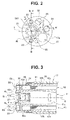
- Fig. 2 is a front view of the distal end face of the distal end portion 17.
- Fig. 3 is a sectional view of a distal end portion of the distal end portion 17 along a line III-III in Fig. 2 .
- Fig. 4 is a sectional view of the distal end portion of the distal end portion 17 along a line IV-IV in Fig. 2 .
- an observation window 21, three illuminating windows 22, 23 and 24, a forward water feeding opening 25, an air/water feeding nozzle 26 and a treatment instrument opening 27 are provided, and disposed.
- a center axis O of the observation window 21 does not correspond to a center axis C of the distal end portion 17.
- the observation window 21 is disposed by being deviated from the center axis C by a predetermined amount.
- the observation window 21 is an end face (hereinafter, an objective face) at an observation target side in an image pickup optical system of an image pickup unit 41 which will be described later.
- the illuminating windows 22, 23 and 24 are respectively end faces (hereinafter, objective faces) at the observation target side in illumination optical systems of light guide units 42, 43 and 44 that will be described later.
- the illuminating windows 22 and 23 are disposed at positions which are point-symmetric or positions which are substantially point-symmetric with respect to a point of the center axis O of the observation window 21, or at positions which are line-symmetric or positions which are substantially line-symmetric with respect to a line LS (shown by a chain double-dashed line in Fig. 2 ) which passes through the center axis O.
- the illuminating windows 22 and 23 are disposed so that distances from an observation optical system of the observation window 21 are equal to each other, in the distal end portion 17, whereas the illuminating window 24 is placed at a position which is farther than the illuminating windows 22 and 23, in the distal end portion 17.
- a distal end rigid member 31 which is in a columnar shape and made of a metal such as stainless steel is provided in the distal end portion 17.
- a plurality of holes are formed, which are for fixing various members corresponding to the observation window 21, the three illuminating windows 22, 23 and 24, the forward water feeding opening 25, the air/water feeding nozzle 26 and the treatment instrument opening 27 which are disposed in the distal end face of the distal end portion 17.
- a distal end cover 32 of a resin is put on a distal end side of the distal end rigid member 31.
- a plurality of holes are also formed in positions corresponding to the plurality of holes which are formed in the distal end rigid member 31.
- An outer sheath 33 is put on the distal end portion 17 to the flexible tube portion 15.
- a distal end portion of the outer sheath 33 is fixed to the distal end rigid member 31 by a thread 34 being wound on the distal end portion, and an adhesive 35 being further applied to the distal end portion.
- the image pickup unit 41 is formed at a rear side from the observation window 21. Further, the light guide units 42, 43 and 44 are formed respectively at rear sides of the three illuminating windows 22, 23 and 24.
- a forward water feeding conduit (not illustrated) is placed at a rear side of the forward water feeding opening 25.
- An air/water feeding conduit (not illustrated) is placed at a rear side of the air/water feeding nozzle 26.
- the treatment instrument opening 27 communicates with a treatment instrument insertion channel (not illustrated).
- the image pickup unit 41 has a frame 41b including an objective lens 41 a at a distal end side. Further, the image pickup unit 41 has the image pickup device 41c such as a CMOS image sensor, which receives light from an object, which passes through the objective lens 41a, subjects the light to photoelectric conversion, and outputs an image pickup signal. A distal end portion of the image pickup unit 41 is fitted into a predetermined hole of the distal end rigid member 31 and is fixed by an adhesive.
- the image pickup device 41c such as a CMOS image sensor
- An observation optical system of the image pickup unit 41 is an optical system capable of enlarged observation optically. That is, the observation optical system including the objective lens 41a is an observation optical system which is placed in the distal end portion 17 of the insertion portion 12 and is capable of enlarged observation optically.
- the light guide unit 42 is formed of an illumination lens 42a formed of a plurality of lenses, and an optical fiber bundle 42b which is a light guide.
- a distal end portion of the optical fiber bundle 42b is fixed into a metal pipe 42c by an adhesive or the like.
- the illumination lens 42a is inserted into a frame 42d to be fixed to the frame 42d, and the metal pipe 42c is internally inserted from a proximal end side of the frame 42d to be fixed to the frame 42d.
- the light guide unit 43 is also formed of an illumination lens 43 a formed of a plurality of lenses, and an optical fiber bundle 43b that is a light guide, similarly to the light guide unit 42.
- a distal end portion of the optical fiber bundle 43b is fixed into a metal pipe 43c by an adhesive or the like.
- the illumination lens 43a is inserted into a frame 43d to be fixed to the frame 43d, and the metal pipe 43c is internally inserted from a proximal end side of the frame 43d to be fixed to the frame 43d.
- the light guide unit 44 is also formed of an illumination lens 44a formed of a plurality of lenses, and an optical fiber bundle 44b that is a light guide, similarly to the light guide unit 42.
- a distal end portion of the optical fiber bundle 44b is fixed into a metal pipe 44c by an adhesive or the like.
- the illumination lens 44a is inserted into a frame 44d, and the metal pipe 44c is internally inserted from a proximal end side of the frame 44d to be fixed to the frame 44d.
- Illumination optical systems of the light guide units 42, 43 and 44 are respectively placed in the distal end portion 17, respectively have illumination lenses and emit illuminating lights.
- Respective distal end portions of the image pickup unit 41 and the light guide units 42, 43 and 44 are inserted into corresponding holes formed in the distal end cover 32, and are fixed by an adhesive.
- Respective distal end faces of the light guide lenses 42, 43 and 44 are exit surfaces ES for illuminating lights, and a distal end face of the objective lens 41a is an incident surface IS for reflection lights from an object.
- the additional illuminating window 24 is provided in addition to the illuminating windows 22 and 23, and the illuminating window 24 is an illuminating window for sufficiently ensuring a light amount of illuminating light at a non-proximal time.
- Fig. 5 is a front view of the distal end face 17a of the distal end portion 17 for explaining an inclined plane portion formed in the distal end face 17a of the distal end portion 17.
- Fig. 6 is a partial sectional view for explaining configurations of the distal end portion of the image pickup unit 41 in the distal end portion 17, and the distal end portion of the light guide unit 42.
- An inclined plane portion DS1 is provided between the observation window 21 and the illuminating windows 22 and 23.
- the inclined plane portion DS1 shown by oblique lines is formed into a U-shape, around the observation window 21 on the distal end face 17a in the distal end cover 32.
- the inclined plane portion DS1 is formed to pass between the observation window 21 and the illuminating window 22 from one edge portion ED1 at the distal end face 17a in the distal end cover 32, surround a part of a peripheral portion of the observation window 21, and pass further between the observation window 21 and the illuminating window 23 to reach another edge portion ED2 at the distal end face 17a.
- a region surrounded by the two edge portions ED1 and ED2 and the observation window 21 is a flat face FS1.
- a concave lens 41a1 at the distal end of the objective lens 41a is fixed to the frame 41b by an adhesive 51.
- the frame 41b in the distal end portion of the image pickup unit 41 is fixed to the distal end cover 32 by an adhesive 52.
- Fig. 6 is a partial sectional view of a part between the image pickup unit 41 and the light guide unit 42, a partial section of a part between the image pickup unit 41 and the light guide unit 43 are similar to Fig. 6 . That is, a configuration of the part between the image pickup unit 41 and the light guide unit 43 is the same as the configuration shown in Fig. 6 .
- a distal end lens 42a1 of the illumination lens 42a is fixed to the frame 42d by an adhesive 53.
- the frame 42d in the distal end portion of the light guide unit 42 is fixed to the distal end cover 32 by an adhesive 54.
- the inclined plane portion DS1 more specifically has an inclined portion r1 that gradually inclines toward an exit surface ES from an end portion of the observation window 21, and an inclined portion r2 that has an inclination angle larger than an inclination angle of the inclined portion r1 toward an outer edge of the adhesive 54 that surrounds the exit surface ES from an outer edge of the inclined portion r1.
- a boundary between the inclined portions r1 and r2 is an edge line portion AL.
- a surface portion r3 of the adhesive 52 for fixing the frame 41b of the image pickup unit 41 to the distal end cover 32 is a part of the inclined portion r1. That is, the surface portion r3 at a distal end side of the adhesive 52 and the inclined portion r1 of the distal end cover 32 have a continuous surface.
- the two inclined portions r1 and r2 are formed at a distal end face side of the distal end cover 32 here, only an inclined portion corresponding to a part of the inclined portion r2 may be formed in the distal end face of the distal end cover 32, and the inclined portion r1 including the edge line portion AL and a part of the inclined portion r2 may be formed in the surface portion r3 at the distal end side of the adhesive 52.
- parts of the inclined portions r1 and r2 of the inclined plane portion DS1 are formed at the distal end cover 32 which is the distal end cover member which is put on the distal end portion 17, and a remaining portion of the inclined portion r1 is formed by the adhesive 52.
- Parts of the inclined portions r1 and r2 of the inclined plane portion DS1 may be formed by the adhesive 52 which connects and fixes the distal end cover 32 which is put on the distal end portion 17 and the image pickup unit 41 which is the observation optical system.
- the two inclined portions r1 and r2 are formed at the distal end face side of the distal end cover 32, because a shape of the edge line portion AL which shields an illuminating light emitted from the exit surface ES is easily formed more accurately by resin molding.
- a peripheral portion of the exit surface ES is provided with the adhesive 54 for fixing the frame 42d of the light guide unit 42 to the distal end cover 32, and a distal end side face of the adhesive 54 and the exit surface ES are on a same plane.
- the inclined portion r1 includes the surface portion r3 at the distal end face side of the adhesive 52, and the inclined portion r2 is formed at the distal end face 17a of the distal end portion 17 so that the inclination angle becomes large from the inclined portion r1 to the inclined portion r2.
- An outer edge portion of the inclined portion r2 is at a same height as the exit surface ES.
- an inclined plane portion DS2 shown by oblique lines is also formed around the illuminating window 24 at the distal end face 17a in the distal end cover 32.
- the inclined plane portion DS2 is formed to pass between the air/water feeding nozzle 26 and the illuminating window 24 from one edge portion ED3 at the distal end face 17a in the distal end cover 32, surround a part of a peripheral portion of the illuminating window 24, pass further between the treatment instrument opening 27 and the illuminating window 24 to reach another edge portion ED4 at the distal end face 17a.
- a region surrounded by the two edge portions ED3 and ED4 and the illuminating window 24 is a flat surface FS2.
- a region between the inclined plane portions DS1 and DS2 is a flat surface FS3. Accordingly, the distal end face 17a is not flat throughout an entire face.
- the inclined plane portion DS1 which is formed between the exit surface ES of the illumination lens 42a and the incident surface IS of the objective lens 41a has the edge line portion AL. Further, on the distal end face 17a, the two exit surfaces ES and the incident surface IS differ from each other in height.
- Heights (that is, distances in a distal end direction of the distal end portion 17 from the flat surface FS3) of the exit surface ES, the edge line portion AL and the incident surface IS with a position of the flat surface FS3 as a predetermined position in a distal end direction of the insertion portion 12 being set as a reference will be compared next.
- the two exit surface ES and the flat surface FS3 are on the same plane. That is, the two exit surfaces ES of the illuminating windows 22 and 23 and the flat surface FS3 are on the same plane. Consequently, the distance to the exit surface ES from the position of the flat surface FS3 is zero. Note that in Fig. 6 , in order to express that the flat surface FS3 and the two exit surfaces ES are functionally different surfaces, the flat surface FS3 and the two exit surfaces ES are shown as different from each other by the heights being caused to differ.
- HLi the heights of the two exit surfaces ES are denoted by HLi (here, i is 1 or 2).
- exit surfaces ES, the edge line portion AL and the incident surface IS are disposed or formed so that the relations of expressions (1) to (3) are established.
- the height H0 is a distance from the flat surface FS3 to the incident surface IS of the objective lens 41 a
- the height HBi is a distance from the flat surface FS3 to the edge line portion AL
- the heights HL1 and NL2 are respectively distances to the exit surfaces ES of the two illumination optical systems.
- the height HBi of the edge line portion AL is equal in the inclined plane portion DS1, but may differ in the inclined plane portion DS1.
- a height HB1 of an edge line portion AL1 at a portion DS 11 (shown by a dotted-line ellipse) between the observation window 21 and the illuminating window 22 in the inclined plane portion DS1 and a height HB2 of an edge line portion AL2 of a portion DS12 (shown by a dotted-line ellipse) between the observation window 21 and the illuminating window 23 in the inclined surface portion DS1 may be different from each other.
- the heights H0, HB1, HB2, HL1 and HL2 have relations that satisfy the following expressions. H 0 > HB 1 > HL 1 H 0 > HB 2 > HL 2
- the portion DS 11 is a portion in a range (a range shown by a dotted line) in which illuminating light advances from the illuminating window 22 to the observation window 21.
- the portion DS 12 is a portion in a range (a range shown by a dotted line) in which illuminating light advances from the illuminating window 23 to the observation window 21.
- a portion DS13 (shown by a dotted-line ellipse) in the inclined plane portion DS1 is an inclined plane portion which is provided between a peripheral edge portion of the objective lens 41 a and the air/water feeding nozzle 26. Consequently, an edge line portion AL3 that is formed to protrude in the distal end direction of the insertion portion 12 is also provided between the peripheral edge portion of the objective lens 41a and the air/water feeding nozzle 26, at an intermediate portion in the portion DS13.
- the inclined plane portions of the three portions DS11, DS12 and DS 13 are included in the inclined plane portion DS1 which is a continuous inclined plane portion, and the inclined plane portion DS1 is formed to connect the two edge portions ED1 and ED2 of the distal end face 17a of the distal end portion 17 and surround a part of the peripheral edge portion of the objective lens 41 a.
- the illuminating window 24 is located at a position more apart from the observation window 21 than the illuminating windows 22 and 23, and the exit surface ES of the illuminating window 24 and the incident surface IS of the observation window 21 are on substantially the same plane.
- a first reason of this is that since the illuminating window 24 is apart from the observation window 21, emission light from the illuminating window 24 cannot be sometimes shielded by the edge line portion AL.
- a second reason is that since the range of a field of view is narrow at a time of proximity photographing, an influence of light distribution unevenness by the illuminating window 24 which is apart from the observation window 21 is small. Further, a third reason is to enable illuminating light to reach a distant spot at a time of non-proximal photographing.
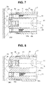
- Fig. 7 is a view showing illuminating lights from the illuminating windows 22 and 23, and an observation range of the observation window 21.
- Fig. 8 is a view showing illuminating lights from the illuminating window 24, and the observation range of the observation window 21.
- Fig. 9 is a view for explaining a light-shielding effect of the edge line portion AL which is formed at the distal end cover 32, at a time of proximity observation by the endoscope.
- illuminating lights emitted from the illuminating windows 22 and 23 are respectively emitted to illumination ranges ER1 and ER2 (shown by dotted lines) in the distal end direction of the distal end portion 17, and illuminate a living tissue TS which is an object.
- illumination ranges ER1 and ER2 shown by dotted lines
- ER3 shown by dotted lines
- Reflection light from living tissue TS of the illuminating light is incident on the observation window 21 in a range IR of a field of view shown by an alternate long and short dash line.
- the respective illumination ranges are enlarged, and even at the time of proximity observation, that is, at the time of enlarged observation, a portion in which the light amount of the illuminating light is small hardly occurs between the two illumination ranges. Consequently, light distribution unevenness by the two illuminating lights from the illuminating windows 22 and 23 decreases.
- the light which advances toward the observation window 21 is shielded by the inclined plane portion DS1 having the edge line portion AL.
- the illuminating light in a range below the edge line portion AL that is, a range lower than the above described height HBi
- the inclined plane portion DS1 is shielded by the inclined plane portion DS1, and is not directly incident on the incident surface IS of the observation window 21.
- an angle ⁇ formed by the inclined portions r1 and r2 of the two inclined planes of the inclined plane portion DS1 which is divided by the edge line portion AL is an obtuse angle. Consequently, the observation window 21 protrudes in the distal end direction from the distal end face 17a, so that favorable cleaning performance for the observation window 21 by the air/water feeding nozzle 26 can be ensured, and favorable water draining performance on the observation window 21 can be also realized.
- the illumination range of the illuminating light emitted from the illuminating window 22 and the illumination range of the illuminating light emitted from the illuminating window 23 are more enlarged on an object than when the exit surfaces ES of the illuminating windows 22 and 23 and the incident surface IS of the observation window 21 are on the same plane, a part between the two illumination ranges, that is, a vicinity of the center of an endoscopic image can be also irradiated with sufficient illuminating light, and light distribution unevenness of the two illuminating lights can be reduced.
- the inclined plane portion DS1 is provided, whereby the illuminating lights from the illuminating windows 22 and 23 are not directly incident on the observation window 21, and therefore, occurrence of a flare on the endoscopic image can be prevented.
- the inclined plane portion DS1 can be provided at least only a range corresponding to the portion DS 11 and a range corresponding to the portion DS12.
- the portion DS 11 of the inclined plane portion DS1 configures a first inclined plane portion which is provided between the peripheral edge portion of the concave lens 41a1 which is the objective lens at the distal end of the observation optical system and the peripheral edge portion of the illumination lens 42a1 at the distal end of the illumination optical system of the illuminating window 22.
- the portion DS12 of the inclined plane portion DS1 configures a second inclined plane portion which is provided between the peripheral edge portion of the concave lens 41a1 which is the objective lens at the distal end of the observation optical system, and the peripheral edge portion of the illumination lens at the distal end of the illuminating optical system of the illuminating window 23.
- the edge line portion AL1 in the portion DS 11 configures a first edge line portion which is formed at an intermediate portion of the first inclined plane portion, and is formed to protrude in the distal end direction of the insertion portion 12, between the peripheral edge portion of the concave lens 41a1 which is the objective lens at the distal end, and the peripheral edge portion of the illumination lens 42a1 of the illuminating window 22.
- the edge line portion AL2 in the portion DS12 configures a second edge line portion that is formed at an intermediate portion of the second inclined plane portion, and is formed to protrude in the distal end direction of the insertion portion 12, between the peripheral edge portion of the concave lens 41a1 which is the objective lens and the peripheral edge portion of the illumination lens of the illuminating window 23.
- the third illuminating window 24 is provided on the distal end face 17a of the distal end portion 17, the illuminating window 24 is disposed in a position which is more apart from the observation window 21 than the illuminating windows 22 and 23, and the exit surface ES of the illuminating window 24 is placed at substantially the same height as the incident surface IS of the observation window 21.
- the third illumination optical system which has the illumination lens at the distal end, and emits illuminating light is placed in the distal end portion 17.
- a shape of an endoscopic image is a long rectangle which is long in a lateral direction. That is, in general, an endoscopic image has a shape in which a width in a vertical direction is shorter as compared with a height in a lateral direction. Therefore, the illuminating windows 22 and 23 are desirably disposed so that centers of the illumination ranges are located in the vertical direction of the obtained endoscopic image.
- Fig. 10 is a view for explaining a range of a field of view of the image pickup device 41 c with respective to dispositions of the illuminating windows 22 and 23.
- U, D, L and R show respective directions of an up, a down, a left and a right of the endoscopic image which is obtained.
- the range RR of the field of view of the image pickup device 41c is a bobbin type which extends laterally by an aberration of a lens.
- the range RR of the field of view differs in accordance with an angle around a center axis (that is, an optical axis of the objective optical system) O of the image pickup device 41c with respect to the objective optical system.
- the angle around the center axis (that is, the optical axis of the objective optical system) O of the image pickup device 41c is set so that the two illumination ranges by the illuminating lights from the illuminating windows 22 and 23 are located in the vertical direction (UD direction) of the endoscopic image.
- the range RR of the field of view of the image pickup device 41 c is set, that is, positioned, so that the two illumination ranges from the illuminating windows 22 and 23 are located in the vertical direction of the endoscopic image which is shorter than the lateral direction of the endoscopic image, and portions CC (shown by dotted-line circles) having the largest light amounts in the centers of the illuminating lights from the illuminating windows 22 and 23 at the time of proximity observation do not enter the range RR of the field of view.
- portions CC shown by dotted-line circles
- the two illumination optical systems corresponding to the illuminating windows 22 and 23 are placed in the distal end portion 17, so that the two illumination ranges of the two illumination optical systems corresponding to the illuminating windows 22 and 23 are disposed along the direction of a shorter length of a length in the vertical direction and a length in the lateral direction of the endoscopic image which is obtained via the observation optical system.
- the two illumination optical systems corresponding to the illuminating windows 22 and 23 are placed in the distal end portion 17 so that the portions CC which are the brightest regions in the respective illumination ranges are located outside the endoscope image, at the time of enlarged observation.
- the inclined plane portion DS1 is also formed between the air/water feeding nozzle 26 and the observation window 21, in a periphery of the observation window 21, a powerful water flows in such a manner as to be pushed out beyond the inclined plane portion DS1 and beyond the surface of the observation window 21 at the time of water feeding, but even if the water remaining in the air/water feeding conduit spouts from the air/water feeding nozzle 26, when water feeding is switched to air feeding, the portion DS 13 of the inclined plane portion DS1 becomes resistance so that water or a water drop does not reach the surface of the observation window 21, and functions as a resistance portion which causes water or a water drop to deviate without flowing toward the observation window 21 as shown by a chain double-dashed line.
- the endoscope in which light distribution unevenness by the two illuminating lights which are respectively emitted from the two illuminating windows is reduced can be provided.
Landscapes
- Health & Medical Sciences (AREA)
- Life Sciences & Earth Sciences (AREA)
- Physics & Mathematics (AREA)
- Surgery (AREA)
- Optics & Photonics (AREA)
- Engineering & Computer Science (AREA)
- Biomedical Technology (AREA)
- General Health & Medical Sciences (AREA)
- Pathology (AREA)
- Nuclear Medicine, Radiotherapy & Molecular Imaging (AREA)
- Biophysics (AREA)
- Heart & Thoracic Surgery (AREA)
- Medical Informatics (AREA)
- Molecular Biology (AREA)
- Animal Behavior & Ethology (AREA)
- Radiology & Medical Imaging (AREA)
- Public Health (AREA)
- Veterinary Medicine (AREA)
- Astronomy & Astrophysics (AREA)
- General Physics & Mathematics (AREA)
- Multimedia (AREA)
- Endoscopes (AREA)
- Instruments For Viewing The Inside Of Hollow Bodies (AREA)
Applications Claiming Priority (2)
| Application Number | Priority Date | Filing Date | Title |
|---|---|---|---|
| JP2014096290 | 2014-05-07 | ||
| PCT/JP2015/059324 WO2015170525A1 (fr) | 2014-05-07 | 2015-03-26 | Endoscope |
Publications (2)
| Publication Number | Publication Date |
|---|---|
| EP3092940A1 true EP3092940A1 (fr) | 2016-11-16 |
| EP3092940A4 EP3092940A4 (fr) | 2017-11-08 |
Family
ID=54392379
Family Applications (1)
| Application Number | Title | Priority Date | Filing Date |
|---|---|---|---|
| EP15789296.9A Withdrawn EP3092940A4 (fr) | 2014-05-07 | 2015-03-26 | Endoscope |
Country Status (5)
| Country | Link |
|---|---|
| US (1) | US20160353983A1 (fr) |
| EP (1) | EP3092940A4 (fr) |
| JP (1) | JP5942046B2 (fr) |
| CN (1) | CN105979846B (fr) |
| WO (1) | WO2015170525A1 (fr) |
Cited By (2)
| Publication number | Priority date | Publication date | Assignee | Title |
|---|---|---|---|---|
| EP3461389A1 (fr) * | 2017-09-28 | 2019-04-03 | FUJIFILM Corporation | Endoscope |
| US20220330802A1 (en) * | 2020-03-23 | 2022-10-20 | Hoya Corporation | Endoscope and method for manufacturing endoscope |
Families Citing this family (8)
| Publication number | Priority date | Publication date | Assignee | Title |
|---|---|---|---|---|
| JP6779100B2 (ja) * | 2016-11-02 | 2020-11-04 | オリンパス株式会社 | 内視鏡 |
| EP3599983B1 (fr) * | 2017-03-24 | 2023-11-15 | Covidien LP | Endoscopes |
| WO2018179791A1 (fr) * | 2017-03-31 | 2018-10-04 | Hoya株式会社 | Endoscope |
| JP2021182948A (ja) * | 2018-06-06 | 2021-12-02 | オリンパス株式会社 | 内視鏡の接着構造 |
| WO2020031261A1 (fr) * | 2018-08-07 | 2020-02-13 | オリンパス株式会社 | Plaquette de lentilles, réseau de lentilles en couches et endoscope |
| DE102020120202A1 (de) * | 2020-07-30 | 2022-02-03 | Schölly Fiberoptic GmbH | Überzug für eine Endoskopspitze und Endoskop |
| WO2023013055A1 (fr) * | 2021-08-06 | 2023-02-09 | オリンパス株式会社 | Système d'éclairage pour endoscope et endoscope équipé de ce système |
| CN116327095A (zh) * | 2023-02-02 | 2023-06-27 | 上海欧太医疗器械有限公司 | 尖端布局装置 |
Family Cites Families (13)
| Publication number | Priority date | Publication date | Assignee | Title |
|---|---|---|---|---|
| CN100339042C (zh) * | 2003-11-12 | 2007-09-26 | 奥林巴斯株式会社 | 内窥镜 |
| JP4554267B2 (ja) * | 2004-04-27 | 2010-09-29 | オリンパス株式会社 | 内視鏡及び内視鏡システム |
| JP3965170B2 (ja) * | 2004-07-27 | 2007-08-29 | オリンパス株式会社 | 内視鏡 |
| US20070038027A1 (en) * | 2005-08-09 | 2007-02-15 | Olympus Medical Systems Corp. | Endoscope hole sealing part |
| JP4868870B2 (ja) * | 2006-02-02 | 2012-02-01 | オリンパスメディカルシステムズ株式会社 | 内視鏡 |
| JP4668831B2 (ja) * | 2006-04-24 | 2011-04-13 | オリンパスメディカルシステムズ株式会社 | 内視鏡 |
| JP5030507B2 (ja) * | 2006-08-30 | 2012-09-19 | オリンパスメディカルシステムズ株式会社 | 内視鏡の先端フードとフード付き内視鏡 |
| JP5317639B2 (ja) * | 2008-11-17 | 2013-10-16 | Hoya株式会社 | 内視鏡 |
| CN102316783B (zh) * | 2009-03-31 | 2015-12-02 | 奥林巴斯株式会社 | 内窥镜 |
| JP2011120863A (ja) * | 2009-11-11 | 2011-06-23 | Fujifilm Corp | 内視鏡 |
| JP5343066B2 (ja) * | 2010-12-15 | 2013-11-13 | 富士フイルム株式会社 | 内視鏡装置 |
| JP5331840B2 (ja) * | 2011-02-28 | 2013-10-30 | 富士フイルム株式会社 | 内視鏡 |
| JP5261518B2 (ja) * | 2011-03-01 | 2013-08-14 | 富士フイルム株式会社 | 内視鏡 |
-
2015
- 2015-03-26 JP JP2015544670A patent/JP5942046B2/ja active Active
- 2015-03-26 EP EP15789296.9A patent/EP3092940A4/fr not_active Withdrawn
- 2015-03-26 CN CN201580007408.4A patent/CN105979846B/zh active Active
- 2015-03-26 WO PCT/JP2015/059324 patent/WO2015170525A1/fr not_active Ceased
-
2016
- 2016-08-16 US US15/238,360 patent/US20160353983A1/en not_active Abandoned
Cited By (4)
| Publication number | Priority date | Publication date | Assignee | Title |
|---|---|---|---|---|
| EP3461389A1 (fr) * | 2017-09-28 | 2019-04-03 | FUJIFILM Corporation | Endoscope |
| US11503983B2 (en) | 2017-09-28 | 2022-11-22 | Fujifilm Corporation | Endoscope |
| US12178408B2 (en) | 2017-09-28 | 2024-12-31 | Fujifilm Corporation | Endoscope |
| US20220330802A1 (en) * | 2020-03-23 | 2022-10-20 | Hoya Corporation | Endoscope and method for manufacturing endoscope |
Also Published As
| Publication number | Publication date |
|---|---|
| CN105979846B (zh) | 2018-04-03 |
| CN105979846A (zh) | 2016-09-28 |
| US20160353983A1 (en) | 2016-12-08 |
| WO2015170525A1 (fr) | 2015-11-12 |
| EP3092940A4 (fr) | 2017-11-08 |
| JPWO2015170525A1 (ja) | 2017-04-20 |
| JP5942046B2 (ja) | 2016-06-29 |
Similar Documents
| Publication | Publication Date | Title |
|---|---|---|
| EP3092940A1 (fr) | Endoscope | |
| US8545400B2 (en) | Endoscope | |
| JP5274719B2 (ja) | 内視鏡及び内視鏡用照明装置 | |
| CN103491854B (zh) | 用于多摄影机内窥镜的多元件罩 | |
| US20150297072A1 (en) | Laryngoscope blade | |
| WO2005055816A1 (fr) | Endoscope | |
| US12171400B2 (en) | Illumination optical system and illumination device | |
| JP5977911B1 (ja) | 内視鏡 | |
| US10695029B2 (en) | Ultrasonic endoscope | |
| CN110769733A (zh) | 内窥镜装置及测量支持方法 | |
| JP5075658B2 (ja) | 内視鏡 | |
| JP2012157577A (ja) | 内視鏡 | |
| EP3103378A1 (fr) | Système d'endoscope | |
| CN110996749A (zh) | 3d视频内窥镜 | |
| US10078209B1 (en) | Optic assembly and light source device for endoscope including the same | |
| US20060217592A1 (en) | Endoscope | |
| CN101360446A (zh) | 喉镜片 | |
| JP2017113079A (ja) | 対物光学ユニット | |
| US20200333580A1 (en) | Image pickup optical system, endoscope, and image pickup apparatus | |
| JPH0560985A (ja) | 内視鏡 | |
| JP2005168606A (ja) | 内視鏡 | |
| JPH08545A (ja) | カバー式内視鏡 | |
| HK40020574B (en) | 3d video endoscope |
Legal Events
| Date | Code | Title | Description |
|---|---|---|---|
| PUAI | Public reference made under article 153(3) epc to a published international application that has entered the european phase |
Free format text: ORIGINAL CODE: 0009012 |
|
| 17P | Request for examination filed |
Effective date: 20160810 |
|
| AK | Designated contracting states |
Kind code of ref document: A1 Designated state(s): AL AT BE BG CH CY CZ DE DK EE ES FI FR GB GR HR HU IE IS IT LI LT LU LV MC MK MT NL NO PL PT RO RS SE SI SK SM TR |
|
| AX | Request for extension of the european patent |
Extension state: BA ME |
|
| DAV | Request for validation of the european patent (deleted) | ||
| DAX | Request for extension of the european patent (deleted) | ||
| A4 | Supplementary search report drawn up and despatched |
Effective date: 20171010 |
|
| RIC1 | Information provided on ipc code assigned before grant |
Ipc: A61B 1/07 20060101ALI20171004BHEP Ipc: A61B 1/05 20060101ALI20171004BHEP Ipc: A61B 1/00 20060101AFI20171004BHEP Ipc: G02B 23/26 20060101ALI20171004BHEP Ipc: A61B 1/06 20060101ALI20171004BHEP |
|
| STAA | Information on the status of an ep patent application or granted ep patent |
Free format text: STATUS: REQUEST FOR EXAMINATION WAS MADE |
|
| STAA | Information on the status of an ep patent application or granted ep patent |
Free format text: STATUS: THE APPLICATION IS DEEMED TO BE WITHDRAWN |
|
| 18D | Application deemed to be withdrawn |
Effective date: 20180508 |