EP3105586B1 - Procédés permettant de déterminer des composés ou compositions pour le traitement de maladies liées à la lipofuscine, et composés ou compositions - Google Patents
Procédés permettant de déterminer des composés ou compositions pour le traitement de maladies liées à la lipofuscine, et composés ou compositions Download PDFInfo
- Publication number
- EP3105586B1 EP3105586B1 EP15708746.1A EP15708746A EP3105586B1 EP 3105586 B1 EP3105586 B1 EP 3105586B1 EP 15708746 A EP15708746 A EP 15708746A EP 3105586 B1 EP3105586 B1 EP 3105586B1
- Authority
- EP
- European Patent Office
- Prior art keywords
- lipofuscin
- compound
- cells
- light
- range
- Prior art date
- Legal status (The legal status is an assumption and is not a legal conclusion. Google has not performed a legal analysis and makes no representation as to the accuracy of the status listed.)
- Active
Links
Images




Classifications
-
- G—PHYSICS
- G01—MEASURING; TESTING
- G01N—INVESTIGATING OR ANALYSING MATERIALS BY DETERMINING THEIR CHEMICAL OR PHYSICAL PROPERTIES
- G01N33/00—Investigating or analysing materials by specific methods not covered by groups G01N1/00 - G01N31/00
- G01N33/48—Biological material, e.g. blood, urine; Haemocytometers
- G01N33/50—Chemical analysis of biological material, e.g. blood, urine; Testing involving biospecific ligand binding methods; Immunological testing
- G01N33/5005—Chemical analysis of biological material, e.g. blood, urine; Testing involving biospecific ligand binding methods; Immunological testing involving human or animal cells
- G01N33/5008—Chemical analysis of biological material, e.g. blood, urine; Testing involving biospecific ligand binding methods; Immunological testing involving human or animal cells for testing or evaluating the effect of chemical or biological compounds, e.g. drugs, cosmetics
- G01N33/5044—Chemical analysis of biological material, e.g. blood, urine; Testing involving biospecific ligand binding methods; Immunological testing involving human or animal cells for testing or evaluating the effect of chemical or biological compounds, e.g. drugs, cosmetics involving specific cell types
-
- G—PHYSICS
- G01—MEASURING; TESTING
- G01N—INVESTIGATING OR ANALYSING MATERIALS BY DETERMINING THEIR CHEMICAL OR PHYSICAL PROPERTIES
- G01N33/00—Investigating or analysing materials by specific methods not covered by groups G01N1/00 - G01N31/00
- G01N33/48—Biological material, e.g. blood, urine; Haemocytometers
- G01N33/50—Chemical analysis of biological material, e.g. blood, urine; Testing involving biospecific ligand binding methods; Immunological testing
- G01N33/92—Chemical analysis of biological material, e.g. blood, urine; Testing involving biospecific ligand binding methods; Immunological testing involving lipids, e.g. cholesterol, lipoproteins, or their receptors
-
- G—PHYSICS
- G01—MEASURING; TESTING
- G01N—INVESTIGATING OR ANALYSING MATERIALS BY DETERMINING THEIR CHEMICAL OR PHYSICAL PROPERTIES
- G01N2500/00—Screening for compounds of potential therapeutic value
- G01N2500/04—Screening involving studying the effect of compounds C directly on molecule A (e.g. C are potential ligands for a receptor A, or potential substrates for an enzyme A)
-
- G—PHYSICS
- G01—MEASURING; TESTING
- G01N—INVESTIGATING OR ANALYSING MATERIALS BY DETERMINING THEIR CHEMICAL OR PHYSICAL PROPERTIES
- G01N2800/00—Detection or diagnosis of diseases
- G01N2800/16—Ophthalmology
- G01N2800/164—Retinal disorders, e.g. retinopathy
Definitions
- the present invention concerns the field of lipofuscin associated diseases of the central nervous system (CNS) and the eye of mammals. It inter alia provides compounds for the treatment of lipofuscin associated diseases of the eye.
- CNS central nervous system
- Lipofuscin is a general term to describe lysosomal deposits of insoluble materials that accumulate in the tissues of organisms in the process of aging or due to genetic deficiencies in common hydrophobic clearance mechanisms (e.g. mutations of ABC transporters). In its broadest sense, the accumulation of critical amounts of lipofuscin is pathologic in any tissue, but especially so in the tissues of the CNS where the loss of cell function through lipofuscin is particularly apparent.
- Lipofuscin is a lipid rich substance which is found to be accumulated in post mitotic cells of e.g. the brain, the heart, or the retinal pigment epithelium in the eye over a life time. The composition is complex and still under investigation. In the eye, one important and well characterized component of lipofuscin is the flurophore N-retinylidene-N-retinylethanolamine (A2E), a byproduct of the visual cycle. It can be detected histologically by its autofluorescence properties.
- A2E flurophore N-retinylidene-N-retinylethanolamine
- Lipofuscin is particularly formed in tissues with high oxidative stress ( A Terman and U T Brunk, 'Lipofuscin: Mechanisms of Formation and Increase with Age', APMIS: acta pathologica, microbiologica, et immunologica Scandinavica, 106 (1998), 265-276 .). It accumulates progressively over time in lysosomes of post mitotic cells, such as neurons and cardiac myocytes and the retinal pigment epithelium (RPE). The exact mechanisms behind this accumulation are still unclear and may vary in different diseases.
- lipofuscin is due to the oxidative alteration of macromolecules by oxygen-derived free radicals generated in reactions catalyzed by redox-active iron of low molecular weight.
- Two principal explanations for the increase of lipofuscin with age have been suggested. The first one is based on the notion that lipofuscin is not totally eliminated (either by degradation or exocytosis) even at a young age, and, thus, accumulates in postmitotic cells as a function of time. Since oxidative reactions are obligatory for life, they would act as age-independent enhancers of lipofuscin accumulation, as well as of many other manifestations of senescence.
- the second explanation is that the increase of lipofuscin is an effect of aging, caused by an age-related enhancement of autophagocytosis, a decline in intralysosomal degradation, and/or a decrease in exocytosis.
- the metabolism is to maintain compounds in solution to allow them to be cleared by solution mechanisms (e.g. urine) or efflux mechanisms as carried out by ABC transporters.
- the cell contains enzymes for oxidising and conjugating even lipophilic compounds.
- lipophilic components such as pigments are susceptible to redox reactions which may lead to cross-linking and consequent precipitation.
- hydrophobic interactions stabilise the precipitate and thereby present few or no sites where the material can interact with the generally deep reaction pockets of hydrolytic enzymes.
- Hydrophobic deposits are, in turn, likely to further interact with and precipitate other hydrophobic species.
- lipofuscin accumulation represents a stabilised form of hydrophobic detritus that appears inaccessible to normal metabolic clearance by enzymes.
- Lipofuscinoses and lipofuscinopathies are, therefore, diseases characterised by high levels of lipofuscin deposits as a result of aging, or metabolic defects.
- Lipofuscin associated degenerative diseases of the eye have in common that lipofuscin is accumulated in the cells of the RPE.
- diseases include age-related macular degeneration, Stargardt's disease, Best's disease and subpopulations of Retinitis pigmentosa.
- AMD age-related macular degeneration
- AMD age-related macular degeneration
- atrophic AMD with geographic atrophy and exudative AMD or synonymous wet, neovascular AMD
- choroidal neovascularization are differentiated.
- atrophic AMD occurs in the eye before development of the exudative form.
- All early stages of AMD and advanced atrophic AMD are usually summarized as dry AMD (see Fig. 1 ). All stages of AMD are characterized by drusen formation and lipofuscin accumulation in RPE cells.
- Advanced dry AMD is in addition characterized by the complete and irreversible degeneration of the neuroretina tissue forming sharply demarcated areas of RPE atrophy, the so called geographic atrophy. Geographic atrophy extending to the macula, the area of the retina responsible for visual acuity ( Fig. 1 ), will seriously affect the ability to read, recognize faces or pursue everyday activities such as walking, driving, or shopping. As such, the impact of AMD on quality of life and patient independence can be devastating. In wet AMD, in addition to the characteristics of early AMD and usually also advanced dry AMD, neovascularization takes place.
- Stargardt's disease (disease code H35.5 according to ICD-10) is a severe inherited juvenile macular degeneration due to autosomal recessive mutation of the ABCA4 gene or autosomal dominant mutation of the ELOVL 4 gene. It begins in late childhood. Along with progression of the disease, lipid rich deposits (lipofuscin) accumulate in the retinal pigment epithelium (RPE) layer beneath the macula. In advanced Stargardt's disease, the build-up of lipofuscin causes atrophy of the RPE and subsequently the macula supplied by this area of the RPE. At the final stage, Stargardt's disease leads to legal blindness.
- Best's disease also termed vitelliform macular dystrophy or vitelliform dystrophy
- vitelliform macular dystrophy is a retinal lipofuscinosis leading to progressive vision loss in the macula.
- the early-onset form, Best disease is caused by mutations of the gene encoding the chloride transporter bestrophin, VMD2, and usually appears in childhood,
- the late-onset form begins in middle age, and tends to be more mild and is associated in ca. 25% of cases with mutations of VMD2 or RDS (peripherin).
- Retinitis pigmentosa is a group of inherited disease of the retina. RP patients develop a degeneration of the photoreceptors and retinal pigment epithelium (RPE) cells. RP culminates in the degeneration of the photoreceptors in the fovea reducing central vision. RP is one of the main causes of acquired blindness in developed countries. Abnormal levels of lipofuscin accumulation are observed in more than one-half of RP patients.
- NCL neuronal ceroid lipofuscinoses
- NCLs neuronal ceroid lipofuscinoses
- the neuronal ceroid-lipofuscinoses (NCLs) are characterized by progressive intellectual and motor deterioration, seizures, and early death. Visual loss is a feature of most forms.
- AD Alzheimer's disease
- a ⁇ protein has been identified as the main component of amyloid of senile plaques, the hallmark lesion of AD, but it is not certain whether the formation of extracellular A ⁇ deposits is the main cause of the series of pathological events in the brain in the course of sporadic AD.
- Lipofuscin is a relatively overlooked age-related product and the hypothesis was formulated that its release into the extracellular space following the death of neurons may contribute to the formation of senile plaques.
- THPEs tetrahydropyridoethers
- (7R, 8R, 9R)-2,3-Dimethyl-8-hydroxy-7-(2-methoxyethoxy)-9-phenyl-7,8,9,10-tetrahydro-imidazo[1,2-h] [1,7]naphthyridine INN Name: Soraprazan
- its salts and related compounds remove natural lipofuscin from RPE cells and can therefore serve as active ingredient in the treatment of AMD degeneration, in particular of dry AMD and Stargardt's disease ( EP 2080513 A1 ).
- Verteporfin has been used for the treatment of neovascular AMD due to verteporfin occluding CNV lesions (see e.g. Scott and Goa, 'Verteporfin', Drugs & Aging, 2000 Feb; 16(2): 139-146 ).
- Wu et al. ('Enzymatic degradation of A2E, a retinal pigment epithelial lipofuscin bisretinoid', J Am Chem Soc, 133 (2001), 849-857 ) disclose that, in a cell-based assay, horseradish peroxidase, when delivered to the cell with the Bioporter system, is able to degrade lipofuscin.
- the invention provides a compound for use in the treatment of lipofuscin associated diseases of the eye by degradation of lipofuscin, wherein the disease is selected from dry AMD, Stargardt's disease, Best's disease and Retinitis pigmentosa, and wherein the compound is verteporfin.
- compositions that comprise the combination of a reactivity factor, as e.g. a radical providing agent, and a targeting factor, as e.g. lipophilicity of the molecule, are able to degrade lipofuscin in cells..
- a reactivity factor as e.g. a radical providing agent
- a targeting factor as e.g. lipophilicity of the molecule
- the reactivity factor is preferably a radical providing agent.
- the reactivity factor may therefore oxidize lipofuscin and consequently cause a degradation and dissolution of lipofuscin aggregates.
- a radical scavenger abolishes the degradation of the lipofuscin caused by the test compound.
- the reactivity factor In order to oxidize the lipofuscin the reactivity factor has to be in proximity to the lipofuscin deposits, and thus has to enter a cell containing lipofuscin and target the lipofuscin in the cell.
- the targeting factor as disclosed herein allows an entry of the compound or composition into a cell and guarantees a targeting of the compound or composition to the lipofuscin in the cell.
- the present invention applies the obtained knowledge about the mechanism of lipofuscin degradation for treating lipofuscin associated diseases in a patient.
- a method for selecting a compound or a composition suitable for treating lipofuscin associated diseases in a patient comprising the steps of
- the method of the first aspect is inter alia based on the unexpected finding that a reactivity factor and a targeting factor are sufficient for a compound or composition to effectively degrade lipofuscin deposits.
- the reactivity factor according to the disclosure is an atom or molecule that may initiate, enhance or undergo a reaction with lipofuscin, or components of lipofuscin.
- the reactivity factor may be an oxidizing agent and/or a radical providing agent.
- reactivity factors enhancing the reaction with lipofuscin are complexed metal ions such as zinc or iron. It is most preferred that the reactivity factor is a radical providing agent. Alternatively, the reactivity factor is an oxidizing agent. Alternatively, the reactivity factor is an agent that is photoactive.
- THPE tetrahydropyridoethers
- the lipofuscin component A2E is degraded by soraprazan also in cell free systems suggesting that an induction of a cellular mechanism by the compound and in particular the action of enzymes is not required.
- members of the THPE family are generators of a superoxide anion as demonstrated by studies using electron paramagnetic resonance and spin trapping of the radical products. Accordingly, a substance capable of generating radical oxygen species is referred to herein as superoxide anion donator (SAD) or reactive oxygen donator (ROD).
- SAD superoxide anion donator
- ROD reactive oxygen donator
- a radical scavenger abolishes the degradation of the lipofuscin induced by an SAD.
- a radical i.e. the superoxide anion is necessary for the degradation of lipofuscin seen in the incubation with the SAD.
- reactive oxygen species mediate the precipitation and accumulation of lipofuscin.
- the transfer of the radical, i.e. the oxygen to the lipofuscin results in the addition of hydroxyl groups, and the destabilization of double bonds in precipitated pigments, which in turn leads to the dissolution of the deposits. Once the solution of the deposits is started, equilibrium favours their export to the blood or medium. In an organism the solubilized lipofuscin is disposed via the normal excretory routes.
- the reactivity factor In order to cause a reaction with the lipofuscin in the cell the reactivity factor has to reach the lipofuscin or get in proximity of the lipofuscin. Accordingly, it has to be taken up by a cell and to be targeted to the lipofuscin deposits.
- a targeting factor or accumulation factor as disclosed herein allows an uptake into a cell and allows targeting of lipofuscin.
- the targeting factor In order to target lipofuscin the targeting factor preferably has an affinity for lipofuscin.
- Soraprazan is for example taken up by a cell and targeted to the lipofuscin deposits due its lipophilic nature.
- the reactivity factor may interact with a metal ion in the lipofuscin structure.
- Metals are components of lipofuscin involved in the coordination and stabilization of the secondary structure of lipofuscin. Accordingly, an interaction of the reactivity factor with metal ions in the lipofuscin structure, such as chelation or coordination of the metal ions may degrade destabilize the lipofuscin agglomerates and increase the solubility of the lipofuscin components.
- the reactivity factor may be a metal interacting agent, in particular a chelator or metal ion coordinator.
- the radical providing agent may be a radical.
- the radical is preferably an oxygen radical.
- the radical providing agent may be a compound with an ability to generate and/or locally coordinate a radical. Methods to determine the ability of a compound to generate and/or locally coordinate a reactive chemical species are known in the art. For example, when observed using electron paramagnetic resonance, the compound mediates transfer of energy to a spin trap molecule used for the purpose of observing such effects.
- the radical may be selected from hydroxyl radical (•OH), superoxide anion (O 2 - ), oxide anion (O 2 - ), nitric oxide (NO - ), hydrogen peroxide (H 2 O 2 ), and singlet oxygen (O 2 *).
- the radical providing agent may generate or locally coordinate a radical selected from hydroxyl radical (•OH), superoxide anion (O 2 - ), oxide anion (O 2 - ), nitric oxide (NO - ), hydrogen peroxide (H 2 O 2 ), and singlet oxygen (O 2 *).
- a radical providing agent as disclosed herein may be one that generates a radical upon energization of the compound. This energization may be caused by illumination by light. Accordingly, the ability of the radical providing agent to generate and/or locally coordinate a radical may be determined in the presence of visible light.
- a light source providing to whole spectrum of visible light may be used.
- light sources providing light of specific wavelengths may be employed.
- the light may have a wavelength in the range from 380 nm to 800 nm.
- the light has a wavelength in the range from 650 nm.
- the light for illumination has a wavelength of less in the range from 380 to 500 nm.
- the ability of the radical providing agent to generate and/or locally coordinate a radical may be determined in the absence of visible light.
- a radical providing agent determined by the method disclosed herein may also be energized by chemical processes such as cellular respiration and reductases.
- the reactivity factor and the targeting factor may be contained in the same or a different compound.
- a targeting factor as disclosed herein most preferably targets lipofuscin due lipophilic interactions.
- Lipofuscin is lipophilic and thus a lipophilic compound will have an affinity to lipofuscin.
- a targeting factor may bind to lipofuscin or components of lipofuscin due to specific interactions that rely on specific moieties of lipofuscin.
- an antibody or a small molecule with affinity to lipofuscin may be a targeting factor that specifically recognizes components of the lipofuscin deposits
- the targeting factor may have an affinity for, or interact with metal ions in the lipofuscin agglomerate.
- the targeting factor may provide an affinity constant to the compound equal to or below 100 ⁇ M.
- affinity constant is equal to or below 10 ⁇ M. More preferably the affinity constant is equal to or below 1 ⁇ M. Most preferably the affinity constant is equal to or below 0.1 ⁇ M. The lower the affinity constant of the compound, the stronger is the binding of the compound to lipofuscin. A stronger binding leads to a longer contact of the compound or composition to lipofuscin and increases the chance to transfer the reactive chemical species to the lipofuscin.
- the ability of the targeting factor to target lipofuscin may be determined by incubating a defined amount of a potential targeting factor with a suspension of lipofuscin in water, removing the water and measuring the concentration of substance in both the water and the lipofuscin fraction.
- a targeting factor as disclosed herein binds at least partially to the lipofuscin fraction. Preferably, at least 5 %, at least 10 %, at least 20 %, at least 30 %, at least 40 %, at least 50 %, at least 60 %, at least 70 %, at least 80 %, at least 90 %, or at least 95 % of the targeting factor binds to the lipofuscin fraction.
- the targeting factor preferably does not only provide a targeting of lipofuscin in the cell.
- the targeting factor may also allow an uptake into the cell.
- the uptake into the cell may for example occur by endocytosis, active or passive membrane transport.
- the uptake of the targeting factor into the cell may be determined by culturing one or more cells in the presence of a potential targeting factor and measuring the concentration increase of the targeting factor in the cell. Such an experiment is also described in the examples. The increase in concentration defines the cellular uptake.
- the uptake of the targeting factor is preferably such that the concentration of the targeting factor in the cell is at least 10 %, at least 20 %, at least 30 %, at least 40 %, at least 50 %, at least 60 %, at least 70 %, at least 80 %, at least 90 % of the concentration of the targeting factor in the environment. Most preferred, the concentration of the targeting factor in the cell is at least 20 % of the concentration of the targeting factor in the environment..
- the uptake rate is the increase in concentration over time.
- a cell of the eye or the CNS of a mammal may be used in the test for determining the cellular uptake of a potential targeting factor.
- the cells are human cells.
- the cell may be a cell of the retinal pigment epithelium (RPE). Most preferably, the cell is a human RPE cell line.
- the initial uptake rate of the targeting factor in a human RPE cell line is defined as the change in concentration in a cell over time preferably at least 50 nM/h, more preferably at least 150 nM/h, most preferably at least 250 nm/h.
- the compounds or composition has to be tolerated by the cell, e.g. has to be non-toxic for the cells.
- a low tolerance of the compounds or compositions might lead to strong side effects.
- the tolerance of the cell of the eye or the CNS to the targeting and the reactivity factor may be determined and a compound or a composition may be selected or compounds may be combined to obtain a compound or composition that comprises a reactivity factor and an targeting factor that are tolerated by the cell.
- Such an additional step helps avoiding the selection of compounds or composition that are for example toxic to the cells.
- the method disclosed herein may comprise the step of determining the elimination of lipofuscin or a lipofuscin marker in vitro by said compound, and selecting a compound additionally based on a predefined elimination threshold.
- the predefined threshold is dependent on the method for determining the elimination rate.
- a reasonable value for the threshold can be obtained by performing a specific method for eliminating lipofuscin in vitro or the lipofuscin marker with an SAD known to eliminate lipofuscin also in vivo. In the examples several methods are described to test the elimination of the lipofuscin in cell culture.
- the step of determining the elimination of lipofuscin may comprise the incubation of the compound with cells selected from retinal epithelial cells that have been incubated with the A2E, retinal epithelial cells that have been incubated with animal lipofuscin, retinal epithelial cells that have been incubated with human lipofuscin, retinal epithelial cells that have been harvested from an animal retinal epithelial cells that have been harvested from human donors.
- A2E N-retinylidene-N-retinylethanolamine
- soraprazan which is known to eliminate lipofuscin in cells also degrades A2E in solution under illumination by visible light.
- the elimination of lipofuscin can be mimicked by A2E in solution.
- the lipofuscin marker is A2E.
- the determination of the degradation of A2E has the advantage to be faster than methods including the growth of cells.
- the use of A2E allows a simple determination of the degradation rate. For example, to A2E in solution a potential compound may be added. This mixture may then be illuminated for a predetermined time.
- the step of determining the elimination of A2E comprises the incubation of the compound with A2E in the presence of light.
- Suitable lamps for illumination are known in the art and do not require further explanation.
- the illumination time is preferably 1 min or 5 min.
- a wavelength of 689 nm for 1 min may be employed.
- the mixture of A2E and the potential compound may be illuminated with white light for 5 min.
- the mixture of A2E and the potential compound may also be incubated with substance that chemically energizes the potential compound.
- the chemically energizing substance may be an oxidizing agent such a hydrogen peroxide (H 2 O 2 ).
- the step of determining the elimination of A2E may comprise the incubation of the compound with A2E in the presence of hydrogen peroxide H 2 O 2 . After illumination or incubation the sample may be analyzed by liquid chromatography and mass spectroscopy. Suitable devices for carrying out a combined liquid chromatography and mass spectroscopy are known in the art.
- a potential candidate compound tested by the method of selection as disclosed herein may be any compound.
- the potential compound is selected from compounds that are able to absorb light and which are cell permeable. More preferably the compound is selected from the classes quinones, flavones, flavins, amino acids, bile acids, aromatic amino acids, pyroles, porphyrins, dioxetanes, photosensitizers, quinolones, quinidines, NSAIDS, psoralens, retinoic acids, and melanizing agents tetracycline antibiotics, redox active proteins, redox active small molecules dyes, and metal interacting agents
- further disclosed herein is a compound or composition comprising at least one reactivity factor and at least one targeting factor that allows an uptake into a cell and provides a targeting of lipofuscin, for use in the treatment of lipofuscin associated diseases
- the compound is not a compound of formula (I) wherein R1 is methyl or hydroxymethyl, one of the substituents R2a and R2b is hydrogen and the other is hydroxy, methoxy, ethoxy, isopropoxy, methoxyethoxy or ethoxypropoxy, one of the substituents R3a and R3b is hydrogen and the other is hydroxy, methoxy, ethoxy, isopropoxy, methoxyethoxy or methoxypropoxy.
- the compound or composition according to the second aspect fulfills in particular the properties defined in the method of selection.
- the reactivity factor of the compound or composition may be an oxidizing agent or a radical providing agent. It is preferred that the radical providing agent is a radical.
- the radical preferably is an oxygen radical.
- the radical providing agent is preferably a molecule with ability to generate and/or locally coordinate a radical.
- the radical may be selected from hydroxyl radical (•OH), superoxide anion (O 2 - ), oxide anion (O 2 -), nitric oxide (NO - ), hydrogen peroxide (H 2 O 2 ), and singlet oxygen (O 2 *).
- the radical providing agent may also generate or locally coordinate a radical selected from hydroxyl radical (•OH), superoxide anion (O 2 - ), oxide anion (O 2 -), nitric oxide (NO - ), hydrogen peroxide (H 2 O 2 ), and singlet oxygen (O 2 *).
- a radical selected from hydroxyl radical (•OH), superoxide anion (O 2 - ), oxide anion (O 2 -), nitric oxide (NO - ), hydrogen peroxide (H 2 O 2 ), and singlet oxygen (O 2 *).
- the radical providing agent of the compound or composition according to the second aspect may be one that generates the radical upon energization of the compound. This energization may be caused by illumination with light.
- the reactivity factor may initiate or only enhance a reaction or participate in a reaction of an atom or molecule with lipofuscin. Examples of reactivity factors enhancing the reaction with lipofuscin are complexed metal ions such as zinc or iron.
- the radical providing agent may also generate and optionally locally coordinate the radical in the presence of visible light.
- a light source providing the whole spectrum of visible light may be used. The light may have a wavelength of less than 800 nm. Preferably the light has a wavelength of less than 650 nm. Most preferably, the light for illumination has a wavelength of less than 500 nm.
- the compound or composition may comprise a reactivity factor, preferably a radical providing agent that generates and optionally locally coordinates the reactive chemical species in the absence of visible light.
- a reactivity factor in particular the radical providing agent may also be energized by chemical processes such as respiration or cellular reductases.
- the targeting factor may provide an affinity constant to the compound or composition that is equal to or below 100 ⁇ M.
- the affinity constant is equal to or below 10 ⁇ M. More preferably the affinity constant is equal to or below 1 ⁇ M. Most preferably the affinity constant is equal to or below 0.1 ⁇ M.
- the ability of the targeting factor to target lipofuscin may be defined by its ability to bind lipofuscin after incubation with a suspension of lipofuscin in water, and separation of the water and the lipofuscin fraction.
- a targeting factor as disclosed herein binds at least partially to the lipofuscin fraction. Preferably, at least 5 %, at least 10 %, at least 20 %, at least 30 %, at least 40 %, at least 50 %, at least 60 %, at least 70 %, at least 80 %, at least 90 %, or at least 95 % of the targeting factor binds to the lipofuscin fraction.
- the uptake of the targeting factor into the cell may be determined by culturing one or more cells in the presence of a potential targeting factor and measuring the concentration increase of the targeting factor in the cell.
- the uptake of the targeting factor is preferably such that the concentration of the targeting factor in the cell is at least 20 % of the concentration of the targeting factor in the environment.
- the uptake rate is the increase in concentration over time.
- a cell of the eye or the CNS of a mammal may be used in a test for determining the cellular uptake of a potential targeting factor.
- the cells are human cells.
- the cell may be a cell of the retinal pigment epithelium (RPE).
- RPE retinal pigment epithelium
- the cell is a human RPE cell line.
- the initial uptake rate (change in concentration following introduction of a substance to the medium) of the targeting factor in a human RPE cell line is preferably at least 50 nM/h, more preferably at least 150 nM/h, most preferably at least 250 nM/h.
- the compound may be selected from the light absorbing agents: quinones, flavones, flavins, amino acids, bile acids, dyes, aromatic amino acids, pyroles, porphyrins, dioxetanes, photosensitizers, quinolones, quinidines, NSAIDS, psoralens, retinoic acids, and melanizing agents, tetracycline antibiotics, redox active proteins, redox active small molecules, and metal interacting agents.
- the light absorbing agents quinones, flavones, flavins, amino acids, bile acids, dyes, aromatic amino acids, pyroles, porphyrins, dioxetanes, photosensitizers, quinolones, quinidines, NSAIDS, psoralens, retinoic acids, and melanizing agents, tetracycline antibiotics, redox active proteins, redox active small molecules, and metal interacting agents.
- Preferred amino acids are cysteine and tryptophan.
- Preferred metabolites are riboflavin and glutathion.
- a preferred quinolone is fleroxacin.
- Preferred dyes are indocyanine green, crystal violet, quinine, fluorescein rose bengal.
- phenothiazine Preferred tetracycline antibiotics are chloramphenicol, doxycycline, and demeclocycline.
- Preferred metal interacting agents are taurodeoxcycholate EDTA and 1,10-phenanthroline.
- the composition may comprise two or more reactivity factors and at least one targeting factor that allows an uptake into a cell and provides a targeting of lipofuscin, for use in the treatment of lipofuscin associated diseases.
- one reactivity factor is a complexed metal ion, such as zinc or iron.
- complexed metal ions in particular Fe (III) may enhance the absorption of radical providing agents such as SADs.
- the first reactivity factor is a complexed metal ion and the second reactivity factor is an SAD.
- the second reactivity factor is a THPE, most preferably the second reactivity factor is soraprazan.
- the composition for use may comprise soraprazan and Fe (III), preferably in a molar ratio of 3:1.
- the composition may comprise a complexed metal ion selected from iron or zinc.
- the iron can be Fe (II) or Fe (III).
- the iron is Fe (III).
- the composition may comprise a compound selected form quinones, flavones, flavins, dyes, aromatic amino acids, pyroles, porphyrins, dioxetanes, photosensitizers, quinolones, quinidines, NSAIDS, psoralens, retinoic acids, and melanizing agents, complexed with a metal ion, in particular Fe (III).
- the Fe (III) may enhance the light absorbance of an SAD, in particular soraprazan.
- the compound may be selected from hypericin, rose bengal diacetate, merocyanine 540, malachite green, 1,10-phenanthroline iodoacetamide, aminolevulinic acid, methyl aminolevulinate, porfimer sodium, talaporfin, temoporfin, methylene blue, riboflavin and tryptophan.
- Riboflavin and tryptophan are natural metabolites of the cells and thus are highly tolerated by the cells. It is preferred that the compound is selected from riboflavin and tryptophan.
- the compound or composition for use in the treatment of lipofuscin related diseases may include a decrease of cellular lipofuscin in the cells of a patient.
- Decrease of cellular lipofuscin may be a decrease in size of lipofuscin aggregates and/or the reduction of the amounts of lipofuscin aggregates.
- the treatment of a lipofuscin related disease includes elimination of lipofuscin aggregates.
- the lipofuscin associated disease as disclosed herein is a disease of the eye or of the CNS of patient.
- the lipofuscin associated disease is a disease of the eye.
- the lipofuscin associated disease is a disease of the eye selected from age-related macular degeneration (AMD), Stargardt's disease, Best's disease and Retinitis pigmentosa.
- AMD age-related macular degeneration
- Stargardt's disease Best's disease
- Retinitis pigmentosa Retinitis pigmentosa.
- the targeting factor is lipophilic and the reactivity factor is a compound selected from hypericin, rose bengal diacetate, merocyanine 540, malachite green, 1,10-phenanthroline iodoacetamide, aminolevulinic acid, methyl aminolevulinate bergapten, methoxsalen, porfimer sodium, psoralene, talaporfin, temoporfin, methylene blue, riboflavin and tryptophan.
- the reactivity factor is a compound selected from hypericin, rose bengal diacetate, merocyanine 540, malachite green, 1,10-phenanthroline iodoacetamide, aminolevulinic acid, methyl aminolevulinate bergapten, methoxsalen, porfimer sodium, psoralene, talaporfin, temoporfin, methylene blue, riboflavin and trypto
- the targeting factor is lipophilic and the reactivity factor is a compound selected from hypericin, rose bengal diacetate, merocyanine 540, malachite green, 1,10-phenanthroline iodoacetamide, aminolevulinic acid, methyl aminolevulinate bergapten, methoxsalen, porfimer sodium, psoralene, talaporfin, temoporfin, methylene blue, riboflavin and tryptophan and the lipofuscin associated disease is age-related macular degeneration (AMD).
- AMD age-related macular degeneration
- the compound or composition may be administered to the patient for treating a lipofuscin associated disease according to any known route of administration.
- routes of administration are oral, parenteral, mucosal, enteral or percutaneous.
- the compound or composition may be administered orally in form of, e.g. a pill, a capsule or in liquid form.
- Parenteral forms of administration include injecting the compound or composition into a vein (intravenous).
- the treatment comprises administering an intravenous injection of verteporfin to a patient with a retinal lipofuscinopathy.
- the compound may be administered by direct injection into the involved tissue e.g. the vitreous (intravitreal) or the conjunctive (conjunctival).
- the treatment comprises administering an intravitreal injection of verteporfin to a patient with a retinal lipofuscinopathy.
- This route of administration is associated with a lower overall dose, a higher concentration at the site of disease and lower systemic exposure.
- Other parenteral forms of administration are e.g. intramuscular, intrarterial, intrperitoneal, intracardiac and subcutaneous.
- the treatment may further comprise an illumination of the eye with visible light.
- a light source providing the whole spectrum of visible light may be used.
- light sources providing light of specific wavelengths may be employed.
- the light has a wavelength of in the range from 380 nm to 800 nm.
- the light has a wavelength in the range from 380 nm to 650 nm.
- the light for illumination has a wavelength in the range from 380 nm to 500 nm.
- the treatment comprises an illumination time that is in the range of 1 min to 72 h, preferably in the range from 2 min to 48 h more preferably in the range from 3 min to 48 h.
- the illumination time of a short illumination treatment is in the range of 1 min to 1 h, preferably in the range from 2 min to 30 min, more preferably in the range from 3 min to 10 min, most preferably in the range from 4 to 6 min.
- the illumination time of a long illumination treatment is in the range of 1 h to 72 h, preferably in the range from 2 h to 48 h more preferably in the range from 3 h to 30 h.
- the radiance of the illumination light is in the range from 0.01 to 1 W/cm 2 , preferably in the range from 0.02 to 0.9 W/cm 2 , more preferably in the range from 0.03 to 0.7 W/cm 2 , most preferably in the range from 0.04 to 0.6 W/m 2 .
- the radiance of the illumination light is in the range from 0.1 to 1 W/cm 2 , preferably in the range from 0.2 to 0.9 W/cm 2 , more preferably in the range from 0.3 to 0.7 W/cm 2 , most preferably in the range from 0.4 to 0.6 W/m 2
- the radiance of the illumination light is in the range from in the range from 0.01 to 0.1 W/cm 2 , preferably in the range from 0.02 to 0.09 W/cm 2 , more preferably in the range from 0.03 to 0.07 W/cm 2 , most preferably in the range from 0.04 to 0.06 W/m 2
- the radiance of the illumination light is in the range from 0.01 to 1 W/cm 2 , preferably in the range from 0.02 to 0.09 W/cm 2 , more preferably in the range from 0.03 to 0.07 W/cm 2 , most preferably in the range from 0.04 to 0.06 W/m 2
- the composition disclosed herein may comprise a photosensitizer in addition to reactivity factor and targeting factor.
- Photosensitizers as disclosed herein are light-sensitive active substances which are photochemically excited by light of appropriate wavelength and intensity and react chemically with other substances, which leads to the degradation of the substance.
- this photosensitizer is a porphyrin, more preferably a porphyrin selected from porfimer sodium, psoralene, talaporfin, temoporfin, photofrin, Porphine, chlorine, bacteriochlorin, expanded porphyrin, verteporfin and phthalocyanine.
- the porphyrin is verteporfin. As shown in the examples a verteporfin enhances the degradation of lipofuscin by the tested SAD.
- the porphyrin photosensitizer may be administered at a dose per eye in mg that is less than 1/10000, less than 1/2000, less than 1/1000, than 1/500, less than 1/250, less than 1/100 of the calculated total body dose in mg.
- the porphyrin photosensitizer may be administered at a dose per eye in mg that is less than 1/10000, preferably less than 1/2000, more preferably less than 1/1000 of the calculated total body dose in mg.
- the treatment comprises administering an intravitreal injection of verteporfin to a patient with a retinal lipofuscinopathy and a light that is different from laser light is applied.
- the compounds or compositions disclosed herein may in addition to the reactivity factor and targeting factor comprise a permeability factor.
- Permeability factors have a lipophilic or amphiphilic character.
- the permeability factor may interact with the lipofuscin promoting the solubility of lipofuscin components and thus the accessibility to cellular degradation mechanisms.
- the permeability factor may interact with the cellular membrane and modulate the transmission of substances through membrane, thus improving the transport of lipofuscin degradation products.
- permeability factors are bile acids, polyethylenglycols (PEG), polysorbates cyclic sugars and uncouplers.
- Polyethylene glycol (PEG) as disclosed herein includes the standard polyether compound with the general structure H-(O-CH2-CH2)n-OH but also any branched or modified PEG such as methoxy PEG. A number behind the PEG identifies the average molecular weight.
- An uncoupler as disclosed herein is a compound which cycles across the membranes causing the dissipation of electrochemical gradient, in particular the proton gradient.
- the permeability factor as disclosed herein may be selected from PEG 400, Tween 80 and cyclodextrin.
- the permeability factor may exhibit a lipofuscin degrading effect even without the reactivity and targeting factor (see Example 16).
- a compound or composition comprising at least one permeability factor for use in the treatment of lipofuscin associated diseases.
- the compound or composition for use in the treatment of lipofuscin associated diseases is not cyclodextrin alone.
- Example 1 Degradation of lipofuscin is dependent on the reactive chemical species
- RPE retinal pigment epithelium
- SAD super oxide anion donor
- soraprazan ((7R,8R,9R)-2,3-Dimethyl-8-hydroxy-7-ethoxy-9-phenyl-7,8,9,10-tetrahydro-imidazo-[1,2-h][1,7]-naphthyridin) - a member of the THPE family - either alone or in combination with a known radical oxygen scavenger, cardioxane.
- test compounds were added to seven cell cultures and the cells were incubated at 37 °C and 5 % CO 2 .
- the SAD was added to the cell cultures in a concentration of 50 ⁇ g/ml either alone or in combination with cardioxane in a concentration of 10 ⁇ M, 50 ⁇ M or 100 ⁇ M.
- three cell cultures were incubated only with cardioxane in a concentration of 10 ⁇ M, 50 ⁇ M or 100 ⁇ M. As control, one cell culture was grown without test compounds.
- N-retinylidene-N-retinylethanolamine (A2E) - component of lipofuscin -in an aqueous solution water was measured in the presence of potential degradation mediators and light illumination.
- A2E was dissolved in deionized H 2 O in a concentration of 20 ⁇ M and the solution was divided into two parts. To one part of the A2E solution verteporfin was added in an amount of 25 ⁇ M. Samples of the A2E solution with and without verteporfin were illuminated with a LED-Lamp SunaECO 1500, Tropic Marin either for 1 min at 689 nm or for 5 min with white light. Non-illuminated samples of A2E solution incubated with and without verteporfin for 5 min served as controls.
- Table 2 also shows that illumination of an A2E suspension containing verteporfin leads to a stronger decrease of A2E. Thus, the photosensitizer verteporfin increases the rate with which A2E is removed or otherwise degraded.
- Table 2 Results of illumination of A2E alone or in the presence of verteporfin Treatment A2E (nM) A2E (% control) A2E non-illuminated, 5 min 32445 100 A2E 689 nm, 83 s * 24605 76 A2E white light, 5 min 2637 8 A2E + Verteporfin non-illuminated, 5 min 28864 89 A2E + Verteporfin 689 nm, 83 s * 14338 44 A2E + verteporfin white light, 5 min 1295 4
- ARPE-19 cells (ca. 10.000 cells) are seeded in DMEM + 10% FBS + 1% P/S in 96-Well-Plates and grown at 37 °C and 0.5% CO 2 After one day 25 ⁇ M of A2E is added to each of the wells and the cells are left for quiescence for three days before treatment with the SADs.
- RPE cells from a human donor are suspended in an aqueous buffer and disrupted by sonication for 25 min at 4 °C using a Soniprep 150 (MSE) fitted with an exponential microprobe.
- the sonicated material is centrifuged at 60 g for 7 min to remove cellular debris and the resultant supernatant is centrifuged at 6000 g for 10 min to sediment the pigment granules.
- This sediment is resuspended in an aqueous solution of sucrose in a concentration of 0.3 M and layered onto a discontinuous sucrose gradient which consisted of eight layers of solutions with a decreasing concentration of sucrose: 2 M; 1.8 M; 1.6 M; 1.55 M; 1.5 M; 1.4 M; 1.2 M and 1 M.
- the overlayered gradient is centrifuged on a swing out head at 103,000 g for 1 hr. At the end of the centrifugation two distinct pigmented fractions are observed.
- the upper fraction being a diffuse light brown band at the interface of the layers with 1.55 and 1.6 M sucrose and the lower fraction constituting a dark brown sediment which passes even the sucrose layer with the highest concentration (2 M).
- the upper band is identified as lipofuscin and the lower as melanin.
- the two fractions are removed from the sucrose gradient, diluted in PBSA and centrifuged at 6000 g for 10 min. Each sediment is resuspended in a 0.3 M sucrose solution and further purified on a second sucrose gradient as described above. Again the fractions are removed from the second gradient, diluted in PBSA and centrifuged at 6,000 g for 10 min. The pellet resulting from each pigmented zone is washed five times in PBSA and stored at - 20' until required.
- ARPE-19 cells (ca. 10.000 cells) are seeded in DMEM + 10% FBS + 1% P/S in 96-Well-Plates and grown at 37 °C and 0.5% CO 2 . After one day 300 lipofuscin granula/cell are added to the wells and the cells are grown for an additional 24 h before treatment with SADs.
- RPE cells from aged human donors contain large amounts of lipofuscin, which can be easily quantified microscopically due to its auto-fluorescence. Therefore, the aged human RPE cells mimic the pathological situation observed in patients, in which RPE cells are completely filled with lipofuscin.
- RPE cells from aged human donors (ca. 10.000 cells) are seeded in DMEM + 10% FBS + 1% P/S in 96-Well-Plates and grown at 37 °C and 5 % CO 2 . When the cells are 80% confluent, the cells are treated with SADs.
- a suspension of 2 mg/ml natural melanin (S. officinalis) is prepared in PBS, pH 7.4.
- the melanin suspension is brought to 37°C and sonicated for 15 min to form a uniform suspension.
- the melanin suspension (0.75 ml) is measured in aliquots into glass test tubes (12 x 75 mm) under continuous shaking to avoid sedimentation.
- the solution of A2E (0.75 ml) is added to the above-mentioned melanin aliquots, and the tubes will be incubated in a shaker incubator at 37°C and 300 rpm for 4 h.
- Controls are prepared by incubating A2E in the absence of melanin in incubation medium and by incubating melanin without A2E. Compounds are added to melanin suspension from stock solutions. After incubation, the suspensions are centrifuged at 13,000 rpm for 15 min at room temperature to remove the melanin granules. The supernatant is collected, diluted with acetonitrile and analyzed by an LC-MS/MS method to determine the concentration of SAD in the supernatant.
- the kinetic parameters of binding study i.e., maximal binding capacity (rmax) and the binding affinity (k) are determined for A2E according to the previously reported method (Cheruvu et al., 2008).
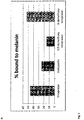
- Fig. 3 A shows the result of the binding experiment of the test compounds to the A2E complex in form of a column diagram.
- a high percentage of soraprazan and demethoxysoraprazan - about 46 % and about 53 %, respectively - are found bound to the melanin A2E particles.
- verteporfin and R/S-demethoxysoraprazan bind to the A2E melanin particles although to a lower percentage.
- a SAD for metal ions is tested.
- the SAD is incubated at various ratios with metal ions such as Fe II or Fe III and the absorbance of light by complexes is measured.
- a difference spectrum suggests interaction between the species, which may explain some aspect of pigment binding.
- Fig. 3 B shows the absorbance in dependence of the illumination wavelength for a soraprazan solution an FeCl 3 solution and a solution of soraprazan and FeCl 3 .
- the graph of Fig. 3 B shows a difference spectrum representing the FeCl 3 absorption subtracted from the soraprazan + FeCl 3 absorption. Comparing the difference spectrum with soraprazan spectrum it can be concluded that the combination of soraprazan + FeCl 3 has a higher absorption above 290 nm than the individual components.
- Example 7 Melanin and lipofuscin increase the cellular uptake of verteporfin
- the cells were treated with 200 ⁇ l of a culture medium that contained 278 nM Verteporfin for the indicated time, the culture medium was discarded, the cells trypsinized, resuspended in 500 ⁇ l medium and counted.
- Table 3 shows the results of verteporfin uptake after 1 and 18 h respectively.
- concentration of verteporfin in HRPE is more than two-fold compared to the concentration in ARPE-19.
- the concentration increased about four-fold in HuRPE cells and about two-fold in ARPE-19 cells.
- the difference in concentration becomes even more pronounced in the two cell lines becomes even more pronounced over time.
- the melanised HuRPE cells exhibit a higher verteporfin uptake than in non-melanised ARPE cells and the difference increases over time. This suggests an uptake rate that is constantly higher that in ARPE-19 cells.
- Table 3 Cellular verteporfin uptake Cell type Incubation time (h) Cell Number after incubation time Verteporfin in the cells after incubation (nM) HuRPE 1 283.750 339 ARPE-19 1 356.250 159 HuRPE 18 243.750 1206 ARPE-19 18 221.250 351
- the method in example 2 is adapted to a 96-well format in which 300 ⁇ L of the test suspension or solution is added to each well in the micro-titre plate.
- Light is provided by an LED array and diffused through a photographic filter. After illumination the plate is centrifuged in a plate centrifuge (1000 g, 1 min) and then placed in a refrigerated HPLC injector where samples are continuously injected onto an LCMS system to determine relative levels of A2E and its epoxides following illumination.
- ARPE19 cells are incubated either with or without A2E (20 ⁇ M) for 3 days and then brought into suspension by light trpysinization. The cells are passed through a cytometer to detect yellow fluorescence. As shown in Fig. 4 upper panel cells incubated with A2E ( Fig. 4 lower panel) are detected with high yellow fluorescence. This indicates a large amount of A2E. Cells incubated without A2E ( Fig. 4 upper panel) have inherently low yellow fluorescence. Treatment with lipofuscin reducing drugs results in an intermediate distribution of A2E in the cells. Thus, this assay may be used to test the elimination of lipofuscin by SADs.
- a patient with putative dry AMD is examined and baseline retinal fluorescence is determined using the blue laser imaging device (Heidelberg engineering).
- the patient is subject to an intravenous dose of 6 mg/m 2 verteporfin by means of infusion of 30 ml over 10 minutes.
- An alternative to intravenous application is the direct intravitreal application of verteporfin. In this case, ca 4 ⁇ g of verteporfin is administered to an affected single eye. Fluorescence after 1, 2 and 4 weeks is compared with fluorescence at baseline. The eyes are treated alternately.
- a laser light at 689 nm delivered 50 J/cm 2 by application of an intensity of 600 mW/cm 2 over 83 seconds using a spot size with a diameter 1000 ⁇ m.
- laser light is not necessary to promote removal of lipofuscin in the presence of a verteporfin.
- low filtering sunglasses can be used as a means to maintain an exposure of the eyes of the patient to 500 lux when outdoors. The exposure should be maintained for a period of at least 6 h per day.
- Blue light laser retinal fluorescence is measured at days 7, 14 and 28 after treatment and compared with pre-treatment values. Specific reductions of retinal fluorescence > 1-20% are on post-treatment examination. In certain patients, the effect is observed in the absence of laser light treatment. The treatment is repeated with intervals of 3 month to reduce the overall levels of lipofuscin.
- Fig. 5 shows electron micrographs of the semi-thin sections of both untreated ( Fig. 5a ) and treated mice ( Fig. 5b ).
- the RPE cell layer is marked by arrows in the Figure.
- Example 12 Quinolone injection into the vitreous of Abca4 (-/-) mice.
- quinolone antibiotics are examples of such compounds.
- Martinez et al 1998 Photochem Photobiol. 1998 Apr;67(4):399-403 . Fluoroquinolone antimicrobials: singlet oxygen, superoxide and phototoxicity.
- Martinez LJ1, Sik RH, Chignell CF. report that the relative photosensitiying activity of quinolones is as follows: "fleroxacin > lomefloxacin, pefloxacin >> ciprofloxacin > enoxacin, norfloxacin and ofloxacin.
- mice One week after injection, the eyes of the mice were enucleated, and one half of the eye was extracted by first sonication in an equal volume of saline solution, and then extraction with 3 volumes of acetonitrile. The other half of the eye was fixed with glutaraldehyde prepared as thin sections as in described in example 11. The left eye was not treated and maintained as a control. Untreated eyes of age-matched mice lacking the Abca4 transporter were an additional control.
- the acetonitrile extracts were subject to analysis by HPLCMSMS using a Sciex 4000 or 4500 triple quadrupole spectrometer as is apparent in Table 1 example 2 to detect A2E and its epoxides.
- Table 1 example 2 To detect A2E and its epoxides.
- Using a standard curve based on authentic A2E we computed the effective concentration of A2E in the eye tissue (not including lens and cornea). The concentration in each treated eye was then expressed as a percentage of the average concentration in all untreated eyes in the mice of the same age. The results are present in Table 4.
- Example 13 Tetracycline antibiotic injection into the vitreous of Abca4 (-/-) mice.
- mice were treated as in example 12 with an intravitreal injection with 2 ⁇ l of a solution containing 20 mg / ml of a solution or a suspension of the test substance.
- One week after injection the eyes were recovered and analysed as in examples 11 and 12. The left eye was not treated and maintained as a control. Untreated eyes of age-matched mice lacking the Abca4 transporter were an additional control.
- Table 6 A2E concentration in mouse eves treated with tetracycline antibiotics Test substance Concentration A2E ( ⁇ M) in the treated eye % of A2E relative to age matched untreated Chloramphenicol 24 70 Doxycycline 28 81 Demeclocycline 37 89 Minocycline 33 100 Vehicle 33 106
- Doxycycline photosensitizer
- minocycline which is not a photosensitizer
- Demeclocycline was notably less soluble which may explain limited effect.
- Example 14 Amino acid or metabolite injection into the vitreous of Abca4 (-/-) mice.
- mice were treated as in example 12 with an intravitreal injection with 2 ⁇ l of a solution containing 40 mg / ml of a solution or a suspension of the test substance.
- One week after injection the eyes were recovered and analyzed as in examples 11 and 12.
- the left eye was not treated and maintained as a control.
- Untreated eyes of age-matched mice lacking the Abca4 transporter were an additional control.
- the photoactive and reactive amino acids appeared to mediate reductions in A2E. Cysteine is able to interact with metals, particularly iron III ( Monatshefte für Chemie / November 1991, Volume 122, Issue 11, pp 887-906 ) and is also redox active in the context of iron.
- Tryptophan is capable of absorbing and re-radiating light energy, as is riboflavin.
- Table 6 A2E concentration in mouse eyes treated with amino acids or metabolites Test substance Concentration A2E ( ⁇ M) in the treated eye % of A2E relative to age matched untreated Cysteine 12 59 Tryptophan 13 66 Glutathione 35 89 Riboflavin 19 91 Vehicle 33 106
- Example 15 Chelator or metal interactor injection into the vitreous of Abca4 (-/-) mice.
- mice were treated as in example 14 with an intravitreal injection with 2 ⁇ l of a solution containing 40 mg / ml of a solution or a suspension of the test substance or a concentration as indicated in the table.
- One week after injection the eyes were recovered and analysed as in examples 11 and 12. The results are presented in Table 7.
- Table 7 A2E concentration in mouse eyes treated with chelators or metal interactors Test substance Concentration A2E ( ⁇ M) in the treated eye % of A2E relative to age matched untreated Cysteine 12 59 Taurodeoxcycholate EDTA 16 75 1,10-phenanthroline 20 91 EDTA cyclodextrin 20 97 Vehicle 33 106
- Example 16 Permeability agent injection into the vitreous of Abca4 (-/-) mice.
- mice were treated as in example 14 with an intravitreal injection with 2 ⁇ l of a solution containing 40 mg / ml of a solution or a suspension of the test substance or a concentration as indicated in the table.
- a solution containing 40 mg / ml of a solution or a suspension of the test substance or a concentration as indicated in the table.
- the eyes were recovered and analysed as in examples 11 and 12. The results are presented in Table 8.
- Table 8 A2E concentration in mouse eves treated with permeability agents Test substance Concentration A2E ( ⁇ M) in the treated eye % of A2E relative to age matched untreated 20% PEG 400 12 56 Cyclodextrin 20 83 1% Tween 80 22 95 Vehicle 33 106
- Example 17 Dye and porphyrins injection into the vitreous of Abca4 (-/-) mice.
- Certain dyes are photosensitisers or are capable of redox reactions.
- porphoryins are in certain instances, photosensitizing.
- Verteporphin is one such example while pthalocyanine is less so.
- mice were treated as in example 14 with an intravitreal injection with 2 ⁇ l of a solution containing 20 mg / ml of a solution or a suspension of the test substance.
- an intravitreal injection with 2 ⁇ l of a solution containing 20 mg / ml of a solution or a suspension of the test substance.
- the eyes were recovered and analysed as in examples 11 and 12.
- Example 18 Redox active and chelating protein injection into the vitreous of Abca4 (- /-) mice.
- mice were treated as in example 14 with an intravitreal injection with 2 ⁇ l of a solution containing 20 mg / ml of a solution or a suspension of the test substance. One week after injection, the eyes were recovered and analysed as in examples 11 and 12. The results are presented in Table 10.
- Table 10 A2E concentration in mouse eyes treated with redox active and chelating protein Test substance Concentration A2E ( ⁇ M) in the treated eye % of A2E relative to age matched untreated Lactoferrin 20 63 Cytochrome C 18 81 Transferrin (apo) 15 82
- Cytochrome C shares some activity of a peroxidase and so was used here as a model peroxidase. It is an electron carrier and is redox active. Peroxidase is also active in reducing lipofuscin. Transferrin and lactoferrin are capable of binding iron with high affinity. They are also taken into cells via endocytosis. We hypothesized that they would destabilize lipofuscin through removal of iron from the aggregates.
- Example 19 Redox active small molecules and chelator injection into the vitreous of Abca4 (-/-) mice.
- mice were treated as in example 14 with an intravitreal injection with 2 ⁇ l of a solution containing 20 mg / ml of a solution or a suspension of the test substance. Unlike in previous examples where the eyes were samples after 7 days, here the eyes were taken at 16 hours after injection, the eyes were recovered and analyzed by HPLC MSMS as in examples 11 and 12. The results are presented in Table 11. Table 11: A2E concentration in mouse eyes treated with Redox active small molecules and chelator Test substance Concentration A2E ( ⁇ M) in the treated eye % of A2E relative to age matched untreated Saline solution (0.9% NaCl) 52 102 Soraprazan 31 61 PEG-EDTA 43 85 Artemesinin 38 75
- Example 20 Calibration of HPLC MSMS detection of A2E from mouse eyes.
- Authentic A2E was dissolved in DMSO to a concentration of 10 ⁇ M and then further diluted in water from 100 ⁇ M to 15 nM in 3-fold steps. Water solutions were diluted 1:3 with acetonitrile and then sealed in vials for injection. 6 ⁇ L of the water acetonitrile solutions were injected onto a 2 x 50 mm Phenyl derived HPLC column (3 ⁇ m) and eluted with a gradient from 20% acetonitrile to 100% acetonitrile over 4 minutes at a flow rate of 500 ⁇ L/minute. The masses detected and the corresponding peak areas are indicated in the following table.
- A2E The relative lack of sensitivity of detection of A2E is related to the fact that A2E fragments easily to many different species of similar and low abundance. There are no main fragments that predominate the fragment spectrum. Peak area A2E A2E A2E - epoxide Molecularion m/z+: 592 592 608 Standard (nM) Fragmention: 418 402 444 15 1430 479 259 46 2020 395 158 137 3370 891 394 412 10000 3860 2110 1235 30400 8060 4560 3704 130000 35000 19100 11111 430000 119000 81500 33333 996000 363000 409000 100000 5310000 1440000 3060000
Landscapes
- Health & Medical Sciences (AREA)
- Life Sciences & Earth Sciences (AREA)
- Engineering & Computer Science (AREA)
- Immunology (AREA)
- Molecular Biology (AREA)
- Biomedical Technology (AREA)
- Chemical & Material Sciences (AREA)
- Urology & Nephrology (AREA)
- Hematology (AREA)
- Cell Biology (AREA)
- Food Science & Technology (AREA)
- General Health & Medical Sciences (AREA)
- Biotechnology (AREA)
- Pathology (AREA)
- General Physics & Mathematics (AREA)
- Medicinal Chemistry (AREA)
- Physics & Mathematics (AREA)
- Analytical Chemistry (AREA)
- Biochemistry (AREA)
- Microbiology (AREA)
- Biophysics (AREA)
- Endocrinology (AREA)
- Bioinformatics & Cheminformatics (AREA)
- Toxicology (AREA)
- Tropical Medicine & Parasitology (AREA)
- Pharmaceuticals Containing Other Organic And Inorganic Compounds (AREA)
- Medicines That Contain Protein Lipid Enzymes And Other Medicines (AREA)
- Acyclic And Carbocyclic Compounds In Medicinal Compositions (AREA)
Claims (8)
- Composé pour utilisation dans le traitement des maladies oculaires associées à la lipofuscine par dégradation de la lipofuscine, ladite maladie étant choisie parmi la DMLA sèche, la maladie de Stargardt, la maladie de Best et la rétinite pigmentaire, et ledit composé étant la vertéporfine.
- Composé pour utilisation selon la revendication 1, ledit traitement comprenant l'administration d'une injection intraveineuse ou intravitréenne du composé ou de la composition à un patient atteint d'une lipofuscinopathie rétinienne.
- Composé pour utilisation selon l'une quelconque des revendications 1 ou 2, ledit traitement comprenant en outre un éclairage par lumière de l'œil.
- Composé pour utilisation selon la revendication 3, le temps d'éclairage se trouvant dans la plage de 1 min à 72 h, de préférence dans la plage de 2 min à 48 h, plus préférablement dans la plage de 3 min à 48 h.
- Composé pour utilisation selon l'une quelconque des revendications 3 à 4, la luminance énergétique de la lumière d'éclairage se trouvant dans la plage de 0,001 à 1 W/cm2, de préférence dans la plage de 0,02 à 0,9 W/cm2, plus préférablement dans la plage de 0,03 à 0,7 W/cm2, idéalement dans la plage de 0,04 à 0,6 W/m2.
- Composé pour utilisation selon l'une quelconque des revendications 3 à 5, ladite lumière ayant une longueur d'onde comprise dans la plage de 380 nm à 800 nm, de préférence ladite lumière ayant une longueur d'onde comprise dans la plage de 380 nm à 650 nm, le plus préférablement ladite lumière d'éclairage ayant une longueur d'onde comprise dans la plage de 380 nm à 500 nm.
- Composé pour utilisation selon l'une quelconque des revendications 3 à 6, ladite lumière étant différente de la lumière laser.
- Composé pour utilisation selon l'une quelconque des revendications 1 à 7, ledit composé étant administré à une dose par œil en mg inférieure à 1/10000, de préférence inférieure à 1/2000, plus préférablement inférieure à 1/1000 de la dose corporelle totale calculée en mg.
Priority Applications (2)
| Application Number | Priority Date | Filing Date | Title |
|---|---|---|---|
| EP20198697.3A EP3855181B1 (fr) | 2014-02-13 | 2015-02-13 | Composés pour le traitement de maladies associées à la lipofuscine |
| DK20198697.3T DK3855181T3 (da) | 2014-02-13 | 2015-02-13 | Forbindelser til behandling af lipofuscinrelaterede sygdomme |
Applications Claiming Priority (2)
| Application Number | Priority Date | Filing Date | Title |
|---|---|---|---|
| EP14155057 | 2014-02-13 | ||
| PCT/EP2015/053148 WO2015121441A1 (fr) | 2014-02-13 | 2015-02-13 | Procédés permettant de déterminer des composés ou compositions pour le traitement de maladies liées à la lipofuscine, et composés ou compositions |
Related Child Applications (2)
| Application Number | Title | Priority Date | Filing Date |
|---|---|---|---|
| EP20198697.3A Division EP3855181B1 (fr) | 2014-02-13 | 2015-02-13 | Composés pour le traitement de maladies associées à la lipofuscine |
| EP20198697.3A Division-Into EP3855181B1 (fr) | 2014-02-13 | 2015-02-13 | Composés pour le traitement de maladies associées à la lipofuscine |
Publications (2)
| Publication Number | Publication Date |
|---|---|
| EP3105586A1 EP3105586A1 (fr) | 2016-12-21 |
| EP3105586B1 true EP3105586B1 (fr) | 2021-08-04 |
Family
ID=50097614
Family Applications (2)
| Application Number | Title | Priority Date | Filing Date |
|---|---|---|---|
| EP15708746.1A Active EP3105586B1 (fr) | 2014-02-13 | 2015-02-13 | Procédés permettant de déterminer des composés ou compositions pour le traitement de maladies liées à la lipofuscine, et composés ou compositions |
| EP20198697.3A Active EP3855181B1 (fr) | 2014-02-13 | 2015-02-13 | Composés pour le traitement de maladies associées à la lipofuscine |
Family Applications After (1)
| Application Number | Title | Priority Date | Filing Date |
|---|---|---|---|
| EP20198697.3A Active EP3855181B1 (fr) | 2014-02-13 | 2015-02-13 | Composés pour le traitement de maladies associées à la lipofuscine |
Country Status (8)
| Country | Link |
|---|---|
| US (1) | US10495630B2 (fr) |
| EP (2) | EP3105586B1 (fr) |
| CA (1) | CA2976364C (fr) |
| DK (1) | DK3855181T3 (fr) |
| ES (2) | ES3012058T3 (fr) |
| HR (1) | HRP20250123T1 (fr) |
| HU (1) | HUE070257T2 (fr) |
| WO (1) | WO2015121441A1 (fr) |
Families Citing this family (5)
| Publication number | Priority date | Publication date | Assignee | Title |
|---|---|---|---|---|
| CN106214628B (zh) * | 2016-08-03 | 2020-02-14 | 四川恒通动保生物科技有限公司 | 一种兽用盐酸多西环素注射液及其制备方法 |
| EA201990560A1 (ru) | 2016-09-08 | 2019-09-30 | Регенера Фарма Лтд. | Композиции, содержащие тритерпеноиды, и их применение для лечения оптической нейропатии |
| EP3949958A1 (fr) | 2020-08-04 | 2022-02-09 | Eberhard Karls Universität Tübingen Medizinische Fakultät | Composé pour le traitement et la prévention d'une maladie associée à la lipofuscine |
| WO2023151788A1 (fr) | 2022-02-09 | 2023-08-17 | Katairo Gmbh | Mélanine pour le traitement de maladies associées à la lipofuscine |
| WO2023213902A1 (fr) * | 2022-05-03 | 2023-11-09 | Katairo Gmbh | Remofuscine (soraprazan) destinée à être utilisée contre un dysfonctionnement lysosomal et le vieillissement |
Family Cites Families (7)
| Publication number | Priority date | Publication date | Assignee | Title |
|---|---|---|---|---|
| US7267830B2 (en) * | 2003-12-19 | 2007-09-11 | Alcon, Inc. | Composition and methods for inhibiting the progression macular degeneration and promoting healthy vision |
| JP2008515778A (ja) | 2004-08-18 | 2008-05-15 | シリオン セラピューティクス, インコーポレイテッド | 眼の状態を13−シス−レチニル誘導体で処置するための組み合わせ方法、組成物および治療 |
| UA81382C2 (en) * | 2005-07-11 | 2007-12-25 | Composition for treating retinol-related diseases by modulation of retinol binding | |
| EP2687208A1 (fr) * | 2007-09-12 | 2014-01-22 | The Trustees of Columbia University in the City of New York | Compositions et procédés pour le traitement de la dégénérescence maculaire |
| EP2080513A1 (fr) | 2008-01-16 | 2009-07-22 | Schraermeyer, Ulrich, Prof. Dr. rer. nat | Tétrahydropyridoéthers pour le traitement de la DMA |
| GB201014340D0 (en) * | 2010-08-27 | 2010-10-13 | Piraee Mahmood | Nutritional supplement formulation comprising saffron and resveratrol |
| WO2013166037A1 (fr) | 2012-05-01 | 2013-11-07 | The Trustees Of Columbia University In The City Of New York | Antagonistes non rétinoïdes pour le traitement de troubles oculaires |
-
2015
- 2015-02-13 CA CA2976364A patent/CA2976364C/fr active Active
- 2015-02-13 HU HUE20198697A patent/HUE070257T2/hu unknown
- 2015-02-13 DK DK20198697.3T patent/DK3855181T3/da active
- 2015-02-13 EP EP15708746.1A patent/EP3105586B1/fr active Active
- 2015-02-13 ES ES20198697T patent/ES3012058T3/es active Active
- 2015-02-13 WO PCT/EP2015/053148 patent/WO2015121441A1/fr not_active Ceased
- 2015-02-13 ES ES15708746T patent/ES2896173T3/es active Active
- 2015-02-13 US US15/118,486 patent/US10495630B2/en active Active
- 2015-02-13 EP EP20198697.3A patent/EP3855181B1/fr active Active
- 2015-02-13 HR HRP20250123TT patent/HRP20250123T1/hr unknown
Non-Patent Citations (2)
| Title |
|---|
| O. ZELPHATI ET AL: "Intracellular Delivery of Proteins with a New Lipid-mediated Delivery System", JOURNAL OF BIOLOGICAL CHEMISTRY, vol. 276, no. 37, 14 September 2001 (2001-09-14), pages 35103 - 35110, XP055381813, ISSN: 0021-9258, DOI: 10.1074/jbc.M104920200 * |
| YALIN WU ET AL: "Enzymatic Degradation of A2E, a Retinal Pigment Epithelial Lipofuscin Bisretinoid", JOURNAL OF THE AMERICAN CHEMICAL SOCIETY, vol. 133, no. 4, 2 February 2011 (2011-02-02), US, pages 849 - 857, XP055515777, ISSN: 0002-7863, DOI: 10.1021/ja107195u * |
Also Published As
| Publication number | Publication date |
|---|---|
| US10495630B2 (en) | 2019-12-03 |
| ES2896173T3 (es) | 2022-02-24 |
| ES3012058T3 (en) | 2025-04-08 |
| EP3855181B1 (fr) | 2024-11-06 |
| EP3855181A1 (fr) | 2021-07-28 |
| CA2976364C (fr) | 2023-02-14 |
| HUE070257T2 (hu) | 2025-05-28 |
| EP3105586A1 (fr) | 2016-12-21 |
| WO2015121441A1 (fr) | 2015-08-20 |
| DK3855181T3 (da) | 2025-02-10 |
| CA2976364A1 (fr) | 2015-08-20 |
| HRP20250123T1 (hr) | 2025-03-28 |
| US20170176416A1 (en) | 2017-06-22 |
Similar Documents
| Publication | Publication Date | Title |
|---|---|---|
| Chen et al. | Tau-targeted multifunctional nanocomposite for combinational therapy of Alzheimer’s disease | |
| Saewanee et al. | Neuroprotective effect of metformin on dopaminergic neurodegeneration and α-synuclein aggregation in C. elegans model of Parkinson’s disease | |
| Wang et al. | Intranasal administration of dauricine loaded on graphene oxide: multi-target therapy for Alzheimer's disease | |
| Crouch et al. | Therapeutic redistribution of metal ions to treat Alzheimer’s disease | |
| US10495630B2 (en) | Methods for the determination of compounds or compositions for the treatment of lipofuscin related diseases and compounds or compositions | |
| Jin et al. | Toxicity of luminescent silica nanoparticles to living cells | |
| Dragicevic et al. | Long-term electromagnetic field treatment enhances brain mitochondrial function of both Alzheimer's transgenic mice and normal mice: a mechanism for electromagnetic field-induced cognitive benefit? | |
| Rogov et al. | Mitochondrial dysfunctions may be one of the major causative factors underlying detrimental effects of benzalkonium chloride | |
| US9572834B2 (en) | Use of carbon nanomaterials with antioxidant properties to treat oxidative stress | |
| Akaiwa et al. | Edaravone suppresses retinal ganglion cell death in a mouse model of normal tension glaucoma | |
| Qian et al. | Multifunctional nano-enabled delivery systems in Alzheimer's disease management | |
| CN121102455A (zh) | 用于改善认知的方法和组合物 | |
| Jo et al. | Activation of lysosomal function ameliorates Amyloid-β-Induced tight junction disruption in the retinal pigment epithelium | |
| US9950007B2 (en) | Neuronal protection by cerium oxide nanoparticles | |
| Ali et al. | Nanotechnology in Parkinson’s Disease: overcoming drug delivery challenges and enhancing therapeutic outcomes | |
| Guo et al. | In situ dual-activated NIRF/PA carrier-free nanoprobe for diagnosis and treatment of Parkinson's disease | |
| Lin et al. | Potential of fluorescent nanoprobe in diagnosis and treatment of Alzheimer’s disease | |
| US20220000804A1 (en) | Intranasal nano inducer for preventing and treating neurodegenerative diseases and method thereof | |
| Xie et al. | Mn3O4 nanozyme-based anti-inflammatory therapy modulates microglial phenotype by downregulating TLR4/NOX2 expression and further alleviates Alzheimer's disease pathology | |
| Olchawa et al. | SuperDopa (SD), SuperDopa amide (SDA) and Thioredoxin-mimetic peptides protect ARPE-19 cells from photic-and non-photic stress | |
| Budhwani et al. | Exosome-mediated delivery of a PLD1 modulator overcomes bioavailability hurdles to target synaptopathy in neurodegenerative diseases | |
| CN106456624A (zh) | Flap抑制剂的降低中枢神经系统内的神经炎症介导的损伤的应用 | |
| JP2019531296A (ja) | 眼疾患の処置方法 | |
| Lo et al. | Lysosome-acidifying nanoparticles rescue A30P α-synuclein induced neuronal death in cellular and Drosophila models of Parkinson’s disease | |
| US10220070B2 (en) | Alphaa-crystallin mimetic peptides and uses thereof |
Legal Events
| Date | Code | Title | Description |
|---|---|---|---|
| PUAI | Public reference made under article 153(3) epc to a published international application that has entered the european phase |
Free format text: ORIGINAL CODE: 0009012 |
|
| STAA | Information on the status of an ep patent application or granted ep patent |
Free format text: STATUS: REQUEST FOR EXAMINATION WAS MADE |
|
| 17P | Request for examination filed |
Effective date: 20160913 |
|
| AK | Designated contracting states |
Kind code of ref document: A1 Designated state(s): AL AT BE BG CH CY CZ DE DK EE ES FI FR GB GR HR HU IE IS IT LI LT LU LV MC MK MT NL NO PL PT RO RS SE SI SK SM TR |
|
| AX | Request for extension of the european patent |
Extension state: BA ME |
|
| DAX | Request for extension of the european patent (deleted) | ||
| STAA | Information on the status of an ep patent application or granted ep patent |
Free format text: STATUS: EXAMINATION IS IN PROGRESS |
|
| 17Q | First examination report despatched |
Effective date: 20181022 |
|
| GRAP | Despatch of communication of intention to grant a patent |
Free format text: ORIGINAL CODE: EPIDOSNIGR1 |
|
| STAA | Information on the status of an ep patent application or granted ep patent |
Free format text: STATUS: GRANT OF PATENT IS INTENDED |
|
| INTG | Intention to grant announced |
Effective date: 20191212 |
|
| GRAJ | Information related to disapproval of communication of intention to grant by the applicant or resumption of examination proceedings by the epo deleted |
Free format text: ORIGINAL CODE: EPIDOSDIGR1 |
|
| STAA | Information on the status of an ep patent application or granted ep patent |
Free format text: STATUS: EXAMINATION IS IN PROGRESS |
|
| GRAP | Despatch of communication of intention to grant a patent |
Free format text: ORIGINAL CODE: EPIDOSNIGR1 |
|
| STAA | Information on the status of an ep patent application or granted ep patent |
Free format text: STATUS: GRANT OF PATENT IS INTENDED |
|
| INTC | Intention to grant announced (deleted) | ||
| INTG | Intention to grant announced |
Effective date: 20200519 |
|
| RAP1 | Party data changed (applicant data changed or rights of an application transferred) |
Owner name: KATAIRO GMBH Owner name: TAKEDA GMBH |
|
| RIN1 | Information on inventor provided before grant (corrected) |
Inventor name: HANAUER, GUIDO Inventor name: SENN-BILFINGER, JOERG Inventor name: SCHRAERMEYER, ULRICH Inventor name: STURM, ERNST Inventor name: BURNET, MICHAEL |
|
| GRAS | Grant fee paid |
Free format text: ORIGINAL CODE: EPIDOSNIGR3 |
|
| GRAA | (expected) grant |
Free format text: ORIGINAL CODE: 0009210 |
|
| STAA | Information on the status of an ep patent application or granted ep patent |
Free format text: STATUS: THE PATENT HAS BEEN GRANTED |
|
| AK | Designated contracting states |
Kind code of ref document: B1 Designated state(s): AL AT BE BG CH CY CZ DE DK EE ES FI FR GB GR HR HU IE IS IT LI LT LU LV MC MK MT NL NO PL PT RO RS SE SI SK SM TR |
|
| REG | Reference to a national code |
Ref country code: GB Ref legal event code: FG4D |
|
| REG | Reference to a national code |
Ref country code: AT Ref legal event code: REF Ref document number: 1417482 Country of ref document: AT Kind code of ref document: T Effective date: 20210815 |
|
| REG | Reference to a national code |
Ref country code: CH Ref legal event code: EP |
|
| REG | Reference to a national code |
Ref country code: DE Ref legal event code: R096 Ref document number: 602015071899 Country of ref document: DE |
|
| REG | Reference to a national code |
Ref country code: IE Ref legal event code: FG4D |
|
| REG | Reference to a national code |
Ref country code: LT Ref legal event code: MG9D |
|
| REG | Reference to a national code |
Ref country code: NL Ref legal event code: MP Effective date: 20210804 |
|
| REG | Reference to a national code |
Ref country code: AT Ref legal event code: MK05 Ref document number: 1417482 Country of ref document: AT Kind code of ref document: T Effective date: 20210804 |
|
| PG25 | Lapsed in a contracting state [announced via postgrant information from national office to epo] |
Ref country code: FI Free format text: LAPSE BECAUSE OF FAILURE TO SUBMIT A TRANSLATION OF THE DESCRIPTION OR TO PAY THE FEE WITHIN THE PRESCRIBED TIME-LIMIT Effective date: 20210804 Ref country code: PT Free format text: LAPSE BECAUSE OF FAILURE TO SUBMIT A TRANSLATION OF THE DESCRIPTION OR TO PAY THE FEE WITHIN THE PRESCRIBED TIME-LIMIT Effective date: 20211206 Ref country code: NO Free format text: LAPSE BECAUSE OF FAILURE TO SUBMIT A TRANSLATION OF THE DESCRIPTION OR TO PAY THE FEE WITHIN THE PRESCRIBED TIME-LIMIT Effective date: 20211104 Ref country code: LT Free format text: LAPSE BECAUSE OF FAILURE TO SUBMIT A TRANSLATION OF THE DESCRIPTION OR TO PAY THE FEE WITHIN THE PRESCRIBED TIME-LIMIT Effective date: 20210804 Ref country code: AT Free format text: LAPSE BECAUSE OF FAILURE TO SUBMIT A TRANSLATION OF THE DESCRIPTION OR TO PAY THE FEE WITHIN THE PRESCRIBED TIME-LIMIT Effective date: 20210804 Ref country code: BG Free format text: LAPSE BECAUSE OF FAILURE TO SUBMIT A TRANSLATION OF THE DESCRIPTION OR TO PAY THE FEE WITHIN THE PRESCRIBED TIME-LIMIT Effective date: 20211104 Ref country code: RS Free format text: LAPSE BECAUSE OF FAILURE TO SUBMIT A TRANSLATION OF THE DESCRIPTION OR TO PAY THE FEE WITHIN THE PRESCRIBED TIME-LIMIT Effective date: 20210804 Ref country code: SE Free format text: LAPSE BECAUSE OF FAILURE TO SUBMIT A TRANSLATION OF THE DESCRIPTION OR TO PAY THE FEE WITHIN THE PRESCRIBED TIME-LIMIT Effective date: 20210804 Ref country code: HR Free format text: LAPSE BECAUSE OF FAILURE TO SUBMIT A TRANSLATION OF THE DESCRIPTION OR TO PAY THE FEE WITHIN THE PRESCRIBED TIME-LIMIT Effective date: 20210804 |
|
| REG | Reference to a national code |
Ref country code: ES Ref legal event code: FG2A Ref document number: 2896173 Country of ref document: ES Kind code of ref document: T3 Effective date: 20220224 |
|
| PG25 | Lapsed in a contracting state [announced via postgrant information from national office to epo] |
Ref country code: PL Free format text: LAPSE BECAUSE OF FAILURE TO SUBMIT A TRANSLATION OF THE DESCRIPTION OR TO PAY THE FEE WITHIN THE PRESCRIBED TIME-LIMIT Effective date: 20210804 Ref country code: LV Free format text: LAPSE BECAUSE OF FAILURE TO SUBMIT A TRANSLATION OF THE DESCRIPTION OR TO PAY THE FEE WITHIN THE PRESCRIBED TIME-LIMIT Effective date: 20210804 Ref country code: GR Free format text: LAPSE BECAUSE OF FAILURE TO SUBMIT A TRANSLATION OF THE DESCRIPTION OR TO PAY THE FEE WITHIN THE PRESCRIBED TIME-LIMIT Effective date: 20211105 |
|
| PG25 | Lapsed in a contracting state [announced via postgrant information from national office to epo] |
Ref country code: NL Free format text: LAPSE BECAUSE OF FAILURE TO SUBMIT A TRANSLATION OF THE DESCRIPTION OR TO PAY THE FEE WITHIN THE PRESCRIBED TIME-LIMIT Effective date: 20210804 |
|
| PG25 | Lapsed in a contracting state [announced via postgrant information from national office to epo] |
Ref country code: DK Free format text: LAPSE BECAUSE OF FAILURE TO SUBMIT A TRANSLATION OF THE DESCRIPTION OR TO PAY THE FEE WITHIN THE PRESCRIBED TIME-LIMIT Effective date: 20210804 |
|
| REG | Reference to a national code |
Ref country code: DE Ref legal event code: R097 Ref document number: 602015071899 Country of ref document: DE |
|
| PG25 | Lapsed in a contracting state [announced via postgrant information from national office to epo] |
Ref country code: SM Free format text: LAPSE BECAUSE OF FAILURE TO SUBMIT A TRANSLATION OF THE DESCRIPTION OR TO PAY THE FEE WITHIN THE PRESCRIBED TIME-LIMIT Effective date: 20210804 Ref country code: SK Free format text: LAPSE BECAUSE OF FAILURE TO SUBMIT A TRANSLATION OF THE DESCRIPTION OR TO PAY THE FEE WITHIN THE PRESCRIBED TIME-LIMIT Effective date: 20210804 Ref country code: RO Free format text: LAPSE BECAUSE OF FAILURE TO SUBMIT A TRANSLATION OF THE DESCRIPTION OR TO PAY THE FEE WITHIN THE PRESCRIBED TIME-LIMIT Effective date: 20210804 Ref country code: EE Free format text: LAPSE BECAUSE OF FAILURE TO SUBMIT A TRANSLATION OF THE DESCRIPTION OR TO PAY THE FEE WITHIN THE PRESCRIBED TIME-LIMIT Effective date: 20210804 Ref country code: CZ Free format text: LAPSE BECAUSE OF FAILURE TO SUBMIT A TRANSLATION OF THE DESCRIPTION OR TO PAY THE FEE WITHIN THE PRESCRIBED TIME-LIMIT Effective date: 20210804 Ref country code: AL Free format text: LAPSE BECAUSE OF FAILURE TO SUBMIT A TRANSLATION OF THE DESCRIPTION OR TO PAY THE FEE WITHIN THE PRESCRIBED TIME-LIMIT Effective date: 20210804 |
|
| PLBE | No opposition filed within time limit |
Free format text: ORIGINAL CODE: 0009261 |
|
| STAA | Information on the status of an ep patent application or granted ep patent |
Free format text: STATUS: NO OPPOSITION FILED WITHIN TIME LIMIT |
|
| 26N | No opposition filed |
Effective date: 20220506 |
|
| PG25 | Lapsed in a contracting state [announced via postgrant information from national office to epo] |
Ref country code: SI Free format text: LAPSE BECAUSE OF FAILURE TO SUBMIT A TRANSLATION OF THE DESCRIPTION OR TO PAY THE FEE WITHIN THE PRESCRIBED TIME-LIMIT Effective date: 20210804 |
|
| PG25 | Lapsed in a contracting state [announced via postgrant information from national office to epo] |
Ref country code: MC Free format text: LAPSE BECAUSE OF FAILURE TO SUBMIT A TRANSLATION OF THE DESCRIPTION OR TO PAY THE FEE WITHIN THE PRESCRIBED TIME-LIMIT Effective date: 20210804 |
|
| REG | Reference to a national code |
Ref country code: CH Ref legal event code: PL |
|
| REG | Reference to a national code |
Ref country code: BE Ref legal event code: MM Effective date: 20220228 |
|
| PG25 | Lapsed in a contracting state [announced via postgrant information from national office to epo] |
Ref country code: LU Free format text: LAPSE BECAUSE OF NON-PAYMENT OF DUE FEES Effective date: 20220213 |
|
| PG25 | Lapsed in a contracting state [announced via postgrant information from national office to epo] |
Ref country code: LI Free format text: LAPSE BECAUSE OF NON-PAYMENT OF DUE FEES Effective date: 20220228 Ref country code: IE Free format text: LAPSE BECAUSE OF NON-PAYMENT OF DUE FEES Effective date: 20220213 Ref country code: CH Free format text: LAPSE BECAUSE OF NON-PAYMENT OF DUE FEES Effective date: 20220228 |
|
| PG25 | Lapsed in a contracting state [announced via postgrant information from national office to epo] |
Ref country code: BE Free format text: LAPSE BECAUSE OF NON-PAYMENT OF DUE FEES Effective date: 20220228 |
|
| PG25 | Lapsed in a contracting state [announced via postgrant information from national office to epo] |
Ref country code: HU Free format text: LAPSE BECAUSE OF FAILURE TO SUBMIT A TRANSLATION OF THE DESCRIPTION OR TO PAY THE FEE WITHIN THE PRESCRIBED TIME-LIMIT; INVALID AB INITIO Effective date: 20150213 |
|
| PG25 | Lapsed in a contracting state [announced via postgrant information from national office to epo] |
Ref country code: MK Free format text: LAPSE BECAUSE OF FAILURE TO SUBMIT A TRANSLATION OF THE DESCRIPTION OR TO PAY THE FEE WITHIN THE PRESCRIBED TIME-LIMIT Effective date: 20210804 Ref country code: CY Free format text: LAPSE BECAUSE OF FAILURE TO SUBMIT A TRANSLATION OF THE DESCRIPTION OR TO PAY THE FEE WITHIN THE PRESCRIBED TIME-LIMIT Effective date: 20210804 |
|
| PG25 | Lapsed in a contracting state [announced via postgrant information from national office to epo] |
Ref country code: MT Free format text: LAPSE BECAUSE OF FAILURE TO SUBMIT A TRANSLATION OF THE DESCRIPTION OR TO PAY THE FEE WITHIN THE PRESCRIBED TIME-LIMIT Effective date: 20210804 |
|
| PGFP | Annual fee paid to national office [announced via postgrant information from national office to epo] |
Ref country code: DE Payment date: 20250319 Year of fee payment: 11 |
|
| PGFP | Annual fee paid to national office [announced via postgrant information from national office to epo] |
Ref country code: ES Payment date: 20250318 Year of fee payment: 11 |
|
| PGFP | Annual fee paid to national office [announced via postgrant information from national office to epo] |
Ref country code: FR Payment date: 20250227 Year of fee payment: 11 |
|
| PGFP | Annual fee paid to national office [announced via postgrant information from national office to epo] |
Ref country code: GB Payment date: 20250324 Year of fee payment: 11 Ref country code: IT Payment date: 20250227 Year of fee payment: 11 |
|
| PG25 | Lapsed in a contracting state [announced via postgrant information from national office to epo] |
Ref country code: TR Free format text: LAPSE BECAUSE OF FAILURE TO SUBMIT A TRANSLATION OF THE DESCRIPTION OR TO PAY THE FEE WITHIN THE PRESCRIBED TIME-LIMIT Effective date: 20210804 |
|
| REG | Reference to a national code |
Ref country code: DE Ref legal event code: R082 Ref document number: 602015071899 Country of ref document: DE Representative=s name: HOFFMANN EITLE PATENT- UND RECHTSANWAELTE PART, DE |