EP3129075B1 - Knochenreparaturzusammensetzungen - Google Patents
Knochenreparaturzusammensetzungen Download PDFInfo
- Publication number
- EP3129075B1 EP3129075B1 EP15777375.5A EP15777375A EP3129075B1 EP 3129075 B1 EP3129075 B1 EP 3129075B1 EP 15777375 A EP15777375 A EP 15777375A EP 3129075 B1 EP3129075 B1 EP 3129075B1
- Authority
- EP
- European Patent Office
- Prior art keywords
- cells
- composition
- another embodiment
- bone
- cell culture
- Prior art date
- Legal status (The legal status is an assumption and is not a legal conclusion. Google has not performed a legal analysis and makes no representation as to the accuracy of the status listed.)
- Active
Links
Images


Classifications
-
- A—HUMAN NECESSITIES
- A61—MEDICAL OR VETERINARY SCIENCE; HYGIENE
- A61L—METHODS OR APPARATUS FOR STERILISING MATERIALS OR OBJECTS IN GENERAL; DISINFECTION, STERILISATION OR DEODORISATION OF AIR; CHEMICAL ASPECTS OF BANDAGES, DRESSINGS, ABSORBENT PADS OR SURGICAL ARTICLES; MATERIALS FOR BANDAGES, DRESSINGS, ABSORBENT PADS OR SURGICAL ARTICLES
- A61L27/00—Materials for grafts or prostheses or for coating grafts or prostheses
- A61L27/36—Materials for grafts or prostheses or for coating grafts or prostheses containing ingredients of undetermined constitution or reaction products thereof, e.g. transplant tissue, natural bone, extracellular matrix
- A61L27/38—Materials for grafts or prostheses or for coating grafts or prostheses containing ingredients of undetermined constitution or reaction products thereof, e.g. transplant tissue, natural bone, extracellular matrix containing added animal cells
- A61L27/3886—Materials for grafts or prostheses or for coating grafts or prostheses containing ingredients of undetermined constitution or reaction products thereof, e.g. transplant tissue, natural bone, extracellular matrix containing added animal cells comprising two or more cell types
- A61L27/3891—Materials for grafts or prostheses or for coating grafts or prostheses containing ingredients of undetermined constitution or reaction products thereof, e.g. transplant tissue, natural bone, extracellular matrix containing added animal cells comprising two or more cell types as distinct cell layers
-
- A—HUMAN NECESSITIES
- A61—MEDICAL OR VETERINARY SCIENCE; HYGIENE
- A61K—PREPARATIONS FOR MEDICAL, DENTAL OR TOILETRY PURPOSES
- A61K31/00—Medicinal preparations containing organic active ingredients
- A61K31/70—Carbohydrates; Sugars; Derivatives thereof
- A61K31/715—Polysaccharides, i.e. having more than five saccharide radicals attached to each other by glycosidic linkages; Derivatives thereof, e.g. ethers, esters
- A61K31/726—Glycosaminoglycans, i.e. mucopolysaccharides
- A61K31/728—Hyaluronic acid
-
- A—HUMAN NECESSITIES
- A61—MEDICAL OR VETERINARY SCIENCE; HYGIENE
- A61K—PREPARATIONS FOR MEDICAL, DENTAL OR TOILETRY PURPOSES
- A61K35/00—Medicinal preparations containing materials or reaction products thereof with undetermined constitution
- A61K35/12—Materials from mammals; Compositions comprising non-specified tissues or cells; Compositions comprising non-embryonic stem cells; Genetically modified cells
- A61K35/28—Bone marrow; Haematopoietic stem cells; Mesenchymal stem cells of any origin, e.g. adipose-derived stem cells
-
- A—HUMAN NECESSITIES
- A61—MEDICAL OR VETERINARY SCIENCE; HYGIENE
- A61K—PREPARATIONS FOR MEDICAL, DENTAL OR TOILETRY PURPOSES
- A61K35/00—Medicinal preparations containing materials or reaction products thereof with undetermined constitution
- A61K35/12—Materials from mammals; Compositions comprising non-specified tissues or cells; Compositions comprising non-embryonic stem cells; Genetically modified cells
- A61K35/44—Vessels; Vascular smooth muscle cells; Endothelial cells; Endothelial progenitor cells
-
- A—HUMAN NECESSITIES
- A61—MEDICAL OR VETERINARY SCIENCE; HYGIENE
- A61L—METHODS OR APPARATUS FOR STERILISING MATERIALS OR OBJECTS IN GENERAL; DISINFECTION, STERILISATION OR DEODORISATION OF AIR; CHEMICAL ASPECTS OF BANDAGES, DRESSINGS, ABSORBENT PADS OR SURGICAL ARTICLES; MATERIALS FOR BANDAGES, DRESSINGS, ABSORBENT PADS OR SURGICAL ARTICLES
- A61L27/00—Materials for grafts or prostheses or for coating grafts or prostheses
- A61L27/02—Inorganic materials
- A61L27/12—Phosphorus-containing materials, e.g. apatite
-
- A—HUMAN NECESSITIES
- A61—MEDICAL OR VETERINARY SCIENCE; HYGIENE
- A61L—METHODS OR APPARATUS FOR STERILISING MATERIALS OR OBJECTS IN GENERAL; DISINFECTION, STERILISATION OR DEODORISATION OF AIR; CHEMICAL ASPECTS OF BANDAGES, DRESSINGS, ABSORBENT PADS OR SURGICAL ARTICLES; MATERIALS FOR BANDAGES, DRESSINGS, ABSORBENT PADS OR SURGICAL ARTICLES
- A61L27/00—Materials for grafts or prostheses or for coating grafts or prostheses
- A61L27/14—Macromolecular materials
- A61L27/20—Polysaccharides
-
- A—HUMAN NECESSITIES
- A61—MEDICAL OR VETERINARY SCIENCE; HYGIENE
- A61L—METHODS OR APPARATUS FOR STERILISING MATERIALS OR OBJECTS IN GENERAL; DISINFECTION, STERILISATION OR DEODORISATION OF AIR; CHEMICAL ASPECTS OF BANDAGES, DRESSINGS, ABSORBENT PADS OR SURGICAL ARTICLES; MATERIALS FOR BANDAGES, DRESSINGS, ABSORBENT PADS OR SURGICAL ARTICLES
- A61L27/00—Materials for grafts or prostheses or for coating grafts or prostheses
- A61L27/36—Materials for grafts or prostheses or for coating grafts or prostheses containing ingredients of undetermined constitution or reaction products thereof, e.g. transplant tissue, natural bone, extracellular matrix
- A61L27/3604—Materials for grafts or prostheses or for coating grafts or prostheses containing ingredients of undetermined constitution or reaction products thereof, e.g. transplant tissue, natural bone, extracellular matrix characterised by the human or animal origin of the biological material, e.g. hair, fascia, fish scales, silk, shellac, pericardium, pleura, renal tissue, amniotic membrane, parenchymal tissue, fetal tissue, muscle tissue, fat tissue, enamel
- A61L27/3608—Bone, e.g. demineralised bone matrix [DBM], bone powder
-
- A—HUMAN NECESSITIES
- A61—MEDICAL OR VETERINARY SCIENCE; HYGIENE
- A61L—METHODS OR APPARATUS FOR STERILISING MATERIALS OR OBJECTS IN GENERAL; DISINFECTION, STERILISATION OR DEODORISATION OF AIR; CHEMICAL ASPECTS OF BANDAGES, DRESSINGS, ABSORBENT PADS OR SURGICAL ARTICLES; MATERIALS FOR BANDAGES, DRESSINGS, ABSORBENT PADS OR SURGICAL ARTICLES
- A61L27/00—Materials for grafts or prostheses or for coating grafts or prostheses
- A61L27/36—Materials for grafts or prostheses or for coating grafts or prostheses containing ingredients of undetermined constitution or reaction products thereof, e.g. transplant tissue, natural bone, extracellular matrix
- A61L27/38—Materials for grafts or prostheses or for coating grafts or prostheses containing ingredients of undetermined constitution or reaction products thereof, e.g. transplant tissue, natural bone, extracellular matrix containing added animal cells
- A61L27/3804—Materials for grafts or prostheses or for coating grafts or prostheses containing ingredients of undetermined constitution or reaction products thereof, e.g. transplant tissue, natural bone, extracellular matrix containing added animal cells characterised by specific cells or progenitors thereof, e.g. fibroblasts, connective tissue cells, kidney cells
- A61L27/3808—Endothelial cells
-
- A—HUMAN NECESSITIES
- A61—MEDICAL OR VETERINARY SCIENCE; HYGIENE
- A61L—METHODS OR APPARATUS FOR STERILISING MATERIALS OR OBJECTS IN GENERAL; DISINFECTION, STERILISATION OR DEODORISATION OF AIR; CHEMICAL ASPECTS OF BANDAGES, DRESSINGS, ABSORBENT PADS OR SURGICAL ARTICLES; MATERIALS FOR BANDAGES, DRESSINGS, ABSORBENT PADS OR SURGICAL ARTICLES
- A61L27/00—Materials for grafts or prostheses or for coating grafts or prostheses
- A61L27/36—Materials for grafts or prostheses or for coating grafts or prostheses containing ingredients of undetermined constitution or reaction products thereof, e.g. transplant tissue, natural bone, extracellular matrix
- A61L27/38—Materials for grafts or prostheses or for coating grafts or prostheses containing ingredients of undetermined constitution or reaction products thereof, e.g. transplant tissue, natural bone, extracellular matrix containing added animal cells
- A61L27/3804—Materials for grafts or prostheses or for coating grafts or prostheses containing ingredients of undetermined constitution or reaction products thereof, e.g. transplant tissue, natural bone, extracellular matrix containing added animal cells characterised by specific cells or progenitors thereof, e.g. fibroblasts, connective tissue cells, kidney cells
- A61L27/3834—Cells able to produce different cell types, e.g. hematopoietic stem cells, mesenchymal stem cells, marrow stromal cells, embryonic stem cells
-
- A—HUMAN NECESSITIES
- A61—MEDICAL OR VETERINARY SCIENCE; HYGIENE
- A61L—METHODS OR APPARATUS FOR STERILISING MATERIALS OR OBJECTS IN GENERAL; DISINFECTION, STERILISATION OR DEODORISATION OF AIR; CHEMICAL ASPECTS OF BANDAGES, DRESSINGS, ABSORBENT PADS OR SURGICAL ARTICLES; MATERIALS FOR BANDAGES, DRESSINGS, ABSORBENT PADS OR SURGICAL ARTICLES
- A61L27/00—Materials for grafts or prostheses or for coating grafts or prostheses
- A61L27/36—Materials for grafts or prostheses or for coating grafts or prostheses containing ingredients of undetermined constitution or reaction products thereof, e.g. transplant tissue, natural bone, extracellular matrix
- A61L27/38—Materials for grafts or prostheses or for coating grafts or prostheses containing ingredients of undetermined constitution or reaction products thereof, e.g. transplant tissue, natural bone, extracellular matrix containing added animal cells
- A61L27/3839—Materials for grafts or prostheses or for coating grafts or prostheses containing ingredients of undetermined constitution or reaction products thereof, e.g. transplant tissue, natural bone, extracellular matrix containing added animal cells characterised by the site of application in the body
- A61L27/3843—Connective tissue
- A61L27/3847—Bones
-
- A—HUMAN NECESSITIES
- A61—MEDICAL OR VETERINARY SCIENCE; HYGIENE
- A61L—METHODS OR APPARATUS FOR STERILISING MATERIALS OR OBJECTS IN GENERAL; DISINFECTION, STERILISATION OR DEODORISATION OF AIR; CHEMICAL ASPECTS OF BANDAGES, DRESSINGS, ABSORBENT PADS OR SURGICAL ARTICLES; MATERIALS FOR BANDAGES, DRESSINGS, ABSORBENT PADS OR SURGICAL ARTICLES
- A61L27/00—Materials for grafts or prostheses or for coating grafts or prostheses
- A61L27/40—Composite materials, i.e. containing one material dispersed in a matrix of the same or different material
- A61L27/44—Composite materials, i.e. containing one material dispersed in a matrix of the same or different material having a macromolecular matrix
- A61L27/46—Composite materials, i.e. containing one material dispersed in a matrix of the same or different material having a macromolecular matrix with phosphorus-containing inorganic fillers
-
- A—HUMAN NECESSITIES
- A61—MEDICAL OR VETERINARY SCIENCE; HYGIENE
- A61L—METHODS OR APPARATUS FOR STERILISING MATERIALS OR OBJECTS IN GENERAL; DISINFECTION, STERILISATION OR DEODORISATION OF AIR; CHEMICAL ASPECTS OF BANDAGES, DRESSINGS, ABSORBENT PADS OR SURGICAL ARTICLES; MATERIALS FOR BANDAGES, DRESSINGS, ABSORBENT PADS OR SURGICAL ARTICLES
- A61L27/00—Materials for grafts or prostheses or for coating grafts or prostheses
- A61L27/50—Materials characterised by their function or physical properties, e.g. injectable or lubricating compositions, shape-memory materials, surface modified materials
- A61L27/58—Materials at least partially resorbable by the body
-
- A—HUMAN NECESSITIES
- A61—MEDICAL OR VETERINARY SCIENCE; HYGIENE
- A61L—METHODS OR APPARATUS FOR STERILISING MATERIALS OR OBJECTS IN GENERAL; DISINFECTION, STERILISATION OR DEODORISATION OF AIR; CHEMICAL ASPECTS OF BANDAGES, DRESSINGS, ABSORBENT PADS OR SURGICAL ARTICLES; MATERIALS FOR BANDAGES, DRESSINGS, ABSORBENT PADS OR SURGICAL ARTICLES
- A61L2400/00—Materials characterised by their function or physical properties
- A61L2400/06—Flowable or injectable implant compositions
-
- A—HUMAN NECESSITIES
- A61—MEDICAL OR VETERINARY SCIENCE; HYGIENE
- A61L—METHODS OR APPARATUS FOR STERILISING MATERIALS OR OBJECTS IN GENERAL; DISINFECTION, STERILISATION OR DEODORISATION OF AIR; CHEMICAL ASPECTS OF BANDAGES, DRESSINGS, ABSORBENT PADS OR SURGICAL ARTICLES; MATERIALS FOR BANDAGES, DRESSINGS, ABSORBENT PADS OR SURGICAL ARTICLES
- A61L2430/00—Materials or treatment for tissue regeneration
- A61L2430/02—Materials or treatment for tissue regeneration for reconstruction of bones; weight-bearing implants
Definitions
- the present invention relates to the field of bone, cartilage and hard tissue prosthetics, and more particularly to the use of cellular-based implants for the preparation of prosthetic implants for bone replacement and repair.
- the tissue harvesting is executed by a surgical procedure usually involved collecting tissue from the iliac crest, the distal femur, the proximal tibia, the fibula, or from other small bones.
- the harvested tissue is restructured and transplanted at the damaged site.
- Tissue harvesting for an autologous grafts or from live donors for an allograft may also result in complications such as inflammation, infection, or even death.
- 3-dimensional (3-D) bone substitutes such as bone extract, polymer or mineral scaffolds as implants has been investigated and porous biocompatible scaffolds have been used for the repair and regeneration of bone tissue.
- Progenitor cells of the osteogenic lineage are seeded onto biocompatible (biodegradable or non-biodegradable) scaffolds in the presence or absence of growth promoting factors ( U.S. Patent Nos. 6,541,024 ; 6,544,290 ; 6,852,330 ).
- Transplantation into affected patients is performed following an ex-vivo expansion phase of the cells on the given scaffold.
- either primary osteogenic cells or expanded Mesenchymal Stromal Cells (MSC) layered upon ceramic scaffolds was able to regenerate bone tissue (Kadiyala et al, 1997; Bruder et al, 1998a,b; Cinotti et al, 2004).
- Living bone is a continuously evolving organ and in the normal course of bone maintenance, a constant remodeling process is being employed. In those procedures, Old bone is being replaced by new bone and the organ responds to its environment changing requirements for strength and elasticity. Therefore, normal remodeling progression requires that the mechanical loading processes of bone resorption and bone formation procedures are tightly coordinated.
- Document US 2007/190101 A1 discloses a flowable bone repair composition
- a flowable bone repair composition comprising particles (diameter of between 10 micron and 1000 millimeter) made of mineralized collagen fibrils in a carrier such as hyaluronic acid.
- a carrier such as hyaluronic acid.
- Combination of cells can be seeded or cultured on the surface of the particles.
- osteoblasts bone resorbing cells
- osteoblasts bone forming cells
- endothelial cell and endothelial cell precursors angioblasts
- HSC Hematopoietic Stem Cells
- the present invention provides a composition
- a composition comprising, (a) a mineral particle having a diameter in the range of 50 microns to 2,000 microns, (b) a 3D cell culture comprising endothelial cells and mesenchymal cells organized in two or more cell layers, wherein said two or more cell layers are attached to said mineral particle, (c) hyaluronic acid, and optionally a biocompatible binder; wherein said two or more cell layers attached to said mineral particle are suspended in the hyaluronic acid, wherein said composition comprises at least 1 ⁇ 10 3 said endothelial cells and said mesenchymal cells per 1 mg of said mineral particle, as defined in the claims.
- the present invention provides a kit comprising a syringe and the composition as defined in the claims.
- the present application discloses a method of treatment, not part of the invention, for filling a gap in a bone of a subject in need thereof, comprising the step of contacting the gap with a composition as defined in the claims, thereby filling a gap in a bone of a subject in need thereof.
- the present invention provides a novel solution for the ex-vivo regeneration of remodeled bone, cartilage and other hard tissue applications.
- the background art describes bone substitutes made from cells of osteogenic lineage cultured on a scaffold.
- the present invention manipulates co-culture and multi-cell cultures made up of two or more independent cell types grown as multi-layered cell culture on mineral particles as scaffolds to optimize the bone regeneration and remodeling processes.
- multi-layered cell culture is obtained by growing the culture in a flow system at a high density.
- cells within one layer are in communication with cells within a second layer.
- a multi-layered cell culture is a heterogeneous cell culture composed of at least two cell types. In another embodiment, a multi-layered cell culture is a heterogeneous cell culture composed of at least three cell types. In another embodiment, a multi-layered cell culture is a heterogeneous cell culture composed of at least four cell types. In another embodiment, a multi-layered cell culture comprises mesenchymal cells. In another embodiment, a multi-layered cell culture comprises osteogenic cells. In another embodiment, a multi-layered cell culture comprises osteoprogenitor cells.
- the multi-layered cell culture is a 3D cell culture of 2 or more cell layers attached to the mineral particle.
- a 3D cell culture of 2 or more cell layers is operably attached to the mineral particle.
- a multi-layered cell culture or a 3D cell culture includes at least 2 layers of cells, wherein at least 10% of the cells in one layer are in contact with at least 10% of the cells in another layer. In some embodiments, a multi-layered cell culture or a 3D cell culture includes at least 3 layers of cells.
- At least 10% of the cells in one layer within a multi-layered cell culture or a 3D cell culture are in contact with at least 10% of the cells in another layer within the same multi-layered cell culture or 3D cell culture.
- at least 20% of the cells in one layer within a multi-layered cell culture or a 3D cell culture are in contact with at least 20% of the cells in another layer within the same multi-layered cell culture or 3D cell culture.
- at least 30% of the cells in one layer within a multi-layered cell culture or a 3D cell culture are in contact with at least 30% of the cells in another layer within the same multi-layered cell culture or 3D cell culture.
- At least 40% of the cells in one layer within a multi-layered cell culture or a 3D cell culture are in contact with at least 40% of the cells in another layer within the same multi-layered cell culture or 3D cell culture.
- at least 50% of the cells in one layer within a multi-layered cell culture or a 3D cell culture are in contact with at least 50% of the cells in another layer within the same multi-layered cell culture or 3D cell culture.
- at least 60% of the cells in one layer within a multi-layered cell culture or a 3D cell culture are in contact with at least 60% of the cells in another layer within the same multi-layered cell culture or 3D cell culture.
- the phrase "in contact" is in physical contact.
- the phrase "in contact” is in cell to cell interaction.
- the phrase "3D culture (three dimensional culture)" refers to a culture in which the cells are disposed to conditions which are compatible with cell growth while allowing the cells to grow in more than one layer.
- cells within the 3D cell culture are held in a complex network of extra cellular matrix nanoscale fibers that allows the establishment of various local microenvironments.
- extra cellular ligands within the ECM mediate not only the attachment to the basal membrane but also access to a variety of vascular and lymphatic vessels.
- cells within the 3D cell culture are exposed to oxygen, hormones and nutrients.
- a 3D cell culture is characterized by cell-cell and cell-ECM interactions.
- a 3D cell culture comprises adipose tissue derived cells.
- adipose tissue derived cells comprise adipose-derived stem cells (ASC) (CD34- CD45- CD11b-, CD19, HLA-DR-, CD105+, CD73+, CD90+).
- ASC adipose-derived stem cells
- adipose tissue derived cells comprise endothelial progenitor cells (CD31+ CD34+ CD45- CD144+ CD146+ CD102+).
- adipose tissue derived cells comprise mature endothelial cells (CD31+ CD34+ CD45- CD90- CD144+ CD146+ CD105+).
- adipose tissue derived cells comprise vascular smooth muscle cells (Smooth muscle alpha-actin positive, Desmin positive, h-caldesmon positive, Smooth muscle myosin heavy chain positive).
- adipose tissue derived cells comprise cells capable of differentiating into adipogenic and osteogenic cells.
- a 3D cell culture comprises: osteoprogenitor cells, osteoblasts, osteocytes, chondroblasts, chondrocytes, osteoclasts, or any combination thereof.
- mesenchymal cells are mesenchymal stem cells.
- mesenchymal cells comprise mesenchymal stem cells and/or osteoprogenitor cells.
- a 3D cell culture comprises Hematopoietic CD34+ cells. In another embodiment, a 3D cell culture comprises osteoprogenitor cells. In another embodiment, adipose derived cells comprise endothelial cells, mesenchymal cells, mesenchymal stem cells, or any combination thereof.
- Adipose derived cells were obtained from adipose tissue (such as of liposuction procedures).
- endothelial cells were collected from peripheral blood.
- cells of the invention are maintained and grown at 37 O C in a tissue culture incubator under humidified condition with 5% CO 2
- mineral particles carrying a multi-layered heterogeneous cell culture are subjected to osteogenic culture differentiation conditions comprising: osteogenic culture differentiation medium composed of one or more of the following molecules in preferred concentration: dexamethasone (10-200 nM) (Sigma), sodium .beta.-glycerophosphate (5-25 mM) (Sigma), 1,25 dihydroxycholecalciferol (calcitriol: 1-50 nM) (Sigma),L-ascorbic acid-2-phosphate (0.05-500 mM) (Sigma)and BMP-2 (10ng/ml-10ug/ml).
- dexamethasone 10-200 nM
- sodium .beta.-glycerophosphate 5-25 mM
- 1,25 dihydroxycholecalciferol calcitriol: 1-50 nM
- L-ascorbic acid-2-phosphate 0.05-500 mM
- BMP-2 (10ng/ml-10ug/ml).
- mineral particles carrying a multi-layered heterogeneous cell culture are subjected to Chondrocyte differentiation conditions comprising: medium with chondrogenic cocktail: DMEM HG, BMP-6 [500 ng/ml] (Sigma), TGF-b3 [10 ng/ml] (Sigma), ITS and premix [dil. :20 of the dil.
- Chondrocyte differentiation conditions comprising: medium with chondrogenic cocktail: DMEM HG, BMP-6 [500 ng/ml] (Sigma), TGF-b3 [10 ng/ml] (Sigma), ITS and premix [dil. :20 of the dil.
- a multi-layered heterogeneous cell culture comprises Z endothelial cells and mesenchymal cells and eventually cell types selected from the group consisting of: osteoprogenitor cells, osteoblasts, osteoclasts, osteocytes, hematopoietic progenitor cells, chondrocytes and chondroblasts.
- cells as described herein are derived from an autologous source, a syngeneic source and an allogeneic source.
- cells as described herein are derived from bone marrow, placenta, adipose tissue, cord blood, peripheral blood, mobilized peripheral blood, embryonic stem cells, or any combination thereof.
- composition as described comprises at least 1 ⁇ 10 3 cells as described herein per 1 mg of mineral particle.
- a composition as described comprises at least 1 ⁇ 10 3 to 1 ⁇ 10 6 cells as described herein per 1 mg of mineral particle.
- a composition as described comprises at least 1 ⁇ 10 3 to 1 ⁇ 10 4 cells as described herein per 1 mg of mineral particle.
- a composition as described comprises at least 1 ⁇ 10 3 to 5 ⁇ 10 4 cells as described herein per 1 mg of mineral particle.
- a composition consisting particles and cells include 2% to 60% v/v cells as described herein. In another embodiment, a composition consisting particles and cells include 20% to 60% v/v cells as described herein. In another embodiment, a composition consisting particles and cells include 2% to 10% v/v cells as described herein. In another embodiment, a composition consisting particles and cells include 20% to 40% v/v cells as described herein. In another embodiment, a composition consisting particles and cells include 30% to 75% v/v cells as described herein. In another embodiment, a composition consisting particles and cells include 40% to 80% v/v cells as described herein.
- the invention provides that the composition further comprises albumin. In another embodiment, the invention provides that the composition further comprises an extra-cellular matrix (ECM) protein. In another embodiment, the invention provides that the composition further comprises fibrin. In another embodiment, the invention provides that the composition further comprises fibronectin. In another embodiment, the invention provides that the composition further comprises collagen type I. In another embodiment, the invention provides that the composition further comprises laminin. In another embodiment, the invention provides that the composition further comprises vitronectin.
- ECM extra-cellular matrix
- the invention provides that the composition further comprises a bone morphogenetic protein. In another embodiment, the invention provides that the composition further comprises insulin like growth factor. In another embodiment, the invention provides that the composition further comprises interleukin-1, interleukin-6, a tumor necrosis factor, RANKL, or any combination thereof. In another embodiment, a composition includes an autologous multicellular 3D cell culture suspended in Human Serum Albumin (HSA) containing medium. In another embodiment, a composition as described herein further comprises an anti-inflammatory agent. In another embodiment, a composition as described herein further comprises an antibiotic.
- HSA Human Serum Albumin
- the invention provides that the composition further comprises a biocompatible binder.
- the biocompatible binder are one or more selected from the group consisting of fibrin adhesive, fibrinogen, thrombin, mussel adhesive protein, silk, elastin, collagen, casein, gellatin, albumin, keratin, chitin and chitosan.
- the biocompatible binder are one or more selected from the group consisting of starch, polylactic acid, polyglycolic acid, polylactic-co-glycolic acid, polydioxanone, polycaprolactone, polycarbonate, polyoxoester, polyamino acid, polyanhydride, polyhydroxybutylate, polyhydroxyvalerate, polypropylene glycol-co-fumaric acid), tyrosine-based-polycarbonate, polyvinylpyrrolidone, cellulose, ethyl cellulose and carboxy methyl cellulose.
- the invention provides that the composition further comprises vitamins. In another embodiment, the invention provides that the composition further comprises a glucosamine. In another embodiment, the invention provides that the composition further comprises a cytokine. In another embodiment, the invention provides that the composition further comprises growth factors.
- the invention provides that the composition comprises mineral particles and cells deposited or attached to the mineral particles wherein the particles carrying the cells are suspended in cell culture media, hyaluronic acid, or a mixture of cell culture media and hyaluronic acid.
- hyaluronic acid (HA) is synonymous with hyaluronan or sodium hyaluronate.
- hyaluronic acid is within a composition comprising a physiological buffer.
- hyaluronic acid has a molecular weight of 200,000 to 850,000 daltons.
- HA is also meant to include its pure or salt form, and also all cross-linked, modified or hybrid forms of HA.
- Cross-linker agents for HA operating via the carboxylic groups or via the amine groups following HA deacetylation include, but are not limited to, aldehydes (e.g., Glutaraldehyde), Dialdehydes, Genipin, Cinnamic acid, or derivatives thereof.
- Hybrid forms of HA include, without limitation, a protein or a carbohydrate polymer with HA, such as, diphenylalanin HA, Albumin HA, Fibrinogen HA, fibrin HA, and Chitosan HA.
- Hyaluronic acid composition for suspending cells deposited or attached to the mineral particles is a composition comprising from 0.5 mg to 50 mg Hyaluronic acid per 1 mL of solution (comprising a buffer). In another embodiment, Hyaluronic acid composition for suspending cells deposited or attached to the mineral particles is a composition comprising from 0.5 mg to 5 mg Hyaluronic acid per 1 mL of solution (comprising a buffer). In another embodiment, Hyaluronic acid composition for suspending cells deposited or attached to the mineral particles is a composition comprising from 5 mg to 20 mg Hyaluronic acid per 1 mL of solution (comprising a buffer).
- Hyaluronic acid composition for suspending cells deposited or attached to the mineral particles is a composition comprising from 10 mg to 30 mg Hyaluronic acid per 1 mL of solution (comprising a buffer). In another embodiment, Hyaluronic acid composition for suspending cells deposited or attached to the mineral particles is a composition comprising from 10 mg to 25 mg Hyaluronic acid per 1 mL of solution (comprising a buffer). In another embodiment, Hyaluronic acid composition for suspending cells deposited or attached to the mineral particles is a composition comprising from 0.05% to 5% by weight Hyaluronic acid.
- Hyaluronic acid composition for suspending cells deposited or attached to the mineral particles is a composition comprising from 0.1% to 1% by weight Hyaluronic acid. In another embodiment, Hyaluronic acid composition for suspending cells deposited or attached to the mineral particles is a composition comprising from 0.1% to 0.5% by weight Hyaluronic acid.
- Hyaluronic acid composition for suspending cells deposited or attached to the mineral particles is a solution. In another embodiment, Hyaluronic acid composition for suspending cells deposited or attached to the mineral particles is a gel.
- a bone-repair composition comprising a mineral particle.
- mineral particles are in the form of a pulverized composition.
- mineral particles are in the form of a micro-pulverized composition.
- mineral particles comprise edges and grooves which provide more cell attachment sites.
- a mineral particle is a bone fiber.
- a bone fiber of the invention has enhanced cell-binding surface.
- a bone fiber of the invention is derived from a bone tissue.
- a bone tissue is cut along its length or along the grain direction of the bone tissue to form a bone fiber.
- a mineral particle is a bone scaffold carrying a 3D cell culture. In another embodiment, a mineral particle is a bone mineral particle. In another embodiment, a mineral particle consists minerals. In another embodiment, a mineral particle comprises calcium phosphate. In another embodiment, a mineral particle comprises a calcium phosphate derivative. In another embodiment, a mineral particle comprises calcium sulfate. In another embodiment, a mineral particle comprises a calcium sulfate derivative. In another embodiment, a mineral particle comprises calcium hydroxyapatite. In another embodiment, a mineral particle comprises a silicate. In another embodiment, a mineral particle comprises a calcium sulfate derivative. In another embodiment, a mineral particle comprises a silicate mineral hydroxyapatite. In another embodiment, a mineral particle comprises beta-3 c alcium p hosphate. In another embodiment, a mineral particle comprises any combination of minerals known to one of skill in the art.
- the mineral particle has a diameter in the range of 50 microns to 2000 microns. In another embodiment, a mineral particle has a diameter in the range of 100 microns to 1000 microns. In another embodiment, a mineral particle has a diameter in the range of 200 microns to 2000 microns.
- a composition comprising particles and cells is grown and/or maintained with cell culture media for a period of at least a day. In another embodiment, a composition comprising particles and cells is grown and/or maintained with cell culture media for a period of at least two days. In another embodiment, a composition comprising particles and cells is grown and/or maintained with cell culture media for a period of 2 to 21 days. In another embodiment, a composition comprising particles and cells is grown and/or maintained with cell culture media for a period of 5 to 21 days. In another embodiment, a composition comprising particles and cells is grown and/or maintained with cell culture media for a period of 6 to 16 days.
- a composition comprising particles and cells is grown and/or maintained with cell culture media for a period of 2 to 16 days. In another embodiment, a composition comprising particles and cells is grown and/or maintained with cell culture media for a period of 8 to 14 days.
- kits comprising: a syringe, a mineral particle comprising endothelial cells and mesenchymal cells organized in 2 or more cell layers attached thereto, and hyaluronic acid as defined in the claims.
- a kit as described herein further comprises a composition comprising an anti-inflammatory agent.
- the pharmaceutical composition for filing a gap within a bone is produced by simply mixing hyaluronic acid and a suspension comprising: mineral particles comprising a 3D cell culture attached thereto suspended in cell culture media.
- a kit for filing a gap within a bone comprises a first part that contains an effective amount of hyaluronic acid, and a second part that contains an effective amount of a suspension comprising: mineral particles comprising a 3D cell culture attached thereto suspended in cell culture media.
- the kit is for injection, and the first and second parts can be in solution form and are separately placed in independent packs (such as plastic bottles or glass bottles like ampoules).
- each pack can comprise multiple dosages, but preferably a single dosage, of the first or second part.
- the two parts prior to injection, are put into the injection syringe according to the information in the instruction (comprising the information such as the operation method of the kit, the mixing ratio of the solutions, etc.) to apply the formulation.
- the two parts prior to injection, are put into a mixing means inside or outside the syringe.
- the two parts prior to injection, are mixed by a mixing means inside or outside the syringe.
- the proposed product contains 20-60% v/v Adipose Tissue derived Cells seeded on bone mineral particles for about 3-14 days.
- the proposed product is based on a previous study which was based on the same materials; the difference is that the cells were not cultivated on the scaffold prior transplantation. The next section will detail results obtained in this study.
- a method for filling a gap in a bone of a subject in need thereof comprising the step of contacting the gap with a composition comprising: a suspension comprising: mineral particles comprising a 3D cell culture attached thereto suspended in cell culture media and mixed in hyaluronic acid as defined in the claims.
- contacting the gap is filling the gap with a composition as described herein.
- contacting the gap is injecting a composition as described herein into the gap.
- the gap is further sealed.
- composition for sealing a gap in a bone are known to one of average skill in the art.
- a gap in a bone is the result of a fracture.
- the method is concerned with expediting bone repair and/or de-novo bone formation within a gap.
- the method is concerned with repairing the periosteum.
- the method is concerned with a vascular damage in proximity to the gap.
- the method is concerned with inhibiting necrosis in the site of bone fracture or gap and inducing bone and vascular repair.
- the method provides for bridging the fracture gap.
- the method is directed to reinforcing and/or adding bone material to a trabecular zone.
- the method further provides minimizing motion by internal or external fixation. In another embodiment, the method further provides rigid fixation where there is absolutely no motion at the fracture site. In another embodiment, the method further provides bone gap healing under rigid fixation. In another embodiment, the method includes filling of the fracture gap with a composition as described herein.
- the method further provides repairing a fracture within a bone. In another embodiment, the method further provides repairing a gap within a bone. In another embodiment, the method further provides repairing a gap within a fractured bone. In another embodiment, the term "gap" is interchangeable with lesion. In another embodiment, a gap is at least 0.05 mm wide. In another embodiment, a gap is from 0.05 to 5 mm wide. In another embodiment, a gap is from 0.1 to 1 mm wide. In another embodiment, a gap is at least 0.05 mm deep. In another embodiment, a gap is from 0.05 to 20 mm deep. In another embodiment, a gap is from 0.1 to 15 mm deep. In another embodiment, a gap is from 0.5 to 10 mm deep. In another embodiment, a gap is from 0.5 to 5 mm deep.
- a gap is from 5 mm to 10 cm wide. In another embodiment, a gap is from 5 mm to 0.1 cm wide. In another embodiment, a gap is from 50 mm to 10 cm wide. In another embodiment, a gap is from 1 to 10 cm wide. In another embodiment, a gap is from 5 to 10 cm wide. -In another embodiment, a gap is from 10 to 80 mm deep. In another embodiment, a gap is from 1 to 50 mm deep. In another embodiment, a gap is from 50 mm to 1 cm deep.
- the present application discloses a method that enhances, induces, and/or increases bone repair and restoration in a fractured bone as described herein.
- the present composition restores bone density at a site formerly characterized by a gap.
- the present composition restores the joint surface at the gap site.
- the present application discloses a method that is used for all kinds of bone fracture, bone-necrosis disease or bone repair. In another embodiment, the present application disclose s a method wherein the bone-repair composition is directly injected into the region of bone loss/gap.
- compositions of the invention comprise liquid solutions, emulsions, suspensions, gels and the like.
- pharmaceutically-acceptable carriers suitable for preparation of such compositions are well known in the art.
- liquid formulations include solutions, suspensions, dispersions, emulsions, oils and the like.
- injectable compositions, of the invention are formulated in aqueous solutions, gels or suspensions.
- injectable compositions of the invention are formulated in physiologically compatible buffers such as Hank's solution, Ringer's solution, or physiological salt buffer.
- formulations for injection are presented in unit dosage form, e.g., in ampoules or in multidose containers with optionally, an added preservative.
- compositions of the present invention are presented in a pack or dispenser device, such as an FDA approved kit, which contain one or more unit dosage forms containing the active composition.
- the pack for example, comprise metal or plastic foil, such as a blister pack.
- the pack or dispenser device is accompanied by instructions for administration.
- the pack or dispenser is accommodated by a notice associated with the container in a form prescribed by a governmental agency regulating the manufacture, use or sale of pharmaceuticals, which notice is reflective of approval by the agency of the form of the compositions or human or veterinary administration.
- a notice associated with the container in a form prescribed by a governmental agency regulating the manufacture, use or sale of pharmaceuticals, which notice is reflective of approval by the agency of the form of the compositions or human or veterinary administration.
- Such notice is labeling approved by the U.S. Food and Drug Administration for prescription drugs or of an approved product insert.
- the present invention also provides, in at least some embodiments, a method of producing the prosthetic implant described above, the method comprising the steps of isolation, expansion and co-cultivation of at least two types of cells onto mineral particles.
- each type of cells is first cultivated and expanded separately.
- the various types of cells are cultivated and co-expanded ex vivo under sterile conditions on the mineral particles, using conventional culture medium, such as DMEM, RPMI, with supplements of human serum (from autologous or allogeneic sources) or animal serum, or in serum-free media that allows the attachment and growth of adherent cells.
- culture medium that supported the initial growth and expansion phase of these cells may optionally be replaced by another cell culture formula that supports the differentiation of these cells and bone formation.
- cells are expanded and co-cultivated in a dedicated bioreactor system.
- a dynamic flow system such as a bioreactor for example, stimulates optimal cell density and cell viability of a 3D cell culture.
- the bioreactor is described in detail in U.S. Pat. No. 6,911,201 .
- a plug flow bioreactor system which allows the growth and prolonged maintenance of high density cell cultures that closely mimics the bone marrow microenvironment.
- the bioreactor comprises a medium reservoir; gas mixture container; gas filters; injection points; plugs or containers of various sized plugs containing the mineral particles; flow monitors; flow valves; conditioned medium collecting and separating container; container for medium exchange; peristaltic pump; sampling point; container for medium exchange; monitor; steering device; and a pH probe.
- a bioreactor that can be used according to the invention is described in United States Patent Publication No. US20120003185 .
- a dynamic flow bioreactor ensures the growth, longevity and differentiation of cell within the 3D cell culture.
- a 3D heterogeneous cell culture grown on a mineral particle is obtained by subjecting the cells attached to the mineral particles to flow-through bioreactor system.
- the growth medium is supplemented with growth factors and cytokines, such as, for example, one or more of: transforming growth factor beta (TGF beta), insulin-like growth factor-1 (IGF-1), osteogenic protein-1 (OP-1), fibroblast growth factor (FGF) members like FGF-2, FGF-9 and FGF-10 and members of bone morphogenic proteins (BMP) especially BMP-2, BMP-4 and BMP-7.
- TGF beta transforming growth factor beta
- IGF-1 insulin-like growth factor-1
- osteogenic protein-1 OP-1
- FGF fibroblast growth factor
- BMP bone morphogenic proteins
- a mineral particle covered by a 3D heterogeneous cell culture is transplanted into a pre-determined site of bone loss or gap.
- Adipose Tissue derived Cells were evaluated for their ability to form new bone after femoral injection into the femur shaft.
- An injectable formulation containing the following components was prepared; the ingredients were 20% PBS containing Adipose Tissue derived Cells and 1ug/ml rhBMP-2 (R&D Systems, US), 20mg OraGraft mineral particles (freeze-dried bone allograft obtained from human bone, LifeNet Health, Inc. VA, USA), 60% hyaluronic acid (Bio-Technology General LTD, Israel).
- Sprague-Dawley rats were anaesthetized. Under aseptic conditions, a longitudinal incision was made over the knee. Skin and muscles were held aside to expose the femoral condyle.
- MicroCT analysis revealed that on an organ level (full compartment), the bone volume density (AVD) of the Scaffold (mineral particle) and Adipose Tissue derived Cells (5 and 3 million cells) groups were higher than AVD value in the Scaffold only group.
- ATD bone volume density
- the scaffold only group had significantly lower BS/TV value, significantly higher trabecular separation (Tb.Sp) value and significantly lower trabecular number (Tb.N) value compared to the scaffold (mineral particle) and Adipose Tissue derived Cells (3 or 5 million) groups. Additionally, connectivity density (Conn.D) was lower in Scaffold only group compared to Scaffold (mineral particle) and 3 or 5 million Adipose Tissue derived Cells groups and BV/TV values were higher in scaffold (mineral particle) and Adipose Tissue derived Cells (3 or 5 million) groups compared to scaffold only group ( Figure 5 ).
- the cortical thickness (Ct. Th) values of scaffold (mineral particle) and Adipose Tissue derived Cells (5 and 3 million cells groups) were higher than the values of the scaffold (mineral particle) only group.
- the difference between the scaffold (mineral particle) and 5 ⁇ 10 6 group and the Scaffold only group was significant.
- BV Bone volume (BV) values were higher in the scaffold (mineral particle) Adipose Tissue derived Cells (5 and 3 million cells) groups compared to scaffold only group ( Figure 6 ).
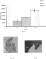
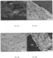
- Femurs used for microCT and mechanical bending tests were taken to histological processing and analysis. Femurs sections were stained with picro serious red stain to highlight new formed bone ( Figure 7A, B ). The histological sections were subjected to histomorphometry and measurements are shown and summarized in Figure 7 , Histomorphometry data reveals that the amount of new formed bone is with concordance to cell number and to the time from transplantation. Scaffold (30 mg of mineral particle) and 5 ⁇ 10 6 cells group surpassed Scaffold (30 mg of mineral particle) and 3 ⁇ 10 6 cells group and significantly surpassed the Scaffold only group in terms of the amount of new bone formed. Moreover, it can be seen that after 16 weeks the amount of grown bone was higher than of the other two groups.
- the proposed product is based on bone mineral particles seeded with multicell Adipose Tissue derived Cells and suspended in hyaluronic acid.
- Histological and scanning electron microscope analysis shows the formation of multicellular layers on top of the bone mineral particles. These layers generate a typical connective tissue structure containing collagen fibers and polysaccharides as seen in Picro Sirius Red stain and Toluidine Blue stain, respectively ( Figure 10 ).
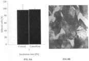
- Adipose derived endothelial cells and mesenchymal stem cells were seeded as a co-culture on the scaffold.
- seeded particles were placed in Matrigel for predetermined time points.
- Adipose Tissue derived endothelial cells were shown to be able to attach to the Matrigel and to form sprouts out of the scaffold and out to the surrounding ( Figure 11 ).
Landscapes
- Health & Medical Sciences (AREA)
- Life Sciences & Earth Sciences (AREA)
- Chemical & Material Sciences (AREA)
- Engineering & Computer Science (AREA)
- Biomedical Technology (AREA)
- Public Health (AREA)
- General Health & Medical Sciences (AREA)
- Veterinary Medicine (AREA)
- Medicinal Chemistry (AREA)
- Epidemiology (AREA)
- Animal Behavior & Ethology (AREA)
- Dermatology (AREA)
- Cell Biology (AREA)
- Oral & Maxillofacial Surgery (AREA)
- Transplantation (AREA)
- Zoology (AREA)
- Chemical Kinetics & Catalysis (AREA)
- Botany (AREA)
- Developmental Biology & Embryology (AREA)
- Urology & Nephrology (AREA)
- Immunology (AREA)
- Pharmacology & Pharmacy (AREA)
- Molecular Biology (AREA)
- Hematology (AREA)
- Inorganic Chemistry (AREA)
- Virology (AREA)
- Biotechnology (AREA)
- Orthopedic Medicine & Surgery (AREA)
- Vascular Medicine (AREA)
- Composite Materials (AREA)
- Materials Engineering (AREA)
- Materials For Medical Uses (AREA)
- Micro-Organisms Or Cultivation Processes Thereof (AREA)
- Medicines Containing Material From Animals Or Micro-Organisms (AREA)
- Polymers & Plastics (AREA)
- Organic Chemistry (AREA)
Claims (14)
- Zusammensetzung, umfassend: (a) ein mineralisches Teilchen mit einem Durchmesser im Bereich von 50 Mikrometern bis 2.000 Mikrometern, (b) eine 3D-Zellkultur, die Endothelzellen und Mesenchymzellen, die in zwei oder mehr Zellschichten organisiert sind, umfasst, wobei die zwei oder mehr Zellschichten an das mineralische Teilchen gebunden sind, (c) Hyaluronsäure und optional ein biokompatibles Bindemittel; wobei die zwei oder mehr an das mineralische Teilchen gebundenen Zellschichten in der Hyaluronsäure suspendiert sind, wobei die Zusammensetzung mindestens 1×10³ der Endothelzellen und Mesenchymzellen pro 1 mg des mineralischen Teilchens umfasst.
- Zusammensetzung nach Anspruch 1, wobei die Zusammensetzung außerdem ein biokompatibles Bindemittel enthält.
- Zusammensetzung nach Anspruch 1, bei der die Endothelzellen und Mesenchymzellen in einem komplexen Netzwerk aus nanoskaligen extrazellulären Matrixfasern gehalten sind, das die Schaffung verschiedener lokaler Mikroumgebungen ermöglicht.
- Zusammensetzung nach Anspruch 2, bei der das biokompatible Bindemittel Albumin ist.
- Zusammensetzung nach Anspruch 1, die aus Fettgewebe abgeleitete Zellen enthält.
- Zusammensetzung nach Anspruch 1, bei der das Mineralteilchen ein Knochenmineralteilchen ist.
- Zusammensetzung nach Anspruch 1, bei der die Mesenchymzellen Mesenchym-Stammzellen umfassen.
- Zusammensetzung nach Anspruch 1, bei der das Mineralteilchen umfasst: ein Calciumphosphatderivat, ein Calciumsulfatderivat, Calciumhydroxyapatit, ein Silikat-Mineral-Hydroxyapatit, Beta-3-Calciumphosphat oder eine beliebige Kombination davon.
- Zusammensetzung nach Anspruch 1, bei der das Mineralteilchen einen Durchmesser im Bereich von 100 Mikrometern bis 1000 Mikrometern hat.
- Zusammensetzung nach Anspruch 5, umfassend 20 % bis 60 % v/v der Endothelzellen und Mesenchymzellen, der aus Fettgewebe abgeleiteten Zellen oder von beiden.
- Zusammensetzung nach Anspruch 1, die 2 bis 16 Tage lang in vitro gehalten worden ist.
- Kit, umfassend: eine Spritze und die Zusammensetzung nach Anspruch 1.
- Kit nach Anspruch 12, das außerdem eine Mischvorrichtung umfasst.
- Zusammensetzung nach Anspruch 1 zur Verwendung beim Füllen einer Lücke in einem Knochen eines Patienten, der dies benötigt.
Applications Claiming Priority (2)
| Application Number | Priority Date | Filing Date | Title |
|---|---|---|---|
| US201461977691P | 2014-04-10 | 2014-04-10 | |
| PCT/IL2015/050380 WO2015155777A1 (en) | 2014-04-10 | 2015-04-09 | Bone repair compositions |
Publications (4)
| Publication Number | Publication Date |
|---|---|
| EP3129075A1 EP3129075A1 (de) | 2017-02-15 |
| EP3129075A4 EP3129075A4 (de) | 2017-12-06 |
| EP3129075B1 true EP3129075B1 (de) | 2024-09-18 |
| EP3129075C0 EP3129075C0 (de) | 2024-09-18 |
Family
ID=54287396
Family Applications (1)
| Application Number | Title | Priority Date | Filing Date |
|---|---|---|---|
| EP15777375.5A Active EP3129075B1 (de) | 2014-04-10 | 2015-04-09 | Knochenreparaturzusammensetzungen |
Country Status (5)
| Country | Link |
|---|---|
| US (2) | US11433163B2 (de) |
| EP (1) | EP3129075B1 (de) |
| AU (1) | AU2015245963B2 (de) |
| CA (1) | CA2945438C (de) |
| WO (1) | WO2015155777A1 (de) |
Families Citing this family (4)
| Publication number | Priority date | Publication date | Assignee | Title |
|---|---|---|---|---|
| US9861482B2 (en) * | 2008-08-26 | 2018-01-09 | Andy Boiangiu | Dental bone implant and implant method |
| EP3129075B1 (de) * | 2014-04-10 | 2024-09-18 | Bonus Therapeutics Ltd. | Knochenreparaturzusammensetzungen |
| US20200268938A1 (en) * | 2016-04-01 | 2020-08-27 | Hs Pharmaceuticals, Llc | Novel bone putty compositions and methods of use thereof |
| KR20190038833A (ko) * | 2016-07-11 | 2019-04-09 | 보누스 테라퓨틱스 리미티드 | 조직 재생용 세포 조성물 |
Family Cites Families (59)
| Publication number | Priority date | Publication date | Assignee | Title |
|---|---|---|---|---|
| NL154600B (nl) | 1971-02-10 | 1977-09-15 | Organon Nv | Werkwijze voor het aantonen en bepalen van specifiek bindende eiwitten en hun corresponderende bindbare stoffen. |
| NL154598B (nl) | 1970-11-10 | 1977-09-15 | Organon Nv | Werkwijze voor het aantonen en bepalen van laagmoleculire verbindingen en van eiwitten die deze verbindingen specifiek kunnen binden, alsmede testverpakking. |
| NL154599B (nl) | 1970-12-28 | 1977-09-15 | Organon Nv | Werkwijze voor het aantonen en bepalen van specifiek bindende eiwitten en hun corresponderende bindbare stoffen, alsmede testverpakking. |
| US3901654A (en) | 1971-06-21 | 1975-08-26 | Biological Developments | Receptor assays of biologically active compounds employing biologically specific receptors |
| US3853987A (en) | 1971-09-01 | 1974-12-10 | W Dreyer | Immunological reagent and radioimmuno assay |
| US3867517A (en) | 1971-12-21 | 1975-02-18 | Abbott Lab | Direct radioimmunoassay for antigens and their antibodies |
| NL171930C (nl) | 1972-05-11 | 1983-06-01 | Akzo Nv | Werkwijze voor het aantonen en bepalen van haptenen, alsmede testverpakkingen. |
| US3850578A (en) | 1973-03-12 | 1974-11-26 | H Mcconnell | Process for assaying for biologically active molecules |
| US3935074A (en) | 1973-12-17 | 1976-01-27 | Syva Company | Antibody steric hindrance immunoassay with two antibodies |
| US3996345A (en) | 1974-08-12 | 1976-12-07 | Syva Company | Fluorescence quenching with immunological pairs in immunoassays |
| US4034074A (en) | 1974-09-19 | 1977-07-05 | The Board Of Trustees Of Leland Stanford Junior University | Universal reagent 2-site immunoradiometric assay using labelled anti (IgG) |
| US3984533A (en) | 1975-11-13 | 1976-10-05 | General Electric Company | Electrophoretic method of detecting antigen-antibody reaction |
| US4186448A (en) | 1976-04-16 | 1980-02-05 | Brekke John H | Device and method for treating and healing a newly created bone void |
| US4098876A (en) | 1976-10-26 | 1978-07-04 | Corning Glass Works | Reverse sandwich immunoassay |
| US4879219A (en) | 1980-09-19 | 1989-11-07 | General Hospital Corporation | Immunoassay utilizing monoclonal high affinity IgM antibodies |
| US5011771A (en) | 1984-04-12 | 1991-04-30 | The General Hospital Corporation | Multiepitopic immunometric assay |
| US4666828A (en) | 1984-08-15 | 1987-05-19 | The General Hospital Corporation | Test for Huntington's disease |
| US4683202A (en) | 1985-03-28 | 1987-07-28 | Cetus Corporation | Process for amplifying nucleic acid sequences |
| US4801531A (en) | 1985-04-17 | 1989-01-31 | Biotechnology Research Partners, Ltd. | Apo AI/CIII genomic polymorphisms predictive of atherosclerosis |
| US5133755A (en) | 1986-01-28 | 1992-07-28 | Thm Biomedical, Inc. | Method and apparatus for diodegradable, osteogenic, bone graft substitute device |
| CA1340581C (en) | 1986-11-20 | 1999-06-08 | Joseph P. Vacanti | Chimeric neomorphogenesis of organs by controlled cellular implantation using artificial matrices |
| GB2215209B (en) | 1988-03-14 | 1992-08-26 | Osmed Inc | Method and apparatus for biodegradable, osteogenic, bone graft substitute device |
| DE3810803A1 (de) | 1988-03-30 | 1989-10-12 | Battelle Institut E V | Verfahren zur herstellung eines synthetischen knochenmaterials mit koerpereigenen eigenschaften |
| US5272057A (en) | 1988-10-14 | 1993-12-21 | Georgetown University | Method of detecting a predisposition to cancer by the use of restriction fragment length polymorphism of the gene for human poly (ADP-ribose) polymerase |
| US5192659A (en) | 1989-08-25 | 1993-03-09 | Genetype Ag | Intron sequence analysis method for detection of adjacent and remote locus alleles as haplotypes |
| JPH07503869A (ja) | 1992-02-14 | 1995-04-27 | ボード・オヴ・リージェンツ,ザ・ユニヴァーシティ・オヴ・テキサス・システム | 多相生侵食性の移植材料または担体並びにその製造および使用方法 |
| US5281521A (en) | 1992-07-20 | 1994-01-25 | The Trustees Of The University Of Pennsylvania | Modified avidin-biotin technique |
| US5514378A (en) | 1993-02-01 | 1996-05-07 | Massachusetts Institute Of Technology | Biocompatible polymer membranes and methods of preparation of three dimensional membrane structures |
| US5522895A (en) | 1993-07-23 | 1996-06-04 | Rice University | Biodegradable bone templates |
| EP0713364A4 (de) | 1993-08-13 | 1996-12-27 | Shalaby W Shalaby | Mikroporöse polymere schäume und mikrostrukturierte oberflächen |
| US5686091A (en) | 1994-03-28 | 1997-11-11 | The Johns Hopkins University School Of Medicine | Biodegradable foams for cell transplantation |
| US5769899A (en) | 1994-08-12 | 1998-06-23 | Matrix Biotechnologies, Inc. | Cartilage repair unit |
| US5716616A (en) | 1995-03-28 | 1998-02-10 | Thomas Jefferson University | Isolated stromal cells for treating diseases, disorders or conditions characterized by bone defects |
| US6132463A (en) | 1995-05-19 | 2000-10-17 | Etex Corporation | Cell seeding of ceramic compositions |
| US5716413A (en) | 1995-10-11 | 1998-02-10 | Osteobiologics, Inc. | Moldable, hand-shapable biodegradable implant material |
| US6200606B1 (en) | 1996-01-16 | 2001-03-13 | Depuy Orthopaedics, Inc. | Isolation of precursor cells from hematopoietic and nonhematopoietic tissues and their use in vivo bone and cartilage regeneration |
| ES2329953T3 (es) | 1996-04-19 | 2009-12-02 | Osiris Therapeutics, Inc. | Regeneracion e incremento de hueso utilizando celulas madre mesenquimales. |
| US5824084A (en) | 1996-07-03 | 1998-10-20 | The Cleveland Clinic Foundation | Method of preparing a composite bone graft |
| US20030114936A1 (en) | 1998-10-12 | 2003-06-19 | Therics, Inc. | Complex three-dimensional composite scaffold resistant to delimination |
| BR0009403A (pt) | 1999-02-04 | 2001-11-27 | Technion Res & Dev Foundation | Método de expansão/conservação das células detronco hemopoiéticas indiferenciadas ou dascélulas progenitoras, método de preparação deum meio condicionado de célula estomacal útil naexpansão/conservação das células de troncohemopoiéticas indiferenciadas ou das célulasprogenitoras, método de transplante de célulasde tronco hemopoiéticas indiferenciadas ou decélulas progenitoras em um recipiente, tampão debiorreator e biorreator |
| US6333029B1 (en) | 1999-06-30 | 2001-12-25 | Ethicon, Inc. | Porous tissue scaffoldings for the repair of regeneration of tissue |
| DE19956503A1 (de) | 1999-11-24 | 2001-06-21 | Universitaetsklinikum Freiburg | Spritzbares Knochenersatzmaterial |
| US6811776B2 (en) | 2000-12-27 | 2004-11-02 | The Regents Of The University Of Michigan | Process for ex vivo formation of mammalian bone and uses thereof |
| WO2002019887A2 (en) | 2000-09-05 | 2002-03-14 | Technion Research And Development Foundation Ltd. | Methods of repairing longitudinal bone defects |
| US6852330B2 (en) | 2000-12-21 | 2005-02-08 | Depuy Mitek, Inc. | Reinforced foam implants with enhanced integrity for soft tissue repair and regeneration |
| US20050074877A1 (en) | 2003-07-28 | 2005-04-07 | Mao Jeremy Jian | Biological engineering of articular structures containing both cartilage and bone |
| US11395865B2 (en) | 2004-02-09 | 2022-07-26 | DePuy Synthes Products, Inc. | Scaffolds with viable tissue |
| US20070190101A1 (en) * | 2004-03-31 | 2007-08-16 | Chunlin Yang | Flowable bone grafts |
| EP1868660B1 (de) | 2005-04-04 | 2016-08-03 | Technion Research & Development Foundation Limited | Medizinisches gerüst, verfahren zu seiner herstellung und seiner verwendung |
| DE602006014534D1 (de) * | 2005-05-30 | 2010-07-08 | Commw Scient Ind Res Org | Herstellung und verwendung von basalmembranpartikeln |
| ES2376222T3 (es) | 2006-03-30 | 2012-03-12 | Mark Clarke | Construcciones óseas tridimensionales mineralizadas. |
| TW200817019A (en) | 2006-07-10 | 2008-04-16 | Univ Columbia | De novo formation and regeneration of vascularized tissue from tissue progenitor cells and vascular progenitor cells |
| US20120028352A1 (en) * | 2008-03-17 | 2012-02-02 | Agency For Science, Technology And Research | Microcarriers for Stem Cell Culture |
| DK2274023T3 (da) * | 2008-04-10 | 2020-03-16 | Bonus Therapeutics Ltd | Knoglelignende proteseimplantater |
| BE1018748A3 (fr) * | 2008-05-07 | 2011-08-02 | Bone Therapeutics Sa | Nouvelles cellules souches mesenchymateuses et cellules osteogeniques . |
| WO2012134540A2 (en) * | 2010-10-22 | 2012-10-04 | Vanderbilt University | Injectable synthetic pur composite |
| US20140086891A1 (en) * | 2011-01-24 | 2014-03-27 | Progenicare, Llc | Compositions and methods for promoting tissue regeneration |
| US20130116190A1 (en) * | 2011-09-06 | 2013-05-09 | Allergan, Inc. | Hyaluronic acid-collagen matrices for tissue engineering |
| EP3129075B1 (de) * | 2014-04-10 | 2024-09-18 | Bonus Therapeutics Ltd. | Knochenreparaturzusammensetzungen |
-
2015
- 2015-04-09 EP EP15777375.5A patent/EP3129075B1/de active Active
- 2015-04-09 US US15/303,011 patent/US11433163B2/en active Active
- 2015-04-09 WO PCT/IL2015/050380 patent/WO2015155777A1/en not_active Ceased
- 2015-04-09 AU AU2015245963A patent/AU2015245963B2/en active Active
- 2015-04-09 CA CA2945438A patent/CA2945438C/en active Active
-
2022
- 2022-07-07 US US17/859,452 patent/US20230001053A1/en not_active Abandoned
Also Published As
| Publication number | Publication date |
|---|---|
| AU2015245963B2 (en) | 2019-02-28 |
| US20230001053A1 (en) | 2023-01-05 |
| EP3129075A4 (de) | 2017-12-06 |
| CA2945438C (en) | 2023-08-22 |
| EP3129075C0 (de) | 2024-09-18 |
| AU2015245963A1 (en) | 2016-10-27 |
| EP3129075A1 (de) | 2017-02-15 |
| CA2945438A1 (en) | 2015-10-15 |
| WO2015155777A1 (en) | 2015-10-15 |
| US20170028104A1 (en) | 2017-02-02 |
| US11433163B2 (en) | 2022-09-06 |
Similar Documents
| Publication | Publication Date | Title |
|---|---|---|
| Wei et al. | Mesenchymal stem cell-loaded porous tantalum integrated with biomimetic 3D collagen-based scaffold to repair large osteochondral defects in goats | |
| Bruder et al. | The effect of implants loaded with autologous mesenchymal stem cells on the healing of canine segmental bone defects | |
| US20230001053A1 (en) | Bone repair compositions | |
| Janicki et al. | What should be the characteristics of the ideal bone graft substitute? Combining scaffolds with growth factors and/or stem cells | |
| Amini et al. | Bone tissue engineering: recent advances and challenges | |
| Arinzeh et al. | Allogeneic mesenchymal stem cells regenerate bone in a critical-sized canine segmental defect | |
| Tang et al. | Posterolateral spinal fusion with nano‐hydroxyapatite–collagen/PLA composite and autologous adipose‐derived mesenchymal stem cells in a rabbit model | |
| S Shekkeris et al. | Clinical applications of mesenchymal stem cells in the treatment of fracture non-union and bone defects | |
| Kanczler et al. | Bone tissue engineering and bone regeneration | |
| WO2005105992A1 (en) | Chondrocyte culture formulations | |
| US20100003222A1 (en) | Methods for bone regeneration using endothelial progenitor cell preparations | |
| Gamie et al. | Skeletal tissue engineering using mesenchymal or embryonic stem cells: clinical and experimental data | |
| Wang et al. | Sheet of osteoblastic cells combined with platelet-rich fibrin improves the formation of bone in critical-size calvarial defects in rabbits | |
| KR102729200B1 (ko) | 지방 유래 줄기세포를 포함하는 생체재료 및 이를 생산하는 방법 | |
| Yang et al. | Combination therapy with BMSCs‑exosomes and porous tantalum for the repair of femur supracondylar defects | |
| US10994050B2 (en) | High yield and high precision bone graft substitute from stem cells | |
| US20200338031A1 (en) | Compositions for the treatment of non-articular cartilage-associated bone conditions | |
| Kasper et al. | Flow perfusion culture of mesenchymal stem cells for bone tissue engineering | |
| Yoshizato et al. | A scaffold-free cartilage construct fabricated using a bio 3D printer accelerates critical-size bone defect regeneration | |
| Yamazaki et al. | Integrated Bone Formation Through In Vivo Endochondral Ossification Using Mesenchymal Stem Cells | |
| Gugjoo | Mesenchymal stem cells therapeutic applications in bone regeneration | |
| WO2012048755A1 (en) | Er-alpha-17p peptide, effects on stem cell differentiation | |
| Ding et al. | 3D perfusion bioreactor‐activated porous granules on implant fixation and early bone formation in sheep | |
| Sharma et al. | Bone tissue engineering | |
| Wang et al. | Extracellular matrix–derived tissues for hard tissue repair |
Legal Events
| Date | Code | Title | Description |
|---|---|---|---|
| STAA | Information on the status of an ep patent application or granted ep patent |
Free format text: STATUS: THE INTERNATIONAL PUBLICATION HAS BEEN MADE |
|
| PUAI | Public reference made under article 153(3) epc to a published international application that has entered the european phase |
Free format text: ORIGINAL CODE: 0009012 |
|
| STAA | Information on the status of an ep patent application or granted ep patent |
Free format text: STATUS: REQUEST FOR EXAMINATION WAS MADE |
|
| 17P | Request for examination filed |
Effective date: 20161010 |
|
| AK | Designated contracting states |
Kind code of ref document: A1 Designated state(s): AL AT BE BG CH CY CZ DE DK EE ES FI FR GB GR HR HU IE IS IT LI LT LU LV MC MK MT NL NO PL PT RO RS SE SI SK SM TR |
|
| AX | Request for extension of the european patent |
Extension state: BA ME |
|
| DAV | Request for validation of the european patent (deleted) | ||
| DAX | Request for extension of the european patent (deleted) | ||
| A4 | Supplementary search report drawn up and despatched |
Effective date: 20171107 |
|
| RIC1 | Information provided on ipc code assigned before grant |
Ipc: C12M 1/00 20060101ALI20171030BHEP Ipc: A61K 35/28 20150101ALI20171030BHEP Ipc: A61K 35/12 20150101ALI20171030BHEP Ipc: A61L 27/38 20060101AFI20171030BHEP Ipc: A61K 31/728 20060101ALI20171030BHEP |
|
| STAA | Information on the status of an ep patent application or granted ep patent |
Free format text: STATUS: EXAMINATION IS IN PROGRESS |
|
| 17Q | First examination report despatched |
Effective date: 20190903 |
|
| GRAP | Despatch of communication of intention to grant a patent |
Free format text: ORIGINAL CODE: EPIDOSNIGR1 |
|
| STAA | Information on the status of an ep patent application or granted ep patent |
Free format text: STATUS: GRANT OF PATENT IS INTENDED |
|
| INTG | Intention to grant announced |
Effective date: 20240430 |
|
| GRAS | Grant fee paid |
Free format text: ORIGINAL CODE: EPIDOSNIGR3 |
|
| GRAA | (expected) grant |
Free format text: ORIGINAL CODE: 0009210 |
|
| STAA | Information on the status of an ep patent application or granted ep patent |
Free format text: STATUS: THE PATENT HAS BEEN GRANTED |
|
| AK | Designated contracting states |
Kind code of ref document: B1 Designated state(s): AL AT BE BG CH CY CZ DE DK EE ES FI FR GB GR HR HU IE IS IT LI LT LU LV MC MK MT NL NO PL PT RO RS SE SI SK SM TR |
|
| REG | Reference to a national code |
Ref country code: GB Ref legal event code: FG4D |
|
| REG | Reference to a national code |
Ref country code: CH Ref legal event code: EP |
|
| REG | Reference to a national code |
Ref country code: IE Ref legal event code: FG4D |
|
| REG | Reference to a national code |
Ref country code: DE Ref legal event code: R096 Ref document number: 602015089935 Country of ref document: DE |
|
| U01 | Request for unitary effect filed |
Effective date: 20241015 |
|
| U07 | Unitary effect registered |
Designated state(s): AT BE BG DE DK EE FI FR IT LT LU LV MT NL PT RO SE SI Effective date: 20241031 |
|
| PG25 | Lapsed in a contracting state [announced via postgrant information from national office to epo] |
Ref country code: NO Free format text: LAPSE BECAUSE OF FAILURE TO SUBMIT A TRANSLATION OF THE DESCRIPTION OR TO PAY THE FEE WITHIN THE PRESCRIBED TIME-LIMIT Effective date: 20241218 |
|
| PG25 | Lapsed in a contracting state [announced via postgrant information from national office to epo] |
Ref country code: GR Free format text: LAPSE BECAUSE OF FAILURE TO SUBMIT A TRANSLATION OF THE DESCRIPTION OR TO PAY THE FEE WITHIN THE PRESCRIBED TIME-LIMIT Effective date: 20241219 |
|
| PG25 | Lapsed in a contracting state [announced via postgrant information from national office to epo] |
Ref country code: HR Free format text: LAPSE BECAUSE OF FAILURE TO SUBMIT A TRANSLATION OF THE DESCRIPTION OR TO PAY THE FEE WITHIN THE PRESCRIBED TIME-LIMIT Effective date: 20240918 |
|
| PG25 | Lapsed in a contracting state [announced via postgrant information from national office to epo] |
Ref country code: RS Free format text: LAPSE BECAUSE OF FAILURE TO SUBMIT A TRANSLATION OF THE DESCRIPTION OR TO PAY THE FEE WITHIN THE PRESCRIBED TIME-LIMIT Effective date: 20241218 |
|
| PG25 | Lapsed in a contracting state [announced via postgrant information from national office to epo] |
Ref country code: RS Free format text: LAPSE BECAUSE OF FAILURE TO SUBMIT A TRANSLATION OF THE DESCRIPTION OR TO PAY THE FEE WITHIN THE PRESCRIBED TIME-LIMIT Effective date: 20241218 Ref country code: NO Free format text: LAPSE BECAUSE OF FAILURE TO SUBMIT A TRANSLATION OF THE DESCRIPTION OR TO PAY THE FEE WITHIN THE PRESCRIBED TIME-LIMIT Effective date: 20241218 Ref country code: HR Free format text: LAPSE BECAUSE OF FAILURE TO SUBMIT A TRANSLATION OF THE DESCRIPTION OR TO PAY THE FEE WITHIN THE PRESCRIBED TIME-LIMIT Effective date: 20240918 Ref country code: GR Free format text: LAPSE BECAUSE OF FAILURE TO SUBMIT A TRANSLATION OF THE DESCRIPTION OR TO PAY THE FEE WITHIN THE PRESCRIBED TIME-LIMIT Effective date: 20241219 |
|
| PG25 | Lapsed in a contracting state [announced via postgrant information from national office to epo] |
Ref country code: IS Free format text: LAPSE BECAUSE OF FAILURE TO SUBMIT A TRANSLATION OF THE DESCRIPTION OR TO PAY THE FEE WITHIN THE PRESCRIBED TIME-LIMIT Effective date: 20250118 |
|
| PG25 | Lapsed in a contracting state [announced via postgrant information from national office to epo] |
Ref country code: SM Free format text: LAPSE BECAUSE OF FAILURE TO SUBMIT A TRANSLATION OF THE DESCRIPTION OR TO PAY THE FEE WITHIN THE PRESCRIBED TIME-LIMIT Effective date: 20240918 |
|
| PG25 | Lapsed in a contracting state [announced via postgrant information from national office to epo] |
Ref country code: ES Free format text: LAPSE BECAUSE OF FAILURE TO SUBMIT A TRANSLATION OF THE DESCRIPTION OR TO PAY THE FEE WITHIN THE PRESCRIBED TIME-LIMIT Effective date: 20240918 |
|
| PG25 | Lapsed in a contracting state [announced via postgrant information from national office to epo] |
Ref country code: PL Free format text: LAPSE BECAUSE OF FAILURE TO SUBMIT A TRANSLATION OF THE DESCRIPTION OR TO PAY THE FEE WITHIN THE PRESCRIBED TIME-LIMIT Effective date: 20240918 Ref country code: CZ Free format text: LAPSE BECAUSE OF FAILURE TO SUBMIT A TRANSLATION OF THE DESCRIPTION OR TO PAY THE FEE WITHIN THE PRESCRIBED TIME-LIMIT Effective date: 20240918 |
|
| PG25 | Lapsed in a contracting state [announced via postgrant information from national office to epo] |
Ref country code: SK Free format text: LAPSE BECAUSE OF FAILURE TO SUBMIT A TRANSLATION OF THE DESCRIPTION OR TO PAY THE FEE WITHIN THE PRESCRIBED TIME-LIMIT Effective date: 20240918 |
|
| U20 | Renewal fee for the european patent with unitary effect paid |
Year of fee payment: 11 Effective date: 20250424 |
|
| PLBE | No opposition filed within time limit |
Free format text: ORIGINAL CODE: 0009261 |
|
| STAA | Information on the status of an ep patent application or granted ep patent |
Free format text: STATUS: NO OPPOSITION FILED WITHIN TIME LIMIT |
|
| 26N | No opposition filed |
Effective date: 20250619 |
|
| REG | Reference to a national code |
Ref country code: CH Ref legal event code: H13 Free format text: ST27 STATUS EVENT CODE: U-0-0-H10-H13 (AS PROVIDED BY THE NATIONAL OFFICE) Effective date: 20251125 |
|
| PG25 | Lapsed in a contracting state [announced via postgrant information from national office to epo] |
Ref country code: MC Free format text: LAPSE BECAUSE OF FAILURE TO SUBMIT A TRANSLATION OF THE DESCRIPTION OR TO PAY THE FEE WITHIN THE PRESCRIBED TIME-LIMIT Effective date: 20240918 |
|
| PG25 | Lapsed in a contracting state [announced via postgrant information from national office to epo] |
Ref country code: CH Free format text: LAPSE BECAUSE OF NON-PAYMENT OF DUE FEES Effective date: 20250430 |
|
| PGFP | Annual fee paid to national office [announced via postgrant information from national office to epo] |
Ref country code: GB Payment date: 20260327 Year of fee payment: 12 |
|
| PG25 | Lapsed in a contracting state [announced via postgrant information from national office to epo] |
Ref country code: IE Free format text: LAPSE BECAUSE OF NON-PAYMENT OF DUE FEES Effective date: 20250409 |