EP3150694A1 - Dispositif, procédé, et programme d'évaluation de cellules - Google Patents
Dispositif, procédé, et programme d'évaluation de cellules Download PDFInfo
- Publication number
- EP3150694A1 EP3150694A1 EP15799811.3A EP15799811A EP3150694A1 EP 3150694 A1 EP3150694 A1 EP 3150694A1 EP 15799811 A EP15799811 A EP 15799811A EP 3150694 A1 EP3150694 A1 EP 3150694A1
- Authority
- EP
- European Patent Office
- Prior art keywords
- cell
- evaluation
- image
- cells
- boundary
- Prior art date
- Legal status (The legal status is an assumption and is not a legal conclusion. Google has not performed a legal analysis and makes no representation as to the accuracy of the status listed.)
- Granted
Links
Images





Classifications
-
- G—PHYSICS
- G06—COMPUTING OR CALCULATING; COUNTING
- G06T—IMAGE DATA PROCESSING OR GENERATION, IN GENERAL
- G06T7/00—Image analysis
- G06T7/0002—Inspection of images, e.g. flaw detection
- G06T7/0012—Biomedical image inspection
-
- C—CHEMISTRY; METALLURGY
- C12—BIOCHEMISTRY; BEER; SPIRITS; WINE; VINEGAR; MICROBIOLOGY; ENZYMOLOGY; MUTATION OR GENETIC ENGINEERING
- C12Q—MEASURING OR TESTING PROCESSES INVOLVING ENZYMES, NUCLEIC ACIDS OR MICROORGANISMS; COMPOSITIONS OR TEST PAPERS THEREFOR; PROCESSES OF PREPARING SUCH COMPOSITIONS; CONDITION-RESPONSIVE CONTROL IN MICROBIOLOGICAL OR ENZYMOLOGICAL PROCESSES
- C12Q1/00—Measuring or testing processes involving enzymes, nucleic acids or microorganisms; Compositions therefor; Processes of preparing such compositions
- C12Q1/02—Measuring or testing processes involving enzymes, nucleic acids or microorganisms; Compositions therefor; Processes of preparing such compositions involving viable microorganisms
-
- C—CHEMISTRY; METALLURGY
- C12—BIOCHEMISTRY; BEER; SPIRITS; WINE; VINEGAR; MICROBIOLOGY; ENZYMOLOGY; MUTATION OR GENETIC ENGINEERING
- C12M—APPARATUS FOR ENZYMOLOGY OR MICROBIOLOGY; APPARATUS FOR CULTURING MICROORGANISMS FOR PRODUCING BIOMASS, FOR GROWING CELLS OR FOR OBTAINING FERMENTATION OR METABOLIC PRODUCTS, i.e. BIOREACTORS OR FERMENTERS
- C12M41/00—Means for regulation, monitoring, measurement or control, e.g. flow regulation
- C12M41/30—Means for regulation, monitoring, measurement or control, e.g. flow regulation of concentration
- C12M41/36—Means for regulation, monitoring, measurement or control, e.g. flow regulation of concentration of biomass, e.g. colony counters or by turbidity measurements
-
- G—PHYSICS
- G06—COMPUTING OR CALCULATING; COUNTING
- G06T—IMAGE DATA PROCESSING OR GENERATION, IN GENERAL
- G06T2207/00—Indexing scheme for image analysis or image enhancement
- G06T2207/10—Image acquisition modality
- G06T2207/10056—Microscopic image
-
- G—PHYSICS
- G06—COMPUTING OR CALCULATING; COUNTING
- G06T—IMAGE DATA PROCESSING OR GENERATION, IN GENERAL
- G06T2207/00—Indexing scheme for image analysis or image enhancement
- G06T2207/30—Subject of image; Context of image processing
- G06T2207/30004—Biomedical image processing
- G06T2207/30024—Cell structures in vitro; Tissue sections in vitro
Definitions
- the present invention relates to a cell evaluation device, a cell evaluation method, and a cell evaluation program that evaluate individual cells in a cell image obtained by imaging a cell group.
- a method for culturing multipotential stem cells such as ES cells, iPS cells or STAP cells, or cells differentiated and induced therefrom or the like, imaging the cells using a microscope, and evaluating a state of the cells by grasping characteristics of an image obtained by the imaging has been proposed.
- the cultured cells are colonized according to the progress of the culture, and are proliferated into a large area.
- the size of a cell is an order of micrometers, and the size of a colony is an order of several millimeters to several centimeters.
- JP2012-231709A discloses a method for comparing an inner optical path length of a cell nucleus with an outer optical path length thereof for an individual cell and evaluating whether the individual cell is a good stem cell based on the comparison result. Further, JP2012-231709A discloses a technique for setting a region where good stem cells in a cell colony are crowded as a good quality region.
- JP2014-39504A discloses a method for determining whether individual cells are nucleated erythrocytes (NRBCs) based on feature amounts calculated from images of the individual cells and learning parameters which are learned in advance.
- NRBCs nucleated erythrocytes
- FIGs. 13A and 13B are enlarged views of a part of a cell image of a colonized cell group.
- a range surrounded by a solid line of Fig. 13A and a range surrounded by a broken line of Fig. 13B correspond to individual cells.
- cells having similar shapes tend to be collected nearby.
- evaluation results of peripheral cells of the evaluation target cell are referenced.
- both the cells in the differentiated state and the cells in the undifferentiated state are present as peripheral cells of the evaluation target cell, and thus, there is a case where an undifferentiated state or differentiated state of the evaluation target cell is not appropriately evaluated.
- an object of the invention is to provide a cell evaluation device, a cell evaluation method, and a cell evaluation program capable of evaluating, when evaluating a predetermined evaluation target cell using evaluation results of peripheral cells thereof, the evaluation target cell with high accuracy.
- a cell evaluation device including: an image acquisition unit that acquires a cell image obtained by imaging a cell group; a cell evaluation unit that specifies an evaluation target cell and peripheral cells around the evaluation target cell in the cell group, and evaluates the evaluation target cell based on evaluation results of the peripheral cells; and a boundary setting unit that sets a boundary in the cell image based on a state of the cell group, in which when specifying the peripheral cells, the cell evaluation unit is capable of specifying only cells are present in a divided region where the evaluation target cell is present among a plurality of divided regions divided by the boundary as the peripheral cells.
- the cell evaluation device may further include: an image pick-up condition acquisition unit that acquires an image pick-up condition different from an image pick-up condition of the cell image used in the evaluation in a case where it is determined that the evaluation is impossible to be performed when evaluating the evaluation target cell based on the evaluation results of the peripheral cells in the cell evaluation unit.
- the image pick-up condition may include at least one of an image pick-up region, an exposure time, an optical magnification, an illumination light wavelength, or an observation light wavelength.
- the cell evaluation unit may add identification information to a region where the peripheral cells specified when evaluating the evaluation target cell are present.
- the image pick-up condition acquisition unit may acquire the image pick-up condition based on the cell image of the region to which the identification information is added.
- the cell evaluation device may further include: a display controller that performs a control for displaying the image pick-up condition acquired by the image pick-up condition acquisition unit.
- the cell evaluation device may further include: an image pick-up controller that outputs an imaging control signal according to the image pick-up condition acquired by the image pick-up condition acquisition unit.
- the boundary setting unit may set the boundary based on at least one of a luminance, a spatial frequency, or a color of the cell image.
- the boundary setting unit may set the boundary of which the number of inflection points or a curvature is limited.
- a cell evaluation method including: acquiring a cell image obtained by imaging a cell group; and when specifying an evaluation target cell and peripheral cells around the evaluation target cell in the cell group and evaluating the evaluation target cell based on evaluation results of the peripheral cells, setting a boundary in the cell image based on a state of the cell group, and specifying only cells that are present in a divided region where the evaluation target cell is present among a plurality of divided regions divided by the boundary, as the peripheral cells.
- a cell evaluation program that causes a computer to function as: an image acquisition unit that acquires a cell image obtained by imaging a cell group; a cell evaluation unit that specifies an evaluation target cell and peripheral cells around the evaluation target cell in the cell group, and evaluates the evaluation target cell based on evaluation results of the peripheral cells; and a boundary setting unit that sets a boundary in the cell image based on a state of the cell group, in which when specifying the peripheral cells, the cell evaluation unit specifies only cells that are present in a divided region where the evaluation target cell is present among a plurality of divided regions divided by the boundary, as the peripheral cells.
- a cell image obtained by imaging a cell group is acquired; and when specifying an evaluation target cell and peripheral cells around the evaluation target cell in the cell group and evaluating the evaluation target cell based on evaluation results of the peripheral cells, a boundary in the cell image is set based on a state of the cell group, and only cells that are present in a divided region where the evaluation target cell is present among a plurality of divided regions divided by the boundary are specified as the peripheral cells.
- a boundary in the cell image is set based on a state of the cell group, and only cells that are present in a divided region where the evaluation target cell is present among a plurality of divided regions divided by the boundary are specified as the peripheral cells.
- Fig. 1 is a block diagram showing a schematic configuration of the cell culture observation system.
- the cell culture observation system of this embodiment includes a cell culture device 1, an image pick-up device 2, a cell evaluation device 3, a display 4, and an input device 5.
- the cell culture device 1 is a device for performing culture of cells.
- culture target cells for example, there are multipotential stem cells such as iPS cells, ES cells or STAP cells, cells such as nerves, skin, cardiac muscles or liver differentiated and induced from stem cells, cancer cells or the like.
- Plural culture vessels in which culture target cells are seeded in a culture medium are accommodated in the cell culture device 1.
- the cell culture device 1 includes a stage 10, a transport unit 11, and a controller 12.
- the stage 10 is a place where a culture vessel of an image pick-up target of the image pick-up device 2 is provided.
- the transport unit 11 selects a culture vessel of an image pick-up target from plural culture vessels which are accommodated in predetermined positions in the cell culture device 1, and transports the selected culture vessel to the stage 10.
- the controller 12 generally controls the cell culture device 1.
- the controller 12 moves the stage 10 in X-Y directions which are orthogonal to each other in an installation surface of a culture vessel.
- An image pick-up region of a phase contrast microscope 20 (which will be described later) is changed according to the movement.
- the controller 12 controls environmental conditions such as temperature, humidity, or CO 2 concentration and or the like in the cell culture device 1, in addition to an operation of the stage 10 or the transport unit 11.
- environmental conditions such as temperature, humidity, or CO 2 concentration and or the like in the cell culture device 1, in addition to an operation of the stage 10 or the transport unit 11.
- a known configuration may be used as a configuration for adjusting the temperature, the humidity, or the CO 2 concentration.
- the image pick-up device 2 captures an image of a cell group in a culture vessel provided on the stage 10.
- the image pick-up device 2 includes the phase contrast microscope 20 that images a cell group and outputs a cell image, and a controller 29 that controls the phase contrast microscope 20.
- the phase contrast microscope 20 captures a phase image of cells in a culture vessel provided on the stage 10.
- Fig. 2 is a diagram showing a schematic configuration of the phase contrast microscope 20.
- the phase contrast microscope 20 includes an illumination light source 21 that emits illumination light, a slit plate 22 that has a ring-shaped slit, receives the illumination light emitted from the illumination light source 21 and incident thereto, and emits ring-shaped illumination light, and an objective lens 23 that receives the ring-shaped illumination light output from the slit plate 22 and incident thereto, and irradiates cells in the culture vessel 15 provided on the stage 10 with the incident ring-shaped illumination light.
- phase contrast lens 24 an image forming lens 27, and an image pick-up element 28 are provided.
- the phase contrast lens 24 includes an objective lens 25 and a phase plate 26.
- the phase plate 26 is an element in which a phase ring is formed with respect to a transparent plate which is transparent to wavelengths of the ring-shaped illumination light.
- the size of the slit of the above-mentioned slit plate 22 is in a conjugate relation with the phase ring.
- the phase ring is an element in which a phase film that shifts a phase of incident light by a 1/4 wavelength and a neutral density filter that dims the incident light are formed in a ring shape.
- Direct light incident to the phase contrast lens 24 is condensed by the objective lens 25 and passes through the phase ring, as a result, its phase shifts by a 1/4 wavelength, and its brightness is weakened.
- diffracted light diffracted by the cells in the culture vessel 15 mostly passes through the transparent plate of the phase plate, and its phase and brightness do not change.
- the phase contrast lens 24 moves in an arrow A direction shown in Fig. 2 by a drive mechanism (not shown). As the phase contrast lens 24 moves in this way, a focus position is changed, and thus, a focus control is performed.
- the drive mechanism moves the phase contrast lens 24 based on a focus control signal output from the controller 29.
- phase contrast microscope 20 of this embodiment is configured so that plural phase contrast lenses 24 having different optical magnifications are exchangeable.
- the exchange of the phase contrast lenses 24 may be automatically performed based on a user's instruction input, or may be manually performed by a user.
- the image forming lens 27 receives direct light and diffracted light passed through the phase contrast lens 24 and incident thereto, and forms an image of the direct light and the diffracted light in the image pick-up element 28.
- the image pick-up element 28 photoelectrically converts the image formed by the image forming lens 27 to image a phase image of cells.
- a charge-coupled device (CCD) image sensor, a complementary metal-oxide semiconductor (CMOS) image sensor or the like may be used as the image pick-up element 28, a charge-coupled device (CCD) image sensor, a complementary metal-oxide semiconductor (CMOS) image sensor or the like may be used.
- CCD charge-coupled device
- CMOS complementary metal-oxide semiconductor
- the phase contrast microscope is used, but the invention is not limited thereto, and for example, a bright field microscope, a differential interference microscope or the like may be used.
- the controller 29 generally controls the image pick-up device 2. Specifically, the controller 29 controls an optical magnification of the phase contrast microscope 20, an exposure time of the image pick-up element 28, an illumination light wavelength of the illumination light source, an observation light wavelength or the like.
- the illumination light wavelength of the illumination light source for example, in a case where the illumination light source is configured by a light emitting diode (LED) or a laser diode (LD), the illumination light wavelength may be changed by changing a driving current thereof. Further, plural illumination light sources having different illumination light wavelengths may be provided and may be switched to change the illumination light wavelengths.
- the observation light wavelength may be changed using a filter (not shown), a spectroscope or the like.
- the cell evaluation device 3 is a device in which a cell evaluation program according to an embodiment of the invention is installed in a computer.
- the cell evaluation device 3 includes a central processing unit, a semiconductor memory, a hard disk and the like.
- the cell evaluation program according to the embodiment of the invention is installed in the hard disk. Further, as the cell evaluation program is executed by the central processing unit, an image acquisition unit 30, a boundary setting unit 31, a cell evaluation unit 32 and a display controller 33 as shown in Fig. 1 are operated.
- the image acquisition unit 30 acquires and stores a cell image of a cell group imaged by the image pick-up device 2.
- the image acquisition unit 30 acquires a cell image obtained by imaging a cell group with the optical magnification of the phase contrast microscope 20 being set to 4 times to 20 times.
- the boundary setting unit 31 sets a boundary in a cell image based on a state of a cell group in the cell image. Specifically, for example, in a case where there are a region where stem cells in a differentiated state are distributed and a region where stem cells in an undifferentiated state are distributed in a cell group of the stem cells, the boundary setting unit 31 of this embodiment determines the differentiated region and the undifferentiated region, and sets a boundary between the differentiated region and the undifferentiated region.
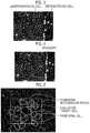
- Fig. 3 is a diagram showing an example of a cell image of a cell group including stem cells in an undifferentiated state and stem cells in a differentiated state.
- the stem cells in the undifferentiated state have the degree of circularity higher than that of the stem cells in the differentiated state, and the stem cells in the differentiated state are more elongated than the stem cells in the undifferentiated state and have a maximum diameter larger than that of the stem cells in the undifferentiated state.
- the boundary setting unit 31 calculates a spatial frequency of a cell image, and then, determines a region where the spatial frequency is equal to or greater than a predetermined threshold value as an undifferentiated region, and determines a region where the spatial frequency is smaller than the threshold value as a differentiated region.
- the boundary setting unit 31 may determine the differentiated region and the undifferentiated region using a luminance change or a color change, instead of the determination based on the spatial frequency of the cell image as described above. For example, as shown in Fig. 3 , in the region where the stem cells in the undifferentiated state are distributed, the stem cells are densely distributed, and halo occurs in a boundary between the stem cells, so that the luminance becomes high. The halo represents a high luminance artifact generated when illumination light pass between cells.
- the boundary setting unit 31 may calculate a luminance change of a cell image, and then, may determine a region where the luminance change is equal to or greater than a threshold value as an undifferentiated region, and may determine a region where the luminance change is smaller than the threshold value as a differentiated region. Further, a color change may be detected instead of the luminance change.
- the boundary setting unit 31 may calculate a color change of a cell image, and then, may determine a region where the color change is equal to or greater than a predetermined threshold value as an undifferentiated region, and may determine a region where the color change is smaller than the threshold value as a differentiated region.
- the boundary setting unit 31 may calculate the luminance of a cell image, and then, may determine a region where the luminance is equal to or greater than a predetermined threshold value as an undifferentiated region, and may determine a region where the luminance is smaller than the threshold value as a differentiated region.
- the boundary setting unit 31 may determine the differentiated region and the undifferentiated region based on a combination of at least two characteristics of spatial frequency, luminance, or color. In this case, for example, the boundary setting unit 31 may calculate one evaluation value by weighting and adding up the plural characteristics, and then, may determine the differentiated region and the undifferentiated region according whether the evaluation value is equal to or greater than a threshold value or is smaller than the threshold value.
- the boundary setting unit 31 of this embodiment calculates a feature amount for an entire cell image such as a spatial frequency, a luminance change, a color change or the like, and sets a boundary for generally dividing a cell group based on the feature amount.
- the boundary between the differentiated region and the undifferentiated region based on at least one of spatial frequency, luminance or color of the cell image as described above, for example, there is a possibility that the halo portion where the luminance between the undifferentiated cells shown in Fig. 3 is high is set as the boundary, according to a threshold value setting method or the like.
- the purpose of setting boundary by the boundary setting unit 31 is for that an inappropriate cell is not included in peripheral cells when evaluating an evaluation target cell using a determination result of the peripheral cells as described above, a boundary for generally dividing the cell group is more favorable than a strict boundary, and it is preferable that a boundary having higher linearity is used.
- the boundary setting unit 31 may set an upper limit of the number of inflection points of the boundary, a curvature thereof or the like, and may set line segments having a smaller number of inflection points or a smaller curvature than the upper limit as the boundary.
- the boundary setting unit 31 may extract plural line segments which are boundary candidates, and may set a line segment that does not exceed the above-mentioned upper limit in the number of inflection points, the curvature or the like from the boundary candidates as a final boundary.
- Fig. 4 is a diagram showing an example of a boundary set in the cell image shown in Fig. 3 .
- a line segment indicated by a single-dot chain line represents a boundary set by the boundary setting unit 31.
- the boundary setting unit 31 sets the boundary between the undifferentiated region and the differentiated region, but the boundary setting is not limited to the boundary between the undifferentiated region and the differentiated region.
- the boundary setting unit 31 may set a boundary according to the degree of differentiation of the cells.
- the boundary setting unit 31 may set a boundary between a region where the degree of differentiation is equal to or greater than a predetermined threshold value and a region where the degree of differentiation is smaller than the threshold value.
- the boundary setting unit 31 may set a boundary according to the degree of malignancy of the cancer cells.
- the boundary setting unit 31 may set a boundary between a region where the degree of malignancy is equal to or greater than a threshold value and a region where the degree of malignancy is smaller than the threshold value.
- the cell evaluation unit 32 specifies an evaluation target cell and peripheral cells around the evaluation target cell in a cell group in a cell image, and evaluates the evaluation target cell based on evaluation results of the specified peripheral cells.
- the cell evaluation unit 32 acquires evaluation results of the peripheral cells either, and evaluates the evaluation target cell using the evaluation results of the peripheral cells and an evaluation result of the evaluation target cell.
- a method for evaluating individual cells will be specifically described.
- the cell evaluation unit 32 specifies individual cells included in a cell image.
- a method for specifying individual cells for example, a method for converting a cell image into a binarized image, detecting edges of the individual cells by performing a filter process, and performing pattern matching with respect to the edges to specify the individual cells may be used. Further, when performing the pattern matching, it is preferable to perform pattern recognition using machine learning.
- the invention is not limited to this method, and may use various known methods.
- the cell evaluation unit 32 evaluates whether the individual cells specified as described above are in a differentiated state or in an undifferentiated state, respectively.
- the cell evaluation unit 32 specifies peripheral cells around an evaluation target cell, and evaluates the evaluation target cell using evaluation results of the peripheral cells.
- a method for specifying peripheral cells around an evaluation target cell will be described.
- a method for setting a rectangular region being in contact with the outline of a predetermined evaluation target cell (indicated by a solid-line oval), and setting rectangular regions having the same size as that of the set rectangular region around the rectangular region of the evaluation target cell, and specifying cells (indicated by dotted-line ovals) that are partly or totally included in the peripheral eight rectangular regions as peripheral cells may be used.
- the cell evaluation unit 32 of this embodiment acquires information about the boundary set in the boundary setting unit 31, and resets peripheral regions based on the boundary information.
- the peripheral regions are reset so that only cells in the differentiated region are specified as the peripheral cells.
- the above-mentioned eight rectangular regions are reset to be all disposed within the differentiated region.
- a method for resetting the peripheral regions is not limited to the example shown in Fig. 7 , and any method may be employed as long as all the peripheral regions can be disposed in the differentiated region by the method.
- the rectangular region being in contact with the outline of the evaluation target cell is set, and the rectangular regions having the same size as that of the set rectangular region are set around the evaluation target cell, that is, the sizes of the peripheral rectangular regions are set based on the size and shape of a cytoplasm of the evaluation target cell, but the invention is not limited thereto.
- a method for detecting a cell nucleus or a nucleolus in an evaluation target cell, setting peripheral regions according to the size or shape of the cell nucleus or the nucleolus, and specifying cells included partly or totally in the peripheral regions as peripheral cells may be used.
- Fig. 8 is a diagram showing an example in which peripheral regions are set to become wider as a cell nucleus becomes larger.
- Fig. 9 is a diagram showing an example in which the shape of a peripheral region is set to be changed according to the shape of the cell nucleus. This is similarly applied to a case where the peripheral regions are set based on the size or shape of the nucleolus.
- peripheral regions are set based on characteristics of cells in one cell image as described above
- information relating to a proliferation rate of a cell, information relating to a migration rate thereof or the like may be acquired from the plural cell images, and peripheral regions may be set based on the information.
- a method for acquiring a cell proliferation rate for example, a method for respectively counting the number of cells per unit area included in cell images captured at different time points and calculating a proliferation rate based on an increment of the number of cells and an image pick-up interval may be used.
- the unit area may be the entirety of a cell image or may be a partial region including an evaluation target cell.
- a method for respectively calculating areas of a cell group included in cell images captured at different time points and acquiring an area increasing rate as information relating to the proliferation rate may be used.
- a method for acquiring a migration rate of a cell for example, a method for calculating a movement distance of an evaluation target cell included in cell images captured at different time points and calculating a migration rate based on the movement distance and an image pick-up interval may be used.
- a method for matching cells having similar shapes which are present in a predetermined range may be used. Further, other known techniques may be used.
- a method for calculating a migration rate based on movement distances of all cells included in cell images may be used.
- a method for calculating a statistic value such as an average value, a maximum value or a minimum value of movement distances of individual cells and calculating a migration rate based on the statistic value and an image pick-up interval may be used.
- a method for acquiring the statistic value of the movement distances described above as information relating to the migration rate may be used.
- each peripheral region may be enlarged until the number of peripheral cells included in the peripheral region reaches a predetermined number.
- an aspect ratio between the enlarged length and width of the rectangular region may be 1, or may be a value different from 1.
- the aspect ratio may be set to 1, and in a case where the shape thereof is an elliptical shape that extends in a longitudinal direction or a transverse direction, the aspect ratio may be set so that an enlarged width in the extending direction is relatively large.
- the aspect ratio may be set so that an enlarged width in a direction where the proliferation rate is high is relatively large.
- the aspect ratio may be set so that an enlarged width in a direction where the migration rate is high is relatively large.
- the number of cells that are investigated in the longitudinal direction and the number of cells that are investigated in the transverse direction may be equal to each other, or may be different from each other.
- the numbers of cells that are investigated in the longitudinal direction and the transverse direction may be set to be equal to each other, and in a case where the shape is an elliptical shape that extends in the longitudinal direction or the transverse direction, the number of cells that are investigated in the extending direction may be relatively large.
- peripheral regions which are rectangular regions are set based on a proliferation rate
- the number of cells that are investigated in a direction where the proliferation rate is high may be relatively large.
- peripheral regions which are rectangular regions are set based on a migration rate
- the number of cells that are investigated in a direction where the migration rate is high may be relatively large.
- peripheral cells are automatically specified as described above, but a user may designate peripheral cells using the input device 5, and the cell evaluation unit 32 receives designated information and thus may specify peripheral cells.
- the cell evaluation unit 32 sequentially specifies individual cells in a cell image as evaluation target cells, and sequentially specifies peripheral cells around the evaluation target cells, and evaluates each evaluation target cell using evaluation results of the peripheral cells.
- the cell evaluation unit 32 of this embodiment evaluates whether an evaluation target cell is in a differentiated state or in an undifferentiated state as described above. Specifically, in this embodiment, the cell evaluation unit 32 calculates the degree of circularity of the evaluation target cell and the peripheral cells, evaluates that the evaluation target cell is in the undifferentiated state in a case where the degree of circularity is equal to or greater than a threshold value, and evaluates that the evaluation target cell is in the differentiated state in a case where the degree of circularity is smaller than the threshold value. Further, evaluation results of the evaluation target cell and the peripheral cells are stored together with positional information about the cells.
- the cell evaluation unit 32 evaluates whether the evaluation target cell is in the undifferentiated state or in the differentiated state based on the degree of circularity, but the evaluation based on the degree of circularity is not limited thereto.
- the cell evaluation unit 32 may evaluate whether the evaluation target cell is in the undifferentiated state or in the differentiated state based on information about the size of a maximum diameter, a minimum diameter, an area or the like of individual cells, the density of a nucleolus included in each cell, or the like.
- the cell evaluation unit 32 may evaluate whether the evaluation target cell is in the differentiated state or in the undifferentiated state based on image information in a predetermined region including individual cells instead of characteristics of the shape of the individual cells as described above. For example, a method for calculating the density of cells in a predetermined region including an evaluation target cell, evaluating that the evaluation target cell is in the undifferentiated state in a case where the density is equal to or greater than a predetermined threshold value, and evaluating that the evaluation target cell is in the differentiated state in a case where the density is smaller than the threshold value may be used.
- a method may be used for calculating a statistic value such as an average value a maximum value, a minimum value or the like of luminance in a predetermined region including an evaluation target cell, and evaluating that the evaluation target cell is in the undifferentiated state in a case where the statistic value of the luminance is smaller than a predetermined threshold value, and evaluating that the evaluation target cell is in the differentiated state in a case where the statistic value of the luminance is equal to or greater than the threshold value.
- the cell evaluation unit 32 may evaluate whether the evaluation target cell is in the undifferentiated state or in the differentiated state using other known methods.
- the cell evaluation unit 32 adds up the evaluation result of the evaluation target cell and the evaluation results of the peripheral cells, for example. In a case where the number of evaluation results in the undifferentiated state is larger is larger, the cell evaluation unit 32 evaluates that the evaluation target cell is in the undifferentiated state, and in a case where the number of evaluation results in the differentiated state is larger is larger, the cell evaluation unit 32 evaluates that the evaluation target cell is in the differentiated state.
- a method for calculating one evaluation value by weighting and adding up the evaluation result of the evaluation target cell and the evaluation results of the peripheral cells and determining an evaluation result of the evaluation target cell based on the evaluation value may be used.
- a method for setting the evaluation value to "2" in a case where the evaluation result is in the undifferentiated state and setting the evaluation value to "1" in a case where the evaluation result is in the differentiated state, calculating one evaluation value by adding up the evaluation value of the evaluation target cell and the evaluation values of the peripheral cells, and then, determining that the evaluation result of the evaluation target cell is in the undifferentiated state in a case where the evaluation value is equal to or greater than a predetermined threshold value and determining that the evaluation result of the evaluation target cell is in the differentiated state in a case where the evaluation value is smaller than the threshold value may be used.
- a weight larger than that of the evaluation values of the peripheral cells may be assigned to the evaluation value of the evaluation target cell for the adding up.
- weights may be changed according to distances from the evaluation target cell. For example, a larger weight of evaluation value of a peripheral cell that is closer to the evaluation target cell may be assigned.
- the evaluation value of the evaluation target cell and the evaluation values of the peripheral cells as described above, for example, in a case where the undifferentiation and differentiation are evaluated based on the degree of circularity, and in a case where the degree of circularity shows an abnormal value out of a normal range, it is determined that the evaluation is impossible to be performed with respect to the concerned cell, and the evaluation value thereof may be set to "0". In this way, by setting the evaluation value of the cell that cannot be evaluated to "0", it is possible to evaluate the evaluation target cell using only evaluation results of cells of which evaluation results are fixed, and thus, it is possible to perform evaluation with higher accuracy.
- a cell cannot be evaluated, for example, there is a case where the cell is a dead cell, or a case where a waste which is not a cell is misrecognized as a cell.
- the evaluation value based on the luminance may become an abnormal value, and thus, it may be determined that the evaluation is impossible.
- the cell evaluation unit 32 sequentially specifies individual cells included in a cell image as evaluation target cells, and sequentially specifies peripheral cells to perform evaluation, but in this case, with respect to a cell which is evaluated once, the cell evaluation unit 32 may not perform evaluation and may use the stored evaluation result. Thus, it is possible to simplify the evaluation process, and to perform the evaluation process at high speed.
- a configuration in which the cell evaluation unit 32 evaluates whether the evaluation target cell is in the undifferentiated state or in the differentiated state is shown, but the invention is not limited thereto.
- a configuration in which in a case where a cell group includes differentiated and induced cells, the degrees of differentiation of the cells are evaluated may be used.
- a method for expressing stages of the degrees of differentiation as numerical values based on shapes or the like of individual cells, calculating a statistic value such as an average value, a maximum value, or a minimum value of the degree of an evaluation target cell and the degrees of differentiation of peripheral cells, and determining the statistic value as the degree of differentiation of the evaluation target cell may be used.
- weighting and adding up may be performed according to distances between the evaluation target cell and the peripheral cells.
- the degree of malignancy of the cells may be evaluated.
- the display controller 33 acquires a cell image read from the image acquisition unit 30, acquires evaluation results of individual cells evaluated in the cell evaluation unit 32, and displays the cell image and the evaluation results on the display 4.
- a method for displaying the evaluation results of the individual cells for example, a method for displaying an evaluation result of a cell designated by a user using the input device 5 as a text may be used, or a method for mapping evaluation results of individual cells using different colors, for example, to generate a cell evaluation image and superimposing the cell evaluation image on the cell image for display may be used.
- a semi-transparent image capable of transmitting the cell image so that the cell image can be observed may be used, or an image in which outlines of individual cells are expressed using different colors or the like may be used.
- the input device 5 includes a mouse, a keyboard or the like, and receives setting inputs from a user.
- the input device 5 is able to receive setting inputs such as image pick-up conditions of an optical magnification or the like of the phase contrast microscope 20, designation information of individual cells in a cell image, or the like.
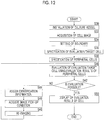
- a culture which is an image pick-up target is selected from plural accommodated culture vessels by the transport unit 11, and the selected culture vessel is installed in the stage 10 (S10).
- an image of a cell colony in the culture vessel is captured by the phase contrast microscope 20 of the image pick-up device 2, and the captured cell image is acquired by the image acquisition unit 30 of the cell evaluation device 3 (S12).
- the boundary setting unit 31 sets a boundary in the cell image based on the state of a cell group in the cell image (S14).
- a boundary between a differentiated region and an undifferentiated region is set as described above.
- Information about the boundary set in the boundary setting unit 31 is output to the cell evaluation unit 32, and the cell evaluation unit 32 specifies individual cells in the cell image (S16), and specifies peripheral cells based on the input boundary information (S18).
- the cell evaluation unit 32 sequentially specifies the individual cells in the cell image as evaluation target cells, sequentially specifies the peripheral cells, and evaluates the evaluation target cells using evaluation results of the peripheral cells (S20).
- Evaluation results of individual cell regions evaluated in the cell evaluation unit 32 are output to the display controller 33, and the display controller 33 displays the cell image and the evaluation results of the individual cells on the display 4 (S22).
- the cell culture observation system when evaluating an evaluation target cell using evaluation results of peripheral cells, an evaluation result of a cell that cannot be evaluated among evaluation results of the evaluation target cell and the peripheral cells is not used.
- the cell that cannot be evaluated in a case where there is a cell that cannot be evaluated, the cell that cannot be evaluated is re-evaluated using cell images captured under image pick-up conditions different from those of a cell image including the cell that cannot be evaluated.
- the cell culture observation system further includes an image pick-up condition acquisition unit 34 and an image pick-up controller 35.
- the cell evaluation unit 32 in the cell culture observation system assigns identification information to an evaluation target region including the evaluation target cell and the peripheral cells for storage.
- the identification information is identification information indicating that a cell that cannot be evaluated is included in an evaluation target region.
- the image pick-up condition acquisition unit 34 acquires image pick-up conditions when re-imaging a cell image used for re-evaluation of the evaluation target region.
- the image pick-up condition acquisition unit 34 acquires an optical magnification higher than an optical magnification when capturing a cell image including a cell that cannot be evaluated.
- a cell image for which an optical magnification is 4 may be used in an initial evaluation, and a cell image for which an optical magnification is 20 may be used in re-evaluation.
- Image pick-up conditions when capturing a cell image including the cell that cannot be evaluated are stored together with the cell image. Further, image pick-up conditions in re-imaging are also set in advance.
- the image pick-up condition acquisition unit 34 may acquire other image pick-up conditions other than the above-described optical magnification.
- image pick-up conditions for example, an image pick-up region, an image pick-up timing, an exposure time of an image pick-up element, an illumination light wavelength, an observation light wavelength, or the like may be used.
- a method for changing the image pick-up region to a narrow range according to change in an optical magnification when performing re-imaging may be used.
- an image pick-up timing for example, a method for setting, in a case where an image pick-up timing when capturing a cell image including a cell that cannot be evaluated is a culture initial stage, an image pick-up timing of re-imaging to a culture termination time may be used. Further, contrarily, a method for acquiring an image pick-up timing prior to the image pick-up timing when the cell image including the cell that cannot be evaluated is captured may be used. That is, re-evaluation may be performed using a previously captured cell image. The image pick-up timing may be measured using a timer or the like, for example. Further, it is preferable that the image pick-up timing is set to a timing that matches a cell division cycle.
- a method for setting the exposure time to be long to raise S/N of a cell image in re-imaging and setting the exposure time to be short to lower the luminance of the cell image in a case where a waste having an abnormally high luminance is present may be used.
- the illumination light wavelength for example, a method for setting the illumination light wavelength to be short to enhance the resolution of a cell image, in re-imaging, may be used. Contrarily, a method for setting the illumination light wavelength to be long to reduce scattered light and to re-capture a cell image with less blur may be used.
- observation light wavelength for example, a method for changing wavelengths using a filter or a spectroscope, in re-imaging, may be used.
- a configuration in which the image pick-up conditions of re-imaging are set in advance with respect to the image pick-up condition acquisition unit 34 but the invention is not limited thereto, and a configuration in which the image pick-up condition acquisition unit 34 automatically determines and acquires image pick-up conditions using a cell image of an evaluation target region including a cell that cannot be evaluated may be used.
- a method for determining and acquiring, in a case where the sizes of individual cells included in the cell image of the evaluation target region are equal to or smaller than a predetermined threshold value, a high optical magnification at which the sizes of individual cells become larger than the predetermined threshold value may be used.
- the image pick-up timing for example, a method for predicting a culture period from densities or the like of individual cells in a cell image of an evaluation target region, and determining and acquiring an image pick-up timing of re-imaging based on a predetermined culture cycle or the like using the culture period as a reference may be used.
- a method for acquiring S/N of a cell image of an evaluation target region and setting, in a case where the S/N is equal to or smaller than a predetermined threshold value, the exposure time to be long to raise the S/N of the cell image may be used.
- a method for acquiring a statistic value such as an average value, a maximum value, a minimum value, or the like of luminance of a cell image of an evaluation target region, and setting, in a case where the statistic value is equal to or greater than a predetermined threshold value, the exposure time to be short to lower the luminance of the cell image may be used.
- a method for acquiring the resolution of a cell image of an evaluation target region and setting, in a case where the resolution is equal or smaller than a predetermined threshold value, the illumination light wavelength to be short to enhance the resolution of the cell image may be used.
- a method for acquiring blur of the cell image of the evaluation target region and setting, in a case where the degree of blur is equal to or greater than a predetermined degree, the illumination light wavelength to be long to reduce the blur of the cell image may be used.
- a method for acquiring the resolution or blur from a cell image to change the resolution or blur may be used.
- the image pick-up controller 35 acquires image pick-up conditions of re-imaging acquired in the image pick-up condition acquisition unit 34, and outputs an imaging control signal to the controller 29 of the image pick-up device 2 or the controller 12 of the cell culture device 1 based on the image pick-up conditions.
- the controller 29 of the image pick-up device 2 controls an optical magnification, an exposure time of the image pick-up element, an image pick-up timing, an illumination light wavelength, and an observation light wavelength based on the imaging control signal output from the image pick-up controller 35.
- the controller 12 of the cell culture device 1 controls an image pick-up region by moving the stage 10 based on the imaging control signal output from the image pick-up controller 35.
- the cell evaluation unit 32 sequentially specifies the individual cells in the cell image as evaluation target cells, sequentially specifies peripheral cells of the evaluation target cells, and evaluates each evaluation target cell using evaluation results of the peripheral cells (S40).
- identification information is assigned to the corresponding evaluation target region for storage (S44).
- positional information or the like of the evaluation target region is output to the image pick-up condition acquisition unit 34 from the cell evaluation unit 32, and the image pick-up condition acquisition unit 34 acquires image pick-up conditions of re-imaging for the evaluation target region (S46).
- the image pick-up conditions acquired by the image pick-up condition acquisition unit 34 are output to the image pick-up controller 35, and the image pick-up controller 35 outputs an imaging control signal to the controller 29 of the image pick-up device 2 or the controller 12 of the cell culture device 1 based on the input image pick-up conditions. Further, re-imaging is performed under the control of the controller 29 of the image pick-up device 2 or the controller 12 of the cell culture device 1, and a cell image having image pick-up conditions different from those of the cell image used in the previous evaluation is acquired (S48).
- the cell image acquired through the re-imaging is input to the cell evaluation unit 32 again, and re-evaluation is performed with respect to an evaluation target region including a cell that cannot be evaluated (S40). Further, in a case where there is no cell that cannot be evaluated (YES in S42), evaluation results of individual cell regions are output to the display controller 33, and the display controller 33 displays the cell image and the evaluation results of the individual cells on the display 4 (S50). Even in a case where a cell that cannot be evaluated through the above-described re-imaging is not removed, a message or the like indicating such a fact may be displayed on the display 4 to then terminate the procedure.
- the cell culture observation system when evaluating individual cells using a cell image captured under predetermined image pick-up conditions, even in a case where a cell that cannot be evaluated are present, since re-imaging is performed under different image pick-up conditions and re-evaluation is performed using a re-captured cell image, it is possible to evaluate individual cells with high accuracy.
- the image pick-up condition acquisition unit 34 automatically acquires image pick-up conditions is shown, but the invention is not limited thereto.
- a configuration in which a user inputs image pick-up conditions of re-imaging using the input device 5 and the image pick-up condition acquisition unit 34 acquires the input image pick-up conditions may be used.
- the image pick-up controller 35 automatically performs re-imaging based on image pick-up conditions acquired the image pick-up condition acquisition unit 34 is shown, but the invention is not limited thereto.
- a configuration in which the display controller 33 displays image pick-up conditions acquired by the image pick-up condition acquisition unit 34 on the display 4 to request a user to perform re-imaging, and accordingly, the user manually performs the re-imaging may be used.
Landscapes
- Engineering & Computer Science (AREA)
- Chemical & Material Sciences (AREA)
- Health & Medical Sciences (AREA)
- Life Sciences & Earth Sciences (AREA)
- Organic Chemistry (AREA)
- General Health & Medical Sciences (AREA)
- Physics & Mathematics (AREA)
- Zoology (AREA)
- Proteomics, Peptides & Aminoacids (AREA)
- Wood Science & Technology (AREA)
- Quality & Reliability (AREA)
- Medical Informatics (AREA)
- Theoretical Computer Science (AREA)
- General Physics & Mathematics (AREA)
- Computer Vision & Pattern Recognition (AREA)
- Radiology & Medical Imaging (AREA)
- Nuclear Medicine, Radiotherapy & Molecular Imaging (AREA)
- Genetics & Genomics (AREA)
- Analytical Chemistry (AREA)
- Molecular Biology (AREA)
- General Engineering & Computer Science (AREA)
- Bioinformatics & Cheminformatics (AREA)
- Biochemistry (AREA)
- Biotechnology (AREA)
- Biophysics (AREA)
- Immunology (AREA)
- Microbiology (AREA)
- Apparatus Associated With Microorganisms And Enzymes (AREA)
- Measuring Or Testing Involving Enzymes Or Micro-Organisms (AREA)
Applications Claiming Priority (2)
| Application Number | Priority Date | Filing Date | Title |
|---|---|---|---|
| JP2014112135A JP6301199B2 (ja) | 2014-05-30 | 2014-05-30 | 細胞評価装置および方法並びにプログラム |
| PCT/JP2015/063884 WO2015182396A1 (fr) | 2014-05-30 | 2015-05-14 | Dispositif, procédé, et programme d'évaluation de cellules |
Publications (3)
| Publication Number | Publication Date |
|---|---|
| EP3150694A1 true EP3150694A1 (fr) | 2017-04-05 |
| EP3150694A4 EP3150694A4 (fr) | 2017-05-17 |
| EP3150694B1 EP3150694B1 (fr) | 2024-12-18 |
Family
ID=54698733
Family Applications (1)
| Application Number | Title | Priority Date | Filing Date |
|---|---|---|---|
| EP15799811.3A Active EP3150694B1 (fr) | 2014-05-30 | 2015-05-14 | Dispositif, procédé, et programme d'évaluation de cellules |
Country Status (4)
| Country | Link |
|---|---|
| US (1) | US10157461B2 (fr) |
| EP (1) | EP3150694B1 (fr) |
| JP (1) | JP6301199B2 (fr) |
| WO (1) | WO2015182396A1 (fr) |
Cited By (1)
| Publication number | Priority date | Publication date | Assignee | Title |
|---|---|---|---|---|
| EP3192861A4 (fr) * | 2014-09-12 | 2017-08-30 | Fujifilm Corporation | Système et procédé d'évaluation de cultures cellulaires |
Families Citing this family (11)
| Publication number | Priority date | Publication date | Assignee | Title |
|---|---|---|---|---|
| JP6071007B2 (ja) * | 2013-08-22 | 2017-02-01 | 富士フイルム株式会社 | 観察画像撮影評価装置および方法並びにプログラム |
| JP6066492B2 (ja) * | 2013-08-22 | 2017-01-25 | 富士フイルム株式会社 | 細胞画像評価装置および方法並びにプログラム |
| JP6143365B2 (ja) * | 2014-03-05 | 2017-06-07 | 富士フイルム株式会社 | 細胞画像評価装置および方法並びにプログラム |
| WO2015182382A1 (fr) * | 2014-05-30 | 2015-12-03 | 富士フイルム株式会社 | Dispositif, procédé, et programme d'évaluation de cellules |
| JP6475134B2 (ja) * | 2015-09-29 | 2019-02-27 | 富士フイルム株式会社 | 細胞評価装置および方法 |
| WO2018012601A1 (fr) * | 2016-07-14 | 2018-01-18 | 大日本印刷株式会社 | Système d'analyse d'image, système de gestion de culture, procédé d'analyse d'image, procédé de gestion de culture, procédé de structure de groupe de cellules, et programme |
| JP7018078B2 (ja) * | 2018-01-31 | 2022-02-09 | ヤマハ発動機株式会社 | 撮像システム |
| WO2019176048A1 (fr) * | 2018-03-15 | 2019-09-19 | オリンパス株式会社 | Dispositif de traitement d'images de cellules |
| WO2021002108A1 (fr) * | 2019-07-03 | 2021-01-07 | 富士フイルム株式会社 | Dispositif, procédé et programme de support d'optimisation |
| WO2023195491A1 (fr) * | 2022-04-06 | 2023-10-12 | 富士フイルム株式会社 | Système de capture d'image et procédé de réglage de condition de culture |
| WO2026048350A1 (fr) * | 2024-08-28 | 2026-03-05 | 国立大学法人東海国立大学機構 | Dispositif de traitement d'informations, système de traitement d'informations, procédé de traitement d'informations et programme informatique |
Family Cites Families (22)
| Publication number | Priority date | Publication date | Assignee | Title |
|---|---|---|---|---|
| WO1996009600A1 (fr) * | 1994-09-20 | 1996-03-28 | Neopath, Inc. | Dispositif d'identification et d'integration de schemas cellulaires multiples |
| AU2003217337A1 (en) * | 2002-02-06 | 2003-09-02 | University Of North Carolina At Chapel Hill | High-throughput cell identification and isolation method and apparatus |
| US20050136509A1 (en) * | 2003-09-10 | 2005-06-23 | Bioimagene, Inc. | Method and system for quantitatively analyzing biological samples |
| US7711174B2 (en) * | 2004-05-13 | 2010-05-04 | The Charles Stark Draper Laboratory, Inc. | Methods and systems for imaging cells |
| JP5355275B2 (ja) * | 2009-07-24 | 2013-11-27 | オリンパス株式会社 | 細胞画像解析装置 |
| JP5740101B2 (ja) * | 2010-04-23 | 2015-06-24 | 国立大学法人名古屋大学 | 細胞評価装置、インキュベータ、細胞評価方法、細胞評価プログラムおよび細胞の培養方法 |
| US20110286654A1 (en) * | 2010-05-21 | 2011-11-24 | Siemens Medical Solutions Usa, Inc. | Segmentation of Biological Image Data |
| JP5167442B2 (ja) | 2011-02-17 | 2013-03-21 | 三洋電機株式会社 | 画像識別装置およびプログラム |
| JP5745919B2 (ja) | 2011-04-28 | 2015-07-08 | 浜松ホトニクス株式会社 | 細胞解析方法、細胞解析装置、および細胞解析プログラム |
| US8934698B2 (en) * | 2011-06-22 | 2015-01-13 | The Johns Hopkins University | System and device for characterizing cells |
| US20130183707A1 (en) * | 2012-01-13 | 2013-07-18 | University Of Pittsburgh - Of The Commonwealth System Of Higher Education | Stem cell bioinformatics |
| JP5333635B1 (ja) | 2012-08-23 | 2013-11-06 | 富士ゼロックス株式会社 | 画像処理装置、プログラム及び画像処理システム |
| US9677869B2 (en) * | 2012-12-05 | 2017-06-13 | Perimeter Medical Imaging, Inc. | System and method for generating a wide-field OCT image of a portion of a sample |
| JP6097952B2 (ja) * | 2013-08-22 | 2017-03-22 | 富士フイルム株式会社 | 観察画像判定装置および方法並びにプログラム |
| JP6066492B2 (ja) * | 2013-08-22 | 2017-01-25 | 富士フイルム株式会社 | 細胞画像評価装置および方法並びにプログラム |
| WO2015041177A1 (fr) * | 2013-09-18 | 2015-03-26 | 株式会社ニコン | Dispositif d'analyse d'images, procédé d'analyse d'images, programme d'analyse d'images, procédé de fabrication de cellules, procédé de culture de cellules et dispositif de fabrication de cellules |
| US9619881B2 (en) * | 2013-09-26 | 2017-04-11 | Cellogy, Inc. | Method and system for characterizing cell populations |
| JP2015146747A (ja) * | 2014-02-05 | 2015-08-20 | 浜松ホトニクス株式会社 | 細胞判定方法 |
| JP6143365B2 (ja) * | 2014-03-05 | 2017-06-07 | 富士フイルム株式会社 | 細胞画像評価装置および方法並びにプログラム |
| JP6130801B2 (ja) * | 2014-03-17 | 2017-05-17 | 富士フイルム株式会社 | 細胞領域表示制御装置および方法並びにプログラム |
| WO2015182382A1 (fr) * | 2014-05-30 | 2015-12-03 | 富士フイルム株式会社 | Dispositif, procédé, et programme d'évaluation de cellules |
| CN106537144A (zh) * | 2014-08-05 | 2017-03-22 | 富士胶片株式会社 | 有核红血球的分选方法 |
-
2014
- 2014-05-30 JP JP2014112135A patent/JP6301199B2/ja active Active
-
2015
- 2015-05-14 EP EP15799811.3A patent/EP3150694B1/fr active Active
- 2015-05-14 WO PCT/JP2015/063884 patent/WO2015182396A1/fr not_active Ceased
-
2016
- 2016-11-10 US US15/348,008 patent/US10157461B2/en active Active
Cited By (1)
| Publication number | Priority date | Publication date | Assignee | Title |
|---|---|---|---|---|
| EP3192861A4 (fr) * | 2014-09-12 | 2017-08-30 | Fujifilm Corporation | Système et procédé d'évaluation de cultures cellulaires |
Also Published As
| Publication number | Publication date |
|---|---|
| EP3150694B1 (fr) | 2024-12-18 |
| US20170061618A1 (en) | 2017-03-02 |
| EP3150694A4 (fr) | 2017-05-17 |
| JP2015223175A (ja) | 2015-12-14 |
| JP6301199B2 (ja) | 2018-03-28 |
| WO2015182396A1 (fr) | 2015-12-03 |
| US10157461B2 (en) | 2018-12-18 |
Similar Documents
| Publication | Publication Date | Title |
|---|---|---|
| EP3150692B1 (fr) | Dispositif, procédé, et programme d'évaluation de cellules | |
| EP3150694B1 (fr) | Dispositif, procédé, et programme d'évaluation de cellules | |
| JP6595156B2 (ja) | 細胞画像取得装置および方法並びにプログラム | |
| JP5707399B2 (ja) | 微生物検出方法、微生物検出装置及びプログラム | |
| US10330907B2 (en) | Cell imaging control device, method, and program | |
| US10538729B2 (en) | Cell imaging control device, method, and program | |
| US10360676B2 (en) | Cell image evaluation device, method, and program | |
| EP3121264B1 (fr) | Dispositif and procédé pour la commande de l'affichage d'une région cellulaire | |
| CN117099128B (zh) | 细胞计数方法、以及计算机可读介质 | |
| US11756190B2 (en) | Cell image evaluation device, method, and program |
Legal Events
| Date | Code | Title | Description |
|---|---|---|---|
| STAA | Information on the status of an ep patent application or granted ep patent |
Free format text: STATUS: THE INTERNATIONAL PUBLICATION HAS BEEN MADE |
|
| PUAI | Public reference made under article 153(3) epc to a published international application that has entered the european phase |
Free format text: ORIGINAL CODE: 0009012 |
|
| STAA | Information on the status of an ep patent application or granted ep patent |
Free format text: STATUS: REQUEST FOR EXAMINATION WAS MADE |
|
| 17P | Request for examination filed |
Effective date: 20161108 |
|
| AK | Designated contracting states |
Kind code of ref document: A1 Designated state(s): AL AT BE BG CH CY CZ DE DK EE ES FI FR GB GR HR HU IE IS IT LI LT LU LV MC MK MT NL NO PL PT RO RS SE SI SK SM TR |
|
| AX | Request for extension of the european patent |
Extension state: BA ME |
|
| A4 | Supplementary search report drawn up and despatched |
Effective date: 20170419 |
|
| RIC1 | Information provided on ipc code assigned before grant |
Ipc: C12M 1/34 20060101AFI20170411BHEP Ipc: C12Q 1/02 20060101ALI20170411BHEP |
|
| DAV | Request for validation of the european patent (deleted) | ||
| DAX | Request for extension of the european patent (deleted) | ||
| RAP3 | Party data changed (applicant data changed or rights of an application transferred) |
Owner name: FUJIFILM CORPORATION |
|
| STAA | Information on the status of an ep patent application or granted ep patent |
Free format text: STATUS: EXAMINATION IS IN PROGRESS |
|
| 17Q | First examination report despatched |
Effective date: 20210705 |
|
| GRAP | Despatch of communication of intention to grant a patent |
Free format text: ORIGINAL CODE: EPIDOSNIGR1 |
|
| STAA | Information on the status of an ep patent application or granted ep patent |
Free format text: STATUS: GRANT OF PATENT IS INTENDED |
|
| INTG | Intention to grant announced |
Effective date: 20240712 |
|
| GRAS | Grant fee paid |
Free format text: ORIGINAL CODE: EPIDOSNIGR3 |
|
| GRAA | (expected) grant |
Free format text: ORIGINAL CODE: 0009210 |
|
| STAA | Information on the status of an ep patent application or granted ep patent |
Free format text: STATUS: THE PATENT HAS BEEN GRANTED |
|
| AK | Designated contracting states |
Kind code of ref document: B1 Designated state(s): AL AT BE BG CH CY CZ DE DK EE ES FI FR GB GR HR HU IE IS IT LI LT LU LV MC MK MT NL NO PL PT RO RS SE SI SK SM TR |
|
| REG | Reference to a national code |
Ref country code: GB Ref legal event code: FG4D |
|
| REG | Reference to a national code |
Ref country code: CH Ref legal event code: EP |
|
| REG | Reference to a national code |
Ref country code: DE Ref legal event code: R096 Ref document number: 602015090666 Country of ref document: DE |
|
| REG | Reference to a national code |
Ref country code: IE Ref legal event code: FG4D |
|
| REG | Reference to a national code |
Ref country code: LT Ref legal event code: MG9D |
|
| PG25 | Lapsed in a contracting state [announced via postgrant information from national office to epo] |
Ref country code: HR Free format text: LAPSE BECAUSE OF FAILURE TO SUBMIT A TRANSLATION OF THE DESCRIPTION OR TO PAY THE FEE WITHIN THE PRESCRIBED TIME-LIMIT Effective date: 20241218 |
|
| PG25 | Lapsed in a contracting state [announced via postgrant information from national office to epo] |
Ref country code: FI Free format text: LAPSE BECAUSE OF FAILURE TO SUBMIT A TRANSLATION OF THE DESCRIPTION OR TO PAY THE FEE WITHIN THE PRESCRIBED TIME-LIMIT Effective date: 20241218 |
|
| PG25 | Lapsed in a contracting state [announced via postgrant information from national office to epo] |
Ref country code: BG Free format text: LAPSE BECAUSE OF FAILURE TO SUBMIT A TRANSLATION OF THE DESCRIPTION OR TO PAY THE FEE WITHIN THE PRESCRIBED TIME-LIMIT Effective date: 20241218 |
|
| PG25 | Lapsed in a contracting state [announced via postgrant information from national office to epo] |
Ref country code: NO Free format text: LAPSE BECAUSE OF FAILURE TO SUBMIT A TRANSLATION OF THE DESCRIPTION OR TO PAY THE FEE WITHIN THE PRESCRIBED TIME-LIMIT Effective date: 20250318 |
|
| REG | Reference to a national code |
Ref country code: NL Ref legal event code: MP Effective date: 20241218 |
|
| PG25 | Lapsed in a contracting state [announced via postgrant information from national office to epo] |
Ref country code: LV Free format text: LAPSE BECAUSE OF FAILURE TO SUBMIT A TRANSLATION OF THE DESCRIPTION OR TO PAY THE FEE WITHIN THE PRESCRIBED TIME-LIMIT Effective date: 20241218 Ref country code: GR Free format text: LAPSE BECAUSE OF FAILURE TO SUBMIT A TRANSLATION OF THE DESCRIPTION OR TO PAY THE FEE WITHIN THE PRESCRIBED TIME-LIMIT Effective date: 20250319 |
|
| PG25 | Lapsed in a contracting state [announced via postgrant information from national office to epo] |
Ref country code: RS Free format text: LAPSE BECAUSE OF FAILURE TO SUBMIT A TRANSLATION OF THE DESCRIPTION OR TO PAY THE FEE WITHIN THE PRESCRIBED TIME-LIMIT Effective date: 20250318 |
|
| PG25 | Lapsed in a contracting state [announced via postgrant information from national office to epo] |
Ref country code: NL Free format text: LAPSE BECAUSE OF FAILURE TO SUBMIT A TRANSLATION OF THE DESCRIPTION OR TO PAY THE FEE WITHIN THE PRESCRIBED TIME-LIMIT Effective date: 20241218 |
|
| REG | Reference to a national code |
Ref country code: AT Ref legal event code: MK05 Ref document number: 1752278 Country of ref document: AT Kind code of ref document: T Effective date: 20241218 |
|
| PG25 | Lapsed in a contracting state [announced via postgrant information from national office to epo] |
Ref country code: SM Free format text: LAPSE BECAUSE OF FAILURE TO SUBMIT A TRANSLATION OF THE DESCRIPTION OR TO PAY THE FEE WITHIN THE PRESCRIBED TIME-LIMIT Effective date: 20241218 |
|
| PG25 | Lapsed in a contracting state [announced via postgrant information from national office to epo] |
Ref country code: PL Free format text: LAPSE BECAUSE OF FAILURE TO SUBMIT A TRANSLATION OF THE DESCRIPTION OR TO PAY THE FEE WITHIN THE PRESCRIBED TIME-LIMIT Effective date: 20241218 |
|
| PGFP | Annual fee paid to national office [announced via postgrant information from national office to epo] |
Ref country code: DE Payment date: 20250402 Year of fee payment: 11 |
|
| PG25 | Lapsed in a contracting state [announced via postgrant information from national office to epo] |
Ref country code: ES Free format text: LAPSE BECAUSE OF FAILURE TO SUBMIT A TRANSLATION OF THE DESCRIPTION OR TO PAY THE FEE WITHIN THE PRESCRIBED TIME-LIMIT Effective date: 20241218 |
|
| PG25 | Lapsed in a contracting state [announced via postgrant information from national office to epo] |
Ref country code: IS Free format text: LAPSE BECAUSE OF FAILURE TO SUBMIT A TRANSLATION OF THE DESCRIPTION OR TO PAY THE FEE WITHIN THE PRESCRIBED TIME-LIMIT Effective date: 20250418 |
|
| PG25 | Lapsed in a contracting state [announced via postgrant information from national office to epo] |
Ref country code: PT Free format text: LAPSE BECAUSE OF FAILURE TO SUBMIT A TRANSLATION OF THE DESCRIPTION OR TO PAY THE FEE WITHIN THE PRESCRIBED TIME-LIMIT Effective date: 20250421 |
|
| PG25 | Lapsed in a contracting state [announced via postgrant information from national office to epo] |
Ref country code: EE Free format text: LAPSE BECAUSE OF FAILURE TO SUBMIT A TRANSLATION OF THE DESCRIPTION OR TO PAY THE FEE WITHIN THE PRESCRIBED TIME-LIMIT Effective date: 20241218 |
|
| PGFP | Annual fee paid to national office [announced via postgrant information from national office to epo] |
Ref country code: FR Payment date: 20250401 Year of fee payment: 11 |
|
| PG25 | Lapsed in a contracting state [announced via postgrant information from national office to epo] |
Ref country code: AT Free format text: LAPSE BECAUSE OF FAILURE TO SUBMIT A TRANSLATION OF THE DESCRIPTION OR TO PAY THE FEE WITHIN THE PRESCRIBED TIME-LIMIT Effective date: 20241218 Ref country code: RO Free format text: LAPSE BECAUSE OF FAILURE TO SUBMIT A TRANSLATION OF THE DESCRIPTION OR TO PAY THE FEE WITHIN THE PRESCRIBED TIME-LIMIT Effective date: 20241218 |
|
| PG25 | Lapsed in a contracting state [announced via postgrant information from national office to epo] |
Ref country code: SK Free format text: LAPSE BECAUSE OF FAILURE TO SUBMIT A TRANSLATION OF THE DESCRIPTION OR TO PAY THE FEE WITHIN THE PRESCRIBED TIME-LIMIT Effective date: 20241218 |
|
| PG25 | Lapsed in a contracting state [announced via postgrant information from national office to epo] |
Ref country code: CZ Free format text: LAPSE BECAUSE OF FAILURE TO SUBMIT A TRANSLATION OF THE DESCRIPTION OR TO PAY THE FEE WITHIN THE PRESCRIBED TIME-LIMIT Effective date: 20241218 |
|
| PG25 | Lapsed in a contracting state [announced via postgrant information from national office to epo] |
Ref country code: IT Free format text: LAPSE BECAUSE OF FAILURE TO SUBMIT A TRANSLATION OF THE DESCRIPTION OR TO PAY THE FEE WITHIN THE PRESCRIBED TIME-LIMIT Effective date: 20241218 |
|
| PG25 | Lapsed in a contracting state [announced via postgrant information from national office to epo] |
Ref country code: SE Free format text: LAPSE BECAUSE OF FAILURE TO SUBMIT A TRANSLATION OF THE DESCRIPTION OR TO PAY THE FEE WITHIN THE PRESCRIBED TIME-LIMIT Effective date: 20241218 |
|
| REG | Reference to a national code |
Ref country code: DE Ref legal event code: R097 Ref document number: 602015090666 Country of ref document: DE |
|
| PG25 | Lapsed in a contracting state [announced via postgrant information from national office to epo] |
Ref country code: DK Free format text: LAPSE BECAUSE OF FAILURE TO SUBMIT A TRANSLATION OF THE DESCRIPTION OR TO PAY THE FEE WITHIN THE PRESCRIBED TIME-LIMIT Effective date: 20241218 |
|
| PLBE | No opposition filed within time limit |
Free format text: ORIGINAL CODE: 0009261 |
|
| STAA | Information on the status of an ep patent application or granted ep patent |
Free format text: STATUS: NO OPPOSITION FILED WITHIN TIME LIMIT |
|
| REG | Reference to a national code |
Ref country code: CH Ref legal event code: L10 Free format text: ST27 STATUS EVENT CODE: U-0-0-L10-L00 (AS PROVIDED BY THE NATIONAL OFFICE) Effective date: 20251029 |
|
| 26N | No opposition filed |
Effective date: 20250919 |
|
| REG | Reference to a national code |
Ref country code: CH Ref legal event code: H13 Free format text: ST27 STATUS EVENT CODE: U-0-0-H10-H13 (AS PROVIDED BY THE NATIONAL OFFICE) Effective date: 20251223 |
|
| PG25 | Lapsed in a contracting state [announced via postgrant information from national office to epo] |
Ref country code: LU Free format text: LAPSE BECAUSE OF NON-PAYMENT OF DUE FEES Effective date: 20250514 |
|
| PG25 | Lapsed in a contracting state [announced via postgrant information from national office to epo] |
Ref country code: CH Free format text: LAPSE BECAUSE OF NON-PAYMENT OF DUE FEES Effective date: 20250531 |
|
| REG | Reference to a national code |
Ref country code: BE Ref legal event code: MM Effective date: 20250531 |
|
| PG25 | Lapsed in a contracting state [announced via postgrant information from national office to epo] |
Ref country code: MC Free format text: LAPSE BECAUSE OF FAILURE TO SUBMIT A TRANSLATION OF THE DESCRIPTION OR TO PAY THE FEE WITHIN THE PRESCRIBED TIME-LIMIT Effective date: 20241218 |
|
| PGFP | Annual fee paid to national office [announced via postgrant information from national office to epo] |
Ref country code: GB Payment date: 20260317 Year of fee payment: 12 |
|
| PG25 | Lapsed in a contracting state [announced via postgrant information from national office to epo] |
Ref country code: IE Free format text: LAPSE BECAUSE OF NON-PAYMENT OF DUE FEES Effective date: 20250514 |
|
| PG25 | Lapsed in a contracting state [announced via postgrant information from national office to epo] |
Ref country code: BE Free format text: LAPSE BECAUSE OF NON-PAYMENT OF DUE FEES Effective date: 20250531 |