EP3190171A1 - Vorrichtung und verfahren zum drucken von biologischem gewebe und organen - Google Patents
Vorrichtung und verfahren zum drucken von biologischem gewebe und organen Download PDFInfo
- Publication number
- EP3190171A1 EP3190171A1 EP15837477.7A EP15837477A EP3190171A1 EP 3190171 A1 EP3190171 A1 EP 3190171A1 EP 15837477 A EP15837477 A EP 15837477A EP 3190171 A1 EP3190171 A1 EP 3190171A1
- Authority
- EP
- European Patent Office
- Prior art keywords
- printing
- module
- gel
- bioink
- radiation
- Prior art date
- Legal status (The legal status is an assumption and is not a legal conclusion. Google has not performed a legal analysis and makes no representation as to the accuracy of the status listed.)
- Withdrawn
Links
Images


Classifications
-
- C—CHEMISTRY; METALLURGY
- C12—BIOCHEMISTRY; BEER; SPIRITS; WINE; VINEGAR; MICROBIOLOGY; ENZYMOLOGY; MUTATION OR GENETIC ENGINEERING
- C12M—APPARATUS FOR ENZYMOLOGY OR MICROBIOLOGY; APPARATUS FOR CULTURING MICROORGANISMS FOR PRODUCING BIOMASS, FOR GROWING CELLS OR FOR OBTAINING FERMENTATION OR METABOLIC PRODUCTS, i.e. BIOREACTORS OR FERMENTERS
- C12M33/00—Means for introduction, transport, positioning, extraction, harvesting, peeling or sampling of biological material in or from the apparatus
-
- C—CHEMISTRY; METALLURGY
- C12—BIOCHEMISTRY; BEER; SPIRITS; WINE; VINEGAR; MICROBIOLOGY; ENZYMOLOGY; MUTATION OR GENETIC ENGINEERING
- C12N—MICROORGANISMS OR ENZYMES; COMPOSITIONS THEREOF; PROPAGATING, PRESERVING, OR MAINTAINING MICROORGANISMS; MUTATION OR GENETIC ENGINEERING; CULTURE MEDIA
- C12N5/00—Undifferentiated human, animal or plant cells, e.g. cell lines; Tissues; Cultivation or maintenance thereof; Culture media therefor
- C12N5/0062—General methods for three-dimensional culture
-
- B—PERFORMING OPERATIONS; TRANSPORTING
- B33—ADDITIVE MANUFACTURING TECHNOLOGY
- B33Y—ADDITIVE MANUFACTURING, i.e. MANUFACTURING OF THREE-DIMENSIONAL [3D] OBJECTS BY ADDITIVE DEPOSITION, ADDITIVE AGGLOMERATION OR ADDITIVE LAYERING, e.g. BY 3D PRINTING, STEREOLITHOGRAPHY OR SELECTIVE LASER SINTERING
- B33Y10/00—Processes of additive manufacturing
-
- B—PERFORMING OPERATIONS; TRANSPORTING
- B33—ADDITIVE MANUFACTURING TECHNOLOGY
- B33Y—ADDITIVE MANUFACTURING, i.e. MANUFACTURING OF THREE-DIMENSIONAL [3D] OBJECTS BY ADDITIVE DEPOSITION, ADDITIVE AGGLOMERATION OR ADDITIVE LAYERING, e.g. BY 3D PRINTING, STEREOLITHOGRAPHY OR SELECTIVE LASER SINTERING
- B33Y30/00—Apparatus for additive manufacturing; Details thereof or accessories therefor
-
- B—PERFORMING OPERATIONS; TRANSPORTING
- B33—ADDITIVE MANUFACTURING TECHNOLOGY
- B33Y—ADDITIVE MANUFACTURING, i.e. MANUFACTURING OF THREE-DIMENSIONAL [3D] OBJECTS BY ADDITIVE DEPOSITION, ADDITIVE AGGLOMERATION OR ADDITIVE LAYERING, e.g. BY 3D PRINTING, STEREOLITHOGRAPHY OR SELECTIVE LASER SINTERING
- B33Y70/00—Materials specially adapted for additive manufacturing
-
- B—PERFORMING OPERATIONS; TRANSPORTING
- B33—ADDITIVE MANUFACTURING TECHNOLOGY
- B33Y—ADDITIVE MANUFACTURING, i.e. MANUFACTURING OF THREE-DIMENSIONAL [3D] OBJECTS BY ADDITIVE DEPOSITION, ADDITIVE AGGLOMERATION OR ADDITIVE LAYERING, e.g. BY 3D PRINTING, STEREOLITHOGRAPHY OR SELECTIVE LASER SINTERING
- B33Y80/00—Products made by additive manufacturing
-
- C—CHEMISTRY; METALLURGY
- C12—BIOCHEMISTRY; BEER; SPIRITS; WINE; VINEGAR; MICROBIOLOGY; ENZYMOLOGY; MUTATION OR GENETIC ENGINEERING
- C12M—APPARATUS FOR ENZYMOLOGY OR MICROBIOLOGY; APPARATUS FOR CULTURING MICROORGANISMS FOR PRODUCING BIOMASS, FOR GROWING CELLS OR FOR OBTAINING FERMENTATION OR METABOLIC PRODUCTS, i.e. BIOREACTORS OR FERMENTERS
- C12M21/00—Bioreactors or fermenters specially adapted for specific uses
- C12M21/08—Bioreactors or fermenters specially adapted for specific uses for producing artificial tissue or for ex-vivo cultivation of tissue
-
- C—CHEMISTRY; METALLURGY
- C12—BIOCHEMISTRY; BEER; SPIRITS; WINE; VINEGAR; MICROBIOLOGY; ENZYMOLOGY; MUTATION OR GENETIC ENGINEERING
- C12N—MICROORGANISMS OR ENZYMES; COMPOSITIONS THEREOF; PROPAGATING, PRESERVING, OR MAINTAINING MICROORGANISMS; MUTATION OR GENETIC ENGINEERING; CULTURE MEDIA
- C12N5/00—Undifferentiated human, animal or plant cells, e.g. cell lines; Tissues; Cultivation or maintenance thereof; Culture media therefor
-
- C—CHEMISTRY; METALLURGY
- C12—BIOCHEMISTRY; BEER; SPIRITS; WINE; VINEGAR; MICROBIOLOGY; ENZYMOLOGY; MUTATION OR GENETIC ENGINEERING
- C12N—MICROORGANISMS OR ENZYMES; COMPOSITIONS THEREOF; PROPAGATING, PRESERVING, OR MAINTAINING MICROORGANISMS; MUTATION OR GENETIC ENGINEERING; CULTURE MEDIA
- C12N2533/00—Supports or coatings for cell culture, characterised by material
- C12N2533/50—Proteins
-
- C—CHEMISTRY; METALLURGY
- C12—BIOCHEMISTRY; BEER; SPIRITS; WINE; VINEGAR; MICROBIOLOGY; ENZYMOLOGY; MUTATION OR GENETIC ENGINEERING
- C12N—MICROORGANISMS OR ENZYMES; COMPOSITIONS THEREOF; PROPAGATING, PRESERVING, OR MAINTAINING MICROORGANISMS; MUTATION OR GENETIC ENGINEERING; CULTURE MEDIA
- C12N2533/00—Supports or coatings for cell culture, characterised by material
- C12N2533/50—Proteins
- C12N2533/52—Fibronectin; Laminin
-
- C—CHEMISTRY; METALLURGY
- C12—BIOCHEMISTRY; BEER; SPIRITS; WINE; VINEGAR; MICROBIOLOGY; ENZYMOLOGY; MUTATION OR GENETIC ENGINEERING
- C12N—MICROORGANISMS OR ENZYMES; COMPOSITIONS THEREOF; PROPAGATING, PRESERVING, OR MAINTAINING MICROORGANISMS; MUTATION OR GENETIC ENGINEERING; CULTURE MEDIA
- C12N2533/00—Supports or coatings for cell culture, characterised by material
- C12N2533/70—Polysaccharides
- C12N2533/74—Alginate
Definitions
- the invention relates to medicine and biology, particularly to the means for artificial manufacturing of biological tissues and organs, and can be used in biotechnology, bioengineering, tissue engineering, regenerative medicine, and in the 3-D printing of biological tissues and organs.
- Technical problem of the invention is to provide printing of live functional biological tissues and organs in high resolution, i.e. precise 3D positioning in accordance with the CAD model of tissue spheroids with high density, as well as to provide cell protection from UV radiation while printing biological tissues and/or organs on the basis of hydrogel materials.
- Apparatus for printing of biological tissues and organs comprising at least
- the apparatus can be designed so as to achieve separation of the bioink printing module from the gel-forming composition printing module either by providing sufficient spacing between the above-mentioned modules or by providing a barrier protecting the bioink printing module from being exposed to UV radiation.
- the apparatus can be designed so as to implement the gel-forming composition module with the nozzles designed for deposition of the printing material.
- the apparatus can be designed so as to implement a UV module whereby the height of the UV light emitting source can be controlled relative to the platform and the intensity of UV radiation emitted by the light source can be controlled as well.
- the apparatus can be designed so as to implement a protection module designed to pick up UV radiation not absorbed by the gel-forming composition.
- the apparatus can be designed so as to implement a hollow-section protection module coated from inside with a UV-absorbing material.
- the apparatus can be designed so as to implement the displacing module comprising a laser system for nozzle positioning with the accuracy up to 5 ⁇ m.
- the apparatus can be designed so as to use tissue spheroids and/or gel-forming composition with living cells as bioink.
- the apparatus can be designed so as to use a gel0forming composition which is a controlled alginate gel-forming system containing liposomes with calcium ions bound in it , and released from the liposome matrix upon exposure to UV light.
- a gel0forming composition which is a controlled alginate gel-forming system containing liposomes with calcium ions bound in it , and released from the liposome matrix upon exposure to UV light.
- the apparatus can be designed so as to implement nozzles with the diameter between 80 ⁇ m and 2,108 ⁇ m.
- the apparatus can be designed so as to implement at least two separate units:
- the apparatus can be designed so as to use wireless data link between the control unit and the printing unit.
- the apparatus can be designed so as to implement a plastic semitransparent enclosure to protect from dirt the internal elements of the apparatus, including nozzles.
- the apparatus can be designed so as to implement a gel-forming composition printing module comprising two nozzles and a bioink printing module comprising three nozzles.
- the apparatus can be designed so as to implement a compressor connected to the nozzles to provide printing pressure between 4,8 bar and 6,2 bar.
- the proposed apparatus can implement a layer by layer method of biological tissue and organ printing using bioink or a gel-forming composition polymerizing when exposed to UV light and involving the following stages of each individual layer forming:
- Implementation of the method can involve spheroids used as the bioink..
- the method whereby the polymerization time is in the range between 5 s and 5 min.
- a gel-forming composition is a controlled alginate gel-forming system containing liposomes with calcium ions bound in it, and released from the liposome matrix upon exposure to UV light.
- Bioprinter a device for printing biological tissues and organs.
- Bioink - printing material used in a bioprinter to allow for the formation of biological tissues and organs.
- bioink materials can include cells, hydrogel with cells or complex systems - spheroids (tissue spheroids) represented by groups of cells forming spherical structures as a result of a contact with each other.
- Spheroids can be protected by a temporary spherical enclosure (consisting for example of hydrogel) preventing tissue spheroids from merging with each other.
- Gel-forming composition (biopaper) - printing biomaterial used for fabrication of a tissue or organ scaffold.
- the basic property of gel-forming compositions in this invention is the ability for polymerization and transformation from the living state allowing free deposition of bioink into a hydrogel to provide scaffold supporting bioink during the time required for the formation of the organ or tissue.
- Different mechanisms can be used to initiate polymerization for hydrogel formation, for example, light initiated polymerization, including that by UV light, chemically initiated polymerization.
- gel-forming composition is represented by a controlled alginate gel-forming system consisting of alginate and liposomes degrading when exposed to UV radiation and releasing calcium ions bound in them.
- Fibrinogen and thrombin is one more example of a gel-forming composition whereby thrombin contacts with fibrinogen to initiate polymerization of the latter and formation of fibrin.
- hydrogel produced from gel-forming composition shall be biologically compatible with the cells, be non-toxic, cause minimum immune response, and be promotive for cell proliferation without causing damage to them. Also, hydrogel shall be biodegradable.
- gel0forming compositions with different polymerization mechanism can be used. Among them are gel-forming compositions with light initiated polymerization, or with polymerization initiated by ions, temperature and acidic Ph value, or by other factors. Listed in Table1 are some gel-forming compositions with different polymerization mechanisms that can be used for printing.
- Resolution the number of cells per volume unit characterizing the accuracy of the digital model replication using bioink and gel-forming composition for printing with a Bioprinter. The higher the resolution, the more accurately the organ or biological tissue can be replicated.
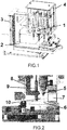
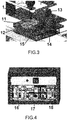
- the developed apparatus - bioprinter for printing biological tissue and organs consists of:
- the printer provides for UV radiation exposure of gel-forming composition at the moment of its spraying which allows extension of the polymerization process and ensures e.g. smooth placement of tissue spheroids layer in the dispersed layer of gel-forming composition.
- a protection unit is used for picking up the non-absorbed UV radiation, while the direction of UV radiation from the unit which can be designed as a light emitting source should be mostly perpendicular to the direction of the material output from the respective nozzle and parallel to the surface of platform with the biological tissue and/or organ, in order to avoid direct UV exposure of cells, biological tissue and/or organs.
- the UV protection unit can be designed as a hollow-section module coated from inside with a UV-absorbing material.
- the UV protection unit design provides for capability of gel-forming composition spraying to create a homogeneous polymerizable layer that serves for spatial placement, fixture and position stabilization of tissue spheroids.
- the gel-forming composition is first dispersed, and then spheroids are built in the produced layer in compliance with the digital model.
- Alginate containing liposomes with calcium ions can be used as gel-forming composition.
- the UV radiation source initiates polymerization through disruption of liposome membranes and release of calcium ions.
- Five nozzles three of which use bioink while the other two print with gel-forming composition allow simultaneous application of various materials including but not limited to the following: cells, cell-containing gel-forming compositions, spheroids.
- different mechanisms can be used for polymerization initiation, e.g.photochemical polymerization including UV radiation exposure or chemical initiation of polymerization.
- This multi-functionality shall be achieved through application of the second nozzle for hydrogel printing.
- I.e. 5 nozzles and UV emission source shall enable implementation of various mechanisms using different hydrogels for fixing and stabilization of tissue spheroids in the model.
- the movement unit (a system for precise laser positioning system for nozzles, with automatic calibration) enables the movement of nozzles with the accuracy up to 5 ⁇ m, therefore e.g. adjacent spheroids can be placed in direct contact with each other thus supporting high print quality.
- the printing process can be monitored in a real-time mode by a digital video camera installed near to the nozzles. Accurate positioning of nozzles ensures high compliance of the printed biological construct with the set digital model.
- the printing process is controlled by the software that allows using different number of nozzles in a variety of combinations as well as to handle file formats for polygonal modeling.
- the software is compatible with various 3D modeling software complexes.
- the designed apparatus includes bioink printing nozzles with the cone size ranging from 100 ⁇ m to 2.108 mm, while gel-forming composition printing nozzle cones range in size from 80 ⁇ m to 0.6414 mm. During the printing process the pressure in nozzles reaches 4.8 - 6.2 bar. Both nozzles for applying bioink and nozzles for applying gel-forming composition can be mounted on the supporting structure.
- Bioprinting control system ( Fig. 4 ) is configured to match the number and type of installed nozzles. Elevation of UV source can be regulated with the help of supporting structure (8), and such parameters as UV emission duration and radiation source operating mode can be regulated with the help of controller (18). Printing parameters for nozzles are regulated with the help of control units 16 and 17.
- Printing control system allows quick switching between different printing modes. Printing convenience and accuracy are provided by automatic calibration and tuning at mode switching.
- the apparatus can be designed so as to implement at least two separate units: (a) printing unit at least including nozzles, UV radiation unit, moving platform (b) control unit at least including a printing unit control system, a system for input of data by the operator. This design of the system allows maximal reduction of the printing unit size and, moreover, provides the required level of sterility during cell handling and protection against pollution. Information between the control unit and printing unit can be transferred by wireless methods.
- the apparatus can be designed so as to implement a plastic semitransparent enclosure to protect the internal components, including the nozzles, from dirt and to provide sterility of the printing process.
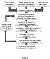
- the printing process includes the following stages: preparation of a model, bioink to be printed and gel-forming composition, setting printing parameters, bioprinter calibration, and printing process itself.
- Block diagram of the complete printing process is shown on Fig. 5 .
- the prepared model is uploaded to CAD/CAM - a specialized software for printing control.
- the digital model can be prepared by using this software itself or imported from some other application capable of three-dimensional modeling.
- Preparation of bioink consists in the formation of homogeneous spheroids of certain size and in sufficient amount. Spheroid sizes range from 100 ⁇ m to 2.108 mm, while the polymerizable layer thickness ranges from 80 ⁇ m to 0.6414 mm. Usually the selected size of spheroids is bigger than the polymerizing hydrogel layer thickness which shall ensure good contact between spheroids in different layers.
- the printing setting parameters the distance between the printing nozzles in X-Y-Z positions, the dosed (dispersed) volume of bioink, gel-forming composition application parameters, duration and intensity of the UV emission source operation.
- Bioprinter calibration is performed with the help of a precision positioning system with a tolerance of 5 ⁇ m. Bioprinter calibration is performed once in the beginning of printing process.
- Bioprinting process is cyclically repeated following the program based on the digital model.
- a nozzle that will apply bioink is selected, then the nozzle with a gel-forming composition is positioned, and then the latter is applied into a Petri cup (11) locked in a holder (15).
- the gel-forming composition (6) is exposed to UV radiation with the wavelength of 365 nm (10), or is mixed with other components initiating the polymerization process which can last from 5 s to 5 min.
- the bioink printing nozzles are positioned, and the bioink is placed in the dispersed layer of polymerizable hydrogel (12).
- the cycle is repeated ( Fig. 5 ) with new coordinates in accordance with the digital model until the whole model is printed.
- this includes both printing with a basic gel-forming composition containing cells and various combinations and sequences of printing.
- Printing process can be continuously monitored online by a digital video camera (12).
- UV module radiation is mostly directed in parallel to the printing platform to prevent exposure of the latter to direct radiation, while the diffuse UV radiation is absorbed by the protection module (7).
- Fig. 6 , 7 printing with gel-forming composition containing cells ( Fig. 6a ), tissue spheroids printing ( Fig. 6b ), encapsulated spheroids printing ( Fig. 6c ), spheroids with lumens printing ( Fig. 6d ), alginate printing with UV ( Fig. 6e ), alginate with calcium chloride printing ( Fig. 6f ), alginate with liposomes containing bound calcium ( Fig. 6g ), thrombin and fibrinogen printing ( Fig. 6h ).
- the list of modes includes, but is not limited to, the following modes:
Landscapes
- Engineering & Computer Science (AREA)
- Health & Medical Sciences (AREA)
- Chemical & Material Sciences (AREA)
- Life Sciences & Earth Sciences (AREA)
- Wood Science & Technology (AREA)
- Organic Chemistry (AREA)
- Bioinformatics & Cheminformatics (AREA)
- Zoology (AREA)
- Biomedical Technology (AREA)
- Genetics & Genomics (AREA)
- Biotechnology (AREA)
- Materials Engineering (AREA)
- Manufacturing & Machinery (AREA)
- Biochemistry (AREA)
- General Engineering & Computer Science (AREA)
- General Health & Medical Sciences (AREA)
- Microbiology (AREA)
- Molecular Biology (AREA)
- Sustainable Development (AREA)
- Cell Biology (AREA)
- Immobilizing And Processing Of Enzymes And Microorganisms (AREA)
- Micro-Organisms Or Cultivation Processes Thereof (AREA)
- Materials For Medical Uses (AREA)
Applications Claiming Priority (2)
| Application Number | Priority Date | Filing Date | Title |
|---|---|---|---|
| EA201401046A EA028039B1 (ru) | 2014-09-05 | 2014-09-05 | Устройство и способы печати биологических тканей и органов |
| PCT/RU2015/000505 WO2016036275A1 (ru) | 2014-09-05 | 2015-08-12 | Устройство и способы печати биологических тканей и органов |
Publications (2)
| Publication Number | Publication Date |
|---|---|
| EP3190171A1 true EP3190171A1 (de) | 2017-07-12 |
| EP3190171A4 EP3190171A4 (de) | 2018-04-18 |
Family
ID=55440179
Family Applications (1)
| Application Number | Title | Priority Date | Filing Date |
|---|---|---|---|
| EP15837477.7A Withdrawn EP3190171A4 (de) | 2014-09-05 | 2015-08-12 | Vorrichtung und verfahren zum drucken von biologischem gewebe und organen |
Country Status (4)
| Country | Link |
|---|---|
| US (2) | US11499135B2 (de) |
| EP (1) | EP3190171A4 (de) |
| EA (1) | EA028039B1 (de) |
| WO (1) | WO2016036275A1 (de) |
Cited By (6)
| Publication number | Priority date | Publication date | Assignee | Title |
|---|---|---|---|---|
| WO2018013737A1 (en) * | 2016-07-12 | 2018-01-18 | Deka Products Limited Partnership | System and method for printing tissue |
| US10782217B2 (en) | 2016-07-12 | 2020-09-22 | Deka Products Limited Partnership | System and method for applying force to a device |
| US11254901B2 (en) | 2016-07-12 | 2022-02-22 | Deka Products Limited Partnership | System and method for printing tissue |
| US11299705B2 (en) | 2016-11-07 | 2022-04-12 | Deka Products Limited Partnership | System and method for creating tissue |
| US11530380B2 (en) | 2017-07-12 | 2022-12-20 | Deka Products Limited Partnership | System and method for transferring tissue |
| US12297416B2 (en) | 2017-07-12 | 2025-05-13 | Deka Products Limited Partneship | System and method for transferring tissue |
Families Citing this family (22)
| Publication number | Priority date | Publication date | Assignee | Title |
|---|---|---|---|---|
| US11304676B2 (en) | 2015-01-23 | 2022-04-19 | The University Of North Carolina At Chapel Hill | Apparatuses, systems, and methods for preclinical ultrasound imaging of subjects |
| KR101795559B1 (ko) * | 2016-01-07 | 2017-11-08 | 주식회사 티앤알바이오팹 | 열민감성 세포프린팅 조성물의 세포 프린팅 장치 |
| EP3409004B1 (de) * | 2016-01-29 | 2021-03-03 | Y Soft Corporation | Sicherer 3d-drucker und 3d-drucker-verwaltungsnetzwerk |
| WO2018100580A1 (en) | 2016-12-01 | 2018-06-07 | Regenesis Biomedical Ltd. | Method and system for 3d printing |
| CN107297894A (zh) * | 2017-08-11 | 2017-10-27 | 江阴司特易生物技术有限公司 | 一种便携式生物3d打印枪 |
| WO2019060680A1 (en) | 2017-09-22 | 2019-03-28 | The Regents Of The University Of Colorado, A Body Corporate | MICROPHYSIOLOGICAL ORGAN-ON-CHIP SYSTEM |
| CN108148754A (zh) * | 2018-01-19 | 2018-06-12 | 扬州大学 | 一种3d生物打印双喷头装置 |
| US11679546B2 (en) | 2018-02-09 | 2023-06-20 | The Regents Of The University Of Colorado, A Body Corporate | Bioprinter and methods of manufacturing an organomimetic device |
| US11480560B2 (en) | 2018-06-11 | 2022-10-25 | The Regents Of The University Of Colorado, A Body Corporate | Delivery of aerosolized respiratory pathogens |
| TWI674201B (zh) * | 2018-09-10 | 2019-10-11 | 財團法人工業技術研究院 | 生物材料列印設備 |
| RU2701330C1 (ru) * | 2018-11-28 | 2019-09-25 | Частное учреждение Лаборатория биотехнологических исследований "3Д Биопринтинг Солюшенс" (ЧУ"3Д Биопринтинг Солюшенс") | Устройство для биопечати одиночными тканевыми сфероидами и используемая в нем печатающая головка |
| US20240158727A1 (en) * | 2019-03-06 | 2024-05-16 | Osaka University | Cell tissue production method, cell tissue production set, and cultivation container containing cell tissue produced by said production method |
| EP3946945A4 (de) * | 2019-04-04 | 2023-11-29 | Ohio State Innovation Foundation | Verfahren zur generativen fertigung mittels eines roboterarms |
| US12150813B2 (en) | 2019-05-10 | 2024-11-26 | The University Of North Carolina At Chapel Hill | Methods, systems, and computer readable media for generating super-resolution images of microvasculature using ultrasound |
| US12296529B2 (en) | 2020-06-09 | 2025-05-13 | 3D Systems, Inc. | Modular print bed for 3D bioprinter |
| US12541180B2 (en) | 2020-09-04 | 2026-02-03 | International Business Machines Corporation | Movement sequence analysis utilizing printed circuits |
| US20220111579A1 (en) * | 2020-10-14 | 2022-04-14 | Applied Materials, Inc. | Hybrid printing platform for 3d bioprinting of live organs |
| WO2022147077A1 (en) * | 2020-12-29 | 2022-07-07 | King Abdulaziz University | 3d printing of polymeric bioceramics for the treatment of bone defects |
| CN112920949B (zh) * | 2021-01-19 | 2022-06-14 | 浙江大学 | 一种基于dlp的载细胞多材料3d打印系统 |
| WO2022240393A1 (en) * | 2021-05-11 | 2022-11-17 | Hewlett-Packard Development Company, L.P. | Bio-ink printing |
| CN113290844B (zh) * | 2021-05-14 | 2022-10-21 | 清华大学 | 一种构建复杂异质组织/器官的多级悬浮打印方法 |
| RU2770558C2 (ru) * | 2021-09-07 | 2022-04-18 | Федеральное государственное бюджетное учреждение «Национальный медицинский исследовательский центр радиологии» Министерства здравоохранения Российской Федерации (ФГБУ «НМИЦ радиологии» Минздрава России) | Способ создания тканеинженерных конструкций методом биопечати биочернилами для регенерации хрящевой ткани в условиях организма |
Family Cites Families (14)
| Publication number | Priority date | Publication date | Assignee | Title |
|---|---|---|---|---|
| US5134569A (en) * | 1989-06-26 | 1992-07-28 | Masters William E | System and method for computer automated manufacturing using fluent material |
| LU90565B1 (en) * | 2000-04-14 | 2001-10-15 | Europ Economic Community | Device for the detection of aflatoxins |
| US8639484B2 (en) | 2003-11-14 | 2014-01-28 | Drexel University | Method and apparatus for computer-aided tissue engineering for modeling, design and freeform fabrication of tissue scaffolds, constructs, and devices |
| US8905669B2 (en) * | 2004-12-06 | 2014-12-09 | Medhesives, Inc. | Dispensing apparatus including an applicator and shield for photocurable resins |
| WO2007124481A2 (en) | 2006-04-21 | 2007-11-01 | Drexel University | Bioprinting three-dimensional structures onto microscale tissue analog devices for pharmacokinetic study and other uses |
| FR2901143B1 (fr) | 2006-05-16 | 2008-10-03 | Centre Nat Rech Scient | "methode de construction de materiaux vivants fonctionnels, materiaux obtenus et applications" |
| US20080089731A1 (en) * | 2006-10-12 | 2008-04-17 | Ezra Szoke | Method and apparatus for using dye-diffusion thermal printing |
| GB0623607D0 (en) * | 2006-11-27 | 2007-01-03 | Haemostatix Ltd | Tissue adhesive |
| US20110212501A1 (en) * | 2008-09-12 | 2011-09-01 | The Brigham And Women's Hospital, Inc. | 3-dimensional multi-layered hydrogels and methods of making the same |
| WO2010087912A1 (en) * | 2009-01-30 | 2010-08-05 | University Of Utah Research Foundation | Hydrogels crosslinked with gold nanoparticles and methods of making and using thereof |
| US20120089238A1 (en) * | 2010-10-06 | 2012-04-12 | Hyun-Wook Kang | Integrated organ and tissue printing methods, system and apparatus |
| CN108619572A (zh) * | 2011-09-12 | 2018-10-09 | 奥加诺沃公司 | 用于体外研究用途的工程化组织、其阵列及其制备方法 |
| US9499779B2 (en) * | 2012-04-20 | 2016-11-22 | Organovo, Inc. | Devices, systems, and methods for the fabrication of tissue utilizing UV cross-linking |
| CN103737931B (zh) * | 2014-01-06 | 2017-01-11 | 上海大学 | 基于数控铣床的生物3d打印试验平台 |
-
2014
- 2014-09-05 EA EA201401046A patent/EA028039B1/ru not_active IP Right Cessation
-
2015
- 2015-08-12 US US15/311,242 patent/US11499135B2/en active Active
- 2015-08-12 EP EP15837477.7A patent/EP3190171A4/de not_active Withdrawn
- 2015-08-12 WO PCT/RU2015/000505 patent/WO2016036275A1/ru not_active Ceased
-
2019
- 2019-10-28 US US16/665,187 patent/US11427802B2/en active Active
Cited By (13)
| Publication number | Priority date | Publication date | Assignee | Title |
|---|---|---|---|---|
| US11543336B2 (en) | 2016-07-12 | 2023-01-03 | Deka Products Limited Partnership | System and method for applying force to a device |
| US10782217B2 (en) | 2016-07-12 | 2020-09-22 | Deka Products Limited Partnership | System and method for applying force to a device |
| US11254901B2 (en) | 2016-07-12 | 2022-02-22 | Deka Products Limited Partnership | System and method for printing tissue |
| US12442740B2 (en) | 2016-07-12 | 2025-10-14 | Deka Products Limited Partneship | System and method for applying force to a device |
| WO2018013737A1 (en) * | 2016-07-12 | 2018-01-18 | Deka Products Limited Partnership | System and method for printing tissue |
| US11939566B2 (en) | 2016-11-07 | 2024-03-26 | Deka Products Limited Partnership | System and method for creating tissue |
| US12024701B2 (en) | 2016-11-07 | 2024-07-02 | Deka Products Limited Partnership | System and method for creating tissue |
| US12365863B2 (en) | 2016-11-07 | 2025-07-22 | Deka Products Limited Partneship | System and method for creating tissue |
| US11299705B2 (en) | 2016-11-07 | 2022-04-12 | Deka Products Limited Partnership | System and method for creating tissue |
| US12618042B2 (en) | 2016-11-07 | 2026-05-05 | Deka Products Limited Partneship | System and method for creating tissue |
| US11530380B2 (en) | 2017-07-12 | 2022-12-20 | Deka Products Limited Partnership | System and method for transferring tissue |
| US11939564B2 (en) | 2017-07-12 | 2024-03-26 | Deka Products Limited Partnership | System and method for transferring tissue |
| US12297416B2 (en) | 2017-07-12 | 2025-05-13 | Deka Products Limited Partneship | System and method for transferring tissue |
Also Published As
| Publication number | Publication date |
|---|---|
| EA028039B1 (ru) | 2017-09-29 |
| US20200056147A1 (en) | 2020-02-20 |
| EA201401046A1 (ru) | 2016-03-31 |
| US11499135B2 (en) | 2022-11-15 |
| US20170198252A1 (en) | 2017-07-13 |
| US11427802B2 (en) | 2022-08-30 |
| WO2016036275A1 (ru) | 2016-03-10 |
| EP3190171A4 (de) | 2018-04-18 |
Similar Documents
| Publication | Publication Date | Title |
|---|---|---|
| US11427802B2 (en) | Method and apparatus for printing biological tissues and organs | |
| JP2022507083A (ja) | 生体材料に対応する三次元構造を印刷するための組成物と方法 | |
| Melchels et al. | A review on stereolithography and its applications in biomedical engineering | |
| Warner et al. | Design and 3D printing of hydrogel scaffolds with fractal geometries | |
| JP6713989B2 (ja) | 3次元多細胞物体を作成するための方法と装置 | |
| US8197743B2 (en) | Hydrogel constructs using stereolithography | |
| JP2025137513A (ja) | 光重合性付加製造のための方法及び組成物 | |
| CN107320779B (zh) | 一种体外三维组织模型的制备方法及装置 | |
| BR112013003863B1 (pt) | Processo de fabricação de um suporte de engenharia de tecidos, processo para fabricação por processamento de luz digital contínua de um implante, processo para fabricação aditiva de um implante reabsorvível e implante | |
| KR101969622B1 (ko) | 고정밀 의료 임플란트용 흡수체 및 반사성 생체적합성 염료 | |
| DE102019132211B3 (de) | 3D-Gerüst aus biokompatiblem Polymer mit einem nach oben offenen Besiedlungsraum für biologische Zellen und mit einem den Besiedlungsraum umgebenden kanalförmigen Gefäß | |
| JP2025156387A (ja) | 三次元構造を作製するパターニング装置及びその製造方法 | |
| CN115916496A (zh) | 通过数字光刻系统和方法的动态微加工 | |
| US20130309706A1 (en) | Selective 3d biopatterning | |
| US11993767B2 (en) | Method for producing 3D, biocompatible polymer scaffold with a cell-filled cavity | |
| AU2024359276A1 (en) | Systems and methods for micromanufacturing and bioprinting in a micro gravity environment thereof | |
| Roh et al. | Fabrication of multi-layered macroscopic hydrogel scaffold composed of multiple components by precise control of UV energy | |
| KR101635964B1 (ko) | 타가 세포 이식용 스캐폴드 제조방법 | |
| Kiratitanaporn et al. | Multi-modal vat photopolymerization for microscale modulation of scaffold stiffness assisted via machine learning | |
| Prabhakar et al. | Printability and Shape Fidelity in Different Bioprinting Processes | |
| Chen et al. | A multi-material bioprinting platform towards stratified articular cartilage tissue fabrication | |
| Ramburrun et al. | 3D Printing of Hydrogels | |
| US20200391017A1 (en) | Method for manufacturing transdermal absorption sheet | |
| Bloomquist | Continuous Liquid Interface Production of Medical Devices for Drug Delivery and Cancer Therapy | |
| Ovsianikov et al. | Two-Photon Polymerization Technique for Bio-Applications |
Legal Events
| Date | Code | Title | Description |
|---|---|---|---|
| STAA | Information on the status of an ep patent application or granted ep patent |
Free format text: STATUS: THE INTERNATIONAL PUBLICATION HAS BEEN MADE |
|
| PUAI | Public reference made under article 153(3) epc to a published international application that has entered the european phase |
Free format text: ORIGINAL CODE: 0009012 |
|
| STAA | Information on the status of an ep patent application or granted ep patent |
Free format text: STATUS: REQUEST FOR EXAMINATION WAS MADE |
|
| 17P | Request for examination filed |
Effective date: 20161116 |
|
| AK | Designated contracting states |
Kind code of ref document: A1 Designated state(s): AL AT BE BG CH CY CZ DE DK EE ES FI FR GB GR HR HU IE IS IT LI LT LU LV MC MK MT NL NO PL PT RO RS SE SI SK SM TR |
|
| AX | Request for extension of the european patent |
Extension state: BA ME |
|
| DAV | Request for validation of the european patent (deleted) | ||
| DAX | Request for extension of the european patent (deleted) | ||
| A4 | Supplementary search report drawn up and despatched |
Effective date: 20180316 |
|
| RIC1 | Information provided on ipc code assigned before grant |
Ipc: C12N 5/00 20060101ALI20180312BHEP Ipc: B33Y 70/00 20150101ALI20180312BHEP Ipc: B33Y 30/00 20150101ALI20180312BHEP Ipc: B33Y 80/00 20150101ALI20180312BHEP Ipc: C12M 3/00 20060101AFI20180312BHEP Ipc: C12M 1/00 20060101ALI20180312BHEP |
|
| STAA | Information on the status of an ep patent application or granted ep patent |
Free format text: STATUS: EXAMINATION IS IN PROGRESS |
|
| 17Q | First examination report despatched |
Effective date: 20210217 |
|
| STAA | Information on the status of an ep patent application or granted ep patent |
Free format text: STATUS: THE APPLICATION IS DEEMED TO BE WITHDRAWN |
|
| 18D | Application deemed to be withdrawn |
Effective date: 20210828 |