EP3194624B1 - Verfahren zur diagnose, prognose und überwachung von brustkrebs und reagenzien dafür - Google Patents
Verfahren zur diagnose, prognose und überwachung von brustkrebs und reagenzien dafür Download PDFInfo
- Publication number
- EP3194624B1 EP3194624B1 EP15841542.2A EP15841542A EP3194624B1 EP 3194624 B1 EP3194624 B1 EP 3194624B1 EP 15841542 A EP15841542 A EP 15841542A EP 3194624 B1 EP3194624 B1 EP 3194624B1
- Authority
- EP
- European Patent Office
- Prior art keywords
- methylation
- tnbc
- nucleic acid
- probes
- subject
- Prior art date
- Legal status (The legal status is an assumption and is not a legal conclusion. Google has not performed a legal analysis and makes no representation as to the accuracy of the status listed.)
- Active
Links
Images























































Classifications
-
- C—CHEMISTRY; METALLURGY
- C12—BIOCHEMISTRY; BEER; SPIRITS; WINE; VINEGAR; MICROBIOLOGY; ENZYMOLOGY; MUTATION OR GENETIC ENGINEERING
- C12Q—MEASURING OR TESTING PROCESSES INVOLVING ENZYMES, NUCLEIC ACIDS OR MICROORGANISMS; COMPOSITIONS OR TEST PAPERS THEREFOR; PROCESSES OF PREPARING SUCH COMPOSITIONS; CONDITION-RESPONSIVE CONTROL IN MICROBIOLOGICAL OR ENZYMOLOGICAL PROCESSES
- C12Q1/00—Measuring or testing processes involving enzymes, nucleic acids or microorganisms; Compositions therefor; Processes of preparing such compositions
- C12Q1/68—Measuring or testing processes involving enzymes, nucleic acids or microorganisms; Compositions therefor; Processes of preparing such compositions involving nucleic acids
- C12Q1/6876—Nucleic acid products used in the analysis of nucleic acids, e.g. primers or probes
- C12Q1/6883—Nucleic acid products used in the analysis of nucleic acids, e.g. primers or probes for diseases caused by alterations of genetic material
- C12Q1/6886—Nucleic acid products used in the analysis of nucleic acids, e.g. primers or probes for diseases caused by alterations of genetic material for cancer
-
- C—CHEMISTRY; METALLURGY
- C12—BIOCHEMISTRY; BEER; SPIRITS; WINE; VINEGAR; MICROBIOLOGY; ENZYMOLOGY; MUTATION OR GENETIC ENGINEERING
- C12Q—MEASURING OR TESTING PROCESSES INVOLVING ENZYMES, NUCLEIC ACIDS OR MICROORGANISMS; COMPOSITIONS OR TEST PAPERS THEREFOR; PROCESSES OF PREPARING SUCH COMPOSITIONS; CONDITION-RESPONSIVE CONTROL IN MICROBIOLOGICAL OR ENZYMOLOGICAL PROCESSES
- C12Q2600/00—Oligonucleotides characterized by their use
- C12Q2600/106—Pharmacogenomics, i.e. genetic variability in individual responses to drugs and drug metabolism
-
- C—CHEMISTRY; METALLURGY
- C12—BIOCHEMISTRY; BEER; SPIRITS; WINE; VINEGAR; MICROBIOLOGY; ENZYMOLOGY; MUTATION OR GENETIC ENGINEERING
- C12Q—MEASURING OR TESTING PROCESSES INVOLVING ENZYMES, NUCLEIC ACIDS OR MICROORGANISMS; COMPOSITIONS OR TEST PAPERS THEREFOR; PROCESSES OF PREPARING SUCH COMPOSITIONS; CONDITION-RESPONSIVE CONTROL IN MICROBIOLOGICAL OR ENZYMOLOGICAL PROCESSES
- C12Q2600/00—Oligonucleotides characterized by their use
- C12Q2600/118—Prognosis of disease development
-
- C—CHEMISTRY; METALLURGY
- C12—BIOCHEMISTRY; BEER; SPIRITS; WINE; VINEGAR; MICROBIOLOGY; ENZYMOLOGY; MUTATION OR GENETIC ENGINEERING
- C12Q—MEASURING OR TESTING PROCESSES INVOLVING ENZYMES, NUCLEIC ACIDS OR MICROORGANISMS; COMPOSITIONS OR TEST PAPERS THEREFOR; PROCESSES OF PREPARING SUCH COMPOSITIONS; CONDITION-RESPONSIVE CONTROL IN MICROBIOLOGICAL OR ENZYMOLOGICAL PROCESSES
- C12Q2600/00—Oligonucleotides characterized by their use
- C12Q2600/154—Methylation markers
Definitions
- the present disclosure generally relates to methods and reagents for the diagnosis, prognosis or the monitoring of breast cancer, including various subtypes of breast cancer including, for example, estrogen receptor (ER) negative breast cancer, ER positive breast cancer, triple negative breast cancer (TNBC) and other subtypes of breast cancer.
- ER estrogen receptor
- TNBC triple negative breast cancer
- Cancer is a leading cause of disease worldwide.
- Breast cancer is one of the most common forms of cancer, affecting both females and males globally.
- Various subtypes of breast cancer have been distinguished based on a number of factors including the histopathological type of tumor, the grade of the tumor, the stage of the tumor, and the expression of genes which are characteristic of particular subtypes of breast cancer. Determination of the particular subtype of cancer in a patient is often of critical importance in determining the most appropriate course of treatment for the patient.
- ER negative (ER-ve) breast cancer and ER positive (ER+ve) breast cancer are two recognised subtypes of breast cancer, defined by the presence or absence of expression of the estrogen receptor gene.
- Triple negative breast cancer (TNBC) is another recognised subtype of breast cancer.
- TNBC The TNBC subtype is clinically defined by the absence of ER and progesterone receptor (PR) expression, and neither overexpression nor amplification of human epidermal growth factor receptor 2 (HER2).
- PR progesterone receptor
- HER2 human epidermal growth factor receptor 2
- TNBC represents approximately 15-20% of all newly diagnosed breast cancer cases and is generally associated with high risk of disease recurrence and shorter overall survival compared to non-TNBC.
- TNBC patients can be categorized into two distinct groups; those that succumb to their disease within 3-5 years regardless of treatment, and those that remain disease free to the extent that their overall survival exceeds that of non-TNBC patients (i.e. approximately >8 to 10 years post-diagnosis).
- DNA methylation signatures are being assessed as potential molecular biomarkers of cancer, for example WO2001/006005 teaches methods for diagnosing/prognosing breast cancer using differential methylation patterns.
- WO2001/006005 teaches methods for diagnosing/prognosing breast cancer using differential methylation patterns.
- the present inventors performed a genome-wide DNA methylation profiling analysis on CpG rich DNA from a number of breast cancer samples. In doing so, the inventors identified novel regions of differential methylation containing one or more CpG dinucleotides, including regional methylation profiles that are specific to breast cancer cells in comparison to healthy cells, including TNBC. Specific regions of differential methylation were validated for this particular subtype of breast cancer using the cancer genome atlas (TCGA) methylation data. The inventors identified at least 822 hypermethylated and at least 43 hypomethylated, statistically significant, differentially methylated regions (DMRs) harboring 64,005 and 623 CpG sites respectively. Of these, a number of DMRs were shown to be characteristic of the particular subtype of breast cancer, TNBC. These markers have been demonstrated to have significant value in the prognosis of TNBC breast cancer.
- DMRs differentially methylated regions
- the inventors identified at least 36 DMRs which were shown to be specific to TNBC samples and which therefore provide significant utility in the diagnosis of TNBC.
- these DMRs therefore also provide significant utility in the stratification of severity of TNBC during prognosis.
- a particular subset of CpG sites in any one or more DMRs selected from within a group of 17 particular DMRs have been shown by the inventors to be associated with strong survival outcomes. Therefore, any one or more CpG sites within this subset of DMRs, in any combination, can be used to determine the likelihood of survival of a subject having TNBC.
- genes and promoters associated with the DMRs identified in the present disclosure include the WT1 and WT1 antisense ( WT1-AS ) gene and its bidirectional promoter. Determining the methylation status associated with any one or more of the specific genes and associated promoters disclosed herein (such as the WT1 and WT1-AS genes) is particularly useful in the prognosis of TNBC.
- the present disclosure provides a method for prognosis of, predicting the therapeutic outcome of and/or monitoring the progression of Triple Negative Breast Cancer (TNBC) in a subject, said method comprising:
- the differential methylation of one or more CpG dinucleotide sequences within the genomic region set forth in Table 3, row 15 relative to the reference level, is associated with a likelihood of survival of the subject.
- the differential methylation of one or more CpG dinucleotide sequences within the genomic region defined in row 15 of Table 3 relative to the reference level of methylation is associated with an increased likelihood of survival of the subject.
- the differential methylation may be increased or decreased methylation relative to the reference level of methylation. In many cases, the differential methylation is increased relative to the reference level of methylation.
- increased methylation of one or more CpG dinucleotide sequences within the chr11-4047 genomic region relative to a reference level of methylation of one or more CpG dinucleotide sequences within that genomic region is associated with an increased likelihood of survival of the subject.
- the likelihood of survival may be determined in accordance with any generally accepted method of determining the likelihood of survival known in the art. In one example, the likelihood of survival is determined as a likelihood that the subject will survive at least 3 years after being diagnosed with TNBC. In another example, the likelihood of survival is determined as a likelihood that the subject will survive at least 5 years after being diagnosed with TNBC.
- methylation status of one or more CpG dinucleotide sequences may be determined according to any suitable method known in the art.
- methylation status of one or more CpG dinucleotide sequences within the one or more genomic regions analysed may be determined by one or more techniques selected from the group consisting of a nucleic acid amplification, polymerase chain reaction (PCR), methylation specific PCR, bisulfite pyrosequencing, single-strand conformation polymorphism (SSCP) analysis, restriction analysis, microarray technology, and proteomics.
- PCR polymerase chain reaction
- SSCP single-strand conformation polymorphism
- methylation status of one or more CpG dinucleotide sequences within the genomic region in the test sample may be determined by one or more of the following:
- the compound that selectively mutates non-methylated cytosine residues may be any compound suitable for that purpose, including, for example, a salt of bisulphite.
- the methods disclosed herein may be performed on any test sample taken from a subject.
- the methylation status of one or more CpG dinucleotides sequence within the one or more genomic regions can be determined in a test sample from the subject comprising tissue and/or a body fluid comprising, or suspected of comprising, a breast cancer cell or components of a breast cancer cell.
- the sample may comprise tissue, a cell and/or an extract thereof taken from a breast or lymph node.
- the body fluid may be selected from the group consisting of whole blood, a fraction of blood such as blood serum or plasma, urine, saliva, breast milk, pleural fluid, sweat, tears and mixtures thereof.
- the reference level of methylation may be a level of methylation determined for one or more CpG dinucleotide sequences within a corresponding genomic region of a sample selected from the group consisting of:
- the reference level of methylation may be a level of methylation determined for one or more CpG dinucleotide sequences within a corresponding genomic region of a healthy breast epithelial cell.
- the normal or healthy tissue may comprise a breast epithelial cell.
- the non-cancerous cell may be a breast epithelial cell.
- the present disclosure also provides a kit for diagnosing breast cancer in a subject, said kit comprising:
- kits for prognosis of, predicting the therapeutic outcome of, and/or monitoring the progression of, Triple Negative Breast Cancer (TNBC) in a subject comprising:
- the reference level of methylation may be a level of methylation determined for one or more CpG dinucleotide sequences within the corresponding genomic region of a sample selected from the group consisting of:
- the reference level of methylation may be a level of methylation determined for one or more CpG dinucleotide sequences within a corresponding genomic region of a healthy breast epithelial cell.
- the normal or healthy tissue may comprise a breast epithelial cell.
- the non-cancerous cell may be a breast epithelial cell.
- the present disclosure also provides any one of the kits disclosed herein when used in any one or more of the claimed methods disclosed herein.
- the present disclosure also provides the use of one or more reagents in the preparation of a medicament for prognosis of, predicting the therapeutic outcome of, and/or monitoring the progression of Triple Negative Breast Cancer (TNBC) in a subject, wherein the one or more reagents is/are configured to determine methylation status of one or more CpG dinucleotide sequences within the claimed genomic region in a test sample obtained from the subject.
- TNBC Triple Negative Breast Cancer
- any or the methods disclosed herein may further comprise a step of administering a therapeutic treatment to a subject.
- a therapeutic treatment for example, the determination of the severity and/or state of progression of TNBC in a subject may lead to the administration of a particular therapeutic treatment to that subject, which therapeutic treatment is particularly tailored to TNBC level of severity or progression of TNBC.
- composition of matter, group of steps or group of compositions of matter shall be taken to encompass one and a plurality (i.e. one or more) of those steps, compositions of matter, groups of steps or group of compositions of matter.
- diagnosis and variants thereof, such as, but not limited to “diagnose” or “diagnosing” shall include, but not be limited to, a primary diagnosis of a clinical state or any primary diagnosis of a clinical state.
- a diagnostic method described herein is also useful for assessing the remission of a subject, or monitoring disease recurrence, or tumor recurrence, such as following surgery, radiation therapy, adjuvant therapy or chemotherapy, or determining the appearance of metastases of a primary tumor. All such uses of the assays described herein are encompassed by the present disclosure.
- prognosis and variants thereof, such as, but not limited to “prognosing” shall refer to the prediction of the likelihood that a cancer patient e.g., a breast cancer patient, will have a cancer-attributable death or that the cancer will progress to a worsening stage in the subject, such as recurrence, metastatic spread or drug resistance of the cancer.
- cancer shall be taken to include a disease that is characterized by uncontrolled growth of cells within a subject.
- the term “cancer” shall not be limited to cancer of a specific tissue or cell type. Those skilled in the art will be aware that as a cancer progresses, metastases occur in organs and tissues outside the site of the primary cancer. Accordingly, the term “cancer” as used herein shall be taken to include a metastasis of a cancer in addition to a primary tumor.
- a particularly preferred cancer in the context of the present disclosure is breast cancer.
- breast cancer shall be understood to include a disease that is characterized by uncontrolled growth of cells from breast tissue of a subject.
- estrogen receptor negative (ER-ve) breast cancer shall be understood to refer to a breast cancer which is characterised by reduced expression of the ER gene when compared to a non-cancerous sample, or an ER+ve cancerous sample, or which is characterised by a level of expression of the ER gene which is not significantly different from the level of expression of a housekeeping gene, or which is characterised by the absence of a detectable level of expression of the ER gene, or which is characterised by the absence of expression of the ER gene.
- TNBC triple negative breast cancer
- ER estrogen receptor
- PR progesterone receptor
- HER-2 human epidermal growth factor receptor 2
- the level of expression of each one of ER, PR and HER-2 may be reduced when compared to a non-cancerous sample, or an ER+ve, PR+ve and HER2 +ve cancerous sample, or which is characterised by a level of expression of each one of ER, PR and HER-2 which is not significantly different from the level of expression of a housekeeping gene, or which is characterised by the absence of a detectable level of expression of each one of ER, PR and HER-2, or which is characterised by the absence of expression of each one of ER, PR and HER-2.
- tumor sample refers to all neoplastic cell growth and proliferation, whether malignant or benign, and all pre-cancerous and cancerous cells and tissues.
- tumor sample or similar in the context of a patient having cancer refers to a sample comprising tumor material obtained from a cancer patient.
- the term encompasses tumor tissue samples, for example, tissue obtained by surgical resection and tissue obtained by biopsy, such as for example, a core biopsy or a fine needle biopsy.
- the tumor sample is a fixed, wax-embedded tissue sample, such as a formalin-fixed, paraffin-embedded tissue sample.
- tumor sample encompasses a sample comprising tumor cells obtained from sites other than the primary tumor, e.g., circulating tumor cells.
- test sample is taken to mean any tissue or body fluid sample taken from a subject having or suspected of having breast cancer. The presence of breast cancer in the subject may therefore already have been determined. Thus, the methods of the present disclosure may be used to determine a particular subtype of breast cancer (such as ER-ve, ER+ve or TNBC) in a subject known to have breast cancer. Thus, the "test sample” may be a "tumor sample” as defined herein. Alternatively, the methods of the present disclosure may be used to determine the presence of breast cancer in a subject in whom the presence of breast cancer has not previously been determined.
- a particular subtype of breast cancer such as ER-ve, ER+ve or TNBC
- methylation will be understood to mean the presence of a methyl group added by the action of a DNA methyl transferase enzyme to a cytosine base or bases in a region of nucleic acid e.g. genomic DNA. Accordingly, the term, "methylation status" as used herein refers to the presence or absence of methylation in a specific nucleic acid region e.g., genomic region. In particular, the present disclosure relates to detection of methylated cytosine (5-methylcytosine).
- a nucleic acid sequence may comprise one or more CpG methylation sites.
- the term “differential methylation” shall be taken to mean a change in the relative amount of methylation of a nucleic acid e.g., genomic DNA, in a biological sample e.g., such as a cell or a cell extract, or a body fluid (such as blood), obtained from a subject.
- a biological sample e.g., such as a cell or a cell extract, or a body fluid (such as blood)
- a body fluid such as blood
- differentiated methylation is generally determined with reference to a baseline level of methylation for a given genomic region, such as a non-cancerous sample, including a non-cancerous matched sample from a subject known to have cancer e.g., breast cancer.
- the level of differential methylation may be at least 2% greater or less than a baseline level of methylation, for example at least 5% greater or less than a baseline level of methylation, or at least 10% greater or less than a baseline level of methylation, or at least 15% greater or less than a baseline level of methylation, or at least 20% greater or less than a baseline level of methylation, or at least 25% greater or less than a baseline level of methylation, or at least 30% greater or less than a baseline level of methylation, or at least 40% greater or less than a baseline level of methylation, or at least 50% greater or less than a baseline level of methylation, or at least 60% greater or less than a baseline level of methylation, or at least 70% greater or less than a baseline level of methylation, or at least 80% greater or less than a baseline level of methylation, or at least 90% greater or less than a baseline level of methylation.
- the level of differential methylation may be at least 10%, at least 15%, at least 20%, or at least 25% greater than or less than a baseline level of methylation.
- the level of differential methylation may be at least 10%, at least 15%, at least 20%, or at least 25% greater than a baseline level of methylation.
- CpG dinucleotide As used herein, a "CpG dinucleotide”, “CpG methylation site” or equivalent, shall be taken to denote a cytosine linked to a guanine by a phosphodiester bond. CpG dinucleotides are targets for methylation of the cytosine residue and may reside within coding or non-coding nucleic acids. Non-coding nucleic acids are understood in the art to include introns, 5'- untranslated regions, 3' untranslated regions, promoter regions of a genomic gene, or intergenic regions.
- a "reference level of methylation” shall be understood to mean a level of methylation detected in a corresponding nucleic acid from a normal or healthy cell or tissue or body fluid, or a data set produced using information from a normal or healthy cell or tissue or body fluid.
- a “reference level of methylation” may be a level of methylation in a corresponding nucleic acid from:
- the reference level of methylation may be a level of methylation determined for one or more CpG dinucleotide sequences within a corresponding genomic region of a healthy breast epithelial cell.
- the normal or healthy cell or tissue may comprise a breast epithelial cell.
- the non-cancerous cell may be a breast epithelial cell.
- the extract of the normal or healthy cell or tissue, or of the non-cancerous cell may be an extract from a breast epithelial cell.
- the term "subject” or "patient” shall be taken to mean any animal including a human, preferably a mammal.
- exemplary subjects include but are not limited to humans, primates, livestock (e.g. sheep, cows, horses, donkeys, pigs), companion animals (e.g. dogs, cats), laboratory test animals (e.g. mice, rabbits, rats, guinea pigs, hamsters), captive wild animals (e.g. fox, deer).
- livestock e.g. sheep, cows, horses, donkeys, pigs
- companion animals e.g. dogs, cats
- laboratory test animals e.g. mice, rabbits, rats, guinea pigs, hamsters
- captive wild animals e.g. fox, deer.
- the mammal is a human or primate. More preferably the mammal is a human.
- the term "survival” as used herein refers to survival of a subject having breast cancer for a particular period of time, such as at least 3 years, and preferably 5 years, from the time of diagnosis or prognosis.
- the term “survival” may refer to survival for at least 3 years, or at least 5 years, or for at least 10 years from the time of diagnosis or prognosis.
- the term “survival” may refer to survival of a subject having breast cancer for a particular period of time, such as at least 3 years, or at least 5 years, or at least 10 years following surgery or other treatment associated with breast cancer.
- the present disclosure provides methods of diagnosing breast cancer comprising detecting the methylation status of one or more CpG dinucleotides within one or more genomic regions set forth in Table 1 in a test sample taken from a subject, and determining differential methylation at said one or more CpG dinucleotides within one or more genomic regions in the test sample relative to a reference level of methylation for the corresponding one or more CpG dinucleotides within one or more corresponding genomic regions, wherein differential methylation at said one or more CpG dinucleotides within one or more genomic regions relative to the reference level of methylation is indicative of the subject having breast cancer.
- the genomic regions set forth in Table 1 are defined with reference to human genome assembly version 18 ("hg18").
- hg18 refers to the March 2006 human reference sequence (NCBI Build 36.1), which was produced by the International Human Genome Sequencing Consortium. Further information about this assembly is provided under the reference NCBI36 in the NCBI Assembly database. Thus, the nucleotide sequences of each of the regions identified in Table 1 (or in any of the Tables disclosed herein) can be identified by reference to hg18, using the "start” and “end” positions described in Table 1 (or in any of the Tables disclosed herein).
- the 865 genomic regions listed in Table 1 encompass 822 hypermethylated regions and 43 hypomethylated regions found to be differentially methylated between breast cancer samples and matched normal samples. For each DMR, the following information is provided:
- detecting increased methylation at any one or more of the DMRs set forth in rows 1-822 of Table 1 in a test sample relative to a reference level of methylation for the corresponding one or more DMRs is indicative of the subject having breast cancer.
- detecting decreased methylation at any one or more of the DMRs set forth in rows 823-865 of Table 1 in a test sample relative to a reference level of methylation for the corresponding one or more DMRs is indicative of the subject having breast cancer.
- Detecting differential methylation at a single CpG dinucleotide sequence within any one of the genomic regions defined in Table 1 may be indicative of the subject having breast cancer.
- detecting differential methylation at two or more CpG dinucleotides within any genomic region defined in Table 1 may be indicative of the subject having breast cancer.
- detecting differential methylation at two or more CpG dinucleotides, or three or more CpG dinucleotides, or four or more CpG dinucleotides, or five or more CpG dinucleotides, or six or more CpG dinucleotides, or seven or more CpG dinucleotides, or eight or more CpG dinucleotides, or nine or more CpG dinucleotides, or 10 or more CpG dinucleotides, or 20 or more CpG dinucleotides, or 30 or more CpG dinucleotides, or 40 or more CpG dinucleotides, or 50 or more CpG dinucleotides within a genomic region set forth in Table 1 may be indicative of the subject having breast cancer.
- the two or more CpG dinucleotides may be consecutive (i.e., contiguous) within a genomic region. Alternatively, the two or more CpG dinucleotides may not be consecutive (i.e., may not be contiguous) within any genomic region.
- Detecting differential methylation of at least one CpG dinucleotide within two or more different genomic regions set forth in Table 1 may be indicative of the subject having breast cancer. For example, detecting differential methylation at a CpG dinucleotide within two or more, or three or more, or four or more, or five or more, or six or more, or seven or more, or eight or more, or nine or more, or 10 or more different genomic regions set forth in Table 1 may be indicative of the subject having breast cancer.
- detecting differential methylation of at least one CpG dinucleotide residing within a CpG island defined in Table 1 is indicative of the subject having breast cancer.
- Table 1 provides details of genomic regions residing in, or overlapping with, one or more CpG islands. Such genomic regions will be recognised by a person skilled in the art.
- detecting differential methylation of at least one CpG dinucleotide residing within a CpG shore defined in Table 1 is indicative of the subject having breast cancer.
- Table 1 provides details of genomic regions residing in, or overlapping with, a CpG shore. Such genomic regions will be recognised by a person skilled in the art.
- detecting differential methylation of at least one CpG dinucleotide within one or more genomic regions defined in Table 1 associated with, or spanning, a promoter region e.g., such as a CpG island promoter of NPY, FERD3L, HMX2, SATB2 and/or C9orf125, is indicative of the subject having breast cancer.
- detecting differential methylation of at least one CpG dinucleotide within one or more genomic regions defined in Table 1 associated with, or spanning, a transcription factor e.g., such as BARHL2, DLX6, OTX2, RUNX1T1 and/or TAC1, is indicative of the subject having breast cancer.
- detecting differential methylation of at least one CpG dinucleotide within one or more genomic regions defined in Table 1 associated with, or spanning, a signalling pathway gene e.g., such as BADRB3, GHSR, NPY and/or ROBO3, is indicative of the subject having breast cancer.
- detecting differential methylation of at least one CpG dinucleotide within one or more genomic regions defined in Table 1 associated with, or spanning, a promoter region e.g., such as in C9orf125, COL14A1, ENPP2, ERG2, PLD5, ROBO3, RUNX1T1, SEMA5A, TBX18, TSHZ3, ZBTB16, and/or ZNF208, is indicative of the subject having breast cancer.
- the promoter includes a mutation and is downregulated.
- detecting differential methylation of at least one CpG dinucleotide within one or more genomic regions defined in Table 1 associated with, or spanning, a gene involved in the axon guidance pathway e.g., such as CRMP1, GDNF, GFRA1, MYL9, ROBO1, ROBO3 and/or SEMA5A is indicative of the subject having breast cancer.
- detecting hypermethylation of at least one CpG dinucleotide within one or more genomic regions defined in Table 1 associated with, or spanning, a gene involved in the axon guidance pathway e.g., such as CRMP1, GDNF, GFRA1, MYL9, ROBO1, ROBO3 and/or SEMA5A is indicative of the subject having breast cancer.
- the methods described herein encompass determining methylation status of any combination of CpG dinucleotide sequences in any combination of genomic regions set forth in Table 1, in any permutation.
- the methods disclosed herein may comprise determining the methylation status of any one or more CpG dinucleotide sequences in any 2 or more, 3 or more, 4 or more, 5 or more, 10 or more, 20 or more, 30 or more, 40 or more, 50 or more, 60 or more, 70 or more, 80 or more, 90 or more, or 100 or more genomic regions set forth in Table 1, in any permutation.
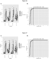
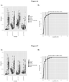
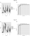
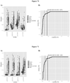
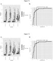
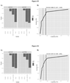
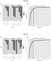
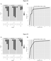
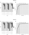
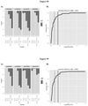
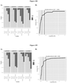
- the inventors have shown e.g., in Examples 5 and 6 herein, that small subsets of probes configured to detect differential methylation at CpG dinucleotide sequences within genomic regions set forth in Table 1 e.g., 2 or 3 or 4 or 5 probes, are capable of discriminating TNBC from non-TNBC with high sensitivity and specificity.
- a subset of probes configured to detect differential methylation at CpG dinucleotide sequences within the 2 or more, 3 or more, 4 or more, 5 or more, 10 or more, 20 or more, 30 or more, 40 or more, 50 or more, 60 or more, 70 or more, 80 or more, 90 or more, or 100 or more genomic regions set forth in Table 1 may include 1 or more, or 2 or more, or 3 or more, or 4 or more, or 5 or more, or 6 or more, or 7 or more or all probes selected from:
- probes configured to detect differential methylation at CpG dinucleotide sequences within the 2 or more, 3 or more, 4 or more, 5 or more, 10 or more, 20 or more, 30 or more, 40 or more, 50 or more, 60 or more, 70 or more, 80 or more, 90 or more, or 100 or more genomic regions set forth in Table 1 may include 1 or more, or 2 or more, or 3 or more, or 4 or more, or 5 or more, or 6 or more, or 7 or more or all probes selected from:
- Particular individual CpG dinucleotides within any of the genomic regions identified in Table 1 may be particularly strong predictors of the presence of breast cancer, or of the presence of a particular subtype of breast cancer e.g., such as ER negative breast cancer or TNBC.
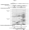
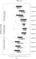
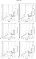
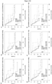
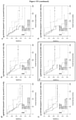
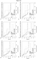
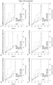
- detecting differential methylation of a CpG dinucleotide sequence within the Wilms tumour protein ( WT1 ) gene and/or its antisense counterpart, WT1-AS e.g., such as within one or more of the DMRs designated chr11-1163, chr11-1210 and/or chr11-4047 (see, e.g., Figure 3F ), is predictive of the presence of breast cancer, including the presence of a TNBC.
- Differential methylation of one or more CpG dinucleotides in any one of more of the genomic regions defined in Table 1 may alter expression of a gene within which a CpG resides. Accordingly, expression levels of genes associated with any of the genomic regions defined in Table 1 may be used as a predictor of the presence of breast cancer, or of the presence of a particular subtype of breast cancer e.g., such as ER negative breast cancer or TNBC.
- the present disclosure also provides methods of diagnosing ER-ve breast cancer specifically, involving determining the methylation status of one or more CpG dinucleotide sequences within one or more genomic regions set forth in Table 1 in a test sample obtained from a subject, and identifying differential methylation at said one or more CpG dinucleotide sequences within one or more genomic regions in the test sample relative to a reference level of methylation for the corresponding one or more CpG dinucleotide sequences.
- the present disclosure also provides methods of diagnosing triple negative breast cancer (TNBC) specifically, involving determining the methylation status of one or more CpG dinucleotides within one or more genomic regions set forth in Table 2 and/or Table 3 in a test sample obtained from a subject, and determining differential methylation of said one or more CpG dinucleotides at said one or more genomic regions in the test sample relative to a reference level of methylation for the corresponding one or more CpG dinucleotides at said one or more genomic regions.
- Differential methylation of said one or more CpG dinucleotides within one or more of the genomics regions set forth in Table 2 relative to the reference level is indicative of the subject having TNBC.
- Table 2 provides a list of 36 regions identified to be more methylated in tumours of TNBC breast cancer subtype as compared to tumours in other breast cancer subtypes.
- the genomic regions set forth in Table 2 are defined with reference to human genome assembly version 18 (hg18). For each DMR, the following information is provided in Table 2:
- detecting differential methylation of at least one CpG dinucleotide residing within a CpG island defined in Table 2 is indicative of the subject having TNBC specifically, as opposed to other breast cancer subtypes.
- Table 2 provides details of genomic regions residing in, or overlapping with, zinc finger proteins e.g., such as ZNF154 and/or ZNF671. Such genomic regions may be silenced as a result of methylation of the one or more CpG dinucleotides.
- the present invention also provides methods of prognosis of, predicting the therapeutic outcome of, and/or monitoring progression of, triple negative breast cancer (TNBC) in a subject, comprising detecting the methylation status of one or more CpG dinucleotides within the genomic region set forth in Table 3, row 15 CHr11-4047 in a test sample obtained from the subject, and determining differential methylation at said genomic region in the test sample relative to a reference level of methylation for the corresponding one or more CpG dinucleotides, wherein differential methylation at said one or more CpG dinucleotides within the genomic region set forth in Table 3, row 15 CHr11-4047 relative to the reference level of methylation is correlated with a prognosis and/or a prediction of the therapeutic outcome of the TNBC.
- TNBC triple negative breast cancer
- the prognosis performed in the inventive methods disclosed herein comprises determining a disease outcome in a subject suffering from TNBC.
- Table 3 provides a list of 17 DMRs identified as being associated with disease outcome, such as an increased or decreased likelihood of survival, in TNBC.
- the genomic regions set forth in Table 3 are defined with reference to human genome assembly version 18 (hg18). For each DMR, the following information is provided in Table 3:
- detecting differential methylation of at least one CpG dinucleotide within the claimed genomic region defined in Table 3 associated with or spanning a promoter e.g., such as a promoter of WT1-AS, and/or associated with or spanning a gene body e.g. WT1, WT1-AS and/or associated with or spanning an intergenic region is associated disease outcome, such as an increased or decreased likelihood of survival, in TNBC.
- Table 14 provides an alternative list of 20 DMRs identified as being associated with disease outcome, such as an increased or decreased likelihood of survival, in TNBC.
- the genomic regions set forth in Table 14 are defined with reference to human genome assembly version 18 (hg18). For each DMR, the following information is provided in Table 3:
- any of the methods disclosed herein may comprise detecting methylation status at one or more CpG dinucleotides within one or more regions set forth in Table 14.
- Detecting differential methylation at a single CpG dinucleotide sequence within the claimed genomic region defined in Table 3 may be predictive of an increased or a decreased likelihood of survival of the subject.
- detecting differential methylation at a single CpG dinucleotide sequence within any one of the genomic regions defined in Table 14 may be predictive of an increased or a decreased likelihood of survival of the subject.
- Examples of CpG dinucleotide sequences within the genomic regions defined in Table 14 which may be predictive of an increased or a decreased likelihood of survival of the subject are provided in Table 15.
- a method of predicting increased or a decreased likelihood of survival of a subject may comprise detecting methylation status of a single CpG dinucleotide sequence set forth in Table 15.
- detecting differential methylation at any two or more CpG dinucleotides within the claimed genomic region set forth in Table 3 may be predictive of an increased or a decreased likelihood of survival of the subject.
- detecting differential methylation at two or more CpG dinucleotides, or three or more CpG dinucleotides, or four or more CpG dinucleotides, or five or more CpG dinucleotides, or six or more CpG dinucleotides, or seven or more CpG dinucleotides, or eight or more CpG dinucleotides, or nine or more CpG dinucleotides, or 10 or more CpG dinucleotides with the claimed genomic region set forth in Table 3 may be predictive of an increased or a decreased likelihood of survival of the subject.
- detecting differential methylation at two or more CpG dinucleotide sequence within any one of the genomic regions defined in Table 14 may be predictive of an increased or a decreased likelihood of survival of the subject.
- detecting differential methylation at two or more CpG dinucleotides, or three or more CpG dinucleotides, or four or more CpG dinucleotides, or five or more CpG dinucleotides, or six or more CpG dinucleotides, or seven or more CpG dinucleotides, or eight or more CpG dinucleotides, or nine or more CpG dinucleotides, or 10 or more CpG dinucleotides with a genomic region set forth in Table 14 may be predictive of an increased or a decreased likelihood of survival of the subject.
- a method of predicting increased or a decreased likelihood of survival of a subject may comprise detecting methylation status of two or more CpG dinucleotide sequences
- detecting differential methylation of at least one CpG dinucleotide within two or more different genomic regions set forth in Table 3, but including the claimed genomic region can be predictive of an increased or a decreased likelihood of survival of the subject.
- detecting differential methylation at a CpG dinucleotide within two or more, or three or more, or four or more, or five or more, or six or more, or seven or more, or eight or more, or nine or more, or 10 or more different genomic regions set forth in Table 3, but including the claimed genomic region is predictive of an increased or a decreased likelihood of survival of the subject.
- detecting differential methylation of at least one CpG dinucleotide within two or more different genomic regions set forth in Table 14 can be predictive of an increased or a decreased likelihood of survival of the subject. For example, detecting differential methylation at a CpG dinucleotide within two or more, or three or more, or four or more, or five or more, or six or more, or seven or more, or eight or more, or nine or more, or 10 or more different genomic regions set forth in Table 14 is predictive of an increased or a decreased likelihood of survival of the subj ect.
- the greater the number of CpG dinucleotides assessed for methylation status the more reliable the prognosis of the subject.
- the greater the number of genomic regions defined in Table 3, but including the claimed genomic region, for which methylation status is determined in the methods disclosed herein the more reliable the prognosis of the subject.
- the greater the number of genomic regions defined in Table 14 for which methylation status is determined in the methods disclosed herein the more reliable the prognosis of the subject.
- Differential methylation of one or more CpG dinucleotides in the claimed genomic region defined in Table 3 may alter expression of a gene within which a CpG resides.
- expression levels of genes associated with the claimed genomic region defined in Table 3 may be used to predict of an increased or a decreased likelihood of survival of the subject.
- differential methylation of one or more CpG dinucleotides in any one of more of the genomics regions defined in Table 14 may alter expression of a gene within which a CpG resides. Accordingly, expression levels of genes associated with any of the genomic regions defined in Table 14 may be used to predict of an increased or a decreased likelihood of survival of the subject.
- detecting decreased methylation e.g., such as hypomethylation, at the DMR set forth in row 15 of Table 3 for a test sample relative to a reference level of methylation for the corresponding one or more DMRs is predictive that the subject will have an increased likelihood of survival relative to a subject in which the corresponding DMR do not show decreased methylation.
- the method of prognosis disclosed herein comprises detecting differential methylation of a CpG dinucleotide sequence within the Wilms tumour protein ( WT1 ) gene and/or its antisense counterpart, WT1-AS.
- WT1 Wilms tumour protein
- detecting increased methylation of a CpG dinucleotide sequence within the DMR designated chr11-4047 relative to the reference level of methylation for that genomic region is predictive of an increased likelihood of survival.
- the present disclosure provides the diagnosis, prognosis, or prediction of therapeutic outcome of any breast cancer, or cancer caused by a malignant cell derived from a breast.
- breast cancers include basal breast cancer, Her2 positive breast cancer, progesterone receptor positive breast cancer, estrogen receptor positive breast cancer, ductal carcinoma in situ, lobular carcinoma in situ, early breast cancer, invasive breast cancer, Paget's disease of the nipple, inflammatory breast cancer, locally advanced breast cancer and secondary breast cancer.
- Breast cancer may also be characterised according to various molecular subtypes which are typically categorized on an immunohistochemical basis. Exemplary molecular subtypes of breast cancer are as follows:
- DMRs identified herein may be particularly useful in the identification of any one or more of these known subtypes of breast cancer.
- the present inventors have identified differentially methylated regions (DMRs) in breast cancer cells compared to non-cancerous cells.
- the present inventors have also identified specific DMRs in breast cancer cells characterised as being ER-ve or TNBC compared to other breast cancer cells.
- the present inventors have demonstrated that a DMR identified in TNBC cells is capable of stratifying TNBC subtypes associated with distinct prognostic profiles e.g., populations of TNBC patients with high, medium or low risk disease outcomes.
- a method for detecting the DMR as described and claimed herein shall be taken to include detecting methylation status of CpG dinucleotide sequences in said claimed genomic region i.e., to determine whether or not a genomic region is differentially methylated relative to a reference level of methylation for the genomic region. Suitable methods for the detection of methylation status are known in the art and/or described herein.
- methylation shall be taken to mean the addition of a methyl group by the action of a DNA methyl transferase enzyme to a CpG island of nucleic acid, e.g., genomic DNA.
- a DNA methyl transferase enzyme e.g., genomic DNA.
- differentiated methylation of a nucleic acid it is meant that there is a deviation in the number of methylated CpG dinucleotides at a genomic region within the subject diagnosed compared to that detected within a corresponding genomic region in a suitable control sample i.e., which provides a reference level of methylation for that genomic region.
- the differentially methylated nucleic acid may have an increased level of methylation within a specific or defined region of nucleic acid e.g., such as hypermethylation, or a decreased level of methylation within a specific or defined region of nucleic acid e.g., such as hypomethylation.
- hypomethylation shall be taken to mean that a plurality of CpG dinucleotides in a specific or defined region of nucleic acid are methylated relative to a reference level.
- hypomethylation shall be taken to mean that a plurality of CpG dinucleotides in a specific or defined region of nucleic acid are unmethylated relative to a reference level.
- the present disclosure is not to be limited by a precise number of methylated residues that are considered to be prognostic of TNBC in a subject or predictive of its outcome, because some variation between patient samples will occur.
- the present disclosure is also not limited by the specific positioning of the methylated residue within a DMR
- the degree of methylation in a subject is determined for one or more genomic regions set forth in Tables 1-3, but including the claimed genomic region.
- probes and/or primers used in, for example, PCR or hybridization.
- Methods for designing probes and/or primers for use in, for example, PCR or hybridization are known in the art and described, for example, in Dieffenbach and Dveksler (Eds) (In: PCR Primer: A Laboratory Manual, Cold Spring Harbor Laboratories, NY, 1995).
- Eds Dieffenbach and Dveksler
- several software packages are publicly available that design optimal probes and/or primers for a variety of assays, e.g. Primer 3 available from the Center for Genome Research, Cambridge, MA, USA.
- the potential use of the probe or primer should be considered during its design.
- the probe or primer be produced for use in, for example, a methylation specific PCR or ligase chain reaction (LCR) assay the nucleotide at the 3' end (or 5' end in the case of LCR) should correspond to a methylated nucleotide in a nucleic acid.
- LCR ligase chain reaction
- Probes and/or primers useful for detection of a marker associated with a cancer are assessed, for example, to determine those that do not form hairpins, self-prime or form primer dimers (e.g. with another probe or primer used in a detection assay).
- oligonucleotide synthesis is described, in Gait (Ed) (In: Oligonucleotide Synthesis: A Practical Approach, IRL Press, Oxford, 1984 ).
- a probe or primer may be obtained by biological synthesis (e.g. by digestion of a nucleic acid with a restriction endonuclease) or by chemical synthesis. For short sequences (up to about 100 nucleotides) chemical synthesis is preferable.
- oligonucleotide synthesis include, for example, phosphotriester and phosphodiester methods ( Narang, et al. Meth. Enzymol 68: 90, 1979 ) and synthesis on a support ( Beaucage, et al Tetrahedron Letters 22: 1859-1862, 1981 ) as well as phosphoramidate technique, Caruthers, M. H., et al., "Methods in Enzymology," Vol. 154, pp. 287-314 (1988 ), and others described in " Synthesis and Applications of DNA and RNA," S. A. Narang, editor, Academic Press, New York, 1987 , and the references cited therein.
- Probes comprising locked nucleic acid are synthesized as described, for example, in Nielsen et al, J. Chem. Soc. Perkin Trans., 1: 3423, 1997 ; Singh and Wengel, Chem. Commun. 1247, 1998 . While, probes comprising peptide-nucleic acid (PNA) are synthesized as described, for example, in Egholm et al., Am. Chem. Soc., 114: 1895, 1992 ; Egholm et al., Nature, 365: 566, 1993 ; and Orum et al., Nucl. Acids Res., 21: 5332, 1993 .
- LNA locked nucleic acid
- the methylation status of one or more genomic regions in a sample is determined using a process comprising treating the nucleic acid with an amount of a methylation-sensitive restriction endonuclease enzyme under conditions sufficient for nucleic acid to be digested and then detecting the fragments produced.
- exemplary methylation-sensitive endonucleases include, for example, Hpa I or Hpa II .
- the digestion of nucleic acid is detected by selective hybridization of a probe or primer to the undigested nucleic acid.
- the probe selectively hybridizes to both digested and undigested nucleic acid but facilitates differentiation between both forms, e.g., by electrophoresis.
- Suitable detection methods for achieving selective hybridization to a hybridization probe include, for example, Southern or other nucleic acid hybridization ( Kawai et al., Mol. Cell. Biol. 14, 7421-7427, 1994 ; Gonzalgo et al., Cancer Res. 57, 594-599, 1997 ).
- the term "selectively hybridizable" means that the probe is used under conditions where a target nucleic acid hybridizes to the probe to produce a signal that is significantly above background (i.e., a high signal-to-noise ratio).
- the intensity of hybridization is measured, for example, by radiolabeling the probe, e.g. by incorporating [ ⁇ - 35 S] and/or [ ⁇ - 32 P]dNTPs, [ ⁇ - 32 P]ATP, biotin, a dye ligand (e.g., FAM or TAMRA), a fluorophore, or other suitable ligand into the probe prior to use and then detecting the ligand following hybridization.
- hybridization reaction conditions should be determined empirically for each probe, although some generalities can be applied.
- hybridizations employing short oligonucleotide probes are performed at low to medium stringency.
- a low stringency is defined herein as being a hybridization and/or a wash carried out in about 6 x SSC buffer and/or about 0.1% (w/v) SDS at about 28°C to about 40°C, or equivalent conditions.
- a moderate stringency is defined herein as being a hybridization and/or washing carried out in about 2 x SSC buffer and/or about 0.1% (w/v) SDS at a temperature in the range of about 45°C to about 65°C, or equivalent conditions.
- a high stringency hybridization and/or wash is preferred.
- a high stringency is defined herein as being a hybridization and/or wash carried out in about 0.1 x SSC buffer and/or about 0.1% (w/v) SDS, or lower salt concentration, and/or at a temperature of at least 65°C, or equivalent conditions.
- Reference herein to a particular level of stringency encompasses equivalent conditions using wash/hybridization solutions other than SSC known to those skilled in the art.
- the stringency is increased by reducing the concentration of SSC buffer, and/or increasing the concentration of SDS and/or increasing the temperature of the hybridization and/or wash.
- the conditions for hybridization and/or wash may vary depending upon the nature of the hybridization matrix used to support the sample DNA, and/or the type of hybridization probe used and/or constituents of any buffer used in a hybridization. For example, formamide reduces the melting temperature of a probe or primer in a hybridization or an amplification reaction.
- a difference in the fragments produced for the test sample and a control sample is indicative of the subject having breast cancer.
- the control sample comprises data from a tumor, cancer tissue, a cancerous cell or pre-cancerous cell e.g., such as from a subject having breast cancer, similarity, albeit not necessarily absolute identity, between the test sample and the control sample is indicative of a positive diagnosis i.e. breast cancer.
- the fragments produced by the restriction enzyme are detected using an amplification system, such as, for example, polymerase chain reaction (PCR), rolling circle amplification (RCA), inverse polymerase chain reaction (iPCR), in situ PCR ( Singer-Sam et al., Nucl. Acids Res. 18, 687,1990 ), strand displacement amplification (SDA) or cycling probe technology.
- amplification system such as, for example, polymerase chain reaction (PCR), rolling circle amplification (RCA), inverse polymerase chain reaction (iPCR), in situ PCR ( Singer-Sam et al., Nucl. Acids Res. 18, 687,1990 ), strand displacement amplification (SDA) or cycling probe technology.
- two non-complementary nucleic acid primer molecules comprising at least about 18 nucleotides in length, and more preferably at least 20-30 nucleotides in length are hybridized to different strands of a nucleic acid template molecule at their respective annealing sites, and specific nucleic acid molecule copies of the template that intervene the annealing sites are amplified enzymatically.
- Amplification products may be detected, for example, using electrophoresis and detection with a detectable marker that binds nucleic acids.
- one or more of the oligonucleotides are labeled with a detectable marker (e.g. a fluorophore) and the amplification product detected using, for example, a lightcycler (Perkin Elmer, Wellesley, MA, USA).
- Strand displacement amplification utilizes oligonucleotide primers, a DNA polymerase and a restriction endonuclease to amplify a target sequence.
- the oligonucleotides are hybridized to a target nucleic acid and the polymerase is used to produce a copy of the region intervening the primer annealing sites.
- the duplexes of copied nucleic acid and target nucleic acid are then nicked with an endonuclease that specifically recognizes a sequence at the beginning of the copied nucleic acid.
- the DNA polymerase recognizes the nicked DNA and produces another copy of the target region at the same time displacing the previously generated nucleic acid.
- SDA Strand displacement amplification
- Cycling Probe Technology uses a chimeric synthetic primer that comprises DNA-RNA-DNA that is capable of hybridizing to a target sequence. Upon hybridization to a target sequence the RNA-DNA duplex formed is a target for RNaseH thereby cleaving the primer. The cleaved primer is then detected, for example, using mass spectrometry or electrophoresis.
- primers that flank or which are adjacent tom a methylation-sensitive endonuclease recognition site, it is preferred that such primers flank only those sites that are hypermethylated in the cancer to ensure that a diagnostic and/or prognostic amplification product is produced.
- an amplification product will only be produced when the restriction site is not cleaved i.e., when it is methylated. Accordingly, detection of an amplification product indicates that the CpG dinucleotide/s of interest is/are methylated.
- This form of analysis may be used to determine the methylation status of a plurality of CpG dinucleotides within a genomic region provided that each dinucleotide is within a methylation sensitive restriction endonuclease site.
- one or more of the primers may be labeled with a detectable marker to facilitate rapid detection of amplified nucleic acid, for example, a fluorescent label (e.g. Cy5 or Cy3) or a radioisotope (e.g. 32 P).
- a detectable marker for example, a fluorescent label (e.g. Cy5 or Cy3) or a radioisotope (e.g. 32 P).
- the amplified nucleic acids are generally analyzed using, for example, non-denaturing agarose gel electrophoresis, non-denaturing polyacrylamide gel electrophoresis, mass spectrometry, liquid chromatography (e.g. HPLC or dHPLC), or capillary electrophoresis. (e.g. MALDI-TOF).
- High throughput detection methods such as, for example, matrix-assisted laser desorption/ionization time of flight (MALDI-TOF), electrospray ionization (ESI), mass spectrometry (including tandem mass spectrometry, e.g.
- amplification of a nucleic acid may be continuously monitored using a melting curve analysis method as described herein and/or in, for example, US 6,174,670 .
- the methylation status of the claimed genomic region in a subject sample is determined using a process comprising treating the nucleic acid with an amount of a compound that selectively mutates a non-methylated cytosine residue within a CpG dinucleotide under conditions sufficient to induce mutagenesis.
- Exemplary compounds mutate cytosine to uracil or thymidine, such as, for example, a salt of bisulfite, e.g., sodium bisulfite or potassium bisulfite ( Frommer et al., Proc. Natl. Acad. Sci. USA 89, 1827-1831, 1992 ).
- Bisulfite treatment of DNA is known to distinguish methylated from non-methylated cytosine residues, by mutating cytosine residues that are not protected by methylation, including cytosine residues that are not within a CpG dinucleotide or that are positioned within a CpG dinucleotide that is not subject to methylation.
- the presence of one or more mutated nucleotides in the claimed genomic region or the number of mutated sequences in a sample is determined by sequencing mutated DNA.
- One form of analysis comprises amplifying mutated nucleic acid or methylated nucleic acid using an amplification reaction described herein, for example, PCR. The amplified product is then directly sequenced or cloned and the cloned product sequenced.
- Methods for sequencing DNA include for example, the dideoxy chain termination method or the Maxam-Gilbert method (see Sambrook et al., Molecular Cloning, A Laboratory Manual (2nd Ed., CSHP, New York 1989 ) or Zyskind et al., Recombinant DNA Laboratory Manual, (Acad. Press, 1988 )).
- nucleic acid with a compound such as, for example, bisulfite results in non-methylated cytosines being mutated to uracil or thymidine
- analysis of the sequence determines the presence or absence of a methylated nucleotide. For example, by comparing the sequence obtained using a control sample or a sample that has not been treated with bisulfite, or the known nucleotide sequence of the region of interest with a treated sample facilitates the detection of differences in the nucleotide sequence. Any thymine residue detected at the site of a cytosine in the treated sample compared to a control or untreated sample may be considered to be caused by mutation as a result of bisulfite treatment.
- Suitable methods for the detection of methylation using sequencing of bisulfite treated nucleic acid are described, for example, in Frommer et al., Proc. Natl. Acad. Sci. USA 89: 1827 - 1831, 1992 or Clark et al., Nucl. Acids Res. 22: 2990-2997, 1994 .
- One example of a commercially available kit for carrying out such methods is the CpGenome TM DNA modification Kit (Millipore).
- Other suitable kits are available from MDX Health SA (Belgium).
- the presence of a mutated or non-mutated nucleotide in a bisulfite treated sample is detected using pyrosequencing, such as, for example, as described in Uhlmann et al., Electrophoresis, 23: 4072 -4079, 2002 .
- this method is a form of real-time sequencing that uses a primer that hybridizes to a site adjacent or close to the site of a cytosine that is methylated in a cancer cell. Following hybridization of the primer and template in the presence of a DNA polymerase each of four modified deoxynucleotide triphosphates are added separately according to a predetermined dispensation order.
- PPi inorganic pyrophosphate
- a related method for determining the sequence of a bisulfite treated nucleotide is methylation-sensitive single nucleotide primer extension (Me-SnuPE) or SNaPmeth. Suitable methods are described, for example, in Gonzalgo and Jones Nucl. Acids Res., 25: 2529-2531 or Uhlmann et al., Electrophoresis, 23: 4072 -4079, 2002 .
- Such methods include, for example, solid phase mini sequencing (as described, for example, in Syvämen et al, Genomics, 13: 1008-1017, 1992 ), or minisequencing with FRET (as described, for example, in Chen and Kwok, Nucleic Acids Res. 25: 347-353, 1997 ).
- the presence of a non-mutated nucleic sequence is detected using combined bisulfite restriction analysis (COBRA) essentially as described in Xiong and Laird, Nucl. Acids Res., 25: 2532-2534, 2001 .
- COBRA combined bisulfite restriction analysis
- This method exploits the differences in restriction enzyme recognition sites between methylated and unmethylated nucleic acid after treatment with a compound that selectively mutates a non-methylated cytosine residue, e.g., bisulfite.
- a region of interest comprising one or more CpG dinucleotides that are methylated in a cancer cell and are included in a restriction endonuclease recognition sequence is amplified using an amplification reaction described herein, e.g., PCR.
- the amplified product is then contacted with the restriction enzyme that cleaves at the site of the CpG dinucleotide for a time and under conditions sufficient for cleavage to occur.
- a restriction site may be selected to indicate the presence or absence of methylation.
- the restriction endonuclease Taq I cleaves the sequence TCGA, following bisulfite treatment of a non-methylated nucleic acid the sequence will be TTGA and, as a consequence, will not be cleaved.
- the digested and/or non-digested nucleic acid is then detected using a detection means known in the art, such as, for example, electrophoresis and/or mass spectrometry.
- the cleavage or non-cleavage of the nucleic acid is indicative of cancer in a subject.
- this method may be employed in either a positive read-out or negative read-out system for the diagnosis of a cancer.
- the assay format of the invention comprises a positive read-out system in which DNA from a cancer sample e.g., breast cancer, that has been treated, for example, with bisulfite is detected as a positive signal.
- a cancer sample e.g., breast cancer
- bisulfite is detected as a positive signal.
- the nonhypermethylated DNA from a healthy or normal control subject is not detected or only weakly detected.
- the enhanced methylation in a subject sample is determined using a process comprising:
- the term “selective hybridization” means that hybridization of a probe or primer to the non-mutated nucleic acid occurs at a higher frequency or rate, or has a higher maximum reaction velocity, than hybridization of the same probe or primer to the corresponding mutated sequence.
- the probe or primer does not hybridize or detectably hybridize (e.g., does not hybridize at a level significantly above background) to the non-methylated sequence carrying the mutation(s) under the reaction conditions used.
- the hybridization is detected using Southern, dot blot, slot blot or other nucleic acid hybridization means ( Kawai et al., Mol. Cell. Biol. 14, 7421-7427, 1994 ; Gonzalgo et al., Cancer Res. 57, 594-599, 1997 ).
- Subject to appropriate probe selection, such assay formats are generally described herein above and apply mutatis mutandis to the presently described selective mutagenesis approach.
- a ligase chain reaction format is employed to distinguish between a mutated and non-mutated nucleic acid.
- Ligase chain reaction (described in EP 320,308 and US 4,883,750 ) uses at least two oligonucleotide probes that anneal to a target nucleic acid in such a way that they are juxtaposed on the target nucleic acid such that they can be linked using a ligase.
- the probes that are not ligated are removed by modifying the hybridization stringency. In this respect, probes that have not been ligated will selectively hybridize under lower stringency hybridization conditions than probes that have been ligated.
- the stringency of the hybridization can be increased to a stringency that is at least as high as the stringency used to hybridize the longer probe, and preferably at a higher stringency due to the increased length contributed by the shorter probe following ligation.
- One exemplary method melts the target-probe duplex, elute the dissociated probe and confirm that is has been ligated, e.g., by determining its length using electrophoresis, mass spectrometry, nucleotide sequence analysis, gel filtration, or other means known to the skilled artisan.
- Methylation specific microarrays are also useful for differentiating between a mutated and non-mutated sequence.
- a suitable method is described, for example, in Adorján et al, Nucl. Acids Res., 30: e21, 2002 .
- MSO uses nucleic acid that has been treated with a compound that selectively mutates a non-methylated cytosine residue (e.g., bisulfite) as template for an amplification reaction that amplifies both mutant and non-mutated nucleic acid.
- the amplification is performed with at least one primer that comprises a detectable label, such as, for example, a fluorophore, e.g., Cy3 or Cy5.
- the labeled amplification products are then hybridized to oligonucleotides on the microarray under conditions that enable detection of single nucleotide differences.
- hybridization is detected using, for example, a microarray scanner.
- this method allows for determination of the methylation status of a large number of CpG dinucleotides, it is also semi-quantitative, enabling determination of the degree of methylation at each CpG dinucleotide analyzed. As there may be some degree of heterogeneity of methylation in a single sample, such quantification may assist in the diagnosis of cancer.
- the hybridization is detected using an amplification system.
- the hybridization is detection using a process comprising amplifying the bisulfite-treated DNA.
- an amplification product is only produced using a sample comprising a methylated nucleotide.
- amplification assay format described herein can be used, such as, for example, polymerase chain reaction (PCR), rolling circle amplification (RCA), inverse polymerase chain reaction (iPCR), in situ PCR ( Singer-Sam et al., Nucl. Acids Res. 18, 687,1990 ), strand displacement amplification, or cycling probe technology.
- PCR polymerase chain reaction
- RCA rolling circle amplification
- iPCR inverse polymerase chain reaction
- in situ PCR Singer-Sam et al., Nucl. Acids Res. 18, 687,1990
- strand displacement amplification or cycling probe technology.
- PCR techniques have been developed for detection of gene mutations ( Kuppuswamy et al., Proc. Natl. Acad. Sci. USA 88:1143-1147, 1991 ) and quantitation of allelic-specific expression ( Szabo and Mann, Genes Dev. 9: 3097-3108, 1995 ; and Singer-Sam et al., PCR Methods Appl. 1: 160-163, 1992 ).
- Such techniques use internal primers, which anneal to a PCR-generated template and terminate immediately 5' of the single nucleotide to be assayed.
- Such as format is readily combined with ligase chain reaction as described herein above.
- Methylation-specific melting-curve analysis (essentially as described in Worm et al., Clin. Chem., 47: 1183-1189, 2001 ) is also contemplated by the present invention.
- This process exploits the difference in melting temperature in amplification products produced using bisulfite treated methylated or unmethylated nucleic acid.
- non-discriminatory amplification of a bisulfite treated sample is performed in the presence of a fluorescent dye that specifically binds to double stranded DNA (e.g., SYBR Green I).
- SYBR Green I fluorescent dye that specifically binds to double stranded DNA
- the temperature at which the fluorescence decreases is indicative of the nucleotide sequence of the amplified nucleic acid, thereby permitting the nucleotide at the site of one or more CpG dinucleotides to be determined.
- the present invention also encompasses the use of real-time quantitative forms of PCR, such as, for example, TaqMan ( Holland et al., Proc. Natl Acad. Sci. USA, 88, 7276-7280, 1991 ; Lee et al., Nucleic Acid Res. 21, 3761-3766, 1993 ) to perform this embodiment.
- TaqMan Holland et al., Proc. Natl Acad. Sci. USA, 88, 7276-7280, 1991 ; Lee et al., Nucleic Acid Res. 21, 3761-3766, 1993
- MethylLight method of Eads et al., Nucl. Acids Res. 28: E32, 2000 uses a modified TaqMan assay to detect methylation of a CpG dinucleotide.
- a probe such as, for example, a Molecular Beacon TM is used (see, for example, Mhlang and Malmberg, Methods 25: 463-471, 2001 ).
- Molecular beacons are single stranded nucleic acid molecules with a stem-and-loop structure.
- the loop structure is complementary to the region surrounding the one or more CpG dinucleotides that are methylated in a cancer sample and not in a control sample.
- the stem structure is formed by annealing two "arms" complementary to each other, which are on either side of the probe (loop).
- a fluorescent moiety is bound to one arm and a quenching moiety that suppresses any detectable fluorescence when the molecular beacon is not bound to a target sequence is bound to the other arm.
- the arms Upon binding of the loop region to its target nucleic acid the arms are separated and fluorescence is detectable. However, even a single base mismatch significantly alters the level of fluorescence detected in a sample.
- the presence or absence of a particular base is determined by the level of fluorescence detected.
- Such an assay facilitates detection of one or more unmutated sites (i.e. methylated nucleotides) in a nucleic acid.
- amplification based assay useful for the detection of a methylated nucleic acid following treatment with a compound that selectively mutates a non-methylated cytosine residue makes use of headloop PCR technology (e.g., WO 03/072810 ).
- This form of amplification uses a probe or primer that comprises a region that binds to a nucleic acid and is capable of amplifying nucleic acid in an amplification reaction whether the nucleic acid is methylated or not.
- the primer additionally comprises a region that is complementary to a portion of the amplified nucleic acid enabling this region of the primer to hybridize to the amplified nucleic acid incorporating the primer thereby forming a hairpin.
- the now 3' terminal nucleotide/s of the annealed region hybridize to the site of one or more mutated cytosine residues (i.e., unmethylated in nucleic acid from a cancer subject). Accordingly, this facilitates self priming of amplification products from unmethylated nucleic acid, the thus formed hairpin structure blocking further amplification of this nucleic acid.
- the complementary region may or may not by capable of hybridizing to an amplification product from methylated (mutated) nucleic acid, but is unable to "self prime” thereby enabling further amplification of this nucleic acid (e.g., by the inability of the now 3' nucleotide to hybridize to the amplification product).
- This method may be performed using a melting curve analysis method to determine the amount of methylated nucleic acid in a biological sample from a subject.
- amplification based methods for detecting methylated nucleic acid following treatment with a compound that selectively mutates a non-methylated cytosine residue include, for example, methylation-specific single stranded conformation analysis (MS-SSCA) ( Bianco et al., Hum. Mutat., 14: 289-293, 1999 ), methylation-specific denaturing gradient gel electrophoresis (MS-DGGE) ( Abrams and Stanton, Methods Enzymol., 212: 71-74, 1992 ) and methylation-specific denaturing high-performance liquid chromatography (MS-DHPLC) ( Deng et al, Chin. J. Cancer Res., 12: 171-191, 2000 ).
- MS-SSCA methylation-specific single stranded conformation analysis
- MS-DGGE methylation-specific denaturing gradient gel electrophoresis
- MS-DHPLC methylation-specific denaturing high-performance liquid chromatography
- the assay format comprises a negative read-out system in which reduced methylation of DNA from a healthy/normal control sample is detected as a positive signal and preferably, methylated DNA from a cancer sample e.g., a breast cancer sample, is not detected or is only weakly detected.
- the reduced methylation is determined using a process comprising:
- the term “selective hybridization” means that hybridization of a probe or primer to the mutated nucleic acid occurs at a higher frequency or rate, or has a higher maximum reaction velocity, than hybridization of the same probe or primer to the corresponding non-mutated sequence.
- the probe or primer does not hybridize or detectably hybridize to the methylated sequence (or non-mutated sequence) under the reaction conditions used.
- the claimed methods disclosed herein may further comprise one or more steps of enriching methylated DNA in a sample.
- the claimed methods disclosed herein may further comprise one or more steps of isolating methylated DNA from a sample.
- the enrichment/isolation step may be performed prior to or concomitant with any other step in the method for detecting the level of methylation of a marker as disclosed herein.
- the methods disclosed herein may comprise a step of enriching methylated DNA in a sample using a commercially available kit such as the CpG MethylQuest DNA Isolation Kit (Millipore), which provides a recombinant protein comprising the methyl binding domain (MBD) of the mouse MBD2b protein fused to a glutathione-S-transferase (GST) protein from S. japonicum via a linker containing a thrombin cleavage site, the recombinant protein being immobilized to a magnetic bead.
- MBD methyl binding domain
- GST glutathione-S-transferase
- the MBD binds to methylated CpG sites with high affinity and in a sequence-independent manner, thereby allowing enrichment of methylated DNA in a sample.
- a method disclosed herein according to any example may also comprise selecting a patient based on the result of a method disclosed herein and performing an additional diagnostic method or recommending performance of an additional diagnostic method.
- the additional diagnostic method may be an ultrasound or a biopsy.
- the present inventors have also demonstrated that the level of expression of nucleic acids within any of a number of genomic regions described herein, but including the claimed region, is varied (e.g., reduced or increased) in breast cancer subjects and in breast cancer cell lines.
- the methods disclosed herein may additionally or alternatively comprise determining the level of expression of any polynucleotides within any of the genomic regions identified in any of the Tables herein.
- the level of expression of a nucleic acid is determined by detecting the level of mRNA transcribed from genomic region described herein.
- the mRNA is detected by hybridizing a nucleic acid probe or primer capable of specifically hybridizing to a transcript of a genomic region described herein to a nucleic acid in a biological sample derived from a subject and detecting the hybridization by a detection means.
- the detection means is an amplification reaction, or a nucleic acid hybridization reaction, such as, for example, as described herein.
- the term “selective hybridization” means that hybridization of a probe or primer to the transcript occurs at a higher frequency or rate, or has a higher maximum reaction velocity, than hybridization of the same probe or primer to any other nucleic acid.
- the probe or primer does not hybridize to another nucleic acid at a detectable level under the reaction conditions used.
- transcripts of a gene or pseudogene described herein are detected using mRNA or cDNA derived therefrom, assays that detect changes in mRNA are preferred (e.g. Northern hybridization, RT-PCR, NASBA, TMA or ligase chain reaction).
- assays that detect changes in mRNA are preferred (e.g. Northern hybridization, RT-PCR, NASBA, TMA or ligase chain reaction).
- RT-PCR Methods of RT-PCR are known in the art and described, for example, in Dieffenbach (ed) and Dveksler (ed) (In: PCR Primer: A Laboratory Manual, Cold Spring Harbour Laboratories, NY, 1995 ). Essentially, this method comprises performing a PCR reaction using cDNA produced by reverse transcribing mRNA from a cell using a reverse transcriptase. Methods of PCR described supra are to be taken to apply mutatis mutandis to this embodiment of the disclosure.
- LCR may be performed using cDNA.
- one or more of the probes or primers used in the reaction specifically hybridize to the transcript of interest. Method of LCR are described supra and are to be taken to apply mutatis mutandis to this embodiment of the disclosure.
- Methods of TMA or self-sustained sequence replication use two or more oligonucleotides that flank a target sequence, a RNA polymerase, RNase H and a reverse transcriptase.
- One oligonucleotide (that also comprises a RNA polymerase binding site) hybridizes to an RNA molecule that comprises the target sequence and the reverse transcriptase produces cDNA copy of this region.
- RNase H is used to digest the RNA in the RNA-DNA complex, and the second oligonucleotide used to produce a copy of the cDNA.
- the RNA polymerase is then used to produce a RNA copy of the cDNA, and the process repeated.
- NASBA systems relies on the simultaneous activity of three enzymes (a reverse transcriptase, RNase H and RNA polymerase) to selectively amplify target mRNA sequences.
- the mRNA template is transcribed to cDNA by reverse transcription using an oligonucleotide that hybridizes to the target sequence and comprises a RNA polymerase binding site at its 5' end.
- the template RNA is digested with RNase H and double stranded DNA is synthesized.
- the RNA polymerase then produces multiple RNA copies of the cDNA and the process is repeated.
- the present disclosure also contemplates the use of a microarray to determine the level of expression of one or more nucleic acids described herein. Such a method enables the detection of a number of different nucleic acids, thereby providing a multi-analyte test and improving the sensitivity and/or accuracy of the diagnostic assay of the disclosure.
- the level of expression of the claimed genomic region is determined by detecting the level of a protein encoded by a nucleic acid within a genomic region described herein.
- the present disclosure is not necessarily limited to the detection of a protein comprising the specific amino acid sequence recited herein. Rather, the present disclosure encompasses the detection of variant sequences (e.g., having at least about 80% or 90% or 95% or 98% amino acid sequence identity) or the detection of an immunogenic fragment or epitope of said protein.
- the amount, level or presence of a polypeptide is determined using any of a variety of techniques known to the skilled artisan such as, for example, a technique selected from the group consisting of, immunohistochemistry, immunofluorescence, an immunoblot, a Western blot, a dot blot, an enzyme linked immunosorbent assay (ELISA), radioimmunoassay (RIA), enzyme immunoassay, fluorescence resonance energy transfer (FRET), matrix-assisted laser desorption/ionization time of flight (MALDI-TOF), electrospray ionization (ESI), mass spectrometry (including tandem mass spectrometry, e.g. LC MS/MS), biosensor technology, evanescent fiber-optics technology or protein chip technology.
- the assay used to determine the amount or level of a protein is a semi-quantitative assay.
- the assay used to determine the amount or level of a protein in a quantitative assay may require the use of a suitable control, e.g. from a normal individual or matched normal control.
- Standard solid-phase ELISA or FLISA formats are particularly useful in determining the concentration of a protein from a variety of samples.
- such an assay involves immobilizing a biological sample onto a solid matrix, such as, for example a polystyrene or polycarbonate microwell or dipstick, a membrane, or a glass support (e.g. a glass slide).
- a solid matrix such as, for example a polystyrene or polycarbonate microwell or dipstick, a membrane, or a glass support (e.g. a glass slide).
- An antibody that specifically binds to a protein described herein is brought into direct contact with the immobilized biological sample, and forms a direct bond with any of its target protein present in said sample.
- This antibody is generally labelled with a detectable reporter molecule, such as for example, a fluorescent label (e.g. FITC or Texas Red) or a fluorescent semiconductor nanocrystal (as described in US 6,306,610 ) in the case of a FLISA or an enzyme (e.g.
- HRP horseradish peroxidase
- AP alkaline phosphatase
- ⁇ -galactosidase ⁇ -galactosidase
- a second labeled antibody can be used that binds to the first antibody.
- the label is detected either directly, in the case of a fluorescent label, or through the addition of a substrate, such as for example hydrogen peroxide, TMB, or toluidine, or 5-bromo-4-chloro-3-indol-beta-D-galaotopyranoside (x-gal) in the case of an enzymatic label.
- a substrate such as for example hydrogen peroxide, TMB, or toluidine, or 5-bromo-4-chloro-3-indol-beta-D-galaotopyranoside (x-gal) in the case of an enzymatic label.
- an ELISA or FLISA comprises immobilizing an antibody or ligand that specifically binds a protein described supra on a solid matrix, such as, for example, a membrane, a polystyrene or polycarbonate microwell, a polystyrene or polycarbonate dipstick or a glass support.
- a sample is then brought into physical relation with said antibody, and the polypeptide is bound or 'captured'.
- the bound protein is then detected using a labeled antibody.
- a labeled antibody that binds to an epitope that is distinct from the first (capture) antibody is used to detect the captured protein.
- a third labeled antibody can be used that binds the second (detecting) antibody.
- the presence or level of a protein is detected in a body fluid using, for example, a biosensor instrument (e.g., BIAcore TM , Pharmacia Biosensor, Piscataway, N.J.).
- a biosensor instrument e.g., BIAcore TM , Pharmacia Biosensor, Piscataway, N.J.
- an antibody or ligand that specifically binds a protein is immobilized onto the surface of a receptor chip.
- the antibody or ligand is covalently attached to dextran fibers that are attached to gold film within the flow cell of the biosensor device.
- a test sample is passed through the cell. Any antigen present in the body fluid sample, binds to the immobilized antibody or ligand, causing a change in the refractive index of the medium over the gold film, which is detected as a change in surface plasmon resonance of the gold film.
- the presence or level of a protein or a fragment or epitope thereof is detected using a protein and/or antibody chip.
- a protein and/or antibody chip an antibody or ligand that binds to the antigen of interest is bound to a solid support such as, for example glass, polycarbonate, polytetrafluoroethylene, polystyrene, silicon oxide, gold or silicon nitride.
- This immobilization is either direct (e.g. by covalent linkage, such as, for example, Schiff's base formation, disulfide linkage, or amide or urea bond formation) or indirect.
- a streptavidin chip is also useful for capturing proteins and/or peptides and/or nucleic acid and/or cells that have been conjugated with biotin (e.g. as described in Pavlickova et al., Biotechniques, 34: 124-130, 2003 ).
- biotin e.g. as described in Pavlickova et al., Biotechniques, 34: 124-130, 2003 .
- a peptide is captured on a microfabricated polyacrylamide gel pad and accelerated into the gel using microelectrophoresis as described in, Arenkov et al. Anal. Biochem. 278:123-131, 2000 .
- assay formats are also contemplated, such as flow-trough immunoassays WO2003/050537 a lateral flow immunoassay ( US20040228761 , US20040248322 or US20040265926 ), a fluorescence polarization immunoassay (FPIA) ( U.S. Pat. Nos. 4,593,089 , 4,492,762 , 4,668,640 , and 4,751,190 ), a homogeneous microparticles immunoassay (“HMI”) (e.g., U.S. Pat. Nos. 5,571,728 , 4,847,209 , 6,514,770 , and 6,248,597 ) or a chemiluminescent microparticle immunoassay ("CMIA").
- HMI homogeneous microparticles immunoassay
- CMIA chemiluminescent microparticle immunoassay
- the present disclosure also contemplates multiplex or multianalyte format assays to improve the accuracy or specificity of prognosis of TNBC. Such assays may also improve the population coverage by an assay.
- an assay described herein is used to analyze a population of test subjects to determine those that will develop cancer. Post-mortem analysis is then used to determine those subjects that did actually determine breast cancer. The number of "true positives" (i.e., subjects that developed breast cancer and were positively identified using the method of the disclosure) and "true negatives” (i.e., subjects that did not develop breast cancer and were not identified using the method of the disclosure) are determined.
- Sensitivity of the assay is then determined by the following formula: No . of true positives/ No . of true positives + No . of false negatives .
- a method of the disclosure has a high degree of sensitivity in detecting of subjects developing or suffering from TNBC.
- the assay detects at least about 50% of subjects developing or suffering from TNBC, for example, at least about 60% of subjects developing or suffering from TNBC, for example, at least about 65% of subjects developing or suffering from TNBC, for example, at least about 70% of subjects developing or suffering from TNBC, for example, at least about 75% of subjects developing or suffering from TNBC, for example, at least about 80% of subjects developing or suffering from TNBC, for example, at least about 85% of subjects developing or suffering from TNBC, for example, at least about 90% of subjects developing or suffering from TNBC, for example, at least about 95% of subjects developing or suffering from TNBC.
- a method of the disclosure has a high degree of sensitivity in stratifying TNBC subtypes associated with distinct prognostic profiles e.g., such as populations of TNBC patients with high, medium or low risk disease outcomes.
- the assay stratifies at least about 50% of subjects having TNBC according to a disease outcome, for example, at least about 60% of subjects having TNBC according to a disease outcome, for example, at least about 70% of subjects having TNBC according to a disease outcome, for example, at least about 80% of subjects having TNBC according to a disease outcome, for example, at least about 85% of subjects having TNBC according to a disease outcome, for example, at least about 90% of subjects having TNBC according to a disease outcome, for example, at least about 95% of subjects having TNBC according to a disease outcome.
- a disease outcome in accordance with this example is a likelihood that the breast cancer patient will survive at least 3 years from the time or diagnosis/prognosis, for example, at least 5 years from the time or diagnosis/prognosis, for example, at least 10 years from the time or diagnosis/prognosis.
- An exemplary multiplex assay comprises, for example, detecting differential methylation of one or more CpG dinucleotides in a plurality of DMRs set forth in Table 3, but including the claimed region.
- the method comprises detecting the level of methylation of one or more CpG dinucleotides in a plurality of DMRs set forth in Table 3, but including the claimed region, to stratify and/or predict disease outcome in a patient suffering from TNBC.
- the multiplex assay of the disclosure is not to be limited to the detection of methylation at a single CpG dinucleotide within a region of interest i.e., each DMR. Rather, the present disclosure contemplates detection of methylation at a sufficient number of CpG dinucleotides in each nucleic acid to provide a prognosis. For example, the disclosure contemplates detection of methylation at 1 or 2 or 3 or 4 or 5 or 7 or 9 or 10 or 15 or 20 or 25 or 30 CpG dinculeotides in each nucleic acid i.e., each DMR. Preferably, the disclosure contemplates detection of methylation at more than 1 CpG dinculeotide in each nucleic acid i.e., each DMR.
- a methylation specific microarray is amenable to such high density analysis.
- up to 232 CpG dinucleotides have been analyzed using such a microarray ( Adorján et al., Nucl. Acids Res. 30: e21, 2002 ).
- the method determines the level of expression of a gene comprising, or comprised in, at least one DMR set forth in Table 3, but including the claimed region, to prognose breast cancer.
- the method determines the level of expression of a gene comprising, or comprised in, at least one DMR set forth in Table 3, but including the claimed region, to stratify and/or predict disease outcome in a patient suffering from TNBC.
- the level of mRNA or protein may be detected.
- the level of mRNA transcribed from one or more genes and the level of one or more proteins expressed by the same or different genes may be determined.
- Each of the previously described detection techniques can be used independently of one another to prognose TNBC. Accordingly, a single sample may be analyzed to determine the level of methylation of one or more CpG dinculeotides in one or more nucleic acids and the level of expression of one or more nucleic acids and/or proteins is also determined. In accordance with this example, enhanced methylation and reduced gene expression is indicative of cancer.
- the present disclosure also contemplates the use of a known diagnostic assay in combination with an assay described herein. For example, detection of a mutation in a BRCA gene using an assay described herein may be used to diagnose breast cancer.
- a sample useful for the method of the present disclosure is, for example, from a tissue suspected of comprising a breast cancer or breast cancer cell.
- the cell is from a region of a tissue thought to comprise a breast cancer or breast cancer cell. This does not exclude cells that have originated in a particular tissue but are isolated from a remote source.
- the sample may be taken from a subject suspected of having or being at risk of developing breast cancer.
- the subject may have a family history of cancer, may have been subjected to tests identifying elevated levels of BRCA gene (which, in one example, may be deemed to indicate an increased likelihood of having or being susceptible to developing breast cancer), or may have been subjected to any other test for detecting and/or determining the likelihood of developing any form of breast cancer.
- the sample may be taken from a subject who has been subjected to any combination of any known test for detecting and/or determining the likelihood of developing any form of breast cancer.
- the sample may be taken from a subject not previously suspected of having breast cancer.
- the sample comprises a body fluid or a derivative of a body fluid or a body secretion.
- the body fluid is selected from the group consisting of whole blood, urine, saliva, breast milk, pleural fluid, sweat, tears and mixtures thereof.
- An example of a derivative of a body fluid is selected from the group consisting of plasma, serum or buffy coat fraction.
- the sample comprises a whole blood sample, a serum sample or a plasma sample.
- DNA is isolated from either; whole blood, plasma, serum, peripheral blood mononucleated cells (PBMC) or enriched epithelial cells derived from the blood of patients diagnosed with breast cancer or healthy controls. DNA may then be bisulfite converted and gene-specific methylated sequences may be detected by either; methylation specific headloop suppression PCR, MALDI-TOF mass spectrometry (sequenom) or other bisulfite based PCR assay.
- PBMC peripheral blood mononucleated cells
- DNA may then be bisulfite converted and gene-specific methylated sequences may be detected by either; methylation specific headloop suppression PCR, MALDI-TOF mass spectrometry (sequenom) or other bisulfite based PCR assay.
- the sample comprises a nucleated cell or an extract thereof. More preferably, the sample comprises a breast cancer cell or an extract thereof.
- the sample comprises nucleic acid and/or protein from a breast cancer cell.
- the nucleic acid and/or protein may be separate need not be isolated with a cell, but rather may be from, for example, a lysed cell.
- breast cancer cell is not to be limited by the stage of a cancer in the subject from which said breast cancer cell is derived (i.e. whether or not the patient is in remission or undergoing disease recurrence or whether or not the breast cancer is a primary tumor or the consequence of metastases).
- breast cancer cell cancer cell or similar to be limited by the stage of the cell cycle of said cancer cell.
- the sample comprises a cell or a plurality of cells derived from a breast.
- the biological sample has been isolated previously from the subject.
- a method of the present disclosure is performed ex vivo.
- the sample may be processed or partially processed into a nucleic acid sample that is substantially free of contaminating protein. All such examples are encompassed by the present disclosure.
- Methods for isolating a sample from a subject include, for example, surgery, biopsy, collection of a body fluid, for example, by paracentesis or thoracentesis or collection of, for example, blood or a fraction thereof. All such methods for isolating a biological sample shall be considered to be within the scope of providing or obtaining a sample.
- a sample is collected, for example, using a fine needle aspiration biopsy, a core needle biopsy, or a surgical biopsy.
- methods provided by the present disclosure involve a degree of quantification to determine elevated or enhanced methylation of nucleic acid in tissue that is suspected of comprising a cancer cell or metastases thereof, or reduced gene expression in tissue that is suspected of comprising a cancer cell or metastases thereof. Such quantification is readily provided by the inclusion of appropriate control samples in the assays as described below.
- control when internal controls are not included in each assay conducted, the control may be derived from an established data set.
- Data pertaining to the control subjects are selected from the group consisting of:
- the data comprising measurements of the degree of methylation and/or gene expression for a healthy subject, individual or population pertains to healthy breast epithelial cell(s) from the subject, individual or population.
- the term "typical population” with respect to subjects known to have breast cancer shall be taken to refer to a population or sample of subjects diagnosed with a specific form of breast cancer that is representative of the spectrum of subjects suffering from breast cancer. This is not to be taken as requiring a strict normal distribution of morphological or clinicopathopathological parameters in the population, since some variation in such a distribution is permissible.
- a "typical population” will exhibit a spectrum of subtypes of breast cancers at different stages of disease progression and with tumors at different stages and having different morphologies or degrees of differentiation.
- the term "healthy individual” shall be taken to mean an individual who is known not to suffer from breast cancer, such knowledge being derived from clinical data on the individual. It is preferred that the healthy individual is asymptomatic with respect to the any symptoms associated with breast cancer.
- normal individual shall be taken to mean an individual having a normal level of methylation at a genomic region and/or gene expression as described herein in a particular sample derived from said individual.
- the level of methylation and/or gene expression as described herein can be determined for any population of individuals, and for any sample derived from said individual, for subsequent comparison to levels determined for a sample being assayed.
- internal controls are preferably included in each assay conducted to control for variation.
- matched sample shall be taken to mean that a control sample is derived from the same subject as the test sample is derived, at approximately the same point in time.
- the control sample provides little or no morphological and/or pathological indications of cancer.
- Matched samples are not applicable to blood-based or serum-based assays. Accordingly, it is preferable that the matched sample is from a region of the same tissue as the test sample e.g., breast tissue, such as breast epithelial tissue, however does not appear to comprise a cancer cell.
- the matched sample does not include malignant cells or exhibit any symptom of the disease.
- the sample comprises less than about 20% malignant cells, such as less than about 10% malignant cells, for example less than about 5% malignant cells, e.g., less than about 1% malignant cells.
- malignant cells such as less than about 10% malignant cells, for example less than about 5% malignant cells, e.g., less than about 1% malignant cells.
- Morphological and pathological indications of malignant cells are known in the art and/or described herein.
- differential methylation of one or more DMRs set forth in Tables 1-3, but including the claimed region, relative to the methylation status of a corresponding one or more DMRs of a control is indicative of a subject having TNBC.
- differential methylation of one or more DMRs set forth in Tables 1-3, but including the claimed region, relative to the methylation status a corresponding one or more DMRs of a control is indicative of a TNBC patient's response to therapy or the progression or recurrence of disease or metastasis.
- differential expression of a gene associated with one or more DMRs set forth in Tables 1-3, but including the claimed region, e.g., a gene comprised within the DMR or comprising the DMR, relative to a corresponding gene expression of a control is indicative of a subject having TNBC.
- differential expression of a gene associated with one or more DMRs set forth in Tables 1-3, but including the claimed region, e.g., a gene comprised within one of the DMR or comprising the DMR, relative to a corresponding gene expression of a control is indicative of a TNBC patient's response to therapy or the progression or recurrence of disease or metastasis.
- the level(s) of differential methylation of the one or more DMRs set forth in Tables 1-3, but including the claimed region are subjected to multivariate analysis to create an algorithm which enables the determination of an index of probability of the presence or absence of TNBC, or metastasis or progression of TNBC or response to treatment.
- the level(s) of differential methylation of the claimed DMR set forth in Table 3 is subjected to multivariate analysis to create an algorithm which enables determination of an index of probability of the presence or absence of TNBC, stratification of TNBC subtypes according to prognostic risk profiles, determination or prediction of metastasis or prediction of progression of TNBC to a worsening stage.
- the present disclosure provides a rule based on the application of a comparison of levels of methylation biomarkers to control samples.
- the rule is based on application of statistical and machine learning algorithms. Such an algorithm uses the relationships between methylation biomarkers and disease status observed in training data (with known disease status) to infer relationships which are then used to predict the status of patients with unknown status. Practitioners skilled in the art of data analysis recognize that many different forms of inferring relationships in the training data may be used without materially changing the present disclosure.
- the term "status" shall be taken to include whether or not a subject suffers from TNBC, whether or not the TNBC has progressed, whether or not the TNBC has metastasized, and/or whether or not a subject is responding to treatment for the TNBC.
- formulae such as statistical classification algorithms can be directly used to both select methylation biomarkers and to generate and train the optimal formula necessary to combine the results from multiple methylation biomarkers into a single index.
- Often techniques such as forward (from zero potential explanatory parameters) and backwards selection (from all available potential explanatory parameters) are used, and information criteria are used to quantify the tradeoff between the performance and diagnostic/prognostic accuracy of the panel and the number of methylation biomarkers used.
- the position of the individual methylation biomarkers on a forward or backwards selected panel can be closely related to its provision of incremental information content for the algorithm, so the order of contribution is highly dependent on the other constituent biomarkers in the panel.
- any formula may be used to combine methylation biomarker results into indices or indexes useful in the practice of the disclosure.
- indices may indicate, among the various other indications, the probability, likelihood, absolute or relative risk, time to or rate of disease, conversion from one to another disease states, or make predictions of future biomarker measurements of cancer. This may be for a specific time period or horizon, or for remaining lifetime risk, or simply be provided as an index relative to another reference subject population.
- the actual model type or formula used may itself be selected from the field of potential models based on the performance and diagnostic accuracy characteristics of its results in a training population.
- the specifics of the formula itself may commonly be derived from biomarker results in the relevant training population.
- such formula may be intended to map the feature space derived from one or more biomarker inputs to a set of subject classes (e.g. useful in predicting class membership of subjects as normal, at risk for having TNBC, recurrence or metastasis thereof or responding/not-responding to treatment), to derive an estimation of a probability function of risk using a Bayesian approach (e.g. the risk of breast cancer or a metastatic or recurrence event), or to estimate the class-conditional probabilities, then use Bayes' rule to produce the class probability function as in the previous case.
- subject classes e.g. useful in predicting class membership of subjects as normal, at risk for having TNBC, recurrence or metastasis thereof or responding/not-responding to treatment
- the index can be transmitted or provided to a third party, e.g., a medical practitioner for assessment.
- the index may be used by the practitioner to assess whether or not additional diagnostic methods are required, e.g., biopsy and histological analysis and/or other assays, or a change in treatment or commencement of treatment.
- the methods described herein are useful for monitoring the progression of TNBC in a subject suffering therefrom.
- the term "determining the progression of cancer” includes determining the stage or grade of the TNBC.
- the method comprises determining differential methylation of the claimed genomic region set forth in Table 3 in a sample from a subject relative to a reference level of methylation for the corresponding claimed genomic region previously determined for the subject or a control sample.
- the method comprises determining differential methylation of the claimed genomic region set forth in Table 3 in a sample from a subject suffering from TNBC relative to a reference level of methylation for the corresponding one or more genomic regions previously determined for the subject or a control sample.
- Comparison to a control sample from a subject having a specific stage or grade permits identification of the stage or grade of the breast cancer e.g., such as TNBC, in the subject.
- the present disclosure is also useful for determining the degree or risk of metastasis of breast cancer, for example, by determining the stage of the breast cancer.
- the present disclosure is useful for determining metastasis of a breast cancer to a tissue, such as, for example, a lymph node, bone or lung.
- the method of the disclosure is useful for monitoring or determining the progression (e.g., stage) of TNBC, it is also useful for determining the efficacy of a therapy for said disease.
- a method described herein is used to determine methylation status of one or more CpG dinucleotides within the claimed genomic region set forth in Table 3 in sample from a subject receiving treatment for breast cancer. This methylation status is then compared to, for example, methylation status for a healthy or control subject. Detection of differential methylation of one or more CpG dinucleotides within the claimed genomic region set forth in Table 3 in the test sample relative to a level of methylation for the corresponding genomic regions in the healthy or control sample is indicative that the subject is not responding to treatment. A similar level of methylation in the test sample and a healthy or control sample indicates that the subject is responding to or has responded to treatment for said disease.
- control sample is derived from a subject suffering from TNBC or from the subject prior to commencing treatment or from a point in time earlier in the treatment.
- the method of the present disclosure is also useful for determining, for example, the risk of an event occurring, or the timing to an event occurring.
- the present disclosure is useful for determining the risk of a patient dying early as a result of TNBC, or determining the risk of metastasis or the timing to metastasis.
- Such methods are also applicable to determining, for example, the risk of or time to development of one or more of the following:
- the level of a methylation biomarker of the disclosure is determined in a series of subjects for which survival data is known.
- a Cox Proportional Hazards model (see, e.g. Cox and Oakes (1984), Analysis of Survival Data, Chapman and Hall, London, N.Y .) is defined with time to death or early death as the dependent variable, and the level of the marker detected as the independent variable.
- the Cox model provides the relative risk (RR) of death for a unit change in the level of the marker.
- the subjects may then be partitioned into subgroups at any threshold value of the level of the marker (on the CT scale), where all subjects with levels above the threshold have higher risk, and all patients with levels below the threshold have lower risk of death or time to death, or vice versa, depending on whether the marker is an indicator of bad (RR>1.01) or good (RR ⁇ 1.01) prognosis.
- any threshold value will define subgroups of patients with respectively increased or decreased risk.
- the Cox proportional hazard model is the most general of the regression models because it is not based on any assumptions concerning the nature or shape of the underlying survival distribution.
- the model assumes that the underlying hazard rate (rather than survival time) is a function of the independent variables (covariates); no assumptions are made about the nature or shape of the hazard function.
- a Cox's Proportional Hazard Model with Time-Dependent Covariates is used to determine the time to or risk of an event in cancer based on a marker described herein in a sample from a subject.
- An assumption of the proportional hazard model is that the hazard function for an individual (i.e., observation in the analysis) depends on the values of the covariates and the value of the baseline hazard. Given two individuals with particular values for the covariates, the ratio of the estimated hazards over time will be constant.
- a level of detection of a methylation biomarker is determined that is predictive of the risk or time to an event. For example, a level of differential methylation of one or more CpG dinucleotides within the claimed genomic region set forth in Table 3 in a TNBC sample relative to a reference level of methylation for the respective genomic regions, is predictive that a subject is likely to live for fewer than a predetermined number of years. For example, fewer than 5 years from the time of diagnosis/prognosis, or fewer than 3 years from the time of diagnosis/prognosis.
- a level of differential methylation of one or more CpG dinucleotides within the claimed genomic region set forth in row 15 of Table 3 in a TNBC sample relative to a reference level of methylation for the respective genomic regions is predictive that a subject is likely to live for greater than 3 years from the time of diagnosis/prognosis, such as greater than 5 years from the time of diagnosis/prognosis.
- This form of analysis is useful for determining the risk of an event occurring in a subject or the time to an event occurring in a subject.
- one example of the disclosure provides a method of determining a time to an event in TNBC, or the risk of an event occurring in a TNBC subject, by determining a level of differential methylation of one or more CpG dinucleotides within the claimed genomic region set forth in Table 3 in a TNBC sample relative to a reference level of methylation for the respective genomic regions, wherein an enhanced level methylation or reduced level of methylation relative to the reference level is indicative of the time to an event in the breast cancer.
- the present disclosure additionally provides a method of treatment of TNBC.
- a method of treatment of TNBC comprises, for example diagnosing TNBC using a method of the disclosure described in any one or more examples described herein and administering a suitable therapeutic and/or prophylactic compound or performing surgery or recommending treatment with a suitable therapeutic/prophylactic agent or recommending performance of surgery.
- kits for use in a method of the disclosure additionally provides a kit for use in a method of the disclosure.
- the kit comprises:
- a kit additionally comprises a reference sample.
- a reference sample may for example, be a polynucleotide sample derived from a sample isolated from one or more subjects suffering from TNBC.
- a reference sample may comprise a sample isolated from one or more normal healthy individuals.
- the kit comprises a probe or primer.
- the probe or primer that is capable of selectively hybridizing to a CpG dinucleotide of the claimed genomic region described herein according to any example.
- probes or primers are known in the art and/or described herein.
- a probe or primer selectively hybridizes to a CpG dinucleotide of the claimed genomic region set forth in Tables 3 that is selectively mutated by, for example, bisulphite treatment if the residue is not methylated.
- a probe or primer selectively hybridizes to a CpG dinucleotide of the claimed genomic region set forth in Tables 3 that can be methylated in a TNBC cell.
- the kit may further comprise instructions for the detection of methylation levels of any of the target genes disclosed herein and for the comparison of those methylation levels with a reference level.
- the instructions may provide one or a series of cut-off values demarcating the likelihood of risk of a subject having, or being predisposed to TNBC.
- the present disclosure additionally provides a kit or an article of manufacture comprising a compound for therapeutic or prophylactic treatment of TNBC packaged with instructions to perform a method substantially as described herein according to any example of the disclosure.
- Knowledge-based computer software and hardware for implementing an algorithm of the disclosure also form part of the present disclosure. Such computer software and/or hardware are useful for performing a method of the disclosure.
- the present disclosure also provides software or hardware programmed to implement an algorithm that processes data obtained by performing the method of the disclosure via an univariate or multivariate analysis to provide a disease index value and provide or permit a prognosis of TNBC and/or determine progression or status of a TNBC or determine whether or not a TNBC has progressed or determine whether or not a subject is responding to treatment for TNBC in accordance with the results of the disease index value in comparison with predetermined values.
- a method of the disclosure may be used in existing knowledge-based architecture or platforms associated with pathology services. For example, results from a method described herein are transmitted via a communications network (e.g. the internet) to a processing system in which an algorithm is stored and used to generate a predicted posterior probability value which translates to the index of disease probability or risk of recurrence or metastasis or responsiveness to treatment which is then forwarded to an end user in the form of a diagnostic or predictive report.
- a communications network e.g. the internet
- the method of the disclosure may, therefore, be in the form of a kit or computer-based system which comprises the reagents necessary to detect the concentration of the biomarkers and the computer hardware and/or software to facilitate determination and transmission of reports to a clinician.
- the assay of the present disclosure permits integration into existing or newly developed pathology architecture or platform systems.
- the present disclosure contemplates a method of allowing a user to determine the status of a subject with respect to a TNBC, the method including:
- the method additionally includes:
- the base station can include first and second processing systems, in which case the method can include:
- the method may also include:
- the method also includes at least one of:
- the second processing system may be coupled to a database adapted to store predetermined data and/or the univariate or multivariate analysis function, the method include: (a) querying the database to obtain at least selected predetermined data or access to the multivariate analysis function from the database; and (b) comparing the selected predetermined data to the subject data or generating a predicted probability index.
- the second processing system can be coupled to a database, the method including storing the data in the database.
- the method can also include having the user determine the data using a secure array, the secure array of elements capable of determining the level of biomarker and having a number of features each located at respective position(s) on the respective code.
- the method typically includes causing the base station to:
- the method can also include causing the base station to:
- the present disclosure also provides a base station for determining the status of a subject with respect to a cancer, the base station including:
- the processing system can be adapted to receive data from a remote end station adapted to determine the data.
- the processing system may include:
- the base station typically includes:
- the processing system can be coupled to a database, the processing system being adapted to store the data in the database.
- FFPE formalin fixed paraffin embedded
- DNA from fresh frozen (FF) tissue was isolated using the Gentra Puregene Genomic DNA purification tissue kit according to the manufacturer's instructions (Qiagen). 5 x 1mm cores or 5 x 10um full faced sections were used for each extraction. Each sample was ground in a 1.5ml tube. 300ul of cell lysis solution was added and the tube incubated for 70°C for 10 mins, followed by addition of 20ul Proteinase K (20mg/ml) to each sample and vortexing for 20 secs and incubation in a 55°C block overnight with constant vortexing. The following day a further 10ul proteinase K was added, vortexed for 20 secs and further incubated at 55°C until the samples appear clear.
- RNase A solution 100mg/ml was added, mixed by inverting 25 times and incubated at 37°C for 1 hr. The sample was placed on ice to quickly cool it. 100ul protein precipitation solution was added to the cell lysates, which was then vortexed for 20 secs, incubated on ice for 5 mins, and centrifuged at full speed for 5 mins at 4°C to pellet the protein precipitate. The supernatant containing the DNA was carefully removed into a clean microcentrifuge tube. The DNA was precipitated with 300ul 100% isopropanol and 2ul glycogen (20mg/ml) were added if low yield was expected ( ⁇ lug).
- the MethylMinerTM Methylated DNA Enrichment Kit (Invitrogen) was used to isolate methylated DNA. 500 ng - 1 ⁇ g of the genomic DNA previously isolated from tissue samples was sonicated to 100-500 bp. MBD-Biotin Protein (3.5 ⁇ g) was coupled to 10 ⁇ l of Dynabeads M-280 Streptavidin according to the manufacturer's instructions. The MBD-magnetic bead con- jugates were washed three times and resuspended in 1 volume of 1x Bind/Wash buffer. The capture reaction was performed by the addition of 500ng-1 ⁇ g sonicated DNA to the MBD-magnetic beads on a rotating mixer for 1 h at room temperature.
- Figure 4 Enrichment of methylated DNA after capture was assessed by quantitative PCR of control genes of known methylation status; namely EN1 (heavily methylated) and GAPDH (unmethylated) ( Perou et al., (2000) Nature, 406:747-752 ) and both showed enrichment ( Figure 4 ).
- MBDCap-Seq can robustly assess the methylation status of 230,655 regions spanning a total of 116Mbp, comprising 5,012,633 CpG dinucleotides, or approximately 18% of the total number of CpG sites in the human genome.
- the assayed CpG sites span 91% of all CpG islands; 84% CpG island shores; 72% RefSeq promoters; 38% introns and 31% exons.
- HMEC human mammary epithelial cells
- MBDCap-Seq coverage of MBDCap-Seq was compared with coverage of the Illumina HumanMethylation450K (HM450K) array, and it was found that MBDCap-Seq interrogates an additional 4,740,327 CpG sites as compared to the highdensity HM450K array.
- a major advantage of the MBDCap-Seq method is the ability to interrogate regional blocks of hypermethylation, that is methylation spanning consecutive CpG sites, which commonly occurs in cancer. Regional MBDCap-Seq coverage was therefore compared to coverage of HM450K arrays, by requiring that each functional element is covered by 3 or more assayed CpG sites.
- FIG. 4(B) illustrates the overlap between CpG sites covered by MBDCap-Seq and HM450K platforms; for MBDCap-Seq covered CpGs are those overlapping ⁇ SssI regions.
- 10ng DNA of MBDCap enriched DNA was prepared for Ilumina sequencing using the Illumina ChIP-Seq DNA sample prep kit (IP-102-1001) according to the manufacturer's instructions.
- the library preparation was analyzed on Agilent High Sensitivity DNA 1000 Chip. Each sample was sequenced on one lane of the GA11x.
- Each reverse primer has a T7-promoter tag (5-CAG TAA TAC GAC TCA CTA TAG GGA GAA GGC T-3) and each forward primer has a 10-mer tag (5-AGG AAG AGA G-3).
- T7-promoter tag 5-CAG TAA TAC GAC TCA CTA TAG GGA GAA GGC T-3
- 10-mer tag 5-AGG AAG AGA G-3.
- the PCRs were optimized and performed in triplicate using the conditions: 95°C for 2 min, 45 cycles of 95°C for 40 sec, 60°C for 1 min and 72°C for 1 min 30 sec and final extension at 72°C for 5 min.
- the triplicates were pooled and a Shrimp Alkaline Phosphatase (SAP) treatment was performed using 5 ⁇ l of the PCR product as template.
- SAP Shrimp Alkaline Phosphatase
- 2 ⁇ l of the SAP-treated PCR product was taken and subjected to in vitro transcription and RNaseA Cleavage for the T-cleavage reaction.
- the samples were purified by resin treatment and spotted on a 384-well SpectroCHIP by a MassARRAY Nanodispenser.
- Sequenom MassARRAY methylation analysis A list of primers used for technical and independent validations with Sequenom.
- MBDCap-Seq sequenced reads were aligned to the hg18 version of the human genome with BOWTIE v1.0. Reads with more than three mismatches and reads mapping to multiple positions were removed. Finally, multiple reads mapping to exactly the same genomic coordinate were eliminated and only one read was retained for downstream analysis to remove redundancy. Alignment statistics for samples used in this study are provided in Table 6. Table 6. Alignment statistics: for each sample the (i) total number of reads in the library, (ii) number of aligned reads, and (iii) number of unique reads are provided.
- a fully methylated sample (SssI blood sample) was used to guide us to the genomic regions attracting sequenced tags. More specifically, the findPeaks peak calling utility from HOMER suite of programs ( Jatoi et al., (2011) Journal of Clinical Oncology 29:2301-2304 ) was applied to the fully methylated sample (with parameter settings of -style histone -size 300 -minDist 300 -tagThreshold 18) to identify 230,655 regions covering approximately 116Mbp of the genome. These regions are interchangeably referred to as regions of interest or SssI regions.
- the number of reads mapping to a particular region of a genome does not depend solely on the average level of methylation in the region, but also on other factors, such as density of methylated CpG nucleotides.
- a fully methylated MBDCap-Seq sample was used to normalize MBDCap-Seq readouts for samples in the discovery cohort. More specifically, let X i be the number of tags overlapping region i and N be the total number of tags overlapping SssI regions in the sample to be normalized and Y i and M be the corresponding numbers in the control sample.
- CpG island annotation for hg18 was obtained from UCSC genome browser.
- the location of CpG island shores was derived from CpG islands by taking +/-2Kb flanking regions and removing any overlaps with CpG islands.
- RefSeq transcript annotation for hg18 was obtained from UCSC genome browser. Promoters were defined as +2000/-100bp around transcription start site (TSS). Intergenic regions were defined as regions complementing transcript regions extended to +/-2Kb around the transcripts.
- HMEC ChromHMM annotations for hg18 were downloaded from ENCODE.
- the original annotation partitions the HMEC genome into 15 functional states (see Fig. 1b in Reis-Filho and Pusztai, (2011) The Lancet, 378:1812-1823 ).
- the three original promoter states were collapsed into one promoter state and the four original enhancer states collapsed into one enhancer state ( Figure 1C and Figure 4B ).
- RNA-Seq expression data (Level 3) was obtained from TCGA data portal in December 2012.
- Expression data spanned 73 normal and 588 tumor ER+ve samples, 19 normal and 174 tumor ER-ve samples, and 12 normal and 119 tumor TNBC samples.
- MBDCap-Seq Computational analysis of SssI MBDCap-Seq revealed that MBDCap-Seq can robustly assess the methylation status of 230,655 regions spanning a total of 116Mbp, comprising 5,012,633 CpG dinucleotides, or approximately 18% of the total number of CpG sites in the human genome ( Figure 4A ).
- the assayed CpG sites span 91% of all CpG islands; 84% CpG island shores; 72% RefSeq promoters; 38% introns and 31% exons.
- MBDCap-Seq A major advantage of the MBDCap-Seq method is its ability to interrogate regional blocks of hypermethylation i.e ., methylation spanning consecutive CpG sites, which commonly occurs in cancer.
- comparison of regional MBDCap-Seq coverage to that of HM450K arrays showed that while MBDCap-Seq and HM450K arrays have similar regional coverage of CpG islands (91% vs. 81%) and RefSeq promoters (71% vs. 83%), MBDCap-Seq regional coverage of shores (77% vs. 28%), enhancers (12% vs. 2%) and insulators (11% vs. 1%) is much greater, highlighting the potential advantage of MBDCap-Seq in screening novel functional regions of the cancer methylome.
- MBDCap-Seq from FFPET was shown to provide equivalent methylation to FF DNA (Pearson Correlation Coefficient of 0.95 and 0.86, respectively) ( Figure 5A ). Furthermore, analyses using MBDCap-Seq and the HM450K array performed on the same FFPET tumor and lymph node DNA were shown to have high concordance (0.79 & 0.77 respectively) ( Figure 5B -D ).
- methylation profiles were determined for FFPET DNA from a discovery cohort of 19 Grade 3 TNBCs tumor and 6 matched normal samples ( Table 4 ) using MBDCap-Seq according to the methods discussed previously. The data obtained was analysed using a novel computational pipeline for comparative statistical analysis of MBDCap-Seq samples as discussed previously ( Figure 6 ).
- One group is annotated with keywords “DNA-BINDING”, “TRANSCRIPTION”, “TRANSCRIPTION REGULATION”, “HOMEOBOX”, “DEVELOPMENTAL PROTEIN”, and “DIFFERENTIATIONS” and contains around 100 genes, mostly transcription factors, such as BARHL2, DLX6, OTX2, RUNX1T1 and TAC1.
- the second group is annotated with keywords “SIGNAL”, “CELL MEMBRANE”, “TRANSDUCER”, “GLYCOPROTEIN”, “G-PROTEIN COUPLED RECEPTOR” and contains genes involved in signaling pathways such as ADRB3, GHSR, NPY and ROBO3. These groups of genes are listed in Table 11 .
- TCGA expression data was assessed for the 308 genes affected by promoter hypermethylation as discussed previously. In doing so, it was shown that genes with promoter hypermethylation are enriched in down-regulated genes (71 out of 245 genes for which expression data is available are down-regulated; fold change (FC) of 1.73; p-value 1e-06) and are depleted in up-regulated genes (28 out of 245 genes are up-regulated; FC of 0.53; p-value le-05) ( Figure 1F ).
- the 308 hypermethylated genes were then overlapped with genes recurrently mutated in breast cancer in TCGA [TCGA ( 2012) Nature, 490:61-70 ] ( Figure 1G ) to identify potential driver events.
- 51 were identified as being mutated (FC of 1.92; p-value 4.12e-06) and 12 ( C9orf125 , COL14A1, ENPP2, ERG2, PLD5, ROBO3, RUNX1T1, SEMA5A, TBX18, TSHZ3, ZBTB16, and ZNF208 ) were both mutated and down-regulated.
- ROBO3 and SEMA5A are part of the axon guidance pathway recently implicated in tumor initiation and progression ( Mehlen et al., (2011) Nat. Rev. Cancer 2011, 11:188-197 ; Neufeld and Kessler (2008) Nat. Rev. Cancer, 8:632-645 ).
- promoter hypermethylation affects, in total, seven members of the axon guidance pathway ( CRMP1 , GDNF , GFRA1 , MYL9 , ROBO1 , ROBO3 and SEMA5A) with four members ( GFRA1 , MYL9 , ROBO3 , and SEMA5A ) being down-regulated.
- Table 11 the members of the axon guidance pathway
- the TCGA breast cancer methylation cohort which comprises HM450K data for 354 ER+ve and 105 ER-ve breast tumors (73 of which are TNBCs) and 83 normal breast samples, was interrogated.
- HM450K data for 354 ER+ve and 105 ER-ve breast tumors (73 of which are TNBCs) and 83 normal breast samples.
- MBDCap-Seq the 822 hypermethylated DMRs identified using MBDCap-Seq. It was determined that 770 are interrogated by a total of 4,987 HM450K probes. It was also determined that whilst the majority of these probes are not methylated in breast normal tissue, they were hypermethylated to various degrees in both ER+ve and ER-ve breast cancers ( Figure 2A ).
- TNBC specific regions are located in the promoters of genes encoding zinc finger proteins ZNF154 and ZNF671 on chromosome19 ( Figure 2B ) . Both promoters have low methylation levels in normal breast and increased levels of methylation in TNBC samples as compared to ER+ve cancer. It was also found that the distribution of expression values mirrors the methylation status, with normal samples showing the highest levels of expression and TNBC tumors showing the lowest levels of expression ( Figure 2C ), suggesting silencing by methylation of both ZNF154 and ZNF671 in TNBC tumors.
- the TCGA TNBC (n 73) tumor samples were clustered based on methylation beta-values of 4,987 HM450K probes overlapping the 822 hypermethylated regions.
- SVD decomposition was used to reduce the dimension of the methylation matrix to R 10 prior to clustering.
- a three-cluster configuration was chosen for downstream survival analysis.
- the BRCA TNBC cohort consists of 73 patients with HM450K methylation data and 12 events. Survival analysis of cluster data was carried out with cluster membership as an explanatory variable. Survival analysis of individual probes was carried out with probe methylation status as explanatory variable (univariate analysis) and age, stage and probe methylation status (multivariate analysis). Methylation status was represented by a binary variable, high (higher that the median beta-value for the probe) and low (smaller or equal to the median beta-value for the probe). Stage was derived from AJCC stage in the clinical annotation of samples. Due to moderate size the cohort we reduced the number of values of the stage variable to two by collapsing stages I, IA, IB, II, IIA, and IIB into one state and stages III, IIIA, IIIB, IIIC, and IV into one state.
- the unsupervised clustering analysis identified three distinct groups of TNBC tumors from the TCGA data sets ( Figure 3A ).
- the medium methylated cluster comprises samples with the worst prognosis (hazard ratio of 8.64 and p-value of 0.005) as compared to the good prognosis cluster (blue cluster).
- the genomic location of the 17 hyper-methylated DMRs vary, with four regions located in a promoter (SLC6A3, C6orf174, WT1-AS and ZNF254), seven in the gene body only (DMRTA2, LHX8, WT1, WT1-AS, HOXB13, ECEL1, SOX2-OT) and five in intergenic regions (Table 3) .
- WT1-AS Wilms tumour protein
- ROC curve analyses were performed using methylation data obtained for the 282 TNBC-specific probes (identified in Example 1) in the TCGA HM450K cohort to determine the accuracy with which those probes can be used to classify tumour samples into TNBC and non-TNBC.
- the model was trained on training set and prediction accuracy assessed on testing set.
- ROC curve analysis showed that the diagnostic methylation signature for the 282 TNBC-specific probes can classify TCGA HM450K tumour samples into TNBC and non-TNBC with high accuracy i.e., sensitivity of 0.72, specificity of 0.94 and AUC of 0.90 ( Figure 13 ).
- Example 4 Validation of the TNBC diagnostic methylation signature on an independent cohort of clinical samples
- Methylation signatures of an independent cohort of TNBC clinical samples obtained from The Garvan Institute, The University of Queensland (UQ) and The University of Newcastle (The NBCF cohort) were determined using HM450K arrays and methodologies described in Example 1.
- the NBCF cohort comprised 47 patient samples which passed quality control (QC); 24 events and 23 non-events.
- the diagnostic methylation signature of the 282 TNBC-specific probes trained on TCGA data was able to classify 93% (44/47) samples from the NBCF cohort as TNBC ( Figures 14 and 15 ).
- Example 1 Based on the 282 TNBC-specific probes identified in Example 1 as having higher average methylation in TNBC tumours relative to non-TNBC tumours, the inventors then sought to identify small subsets of probes that could be used in combination with Machine Learning Techniques to achieve accurate separation between TNBC and non-TNBC tumours.
- PLS Partial Least Squares
- Model selection and parameter estimation was performed on the train dataset and final model evaluation was performed on the test dataset.
- Two-probe signatures were formed by training PLS models on all three possible combinations of two probes out of three most important probes. The two-probe signatures were then extended to three-probe signatures by adding one probe at a time and keeping solutions that resulted in statistically significant improvement of AUC on train dataset (function pROC::roc.test). To reduce the amount of computation, only the twenty most important probes were considered during the extension step. Using this approach, four-probe signatures were also created by extending three-probe signatures in a similar manner. This procedure resulted in 56 possible signatures as shown in Figure 17 .
- Probes in subsets AUC No. of Probes Figure 1 cg08048222 + cg09368188 0.938 2 18 2 cg00421363 + cg09368188 0.929 2 19 3 cg00421363 + cg08048222 0.908 2 20 4 cg04781584 + cg08048222 + cg09368188 0.959 3 21 5 cg03559454 + cg08048222 + cg09368188 0.955 3 22 6 cg08048222 + cg09368188 + cg23524195 0.952 3 23 7 cg08048222 + cg09368188 +cg10884788 0.952 3 24 8 cg08048222 + cg09368188 + cg13473196 0.951 3 25 9 cg00421363 +
- Probes Figure 18 cg05650260 + cg08048222 + cg09368188 0.943 3 35 19 cg00421363 + cg04416734 + cg09368188 0.942 3 36 20 cg00421363 + cg05650260 + cg09368188 0.942 3 37 21 cg00421363 + cg01126567 + cg09368188 0.940 3 38 22 cg04416734 + cg08048222 + cg09368188 0.940 3 39 23 cg00421363 + cg07385362 + cg09368188 0.939 3 40 24 cg00421363 + cg01926238 + cg09368188 0.936 3 41 25 cg00421363 + cg08048222 + cg23524195 0.917 3 42 26 cg00421363 + cg04416734 + cg080
- Figures 18-20 Detailed performance summaries of (i) three representative two-probe signatures which performed particularly well are provided in Figures 18-20 , respectively, (ii) three representative three-probe signatures which performed particularly well are provided in Figures 21-23 , respectively, and (iii) three representative four-probe signatures which performed particularly well are provided in Figures 44-46 , respectively.
- Figures 18-23 and 44-46 show the distribution of methylation values across the probes in the model, as well as ROC curves and associated AUC statistics for diagnosing TNBC using the respective probe subset. Similar performance summaries for the remaining unique diagnostic signatures described in Table 12 are provided in Figures 24-43 and 45-78 .
- the inventors determined whether the numerical methylation values (fraction of methylated molecules which is between 0 and 1) could be reduced from the HM450K array to three methylation categories (Category_1: 0.00-0.25, Category_2: 0.25-0.50, and Category_3: 0.50-1.00) to increase the applicability in the clinical setting.
- Figures 80-82 Detailed performance summaries of the (i) three representative two-probe diagnostic signatures which performed particularly well are provided in Figures 80-82 , respectively, (ii) three representative three-probe diagnostic signatures which performed particularly well are provided in Figures 86-88 , respectively, and (iii) three representative four-probe diagnostic signatures which performed particularly well are provided in Figures 97-99 , respectively.
- Figures 80-82 , 86-88 and 97-99 provides the distribution of methylation values across probes in the respective model, as well as ROC curves and associated AUC statistics for diagnosing TNBC using the respective probe subset.
- Similar performance summaries for the remaining unique diagnostic signatures described in Table 13 are provided in Figures 83-85 , 89-96 and 100-118.
- Example 7 Validation of the TNBC prognostic methylation signature on an independent cohort of clinical samples
- Methylation signatures were determined for the NBCF cohort using HM450K arrays and methodologies described in Example 1.
- the NBCF cohort used to validate TNBC prognostic methylation signatures comprised a total of 62 patients which passed HM450 array QC and had a complete set of clinical data. Events had a median time to death of 28.5 months (range 7 - 82 months) and non-events had a median follow up time of 61.5 months (range 7 - 194 months) ( Figure 125 ).
- prognostic regions were limited to a set of 20 loci (Table 14). This final set of prognostic regions contained 39 probes that were statistically significant in the discovery cohort and 35 probes significant in the NBCF cohort.
- Figures 127-146 show methylation levels in the good and poor prognosis groups for each of the probes in the prognostic regions identified in Table 14.
- Figures 147-166 provide Kaplan Meier plots for each of the probes in the prognostic regions based on methylation between survival groups in the NBCF cohort. Detailed performance summaries are provided in Table 15 for the 35 probes showing significant differential methylation between survival groups in the NBCF cohort. The Figures providing Kaplan Meier plots for those 35 probes are also identified in Table 15. All significant probes in the NBCF cohort exhibited an association between increased methylation and poorer prognosis.
Landscapes
- Chemical & Material Sciences (AREA)
- Life Sciences & Earth Sciences (AREA)
- Health & Medical Sciences (AREA)
- Organic Chemistry (AREA)
- Proteomics, Peptides & Aminoacids (AREA)
- Engineering & Computer Science (AREA)
- Immunology (AREA)
- Pathology (AREA)
- Analytical Chemistry (AREA)
- Zoology (AREA)
- Genetics & Genomics (AREA)
- Wood Science & Technology (AREA)
- Physics & Mathematics (AREA)
- Biotechnology (AREA)
- Microbiology (AREA)
- Molecular Biology (AREA)
- Hospice & Palliative Care (AREA)
- Biophysics (AREA)
- Oncology (AREA)
- Biochemistry (AREA)
- Bioinformatics & Cheminformatics (AREA)
- General Engineering & Computer Science (AREA)
- General Health & Medical Sciences (AREA)
- Measuring Or Testing Involving Enzymes Or Micro-Organisms (AREA)
Claims (8)
- Verfahren zur Prognose, zum Vorhersagen des therapeutischen Ergebnisses und/oder zum Überwachen des Fortschreitens von dreifach negativem Brustkrebs (Triple Negative Breast Cancer - TNBC) bei einem Patienten, wobei das Verfahren umfasst:(i) Bestimmen der differentiellen Methylierung einer oder mehrerer CpG-Dinukleotidsequenzen innerhalb des Wilms-Tumor-Proteins (WT1)/dessen Antisense-Gegenstücks, WT1/WT1-AS, in der Region, die in Tabelle 3, Zeile 15 CHr11-4047 definiert ist, in dem Patienten relativ zu einem Referenzgrad der Methylierung für die entsprechende(n) CpG-Dinukleotidsequenz(en); und(ii) Korrelieren der unter (i) bestimmten differentiellen Methylierung mit einer Prognose des TNBC in dem Patienten.
- Verfahren nach Anspruch 1, wobei die differentielle Methylierung einer oder mehrerer CpG-Dinukleotidsequenzen innerhalb der genomischen Region relativ zum Referenzgrad mit einer Überlebenswahrscheinlichkeit des Patienten assoziiert ist.
- Verfahren nach Anspruch 2, wobei die differentielle Methylierung einer oder mehrerer CpG-Dinukleotidsequenzen innerhalb der in Zeile 15 von Tabelle 3 definierten genomischen Region relativ zum Referenzgrad der Methylierung mit einer erhöhten Überlebenswahrscheinlichkeit des Patienten assoziiert ist.
- Verfahren nach einem der Ansprüche 1 bis 3, wobei eine erhöhte Methylierung einer oder mehrerer CpG-Dinukleotidsequenzen innerhalb der genomischen Region chr11-4047 relativ zu einem Referenzgrad der Methylierung einer oder mehrerer CpG-Dinukleotidsequenzen innerhalb dieser genomischen Region mit einer erhöhten Überlebenswahrscheinlichkeit des Patienten assoziiert ist.
- Verfahren nach einem der Ansprüche 1 bis 4, wobei:(i) die Überlebenswahrscheinlichkeit eine Wahrscheinlichkeit ist, dass der Patient weniger als 5 Jahre ab dem Zeitpunkt der TNBC-Diagnosestellung leben wird; oder(ii) die Überlebenswahrscheinlichkeit eine Wahrscheinlichkeit ist, dass der Patient mindestens 5 Jahre nach der TNBC-Diagnosestellung leben wird.
- Verfahren nach einem der Ansprüche 1 bis 5, wobei:(a) der Methylierungsstatus einer oder mehrerer CpG-Dinukleotidsequenzen innerhalb der genomischen Region(en) durch eine oder mehrere Techniken bestimmt wird, die aus der Gruppe ausgewählt sind, die aus einer Nukleinsäureamplifikation, einer Polymerase-Kettenreaktion (polymerase chain reaction - PCR), einer methylierungsspezifischen PCR, einer Bisulfit-Pyrosequenzierung, einer Einzelstrang-Konformations-Polymorphismus (single-strand conformation polymorphism - SSCP)-Analyse, einer Restriktionsanalyse, einer Microarray-Technologie und einer Proteomik besteht; und/oder(b) der Methylierungsstatus einer oder mehrerer CpG-Dinukleotidsequenzen innerhalb der genomischen Region in der Testprobe durch ein oder mehrere der folgenden Schritte bestimmt wird:(i) Durchführen eines methylierungssensitiven Endonuklease-Verdaus der DNA des Patienten;(ii) Behandeln von Nukleinsäure aus dem Patienten mit einer Menge einer Verbindung, die selektiv nicht-methylierte Cytosinreste in Nukleinsäure unter Bedingungen mutiert, die ausreichen, um deren Mutagenese zu induzieren und eine mutierte Nukleinsäure zu erzeugen, und Amplifizieren der mutierten Nukleinsäure unter Verwendung mindestens eines Primers, der selektiv an die mutierte Nukleinsäure hybridisiert;(iii) Behandeln von Nukleinsäure aus dem Patienten mit einer Menge einer Verbindung, die selektiv nicht-methylierte Cytosinreste in Nukleinsäure unter Bedingungen mutiert, die ausreichen, um deren Mutagenese zu induzieren und eine mutierte Nukleinsäure zu erzeugen, Hybridisieren einer Nukleinsäuresonde oder eines Primers, die in der Lage sind, spezifisch mit der mutierten Nukleinsäure zu hybridisieren, und Nachweisen der hybridisierten Sonde oder des Primers;(iv) Behandeln von Nukleinsäure aus dem Patienten mit einer Menge einer Verbindung, die selektiv nicht-methylierte Cytosinreste in Nukleinsäure unter Bedingungen mutiert, die ausreichen, um deren Mutagenese zu induzieren und eine mutierte Nukleinsäure zu erzeugen, Amplifizieren der mutierten Nukleinsäure mit Promotor-markierten Primern, Transkribieren der mutierten Nukleinsäure in vitro, um ein Transkript zu erzeugen, Unterziehen des Transkripts einer enzymatischen basenspezifischen Spaltung und Bestimmen von Unterschieden in der Masse und/oder Größe jeglicher gespaltener Fragmente, die aus mutierten Cysteinresten resultieren, wie beispielsweise durch MALDI-TOF-Massenspektrometrie; und(v) Behandeln von Nukleinsäure aus dem Patienten mit einer Menge einer Verbindung, die selektiv nicht-methylierte Cytosinreste in Nukleinsäure unter Bedingungen mutiert, die ausreichen, um deren Mutagenese zu induzieren, wodurch eine mutierte Nukleinsäure erzeugt wird, und Bestimmen der Nukleotidsequenz der mutierten Nukleinsäure.
- Verfahren nach einem der Ansprüche 1 bis 6, wobei der Methylierungsstatus einer oder mehrerer CpG-Dinukleotidsequenzen innerhalb der genomischen Region in einer Testprobe des Patienten bestimmt wird, die Gewebe und/oder eine Körperflüssigkeit umfasst, das/die eine Brustkrebszelle oder Komponenten einer Brustkrebszelle umfasst oder vermutlich umfasst, wobei die Gewebe umfassende Testprobe vorzugsweise Gewebe, eine Zelle und/oder einen Extrakt davon umfasst, die/der aus einer Brust oder einem Lymphknoten entnommen wurde, und wobei die Körperflüssigkeit vorzugsweise aus der Gruppe ausgewählt ist, die aus Vollblut, einer Blutfraktion wie Blutserum oder Plasma, Urin, Speichel, Muttermilch, Pleuraflüssigkeit, Schweiß, Tränen und Mischungen davon besteht.
- Verfahren nach einem der Ansprüche 1 bis 7, wobei der Referenzgrad der Methylierung ein Grad der Methylierung ist, der für eine oder mehrere CpG-Dinukleotidsequenzen innerhalb einer entsprechenden genomischen Region einer Probe bestimmt wird, die aus der Gruppe ausgewählt ist, die aus Folgendem besteht:(i) einer Probe aus einem normalen oder gesunden Gewebe;(ii) einer Probe, die eine nicht krebsartige Zelle umfasst;(iii) einer Probe, die eine krebsartige Zelle umfasst, die keine TNBC-Zelle ist;(iv) einem Extrakt von (i), (ii) und/oder (iii);(v) einem Datensatz, der Methylierungsgrade für die eine oder mehrere CpG-Dinukleotidsequenzen innerhalb der entsprechenden genomischen Region in einem normalen oder gesunden Individuum oder einer Population von normalen oder gesunden Individuen umfasst;(vi) einem Datensatz, der Methylierungsgrade für die eine oder mehrere CpG-Dinukleotidsequenzen innerhalb der entsprechenden genomischen Region in einem Individuum oder einer Population von Individuen mit Krebs eines Nicht-TNBC-Typs umfasst; und(vii) einem Datensatz, der Methylierungsgrade für die eine oder mehrere CpG-Dinukleotidsequenzen innerhalb der entsprechenden genomischen Region in dem getesteten Patienten umfasst, wobei die Methylierungsgrade für eine abgeglichene Probe mit normalen Zellen bestimmt werden.
Applications Claiming Priority (2)
| Application Number | Priority Date | Filing Date | Title |
|---|---|---|---|
| AU2014903680A AU2014903680A0 (en) | 2014-09-15 | Methods for diagnosis, prognosis and monitoring of breast cancer and reagents therefor | |
| PCT/AU2015/050549 WO2016041010A1 (en) | 2014-09-15 | 2015-09-15 | Methods for diagnosis, prognosis and monitoring of breast cancer and reagents therefor |
Publications (3)
| Publication Number | Publication Date |
|---|---|
| EP3194624A1 EP3194624A1 (de) | 2017-07-26 |
| EP3194624A4 EP3194624A4 (de) | 2018-08-29 |
| EP3194624B1 true EP3194624B1 (de) | 2022-02-16 |
Family
ID=55532339
Family Applications (1)
| Application Number | Title | Priority Date | Filing Date |
|---|---|---|---|
| EP15841542.2A Active EP3194624B1 (de) | 2014-09-15 | 2015-09-15 | Verfahren zur diagnose, prognose und überwachung von brustkrebs und reagenzien dafür |
Country Status (4)
| Country | Link |
|---|---|
| US (1) | US11352672B2 (de) |
| EP (1) | EP3194624B1 (de) |
| AU (2) | AU2015318823A1 (de) |
| WO (1) | WO2016041010A1 (de) |
Families Citing this family (27)
| Publication number | Priority date | Publication date | Assignee | Title |
|---|---|---|---|---|
| CA2902916C (en) | 2013-03-14 | 2018-08-28 | Mayo Foundation For Medical Education And Research | Detecting neoplasm |
| US10435755B2 (en) | 2015-03-27 | 2019-10-08 | Exact Sciences Development Company, Llc | Detecting esophageal disorders |
| CN114574585A (zh) | 2015-08-31 | 2022-06-03 | 梅约医药教育及研究基金会 | 检测胃肿瘤 |
| EP3481941A4 (de) * | 2016-07-06 | 2020-04-15 | Youhealth Biotech, Limited | Methylierungsmarker für brust- und ovarialkrebs und deren verwendung |
| EP3382033B1 (de) * | 2017-03-30 | 2020-08-05 | Rheinisch-Westfälische Technische Hochschule (RWTH) Aachen | Verfahren zur bestimmung von blutkörperchenzahlen auf der basis der dns methylierung |
| WO2019055505A1 (en) * | 2017-09-13 | 2019-03-21 | Christiana Care Health Services, Inc. | IDENTIFICATION OF EPIGENETIC SIGNATURES INDICATING BREAST CANCER |
| JP7277460B2 (ja) | 2017-11-30 | 2023-05-19 | マヨ ファウンデーション フォア メディカル エデュケーション アンド リサーチ | 乳癌の検出 |
| PL3790984T3 (pl) * | 2018-05-08 | 2024-04-02 | EG BioMed Co., Ltd. | Sposoby wczesnego przewidywania, monitorowania odpowiedzi na leczenie, nawrotu i prognozowania raka piersi |
| CN108676879A (zh) * | 2018-05-24 | 2018-10-19 | 中国科学院北京基因组研究所 | 特异甲基化位点作为乳腺癌分子分型诊断标志物的应用 |
| WO2019233792A1 (en) * | 2018-06-04 | 2019-12-12 | Universitat De València | In vitro method for the prediction of response to chemotherapy in triple negative breast cancer patients |
| CN109468381B (zh) * | 2018-11-19 | 2022-11-25 | 兰州市第一人民医院 | 用于宫颈癌筛查的甲基化位点及其检测引物 |
| GB201908591D0 (en) * | 2019-06-14 | 2019-07-31 | Ucl Business Plc | Methods for cancer diagnosis |
| CN111187841A (zh) * | 2020-03-11 | 2020-05-22 | 华东医院 | 一种诊断肺腺癌的甲基化分子标志物及其应用 |
| CN112391466B (zh) * | 2020-05-19 | 2025-09-05 | 广州市基准医疗有限责任公司 | 用于检测乳腺癌的甲基化生物标记物或其组合和应用 |
| WO2021255460A1 (en) * | 2020-06-17 | 2021-12-23 | Ucl Business Ltd | Methods for detecting and predicting grade 3 cervical epithelial neoplasia (cin3) and/or cancer |
| IL299142A (en) * | 2020-06-17 | 2023-02-01 | Ucl Business Ltd | Methods for detecting and predicting cancer and/or cin3 |
| GB202009217D0 (en) * | 2020-06-17 | 2020-07-29 | Ucl Business Ltd | Methods for detecting and predicting breast cancer |
| GB202009220D0 (en) * | 2020-06-17 | 2020-07-29 | Ucl Business Ltd | Methods for detecting and predicting cancer |
| CN113444802B (zh) * | 2021-07-01 | 2024-02-06 | 复旦大学附属肿瘤医院 | Htr1a在乳腺癌诊断、治疗和预后中的应用 |
| GB202116412D0 (en) * | 2021-11-15 | 2021-12-29 | Sola Diagnostics Gmbh | Methods for detecting and predicting breast cancer |
| CN114561465B (zh) * | 2021-12-20 | 2024-01-26 | 上海锐翌生物科技有限公司 | 一种用于检测结直肠腺瘤的标志组合物及其早期诊断试剂 |
| WO2024027796A1 (zh) * | 2022-08-04 | 2024-02-08 | 江苏鹍远生物科技股份有限公司 | 标志物在诊断乳腺癌或预测乳腺癌风险中的用途 |
| CN115478106B (zh) * | 2022-08-18 | 2023-07-07 | 南方医科大学南方医院 | 一种基于lr对对三阴性乳腺癌进行分型的方法及其应用 |
| CN116705159A (zh) * | 2023-06-15 | 2023-09-05 | 杭州布平医学检验实验室有限公司 | 甲基化标志物的筛选方法、识别甲基化特征的方法和装置 |
| CN117512111A (zh) * | 2023-07-11 | 2024-02-06 | 南京医科大学 | 一种原发性肺癌辅助诊断panel、试剂盒及其应用 |
| CN117210567A (zh) * | 2023-10-26 | 2023-12-12 | 中国人民解放军总医院第一医学中心 | 检测zscan23基因启动子区dna甲基化状态的引物对及试剂盒 |
| CN121354681B (zh) * | 2025-12-18 | 2026-04-03 | 苏州大学 | 一种基于血浆中游离dna测序片段的甲基化信号检测方法、装置、设备及存储介质 |
Family Cites Families (31)
| Publication number | Priority date | Publication date | Assignee | Title |
|---|---|---|---|---|
| US4593089A (en) | 1980-07-30 | 1986-06-03 | Abbott Laboratories | Fluorescent polarization immunoassay utilizing substituted triazinylaminofluorescein aminoglycosides |
| US4492762A (en) | 1981-12-11 | 1985-01-08 | Abbott Laboratories | Fluorescent polarization immunoassays |
| US4668640A (en) | 1981-12-11 | 1987-05-26 | Abbott Laboratories | Fluorescence polarization immunoassay utilizing substituted carboxyfluoresceins |
| US4883750A (en) | 1984-12-13 | 1989-11-28 | Applied Biosystems, Inc. | Detection of specific sequences in nucleic acids |
| US4751190A (en) | 1985-07-22 | 1988-06-14 | Abbott Laboratories | Fluorescence polarization immunoassay and reagents for use therein |
| US4847209A (en) | 1987-11-09 | 1989-07-11 | Miles Inc. | Latex agglutination immunoassay in the presence of hemoglobin |
| CA1323293C (en) | 1987-12-11 | 1993-10-19 | Keith C. Backman | Assay using template-dependent nucleic acid probe reorganization |
| US5143854A (en) | 1989-06-07 | 1992-09-01 | Affymax Technologies N.V. | Large scale photolithographic solid phase synthesis of polypeptides and receptor binding screening thereof |
| DE4211351A1 (de) | 1992-04-04 | 1993-10-07 | Behringwerke Ag | Verfahren zur Analyse partikelverstärkter Agglutinationsreaktionen auf Zentrifugalanalysatoren durch Bestimmung der Trübungsaufhellung |
| US5837832A (en) | 1993-06-25 | 1998-11-17 | Affymetrix, Inc. | Arrays of nucleic acid probes on biological chips |
| US5830645A (en) | 1994-12-09 | 1998-11-03 | The Regents Of The University Of California | Comparative fluorescence hybridization to nucleic acid arrays |
| JP4540754B2 (ja) | 1996-06-04 | 2010-09-08 | ユニバーシティ オブ ユタ リサーチ ファウンデーション | Pcr中のハイブリダイゼーションのモニタリング |
| WO1998049557A1 (en) | 1997-04-28 | 1998-11-05 | B-E Safe, Inc. | Taxonomic identification of microorganisms, proteins and peptides involved in vertebrate disease states |
| CA2244326C (en) | 1997-08-11 | 2006-03-28 | Shinichi Eda | Microparticle enhanced light scattering agglutination assay and microparticle reagents therefor |
| US6306610B1 (en) | 1998-09-18 | 2001-10-23 | Massachusetts Institute Of Technology | Biological applications of quantum dots |
| US6566051B1 (en) | 1999-01-15 | 2003-05-20 | Medtox Scientific, Inc. | Lateral flow test strip |
| AU6000600A (en) | 1999-07-15 | 2001-02-05 | University Of Bristol, The | Diagnostic method |
| US6514770B1 (en) | 1999-07-30 | 2003-02-04 | Mitsubishi Chemical Corporation | Immunoassay |
| IL154663A0 (en) * | 2000-09-01 | 2003-09-17 | Epigenomics Ag | Method for determining the degree of methylation of defined cytosines in genomic dna in the sequence context 5'-cpg-3' |
| US6756200B2 (en) * | 2001-01-26 | 2004-06-29 | The Johns Hopkins University School Of Medicine | Aberrantly methylated genes as markers of breast malignancy |
| AUPS076802A0 (en) | 2002-02-26 | 2002-03-21 | Commonwealth Scientific And Industrial Research Organisation | Selective nucleic acid amplification |
| EP1554407B1 (de) * | 2002-10-01 | 2009-07-29 | Epigenomics AG | Verfahren für die behandlung von proliferativen erkrankungen von brustzellen |
| US7250496B2 (en) * | 2002-11-14 | 2007-07-31 | Rosetta Genomics Ltd. | Bioinformatically detectable group of novel regulatory genes and uses thereof |
| WO2004046729A2 (en) | 2002-11-21 | 2004-06-03 | The University Of Leicester | Bodily fluid markers of tissue hypoxia |
| US7393697B2 (en) | 2003-06-06 | 2008-07-01 | Advantage Diagnostics Corporation | Diagnostic test for analytes in a sample |
| EP2446033B1 (de) * | 2009-06-22 | 2013-10-23 | Université de Strasbourg | Methode zur herstellung eines adduktes |
| US20140018251A1 (en) * | 2010-09-20 | 2014-01-16 | Stichting Het Nederlands Kanker Instituut | Methods for Predicting Response to Anti-Cancer Therapy in Cancer Patients |
| US20130296328A1 (en) | 2011-01-20 | 2013-11-07 | Université Libre de Bruxelles | Epigenetic portraits of human breast cancers |
| WO2012106559A1 (en) | 2011-02-02 | 2012-08-09 | Translational Genomics Research Institute | Biomarkers and methods of use thereof |
| WO2012129352A1 (en) * | 2011-03-21 | 2012-09-27 | Yale University | The kras variant and tumor biology |
| WO2013012781A2 (en) | 2011-07-15 | 2013-01-24 | The Johns Hopkins University | Genome-wide methylation analysis and use to identify genes specific to breast cancer hormone receptor status and risk of recurrence |
-
2015
- 2015-09-15 EP EP15841542.2A patent/EP3194624B1/de active Active
- 2015-09-15 AU AU2015318823A patent/AU2015318823A1/en not_active Abandoned
- 2015-09-15 US US15/510,956 patent/US11352672B2/en active Active
- 2015-09-15 WO PCT/AU2015/050549 patent/WO2016041010A1/en not_active Ceased
-
2021
- 2021-12-24 AU AU2021290386A patent/AU2021290386A1/en not_active Abandoned
Non-Patent Citations (1)
| Title |
|---|
| None * |
Also Published As
| Publication number | Publication date |
|---|---|
| US11352672B2 (en) | 2022-06-07 |
| AU2015318823A1 (en) | 2016-03-24 |
| EP3194624A1 (de) | 2017-07-26 |
| US20170283886A1 (en) | 2017-10-05 |
| AU2021290386A1 (en) | 2022-02-03 |
| WO2016041010A1 (en) | 2016-03-24 |
| EP3194624A4 (de) | 2018-08-29 |
Similar Documents
| Publication | Publication Date | Title |
|---|---|---|
| EP3194624B1 (de) | Verfahren zur diagnose, prognose und überwachung von brustkrebs und reagenzien dafür | |
| US12359258B2 (en) | Lung cancer methylation markers and uses thereof | |
| EP2280084B1 (de) | Verfahren und Nukleinsäuren zur Analyse von mit der Prognose von Störungen der Proliferation von Prostatazellen assoziierter Genexpression | |
| EP2250287B1 (de) | Nachweis und prognose von lungenkrebs | |
| EP2885427B1 (de) | Kolorektalkrebsmethylierungsmarker | |
| EP2707506B1 (de) | Verfahren zum nachweis von krebs durch allgemeinen stabilitätsverlust epigenetischer domänen und zusammensetzungen dafür | |
| US20140113286A1 (en) | Epigenomic Markers of Cancer Metastasis | |
| US9540697B2 (en) | Prostate cancer markers | |
| EP3147375A1 (de) | Neuartige marker zur blasenkrebsdetektion | |
| EP3322823A1 (de) | Verfahren zur diagnose, prognose und überwachung von brustkrebs und reagenzien dafür | |
| WO2007085497A2 (en) | Markers for the prediction of outcome of anthracycline treatment | |
| US20140106354A1 (en) | Method of Diagnosing Cancer | |
| US20180363062A1 (en) | Methods for Diagnosis, Prognosis and Monitoring of Breast Cancer and Reagents Therefor | |
| WO2014173905A2 (en) | Methods and kits for prognosis of stage i nsclc by determining the methylation pattern of cpg dinucleotides | |
| WO2014080182A1 (en) | Materials and methods for determining susceptibility or predisposition to cancer | |
| JP2011500017A (ja) | Brca1関連腫瘍及び散発性腫瘍の差別化 | |
| KR20250124872A (ko) | 식도암 발견을 위한 조성물 및 방법 | |
| EP3999662A1 (de) | Charakterisierung von prostatakrebs unter verwendung eines dna-methylierungsassay-systems | |
| KR20240104309A (ko) | 폐암 특이적 메틸화 마커 유전자를 이용한 폐암 검출 방법 | |
| Kwan | Epigenetic Study of Plasma Circulating DNA in Prostate Cancer |
Legal Events
| Date | Code | Title | Description |
|---|---|---|---|
| STAA | Information on the status of an ep patent application or granted ep patent |
Free format text: STATUS: THE INTERNATIONAL PUBLICATION HAS BEEN MADE |
|
| PUAI | Public reference made under article 153(3) epc to a published international application that has entered the european phase |
Free format text: ORIGINAL CODE: 0009012 |
|
| STAA | Information on the status of an ep patent application or granted ep patent |
Free format text: STATUS: REQUEST FOR EXAMINATION WAS MADE |
|
| 17P | Request for examination filed |
Effective date: 20170407 |
|
| AK | Designated contracting states |
Kind code of ref document: A1 Designated state(s): AL AT BE BG CH CY CZ DE DK EE ES FI FR GB GR HR HU IE IS IT LI LT LU LV MC MK MT NL NO PL PT RO RS SE SI SK SM TR |
|
| AX | Request for extension of the european patent |
Extension state: BA ME |
|
| DAV | Request for validation of the european patent (deleted) | ||
| DAX | Request for extension of the european patent (deleted) | ||
| RIC1 | Information provided on ipc code assigned before grant |
Ipc: C12Q 1/68 20060101AFI20180406BHEP |
|
| A4 | Supplementary search report drawn up and despatched |
Effective date: 20180727 |
|
| RIC1 | Information provided on ipc code assigned before grant |
Ipc: C12Q 1/68 20060101AFI20180723BHEP |
|
| STAA | Information on the status of an ep patent application or granted ep patent |
Free format text: STATUS: EXAMINATION IS IN PROGRESS |
|
| 17Q | First examination report despatched |
Effective date: 20190910 |
|
| REG | Reference to a national code |
Ref country code: DE Ref legal event code: R079 Ref document number: 602015076989 Country of ref document: DE Free format text: PREVIOUS MAIN CLASS: C12Q0001680000 Ipc: C12Q0001688600 |
|
| GRAJ | Information related to disapproval of communication of intention to grant by the applicant or resumption of examination proceedings by the epo deleted |
Free format text: ORIGINAL CODE: EPIDOSDIGR1 |
|
| GRAP | Despatch of communication of intention to grant a patent |
Free format text: ORIGINAL CODE: EPIDOSNIGR1 |
|
| RIC1 | Information provided on ipc code assigned before grant |
Ipc: C12Q 1/6886 20180101AFI20210809BHEP |
|
| GRAP | Despatch of communication of intention to grant a patent |
Free format text: ORIGINAL CODE: EPIDOSNIGR1 |
|
| STAA | Information on the status of an ep patent application or granted ep patent |
Free format text: STATUS: GRANT OF PATENT IS INTENDED |
|
| INTG | Intention to grant announced |
Effective date: 20210825 |
|
| INTC | Intention to grant announced (deleted) | ||
| INTG | Intention to grant announced |
Effective date: 20210917 |
|
| GRAS | Grant fee paid |
Free format text: ORIGINAL CODE: EPIDOSNIGR3 |
|
| GRAA | (expected) grant |
Free format text: ORIGINAL CODE: 0009210 |
|
| STAA | Information on the status of an ep patent application or granted ep patent |
Free format text: STATUS: THE PATENT HAS BEEN GRANTED |
|
| AK | Designated contracting states |
Kind code of ref document: B1 Designated state(s): AL AT BE BG CH CY CZ DE DK EE ES FI FR GB GR HR HU IE IS IT LI LT LU LV MC MK MT NL NO PL PT RO RS SE SI SK SM TR |
|
| REG | Reference to a national code |
Ref country code: GB Ref legal event code: FG4D |
|
| REG | Reference to a national code |
Ref country code: CH Ref legal event code: EP |
|
| REG | Reference to a national code |
Ref country code: DE Ref legal event code: R096 Ref document number: 602015076989 Country of ref document: DE |
|
| REG | Reference to a national code |
Ref country code: AT Ref legal event code: REF Ref document number: 1468904 Country of ref document: AT Kind code of ref document: T Effective date: 20220315 |
|
| REG | Reference to a national code |
Ref country code: IE Ref legal event code: FG4D |
|
| REG | Reference to a national code |
Ref country code: LT Ref legal event code: MG9D |
|
| REG | Reference to a national code |
Ref country code: NL Ref legal event code: MP Effective date: 20220216 |
|
| REG | Reference to a national code |
Ref country code: AT Ref legal event code: MK05 Ref document number: 1468904 Country of ref document: AT Kind code of ref document: T Effective date: 20220216 |
|
| PG25 | Lapsed in a contracting state [announced via postgrant information from national office to epo] |
Ref country code: SE Free format text: LAPSE BECAUSE OF FAILURE TO SUBMIT A TRANSLATION OF THE DESCRIPTION OR TO PAY THE FEE WITHIN THE PRESCRIBED TIME-LIMIT Effective date: 20220216 Ref country code: RS Free format text: LAPSE BECAUSE OF FAILURE TO SUBMIT A TRANSLATION OF THE DESCRIPTION OR TO PAY THE FEE WITHIN THE PRESCRIBED TIME-LIMIT Effective date: 20220216 Ref country code: PT Free format text: LAPSE BECAUSE OF FAILURE TO SUBMIT A TRANSLATION OF THE DESCRIPTION OR TO PAY THE FEE WITHIN THE PRESCRIBED TIME-LIMIT Effective date: 20220616 Ref country code: NO Free format text: LAPSE BECAUSE OF FAILURE TO SUBMIT A TRANSLATION OF THE DESCRIPTION OR TO PAY THE FEE WITHIN THE PRESCRIBED TIME-LIMIT Effective date: 20220516 Ref country code: NL Free format text: LAPSE BECAUSE OF FAILURE TO SUBMIT A TRANSLATION OF THE DESCRIPTION OR TO PAY THE FEE WITHIN THE PRESCRIBED TIME-LIMIT Effective date: 20220216 Ref country code: LT Free format text: LAPSE BECAUSE OF FAILURE TO SUBMIT A TRANSLATION OF THE DESCRIPTION OR TO PAY THE FEE WITHIN THE PRESCRIBED TIME-LIMIT Effective date: 20220216 Ref country code: HR Free format text: LAPSE BECAUSE OF FAILURE TO SUBMIT A TRANSLATION OF THE DESCRIPTION OR TO PAY THE FEE WITHIN THE PRESCRIBED TIME-LIMIT Effective date: 20220216 Ref country code: ES Free format text: LAPSE BECAUSE OF FAILURE TO SUBMIT A TRANSLATION OF THE DESCRIPTION OR TO PAY THE FEE WITHIN THE PRESCRIBED TIME-LIMIT Effective date: 20220216 Ref country code: BG Free format text: LAPSE BECAUSE OF FAILURE TO SUBMIT A TRANSLATION OF THE DESCRIPTION OR TO PAY THE FEE WITHIN THE PRESCRIBED TIME-LIMIT Effective date: 20220516 |
|
| PG25 | Lapsed in a contracting state [announced via postgrant information from national office to epo] |
Ref country code: PL Free format text: LAPSE BECAUSE OF FAILURE TO SUBMIT A TRANSLATION OF THE DESCRIPTION OR TO PAY THE FEE WITHIN THE PRESCRIBED TIME-LIMIT Effective date: 20220216 Ref country code: LV Free format text: LAPSE BECAUSE OF FAILURE TO SUBMIT A TRANSLATION OF THE DESCRIPTION OR TO PAY THE FEE WITHIN THE PRESCRIBED TIME-LIMIT Effective date: 20220216 Ref country code: GR Free format text: LAPSE BECAUSE OF FAILURE TO SUBMIT A TRANSLATION OF THE DESCRIPTION OR TO PAY THE FEE WITHIN THE PRESCRIBED TIME-LIMIT Effective date: 20220517 Ref country code: FI Free format text: LAPSE BECAUSE OF FAILURE TO SUBMIT A TRANSLATION OF THE DESCRIPTION OR TO PAY THE FEE WITHIN THE PRESCRIBED TIME-LIMIT Effective date: 20220216 Ref country code: AT Free format text: LAPSE BECAUSE OF FAILURE TO SUBMIT A TRANSLATION OF THE DESCRIPTION OR TO PAY THE FEE WITHIN THE PRESCRIBED TIME-LIMIT Effective date: 20220216 |
|
| PG25 | Lapsed in a contracting state [announced via postgrant information from national office to epo] |
Ref country code: IS Free format text: LAPSE BECAUSE OF FAILURE TO SUBMIT A TRANSLATION OF THE DESCRIPTION OR TO PAY THE FEE WITHIN THE PRESCRIBED TIME-LIMIT Effective date: 20220617 |
|
| PG25 | Lapsed in a contracting state [announced via postgrant information from national office to epo] |
Ref country code: SM Free format text: LAPSE BECAUSE OF FAILURE TO SUBMIT A TRANSLATION OF THE DESCRIPTION OR TO PAY THE FEE WITHIN THE PRESCRIBED TIME-LIMIT Effective date: 20220216 Ref country code: SK Free format text: LAPSE BECAUSE OF FAILURE TO SUBMIT A TRANSLATION OF THE DESCRIPTION OR TO PAY THE FEE WITHIN THE PRESCRIBED TIME-LIMIT Effective date: 20220216 Ref country code: RO Free format text: LAPSE BECAUSE OF FAILURE TO SUBMIT A TRANSLATION OF THE DESCRIPTION OR TO PAY THE FEE WITHIN THE PRESCRIBED TIME-LIMIT Effective date: 20220216 Ref country code: EE Free format text: LAPSE BECAUSE OF FAILURE TO SUBMIT A TRANSLATION OF THE DESCRIPTION OR TO PAY THE FEE WITHIN THE PRESCRIBED TIME-LIMIT Effective date: 20220216 Ref country code: DK Free format text: LAPSE BECAUSE OF FAILURE TO SUBMIT A TRANSLATION OF THE DESCRIPTION OR TO PAY THE FEE WITHIN THE PRESCRIBED TIME-LIMIT Effective date: 20220216 Ref country code: CZ Free format text: LAPSE BECAUSE OF FAILURE TO SUBMIT A TRANSLATION OF THE DESCRIPTION OR TO PAY THE FEE WITHIN THE PRESCRIBED TIME-LIMIT Effective date: 20220216 |
|
| REG | Reference to a national code |
Ref country code: DE Ref legal event code: R097 Ref document number: 602015076989 Country of ref document: DE |
|
| PG25 | Lapsed in a contracting state [announced via postgrant information from national office to epo] |
Ref country code: AL Free format text: LAPSE BECAUSE OF FAILURE TO SUBMIT A TRANSLATION OF THE DESCRIPTION OR TO PAY THE FEE WITHIN THE PRESCRIBED TIME-LIMIT Effective date: 20220216 |
|
| PLBE | No opposition filed within time limit |
Free format text: ORIGINAL CODE: 0009261 |
|
| STAA | Information on the status of an ep patent application or granted ep patent |
Free format text: STATUS: NO OPPOSITION FILED WITHIN TIME LIMIT |
|
| 26N | No opposition filed |
Effective date: 20221117 |
|
| PG25 | Lapsed in a contracting state [announced via postgrant information from national office to epo] |
Ref country code: SI Free format text: LAPSE BECAUSE OF FAILURE TO SUBMIT A TRANSLATION OF THE DESCRIPTION OR TO PAY THE FEE WITHIN THE PRESCRIBED TIME-LIMIT Effective date: 20220216 |
|
| PG25 | Lapsed in a contracting state [announced via postgrant information from national office to epo] |
Ref country code: MC Free format text: LAPSE BECAUSE OF FAILURE TO SUBMIT A TRANSLATION OF THE DESCRIPTION OR TO PAY THE FEE WITHIN THE PRESCRIBED TIME-LIMIT Effective date: 20220216 |
|
| REG | Reference to a national code |
Ref country code: CH Ref legal event code: PL |
|
| REG | Reference to a national code |
Ref country code: BE Ref legal event code: MM Effective date: 20220930 |
|
| PG25 | Lapsed in a contracting state [announced via postgrant information from national office to epo] |
Ref country code: LU Free format text: LAPSE BECAUSE OF NON-PAYMENT OF DUE FEES Effective date: 20220915 |
|
| PG25 | Lapsed in a contracting state [announced via postgrant information from national office to epo] |
Ref country code: LI Free format text: LAPSE BECAUSE OF NON-PAYMENT OF DUE FEES Effective date: 20220930 Ref country code: IT Free format text: LAPSE BECAUSE OF FAILURE TO SUBMIT A TRANSLATION OF THE DESCRIPTION OR TO PAY THE FEE WITHIN THE PRESCRIBED TIME-LIMIT Effective date: 20220216 Ref country code: IE Free format text: LAPSE BECAUSE OF NON-PAYMENT OF DUE FEES Effective date: 20220915 Ref country code: CH Free format text: LAPSE BECAUSE OF NON-PAYMENT OF DUE FEES Effective date: 20220930 |
|
| PG25 | Lapsed in a contracting state [announced via postgrant information from national office to epo] |
Ref country code: BE Free format text: LAPSE BECAUSE OF NON-PAYMENT OF DUE FEES Effective date: 20220930 |
|
| PG25 | Lapsed in a contracting state [announced via postgrant information from national office to epo] |
Ref country code: HU Free format text: LAPSE BECAUSE OF FAILURE TO SUBMIT A TRANSLATION OF THE DESCRIPTION OR TO PAY THE FEE WITHIN THE PRESCRIBED TIME-LIMIT; INVALID AB INITIO Effective date: 20150915 |
|
| PG25 | Lapsed in a contracting state [announced via postgrant information from national office to epo] |
Ref country code: CY Free format text: LAPSE BECAUSE OF FAILURE TO SUBMIT A TRANSLATION OF THE DESCRIPTION OR TO PAY THE FEE WITHIN THE PRESCRIBED TIME-LIMIT Effective date: 20220216 |
|
| PG25 | Lapsed in a contracting state [announced via postgrant information from national office to epo] |
Ref country code: MK Free format text: LAPSE BECAUSE OF FAILURE TO SUBMIT A TRANSLATION OF THE DESCRIPTION OR TO PAY THE FEE WITHIN THE PRESCRIBED TIME-LIMIT Effective date: 20220216 |
|
| PG25 | Lapsed in a contracting state [announced via postgrant information from national office to epo] |
Ref country code: TR Free format text: LAPSE BECAUSE OF FAILURE TO SUBMIT A TRANSLATION OF THE DESCRIPTION OR TO PAY THE FEE WITHIN THE PRESCRIBED TIME-LIMIT Effective date: 20220216 |
|
| PG25 | Lapsed in a contracting state [announced via postgrant information from national office to epo] |
Ref country code: MT Free format text: LAPSE BECAUSE OF FAILURE TO SUBMIT A TRANSLATION OF THE DESCRIPTION OR TO PAY THE FEE WITHIN THE PRESCRIBED TIME-LIMIT Effective date: 20220216 |
|
| PGFP | Annual fee paid to national office [announced via postgrant information from national office to epo] |
Ref country code: DE Payment date: 20250819 Year of fee payment: 11 |
|
| PGFP | Annual fee paid to national office [announced via postgrant information from national office to epo] |
Ref country code: GB Payment date: 20250821 Year of fee payment: 11 |
|
| PGFP | Annual fee paid to national office [announced via postgrant information from national office to epo] |
Ref country code: FR Payment date: 20250821 Year of fee payment: 11 |