EP3229023B1 - Procédé de détection d'une forme agrégée d'un polypeptide formant des agrégats - Google Patents
Procédé de détection d'une forme agrégée d'un polypeptide formant des agrégats Download PDFInfo
- Publication number
- EP3229023B1 EP3229023B1 EP15865332.9A EP15865332A EP3229023B1 EP 3229023 B1 EP3229023 B1 EP 3229023B1 EP 15865332 A EP15865332 A EP 15865332A EP 3229023 B1 EP3229023 B1 EP 3229023B1
- Authority
- EP
- European Patent Office
- Prior art keywords
- aggregate
- forming polypeptide
- label
- amino acid
- days
- Prior art date
- Legal status (The legal status is an assumption and is not a legal conclusion. Google has not performed a legal analysis and makes no representation as to the accuracy of the status listed.)
- Active
Links
Images



Classifications
-
- G—PHYSICS
- G01—MEASURING; TESTING
- G01N—INVESTIGATING OR ANALYSING MATERIALS BY DETERMINING THEIR CHEMICAL OR PHYSICAL PROPERTIES
- G01N33/00—Investigating or analysing materials by specific methods not covered by groups G01N1/00 - G01N31/00
- G01N33/48—Biological material, e.g. blood, urine; Haemocytometers
- G01N33/50—Chemical analysis of biological material, e.g. blood, urine; Testing involving biospecific ligand binding methods; Immunological testing
- G01N33/68—Chemical analysis of biological material, e.g. blood, urine; Testing involving biospecific ligand binding methods; Immunological testing involving proteins, peptides or amino acids
-
- G—PHYSICS
- G01—MEASURING; TESTING
- G01N—INVESTIGATING OR ANALYSING MATERIALS BY DETERMINING THEIR CHEMICAL OR PHYSICAL PROPERTIES
- G01N33/00—Investigating or analysing materials by specific methods not covered by groups G01N1/00 - G01N31/00
- G01N33/48—Biological material, e.g. blood, urine; Haemocytometers
- G01N33/50—Chemical analysis of biological material, e.g. blood, urine; Testing involving biospecific ligand binding methods; Immunological testing
- G01N33/68—Chemical analysis of biological material, e.g. blood, urine; Testing involving biospecific ligand binding methods; Immunological testing involving proteins, peptides or amino acids
- G01N33/6893—Chemical analysis of biological material, e.g. blood, urine; Testing involving biospecific ligand binding methods; Immunological testing involving proteins, peptides or amino acids related to diseases not provided for elsewhere
- G01N33/6896—Neurological disorders, e.g. Alzheimer's disease
-
- G—PHYSICS
- G01—MEASURING; TESTING
- G01N—INVESTIGATING OR ANALYSING MATERIALS BY DETERMINING THEIR CHEMICAL OR PHYSICAL PROPERTIES
- G01N33/00—Investigating or analysing materials by specific methods not covered by groups G01N1/00 - G01N31/00
- G01N33/48—Biological material, e.g. blood, urine; Haemocytometers
- G01N33/50—Chemical analysis of biological material, e.g. blood, urine; Testing involving biospecific ligand binding methods; Immunological testing
- G01N33/53—Immunoassay; Biospecific binding assay; Materials therefor
-
- C—CHEMISTRY; METALLURGY
- C07—ORGANIC CHEMISTRY
- C07K—PEPTIDES
- C07K14/00—Peptides having more than 20 amino acids; Gastrins; Somatostatins; Melanotropins; Derivatives thereof
- C07K14/435—Peptides having more than 20 amino acids; Gastrins; Somatostatins; Melanotropins; Derivatives thereof from animals; from humans
- C07K14/46—Peptides having more than 20 amino acids; Gastrins; Somatostatins; Melanotropins; Derivatives thereof from animals; from humans from vertebrates
- C07K14/47—Peptides having more than 20 amino acids; Gastrins; Somatostatins; Melanotropins; Derivatives thereof from animals; from humans from vertebrates from mammals
-
- C—CHEMISTRY; METALLURGY
- C07—ORGANIC CHEMISTRY
- C07K—PEPTIDES
- C07K14/00—Peptides having more than 20 amino acids; Gastrins; Somatostatins; Melanotropins; Derivatives thereof
- C07K14/435—Peptides having more than 20 amino acids; Gastrins; Somatostatins; Melanotropins; Derivatives thereof from animals; from humans
- C07K14/46—Peptides having more than 20 amino acids; Gastrins; Somatostatins; Melanotropins; Derivatives thereof from animals; from humans from vertebrates
- C07K14/47—Peptides having more than 20 amino acids; Gastrins; Somatostatins; Melanotropins; Derivatives thereof from animals; from humans from vertebrates from mammals
- C07K14/4701—Peptides having more than 20 amino acids; Gastrins; Somatostatins; Melanotropins; Derivatives thereof from animals; from humans from vertebrates from mammals not used
- C07K14/4711—Alzheimer's disease; Amyloid plaque core protein
-
- G—PHYSICS
- G01—MEASURING; TESTING
- G01N—INVESTIGATING OR ANALYSING MATERIALS BY DETERMINING THEIR CHEMICAL OR PHYSICAL PROPERTIES
- G01N33/00—Investigating or analysing materials by specific methods not covered by groups G01N1/00 - G01N31/00
- G01N33/48—Biological material, e.g. blood, urine; Haemocytometers
- G01N33/50—Chemical analysis of biological material, e.g. blood, urine; Testing involving biospecific ligand binding methods; Immunological testing
- G01N33/53—Immunoassay; Biospecific binding assay; Materials therefor
- G01N33/543—Immunoassay; Biospecific binding assay; Materials therefor with an insoluble carrier for immobilising immunochemicals
- G01N33/54306—Solid-phase reaction mechanisms
-
- G—PHYSICS
- G01—MEASURING; TESTING
- G01N—INVESTIGATING OR ANALYSING MATERIALS BY DETERMINING THEIR CHEMICAL OR PHYSICAL PROPERTIES
- G01N33/00—Investigating or analysing materials by specific methods not covered by groups G01N1/00 - G01N31/00
- G01N33/48—Biological material, e.g. blood, urine; Haemocytometers
- G01N33/50—Chemical analysis of biological material, e.g. blood, urine; Testing involving biospecific ligand binding methods; Immunological testing
- G01N33/58—Chemical analysis of biological material, e.g. blood, urine; Testing involving biospecific ligand binding methods; Immunological testing involving labelled substances
-
- G—PHYSICS
- G01—MEASURING; TESTING
- G01N—INVESTIGATING OR ANALYSING MATERIALS BY DETERMINING THEIR CHEMICAL OR PHYSICAL PROPERTIES
- G01N33/00—Investigating or analysing materials by specific methods not covered by groups G01N1/00 - G01N31/00
- G01N33/48—Biological material, e.g. blood, urine; Haemocytometers
- G01N33/50—Chemical analysis of biological material, e.g. blood, urine; Testing involving biospecific ligand binding methods; Immunological testing
- G01N33/68—Chemical analysis of biological material, e.g. blood, urine; Testing involving biospecific ligand binding methods; Immunological testing involving proteins, peptides or amino acids
- G01N33/6854—Immunoglobulins
-
- G—PHYSICS
- G01—MEASURING; TESTING
- G01N—INVESTIGATING OR ANALYSING MATERIALS BY DETERMINING THEIR CHEMICAL OR PHYSICAL PROPERTIES
- G01N2333/00—Assays involving biological materials from specific organisms or of a specific nature
- G01N2333/435—Assays involving biological materials from specific organisms or of a specific nature from animals; from humans
- G01N2333/46—Assays involving biological materials from specific organisms or of a specific nature from animals; from humans from vertebrates
- G01N2333/47—Assays involving proteins of known structure or function as defined in the subgroups
-
- G—PHYSICS
- G01—MEASURING; TESTING
- G01N—INVESTIGATING OR ANALYSING MATERIALS BY DETERMINING THEIR CHEMICAL OR PHYSICAL PROPERTIES
- G01N2333/00—Assays involving biological materials from specific organisms or of a specific nature
- G01N2333/435—Assays involving biological materials from specific organisms or of a specific nature from animals; from humans
- G01N2333/46—Assays involving biological materials from specific organisms or of a specific nature from animals; from humans from vertebrates
- G01N2333/47—Assays involving proteins of known structure or function as defined in the subgroups
- G01N2333/4701—Details
- G01N2333/4709—Amyloid plaque core protein
-
- G—PHYSICS
- G01—MEASURING; TESTING
- G01N—INVESTIGATING OR ANALYSING MATERIALS BY DETERMINING THEIR CHEMICAL OR PHYSICAL PROPERTIES
- G01N2800/00—Detection or diagnosis of diseases
- G01N2800/28—Neurological disorders
- G01N2800/2814—Dementia; Cognitive disorders
- G01N2800/2821—Alzheimer
-
- G—PHYSICS
- G01—MEASURING; TESTING
- G01N—INVESTIGATING OR ANALYSING MATERIALS BY DETERMINING THEIR CHEMICAL OR PHYSICAL PROPERTIES
- G01N2800/00—Detection or diagnosis of diseases
- G01N2800/28—Neurological disorders
- G01N2800/2835—Movement disorders, e.g. Parkinson, Huntington, Tourette
Definitions
- the present invention relates to a method for detecting an aggregate form of an aggregate-forming polypeptide in a biosample as defined in the appended set of claims.
- polypeptides constituting proteins make functional proteins by forming multimers.
- polypeptides present as monomers in a normal state form multimers, they aggregate abnormally (e.g., being converted into a misfolded form), and cause diseases ( Massimo Stefani, et al., J. Mol. Med. 81:678-699(2003 ); and Radford SE, et al., Cell. 97:291-298(1999 )) .
- the diseases or disorders associated with abnormal aggregation or misfolding of proteins include Alzheimer's disease, Creutzfeldt-Jakob disease, spongiform encephalopathies, Parkinson's disease, Huntington's disease, amyotrophic lateral sclerosis, Serpin deficiency, emphysema, cirrhosis, type II diabetes, primary systemic amyloidosis, secondary systemic amyloidosis, frontotemporal dementias, senile systemic amyloidosis, familial amyloid polyneuropathy, hereditary cerebral amyloid angiopathy, and haemodialysis-related amyloidosis.
- Alzheimer's disease Creutzfeldt-Jakob disease, spongiform encephalopathies, Parkinson's disease, Huntington's disease, amyotrophic lateral sclerosis, Serpin deficiency, emphysema, cirrhosis, type II diabetes, primary systemic amyloidos
- the antigen to be measured is too small in size or too small in amount, and thus, the diagnosis of diseases is often not easy by sandwich ELISA.
- Kang et al (“Blood detection of Alzheimer's diseases with multimer detection system-ad")
- ALZHEIMER'S & DEMENTIA THE JOURNAL OF THE ALZHEIMER'S ASSOCIATION, ELSEVIER, NEW YORK, NY, US, vol. 6, no. 4, 1 July 2010 (2010-07-01 ), page e43) discloses a multimer detection system for Alzheimer's disease that is tested against spiked synthetic amyloid beta oligomers in blood. Plasma samples from patients having Alzheimer's disease and from healthy control are analysed.
- WO 2007/123345 A1 discloses a method for differentially detecting a multimeric form from a monomeric form of a multimer-forming polypeptide in a biosample. For this, a plasma sample that comprises a multimer forming polypeptides is prepared and a capturing antibody bound to a magnetic bead is added to the sample. A detection antibody is also added to the sample and the multimer is detected by ECL. Detection of amyloid-13 and ⁇ -synuclein is disclosed.
- EP 2579042 A1 discloses a method for detecting A ⁇ -specific antibodies in a biological sample, especially in connection with and for diagnosing of Alzheimer's disease.
- the method comprises contacting the sample with A ⁇ -aggregates and allowing the A ⁇ -specific antibodies to bind to the A ⁇ -aggregates, and detecting the A ⁇ -specific antibodies bound to the A ⁇ -aggregates by a single particle detection technique.
- US 2010/009388 A1 discloses a method for differentially detecting a polypeptide in a biosample.
- the method comprises the steps of mixing plasma sample with the human recombinant PrP in the presence of plasminogen inhibitors and incubated for 15 minutes at room temperature and afterwards the samples are applied to MDS and incubated again for 1h before performing the measurement.
- the present inventors recognized the need for the development of a method for detecting an aggregate form of an aggregate-forming polypeptide, the method maximizing a differentiation in the diagnostic signal between a patient and a normal subject.
- the present inventors have conducted extensive research to develop a novel method for detecting an aggregate form of an aggregate-forming polypeptide, and as a result have developed a method for detecting an aggregate form of an aggregate-forming polypeptide, the method maximizing a difference in the diagnostic signal between a patient and a normal subject using a difference in the clearing system suppressing the formation of an aggregate form of a polypeptide or a difference in hydrophobic interaction.
- an aspect of the present invention is to provide a method for detecting an aggregate form of an aggregate-forming polypeptide in a biosample as defined in the appended set of claims.
- a method for detecting an aggregate form of an aggregate-forming polypeptide in a biosample including the steps of: (a) spiking, with a biosample to be analyzed, (i) a monomeric or multimeric form of the aggregate-forming polypeptide, (ii) a hydrophobic deleted derivative of the aggregate-forming polypeptide, or (iii) a monomeric or multimeric form of the aggregate-forming polypeptide and a hydrophobic deleted derivative of the aggregate-forming polypeptide; (b) additionally forming an aggregate form of the aggregate-forming polypeptide by incubating a product of step (a) at 25-40°C for 2-6 days; (c) contacting, with a product of step (b), a binder-label in which a signal generation label is conjugated to a binder binding to the aggregate form of the aggregate-forming polypeptide; and (d) detecting a signal generated from the binder-label bound to the aggregate
- the present invention is directed to a method for detecting an aggregate form of an aggregate-forming polypeptide, the method maximizing a difference in the diagnostic signal between a patient and a normal subject using a difference in the clearing system suppressing the formation of an aggregate form of a polypeptide or a difference in hydrophobic interaction.
- aggregate-forming polypeptide refers to a polypeptide capable of forming a multimeric form (oligomeric form) or forming an aggregate form through hydrophobic interaction with a monomer.
- oligomeric form oligomeric form
- aggregate form through hydrophobic interaction with a monomer.
- structural changes above cause various diseases.
- the diseases include Alzheimer's disease, Creutzfeldt-Jakob disease, spongiform encephalopathies, Parkinson's disease, Huntington's disease, amyotrophic lateral sclerosis, Serpin deficiency, emphysema, cirrhosis, type II diabetes, primary systemic amyloidosis, secondary systemic amyloidosis, frontotemporal dementias, senile systemic amyloidosis, familial amyloid polyneuropathy, hereditary cerebral amyloid angiopathy, and haemodialysis-related amyloidosis.
- non-monomeric forms of the aggregate-forming polypeptide are normal, but an aggregate form thereof causes a neurodegenerative disease, such as, especially Alzheimer's disease, Creutzfeldt-Jakob disease, or Parkinson's disease.
- the method of the present invention includes spiking, with a biosample to be analyzed, (i) a monomeric or multimeric form (oligomeric form) of the aggregate-forming polypeptide, (ii) a hydrophobic deleted derivative of the aggregate-forming polypeptide, or (iii) a monomeric or multimeric form (oligomeric form) of the aggregate-forming polypeptide and a hydrophobic deleted derivative of the aggregate-forming polypeptide.
- biosample refers to an organism-originated sample to be analyzed.
- the biosample is plasma.
- the aggregate-forming polypeptide include A ⁇ peptide and tau protein involved in Alzheimer's disease, prion involved in Creutzfeldt-Jakob disease and sponge foam brain disease, ⁇ -synuclein involved in Parkinson's disease, in Ig light chain involved in primary systemic amyloidosis, serum amyloid A involved in secondary systemic amyloidosis, tau protein involved in frontotemporal dementias, transthyretin involved in senile systemic amyloidosis, transthyretin involved in familial amyloid multiple neuropathy, cystatin C involved in hereditary cerebral amyloid angiopathy, ⁇ 2-microglobulin involved in haemodialysis-related amyloidosis, Huntingtin involved in Huntington's disease, superoxide dismutase involved in amyotrophic lateral sclerosis, serpin involved in serpin deficiency, pulmonary emphysema, and cirrhosis, and am
- the aggregate-forming polypeptide is A ⁇ peptide or tau protein involved in Alzheimer's disease, or ⁇ -synuclein involved in Parkinson's disease, most preferably, A ⁇ peptide or ⁇ -synuclein.
- spiking refers to a procedure of adding to or adding to and then mixing with a biosample to be analyzed, a monomeric form (or multimeric form) of an aggregate-forming polypeptide and/or a hydrophobic deleted derivative of an aggregate-forming polypeptide.
- multimer is one that is formed through a combination of two or more monomers, and also includes an oligomer.
- the difference in the diagnostic signal between a patient and a normal subject is intended to be maximized using a difference in the clearing system suppressing the formation of an aggregate form of an aggregate-forming polypeptide, that is, a biosample of the patient has a low degree of the clearing system, promoting the formation of an aggregate form of an aggregate-forming polypeptide, but a biosample of a normal subject has a high degree of the clearing system, reducing the formation of an aggregate form of a aggregate-forming polypeptide, thereby maximizing the difference in the diagnostic signal.
- the monomeric form of the aggregate-forming polypeptide is A ⁇ peptide including the amino acid sequence of SEQ ID NO: 1 or ⁇ -synuclein including the amino acid sequence of SEQ ID NO: 2.
- the difference in the diagnostic signal between a patient and a normal subject is intended to be maximized using a difference in hydrophobic interaction between a hydrophobic deleted derivative of an aggregate-forming polypeptide including hydrophobic amino acid residues and a monomeric or multimeric form (oligomeric form) of an aggregate-forming polypeptide existing in the biosample, that is, a monomeric or multimeric form (oligomeric form) of an aggregate-forming polypeptide existing in the biosample of the patient and a hydrophobic deleted derivative of an aggregate-forming polypeptide form a large amount of aggregate forms through hydrophobic interaction, or a monomeric form of an aggregate-forming polypeptide existing in a biosample of a normal subject and a hydrophobic deleted derivative of an aggregate-forming polypeptide form aggregate forms through hydrophobic interaction, but form a smaller amount of aggregate forms
- hydrophobic deleted derivative of an aggregate-forming polypeptide refers to a derivative, in which amino acid residues are deleted to include a plurality of hydrophobic amino acid residues in the amino acid sequence of an aggregate-forming polypeptide so as to form an aggregate form through a hydrophobic interaction with a monomeric or multimeric form (oligomeric form) of an aggregate-forming polypeptide.
- the hydrophobic deleted derivative of the aggregate-forming polypeptide may be selected in consideration of the length (molecular weight) and/or hydrophobic amino acid residue for hydrophobic interaction with a monomeric or multimeric form (oligomeric form) of an aggregate-forming polypeptide.
- the hydrophobic deleted derivative of the aggregate-forming polypeptide is A ⁇ delete peptide including the 37th to 42nd amino acid residues in the amino acid sequence of SEQ ID NO: 1. More preferably, the hydrophobic deleted derivative of the aggregate-forming polypeptide is A ⁇ delete peptide including the 29th to 42nd amino acid residues in the amino acid sequence of SEQ ID NO: 1.
- the hydrophobic deleted derivative of the aggregate-forming polypeptide is A ⁇ delete peptide including the 17th to 42nd amino acid residues in the amino acid sequence of SEQ ID NO: 1.
- the hydrophobic deleted derivative of the aggregate-forming polypeptide is A ⁇ delete peptide including the 9th to 42nd amino acid residues in the amino acid sequence of SEQ ID NO: 1.
- both effects attained by spiking are intended to be employed, that is, the difference in the diagnostic signal between a patient and a normal subject is intended to be maximized using the difference in the clearing system suppressing the formation of an aggregate form of a polypeptide and the difference in hydrophobic interaction.
- a buffer is additionally added to the product in step (a). More preferably, the buffer is added in an amount of 3-15 times (v/v) to a biosample, still more preferably, 5-13 times (v/v), yet more preferably, 7-11 times (v/v), and even more preferably 8-10 times (v/v).
- the buffer used in the present invention, various buffers known in the art may be used, but preferably, the buffer is a non-ionic surfactantcontaining phosphate buffer.
- non-ionic surfactant contained in the phosphate buffer used in the present invention various non-ionic surfactants known in the art may be used, and preferably the non-ionic surfactant includes alkoxylated alkyl ethers, alkoxylated alkyl esters, alkyl polyglycosides, polyglyceryl esters, polysorbates, and sugar esters. More preferably, Tween-20 or Triton X-100 is used, and most preferably, Tween-20 is used.
- the method of the present invention includes step (b) of additionally forming an aggregate form of the aggregate-forming polypeptide by incubating the product of step (a).
- one of the greatest features of the present invention is that, in cases where the measurement is difficult since the amount of an aggregate form of an aggregate-forming polypeptide (antigen) to be measured is very small in a biosample or the size of an aggregate form of an aggregate-forming polypeptide is very small, or in cases where the amount of the aggregate form of the aggregate-forming polypeptide (antigen) in the body is not proportional to the amount of the aggregate form of the aggregate-forming polypeptide (antigen) in the biosample, (i) the monomeric or multimeric form of the aggregate-forming polypeptide, (ii) the hydrophobic deleted derivative of the aggregate-forming polypeptide, or (iii) the monomeric or multimeric form of the aggregate-forming polypeptide and the hydrophobic deleted derivative of the aggregate-forming polypeptide are spiked with the biosample to additionally form an aggregate form of the aggregate-forming polypeptide, so that the presence or absence or the progress of a disease or disorder can be measured.
- the additional forming of the aggregate form of the aggregate-forming polypeptide in step (b) is conducted by incubating the production in step (a) at a temperature of 25-40°C, and even more preferably 25-38°C.
- the additional formation of the aggregate form of the aggregate-forming polypeptide in step (b) is conducted by incubation of the production in step (a) for 2 days to 6 days, even more preferably for 3 days to 6 days, yet even more preferably for 4 days to 6 days, and most preferably for 5 days to 6 days.
- the term “incubation” refers to standing or shaking a biosample to be analyzed at a predetermined temperature for a predetermined period of time, and such shaking is, preferably, mild shaking.
- a biosample is allowed to stand (i.e., incubation) at a predetermined temperature for a predetermined period of time, so that the monomeric form (or multimeric form) of the aggregate-forming polypeptide and/or the hydrophobic deleted derivative of the aggregate-forming polypeptide and the aggregate-forming polypeptide, which exist in the biosample, aggregate well together, thereby maximizing the difference in the diagnostic signal between a patient and a normal subject.
- the method of the present invention includes step (c) of contacting, with a production of step (b), a binder-label in which a signal generation label is conjugated to the binder binding to the aggregate form of the aggregate-forming polypeptide.
- the binder binding to the aggregate form of the aggregate-forming polypeptide in the present invention includes an antibody, a peptide aptamer, an AdNectin, an affibody ( USP No. 5,831,012 ), an avimer ( Silverman, J. et al, Nature Biotechnology 23(12) :1556(2005 )) or a Kunitz domain ( Arnoux B et al., Acta Crystallogr. D Biol. Crystallogr. 58(Pt 7):12524(2002 ), and Nixon, AE, Current opinion in drug discovery & development 9(2):2618(2006 )).
- a signal generation label which is conjugated to a binder binding to the aggregate form of the aggregate-forming polypeptide, includes a compound label (e.g., biotin), an enzyme label (e.g., alkaline phosphatase, peroxidase, ⁇ -galactosidase, and ⁇ -glucosidase), a radioactive label (e.g., I 125 and C 14 ), a fluorescent label (e.g., fluorescein), a luminescent label, a chemiluminescent label, and a fluorescence resonance energy transfer (FRET) label, but is not limited thereto.
- a compound label e.g., biotin
- an enzyme label e.g., alkaline phosphatase, peroxidase, ⁇ -galactosidase, and ⁇ -glucosidase
- a radioactive label e.g., I 125 and C 14
- a fluorescent label e.g.
- the method of the present invention includes step (d) of detecting a signal generated from the binder-label binding to the aggregate form of the aggregate-forming polypeptide.
- the detecting of the signal generated from the binder-label binding to the aggregate form of the aggregate-forming polypeptide may be conducted by various methods known in the art, and for example, an immunoassay method associated with an antigen-antibody reaction may be used.
- steps (c) and (d) are performed by including the following steps: (c-1) contacting the product of step (b) with a capture antibody recognizing an epitope on the aggregate-forming polypeptide capturing the aggregate form; (c-2) contacting the captured aggregate form with a detection antibody recognizing an epitope on the aggregate-forming polypeptide; and (c-3) detecting an aggregate form-detection antibody complex.
- Such a detection method employs two types of antibodies, namely, a capture antibody and a detection antibody.
- capture antibody refers to an antibody that can bind to an aggregate-forming polypeptide to be detected in a biosample.
- detection antibody refers to an antibody that can bind to an aggregate-forming polypeptide captured by the capture antibody.
- antibody refers to an immunoglobulin protein that can bind to an antigen.
- the antibody used herein includes antibody fragments (e.g., F(ab')2, Fab', Fab, Fv) as well as a whole antibody that can bind to an epitope, an antigen, or an antigen fragment.
- the detection method employs one set of a capture antibody and a detection antibody, which specifically recognizes epitopes on an aggregate-forming polypeptide, and the epitopes specifically recognized by the capture antibody and the detection antibody are identical to or overlapped with each other.
- the term “overlapped with” encompasses epitopes having completely or partially overlapped amino acid sequences.
- the epitopes to 6E10 and WO2 antibodies have amino acid sequences including amino acids residue 3-8 and 4-10, respectively, of the human A ⁇ peptide sequence
- the epitopes to 6E10 and FF51 antibodies have amino acid sequences including amino acids residues 3-8 and 1-4, respectively, of the human A ⁇ peptide sequence
- the epitopes to 1E11 and WO2 antibodies have amino acid sequences including amino acid residues 1-8 and 4-10, respectively, of the human A ⁇ peptide sequence
- the epitopes to 1E11 and FF51 antibodies have amino acid sequences including amino acid residues 1-8 and 1-4, respectively, of the human A ⁇ peptide sequence.
- Such epitopes may be described as completely overlapped epitopes.
- epitopes to 3B6 and 3B6 biotin antibodies have sequences including amino acid residues 119-140 of the ⁇ -synuclein protein sequence.
- the epitope As expressed herein to recite the human A ⁇ peptide sequence, the epitope has an amino acid sequence including amino acid residues 1-8, 3-8, 1-4, or 4-10; and, as expressed herein to recite the ⁇ -synuclein protein sequence, the epitope has an amino acid sequence including amino acids 119-140.
- the epitope recognized by the capture antibody has a sequence that is not repeated in the aggregate-forming polypeptide
- the epitope recognized by the detection antibody has a sequence that is not repeated in the aggregate-forming polypeptide. According to the detection method of the present invention, the aggregate-forming polypeptide bound to the capture antibody cannot further bind to the detection antibody, and the reason is that there is no additional epitope recognized by the detection antibody.
- the capture antibody and the detection antibody can be identical to each other. That is, the epitopes, specifically bound to the capture antibody and the detection antibody, are preferably identical to each other.
- the capture antibody is bound to a solid substrate.
- a solid substrate includes polystyrene, polypropylene, glass, metal, and a hydrocarbon polymer, such as a gel.
- the solid substrate may be present in the form of a dipstick, a microtiter plate, a particle (e.g., bead), an affinity column, and an immunoblot membrane (e.g., a polyvinylidene fluoride membrane) (see, USP 5,143,825 , 5,374,530 , 4,908,305 , and 5,498,551 ).
- the detection antibody has a label generating a detectable signal.
- the label includes a compound label (e.g., biotin), an enzyme label (e.g., alkaline phosphatase, peroxidase, ⁇ -galactosidase, and ⁇ -glucosidase), a radioactive label (e.g., I 125 and C 14 ), a fluorescent label (e.g., fluorescein), a luminescent label, a chemiluminescent label, and a fluorescence resonance energy transfer (FRET) label, but is not limited thereto.
- Various labels and methods for labeling antibodies are known in the art ( Harlow and Lane, eds. Antibodies: A Laboratory Manual (1988) Cold Spring Harbor Laboratory Press, Cold Spring Harbor, N.Y .).
- the antibodies that can be bound to aggregate-forming polypeptides may be prepared using epitopes that are conventionally described as immunogens according to the prior art, such as a fusion method ( Kohler and Milstein, European Journal of Immunology, 6:511-519(1976 )), a recombinant DNA method ( USP 4,816,567 ), or a phage antibody library method ( Clackson et al, Nature, 352:624-628(1991 ) and Marks et al, J. Mol. Biol., 222:58, 1-597(1991 )).
- a fusion method Kohler and Milstein, European Journal of Immunology, 6:511-519(1976 )
- a recombinant DNA method USP 4,816,567
- a phage antibody library method Clackson et al, Nature, 352:624-628(1991 ) and Marks et al, J. Mol. Biol., 222:58, 1-597(19
- the preparation of hybridoma cell lines for the production of monoclonal antibodies is conducted by the fusion of an immortal cell line and antibody-producing lymphocytes.
- the preparation of monoclonal antibodies may be conducted using techniques known in the art.
- the polyclonal antibodies may be prepared by injecting the foregoing antigen into a suitable animal, collecting anti-serum containing an antibody, and then isolating the antibody by a method for isolating an antibody through a known affinity technique.
- the detection of the aggregate form-detection antibody complex may be conducted by various methods known in the art.
- the formation of the aggregate form-detection antibody complex shows the presence of the aggregate form in the biosample.
- the step above may be quantitatively or qualitatively conducted using various detectable label/substrate pairs disclosed in, for example, Enzyme Immunoassay, E. T. Maggio, ed., CRC Press, Boca Raton, Florida, 1980 and Harlow and Lane, eds. Antibodies: A Laboratory Manual (1988) Cold Spring Harbor Laboratory Press, Cold Spring Harbor, N.Y ., by the conventional method.
- the detection antibody is labeled with alkaline phosphatase, bromochloroindolylphosphate (BCIP), nitro blue tetrazolium (NBT), or ECF may be used as a substrate for a color development reaction; in cases where the detection antibody is labeled with horseradish peroxidase, chloronaphthol, aminoethyl carbazole, diaminobenzidine, D-luciferin, lucigenin (bis-N-methylacridinium nitrate), resorufin benzyl ether, luminol, Amplex Red reagent (10-acetyl-3,7-dihydroxyphenoxazine), TMB (3,3,5,5-tetramethylbenzidine), enhanced chemiluminescence (ECL), or ABTS (2,2-azine-di[3-ethylbenzthiazoline sulfonate]) may be used as a substrate.
- BCIP bromochloroindo
- the signal generated using a biosample from a human being having a disease involving a multimeric form of an aggregate-forming polypeptide can be increased by 1.5-20 times compared with the signal generated using a biosample of a normal human being, preferably by 1.5-10 times, and more preferably by 1.6-10 times.
- kits for detecting an aggregate form of an aggregate-forming polypeptide in a biosample including: (i) a monomeric or multimeric form of the aggregate-forming polypeptide, (ii) a hydrophobic deleted derivative of the aggregate-forming polypeptide, or (iii) a monomeric or multimeric form of the aggregate-forming polypeptide and a hydrophobic deleted derivative of the aggregate-forming polypeptide.
- the kit uses the foregoing method for detecting an aggregate form of an aggregate-forming polypeptide in a biosample, and thus the description of overlapping contents therebetween will be omitted to avoid excessive complexity of the specification due to repetitive descriptions thereof.
- the kit further includes: a capture antibody recognizing an epitope on the aggregate-forming polypeptide; and a detection antibody recognizing the epitope recognized by the capture antibody.
- Carbonate-Bicarbonate Buffer, PBST, TBST, and PBS were purchased from Sigma.
- Block Ace was purchased from Bio-rad.
- Buffer A was prepared by diluting Block Ace to 0.4% in TBST.
- a blocking buffer was prepared by diluting 1% Block Ace to 0.4% in TBST.
- 6E10 antibody was purchased from Biolegend.
- 3B6 antibody was purchased from Novus Biologicals.
- Streptavidin-HRP was purchased from Thermo Scientific.
- HBR1 was purchased from Scantibodies Laboratory.
- FF51-HRP was purchased from The H lab.
- Recombinant A ⁇ 1-42 was purchased from Biolegend.
- Recombinant A ⁇ 9-42 biotin was purchased from Anaspec. Recombinant A ⁇ 9-42 was purchased from Anaspec. Recombinant ⁇ -synuclein was purchased from Millipore. Plasma samples were obtained from Seoul National University Bundang Hospital and Chungang University Hospital. ECL solution was purchased from Rockland. Plates were purchased from Nunc.
- the epitopes to 6E10 and FF51 antibodies have the amino acid sequences including amino acids 3-8 and 1-4, respectively, of the the human A ⁇ peptide sequence.
- the epitopes to 3B6 and 3B6 biotin antibodies have sequences including 119-140 amino acid residues of the ⁇ -synuclein protein sequence.
- 6E10 antibody anti-A ⁇ protein, Biolegend
- a coating buffer Sigma
- 100 ⁇ l was dispensed into each well in a plate (Nunc), followed by reaction in a 4°C refrigerator for one day.
- the plate was washed three times with PBS, and 240 ⁇ l of the blocking buffer in which 1% block Ace was dissolved in D.W. was dispensed, followed by reaction at room temperature for 2 hours or more.
- the plate was washed three times with BPS and was then dried at room temperature for 30 minutes before.
- 3B6 antibody anti- ⁇ -synuclein protein, Novus Biologicals
- a coating buffer Sigma
- the plate was washed three times with PBS, and 240 ⁇ l of the blocking buffer in which 1% block Ace was dissolved in D.W. was dispensed, followed by reaction at room temperature for 2 hours or more.
- the plate was washed with three times with BPS, and was then dried at room temperature for 30 minutes before.
- a positive control 990 ⁇ l of PBST was added to 10 ⁇ l of recombinant A ⁇ 1-42 (rec. A ⁇ ) (1 ⁇ g/ml), and 100 ⁇ l was used.
- 990 ⁇ l of PBST was added to 10 ⁇ l of ⁇ -synuclein (1 mg/ml), and 100 ⁇ l was used.
- 100 ⁇ l of PBS was used.
- Samples were prepared based on two samples. Frozen plasma samples were dissolved in a 37°C heat block for 15 minutes, followed by vortexing for 30 seconds before use.
- a ⁇ 1-42 (1 ng)-spiked samples 8.08 ⁇ l of HBR1 (0.123 mg/ml), 180 ⁇ l of PBST, and 20 ⁇ l of rec.
- a ⁇ 1-42 (1 ng/10 ul) were mixed with 20 ⁇ l of plasma to prepare a total of 228.08 ⁇ l.
- a ⁇ 9-42 biotin (1 ng)-spiked samples 8.08 ⁇ l of HBR1 (0.123 mg/ml), 180 ⁇ l of PBST, and 20 ⁇ l of rec.
- a ⁇ 9-42 biotin (1 ng/10 ul) were mixed with 20 ⁇ l of plasma to prepare a total of 228.08 ⁇ l.
- a ⁇ 9-42 (1 ng)-spiked samples 8.08 ⁇ l of HBR1 (0.123 mg/ml), 180 ⁇ l of PBST, and 20 ⁇ l of rec.
- a ⁇ 9-42 (1 ng/10 ul) were mixed with 20 ⁇ l of plasma to prepare a total of 228.08 ⁇ l.
- ⁇ -synuclein (1 ⁇ g)-spiked samples 8.08 ⁇ l of HBR1 (0.123 mg/ml), 180 ⁇ l of PBST, and 20 ⁇ l of recombinant ⁇ -synuclein (1 pg/10 ul) were mixed with 20 ⁇ l of plasma to prepare a total of 228.08 ⁇ l.
- 8.08 ⁇ l of HBR1 (0.123 mg/ml) and 200 ⁇ l of PBST were mixed with 20 ⁇ l of plasma to prepare a total of 228.08 ⁇ l.
- the samples prepared by treatment with rec. A ⁇ 1-42 were incubated in a 37°C incubator for 4 days and 6 days, respectively.
- the samples prepared by treatment with rec. A ⁇ 1-42 were incubated in a 37°C incubator for 2 days, 3 days, 4 days, and 5 days, respectively.
- the samples prepared by treatment without rec. A ⁇ 1-42 were incubated in a 37°C incubator for 0 days and 5 days, respectively.
- the samples prepared by treatment with rec. A ⁇ 9-42 biotin were incubated for 6 days.
- the samples prepared by treatment with rec. A ⁇ 9-42 were incubated for 2 days, 3 days, and 4 days, respectively.
- the samples prepared by treatment with recombinant ⁇ -synuclein were incubated for 0 day, 2 days, 4 days, and 6 days, respectively.
- Example 7 Detection of A ⁇ oligomers in samples treated with rec. A ⁇ 1-42 and incubated for 4 days and 6 days using multimer detection system (MDS)
- MDS multimer detection system
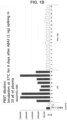
- FIGS. 1A and 1B show that the signal of the AD sample is increased compared with the signal of the Non AD sample according to the incubation time after the addition of rec. A ⁇ 1-42. The difference between AD and Non AD is shown in each condition after incubation for 4 days and 6 days.
- the reason why the signal of the A ⁇ oligomer was higher in the AD patient samples compared with the Non AD patient samples is considered to be that the clearing system suppressing the formation of A ⁇ oligomer in the AD patient samples was activated less than that in the Non AD patient samples.
- Example 8 Detection of A ⁇ oligomers in samples treated with rec. A ⁇ 1-42 and incubated for 2 days, 3 days, 4 days, and 5 days using multimer detection system (MDS)
- MDS multimer detection system
- the positive control, the negative control, and the samples treated with rec. A ⁇ 1-42 and incubated for 2 days, 3 days, 4 days, and 6 days were dispensed in 100 ⁇ l each on 6E10 coated plate (3 pg/ml), followed by reaction in a 27°C incubator in a standing state. After the plate was washed three times with TBST, the FF51-HRP antibody was added to buffer A to reach a concentration of 10 ng/ml and then 100 ⁇ l of each was dispensed. The plate was subjected to reaction in a 27°C incubator in a standing state for 1 hour, and washed three times with TBST, and then 100 ul of the ECL solution was dispensed. The plate reacted with ELC was inserted into a luminometer (PerkinElmer) to measure a luminescent signal. The results are shown in FIG. 1C .
- FIG. 1C shows that the signal of the AD samples is increased by 1.15 times, 1.34 times, 1.65 times, and 1.84 times compared with the signal of the Non AD samples according to the incubation time for 2 days, 3 days, 4 days, and 5 days after the addition of rec. A ⁇ 1-42. It shows that, as the number of days of incubation increased, the difference between AD and Non AD was gradually increased.
- the reason why the signal of the A ⁇ oligomer was higher in AD patient samples compared with Non AD patient samples is considered to be that the clearing system suppressing the formation of the A ⁇ oligomer in the AD patient samples was activated less than that in the Non AD patient samples.
- Example 9 Detection of A ⁇ oligomers in samples treated with rec. A ⁇ 1-42 and incubated for 5 days and samples treated without rec. A ⁇ 1-42 and incubation for 0 days and 5 days using multimer detection system (MDS)
- MDS multimer detection system
- the plate was subjected to reaction in a 27°C incubator in a standing state for 1 hour and washed three times with TBST, and then 100 ul of the ECL solution was dispensed.
- the plate reacted with ELC was inserted into a luminometer (PerkinElmer) to measure a luminescent signal. The results are shown in FIG. 1D .
- FIG. 1D shows A ⁇ oligomer measurement data in the samples prepared by the addition of rec. A ⁇ 1-42 and incubation for 5 days and the samples prepared by the non-addition of rec. A ⁇ 1-42 and incubation for 0 day and 5 days, and illustrates the increase states of the signal of the AD sample and the signal of the Non AD sample over time when A ⁇ 1-42 was spiked and was not spiked, indicating that a differentiation between AD and Non AD was shown only in the samples prepared by the addition of rec. A ⁇ 1-42 and incubation for 5 days.
- the reason why the signal of the A ⁇ oligomer was high in AD patient samples compared with Non AD patient samples is considered that the clearing system, which suppresses the formation of A ⁇ oligomer in the AD patient samples, is activated less than that in the Non AD patient samples, showing an effect of the clearing system that suppresses the formation of A ⁇ oligomer when A ⁇ 1-42 was spiked.
- Example 10 Detection of A ⁇ oligomers in samples treated with rec. A ⁇ 9-42 biotin and incubated for 6 days using multimer detection system (MDS)
- MDS multimer detection system
- a luminometer PerkinElmer
- FIG. 2a shows that the signal of the AD patient group was higher than that of the normal group after the spiking of the rec. A ⁇ 9-42 biotin binding to A ⁇ and then incubation for 6 days.
- Example 11 Detection of A ⁇ oligomers in samples treated with rec. A ⁇ 9-42 biotin and incubated for 2 days, 3 days, and 4 days using multimer detection system (MDS)
- MDS multimer detection system
- the positive control, the negative control, and the samples treated with rec. were dispensed in 100 ⁇ l each on 6E10 coated plate (3 ⁇ g/ml), followed by reaction in a 27°C incubator in a standing state. After the plate was washed three times with TBST, the FF51-HRP antibody was added to buffer A to reach a concentration of 10 ng/ml, and then 100 ⁇ l of each was dispensed. The plate was subjected to reaction in a 27°C incubator in a standing state for 1 hour and washed three times with TBST, and then 100 ul of the ECL solution was dispensed. The plate reacted with ELC was inserted into a luminometer (PerkinElmer) to measure a luminescent signal, and the results are summarized in FIG. 2b .
- a luminometer PerkinElmer
- FIG. 2b shows that, when rec. A ⁇ 9-42 binding to A ⁇ was spiked and incubated for 2 days, 3 days, and 4 days, the signal of the AD patient group was 1.1 times, 1.52 times, and 1.84 times higher than the signal of the normal group with the increased time.
- Example 12 Detection of ⁇ -synuclein oligomers in samples treated with recombinant ⁇ -synuclein and incubated for 0 days, 2 days, 4 days, and 6 days using multimer detection system (MDS)
- MDS multimer detection system
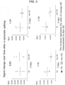
- Streptavidin-HRP was diluted 1/5000 in buffer A, and 100 ⁇ l each was dispensed. Thereafter, the plate was reacted in a 27°C incubator in a standing state for 1 hour and washed three times with TBST. 100 ⁇ l of the ECL solution was dispensed. The plate reacted with ELC was inserted into a luminometer (PerkinElmer) to measure a luminescent signal, and the results are summarized in FIG. 3 .
- FIG. 3 shows signal change data of the signal of the PD sample and the signal of the Non PD sample with increased time after the addition of ⁇ -synuclein and incubation for 0 days, 2 days, 4 days, and 6 days, and illustrates that the ratio of the signal of the PD sample to the signal of the Non PD sample was 9.2 times on day 0, increased to 1.34 times, 1.76 times, and 1.78 times for the incubation for 2 days, 4 days, and 6 days.
- the reason why the signal of ⁇ -synuclein was higher in the PD patient samples compared with the Non AD patient samples is considered to be that the clearing system suppressing the formation of ⁇ -synuclein oligomer in the PD patient samples was activated less than that in the Non PD patient samples.
Landscapes
- Health & Medical Sciences (AREA)
- Life Sciences & Earth Sciences (AREA)
- Engineering & Computer Science (AREA)
- Chemical & Material Sciences (AREA)
- Immunology (AREA)
- Biomedical Technology (AREA)
- Molecular Biology (AREA)
- Hematology (AREA)
- Urology & Nephrology (AREA)
- General Health & Medical Sciences (AREA)
- Biochemistry (AREA)
- Medicinal Chemistry (AREA)
- Cell Biology (AREA)
- Microbiology (AREA)
- Biotechnology (AREA)
- Food Science & Technology (AREA)
- Physics & Mathematics (AREA)
- Analytical Chemistry (AREA)
- General Physics & Mathematics (AREA)
- Pathology (AREA)
- Proteomics, Peptides & Aminoacids (AREA)
- Organic Chemistry (AREA)
- Neurology (AREA)
- Genetics & Genomics (AREA)
- Toxicology (AREA)
- Zoology (AREA)
- Gastroenterology & Hepatology (AREA)
- Biophysics (AREA)
- Neurosurgery (AREA)
- Chemical Kinetics & Catalysis (AREA)
- Peptides Or Proteins (AREA)
- Investigating Or Analysing Biological Materials (AREA)
- Measuring Or Testing Involving Enzymes Or Micro-Organisms (AREA)
Claims (12)
- Procédé pour détecter une forme agrégée d'un polypeptide formant un agrégat dans un échantillon biologique, le procédé comprenant les étapes suivantes :(a) l'ensemencement, avec un échantillon biologique devant être analysé, (i) d'une forme monomère ou multimère du polypeptide formant un agrégat, (ii) d'un dérivé à délétion hydrophobe du polypeptide formant un agrégat, ou (iii) d'une forme monomère ou multimère du polypeptide formant un agrégat et d'un dérivé à délétion hydrophobe du polypeptide formant un agrégat ;(b) de plus la formation d'une forme agrégée du polypeptide formant un agrégat par incubation du produit de l'étape a) à 25 à 40°C pendant 2 à 6 jours ;(c) la mise en contact, avec un produit de l'étape (b), d'un lieur-marqueur dans lequel un marqueur générant un signal est couplé à un lieur se liant à la forme agrégée du polypeptide formant un agrégat ; et(d) la détection d'un signal généré par le lieur-marqueur lié à la forme agrégée du polypeptide formant un agrégat,dans lequel l'incubation dans l'étape (b) effectué pendant un temps d'incubation suffisant pour une multimérisation du (i), (ii) ou (iii) ensemencé par l'échantillon,dans lequel l'échantillon biologique est un plasma, etdans lequel l'échantillon biologique pour effectuer la multimérisation du (i), (ii) ou (iii) ensemencé est un échantillon biologique d'un être humain ayant une maladie impliquant la forme multimère du polypeptide formant un agrégat.
- Procédé selon la revendication 1, dans lequel le polypeptide formant un agrégat est choisi dans le groupe constitué par le peptide Ab, la protéine tau, un prion, l'a-synucléine, une chaîne légère d'Ig, l'amyloïde sérique A, la transthyrétine, la cystatine C, la microglobuline b2, la huntingtine, la superoxyde dismutase, la serpine, et l'amyline.
- Procédé selon la revendication 2, dans lequel le polypeptide formant un agrégat est le peptide Ab, la protéine tau, ou l'a-synucléine.
- Procédé selon la revendication 1, dans lequel la forme monomère du polypeptide formant un agrégat est le peptide Ab comprenant la séquence d'acides aminés de la SEQ ID NO : 1 ou l'a-synucléine comprenant la séquence d'acides aminés de la SEQ ID NO : 2.
- Procédé selon la revendication 1, dans lequel le dérivé à délétion hydrophobe du polypeptide formant un agrégat est le peptide Abdelete comprenant du 37ème résidu d'acide aminé au 42ème résidu d'acide aminé dans la séquence d'acides aminés de la SEQ ID NO : 1.
- Procédé selon la revendication 5, dans lequel le peptide Abdelete est un peptide comprenant du 29ème résidu d'acide aminé au 42ème résidu d'acide aminé dans la séquence d'acides aminés de la SEQ ID NO : 1.
- Procédé selon la revendication 6, dans lequel le peptide Abdelete est un peptide comprenant du 9ème résidu d'acide aminé au 42ème résidu d'acide aminé dans la séquence d'acides aminés de la SEQ ID NO : 1.
- Procédé selon la revendication 1, dans lequel les étapes (c) et (d) sont effectuées en comprenant les étapes suivantes :(c-1) mise en contact du produit de l'étape (b) avec un anticorps de capture reconnaissant un épitope sur le polypeptide formant un agrégat capturant la forme agrégée ;(c-2) mise en contact de la forme agrégée capturée avec un anticorps de détection comprenant un épitope sur le polypeptide formant un agrégat ; et(c-3) détection d'un complexe de forme agrégée-anticorps de détection.
- Procédé selon la revendication 8, dans lequel l'anticorps de détection est un anticorps de détection reconnaissant un épitope identique à ou en chevauchement avec l'épitope dans l'étape (c-1).
- Procédé selon la revendication 8, dans lequel l'anticorps de capture est lié à un substrat solide.
- Procédé selon la revendication 8, dans lequel l'anticorps de détection a un marqueur générant un signal détectable.
- Procédé selon la revendication 11, dans lequel le marqueur lié à l'anticorps de détection comprend un marqueur de type composé, un marqueur enzymatique, un marqueur radioactif, un marqueur fluorescent, un marqueur luminescent, un marqueur chimioluminescent, et un marqueur FRET.
Applications Claiming Priority (2)
| Application Number | Priority Date | Filing Date | Title |
|---|---|---|---|
| KR20140170608 | 2014-12-02 | ||
| PCT/KR2015/010558 WO2016088999A1 (fr) | 2014-12-02 | 2015-10-06 | Procédé de détection d'une forme agrégée d'un polypeptide formant des agrégats |
Publications (4)
| Publication Number | Publication Date |
|---|---|
| EP3229023A4 EP3229023A4 (fr) | 2017-10-11 |
| EP3229023A1 EP3229023A1 (fr) | 2017-10-11 |
| EP3229023B1 true EP3229023B1 (fr) | 2023-06-07 |
| EP3229023C0 EP3229023C0 (fr) | 2023-06-07 |
Family
ID=56091912
Family Applications (1)
| Application Number | Title | Priority Date | Filing Date |
|---|---|---|---|
| EP15865332.9A Active EP3229023B1 (fr) | 2014-12-02 | 2015-10-06 | Procédé de détection d'une forme agrégée d'un polypeptide formant des agrégats |
Country Status (7)
| Country | Link |
|---|---|
| US (1) | US20170261521A1 (fr) |
| EP (1) | EP3229023B1 (fr) |
| JP (1) | JP6803334B2 (fr) |
| KR (2) | KR101658620B1 (fr) |
| CN (1) | CN107110872A (fr) |
| ES (1) | ES2946900T3 (fr) |
| WO (1) | WO2016088999A1 (fr) |
Families Citing this family (5)
| Publication number | Priority date | Publication date | Assignee | Title |
|---|---|---|---|---|
| KR20170108203A (ko) * | 2016-03-16 | 2017-09-27 | 주식회사 피플바이오 | 응집형-형성 폴리펩타이드의 응집형을 검출하는 방법 |
| CA3051839A1 (fr) | 2017-02-17 | 2018-08-23 | Bristol-Myers Squibb Company | Anticorps anti-alpha-synucleine et leurs utilisations |
| KR20220125640A (ko) | 2021-03-05 | 2022-09-14 | 주식회사 피플바이오 | 알츠하이머병의 진단을 위한 트로포마이오신 알파-4 사슬 및 혈소판 당단백질 Ib 베타 사슬의 용도 |
| US20250321237A1 (en) * | 2022-05-01 | 2025-10-16 | Western Michigan University Homer Stryker M.D. School Of Medicine | Assay for rapid protein multimer detection, characterization and quantification |
| CN116593720B (zh) * | 2023-03-17 | 2025-12-19 | 中元汇吉生物技术股份有限公司 | 一种抗原单体检测的校准质控品及其应用 |
Citations (1)
| Publication number | Priority date | Publication date | Assignee | Title |
|---|---|---|---|---|
| US20100009388A1 (en) * | 2005-02-19 | 2010-01-14 | Peoplebio, Inc. | Differential detection of multimeric and monomeric forms of multimer-forming polypeptides |
Family Cites Families (7)
| Publication number | Priority date | Publication date | Assignee | Title |
|---|---|---|---|---|
| CN101124342A (zh) * | 2005-02-19 | 2008-02-13 | 人民生物公司 | 示差检测多聚体形成的多肽的单体形式中多聚体形式的方法 |
| FR2888937B1 (fr) * | 2005-07-21 | 2012-10-26 | Biomerieux Sa | Procede de detection des fcpa utilisant un agent d'agregation des fcpa et un agent de capture des agregats formes |
| WO2007123345A1 (fr) * | 2006-04-21 | 2007-11-01 | Peoplebio, Inc. | Procédé de détection différentielle d'une forme multimère et d'une forme monomère dans des polypeptides formant des multimères par interactions tridimensionnelles |
| CN101427132A (zh) * | 2006-04-21 | 2009-05-06 | 人民生物公司 | 通过三维相互作用从多聚体形成多肽的单体形式中示差检测多聚体形式的方法 |
| CN101308144A (zh) * | 2007-05-16 | 2008-11-19 | 首都医科大学宣武医院 | 检测受试者体液内疾病相关蛋白聚合能力的方法 |
| US9797886B2 (en) * | 2011-06-07 | 2017-10-24 | Wako Pure Chemical Industries, Ltd. | Agglutination enhancer |
| PT2579042E (pt) * | 2011-10-04 | 2014-09-09 | Affiris Ag | Método para detecção de anticorpos específicos para a numa amostra biológica. |
-
2015
- 2015-10-01 KR KR1020150138507A patent/KR101658620B1/ko active Active
- 2015-10-06 EP EP15865332.9A patent/EP3229023B1/fr active Active
- 2015-10-06 JP JP2017530194A patent/JP6803334B2/ja active Active
- 2015-10-06 US US15/532,185 patent/US20170261521A1/en not_active Abandoned
- 2015-10-06 CN CN201580065540.0A patent/CN107110872A/zh active Pending
- 2015-10-06 WO PCT/KR2015/010558 patent/WO2016088999A1/fr not_active Ceased
- 2015-10-06 ES ES15865332T patent/ES2946900T3/es active Active
-
2016
- 2016-04-11 KR KR1020160044232A patent/KR101829026B1/ko active Active
Patent Citations (1)
| Publication number | Priority date | Publication date | Assignee | Title |
|---|---|---|---|---|
| US20100009388A1 (en) * | 2005-02-19 | 2010-01-14 | Peoplebio, Inc. | Differential detection of multimeric and monomeric forms of multimer-forming polypeptides |
Also Published As
| Publication number | Publication date |
|---|---|
| US20170261521A1 (en) | 2017-09-14 |
| KR20160067026A (ko) | 2016-06-13 |
| JP6803334B2 (ja) | 2020-12-23 |
| EP3229023C0 (fr) | 2023-06-07 |
| ES2946900T3 (es) | 2023-07-28 |
| WO2016088999A1 (fr) | 2016-06-09 |
| KR20160067065A (ko) | 2016-06-13 |
| EP3229023A4 (fr) | 2017-10-11 |
| EP3229023A1 (fr) | 2017-10-11 |
| JP2018503807A (ja) | 2018-02-08 |
| CN107110872A (zh) | 2017-08-29 |
| KR101829026B1 (ko) | 2018-02-20 |
| KR101658620B1 (ko) | 2016-09-23 |
Similar Documents
| Publication | Publication Date | Title |
|---|---|---|
| EP2010904B1 (fr) | Procédé de detection differentielle d'une forme multimere et d'une forme monomere dans des polypeptides formant des multimeres par interactions tridimensionnelles | |
| EP3229023B1 (fr) | Procédé de détection d'une forme agrégée d'un polypeptide formant des agrégats | |
| US20230152334A1 (en) | Detection of Structural Forms of Proteins | |
| WO2017178526A1 (fr) | Compositions et méthodes pour la détection de protéines de cellules hôtes | |
| EP3431999B1 (fr) | Procédé de détection d'un agrégat d'un polypeptide formant un agrégat | |
| US20220260593A1 (en) | Targets and methods of diagnosing, monitoring and treating frontotemporal dementia | |
| KR102086852B1 (ko) | 응집형-형성 폴리펩타이드의 응집형을 검출하는 방법 | |
| HK1260029A1 (en) | Method for detecting aggregate of aggregate-forming polypeptide | |
| HK1260029B (zh) | 用於形成聚集体的多肽的聚集体检测方法 | |
| US20230314450A1 (en) | METHOD FOR DETECTING A ß-SHEET AGGREGATE FORM OF A PROTEIN FORMING ß-SHEET AGGREGATES | |
| HK1241020A1 (en) | Method for detecting aggregate form of aggregate-forming polypeptide | |
| WO2009011500A2 (fr) | Procédés pour diminuer de faux signaux dans un essai immunologique pour détecter une forme multimère d'une forme monomère de polypeptides formant des multimères | |
| US20090162943A1 (en) | Reagents and Methods for the Detection of Transmissible Spongiform Encephalopathy |
Legal Events
| Date | Code | Title | Description |
|---|---|---|---|
| STAA | Information on the status of an ep patent application or granted ep patent |
Free format text: STATUS: THE INTERNATIONAL PUBLICATION HAS BEEN MADE |
|
| PUAI | Public reference made under article 153(3) epc to a published international application that has entered the european phase |
Free format text: ORIGINAL CODE: 0009012 |
|
| STAA | Information on the status of an ep patent application or granted ep patent |
Free format text: STATUS: REQUEST FOR EXAMINATION WAS MADE |
|
| 17P | Request for examination filed |
Effective date: 20170628 |
|
| A4 | Supplementary search report drawn up and despatched |
Effective date: 20170818 |
|
| AK | Designated contracting states |
Kind code of ref document: A1 Designated state(s): AL AT BE BG CH CY CZ DE DK EE ES FI FR GB GR HR HU IE IS IT LI LT LU LV MC MK MT NL NO PL PT RO RS SE SI SK SM TR |
|
| AX | Request for extension of the european patent |
Extension state: BA ME |
|
| RAP1 | Party data changed (applicant data changed or rights of an application transferred) |
Owner name: PEOPLEBIO INC. |
|
| DAV | Request for validation of the european patent (deleted) | ||
| DAX | Request for extension of the european patent (deleted) | ||
| STAA | Information on the status of an ep patent application or granted ep patent |
Free format text: STATUS: EXAMINATION IS IN PROGRESS |
|
| 17Q | First examination report despatched |
Effective date: 20181207 |
|
| GRAP | Despatch of communication of intention to grant a patent |
Free format text: ORIGINAL CODE: EPIDOSNIGR1 |
|
| STAA | Information on the status of an ep patent application or granted ep patent |
Free format text: STATUS: GRANT OF PATENT IS INTENDED |
|
| INTG | Intention to grant announced |
Effective date: 20221219 |
|
| GRAS | Grant fee paid |
Free format text: ORIGINAL CODE: EPIDOSNIGR3 |
|
| GRAA | (expected) grant |
Free format text: ORIGINAL CODE: 0009210 |
|
| STAA | Information on the status of an ep patent application or granted ep patent |
Free format text: STATUS: THE PATENT HAS BEEN GRANTED |
|
| AK | Designated contracting states |
Kind code of ref document: B1 Designated state(s): AL AT BE BG CH CY CZ DE DK EE ES FI FR GB GR HR HU IE IS IT LI LT LU LV MC MK MT NL NO PL PT RO RS SE SI SK SM TR |
|
| REG | Reference to a national code |
Ref country code: GB Ref legal event code: FG4D |
|
| REG | Reference to a national code |
Ref country code: CH Ref legal event code: EP Ref country code: AT Ref legal event code: REF Ref document number: 1576623 Country of ref document: AT Kind code of ref document: T Effective date: 20230615 |
|
| REG | Reference to a national code |
Ref country code: DE Ref legal event code: R096 Ref document number: 602015084048 Country of ref document: DE |
|
| U01 | Request for unitary effect filed |
Effective date: 20230607 |
|
| U07 | Unitary effect registered |
Designated state(s): AT BE BG DE DK EE FI FR IT LT LU LV MT NL PT SE SI Effective date: 20230612 |
|
| REG | Reference to a national code |
Ref country code: ES Ref legal event code: FG2A Ref document number: 2946900 Country of ref document: ES Kind code of ref document: T3 Effective date: 20230728 |
|
| REG | Reference to a national code |
Ref country code: LT Ref legal event code: MG9D |
|
| U20 | Renewal fee for the european patent with unitary effect paid |
Year of fee payment: 9 Effective date: 20230905 |
|
| PG25 | Lapsed in a contracting state [announced via postgrant information from national office to epo] |
Ref country code: NO Free format text: LAPSE BECAUSE OF FAILURE TO SUBMIT A TRANSLATION OF THE DESCRIPTION OR TO PAY THE FEE WITHIN THE PRESCRIBED TIME-LIMIT Effective date: 20230907 |
|
| PG25 | Lapsed in a contracting state [announced via postgrant information from national office to epo] |
Ref country code: RS Free format text: LAPSE BECAUSE OF FAILURE TO SUBMIT A TRANSLATION OF THE DESCRIPTION OR TO PAY THE FEE WITHIN THE PRESCRIBED TIME-LIMIT Effective date: 20230607 Ref country code: HR Free format text: LAPSE BECAUSE OF FAILURE TO SUBMIT A TRANSLATION OF THE DESCRIPTION OR TO PAY THE FEE WITHIN THE PRESCRIBED TIME-LIMIT Effective date: 20230607 Ref country code: GR Free format text: LAPSE BECAUSE OF FAILURE TO SUBMIT A TRANSLATION OF THE DESCRIPTION OR TO PAY THE FEE WITHIN THE PRESCRIBED TIME-LIMIT Effective date: 20230908 |
|
| PG25 | Lapsed in a contracting state [announced via postgrant information from national office to epo] |
Ref country code: SK Free format text: LAPSE BECAUSE OF FAILURE TO SUBMIT A TRANSLATION OF THE DESCRIPTION OR TO PAY THE FEE WITHIN THE PRESCRIBED TIME-LIMIT Effective date: 20230607 |
|
| PG25 | Lapsed in a contracting state [announced via postgrant information from national office to epo] |
Ref country code: IS Free format text: LAPSE BECAUSE OF FAILURE TO SUBMIT A TRANSLATION OF THE DESCRIPTION OR TO PAY THE FEE WITHIN THE PRESCRIBED TIME-LIMIT Effective date: 20231007 |
|
| PG25 | Lapsed in a contracting state [announced via postgrant information from national office to epo] |
Ref country code: SM Free format text: LAPSE BECAUSE OF FAILURE TO SUBMIT A TRANSLATION OF THE DESCRIPTION OR TO PAY THE FEE WITHIN THE PRESCRIBED TIME-LIMIT Effective date: 20230607 Ref country code: SK Free format text: LAPSE BECAUSE OF FAILURE TO SUBMIT A TRANSLATION OF THE DESCRIPTION OR TO PAY THE FEE WITHIN THE PRESCRIBED TIME-LIMIT Effective date: 20230607 Ref country code: RO Free format text: LAPSE BECAUSE OF FAILURE TO SUBMIT A TRANSLATION OF THE DESCRIPTION OR TO PAY THE FEE WITHIN THE PRESCRIBED TIME-LIMIT Effective date: 20230607 Ref country code: IS Free format text: LAPSE BECAUSE OF FAILURE TO SUBMIT A TRANSLATION OF THE DESCRIPTION OR TO PAY THE FEE WITHIN THE PRESCRIBED TIME-LIMIT Effective date: 20231007 Ref country code: CZ Free format text: LAPSE BECAUSE OF FAILURE TO SUBMIT A TRANSLATION OF THE DESCRIPTION OR TO PAY THE FEE WITHIN THE PRESCRIBED TIME-LIMIT Effective date: 20230607 |
|
| PG25 | Lapsed in a contracting state [announced via postgrant information from national office to epo] |
Ref country code: PL Free format text: LAPSE BECAUSE OF FAILURE TO SUBMIT A TRANSLATION OF THE DESCRIPTION OR TO PAY THE FEE WITHIN THE PRESCRIBED TIME-LIMIT Effective date: 20230607 |
|
| REG | Reference to a national code |
Ref country code: DE Ref legal event code: R097 Ref document number: 602015084048 Country of ref document: DE |
|
| PLBE | No opposition filed within time limit |
Free format text: ORIGINAL CODE: 0009261 |
|
| STAA | Information on the status of an ep patent application or granted ep patent |
Free format text: STATUS: NO OPPOSITION FILED WITHIN TIME LIMIT |
|
| 26N | No opposition filed |
Effective date: 20240308 |
|
| PG25 | Lapsed in a contracting state [announced via postgrant information from national office to epo] |
Ref country code: MC Free format text: LAPSE BECAUSE OF FAILURE TO SUBMIT A TRANSLATION OF THE DESCRIPTION OR TO PAY THE FEE WITHIN THE PRESCRIBED TIME-LIMIT Effective date: 20230607 |
|
| REG | Reference to a national code |
Ref country code: CH Ref legal event code: PL |
|
| PG25 | Lapsed in a contracting state [announced via postgrant information from national office to epo] |
Ref country code: CH Free format text: LAPSE BECAUSE OF NON-PAYMENT OF DUE FEES Effective date: 20231031 |
|
| PG25 | Lapsed in a contracting state [announced via postgrant information from national office to epo] |
Ref country code: CH Free format text: LAPSE BECAUSE OF NON-PAYMENT OF DUE FEES Effective date: 20231031 |
|
| PG25 | Lapsed in a contracting state [announced via postgrant information from national office to epo] |
Ref country code: IE Free format text: LAPSE BECAUSE OF NON-PAYMENT OF DUE FEES Effective date: 20231006 |
|
| U20 | Renewal fee for the european patent with unitary effect paid |
Year of fee payment: 10 Effective date: 20240906 |
|
| PG25 | Lapsed in a contracting state [announced via postgrant information from national office to epo] |
Ref country code: IE Free format text: LAPSE BECAUSE OF NON-PAYMENT OF DUE FEES Effective date: 20231006 |
|
| PG25 | Lapsed in a contracting state [announced via postgrant information from national office to epo] |
Ref country code: CY Free format text: LAPSE BECAUSE OF FAILURE TO SUBMIT A TRANSLATION OF THE DESCRIPTION OR TO PAY THE FEE WITHIN THE PRESCRIBED TIME-LIMIT; INVALID AB INITIO Effective date: 20151006 |
|
| PG25 | Lapsed in a contracting state [announced via postgrant information from national office to epo] |
Ref country code: HU Free format text: LAPSE BECAUSE OF FAILURE TO SUBMIT A TRANSLATION OF THE DESCRIPTION OR TO PAY THE FEE WITHIN THE PRESCRIBED TIME-LIMIT; INVALID AB INITIO Effective date: 20151006 |
|
| PGFP | Annual fee paid to national office [announced via postgrant information from national office to epo] |
Ref country code: GB Payment date: 20250925 Year of fee payment: 11 |
|
| U20 | Renewal fee for the european patent with unitary effect paid |
Year of fee payment: 11 Effective date: 20251008 |
|
| PG25 | Lapsed in a contracting state [announced via postgrant information from national office to epo] |
Ref country code: TR Free format text: LAPSE BECAUSE OF FAILURE TO SUBMIT A TRANSLATION OF THE DESCRIPTION OR TO PAY THE FEE WITHIN THE PRESCRIBED TIME-LIMIT Effective date: 20230607 |
|
| PGFP | Annual fee paid to national office [announced via postgrant information from national office to epo] |
Ref country code: ES Payment date: 20251107 Year of fee payment: 11 |