EP3287768B1 - Erkennung von unterschiedlichen molekülen mit glasfasersensoren - Google Patents
Erkennung von unterschiedlichen molekülen mit glasfasersensoren Download PDFInfo
- Publication number
- EP3287768B1 EP3287768B1 EP17188082.6A EP17188082A EP3287768B1 EP 3287768 B1 EP3287768 B1 EP 3287768B1 EP 17188082 A EP17188082 A EP 17188082A EP 3287768 B1 EP3287768 B1 EP 3287768B1
- Authority
- EP
- European Patent Office
- Prior art keywords
- fiber optic
- affinity
- reagent
- target molecules
- fluid
- Prior art date
- Legal status (The legal status is an assumption and is not a legal conclusion. Google has not performed a legal analysis and makes no representation as to the accuracy of the status listed.)
- Active
Links
Images
Classifications
-
- G—PHYSICS
- G01—MEASURING; TESTING
- G01N—INVESTIGATING OR ANALYSING MATERIALS BY DETERMINING THEIR CHEMICAL OR PHYSICAL PROPERTIES
- G01N21/00—Investigating or analysing materials by the use of optical means, i.e. using sub-millimetre waves, infrared, visible or ultraviolet light
- G01N21/75—Systems in which material is subjected to a chemical reaction, the progress or the result of the reaction being investigated
- G01N21/77—Systems in which material is subjected to a chemical reaction, the progress or the result of the reaction being investigated by observing the effect on a chemical indicator
- G01N21/7703—Systems in which material is subjected to a chemical reaction, the progress or the result of the reaction being investigated by observing the effect on a chemical indicator using reagent-clad optical fibres or optical waveguides
-
- C—CHEMISTRY; METALLURGY
- C12—BIOCHEMISTRY; BEER; SPIRITS; WINE; VINEGAR; MICROBIOLOGY; ENZYMOLOGY; MUTATION OR GENETIC ENGINEERING
- C12Q—MEASURING OR TESTING PROCESSES INVOLVING ENZYMES, NUCLEIC ACIDS OR MICROORGANISMS; COMPOSITIONS OR TEST PAPERS THEREFOR; PROCESSES OF PREPARING SUCH COMPOSITIONS; CONDITION-RESPONSIVE CONTROL IN MICROBIOLOGICAL OR ENZYMOLOGICAL PROCESSES
- C12Q1/00—Measuring or testing processes involving enzymes, nucleic acids or microorganisms; Compositions therefor; Processes of preparing such compositions
- C12Q1/68—Measuring or testing processes involving enzymes, nucleic acids or microorganisms; Compositions therefor; Processes of preparing such compositions involving nucleic acids
- C12Q1/6813—Hybridisation assays
- C12Q1/6816—Hybridisation assays characterised by the detection means
- C12Q1/6825—Nucleic acid detection involving sensors
-
- G—PHYSICS
- G01—MEASURING; TESTING
- G01N—INVESTIGATING OR ANALYSING MATERIALS BY DETERMINING THEIR CHEMICAL OR PHYSICAL PROPERTIES
- G01N21/00—Investigating or analysing materials by the use of optical means, i.e. using sub-millimetre waves, infrared, visible or ultraviolet light
- G01N21/17—Systems in which incident light is modified in accordance with the properties of the material investigated
- G01N21/55—Specular reflectivity
- G01N21/552—Attenuated total reflection
- G01N21/553—Attenuated total reflection and using surface plasmons
- G01N21/554—Attenuated total reflection and using surface plasmons detecting the surface plasmon resonance of nanostructured metals, e.g. localised surface plasmon resonance
-
- G—PHYSICS
- G01—MEASURING; TESTING
- G01N—INVESTIGATING OR ANALYSING MATERIALS BY DETERMINING THEIR CHEMICAL OR PHYSICAL PROPERTIES
- G01N21/00—Investigating or analysing materials by the use of optical means, i.e. using sub-millimetre waves, infrared, visible or ultraviolet light
- G01N21/84—Systems specially adapted for particular applications
- G01N21/85—Investigating moving fluids or granular solids
- G01N21/8507—Probe photometers, i.e. with optical measuring part dipped into fluid sample
-
- G—PHYSICS
- G01—MEASURING; TESTING
- G01N—INVESTIGATING OR ANALYSING MATERIALS BY DETERMINING THEIR CHEMICAL OR PHYSICAL PROPERTIES
- G01N33/00—Investigating or analysing materials by specific methods not covered by groups G01N1/00 - G01N31/00
- G01N33/48—Biological material, e.g. blood, urine; Haemocytometers
- G01N33/50—Chemical analysis of biological material, e.g. blood, urine; Testing involving biospecific ligand binding methods; Immunological testing
- G01N33/53—Immunoassay; Biospecific binding assay; Materials therefor
- G01N33/543—Immunoassay; Biospecific binding assay; Materials therefor with an insoluble carrier for immobilising immunochemicals
- G01N33/54313—Immunoassay; Biospecific binding assay; Materials therefor with an insoluble carrier for immobilising immunochemicals the carrier being characterised by its particulate form
- G01N33/54346—Nanoparticles
-
- G—PHYSICS
- G01—MEASURING; TESTING
- G01N—INVESTIGATING OR ANALYSING MATERIALS BY DETERMINING THEIR CHEMICAL OR PHYSICAL PROPERTIES
- G01N33/00—Investigating or analysing materials by specific methods not covered by groups G01N1/00 - G01N31/00
- G01N33/48—Biological material, e.g. blood, urine; Haemocytometers
- G01N33/50—Chemical analysis of biological material, e.g. blood, urine; Testing involving biospecific ligand binding methods; Immunological testing
- G01N33/53—Immunoassay; Biospecific binding assay; Materials therefor
- G01N33/543—Immunoassay; Biospecific binding assay; Materials therefor with an insoluble carrier for immobilising immunochemicals
- G01N33/54366—Apparatus specially adapted for solid-phase testing
- G01N33/54373—Apparatus specially adapted for solid-phase testing involving physiochemical end-point determination, e.g. wave-guides, FETS, gratings
-
- B—PERFORMING OPERATIONS; TRANSPORTING
- B82—NANOTECHNOLOGY
- B82Y—SPECIFIC USES OR APPLICATIONS OF NANOSTRUCTURES; MEASUREMENT OR ANALYSIS OF NANOSTRUCTURES; MANUFACTURE OR TREATMENT OF NANOSTRUCTURES
- B82Y15/00—Nanotechnology for interacting, sensing or actuating, e.g. quantum dots as markers in protein assays or molecular motors
Definitions
- the invention relates to the field of detection of molecules in a sample using a mass-sensitive sensor. More specifically it relates to a method for detecting a plurality of distinct target molecules in a sample fluid using at least one fiber optic sensor, a fiber optic sensor adapted for detecting a plurality of distinct molecules in a sample.
- a fast, simple and reliable analysis of molecules e.g. biomolecules, such as the detection and/or quantification of various proteins in a sample
- Diagnostic tests for allergens, contaminants or diseases may rely on such analysis.
- a pathological condition may be indicated by the presence of small quantities of specific biomolecules.
- Multiplex assays are known in the art for detecting and/or quantifying a plurality of different analytes in a sample in a single test operation.
- nucleic acid analysis techniques such as DNA melting analysis using high resolution instrumentation and specialized fluorescent DNA-binding dyes are used to determine the presence and identity of different nucleic acids in the same solution.
- Another technique based on qPCR powerfully combines sensitive nucleic acid detection in real-time with multiplexing capacity, e.g. as disclosed by Espy et al., Clin Microbiol Rev, 2006, 1 9, pp. 165-256 .
- these techniques require the use of multiple fluorescent dyes to monitor the amplification of target nucleic acids in real-time. Because resonance energy transfer occurs between the dyes, the sensitivity of these reactions is hampered, and most qPCR assays can simultaneously detect only 2 or 3 targets.
- T m characteristic melting temperature
- multiplex PCR reactions may be achieved by using multiple sensor probes, e.g. an individually functionalized optic fiber probe for each amplification reaction to be studied.
- SPR surface plasmon resonance
- FO-SPR fiber optic SPR
- An FO-SPR device can be used for monitoring solid phase PCR amplification reactions, e.g. as disclosed by Pollet et al., Small, 2011, 7, 1003-1006 .
- a solid phase PCR reaction is performed using one primer which is attached to the sensor and one primer which is attached to a gold particle.
- the probes on the surface may be extended during PCR and determination of the melting point can thus be dictated by the behaviour of the entire amplicon and not just by primer template hybridisation.
- multiplex PCR reactions would be achieved by using multiple fibers, e.g. an individually functionalized fiber for each amplification reaction.
- Another fiber optic surface plasmon resonance sensor was disclosed by Pollet et al. in "Fiber optic SPR biosensing of DNA hybridization and DNA-protein interactions," published in Biosens. Bioelectron. 25(4), pp. 864-869 .
- This reusable, label-free fiber optic SPR biosensor can be used for measuring DNA hybridization and DNA-protein interactions.
- a fiber-based SPR system is combined with DNA aptamer bioreceptors.
- Fiber optic-SPR platform for fast and sensitive infliximab detection in serum of inflammatory bowel disease patients Biosens Bioelectron. 79, pp. 173-179, Lu et al. disclosed a fast bioassay for determining infliximab concentration in serum using a fiber-optic surface plasmon resonance (FO-SPR) biosensor, in which an infliximab-specific antibody is covalently immobilized on the sensor surface of the FO-SPR biosensor.
- FO-SPR fiber-optic surface plasmon resonance
- LA-ICPMS laser ablation inductively coupled plasma mass spectrometry
- WO2015/071338 A1 discloses detection of a target nucleic acid in a sample using a mass sensitive sensor functionalized with a first nucleic acid probe hybridizing to the target nucleic acid, and metal nanoparticles functionalized with a second nucleic acid probe hybridizing to the target nucleic acid.
- a nucleic acid amplification of the target nucleic acid is performed using non-immobilized probes complementary to the target nucleic DNA.
- a fiber optic sensor e.g. a fiber optic sensor, e.g. fiber optic biosensor, e.g. for multiplex analyte detection using a fiber optic biosensor.
- the fiber optic sensor may comprise a configuration configuration as known in the art, e.g. described in Sensors & Bioelectronics Vol. 12. No. 4, pp. 329-336, 1997 .
- target molecules not present in the sample fluid can be eliminated quickly by stepwise testing.
- a method according to embodiments of the present invention may further comprise exposing the or any of the at least one fiber optic sensor to a further reagent fluid of the plurality of reagent fluids, and detecting a further signal generated by the thus exposed fiber optic sensor to determine whether at least one further target molecule or any of a further combination of target molecules was present in the sample fluid.
- the affinity reagent or affinity reagents in the further reagent fluid may form a second proper subset of the affinity reagents in the first reagent fluid, in which this second proper subset is complimentary to the first proper subset.
- the number of affinity reagents in the second proper subset may be in the range of 50% to 150% of the number of affinity reagents in the first proper subset.
- a fiber optic sensor can be easily reused for further testing if no binding was to molecules to be tested was observed.
- the at least one fiber optic sensor may comprise a plurality of fiber optic sensors, wherein the second fiber optic sensor is exposed to the second reagent fluid if the first signal indicated a presence of the first target molecule or of any target molecule of the first combination of target molecules.
- the first reagent fluid may comprise, for each target molecule of the plurality of target molecules, a corresponding affinity reagent for binding simultaneously with a corresponding affinity probe of the plurality of distinct affinity probes to the target molecule, such that the first detected signal forms a global screening signal indicative of the presence of any of the plurality of target molecules in the sample liquid.
- the target molecules may comprise proteins to be detected in the sample fluid.
- the optical signal is monitored while increasing the temperature of the detection surface (directly or indirectly, for example by heating a fluid, e.g. the sample fluid, in which the detection surface is immersed) and a change in the monitored optical signal, which change is representative of a temperature-induced release of target molecules from the detection surface, is detected to determine whether the target molecules were present in the sample and were bound to the detection surface by a corresponding affinity probe and affinity reagent.
- a fluid e.g. the sample fluid, in which the detection surface is immersed
- the detection surface of the at least one fiber optic sensor may be provided with a surface texturing structure to enable binding of the target molecules on the corresponding affinity probes in a predetermined orientation.
- the surface texturing structure may comprise self-assembled DNA structures, e.g. self-assembled DNA nanostructures or self-assembled DNA origami, such as tetrahaeder structures and/or pillar structures.
- the exposing of the fiber optic sensor to the sample fluid may comprise contacting the fiber optic sensor to the sample fluid in a reaction chamber.
- contacting the mass sensitive sensor to the sample fluid in a reaction chamber may comprise dipping the mass sensitive sensor, e.g. the fiber optic probe, in the sample fluid.
- exposing of the fiber optic sensor to the sample fluid may comprise performing a nucleic acid amplification of the plurality of different target molecules, such that the amplified target nucleic acids of the plurality of different target molecules form complexes with the corresponding plurality of distinct affinity probes.
- the nucleic acid amplification may comprise a Polymerase Chain Reaction, a Ligation Chain Reaction, a Rolling Circle Amplification, and/or an MNAzyme amplification.
- a “sensor” refers to a sensing device, or sensing part thereof, to which target molecules of interest can bind such that the presence or absence of this bound molecule can be determined.
- “sensor” may refer specifically to a mass-sensitive sensor, i.e. a device for measuring a property that functionally relates, e.g. bijectively relates, e.g. that scales proportionally, to mass associated with or bound to a sensitive surface thereof, e.g. a detection surface assembled with capture probes.
- the sensor may be adapted for submersion in a reaction chamber when the binding reaction occurs with such target molecules in a fluid sample to be analysed.
- the term "sensor” refers in present description specifically to a sensor used for analysis or detection of molecules whereby a target analyte interacts with an affinity probe and signals generated by this interaction are transformed by a physicochemical detection mechanism into a form that can be measured and quantified.
- An exemplary sensor may be an optical sensor, e.g. an optical biosensor, based on the principle of surface plasmon resonance (SPR), e.g. a fiber optic SPR (FO-SPR) device.
- the fiber optic sensor may comprise a fiber optic surface plasmon resonance device.
- the present invention relates to a method for detecting a plurality of distinct target molecules in a sample fluid.
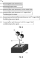
- an exemplary method 10 for detecting a plurality of distinct target molecules 21 in a sample fluid 22 is illustrated.
- This method may be a method for multiplex target analysis to identify the target molecules of the plurality of distinct target molecules that are present in the sample fluid.
- the target molecules may comprise biomolecules, e.g. chemical entities produced by biological processes, and semi-synthetic or synthetic analogs thereof.
- biomolecules may comprise large macromolecules such as proteins (e.g. recombinant and/or natural proteins), carbohydrates, lipids, and/or nucleic acids.
- biomolecules may comprise small molecules such as primary metabolites, secondary metabolites, and natural products.
- Biomolecules may be endogenous or exogenous, e.g. the target molecules may comprise synthetic, semisynthetic and/or natural products, e.g. synthetic pharmaceuticals and/or biopharmaceuticals.
- the target molecules may comprise proteins to be detected in the sample fluid. It is an advantage of embodiments of the present invention that an efficient and specific detection of proteins is provided using fiber optic sensor probes, e.g. where disambiguation of binding of different target proteins on the sensor may be difficult using different melting curve characteristics.
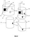
- the method 10 comprises the step of providing 11 at least one fiber optic sensor 23 functionalized with a plurality of distinct affinity probes 24 immobilized on a detection surface 25 of the fiber optic sensor 23.
- Such sensor 23 may combine affinity probes, e.g. comprising biologically-derived materials, with a physicochemical detector.
- Target analytes may interact with the affinity probes, and signals generated by this interaction may be measured using an optical sensor based on the principle of surface plasmon resonance (SPR), e.g. a fiber optic SPR (FO-SPR) device.
- SPR surface plasmon resonance
- the fiber optic sensor may comprise a fiber optic surface plasmon resonance device.
- the sensor may be a mass-based sensor.
- target molecules may be identified by DNA melting peak analysis using such mass based sensor, as known in the art.
- providing the at least one fiber optic sensor 11 may comprise providing a fiber optic sensor configured for detecting an optical signal sensitive to surface plasmon resonance on the detection surface due to simultaneous binding of the target molecules 21 with the corresponding distinct affinity probes 24 and the corresponding affinity reagents discussed further hereinbelow, e.g. antibodies may be bound to DNA and this DNA may hybridize on the detection surface.
- a signal 29, e.g. the first signal, the second signal and/or any further signal referred to hereinbelow, may be generated by increasing a temperature of the fiber optic sensor, e.g.
- a fiber optic surface plasmon resonance sensor and determining a change in the optical signal representative of a temperature-induced release of the target molecules 21 from the detection surface 25, e.g. a melting of the hybridized DNA.
- This increasing of the temperature of the fiber optic sensor may furthermore be used advantageously to regenerate the detection surface of the fiber optic sensor, e.g. such that the same fiber optic sensor may be used in a further detection step.
- the detection surface 25 of the at least one fiber optic sensor may be provided with a surface texturing structure to enable binding of the target molecules on the corresponding affinity probes in a predetermined orientation.
- a surface texturing structure may comprise self-assembled DNA structures, e.g. self-assembled DNA nanostructures or self-assembled DNA origami, such as tetrahaeder structures and/or pillar structures.
- Each affinity probe 24 specifically binds to a corresponding target molecule 21 of the plurality of distinct target molecules to be detected in the sample fluid 22.
- each of the plurality of fiber optic sensors 23 is functionalized with a same plurality of distinct affinity probes, e.g. comprises the same set of distinct affinity probes for specifically binding to the same set of corresponding target molecules. It is an advantage of embodiments of the present invention that identically manufactured fiber optic sensors 23 can be used to test for a possibly large plurality of target molecules in a target-specific manner.
- providing the at least one fiber optic sensor comprises providing the at least one fiber optic sensor functionalized with a plurality of distinct affinity probes comprising an antibody, a monoclonal antibody, an aptamer, an affimer, a peptide, a nucleic acid and/or another small molecule that specifically binds to a target molecule of the plurality of target molecules.
- a high efficiency can be achieved due to the use of multi-purpose, e.g. identically manufactured, sensors, and/or by using a single fluid sample to test for a possibly large plurality of different target molecules. It is, for example, an advantage of a single fluid sample to test for a plurality of different target molecules that measurement errors and/or systematic errors induced by the preparation of different samples of the same material can be avoided. It is a further advantage that, in accordance with embodiments of the present invention, a reference measurement and/or positive control can be performed within the same measurement.
- FIG 3 illustrates an exemplary sensor for use in embodiments of the present invention.
- This sensor comprises a fiber optic surface plasmon resonance (FO-SPR) tip 35 for exposing to the sample fluid, e.g. by means of dipping in a reservoir.
- This tip 35 may be connected to a bifurcated optical fiber 36, one bifurcation of this fiber being connected to a light source 37, and the other bifurcation of this fiber being connected to a detector for generating a detection signal 29, e.g. a spectrometer 38.
- the tip 35 may be directly or indirectly, e.g. via a fluid medium, in thermal contact with a controlled heat source, e.g. a thermocycler 39, e.g. such as to obtain a detection and/or quantitative measurement by melting curve analysis.
- a controlled heat source e.g. a thermocycler 39
- the method further comprises providing 12 a plurality of reagent fluids 26.
- Each reagent fluid 26 comprises at least one affinity reagent 27 for binding, simultaneously with a corresponding affinity probe 24 of the plurality of distinct affinity probes, to the corresponding target molecule 21.
- the affinity reagent 27 may bind to a corresponding target molecule 21, while this target molecule 21 can bind in parallel to a corresponding affinity probe 24 immobilized on the detection surface 25 of the sensor 23.
- Each affinity reagent 27 is bound to a metal nanoparticle 28 for influencing a signal 29 generated by the fiber optic sensor 23 when the affinity reagent 27 and the corresponding affinity probe 24 on the detection surface 25 of the fiber optic sensor 23 are simultaneously bound to the corresponding target molecule 21.
- Such nanoparticle 28 may be a mass element adapted for increasing the sensitivity of a binding assay.
- the nanoparticle 28 may enhance the signal 29 which may be detected by the sensor.
- the nanoparticle may comprise a gold nanoparticle (Au NP), which may be advantageous for enhancing a signal generated by a fiber optic surface plasmon resonance sensor.
- Au NP gold nanoparticle
- Other exemplary nanoparticle materials which may also be advantageously used in combination with a fiber optic SPR sensor, comprise silver, copper, platinum, ruthenium, palladium, or an alloy or combination of such metals.
- the nanoparticle may have a diameter in the range of 2 nm to 100 nm.
- the at least one affinity reagent comprises an antibody, a monoclonal antibody, an aptamer, an affimer, a peptide, a nucleic acid and/or another small molecule that specifically binds to a target molecule of the plurality of target molecules in parallel with the binding of a corresponding affinity probe of the plurality of distinct affinity probes to the target molecule.
- Each reagent fluid 26 has a different composition than each other reagent fluid such as to enable binding of the affinity reagent 27 or affinity reagents in each reagent fluid 26 to a different target molecule 21 or to a different combination of target molecules.
- the method 10 also comprises exposing 13 the or each fiber optic sensor 23 to the sample fluid 22 to bind the different target molecules 21, when present in the sample fluid 22, to the corresponding plurality of distinct affinity probes 24 immobilized on the detection surface 25 of the fiber optic sensor 23.
- Exposing 13 the fiber optic sensor to the sample fluid may comprise contacting the fiber optic sensor to the sample fluid in a reaction chamber.
- contacting the sensor to the sample fluid in a reaction chamber may comprise dipping the fiber optic sensor, e.g. a mass sensitive sensor such as a fiber optic surface plasmon resonance sensor, in the sample fluid.
- exposing of the fiber optic sensor to the sample fluid may comprise performing a nucleic acid amplification of the plurality of different target molecules, such that the amplified target nucleic acids of the plurality of different target molecules form complexes with the corresponding plurality of distinct affinity probes.
- nucleic acid amplification may comprise a Polymerase Chain Reaction, a Ligation Chain Reaction, a Rolling Circle Amplification, and/or an MNAzyme amplification.
- the method 10 further comprises exposing 14 a first fiber optic sensor of the at least one fiber optic sensor 23 to a first reagent fluid 30 of the plurality of reagent fluids 26, and detecting a first signal 29 generated by the first fiber optic sensor 27 to determine whether a first target molecule or any of a first combination of target molecules was present in the sample fluid 26.
- This step of exposing 14 the first sensor to the first reagent fluid may be performed after the step of exposing 13 at least the first sensor to the sample fluid.
- the first reagent fluid comprises, for each target molecule of the plurality of target molecules, a corresponding affinity reagent for binding simultaneously with a corresponding affinity probe of the plurality of distinct affinity probes to the target molecule, such that the first detected signal forms a signal indicative of the presence of any of the plurality of target molecules in the sample liquid.
- the method also comprises exposing 15 the first fiber optic sensor, or a second fiber optic sensor of the at least one fiber optic sensor 23, e.g. the same or an identical fiber optic sensor as was exposed 14 to the first reagent fluid 30, to a second reagent fluid 31 of the plurality of reagent fluids 26, and detecting a second signal 29 generated by that optic sensor, e.g. the first fiber optic sensor or respectively of the second fiber optic sensor, to determine whether a second target molecule or any of a second combination of target molecules was present in the sample fluid.
- the steps of exposing 14 to the first reagent fluid and exposing 15 to the second reagent fluid may be performed consecutively.
- the steps of exposing the sensor or sensors to the sample fluid, exposing 14 the first sensor to the first reagent fluid and exposing 15 the first or second sensor to the second reagent fluid may be performed consecutively.
- the step of exposing 15 the first or second sensor to the second reagent fluid may take the first signal generated by exposing the first sensor to the first reagent fluid into account, for example to determine whether the same or another identical sensor is exposed to the second reagent fluid, and/or to select the second reagent fluid from the plurality of reagent fluids taking the first signal into account.
- the specific target molecule or molecules of the first combination that are present in the sample fluid may be further determined by the second signal, wherein the affinity reagent or affinity reagents in the second reagent fluid form a first proper subset of the affinity reagents in the first reagent fluid.
- a proper subset refers to the mathematical concept in which every component affinity reagent present in the second reagent fluid was also present in the first reagent fluid, but there exists at least one component affinity reagent in the first reagent fluid that is not present in the second reagent fluid. It is an advantage of embodiments of the present invention that molecules not present in the sample fluid can be eliminated quickly by stepwise testing.
- the first, or respectively the second, fiber optic sensor may be exposed to the second reagent fluid if, e.g. if and only if, the first signal indicated a presence of the first target molecule or of any of the target molecules of the first combination of target molecules, e.g. to determine the target molecules present in the sample more accurately.
- the at least one fiber optic sensor may comprise a plurality of fiber optic sensors, wherein the second fiber optic sensor is exposed to the second reagent fluid if the first signal indicated a presence of the first target molecule or of any target molecule of the first combination of target molecules.
- the first fiber optic sensor may be exposed to the second reagent fluid if, e.g. if and only if, the first signal indicated an absence of the first target molecule or of all target molecules of the first combination of target molecules.
- a fiber optic sensor can be easily reused for further testing if no binding was to molecules to be tested was observed. Furthermore, this may apply equally to an exposure to a further reagent fluid as described further hereinbelow.
- the first fiber optic sensor may be exposed to the second reagent fluid if, e.g. if and only if, the first signal indicated a presence of the first target molecule or of any of the target molecules of the first combination of target molecules, e.g. to determine the target molecules present in the sample more accurately.
- the second reagent fluid may contain a proper subset of the affinity reagents present in the first reagent fluid.
- a further reagent fluid may contain a proper subset of the affinity reagents present in the second reagent fluid.
- a further reagent fluid may contain a complimentary proper subset of the affinity reagents present in the first reagent fluid, e.g. complimentary to the first proper subset formed by the affinity reagents present in the second reagent fluid.
- This principle of stepwise elimination and/or more specific testing may be applied in yet further steps to test yet further reagent fluids.
- the first reagent fluid may comprise, for each target molecule of the plurality of target molecules, a corresponding affinity reagent for binding simultaneously with a corresponding affinity probe of the plurality of distinct affinity probes to the target molecule, such that the first detected signal forms a global screening signal indicative of the presence of any of the plurality of target molecules in the sample liquid.
- a method according to embodiments of the present invention may further comprise exposing 16 the, or any of the, at least one fiber optic sensor to a further reagent fluid 32 of the plurality of reagent fluids, and detecting a further signal 29 generated by the thus exposed fiber optic sensor to determine whether at least one further target molecule or any of a further combination of target molecules was present in the sample fluid.
- This further step may be performed after the step of exposing 15 the sensor to the second reagent fluid.
- the specific target molecule or molecules of the second combination that are present in the sample fluid may be further determined by the further signal, in which the affinity reagent or affinity reagents in the further reagent fluid form a second proper subset of the affinity reagents in the second reagent fluid.
- the affinity reagent or affinity reagents in the further reagent fluid may form a second proper subset of the affinity reagents in the first reagent fluid, in which this second proper subset is complimentary to the first proper subset.
- the number of affinity reagents in the second proper subset may be in the range of 50% to 150% of the number of affinity reagents in the first proper subset.
- the second proper subset formed by the affinity reagents in the further reagent fluid may be selected as a complimentary proper subset of the affinity reagents in the first reagent fluid.
- each of the first proper subset and the second proper subset may consist of complimentary subsets of the affinity reagents present in the first reagent fluid, in which the first proper subset and the second proper subset contain an about equal number of affinity reagents.
- the fiber optic sensor e.g. comprising an optic fiber tip having a detection surface on which the plurality of distinct affinity probes are immobilized, may be exposed to the sample fluid to bind the different target molecules, e.g. to bind the target molecules in the sample fluid to the distinct affinity probes by exposing the optic fiber tip to the sample fluid.
- the method may comprise, after exposing the fiber optic sensor to the sample fluid, e.g. dipping the fiber optic tip in the sample fluid, a step of moving the fiber optic sensor, e.g. the optic fiber tip, to the first reagent fluid and exposing the fiber optic sensor, e.g. the optic fiber tip, to the first reagent fluid.
- This moving and exposing may comprise a sequential exposure of the fiber optic sensor to a plurality of different reagent fluids.
- this moving and exposing may be performed using a three-dimensional positioning system, e.g. an actuated frame having at least three degrees of freedom, e.g. three degrees of translation.
- a three-dimensional positioning system e.g. an actuated frame having at least three degrees of freedom, e.g. three degrees of translation.
- Such positioning system may comprise a controller, controlling the motion of the fiber optic sensor from the sample fluid to the reagent fluid or along a sequence of reagent fluids.
- another fiber optic sensor may be moved between the sample (or another sample) and the reagent fluids, and exposed thereto, in an synchronized manner, e.g. such as to avoid collision of the former fiber optic sensor and the other fiber optic sensor. Therefore, a three-dimensional positioning system, e.g. a positioning robot, may be controlled to move one or more of fiber optic sensors.
Landscapes
- Health & Medical Sciences (AREA)
- Chemical & Material Sciences (AREA)
- Life Sciences & Earth Sciences (AREA)
- Immunology (AREA)
- Engineering & Computer Science (AREA)
- Analytical Chemistry (AREA)
- Physics & Mathematics (AREA)
- General Health & Medical Sciences (AREA)
- Biochemistry (AREA)
- General Physics & Mathematics (AREA)
- Pathology (AREA)
- Molecular Biology (AREA)
- Hematology (AREA)
- Urology & Nephrology (AREA)
- Biomedical Technology (AREA)
- Microbiology (AREA)
- Organic Chemistry (AREA)
- Biotechnology (AREA)
- Nanotechnology (AREA)
- Cell Biology (AREA)
- Zoology (AREA)
- Medicinal Chemistry (AREA)
- Food Science & Technology (AREA)
- Wood Science & Technology (AREA)
- Proteomics, Peptides & Aminoacids (AREA)
- Plasma & Fusion (AREA)
- Biophysics (AREA)
- Genetics & Genomics (AREA)
- Chemical Kinetics & Catalysis (AREA)
- General Engineering & Computer Science (AREA)
- Bioinformatics & Cheminformatics (AREA)
- Investigating Or Analysing Materials By Optical Means (AREA)
- Apparatus Associated With Microorganisms And Enzymes (AREA)
- Investigating, Analyzing Materials By Fluorescence Or Luminescence (AREA)
- Investigating Or Analyzing Materials By The Use Of Electric Means (AREA)
Claims (11)
- Verfahren (10) zum Nachweis einer Vielzahl unterschiedlicher Zielmoleküle (21) in einer Probenflüssigkeit (26), wobei das Verfahren umfasst:- Bereitstellen (11) mindestens eines faseroptischen Sensors (23) mit einer Detektionsoberfläche (25), wobei der Sensor zum Detektieren eines optischen Signals (29) konfiguriert ist, das für Oberflächenplasmonenresonanz auf der Detektionsoberfläche (25) empfindlich ist, und funktionalisiert ist mit a Vielzahl unterschiedlicher Affinitätssonden (24), ausgewählt aus einem Antikörper, einem monoklonalen Antikörper, einem Aptamer, einem Affimer, einem Peptid, einer Nukleinsäure und/oder einem anderen kleinen Molekül, das spezifisch an ein Zielmolekül der Vielzahl unterschiedlicher Zielmoleküle bindet ( 21), wobei die Affinitätssonden (24) auf der Detektionsoberfläche (25) des faseroptischen Sensors (23) immobilisiert sind, wobei jede Affinitätssonde (24) spezifisch an ein entsprechendes Zielmolekül der Vielzahl von unterschiedlichen Zielmolekülen (21) bindet, in dem Probenfluid (26) zu detektieren, wobei, wenn der mindestens eine faseroptische Sensor (23) eine Vielzahl von faseroptischen Sensoren umfasst, jeder der Vielzahl von faseroptischen Sensoren mit einer gleichen Vielzahl von unterschiedlichen Affinitätssonden funktionalisiert ist (24);- Bereitstellen (12) einer Vielzahl von Reagenzflüssigkeiten (26), wobei jede Reagenzflüssigkeit eine Vielzahl von Affinitätsreagenzien (27) umfasst, ausgewählt aus einem Antikörper, einem monoklonalen Antikörper, einem Aptamer, einem Affimer, einem Peptid, einer Nukleinsäure und/oder ein weiteres kleines Molekül, das spezifisch an ein Zielmolekül der mehreren unterschiedlichen Zielmoleküle (21) parallel zu der Bindung einer entsprechenden Affinitätssonde der mehreren unterschiedlichen Affinitätssonden (24) an das Zielmolekül (21, jedes Affinitätsreagenz) bindet an ein Metallnanopartikel (28) gebunden ist, um das optische Signal (29) aufgrund gleichzeitiger Bindung eines Zielmoleküls (21) mit einer entsprechenden unterschiedlichen Affinitätssonde (24) und mindestens einem der entsprechenden Affinitätsreagenzien (27) zu verstärken, wobei jedes Reagenzfluid (26) eine andere Zusammensetzung als jedes andere Reagenzfluid hat, um eine Bindung des Affinitätsreagenz oder der Affinitätsreagenzien (27) in jedem Reagenzfluid (26) an ein anderes Zielmolekül (21) oder an ein anderes zu ermöglichen Kombination von Zielmolekülen;- Aussetzen (13) des oder jedes faseroptischen Sensors (23) an die Probenflüssigkeit (22), um die unterschiedlichen Zielmoleküle (21), wenn sie in der Probenflüssigkeit vorhanden sind, an die entsprechende Vielzahl von unterschiedlichen Affinitätssonden (24) zu binden;- Aussetzen (14) eines ersten faseroptischen Sensors des mindestens einen faseroptischen Sensors (23) einem ersten Reagensfluid (30) der mehreren Reagensfluide (26) und Erfassen eines ersten optischen Signals, das von der ersten Faser erzeugt wird optischer Sensor (23) zum Bestimmen, ob ein erstes Zielmolekül oder irgendeines einer ersten Kombination von Zielmolekülen in der Probenflüssigkeit (22) vorhanden ist; Und- Aussetzen (15) desselben ersten faseroptischen Sensors oder eines identischen zweiten faseroptischen Sensors des mindestens einen faseroptischen Sensors (23) einem zweiten Reagenzfluid (31) der Vielzahl von Reagenzfluiden (26), und Detektieren eines zweiten optischen Signals, das von dem ersten faseroptischen Sensor bzw. von dem zweiten faseroptischen Sensor erzeugt wird, um zu bestimmen, ob ein zweites Zielmolekül oder eines einer zweiten Kombination von Zielmolekülen in der Probenflüssigkeit (22) vorhanden ist,wobei die Reagensflüssigkeiten (30, 31) in thermischem Kontakt mit einer gesteuerten Wärmequelle (39) sind und die optischen Signale (29) überwacht werden, während eine Temperatur der Erfassungsoberfläche (25) erhöht wird, und wobei eine Änderung des überwachten optischen Signals erfolgt (29), das repräsentativ für eine temperaturinduzierte Freisetzung der Zielmoleküle (21) von der Detektionsoberfläche (25) ist, erfasst wird, um zu bestimmen, ob die Zielmoleküle in der Probenflüssigkeit (22) vorhanden und an die Detektionsoberfläche gebunden sind durch Affinitätssonden und Affinitätsreagenzien,wobei, wenn das erste Signal das Vorhandensein irgendeines der ersten Kombination von Zielmolekülen anzeigt, das spezifische Zielmolekül oder die spezifischen Zielmoleküle der ersten Kombination, die in der Probenflüssigkeit (22) vorhanden sind, ferner durch das zweite Signal bestimmt wird, wobei die Affinitätsreagenzien in das zweite Reagenzfluid (31) eine erste geeignete Teilmenge der Affinitätsreagenzien in dem ersten Reagenzfluid (30) bildet.
- Verfahren nach Anspruch 1, ferner umfassend das Aussetzen des oder irgendeines der mindestens einen faseroptischen Sensoren (23) einem weiteren Reagenzfluid (32) der Vielzahl von Reagenzfluiden und Detektieren eines weiteren optischen Signals, das dadurch erzeugt wird den so freigelegten faseroptischen Sensor, um zu bestimmen, ob mindestens ein weiteres Zielmolekül oder irgendeine weitere Kombination von Zielmolekülen in der Probenflüssigkeit (22) vorhanden ist.
- Verfahren nach Anspruch 2, wobei die Affinitätsreagenzien in der weiteren Reagenzflüssigkeit (32) eine zweite richtige Teilmenge der Affinitätsreagenzien in der ersten Reagenzflüssigkeit (30) bilden, wobei die zweite richtige Teilmenge komplementär zu der ersten richtigen Teilmenge ist.
- Verfahren nach Anspruch 3, wobei die Anzahl der Affinitätsreagenzien in der zweiten richtigen Untergruppe im Bereich von 50% bis 150% der Anzahl der Affinitätsreagenzien in der ersten richtigen Untergruppe liegt.
- Verfahren nach einem der vorhergehenden Ansprüche, wobei das erste Reagenzfluid (30) für jedes Zielmolekül der Vielzahl von Zielmolekülen (21) ein entsprechendes Affinitätsreagenz zum gleichzeitigen Binden mit einer entsprechenden Affinitätssonde umfasst eine Vielzahl unterschiedlicher Affinitätssonden (24) zu dem Zielmolekül, so dass das erste detektierte Signal ein Screening-Signal bildet, das die Anwesenheit irgendeines der Vielzahl von Zielmolekülen (21) in der Probenflüssigkeit (22) anzeigt.
- Verfahren nach einem der vorhergehenden Ansprüche, wobei die Zielmoleküle (21) in der Probenflüssigkeit (22) nachzuweisende Proteine umfassen.
- Verfahren nach Anspruch 1, wobei die Detektionsoberfläche (25) des mindestens einen faseroptischen Sensors (23) mit einer Oberflächentexturierungsstruktur versehen ist, um eine Bindung der Zielmoleküle (21) an die entsprechenden Affinitätssonden (21) zu ermöglichen. 24) in einer vorbestimmten Ausrichtung.
- Verfahren nach Anspruch 7, wobei die Oberflächentexturierungsstruktur selbstorganisierte DNA-Strukturen umfasst.
- Verfahren nach einem der vorhergehenden Ansprüche, bei dem das Aussetzen (13) des faseroptischen Sensors (23) dem Probenfluid (22) das Kontaktieren des faseroptischen Sensors (23) mit dem Probenfluid (22) umfasst eine Reaktionskammer.
- Verfahren nach einem der vorhergehenden Ansprüche, wobei das Aussetzen ( 13 ) des faseroptischen Sensors ( 23 ) dem Probenfluid ( 22 ) das Durchführen einer Nukleinsäureamplifikation der Vielzahl unterschiedlicher Zielmoleküle umfasst, so dass die amplifizierte Zielnucleinsäuren der Vielzahl unterschiedlicher Zielmoleküle (21) bilden Komplexe mit der entsprechenden Vielzahl unterschiedlicher Affinitätssonden (24).
- Verfahren nach Anspruch 10, wobei die Nukleinsäure-Amplifikation eine Polymerase-Kettenreaktion, eine Ligations-Kettenreaktion, eine Rolling-Circle-Amplifikation und/oder eine MNAzyme-Amplifikation umfasst.
Applications Claiming Priority (1)
| Application Number | Priority Date | Filing Date | Title |
|---|---|---|---|
| GBGB1614597.1A GB201614597D0 (en) | 2016-08-26 | 2016-08-26 | Detection of distinct molecules using fiber optic sensor |
Publications (3)
| Publication Number | Publication Date |
|---|---|
| EP3287768A1 EP3287768A1 (de) | 2018-02-28 |
| EP3287768B1 true EP3287768B1 (de) | 2023-08-09 |
| EP3287768C0 EP3287768C0 (de) | 2023-08-09 |
Family
ID=57119780
Family Applications (1)
| Application Number | Title | Priority Date | Filing Date |
|---|---|---|---|
| EP17188082.6A Active EP3287768B1 (de) | 2016-08-26 | 2017-08-28 | Erkennung von unterschiedlichen molekülen mit glasfasersensoren |
Country Status (4)
| Country | Link |
|---|---|
| EP (1) | EP3287768B1 (de) |
| ES (1) | ES2958416T3 (de) |
| GB (1) | GB201614597D0 (de) |
| PL (1) | PL3287768T3 (de) |
Citations (1)
| Publication number | Priority date | Publication date | Assignee | Title |
|---|---|---|---|---|
| WO2015071338A1 (en) * | 2013-11-13 | 2015-05-21 | Katholieke Universiteit Leuven | Monitoring dna amplification |
Family Cites Families (1)
| Publication number | Priority date | Publication date | Assignee | Title |
|---|---|---|---|---|
| US8563328B2 (en) * | 2004-07-26 | 2013-10-22 | University Of Louisville Research Foundation, Inc. | Fiber-optic biosensor and biosensing methods |
-
2016
- 2016-08-26 GB GBGB1614597.1A patent/GB201614597D0/en not_active Ceased
-
2017
- 2017-08-28 PL PL17188082.6T patent/PL3287768T3/pl unknown
- 2017-08-28 EP EP17188082.6A patent/EP3287768B1/de active Active
- 2017-08-28 ES ES17188082T patent/ES2958416T3/es active Active
Patent Citations (1)
| Publication number | Priority date | Publication date | Assignee | Title |
|---|---|---|---|---|
| WO2015071338A1 (en) * | 2013-11-13 | 2015-05-21 | Katholieke Universiteit Leuven | Monitoring dna amplification |
Non-Patent Citations (1)
| Title |
|---|
| KAREL KNEZ ET AL: "Spherical Nucleic Acid Enhanced FO-SPR DNA Melting for Detection of Mutations in Legionella pneumophila", ANALYTICAL CHEMISTRY, vol. 85, no. 3, 5 February 2013 (2013-02-05), US, pages 1734 - 1742, XP055580936, ISSN: 0003-2700, DOI: 10.1021/ac303008f * |
Also Published As
| Publication number | Publication date |
|---|---|
| PL3287768T3 (pl) | 2024-03-04 |
| GB201614597D0 (en) | 2016-10-12 |
| EP3287768A1 (de) | 2018-02-28 |
| ES2958416T3 (es) | 2024-02-08 |
| EP3287768C0 (de) | 2023-08-09 |
Similar Documents
| Publication | Publication Date | Title |
|---|---|---|
| Chen et al. | SERS-based dual-mode DNA aptasensors for rapid classification of SARS-CoV-2 and influenza A/H1N1 infection | |
| JP6568949B2 (ja) | 競合アッセイによる小分子のナノポア検出法 | |
| US20040091862A1 (en) | Method and device for detecting temperature-dependent parameters, such as the association/dissociation parameters and/or the equilibrium constant of complexes comprising at least two components | |
| Charoenkitamorn et al. | Electrochemical and optical biosensors for biological sensing applications | |
| Takano et al. | Pipette tip biosensors for bacterial double-stranded DNA using bioluminescence induced by zinc finger luciferase | |
| Ebrahimi et al. | Cardiac troponin I biosensors: innovations in real‐time diagnosis of cardiovascular diseases | |
| US11091803B2 (en) | Nucleic acid quantification method | |
| US9873119B2 (en) | Multiple- analyte assay device and system | |
| Lecaruyer et al. | Surface plasmon resonance imaging as a multidimensional surface characterization instrument—application to biochip genotyping | |
| EP3287768A1 (de) | Erkennung von unterschiedlichen molekülen mit glasfasersensoren | |
| Inoue et al. | A reliable aptamer array prepared by repeating inkjet-spotting toward on-site measurement | |
| EP1586659A1 (de) | Oberflächenverstärkte Raman Spektroskopie für Biosensor-Systeme und Methoden zur Detektion von Biomolekülen | |
| JP4626377B2 (ja) | 標的分子の検出方法、遺伝子多型の検出方法、ならびにこれらの検出方法に用いられる基板およびキット | |
| US9989526B2 (en) | Method and device for bioassays | |
| NL2007328C2 (en) | A nanopore sensor and method for selective detection of analytes in a sample. | |
| EP2205973B1 (de) | Auf hochempfindlicher nanotechnologie basierendes multiplex-bioassay-verfahren und vorrichtung | |
| JP3621927B2 (ja) | 生体試料測定方法及び装置 | |
| US20250312785A1 (en) | Integrated microfluidic devices with isothermal amplification and electrochemical sensing for rapid molecular diagnostics | |
| EP3343220B1 (de) | Biopartikelcharakterisierung | |
| RU2762360C1 (ru) | Способ автоматизации параллельной безметочной детекции биологического маркера и устройство для его реализации | |
| Leroy et al. | Biological Applications of Surface Plasmon Resonance Imaging | |
| EP3677690A1 (de) | Verfahren und vorrichtung zur analyse von nukleinsäuren | |
| Delport et al. | Fiber optic biosensing for allergen and pathogen screening in food | |
| WO2025137743A1 (en) | Aptamer screening method | |
| EP2131196A1 (de) | Erkennung von Analyten in einem Durchlauf-Biosensor |
Legal Events
| Date | Code | Title | Description |
|---|---|---|---|
| PUAI | Public reference made under article 153(3) epc to a published international application that has entered the european phase |
Free format text: ORIGINAL CODE: 0009012 |
|
| STAA | Information on the status of an ep patent application or granted ep patent |
Free format text: STATUS: THE APPLICATION HAS BEEN PUBLISHED |
|
| AK | Designated contracting states |
Kind code of ref document: A1 Designated state(s): AL AT BE BG CH CY CZ DE DK EE ES FI FR GB GR HR HU IE IS IT LI LT LU LV MC MK MT NL NO PL PT RO RS SE SI SK SM TR |
|
| AX | Request for extension of the european patent |
Extension state: BA ME |
|
| STAA | Information on the status of an ep patent application or granted ep patent |
Free format text: STATUS: REQUEST FOR EXAMINATION WAS MADE |
|
| 17P | Request for examination filed |
Effective date: 20180828 |
|
| RBV | Designated contracting states (corrected) |
Designated state(s): AL AT BE BG CH CY CZ DE DK EE ES FI FR GB GR HR HU IE IS IT LI LT LU LV MC MK MT NL NO PL PT RO RS SE SI SK SM TR |
|
| RAP1 | Party data changed (applicant data changed or rights of an application transferred) |
Owner name: FOX BIOSYSTEMS NV |
|
| RAP1 | Party data changed (applicant data changed or rights of an application transferred) |
Owner name: FOX BIOSYSTEMS NV |
|
| STAA | Information on the status of an ep patent application or granted ep patent |
Free format text: STATUS: EXAMINATION IS IN PROGRESS |
|
| 17Q | First examination report despatched |
Effective date: 20210511 |
|
| GRAP | Despatch of communication of intention to grant a patent |
Free format text: ORIGINAL CODE: EPIDOSNIGR1 |
|
| STAA | Information on the status of an ep patent application or granted ep patent |
Free format text: STATUS: GRANT OF PATENT IS INTENDED |
|
| RIC1 | Information provided on ipc code assigned before grant |
Ipc: G01N 33/543 20060101ALI20230118BHEP Ipc: C12Q 1/6825 20180101ALI20230118BHEP Ipc: G01N 21/77 20060101ALI20230118BHEP Ipc: G01N 21/85 20060101ALI20230118BHEP Ipc: B82Y 15/00 20110101ALI20230118BHEP Ipc: C12Q 1/68 20060101ALI20230118BHEP Ipc: G01N 21/552 20140101AFI20230118BHEP |
|
| INTG | Intention to grant announced |
Effective date: 20230207 |
|
| GRAS | Grant fee paid |
Free format text: ORIGINAL CODE: EPIDOSNIGR3 |
|
| GRAA | (expected) grant |
Free format text: ORIGINAL CODE: 0009210 |
|
| STAA | Information on the status of an ep patent application or granted ep patent |
Free format text: STATUS: THE PATENT HAS BEEN GRANTED |
|
| AK | Designated contracting states |
Kind code of ref document: B1 Designated state(s): AL AT BE BG CH CY CZ DE DK EE ES FI FR GB GR HR HU IE IS IT LI LT LU LV MC MK MT NL NO PL PT RO RS SE SI SK SM TR |
|
| REG | Reference to a national code |
Ref country code: GB Ref legal event code: FG4D |
|
| REG | Reference to a national code |
Ref country code: CH Ref legal event code: EP |
|
| REG | Reference to a national code |
Ref country code: IE Ref legal event code: FG4D |
|
| REG | Reference to a national code |
Ref country code: DE Ref legal event code: R096 Ref document number: 602017072395 Country of ref document: DE |
|
| U01 | Request for unitary effect filed |
Effective date: 20230831 |
|
| U07 | Unitary effect registered |
Designated state(s): AT BE BG DE DK EE FI FR IT LT LU LV MT NL PT SE SI Effective date: 20230912 |
|
| U20 | Renewal fee for the european patent with unitary effect paid |
Year of fee payment: 7 Effective date: 20230914 |
|
| PG25 | Lapsed in a contracting state [announced via postgrant information from national office to epo] |
Ref country code: GR Free format text: LAPSE BECAUSE OF FAILURE TO SUBMIT A TRANSLATION OF THE DESCRIPTION OR TO PAY THE FEE WITHIN THE PRESCRIBED TIME-LIMIT Effective date: 20231110 |
|
| PG25 | Lapsed in a contracting state [announced via postgrant information from national office to epo] |
Ref country code: IS Free format text: LAPSE BECAUSE OF FAILURE TO SUBMIT A TRANSLATION OF THE DESCRIPTION OR TO PAY THE FEE WITHIN THE PRESCRIBED TIME-LIMIT Effective date: 20231209 |
|
| PG25 | Lapsed in a contracting state [announced via postgrant information from national office to epo] |
Ref country code: RS Free format text: LAPSE BECAUSE OF FAILURE TO SUBMIT A TRANSLATION OF THE DESCRIPTION OR TO PAY THE FEE WITHIN THE PRESCRIBED TIME-LIMIT Effective date: 20230809 Ref country code: NO Free format text: LAPSE BECAUSE OF FAILURE TO SUBMIT A TRANSLATION OF THE DESCRIPTION OR TO PAY THE FEE WITHIN THE PRESCRIBED TIME-LIMIT Effective date: 20231109 Ref country code: IS Free format text: LAPSE BECAUSE OF FAILURE TO SUBMIT A TRANSLATION OF THE DESCRIPTION OR TO PAY THE FEE WITHIN THE PRESCRIBED TIME-LIMIT Effective date: 20231209 Ref country code: HR Free format text: LAPSE BECAUSE OF FAILURE TO SUBMIT A TRANSLATION OF THE DESCRIPTION OR TO PAY THE FEE WITHIN THE PRESCRIBED TIME-LIMIT Effective date: 20230809 Ref country code: GR Free format text: LAPSE BECAUSE OF FAILURE TO SUBMIT A TRANSLATION OF THE DESCRIPTION OR TO PAY THE FEE WITHIN THE PRESCRIBED TIME-LIMIT Effective date: 20231110 |
|
| REG | Reference to a national code |
Ref country code: ES Ref legal event code: FG2A Ref document number: 2958416 Country of ref document: ES Kind code of ref document: T3 Effective date: 20240208 |
|
| PG25 | Lapsed in a contracting state [announced via postgrant information from national office to epo] |
Ref country code: SM Free format text: LAPSE BECAUSE OF FAILURE TO SUBMIT A TRANSLATION OF THE DESCRIPTION OR TO PAY THE FEE WITHIN THE PRESCRIBED TIME-LIMIT Effective date: 20230809 Ref country code: RO Free format text: LAPSE BECAUSE OF FAILURE TO SUBMIT A TRANSLATION OF THE DESCRIPTION OR TO PAY THE FEE WITHIN THE PRESCRIBED TIME-LIMIT Effective date: 20230809 Ref country code: CZ Free format text: LAPSE BECAUSE OF FAILURE TO SUBMIT A TRANSLATION OF THE DESCRIPTION OR TO PAY THE FEE WITHIN THE PRESCRIBED TIME-LIMIT Effective date: 20230809 Ref country code: SK Free format text: LAPSE BECAUSE OF FAILURE TO SUBMIT A TRANSLATION OF THE DESCRIPTION OR TO PAY THE FEE WITHIN THE PRESCRIBED TIME-LIMIT Effective date: 20230809 |
|
| REG | Reference to a national code |
Ref country code: DE Ref legal event code: R097 Ref document number: 602017072395 Country of ref document: DE |
|
| REG | Reference to a national code |
Ref country code: IE Ref legal event code: MM4A |
|
| PG25 | Lapsed in a contracting state [announced via postgrant information from national office to epo] |
Ref country code: MC Free format text: LAPSE BECAUSE OF FAILURE TO SUBMIT A TRANSLATION OF THE DESCRIPTION OR TO PAY THE FEE WITHIN THE PRESCRIBED TIME-LIMIT Effective date: 20230809 |
|
| PLBE | No opposition filed within time limit |
Free format text: ORIGINAL CODE: 0009261 |
|
| STAA | Information on the status of an ep patent application or granted ep patent |
Free format text: STATUS: NO OPPOSITION FILED WITHIN TIME LIMIT |
|
| PG25 | Lapsed in a contracting state [announced via postgrant information from national office to epo] |
Ref country code: IE Free format text: LAPSE BECAUSE OF NON-PAYMENT OF DUE FEES Effective date: 20230828 |
|
| 26N | No opposition filed |
Effective date: 20240513 |
|
| PG25 | Lapsed in a contracting state [announced via postgrant information from national office to epo] |
Ref country code: IE Free format text: LAPSE BECAUSE OF NON-PAYMENT OF DUE FEES Effective date: 20230828 |
|
| U20 | Renewal fee for the european patent with unitary effect paid |
Year of fee payment: 8 Effective date: 20240827 |
|
| PGFP | Annual fee paid to national office [announced via postgrant information from national office to epo] |
Ref country code: ES Payment date: 20240902 Year of fee payment: 8 |
|
| PGFP | Annual fee paid to national office [announced via postgrant information from national office to epo] |
Ref country code: PL Payment date: 20240801 Year of fee payment: 8 |
|
| U1K | Transfer of rights of the unitary patent after the registration of the unitary effect |
Owner name: CARTERRA INCORPORATED; US |
|
| REG | Reference to a national code |
Ref country code: GB Ref legal event code: 732E Free format text: REGISTERED BETWEEN 20250508 AND 20250514 |
|
| PG25 | Lapsed in a contracting state [announced via postgrant information from national office to epo] |
Ref country code: CY Free format text: LAPSE BECAUSE OF FAILURE TO SUBMIT A TRANSLATION OF THE DESCRIPTION OR TO PAY THE FEE WITHIN THE PRESCRIBED TIME-LIMIT; INVALID AB INITIO Effective date: 20170828 |
|
| PG25 | Lapsed in a contracting state [announced via postgrant information from national office to epo] |
Ref country code: HU Free format text: LAPSE BECAUSE OF FAILURE TO SUBMIT A TRANSLATION OF THE DESCRIPTION OR TO PAY THE FEE WITHIN THE PRESCRIBED TIME-LIMIT; INVALID AB INITIO Effective date: 20170828 |
|
| U20 | Renewal fee for the european patent with unitary effect paid |
Year of fee payment: 9 Effective date: 20250827 |
|
| PGFP | Annual fee paid to national office [announced via postgrant information from national office to epo] |
Ref country code: GB Payment date: 20250827 Year of fee payment: 9 |
|
| PGFP | Annual fee paid to national office [announced via postgrant information from national office to epo] |
Ref country code: CH Payment date: 20250901 Year of fee payment: 9 |
|
| PG25 | Lapsed in a contracting state [announced via postgrant information from national office to epo] |
Ref country code: TR Free format text: LAPSE BECAUSE OF FAILURE TO SUBMIT A TRANSLATION OF THE DESCRIPTION OR TO PAY THE FEE WITHIN THE PRESCRIBED TIME-LIMIT Effective date: 20230809 |