EP3298977A1 - System und verfahren zur durchführung eines chirurgischen navigationsverfahrens - Google Patents
System und verfahren zur durchführung eines chirurgischen navigationsverfahrens Download PDFInfo
- Publication number
- EP3298977A1 EP3298977A1 EP17193197.5A EP17193197A EP3298977A1 EP 3298977 A1 EP3298977 A1 EP 3298977A1 EP 17193197 A EP17193197 A EP 17193197A EP 3298977 A1 EP3298977 A1 EP 3298977A1
- Authority
- EP
- European Patent Office
- Prior art keywords
- surgical tool
- target tissue
- image data
- imaging device
- target
- Prior art date
- Legal status (The legal status is an assumption and is not a legal conclusion. Google has not performed a legal analysis and makes no representation as to the accuracy of the status listed.)
- Pending
Links
Images




Classifications
-
- A—HUMAN NECESSITIES
- A61—MEDICAL OR VETERINARY SCIENCE; HYGIENE
- A61B—DIAGNOSIS; SURGERY; IDENTIFICATION
- A61B34/00—Computer-aided surgery; Manipulators or robots specially adapted for use in surgery
- A61B34/20—Surgical navigation systems; Devices for tracking or guiding surgical instruments, e.g. for frameless stereotaxis
-
- A—HUMAN NECESSITIES
- A61—MEDICAL OR VETERINARY SCIENCE; HYGIENE
- A61B—DIAGNOSIS; SURGERY; IDENTIFICATION
- A61B10/00—Instruments for taking body samples for diagnostic purposes; Other methods or instruments for diagnosis, e.g. for vaccination diagnosis, sex determination or ovulation-period determination; Throat striking implements
- A61B10/02—Instruments for taking cell samples or for biopsy
-
- A—HUMAN NECESSITIES
- A61—MEDICAL OR VETERINARY SCIENCE; HYGIENE
- A61B—DIAGNOSIS; SURGERY; IDENTIFICATION
- A61B18/00—Surgical instruments, devices or methods for transferring non-mechanical forms of energy to or from the body
- A61B18/04—Surgical instruments, devices or methods for transferring non-mechanical forms of energy to or from the body by heating
- A61B18/12—Surgical instruments, devices or methods for transferring non-mechanical forms of energy to or from the body by heating by passing a current through the tissue to be heated, e.g. high-frequency current
- A61B18/14—Probes or electrodes therefor
- A61B18/1492—Probes or electrodes therefor having a flexible, catheter-like structure, e.g. for heart ablation
-
- A—HUMAN NECESSITIES
- A61—MEDICAL OR VETERINARY SCIENCE; HYGIENE
- A61B—DIAGNOSIS; SURGERY; IDENTIFICATION
- A61B34/00—Computer-aided surgery; Manipulators or robots specially adapted for use in surgery
- A61B34/25—User interfaces for surgical systems
-
- A—HUMAN NECESSITIES
- A61—MEDICAL OR VETERINARY SCIENCE; HYGIENE
- A61B—DIAGNOSIS; SURGERY; IDENTIFICATION
- A61B6/00—Apparatus or devices for radiation diagnosis; Apparatus or devices for radiation diagnosis combined with radiation therapy equipment
- A61B6/02—Arrangements for diagnosis sequentially in different planes; Stereoscopic radiation diagnosis
- A61B6/03—Computed tomography [CT]
- A61B6/032—Transmission computed tomography [CT]
-
- A—HUMAN NECESSITIES
- A61—MEDICAL OR VETERINARY SCIENCE; HYGIENE
- A61B—DIAGNOSIS; SURGERY; IDENTIFICATION
- A61B90/00—Instruments, implements or accessories specially adapted for surgery or diagnosis and not covered by any of the groups A61B1/00 - A61B50/00, e.g. for luxation treatment or for protecting wound edges
- A61B90/36—Image-producing devices or illumination devices not otherwise provided for
- A61B90/361—Image-producing devices, e.g. surgical cameras
-
- A—HUMAN NECESSITIES
- A61—MEDICAL OR VETERINARY SCIENCE; HYGIENE
- A61B—DIAGNOSIS; SURGERY; IDENTIFICATION
- A61B90/00—Instruments, implements or accessories specially adapted for surgery or diagnosis and not covered by any of the groups A61B1/00 - A61B50/00, e.g. for luxation treatment or for protecting wound edges
- A61B90/36—Image-producing devices or illumination devices not otherwise provided for
- A61B90/37—Surgical systems with images on a monitor during operation
-
- A—HUMAN NECESSITIES
- A61—MEDICAL OR VETERINARY SCIENCE; HYGIENE
- A61M—DEVICES FOR INTRODUCING MEDIA INTO, OR ONTO, THE BODY; DEVICES FOR TRANSDUCING BODY MEDIA OR FOR TAKING MEDIA FROM THE BODY; DEVICES FOR PRODUCING OR ENDING SLEEP OR STUPOR
- A61M25/00—Catheters; Hollow probes
- A61M25/01—Introducing, guiding, advancing, emplacing or holding catheters
-
- A—HUMAN NECESSITIES
- A61—MEDICAL OR VETERINARY SCIENCE; HYGIENE
- A61B—DIAGNOSIS; SURGERY; IDENTIFICATION
- A61B18/00—Surgical instruments, devices or methods for transferring non-mechanical forms of energy to or from the body
- A61B2018/00571—Surgical instruments, devices or methods for transferring non-mechanical forms of energy to or from the body for achieving a particular surgical effect
- A61B2018/00577—Ablation
-
- A—HUMAN NECESSITIES
- A61—MEDICAL OR VETERINARY SCIENCE; HYGIENE
- A61B—DIAGNOSIS; SURGERY; IDENTIFICATION
- A61B34/00—Computer-aided surgery; Manipulators or robots specially adapted for use in surgery
- A61B34/20—Surgical navigation systems; Devices for tracking or guiding surgical instruments, e.g. for frameless stereotaxis
- A61B2034/2046—Tracking techniques
- A61B2034/2051—Electromagnetic tracking systems
-
- A—HUMAN NECESSITIES
- A61—MEDICAL OR VETERINARY SCIENCE; HYGIENE
- A61B—DIAGNOSIS; SURGERY; IDENTIFICATION
- A61B34/00—Computer-aided surgery; Manipulators or robots specially adapted for use in surgery
- A61B34/20—Surgical navigation systems; Devices for tracking or guiding surgical instruments, e.g. for frameless stereotaxis
- A61B2034/2046—Tracking techniques
- A61B2034/2055—Optical tracking systems
-
- A—HUMAN NECESSITIES
- A61—MEDICAL OR VETERINARY SCIENCE; HYGIENE
- A61B—DIAGNOSIS; SURGERY; IDENTIFICATION
- A61B34/00—Computer-aided surgery; Manipulators or robots specially adapted for use in surgery
- A61B34/20—Surgical navigation systems; Devices for tracking or guiding surgical instruments, e.g. for frameless stereotaxis
- A61B2034/2046—Tracking techniques
- A61B2034/2055—Optical tracking systems
- A61B2034/2057—Details of tracking cameras
-
- A—HUMAN NECESSITIES
- A61—MEDICAL OR VETERINARY SCIENCE; HYGIENE
- A61B—DIAGNOSIS; SURGERY; IDENTIFICATION
- A61B34/00—Computer-aided surgery; Manipulators or robots specially adapted for use in surgery
- A61B34/20—Surgical navigation systems; Devices for tracking or guiding surgical instruments, e.g. for frameless stereotaxis
- A61B2034/2046—Tracking techniques
- A61B2034/2065—Tracking using image or pattern recognition
-
- A—HUMAN NECESSITIES
- A61—MEDICAL OR VETERINARY SCIENCE; HYGIENE
- A61B—DIAGNOSIS; SURGERY; IDENTIFICATION
- A61B90/00—Instruments, implements or accessories specially adapted for surgery or diagnosis and not covered by any of the groups A61B1/00 - A61B50/00, e.g. for luxation treatment or for protecting wound edges
- A61B90/36—Image-producing devices or illumination devices not otherwise provided for
- A61B90/37—Surgical systems with images on a monitor during operation
- A61B2090/376—Surgical systems with images on a monitor during operation using X-rays, e.g. fluoroscopy
-
- A—HUMAN NECESSITIES
- A61—MEDICAL OR VETERINARY SCIENCE; HYGIENE
- A61B—DIAGNOSIS; SURGERY; IDENTIFICATION
- A61B90/00—Instruments, implements or accessories specially adapted for surgery or diagnosis and not covered by any of the groups A61B1/00 - A61B50/00, e.g. for luxation treatment or for protecting wound edges
- A61B90/36—Image-producing devices or illumination devices not otherwise provided for
- A61B90/37—Surgical systems with images on a monitor during operation
- A61B2090/376—Surgical systems with images on a monitor during operation using X-rays, e.g. fluoroscopy
- A61B2090/3762—Surgical systems with images on a monitor during operation using X-rays, e.g. fluoroscopy using computed tomography systems [CT]
Definitions
- patient data including X-ray data, computed tomography (CT) scan data, magnetic resonance imaging (MRI) data, or other imaging data that allows the clinician to view the internal anatomy of a patient.
- CT computed tomography
- MRI magnetic resonance imaging
- the clinician utilizes the patient data to identify targets of interest and to develop strategies for accessing the targets of interest for the surgical procedure.
- the system and methods described herein provide solutions to the time-consuming, multi-iteration, approach of performing a navigation procedure described above.
- the system and methods performed by the system utilize an imaging device such as a CT, cone beam CT, or standard fluoroscope, to automatically capture a video or series or images of a target area from multiple angles relative to the target area, enabling a clinician to visualize placement of the surgical tool relative to the target tissue from multiple angles while in the process of navigating the surgical tool.
- an imaging device such as a CT, cone beam CT, or standard fluoroscope
- the user interface may be used by a clinician to set one or more parameters of the imaging device, for example, the length of the path for which the imaging device will traverse or the speed at which the imaging device traverses the path.
- the system may additionally include an electromagnetic field generator operably coupled to the computing device and configured to generate an electromagnetic field such that the computing device can determine a location of the surgical tool based on the electromagnetic field.
- the method includes initiating navigation of a surgical tool to a target tissue located in a target area and acquiring image data of the target area during navigation of the surgical tool to the target tissue by automatically traversing an imaging device back and forth along a path relative to the target area and acquiring image data of the target area while traversing the path.
- the method may also include displaying the acquired image data during navigation of the surgical tool to the target tissue such that a position of the surgical tool relative to the target tissue is displayed from multiple angles relative to the target area, and determining that navigation of the surgical tool to the target tissue is complete when the position of the surgical tool displayed in each of the multiple angles is at a desired location from the perspective of each of the multiple angles.
- the method may further include performing a procedure on the target tissue.
- the method may include receiving parameters controlling at least one of a format of the image data captured, a length of the path, or a speed at which the imaging device traverses the path. Additionally, or alternatively, the method may further include generating an electromagnetic field about the target area and determining a location of the surgical tool based on the electromagnetic field. In one aspect, determining that navigation of the surgical tool to the target tissue is complete includes determining that a distance between the surgical tool and the target tissue in each of the multiple angles is below a threshold.
- a surgical navigation procedure for ablation treatment or other treatments is generally divided into two phases: (1) a planning phase, and (2) a procedure phase.
- the planning phase of a microwave ablation treatment is more fully described in U.S. Patent Application Publication No. 2016/0038248 , entitled TREATMENT PROCEDURE PLANNING SYSTEM AND METHOD, filed on August 11, 2014 by Bharadwaj et al., the entire contents of which are incorporated by reference herein.
- a planning and procedure system for example, for a microwave ablation procedure may be a unitary system configured to perform both the planning phase and the procedure phase, or the system may include separate devices and software programs for the various phases.
- An example of the latter may be a system wherein a first computing device with one or more specialized software programs is used during the planning phase, and a second computing device with one or more specialized software programs may import data from the first computing device to be used during the procedure phase.
- System 10 includes a computing device 100, a display 110, a table 120, a surgical tool 130, and an imaging device 140.
- Computing device 100 may be, for example, a laptop computer, desktop computer, tablet computer, or other similar device.
- Computing device 100 may be configured to control an electrosurgical generator, a fluid pump, a power supply, and/or any other accessories and peripheral devices relating to, or forming part of, system 10.
- computing device 100 may include an electrosurgical generator 134 configured to generate electrosurgical energy such as microwave ablation energy.
- Display 110 is configured to display user interfaces which include parameter setting inputs, output instructions, images, messages relating to the performance of the procedure, and other useful user interfaces.
- the target may be tissue of interest or a region of interest identified during review of the CT image data during the planning phase.
- the procedure performed on the target may include obtaining a tissue sample from the tissue located at or proximate to, the target, delivering items, such as radio-opaque markers and/or therapeutic agents to the region of the target, or treating the target using electrosurgical or microwave energy.
- surgical tools 130 that may be used with system 10 include catheter guide assemblies 200 ( Fig. 2 ) configured to navigate to targets through a natural orifice, such as those currently marketed and sold by Medtronic PLC under the brand names SUPERDIMENSION® Procedure Kits or EDGETM Procedure Kits.
- catheter guide assemblies 200 Fig. 2
- Fig. 2 catheter guide assemblies 200
- SUPERDIMENSION® Procedure Kits or EDGETM Procedure Kits.
- system 10 may be used with surgical tool 130 such as ablation tool 300 ( Fig. 3 ) configured to percutaneously access a target tissue site and deliver ablation energy to the target tissue.
- surgical tool 130 may include biopsy tools, chemotherapy catheters for localized treatment, balloon catheters or any other such device.
- surgical tool 130 may be a device used to navigate any of the above-described tools or may be the actual device itself.
- table 120 may be, for example, an operating table or other table suitable for use during a surgical procedure, which includes an electromagnetic field generator 121.
- Electromagnetic field generator 121 is used to generate an electromagnetic field during the navigation procedure and forms part of an electromagnetic tracking system which is used to track the positions of surgical instruments within the body of a patient.
- Electromagnetic field generator 121 may include various components, such as a specially designed pad to be placed under, or integrated into, an operating table or patient bed.
- An example of such an electromagnetic tracking system is the AURORATM system sold by Northern Digital Inc.
- Surgical tool 130 may be a surgical instrument having a microwave ablation antenna which is used to ablate tissue.
- Surgical tool 130 can used to ablate a target tissue (for example, a lesion or tumor) by using electromagnetic radiation or microwave energy to heat tissue in order to denature or kill cancerous cells.
- a target tissue for example, a lesion or tumor
- the construction and use of a system including such a surgical tool 130 is more fully described in co-pending U.S. Patent Application Publication No. 2016/0058507 entitled MICROWAVE ABLATION SYSTEM, filed on August 18, 2015, by Dickhans, U.S. Patent No. 9,247,992 entitled MICROWAVE ABLATION CATHETER AND METHOD OF UTILIZING THE SAME, filed on March 15, 2013, by Ladtkow et al., and U.S. Patent No. 9,119,650 entitled MICROWAVE ENERGY-DELIVERY DEVICE AND SYSTEM, filed on March 15, 2013, by Brannan et al.,
- imaging device 140 may be used to visualize the location of the surgical tool 130 relative to a target inside the patient's body.
- Imaging device 140 may be any device suitable for visualizing the navigated surgical tool 130 relative to the target tissue.
- imaging device 140 may include a CT imaging device, a cone beam CT imaging device, or a fluoroscopic imaging device.
- Fig. 2 illustrates an example surgical tool 130 ( Fig. 1 ) as a catheter guide assembly 200 usable with the system 10 and introducible through a natural orifice of a patient.
- Catheter guide assembly 200 includes a handle 210 and an extended working channel (EWC) 220 which extends distally from handle 210.
- EWC 220 is inserted into a bronchoscope for access to a luminal network of the patient.
- EWC 220 of catheter guide assembly 200 may be inserted into a working channel of a bronchoscope for navigation through a patient's luminal network.
- a locatable guide (LG) 230 including a sensor 240 disposed thereon, is inserted into the EWC 220 and locked into position such that the sensor 240 extends a desired distance beyond the distal tip of the EWC 220.
- EWC 220 includes sensor 240 disposed thereon (for example, on the distal portion of EWC 220), eliminating the need for a separate LG 230.
- the position and orientation of the sensor 240 relative to a reference coordinate system, and thus the distal end of the EWC 220, within an electromagnetic field can be derived by system 10 using the electromagnetic field generator 121.
- catheter guide assemblies 200 are currently marketed and sold by Medtronic PLC under the brand names SUPERDIMENSION® Procedure Kits or EDGETM Procedure Kits, and are contemplated as useable with the present disclosure.
- a more detailed description of these catheter guide assemblies reference is made to commonly-owned U.S. Patent No. 9,247,992, filed on March 15, 2013, by Ladtkow et al. , U.S. Patent No. 7,233,820, filed on April 16, 2003 , and U.S. Patent No. 9,044,254, filed on March 15, 2013 , the entire contents of each of which are incorporated by reference herein.
- Fig. 3 illustrates an example of a surgical tool 130 ( Fig. 1 ) as an ablation tool 300 usable with the system 10 which is used to percutaneously access a target tissue site of the patient.
- a surgical tool 130 is more fully described in co-pending U.S. Patent Application Publication No. 2016/0058507 entitled MICROWAVE ABLATION SYSTEM, filed on August 18, 2015, by Dickhans, U.S. Patent No. 9,247,992 entitled MICROWAVE ABLATION CATHETER AND METHOD OF UTILIZING THE SAME, filed on March 15, 2013, by Ladtkow et al., and U.S. Patent No. 9,119,650 entitled MICROWAVE ENERGY-DELI VERY DEVICE AND SYSTEM, filed on March 15, 2013, by Brannan et al., the entire contents of each of which are incorporated by reference herein.
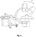
- an imaging device 140 (also referred to herein as a "CT imaging device 140") is shown in a position relative to the patient during or after navigation of a surgical tool 130 (either catheter guide assembly 200 of Fig. 2 or ablation tool 300 of Fig. 3 ) to a desired target tissue site.
- the CT imaging device 140 moves along path "P" while capturing image data (for example, a video or a series of images) along path "P" from different perspectives relative to the patient or target area.
- CT imaging device 140 slowly reciprocates back and forth along path "P" and transmits the acquired image data to computing device 100 for processing and/or display of the image data on display 110.
- CT imaging device 140 includes a C-arm and a motor for moving an imager of the CT imaging device 140 along the C-arm.
- the image data may include fluoroscopic images, fluoroscopic video, or other CT data.
- Computing device 100 displays the acquired CT data on display 110 such that a clinician can visualize the position of the surgical tool 130 relative to the target tissue from multiple perspectives, without the need to iteratively adjust the position of the CT imaging device 140, and readjust the position of the surgical tool 130 multiple times before reaching the desired target tissue.
- computing device 100 provides the clinician with a continuous multi-perspective or multi-angle visualization of the target region, thereby enabling the clinician to interpolate the three-dimensional space and simultaneously navigate the surgical tool 130 to the target tissue without being required to repeatedly retract the surgical tool 130 and correct its trajectory.
- the speed at which the imaging device 140 traverses the path "P" may also be selected or modified by the computing device 100 or the clinician.
- a clinician may choose the speed at which the imaging device 140 traverses along the path "P" in order to suit the particular needs of the navigation procedure. That is, in some instances it may be useful for the clinician to visualize the target area while receiving images or video of the target area while the imaging device 140 traverses back and forth along path "P" at a fast speed and in some other instances it may be useful for the clinician to visualize the target area while receiving images or video of the target area while the imaging device 140 traverses back and forth along path "P" at a slow speed.
- the type or format of the image data acquired by the imaging device 140 may be selected or modified by the computing device 100 or may be selected or modified by a clinician.
- the clinician may select the image data to be a series of images, or a combination of a series of images and video along different portions of the path "P.”
- Method 500 begins at step 501 where navigation of a surgical tool 130 to a target region is initiated.
- Step 501 may include using a catheter guide assembly 200 ( Fig. 2 ) using a navigation pathway previously planned, as described above.
- step 501 may include percutaneously inserting an ablation tool 300 ( Fig. 3 ) to access the target region along a previously determined trajectory.
- computing device 100 receives parameters for the path "P" ( Fig. 4 ) or parameters of the imaging device 140.
- a user may input the distance or length of path "P" for which the imaging device 140 will traverse. Setting the length of path “P” sets the range of angular positions of the imaging device 140 relative to the target.
- a user may input the speed at which the imaging device 140 traverses along path "P.”
- a user may also input the type of image data desired to be acquired. For example, in certain instances a clinician may desire to generate a video stream of the target area.
- this imaging at different angles relative to the patient as the imaging device 140 traverses the path "P" provides for enhanced imaging of the position of the surgical tool 130 relative to the target at a number of angles.
- the clinician observes the images, either still or video the effect is the impression of a three-dimensional-like image which is viewable by the clinician.
- the clinician is able to observe the relative position of the surgical tool 130 and the target from multiple perspective angles.
- Such a continuous multi-perspective visualization of the target region enables the clinician to interpolate the three-dimensional space and simultaneously navigate the surgical tool 130 to the target tissue without being required to repeatedly retract the surgical tool 130 and correct its trajectory.
- step 509 it is determined if the surgical tool 130 is at a desired location (for example, the target tissue or target region).
- step 509 is accomplished by determining whether a distance between a portion of the surgical tool 130 and an edge, or center, of the target tissue is within a predetermined threshold. For example, when the distance between the surgical tool 130 and the target tissue is more than the predetermined threshold, then it is determined that the surgical tool 130 is not at the desired position (no in step 509) and method 500 proceeds to step 511 where the clinician can adjust the position of the surgical tool 130. Specifically, in step 511, the clinician can continue to advance the surgical tool 130 while simultaneously visualizing the position of the surgical tool 130 relative to the target tissue from multiple angles with the image data being displayed on display 110.
- the desired position is the center of the target tissue.
- step 513 the procedure is performed on the target tissue.
- method 500 may be used to navigate to a target region for various purposes. That is, step 513 may include any type of procedure, which may include for example, biopsy collection, marker placement, device placement, therapeutic treatments, agent delivery, ablation treatments including radiofrequency and microwave ablations, and any other such procedure that may benefit from enhanced and more accurate navigation to a target within a patient.
- FIG. 6 an example user interface 600 is illustrated which can be displayed on display 110 ( Fig. 1 ).
- User interface 600 includes a split screen view which is presented to the user to assist the user in visualizing the position of the surgical tool 130 relative to the target from multiple perspectives while simultaneously advancing the surgical tool 130 to the target.
- section 610 includes an axial slice of the target "T" which is generated from previously acquired CT image data. The particular axial slice of the target "T" displayed in section 610 corresponds to the axial position of the imaging device 140 relative to the patient.
- Icon 611 is a virtual representation of the position of the imaging device 140 at a given point in time.
- Section 611 also includes setting input region 613.
- Setting input region 613 may be used by an operator to input parameters associate with the imaging device 140 and path "P.”
- a user may utilize input region 613 to set or modify the length of the path "P" (which in turn sets or modifies the angular range a-e of images to acquire), the speed at which the imaging device 140 traverses back and forth along the path "P," the image format to acquire, and the portions of the path "P” for which to capture different types of images formats.
- a graphical representation of the path "P” may also be included in section 610 and when the length of the path "P" is adjusted using region 613, the graphical representation is adjusted accordingly.
- User interface 600 also includes section 620 which displays a continuous stream of images or video of the image data acquired by the imaging device 140.
- section 620 includes an image of target "T” and surgical tool 130 which continuously changes to a different perspective captured by the imaging device 140. Continuously displaying the target “T” and the position of the surgical tool 130 relative to the target “T” while simultaneously navigating the surgical tool enables the clinician to visualize the position of the surgical tool relative to the target "T” from multiple perspectives while still in the process of advancing the surgical tool 130 to the target "T.” In this manner, as the clinician observes the display in section 620, the effect is the impression of a three-dimensional-like image which is being viewed by the clinician.
Landscapes
- Health & Medical Sciences (AREA)
- Life Sciences & Earth Sciences (AREA)
- Engineering & Computer Science (AREA)
- Surgery (AREA)
- Veterinary Medicine (AREA)
- Biomedical Technology (AREA)
- Heart & Thoracic Surgery (AREA)
- Medical Informatics (AREA)
- Animal Behavior & Ethology (AREA)
- General Health & Medical Sciences (AREA)
- Public Health (AREA)
- Molecular Biology (AREA)
- Nuclear Medicine, Radiotherapy & Molecular Imaging (AREA)
- Pathology (AREA)
- Robotics (AREA)
- Oral & Maxillofacial Surgery (AREA)
- Physics & Mathematics (AREA)
- Radiology & Medical Imaging (AREA)
- Biophysics (AREA)
- Pulmonology (AREA)
- Human Computer Interaction (AREA)
- Otolaryngology (AREA)
- Plasma & Fusion (AREA)
- Cardiology (AREA)
- Gynecology & Obstetrics (AREA)
- Theoretical Computer Science (AREA)
- Optics & Photonics (AREA)
- High Energy & Nuclear Physics (AREA)
- Anesthesiology (AREA)
- Hematology (AREA)
- Apparatus For Radiation Diagnosis (AREA)
Applications Claiming Priority (1)
| Application Number | Priority Date | Filing Date | Title |
|---|---|---|---|
| US15/277,742 US11051886B2 (en) | 2016-09-27 | 2016-09-27 | Systems and methods for performing a surgical navigation procedure |
Publications (1)
| Publication Number | Publication Date |
|---|---|
| EP3298977A1 true EP3298977A1 (de) | 2018-03-28 |
Family
ID=60037361
Family Applications (1)
| Application Number | Title | Priority Date | Filing Date |
|---|---|---|---|
| EP17193197.5A Pending EP3298977A1 (de) | 2016-09-27 | 2017-09-26 | System und verfahren zur durchführung eines chirurgischen navigationsverfahrens |
Country Status (5)
| Country | Link |
|---|---|
| US (1) | US11051886B2 (de) |
| EP (1) | EP3298977A1 (de) |
| CN (1) | CN107865694B (de) |
| AU (1) | AU2017232099B2 (de) |
| CA (1) | CA2980211A1 (de) |
Families Citing this family (13)
| Publication number | Priority date | Publication date | Assignee | Title |
|---|---|---|---|---|
| US10448910B2 (en) * | 2016-02-03 | 2019-10-22 | Globus Medical, Inc. | Portable medical imaging system |
| US11026752B2 (en) * | 2018-06-04 | 2021-06-08 | Medtronic Navigation, Inc. | System and method for performing and evaluating a procedure |
| US11918423B2 (en) | 2018-10-30 | 2024-03-05 | Corindus, Inc. | System and method for navigating a device through a path to a target location |
| US11701181B2 (en) * | 2019-04-24 | 2023-07-18 | Warsaw Orthopedic, Inc. | Systems, instruments and methods for surgical navigation with verification feedback |
| CN112221006A (zh) * | 2020-10-19 | 2021-01-15 | 湖北工业大学 | 一种用于快速诊断急性肺栓塞的导管介入电磁导引装置 |
| US12193759B2 (en) * | 2020-12-30 | 2025-01-14 | Canon U.S.A., Inc. | Real-time correction of regional tissue deformation during endoscopy procedure |
| WO2022197580A1 (en) * | 2021-03-18 | 2022-09-22 | Covidien Lp | Catheter with multiple working channels |
| CN115120348B (zh) * | 2021-03-24 | 2025-08-15 | 上海微创医疗机器人(集团)股份有限公司 | 一种计算机可读存储介质、电子设备及手术机器人系统 |
| CN113081268A (zh) * | 2021-03-31 | 2021-07-09 | 上海电气集团股份有限公司 | 基于AR和IoT的手术引导系统 |
| CN113101549B (zh) * | 2021-05-19 | 2023-07-14 | 深圳市鹰眼在线电子科技有限公司 | 放疗系统和放疗3d视觉实时监测姿态保持方法 |
| CN113729940B (zh) * | 2021-09-23 | 2023-05-23 | 上海卓昕医疗科技有限公司 | 手术辅助定位系统及其控制方法 |
| US11806093B1 (en) | 2022-10-14 | 2023-11-07 | Verdure Imaging, Inc. | Apparatus and method for tracking hand-held surgical tools |
| WO2024080997A1 (en) * | 2022-10-14 | 2024-04-18 | Verdure Imaging, Inc. | Apparatus and method for tracking hand-held surgical tools |
Citations (10)
| Publication number | Priority date | Publication date | Assignee | Title |
|---|---|---|---|---|
| US20060058647A1 (en) * | 1999-05-18 | 2006-03-16 | Mediguide Ltd. | Method and system for delivering a medical device to a selected position within a lumen |
| WO2006116749A2 (en) * | 2005-04-28 | 2006-11-02 | Boston Scientific Scimed, Inc. | Automated activation/deactivation of imaging device based on tracked medical device position |
| US7233820B2 (en) | 2002-04-17 | 2007-06-19 | Superdimension Ltd. | Endoscope structures and techniques for navigating to a target in branched structure |
| US20120250818A1 (en) * | 2011-03-30 | 2012-10-04 | Medtronic Navigation, Inc | System and Method for Off-Center Imaging |
| US9044254B2 (en) | 2012-08-07 | 2015-06-02 | Covidien Lp | Microwave ablation catheter and method of utilizing the same |
| US20150196260A1 (en) * | 2014-01-14 | 2015-07-16 | Samsung Electronics Co., Ltd. | X-ray imaging apparatus and control method thereof |
| US9119650B2 (en) | 2013-03-15 | 2015-09-01 | Covidien Lp | Microwave energy-delivery device and system |
| US20160038248A1 (en) | 2014-08-11 | 2016-02-11 | Covidien Lp | Treatment procedure planning system and method |
| US20160058507A1 (en) | 2014-08-26 | 2016-03-03 | Covidien Lp | Microwave ablation system |
| US20160120522A1 (en) * | 2014-10-31 | 2016-05-05 | Covidien Lp | Computed tomography enhanced fluoroscopic system, device, and method of utilizing the same |
Family Cites Families (161)
| Publication number | Priority date | Publication date | Assignee | Title |
|---|---|---|---|---|
| DE19620371A1 (de) | 1996-05-21 | 1997-12-04 | Philips Patentverwaltung | Röntgenaufnahme-Verfahren |
| DE19703556A1 (de) | 1997-01-31 | 1998-08-06 | Philips Patentverwaltung | Verfahren und Anordnung zur Positionsbestimmung bei der Röntgenbildgebung |
| US6038282A (en) | 1997-04-30 | 2000-03-14 | Siemens Aktiengesellschaft | X-ray imaging system |
| US6055449A (en) | 1997-09-22 | 2000-04-25 | Siemens Corporate Research, Inc. | Method for localization of a biopsy needle or similar surgical tool in a radiographic image |
| US5930329A (en) | 1997-09-22 | 1999-07-27 | Siemens Corporate Research, Inc. | Apparatus and method for detection and localization of a biopsy needle or similar surgical tool in a radiographic image |
| DE19746093C2 (de) | 1997-10-17 | 2002-10-17 | Siemens Ag | C-Bogen-Röntgengerät |
| US5963612A (en) | 1997-12-31 | 1999-10-05 | Siemens Corporation Research, Inc. | Apparatus for C-arm calibration for 3D reconstruction in an imaging system utilizing planar transformation |
| FR2781140B1 (fr) | 1998-07-17 | 2000-11-10 | Ge Medical Syst Sa | Procede de positionnement d'un appareil de radiologie |
| US6081577A (en) | 1998-07-24 | 2000-06-27 | Wake Forest University | Method and system for creating task-dependent three-dimensional images |
| US6477400B1 (en) | 1998-08-20 | 2002-11-05 | Sofamor Danek Holdings, Inc. | Fluoroscopic image guided orthopaedic surgery system with intraoperative registration |
| JP2000201920A (ja) | 1999-01-19 | 2000-07-25 | Fuji Photo Film Co Ltd | 撮影画像デ―タ取得方法および撮影画像デ―タ取得装置 |
| JP4473358B2 (ja) | 1999-01-21 | 2010-06-02 | 株式会社東芝 | 診断装置 |
| US6285902B1 (en) * | 1999-02-10 | 2001-09-04 | Surgical Insights, Inc. | Computer assisted targeting device for use in orthopaedic surgery |
| DE19919907C2 (de) | 1999-04-30 | 2003-10-16 | Siemens Ag | Verfahren und Vorrichtung zur Katheter-Navigation in dreidimensionalen Gefäßbaum-Aufnahmen |
| US7343195B2 (en) | 1999-05-18 | 2008-03-11 | Mediguide Ltd. | Method and apparatus for real time quantitative three-dimensional image reconstruction of a moving organ and intra-body navigation |
| US6139544A (en) | 1999-05-26 | 2000-10-31 | Endocare, Inc. | Computer guided cryosurgery |
| US6236704B1 (en) | 1999-06-30 | 2001-05-22 | Siemens Corporate Research, Inc. | Method and apparatus using a virtual detector for three-dimensional reconstruction from x-ray images |
| DE19936408B4 (de) | 1999-08-03 | 2005-09-01 | Siemens Ag | Verfahrbares Röntgengerät |
| FR2799028B1 (fr) | 1999-09-27 | 2002-05-03 | Ge Medical Syst Sa | Procede de reconstitution d'une image tridimentionnelle d'elements de fort contraste |
| US7689014B2 (en) | 2000-01-18 | 2010-03-30 | Z-Kat Inc | Apparatus and method for measuring anatomical objects using coordinated fluoroscopy |
| DE10003524B4 (de) * | 2000-01-27 | 2006-07-13 | Siemens Ag | Verfahrbares Röntgengerät und Verfahren zur Bestimmung von Projektionsgeometrien |
| US6856826B2 (en) | 2000-04-28 | 2005-02-15 | Ge Medical Systems Global Technology Company, Llc | Fluoroscopic tracking and visualization system |
| US6856827B2 (en) | 2000-04-28 | 2005-02-15 | Ge Medical Systems Global Technology Company, Llc | Fluoroscopic tracking and visualization system |
| US6490475B1 (en) | 2000-04-28 | 2002-12-03 | Ge Medical Systems Global Technology Company, Llc | Fluoroscopic tracking and visualization system |
| US6782287B2 (en) | 2000-06-27 | 2004-08-24 | The Board Of Trustees Of The Leland Stanford Junior University | Method and apparatus for tracking a medical instrument based on image registration |
| US6389104B1 (en) | 2000-06-30 | 2002-05-14 | Siemens Corporate Research, Inc. | Fluoroscopy based 3-D neural navigation based on 3-D angiography reconstruction data |
| US6351513B1 (en) | 2000-06-30 | 2002-02-26 | Siemens Corporate Research, Inc. | Fluoroscopy based 3-D neural navigation based on co-registration of other modalities with 3-D angiography reconstruction data |
| DE10033063A1 (de) | 2000-07-07 | 2002-01-24 | Brainlab Ag | Verfahren zur atmungskompensierten Strahlenbehandlung |
| US6837892B2 (en) * | 2000-07-24 | 2005-01-04 | Mazor Surgical Technologies Ltd. | Miniature bone-mounted surgical robot |
| US6823207B1 (en) | 2000-08-26 | 2004-11-23 | Ge Medical Systems Global Technology Company, Llc | Integrated fluoroscopic surgical navigation and imaging workstation with command protocol |
| US6714810B2 (en) | 2000-09-07 | 2004-03-30 | Cbyon, Inc. | Fluoroscopic registration system and method |
| DE10051370A1 (de) | 2000-10-17 | 2002-05-02 | Brainlab Ag | Verfahren und Vorrichtung zur exakten Patientenpositionierung in der Strahlentherapie und Radiochirurgie |
| EP1341443B1 (de) | 2000-10-18 | 2010-12-29 | Paieon Inc. | System zur positionierung einer einrichtung in einem rohrförmigen organ |
| JP4488678B2 (ja) | 2001-02-07 | 2010-06-23 | アーオー テクノロジー アクチエンゲゼルシャフト | 骨のx線画像の三次元表示の確立方法 |
| US6785571B2 (en) | 2001-03-30 | 2004-08-31 | Neil David Glossop | Device and method for registering a position sensor in an anatomical body |
| US6546279B1 (en) | 2001-10-12 | 2003-04-08 | University Of Florida | Computer controlled guidance of a biopsy needle |
| WO2006103644A1 (en) | 2005-03-31 | 2006-10-05 | Paieon Inc. | Method and apparatus for positioning a device in a tubular organ |
| US6768784B1 (en) | 2001-11-07 | 2004-07-27 | Koninklijke Philips Electronics N.V. | X-ray image enhancement |
| DE10155590A1 (de) | 2001-11-13 | 2003-05-15 | Philips Corp Intellectual Pty | Fluoroskopisches Computertomographie-Verfahren |
| US7010152B2 (en) | 2002-01-22 | 2006-03-07 | Canon Kabushiki Kaisha | Radiographic image composition and use |
| US6774624B2 (en) | 2002-03-27 | 2004-08-10 | Ge Medical Systems Global Technology Company, Llc | Magnetic tracking system |
| US6707878B2 (en) | 2002-04-15 | 2004-03-16 | General Electric Company | Generalized filtered back-projection reconstruction in digital tomosynthesis |
| US8244327B2 (en) | 2002-04-22 | 2012-08-14 | The Johns Hopkins University | Apparatus for insertion of a medical device during a medical imaging process |
| US7787932B2 (en) | 2002-04-26 | 2010-08-31 | Brainlab Ag | Planning and navigation assistance using two-dimensionally adapted generic and detected patient data |
| US7165362B2 (en) | 2002-07-15 | 2007-01-23 | Apple Computer, Inc. | Glass support member |
| US7356115B2 (en) | 2002-12-04 | 2008-04-08 | Varian Medical Systems Technology, Inc. | Radiation scanning units including a movable platform |
| AU2003262726A1 (en) | 2002-08-21 | 2004-03-11 | Breakaway Imaging, Llc | Apparatus and method for reconstruction of volumetric images in a divergent scanning computed tomography system |
| US7251522B2 (en) | 2002-09-12 | 2007-07-31 | Brainlab Ag | X-ray image-assisted navigation using original, two-dimensional x-ray images |
| DE10243162B4 (de) | 2002-09-17 | 2005-10-06 | Siemens Ag | Rechnergestütztes Darstellungsverfahren für ein 3D-Objekt |
| US7697972B2 (en) * | 2002-11-19 | 2010-04-13 | Medtronic Navigation, Inc. | Navigation system for cardiac therapies |
| US6898263B2 (en) | 2002-11-27 | 2005-05-24 | Ge Medical Systems Global Technology Company, Llc | Method and apparatus for soft-tissue volume visualization |
| JP4200811B2 (ja) | 2003-05-16 | 2008-12-24 | 株式会社島津製作所 | 放射線治療計画装置 |
| DE10322738A1 (de) | 2003-05-20 | 2004-12-16 | Siemens Ag | Verfahren zur markerlosen automatischen Fusion von 2D-Fluoro-C-Bogen-Bildern mit präoperativen 3D-Bildern unter Verwendung eines intraoperativ gewonnenen 3D-Datensatzes |
| US7099431B2 (en) | 2003-06-09 | 2006-08-29 | Canon Kabushiki Kaisha | Radiation imaging apparatus |
| FR2856170B1 (fr) | 2003-06-10 | 2005-08-26 | Biospace Instr | Procede d'imagerie radiographique pour la reconstruction tridimensionnelle, dispositif et programme d'ordinateur pour mettre en oeuvre ce procede |
| US7907989B2 (en) | 2003-06-16 | 2011-03-15 | Koninklijke Philips Electronics N.V. | Imaging system for interventional radiology |
| US6944260B2 (en) | 2003-11-11 | 2005-09-13 | Ge Medical Systems Global Technology Company, Llc | Methods and apparatus for artifact reduction in computed tomography imaging systems |
| US8126224B2 (en) | 2004-02-03 | 2012-02-28 | Ge Medical Systems Global Technology Company, Llc | Method and apparatus for instrument tracking on a scrolling series of 2D fluoroscopic images |
| US7035371B2 (en) | 2004-03-22 | 2006-04-25 | Siemens Aktiengesellschaft | Method and device for medical imaging |
| US7620223B2 (en) | 2004-04-22 | 2009-11-17 | Siemens Medical Solutions Usa, Inc. | Method and system for registering pre-procedural images with intra-procedural images using a pre-computed knowledge base |
| DE102004030836A1 (de) | 2004-06-25 | 2006-01-26 | Siemens Ag | Verfahren zur Bilddarstellung eines in einen sich rhythmisch oder arhythmisch bewegenden Untersuchungsbereich eines Patienten eingebrachten medizinischen Instruments, insbesondere eines Katheters |
| US8515527B2 (en) | 2004-10-13 | 2013-08-20 | General Electric Company | Method and apparatus for registering 3D models of anatomical regions of a heart and a tracking system with projection images of an interventional fluoroscopic system |
| US7327872B2 (en) | 2004-10-13 | 2008-02-05 | General Electric Company | Method and system for registering 3D models of anatomical regions with projection images of the same |
| US7720520B2 (en) | 2004-12-01 | 2010-05-18 | Boston Scientific Scimed, Inc. | Method and system for registering an image with a navigation reference catheter |
| WO2006063141A2 (en) | 2004-12-07 | 2006-06-15 | Medical Metrx Solutions, Inc. | Intraoperative c-arm fluoroscope datafusion system |
| JP4649219B2 (ja) | 2005-02-01 | 2011-03-09 | キヤノン株式会社 | 立体画像生成装置 |
| CN101138010B (zh) | 2005-03-10 | 2011-05-18 | 皇家飞利浦电子股份有限公司 | 用于将二维和三维图像数据对准的图像处理系统和方法 |
| EP1876988B1 (de) | 2005-04-26 | 2016-06-08 | Koninklijke Philips N.V. | Medizinisches abbildungssystem und verfahren zur detektion und verbesserung statischer strukturen bei verrauschten bildern mittels bewegung der bildaufnahmevorrichtung |
| DE102005023194A1 (de) | 2005-05-19 | 2006-11-23 | Siemens Ag | Verfahren zur Erweiterung des Darstellungsbereiches von 2D-Bildaufnahmen eines Objektbereiches |
| DE102005023167B4 (de) | 2005-05-19 | 2008-01-03 | Siemens Ag | Verfahren und Vorrichtung zur Registrierung von 2D-Projektionsbildern relativ zu einem 3D-Bilddatensatz |
| US7603155B2 (en) | 2005-05-24 | 2009-10-13 | General Electric Company | Method and system of acquiring images with a medical imaging device |
| DE102005030646B4 (de) | 2005-06-30 | 2008-02-07 | Siemens Ag | Verfahren zur Kontur-Visualisierung von zumindest einer interessierenden Region in 2D-Durchleuchtungsbildern |
| DE102005032523B4 (de) | 2005-07-12 | 2009-11-05 | Siemens Ag | Verfahren zur prä-interventionellen Planung einer 2D-Durchleuchtungsprojektion |
| DE102005032755B4 (de) | 2005-07-13 | 2014-09-04 | Siemens Aktiengesellschaft | System zur Durchführung und Überwachung minimal-invasiver Eingriffe |
| DE102005036322A1 (de) | 2005-07-29 | 2007-02-15 | Siemens Ag | Registrieren intraoperativer Bilddatensätze mit präoperativen 3D-Bilddatensätzen auf Basis optischer Oberflächenextraktion |
| DE102005059804A1 (de) | 2005-12-14 | 2007-07-05 | Siemens Ag | Verfahren und Vorrichtung zur Bewegungskorrektur bei der Bildgebung während einer medizinischen Intervention |
| US8050739B2 (en) | 2005-12-15 | 2011-11-01 | Koninklijke Philips Electronics N.V. | System and method for visualizing heart morphology during electrophysiology mapping and treatment |
| DE102006008042A1 (de) | 2006-02-21 | 2007-07-19 | Siemens Ag | Medizinisches Gerät mit im medizinischen Gerät kombinierter Bilderfassungs- und Positionsbestimmungsvorrichtung |
| US8526688B2 (en) | 2006-03-09 | 2013-09-03 | General Electric Company | Methods and systems for registration of surgical navigation data and image data |
| DE102006011242B4 (de) | 2006-03-10 | 2012-03-29 | Siemens Ag | Verfahren zur Rekonstruktion einer 3D-Darstellung |
| US9636188B2 (en) | 2006-03-24 | 2017-05-02 | Stryker Corporation | System and method for 3-D tracking of surgical instrument in relation to patient body |
| WO2007113703A2 (en) | 2006-03-30 | 2007-10-11 | Koninklijke Philips Electronics N. V. | Targeting method, targeting device, computer readable medium and program element |
| WO2007130433A2 (en) * | 2006-05-02 | 2007-11-15 | Wisconsin Alumni Research Foundation | X-ray system for use in image guided procedures |
| US7467007B2 (en) | 2006-05-16 | 2008-12-16 | Siemens Medical Solutions Usa, Inc. | Respiratory gated image fusion of computed tomography 3D images and live fluoroscopy images |
| FR2904750B1 (fr) | 2006-08-03 | 2008-10-17 | Gen Electric | Procede de reconstruction tridimensionnelle d'une enveloppe exterieure d'un corps d'un appareil a rayons x |
| DE102006041033B4 (de) | 2006-09-01 | 2017-01-19 | Siemens Healthcare Gmbh | Verfahren zur Rekonstruktion eines dreidimensionalen Bildvolumens |
| US8248413B2 (en) | 2006-09-18 | 2012-08-21 | Stryker Corporation | Visual navigation system for endoscopic surgery |
| CN103961130B (zh) | 2006-09-25 | 2017-08-15 | 马佐尔机器人有限公司 | 使得c型臂系统适应以提供三维成像信息的方法 |
| DE102006046735A1 (de) | 2006-09-29 | 2008-04-10 | Siemens Ag | Vorrichtung zur Fusion eines 2D-Durchleuchtungsbildes mit einem Bild aus einem 3D-Bilddatensatz |
| US7711409B2 (en) | 2006-10-04 | 2010-05-04 | Hampton University | Opposed view and dual head detector apparatus for diagnosis and biopsy with image processing methods |
| US8320992B2 (en) | 2006-10-05 | 2012-11-27 | Visionsense Ltd. | Method and system for superimposing three dimensional medical information on a three dimensional image |
| US8111894B2 (en) | 2006-11-16 | 2012-02-07 | Koninklijke Philips Electronics N.V. | Computer Tomography (CT) C-arm system and method for examination of an object |
| US7831096B2 (en) | 2006-11-17 | 2010-11-09 | General Electric Company | Medical navigation system with tool and/or implant integration into fluoroscopic image projections and method of use |
| US8111895B2 (en) | 2006-12-06 | 2012-02-07 | Siemens Medical Solutions Usa, Inc. | Locally adaptive image enhancement for digital subtraction X-ray imaging |
| IL188569A (en) | 2007-01-17 | 2014-05-28 | Mediguide Ltd | Method and system for coordinating a 3D image coordinate system with a medical position coordinate system and a 2D image coordinate system |
| US8781193B2 (en) | 2007-03-08 | 2014-07-15 | Sync-Rx, Ltd. | Automatic quantitative vessel analysis |
| DE102007013807B4 (de) | 2007-03-22 | 2014-11-13 | Siemens Aktiengesellschaft | Verfahren zur Unterstützung der Navigation interventioneller Werkzeuge bei Durchführung von CT- bzw. MRT-gesteuerten Interventionen in einer vorgegebenen Interventionsebene |
| US7899226B2 (en) | 2007-04-03 | 2011-03-01 | General Electric Company | System and method of navigating an object in an imaged subject |
| US7853061B2 (en) | 2007-04-26 | 2010-12-14 | General Electric Company | System and method to improve visibility of an object in an imaged subject |
| WO2008139354A2 (en) | 2007-05-10 | 2008-11-20 | Koninklijke Philips Electronics N. V. | Targeting method, targeting device, computer readable medium and program element |
| DE102007026115B4 (de) | 2007-06-05 | 2017-10-12 | Siemens Healthcare Gmbh | Verfahren zum Erzeugen einer 3D-Rekonstruktion eines Körpers |
| US7991450B2 (en) | 2007-07-02 | 2011-08-02 | General Electric Company | Methods and systems for volume fusion in diagnostic imaging |
| FR2919096A1 (fr) | 2007-07-19 | 2009-01-23 | Gen Electric | Procede de correction de recalage d'images radiographiques |
| DE102007042333A1 (de) | 2007-09-06 | 2009-03-12 | Siemens Ag | Verfahren zum Ermitteln einer Abbildungsvorschrift und Verfahren zum Erzeugen einer 3D-Rekonstruktion |
| US8271068B2 (en) | 2007-10-02 | 2012-09-18 | Siemens Aktiengesellschaft | Method for dynamic road mapping |
| US8270691B2 (en) | 2007-10-09 | 2012-09-18 | Siemens Aktiengesellschaft | Method for fusing images acquired from a plurality of different image acquiring modalities |
| US8090168B2 (en) | 2007-10-15 | 2012-01-03 | General Electric Company | Method and system for visualizing registered images |
| JP5106978B2 (ja) | 2007-10-15 | 2012-12-26 | ジーイー・メディカル・システムズ・グローバル・テクノロジー・カンパニー・エルエルシー | X線ct装置 |
| US8195271B2 (en) | 2007-11-06 | 2012-06-05 | Siemens Aktiengesellschaft | Method and system for performing ablation to treat ventricular tachycardia |
| US9001121B2 (en) | 2007-11-15 | 2015-04-07 | The Boeing Company | Method and apparatus for generating data for three-dimensional models from x-rays |
| JP5229865B2 (ja) | 2007-11-30 | 2013-07-03 | ジーイー・メディカル・システムズ・グローバル・テクノロジー・カンパニー・エルエルシー | X線ct装置 |
| RU2568635C2 (ru) | 2007-12-18 | 2015-11-20 | Конинклейке Филипс Электроникс, Н.В. | Регистрация двумерных/трехмерных изображений на основе признаков |
| EP2231016B1 (de) * | 2007-12-21 | 2016-04-06 | Koning Corporation | Vorrichtung für 3d-computertomografie |
| DE102007061935A1 (de) | 2007-12-21 | 2009-06-25 | Siemens Aktiengesellschaft | Verfahren zur Qualitätssteigerung von computertomographischen Aufnahmeserien durch Bildverarbeitung und CT-System mit Recheneinheit |
| US9445772B2 (en) | 2007-12-31 | 2016-09-20 | St. Jude Medical, Atrial Fibrillatin Division, Inc. | Reduced radiation fluoroscopic system |
| DE102008003173B4 (de) | 2008-01-04 | 2016-05-19 | Siemens Aktiengesellschaft | Verfahren und Vorrichtung für die Computertomographie zur Be- |
| EP2082686B1 (de) | 2008-01-22 | 2015-01-21 | Brainlab AG | Orientierte Wiedergabe von Aufnahmen |
| US8340379B2 (en) | 2008-03-07 | 2012-12-25 | Inneroptic Technology, Inc. | Systems and methods for displaying guidance data based on updated deformable imaging data |
| DE102008018269A1 (de) | 2008-04-10 | 2009-10-29 | Fraunhofer-Gesellschaft zur Förderung der angewandten Forschung e.V. | Vorrichtung und Verfahren zur rotationsfreien Computertomographie |
| US8532259B2 (en) | 2008-04-17 | 2013-09-10 | University Of Florida Research Foundation, Inc. | Method and apparatus for computed imaging backscatter radiography |
| DE102008020948A1 (de) | 2008-04-25 | 2009-11-26 | Fraunhofer-Gesellschaft zur Förderung der angewandten Forschung e.V. | Röntgencomputertomograph und Verfahren zur Untersuchung eines Bauteils mittels Röntgencomputertomographie |
| US9050470B2 (en) | 2008-05-15 | 2015-06-09 | Intelect Medical, Inc. | Clinician programmer system interface for monitoring patient progress |
| CN102112055B (zh) * | 2008-08-04 | 2016-06-22 | 皇家飞利浦电子股份有限公司 | 介入性成像和数据处理 |
| DE102008050844B4 (de) | 2008-10-08 | 2016-04-21 | Siemens Aktiengesellschaft | Vorrichtung und Verfahren zur Lokalisation einer Gewebeveränderung für eine Biopsie |
| CN102202576B (zh) | 2008-10-10 | 2015-04-08 | 皇家飞利浦电子股份有限公司 | 用于为了在微创的x射线引导的介入中减少x射线剂量而产生覆盖经分割的目标结构或病变的减小视场的、具有自动快门适应的血管造影图像采集系统和方法 |
| JP2012515592A (ja) | 2009-01-21 | 2012-07-12 | コーニンクレッカ フィリップス エレクトロニクス エヌ ヴィ | 大きい視野のイメージング並びに動きのアーチファクトの検出及び補償ための方法及び装置 |
| US7912180B2 (en) | 2009-02-19 | 2011-03-22 | Kabushiki Kaisha Toshiba | Scattered radiation correction method and scattered radiation correction apparatus |
| US8423117B2 (en) | 2009-06-22 | 2013-04-16 | General Electric Company | System and method to process an acquired image of a subject anatomy to differentiate a portion of subject anatomy to protect relative to a portion to receive treatment |
| US8675996B2 (en) | 2009-07-29 | 2014-03-18 | Siemens Aktiengesellschaft | Catheter RF ablation using segmentation-based 2D-3D registration |
| JP5662447B2 (ja) | 2009-08-20 | 2015-01-28 | コーニンクレッカ フィリップス エヌ ヴェ | 関心領域画像の再構成 |
| CN102483853B (zh) | 2009-09-07 | 2015-08-26 | 皇家飞利浦电子股份有限公司 | 用于处理投影数据的装置和方法 |
| DE102009049818A1 (de) | 2009-10-19 | 2011-04-21 | Siemens Aktiengesellschaft | Verfahren zur Ermittlung der Projektionsgeometrie einer Röntgenanlage |
| US8694075B2 (en) | 2009-12-21 | 2014-04-08 | General Electric Company | Intra-operative registration for navigated surgical procedures |
| JP5677738B2 (ja) | 2009-12-24 | 2015-02-25 | 株式会社東芝 | X線コンピュータ断層撮影装置 |
| CN102711650B (zh) | 2010-01-13 | 2015-04-01 | 皇家飞利浦电子股份有限公司 | 用于内窥镜手术的基于图像整合的配准和导航 |
| US9104902B2 (en) | 2010-04-15 | 2015-08-11 | Koninklijke Philips N.V. | Instrument-based image registration for fusing images with tubular structures |
| US9401047B2 (en) | 2010-04-15 | 2016-07-26 | Siemens Medical Solutions, Usa, Inc. | Enhanced visualization of medical image data |
| DE102010019632A1 (de) | 2010-05-06 | 2011-11-10 | Siemens Aktiengesellschaft | Verfahren zur Aufnahme und Rekonstruktion eines dreidimensionalen Bilddatensatzes und Röntgeneinrichtung |
| US8625869B2 (en) | 2010-05-21 | 2014-01-07 | Siemens Medical Solutions Usa, Inc. | Visualization of medical image data with localized enhancement |
| JP5899208B2 (ja) | 2010-05-27 | 2016-04-06 | コーニンクレッカ フィリップス エヌ ヴェKoninklijke Philips N.V. | 偏心フラットパネル検出器による、コーンビーム・コンピュータ断層撮影画像形成のための改善された再構成 |
| US9282944B2 (en) | 2010-06-22 | 2016-03-15 | Queen's University At Kingston | C-arm pose estimation using intensity-based registration of imaging modalities |
| US8526700B2 (en) * | 2010-10-06 | 2013-09-03 | Robert E. Isaacs | Imaging system and method for surgical and interventional medical procedures |
| US8718346B2 (en) | 2011-10-05 | 2014-05-06 | Saferay Spine Llc | Imaging system and method for use in surgical and interventional medical procedures |
| RU2586448C2 (ru) | 2011-03-04 | 2016-06-10 | Конинклейке Филипс Н.В. | Совмещение двумерных/трехмерных изображений |
| DE102011005119A1 (de) | 2011-03-04 | 2012-09-06 | Siemens Aktiengesellschaft | Verfahren zum Bereitstellen eines 3D-Bilddatensatzes eines physiologischen Objekts mit Metallobjekt darin |
| US9232924B2 (en) | 2011-04-01 | 2016-01-12 | Koninklijke Philips N.V. | X-ray pose recovery |
| DE102011006991B4 (de) | 2011-04-07 | 2018-04-05 | Siemens Healthcare Gmbh | Röntgenverfahren und Röntgenvorrichtung zum Zusammenfügen von Röntgenaufnahmen und Ermitteln dreidimensionaler Volumendaten |
| US9265468B2 (en) | 2011-05-11 | 2016-02-23 | Broncus Medical, Inc. | Fluoroscopy-based surgical device tracking method |
| JP2012249960A (ja) | 2011-06-06 | 2012-12-20 | Toshiba Corp | 医用画像処理装置 |
| KR101146833B1 (ko) | 2011-12-09 | 2012-05-21 | 전남대학교산학협력단 | 무회전 ct 시스템 |
| US9031188B2 (en) | 2012-02-08 | 2015-05-12 | Georgetown Rail Equipment Company | Internal imaging system |
| DE102012204019B4 (de) | 2012-03-14 | 2018-02-08 | Siemens Healthcare Gmbh | Verfahren zur Reduzierung von Bewegungsartefakten |
| JP5787030B2 (ja) | 2012-03-29 | 2015-09-30 | 株式会社島津製作所 | 医療用x線装置 |
| WO2014008613A1 (de) | 2012-07-12 | 2014-01-16 | Ao Technology Ag | Verfahren zur herstellung eines graphischen 3d computermodells mindestens einer anatomischen struktur in einem wählbaren prä-, intra- oder post-operativen status |
| US9001962B2 (en) | 2012-12-20 | 2015-04-07 | Triple Ring Technologies, Inc. | Method and apparatus for multiple X-ray imaging applications |
| KR20140092437A (ko) | 2012-12-27 | 2014-07-24 | 삼성전자주식회사 | 엑스선 영상 장치 및 엑스선 영상 장치의 제어 방법 |
| EP2928408B1 (de) | 2013-03-15 | 2019-03-06 | St. Jude Medical International Holding S.à r.l. | Navigationssystem für eine medizinische vorrichtung |
| US10201265B2 (en) * | 2013-09-06 | 2019-02-12 | Covidien Lp | Microwave ablation catheter, handle, and system |
| ES2865049T3 (es) | 2013-09-06 | 2021-10-14 | Covidien Lp | Catéter, mango y sistema de ablación por microondas |
| US9171365B2 (en) | 2013-11-29 | 2015-10-27 | Kabushiki Kaisha Toshiba | Distance driven computation balancing |
| WO2015100310A1 (en) | 2013-12-23 | 2015-07-02 | Camplex, Inc. | Surgical visualization systems |
-
2016
- 2016-09-27 US US15/277,742 patent/US11051886B2/en active Active
-
2017
- 2017-09-20 AU AU2017232099A patent/AU2017232099B2/en not_active Ceased
- 2017-09-25 CA CA2980211A patent/CA2980211A1/en not_active Abandoned
- 2017-09-26 EP EP17193197.5A patent/EP3298977A1/de active Pending
- 2017-09-27 CN CN201710885356.1A patent/CN107865694B/zh not_active Expired - Fee Related
Patent Citations (11)
| Publication number | Priority date | Publication date | Assignee | Title |
|---|---|---|---|---|
| US20060058647A1 (en) * | 1999-05-18 | 2006-03-16 | Mediguide Ltd. | Method and system for delivering a medical device to a selected position within a lumen |
| US7233820B2 (en) | 2002-04-17 | 2007-06-19 | Superdimension Ltd. | Endoscope structures and techniques for navigating to a target in branched structure |
| WO2006116749A2 (en) * | 2005-04-28 | 2006-11-02 | Boston Scientific Scimed, Inc. | Automated activation/deactivation of imaging device based on tracked medical device position |
| US20120250818A1 (en) * | 2011-03-30 | 2012-10-04 | Medtronic Navigation, Inc | System and Method for Off-Center Imaging |
| US9044254B2 (en) | 2012-08-07 | 2015-06-02 | Covidien Lp | Microwave ablation catheter and method of utilizing the same |
| US9247992B2 (en) | 2012-08-07 | 2016-02-02 | Covidien, LP | Microwave ablation catheter and method of utilizing the same |
| US9119650B2 (en) | 2013-03-15 | 2015-09-01 | Covidien Lp | Microwave energy-delivery device and system |
| US20150196260A1 (en) * | 2014-01-14 | 2015-07-16 | Samsung Electronics Co., Ltd. | X-ray imaging apparatus and control method thereof |
| US20160038248A1 (en) | 2014-08-11 | 2016-02-11 | Covidien Lp | Treatment procedure planning system and method |
| US20160058507A1 (en) | 2014-08-26 | 2016-03-03 | Covidien Lp | Microwave ablation system |
| US20160120522A1 (en) * | 2014-10-31 | 2016-05-05 | Covidien Lp | Computed tomography enhanced fluoroscopic system, device, and method of utilizing the same |
Also Published As
| Publication number | Publication date |
|---|---|
| US20180085173A1 (en) | 2018-03-29 |
| AU2017232099B2 (en) | 2019-02-21 |
| CN107865694A (zh) | 2018-04-03 |
| CA2980211A1 (en) | 2018-03-27 |
| AU2017232099A1 (en) | 2018-04-12 |
| US11051886B2 (en) | 2021-07-06 |
| CN107865694B (zh) | 2020-12-11 |
Similar Documents
| Publication | Publication Date | Title |
|---|---|---|
| AU2017232099B2 (en) | Systems and methods for performing a surgical navigation procedure | |
| US11172989B2 (en) | Dynamic 3D lung map view for tool navigation inside the lung | |
| US11974865B2 (en) | System and method of providing distance and orientation feedback while navigating in 3D | |
| US12064280B2 (en) | System and method for identifying and marking a target in a fluoroscopic three-dimensional reconstruction | |
| US11596475B2 (en) | Systems and methods for ultrasound image-guided ablation antenna placement | |
| EP3291736B1 (de) | Planungs- und verfahrenssysteme für mikrowellenablation | |
| EP3212112B1 (de) | Computertomographieverbessertes durchleuchtungssystem | |
| US8165660B2 (en) | System and method for selecting a guidance mode for performing a percutaneous procedure | |
| CN112618015A (zh) | 用于经皮插入的设备的图像引导导航的系统和方法 | |
| EP4179994A2 (de) | Planung eines verfahrens, führung eines verfahrens innerhalb einer biopsie und tumorablation mit und ohne kegelstrahlcomputertomografie oder fluoroskopischer bildgebung |
Legal Events
| Date | Code | Title | Description |
|---|---|---|---|
| PUAI | Public reference made under article 153(3) epc to a published international application that has entered the european phase |
Free format text: ORIGINAL CODE: 0009012 |
|
| STAA | Information on the status of an ep patent application or granted ep patent |
Free format text: STATUS: REQUEST FOR EXAMINATION WAS MADE |
|
| 17P | Request for examination filed |
Effective date: 20170926 |
|
| AK | Designated contracting states |
Kind code of ref document: A1 Designated state(s): AL AT BE BG CH CY CZ DE DK EE ES FI FR GB GR HR HU IE IS IT LI LT LU LV MC MK MT NL NO PL PT RO RS SE SI SK SM TR |
|
| AX | Request for extension of the european patent |
Extension state: BA ME |
|
| RBV | Designated contracting states (corrected) |
Designated state(s): AL AT BE BG CH CY CZ DE DK EE ES FI FR GB GR HR HU IE IS IT LI LT LU LV MC MK MT NL NO PL PT RO RS SE SI SK SM TR |
|
| R17P | Request for examination filed (corrected) |
Effective date: 20170926 |
|
| STAA | Information on the status of an ep patent application or granted ep patent |
Free format text: STATUS: EXAMINATION IS IN PROGRESS |
|
| 17Q | First examination report despatched |
Effective date: 20230328 |