EP3301443A1 - Dispositif d'analyse médicale et procédé d'analyse cellulaire - Google Patents
Dispositif d'analyse médicale et procédé d'analyse cellulaire Download PDFInfo
- Publication number
- EP3301443A1 EP3301443A1 EP17189342.3A EP17189342A EP3301443A1 EP 3301443 A1 EP3301443 A1 EP 3301443A1 EP 17189342 A EP17189342 A EP 17189342A EP 3301443 A1 EP3301443 A1 EP 3301443A1
- Authority
- EP
- European Patent Office
- Prior art keywords
- hydrophilic polymer
- blood
- analysis device
- cells
- polymer layer
- Prior art date
- Legal status (The legal status is an assumption and is not a legal conclusion. Google has not performed a legal analysis and makes no representation as to the accuracy of the status listed.)
- Granted
Links
Images
Classifications
-
- G—PHYSICS
- G01—MEASURING; TESTING
- G01N—INVESTIGATING OR ANALYSING MATERIALS BY DETERMINING THEIR CHEMICAL OR PHYSICAL PROPERTIES
- G01N33/00—Investigating or analysing materials by specific methods not covered by groups G01N1/00 - G01N31/00
- G01N33/48—Biological material, e.g. blood, urine; Haemocytometers
-
- G—PHYSICS
- G01—MEASURING; TESTING
- G01N—INVESTIGATING OR ANALYSING MATERIALS BY DETERMINING THEIR CHEMICAL OR PHYSICAL PROPERTIES
- G01N33/00—Investigating or analysing materials by specific methods not covered by groups G01N1/00 - G01N31/00
- G01N33/48—Biological material, e.g. blood, urine; Haemocytometers
- G01N33/50—Chemical analysis of biological material, e.g. blood, urine; Testing involving biospecific ligand binding methods; Immunological testing
- G01N33/5005—Chemical analysis of biological material, e.g. blood, urine; Testing involving biospecific ligand binding methods; Immunological testing involving human or animal cells
-
- C—CHEMISTRY; METALLURGY
- C12—BIOCHEMISTRY; BEER; SPIRITS; WINE; VINEGAR; MICROBIOLOGY; ENZYMOLOGY; MUTATION OR GENETIC ENGINEERING
- C12M—APPARATUS FOR ENZYMOLOGY OR MICROBIOLOGY; APPARATUS FOR CULTURING MICROORGANISMS FOR PRODUCING BIOMASS, FOR GROWING CELLS OR FOR OBTAINING FERMENTATION OR METABOLIC PRODUCTS, i.e. BIOREACTORS OR FERMENTERS
- C12M23/00—Constructional details, e.g. recesses, hinges
- C12M23/02—Form or structure of the vessel
- C12M23/12—Well or multiwell plates
-
- B—PERFORMING OPERATIONS; TRANSPORTING
- B01—PHYSICAL OR CHEMICAL PROCESSES OR APPARATUS IN GENERAL
- B01L—CHEMICAL OR PHYSICAL LABORATORY APPARATUS FOR GENERAL USE
- B01L3/00—Containers or dishes for laboratory use, e.g. laboratory glassware; Droppers
- B01L3/50—Containers for the purpose of retaining a material to be analysed, e.g. test tubes
- B01L3/508—Rigid containers without fluid transport within
- B01L3/5085—Rigid containers without fluid transport within for multiple samples, e.g. microtitration plates
-
- C—CHEMISTRY; METALLURGY
- C08—ORGANIC MACROMOLECULAR COMPOUNDS; THEIR PREPARATION OR CHEMICAL WORKING-UP; COMPOSITIONS BASED THEREON
- C08F—MACROMOLECULAR COMPOUNDS OBTAINED BY REACTIONS ONLY INVOLVING CARBON-TO-CARBON UNSATURATED BONDS
- C08F120/00—Homopolymers of compounds having one or more unsaturated aliphatic radicals, each having only one carbon-to-carbon double bond, and only one being terminated by only one carboxyl radical or a salt, anhydride, ester, amide, imide or nitrile thereof
- C08F120/02—Monocarboxylic acids having less than ten carbon atoms; Derivatives thereof
- C08F120/10—Esters
- C08F120/26—Esters containing oxygen in addition to the carboxy oxygen
- C08F120/28—Esters containing oxygen in addition to the carboxy oxygen containing no aromatic rings in the alcohol moiety
-
- C—CHEMISTRY; METALLURGY
- C12—BIOCHEMISTRY; BEER; SPIRITS; WINE; VINEGAR; MICROBIOLOGY; ENZYMOLOGY; MUTATION OR GENETIC ENGINEERING
- C12M—APPARATUS FOR ENZYMOLOGY OR MICROBIOLOGY; APPARATUS FOR CULTURING MICROORGANISMS FOR PRODUCING BIOMASS, FOR GROWING CELLS OR FOR OBTAINING FERMENTATION OR METABOLIC PRODUCTS, i.e. BIOREACTORS OR FERMENTERS
- C12M23/00—Constructional details, e.g. recesses, hinges
- C12M23/20—Material Coatings
-
- C—CHEMISTRY; METALLURGY
- C12—BIOCHEMISTRY; BEER; SPIRITS; WINE; VINEGAR; MICROBIOLOGY; ENZYMOLOGY; MUTATION OR GENETIC ENGINEERING
- C12M—APPARATUS FOR ENZYMOLOGY OR MICROBIOLOGY; APPARATUS FOR CULTURING MICROORGANISMS FOR PRODUCING BIOMASS, FOR GROWING CELLS OR FOR OBTAINING FERMENTATION OR METABOLIC PRODUCTS, i.e. BIOREACTORS OR FERMENTERS
- C12M25/00—Means for supporting, enclosing or fixing the microorganisms, e.g. immunocoatings
- C12M25/02—Membranes; Filters
- C12M25/04—Membranes; Filters in combination with well or multiwell plates, i.e. culture inserts
-
- C—CHEMISTRY; METALLURGY
- C12—BIOCHEMISTRY; BEER; SPIRITS; WINE; VINEGAR; MICROBIOLOGY; ENZYMOLOGY; MUTATION OR GENETIC ENGINEERING
- C12M—APPARATUS FOR ENZYMOLOGY OR MICROBIOLOGY; APPARATUS FOR CULTURING MICROORGANISMS FOR PRODUCING BIOMASS, FOR GROWING CELLS OR FOR OBTAINING FERMENTATION OR METABOLIC PRODUCTS, i.e. BIOREACTORS OR FERMENTERS
- C12M3/00—Tissue, human, animal or plant cell, or virus culture apparatus
- C12M3/04—Tissue, human, animal or plant cell, or virus culture apparatus with means providing thin layers
-
- B—PERFORMING OPERATIONS; TRANSPORTING
- B01—PHYSICAL OR CHEMICAL PROCESSES OR APPARATUS IN GENERAL
- B01L—CHEMICAL OR PHYSICAL LABORATORY APPARATUS FOR GENERAL USE
- B01L2300/00—Additional constructional details
- B01L2300/08—Geometry, shape and general structure
- B01L2300/0809—Geometry, shape and general structure rectangular shaped
- B01L2300/0829—Multi-well plates; Microtitration plates
-
- B—PERFORMING OPERATIONS; TRANSPORTING
- B01—PHYSICAL OR CHEMICAL PROCESSES OR APPARATUS IN GENERAL
- B01L—CHEMICAL OR PHYSICAL LABORATORY APPARATUS FOR GENERAL USE
- B01L2300/00—Additional constructional details
- B01L2300/16—Surface properties and coatings
-
- B—PERFORMING OPERATIONS; TRANSPORTING
- B01—PHYSICAL OR CHEMICAL PROCESSES OR APPARATUS IN GENERAL
- B01L—CHEMICAL OR PHYSICAL LABORATORY APPARATUS FOR GENERAL USE
- B01L2300/00—Additional constructional details
- B01L2300/16—Surface properties and coatings
- B01L2300/161—Control and use of surface tension forces, e.g. hydrophobic, hydrophilic
-
- B—PERFORMING OPERATIONS; TRANSPORTING
- B01—PHYSICAL OR CHEMICAL PROCESSES OR APPARATUS IN GENERAL
- B01L—CHEMICAL OR PHYSICAL LABORATORY APPARATUS FOR GENERAL USE
- B01L3/00—Containers or dishes for laboratory use, e.g. laboratory glassware; Droppers
- B01L3/50—Containers for the purpose of retaining a material to be analysed, e.g. test tubes
- B01L3/502—Containers for the purpose of retaining a material to be analysed, e.g. test tubes with fluid transport, e.g. in multi-compartment structures
- B01L3/5021—Test tubes specially adapted for centrifugation purposes
-
- C—CHEMISTRY; METALLURGY
- C12—BIOCHEMISTRY; BEER; SPIRITS; WINE; VINEGAR; MICROBIOLOGY; ENZYMOLOGY; MUTATION OR GENETIC ENGINEERING
- C12N—MICROORGANISMS OR ENZYMES; COMPOSITIONS THEREOF; PROPAGATING, PRESERVING, OR MAINTAINING MICROORGANISMS; MUTATION OR GENETIC ENGINEERING; CULTURE MEDIA
- C12N2533/00—Supports or coatings for cell culture, characterised by material
- C12N2533/50—Proteins
- C12N2533/56—Fibrin; Thrombin
Definitions
- the present invention relates to a medical analysis device and a cell analysis method, which can capture specific cells present in blood or biological fluid (e.g., cancer cells present in blood or biological fluid).
- specific cells present in blood or biological fluid e.g., cancer cells present in blood or biological fluid.
- cancer cells When cancer cells are formed, they are known to appear in due course in blood or biological fluid. Such cancer cells in blood are called “circulating tumor cells (CTCs)". Thus, it can be expected that the circulating tumor cells are examined, e.g., to confirm the cancer-treating effect, predict prognosis life expectancy, predict the effect of anticancer drugs before administration, or examine treatment methods through genetic analysis of cancer cells.
- CTCs circulating tumor cells
- the CellSearch System is known as a technique for capturing circulating tumor cells.
- This technique which utilizes an antigen-antibody reaction (capture by EpCAM antibody), can only capture cancer cells expressing EpCAM, and the types of capturable cancer cells are limited.
- Patent Literature 1 JP 2005-523981 T
- the present invention aims to solve the above problem and provide a medical analysis device and a cell analysis method, which capture many types of cancer cells, including cancer cells not expressing EpCAM.
- the present invention relates to a medical analysis device having a well portion, the well portion having a hydrophilic polymer layer formed at least partly on an inner surface thereof, the hydrophilic polymer layer having fibronectin adsorbed thereto.
- the hydrophilic polymer layer is preferably formed of at least one hydrophilic polymer selected from the group consisting of poly(meth)acryloylmorpholine and a polymer represented by the formula (I): wherein R 1 represents a hydrogen atom or a methyl group, R 2 represents an alkyl group, m represents 1 to 5, and n represents the number of repetitions.
- the hydrophilic polymer layer is preferably formed of a copolymer of at least one hydrophilic monomer selected from the group consisting of (meth)acryloylmorpholine and a compound represented by the formula (I-1): wherein R 1 , R 2 , and m are as defined above, with a second monomer.
- the medical analysis device preferably further includes a component having fibrinogen adsorbed thereto.
- the present invention also relates to a cell analysis method for examining cells present in blood or biological fluid using the medical analysis device described above.
- the blood or biological fluid is preferably brought into contact with a component having fibrinogen adsorbed thereto before being introduced into the medical analysis device.
- the component having fibrinogen adsorbed thereto is preferably a blood collection syringe, a blood collection tube, or a centrifuge tube.
- the blood or biological fluid is preferably centrifuged to remove at least one of a supernatant or a sediment before being introduced into the medical analysis device.
- the medical analysis device has a well portion.
- the well portion has a hydrophilic polymer layer formed at least partly on the inner surface thereof, and the hydrophilic polymer layer has fibronectin adsorbed thereto.
- Such a medical analysis device can capture many types of cancer cells, including cancer cells not expressing EpCAM.
- cancer cells including cancer cells not expressing EpCAM.
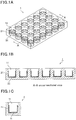
- Figs. 1 are exemplary schematic views of a multi-well plate or a single well (medical analysis device) having a well portion having a fibronectin-adsorbed hydrophilic polymer layer formed on the inner surface thereof.
- the present invention relates to a medical analysis device having a well portion.
- the well portion has a hydrophilic polymer layer formed at least partly on the inner surface thereof, and the hydrophilic polymer layer has fibronectin adsorbed thereto.
- hydrophilic polymer layer is not simply formed at least partly on the inner surface of the well portion but fibronectin is further adsorbed to the hydrophilic polymer layer, the hydrophilic polymer displays a significantly improved ability to adhere (adsorb) to specific cells such as cancer cells. Accordingly, the ability to capture specific cells such as cancer cells is greatly improved while reducing the ability to capture other cells such as platelets. As a result, an effect which could never be produced when proteins are present at high levels is achieved in selectively capturing specific cells.
- tumor cells e.g. cancer cells
- biological fluid such as circulating tumor cells (several to hundreds of cells/1 mL of blood)
- fibronectin that promotes adhesion (adsorption) of tumor cells is further adsorbed to the surface of the hydrophilic polymer layer
- more tumor cells or other specific cells in biological fluid such as blood can be adsorbed or adhered to the hydrophilic polymer layer by bringing the biological fluid into contact with the hydrophilic polymer layer.
- the number of adsorbed specific cells such as tumor cells is counted to determine the number of specific cells in the blood or biological fluid, e.g., in order to confirm the cancer-treating effect.
- the captured specific cells may be cultured and then used to determine the effect of drugs such as anticancer drugs. This allows us to determine the effect of drugs such as anticancer drugs ex vivo before administration, and also helps to screen drugs such as anticancer drugs.
- a medical analysis device 1 illustrated in Figs. 1A and 1B is a device intended to capture specific cells such as cancer cells in which wells 11 are arranged in so-called matrix form.
- the multi-well plate 1 has multiple wells 11 having a circular opening.
- the wells 11, which are recesses into which blood, biological fluid, or others are injected, can be used to confirm the presence or absence of specific cells in the injected blood or biological fluid, count the number of specific cells, culture the specific cells, determine the effect of drugs, and screen the drugs.
- Figs. 1A and 1B show a 24-well plate having 24 wells 11 arranged in 4 rows by 6 columns as an example, it is sufficient for the multi-well plate 1 to have at least two wells 11, and any number of wells 11 may be provided. Examples other than the 24-well plate include general multi-well plates in which the number of wells 11 is 6, 96, 384, etc.
- a single well 2 (medical analysis device 2) ( Fig. 1C ).
- the material of the multi-well plate 1 or single well 2 should be highly transparent for observation during the cell analysis, and examples include acrylic resins (polyacrylic resins) such as polymethyl acrylate, polymethyl methacrylate, polyacrylic acid, and polymethacrylic acid, cycloolefin resins (polycycloolefins), carbonate resins (polycarbonates), styrene resins (polystyrenes), polyester resins such as polyethylene terephthalate (PET), polydimethylsiloxanes, and glass (e.g. borosilicate glass, soda-lime glass).
- the material should be highly hydrophilic for coating with a hydrophilic polymer, and preferred are polyacrylic resins and soda-lime glass.
- Each well 11 is a non-through hole which is opened at the surface of the multi-well plate 1 or single well 2. Blood or biological fluid is injected into the wells 11 through the respective openings. If the presence of specific cells such as cancer cells is confirmed, a culture fluid for culturing the specific cells may also be injected.
- the diameter R and depth D of the opening of each well 11 are not particularly limited, and may be those of a conventional multi-well plate 1. Although in Figs. 1 , the inner side surface of each well 11 is substantially vertical to the opposite sides of the multi-well plate 1 or single well 2, the inner side surface of each well 11 may be inclined to taper from the opening to the bottom. Alternatively, the inner side surface may be inclined to flare out from the opening to the bottom.
- the opening of the wells 11 may be of any shape such as a quadrangle.
- the multi-well plate 1 may suitably be one in which the multiple wells 11 are separable. Since multiple wells are provided, they can be separated into wells for counting the number of specific cells and for culturing specific cells. For example, the presence or absence of cancer cells is first confirmed in the wells for counting, and if the presence is confirmed, the cancer cells are cultured in the wells for culturing and then used to determine the effect of drugs.
- each well 11 has a hydrophilic polymer layer formed at least partly on the inner surface thereof, and the hydrophilic polymer layer has fibronectin adsorbed thereto.
- Figs. 1 illustrate a case where a hydrophilic polymer layer 21 is formed on the bottom surface and a part of the side surface of the wells, and fibronectin 31 is adsorbed to the hydrophilic polymer layer 21.
- specific cells such as cancer cells present in the blood or biological fluid are adsorbed onto the hydrophilic polymer layer 21 with adsorbed fibronectin 31, while the adsorption of other cells such as platelets and erythrocytes is reduced.
- specific cells can be adsorbed onto the hydrophilic polymer layer 21 by introducing and retaining blood or biological fluid in the wells for a predetermined time, followed by washing. Then, it can be expected that the number of adsorbed specific cells is counted to determine the number of specific cells such as cancer cells in the blood or biological fluid, e.g., in order to confirm the cancer-treating effect.
- the thickness (film thickness) of the hydrophilic polymer layer 21 is preferably 2 to 200 nm, more preferably 20 to 180 nm. When the thickness is adjusted within the range indicated above, selective adsorption or adhesion of cancer cells and low adsorption of other proteins and cells can be well achieved.
- the hydrophilic polymer may be appropriately selected from polymers having hydrophilicity.
- it may be a homopolymer or copolymer of one or two or more hydrophilic monomers, or a copolymer of one or two or more hydrophilic monomers with a second monomer.
- the homopolymer and copolymers include polyacrylic acid, polyacrylic acid esters, polymethacrylic acid, polymethacrylic acid esters, polyacryloylmorpholine, polymethacryloylmorpholine, polyacrylamide, and polymethacrylamide.
- the hydrophilic monomer may be any monomer containing a hydrophilic group.
- the hydrophilic group include known hydrophilic groups such as an amide group, a sulfuric acid group, a sulfonic acid group, a carboxylic acid group, a hydroxy group, an amino group, and an oxyethylene group.
- hydrophilic monomer examples include (meth)acrylic acid, (meth)acrylic acid esters (e.g. alkoxyalkyl (meth)acrylates such as methoxyethyl (meth)acrylate, and hydroxyalkyl (meth)acrylates such as hydroxyethyl (meth)acrylate), (meth)acrylamide, and (meth)acrylamide derivatives containing cyclic groups (e.g. (meth)acryloylmorpholine).
- alkoxyalkyl (meth)acrylates such as methoxyethyl (meth)acrylate
- hydroxyalkyl (meth)acrylates such as hydroxyethyl (meth)acrylate
- (meth)acrylamide, and (meth)acrylamide derivatives containing cyclic groups e.g. (meth)acryloylmorpholine).
- the second monomer may be appropriately selected as long as it does not inhibit the effect of the hydrophilic polymer.
- aromatic monomers such as styrene, vinyl acetate, and N-isopropylacrylamide which can impart temperature responsiveness.
- the hydrophilic polymer is preferably at least one selected from the group consisting of poly(meth)acryloylmorpholine and a polymer represented by the following formula (I): wherein R 1 represents a hydrogen atom or a methyl group, R 2 represents an alkyl group, m represents 1 to 5, and n represents the number of repetitions.
- the alkyl group represented by R 2 preferably has a carbon number of 1 to 10, more preferably 1 to 5.
- R 2 is particularly preferably a methyl group or an ethyl group.
- the symbol m is preferably 1 to 3, while n (number of repeating units) is preferably 15 to 1,000, more preferably 30 to 500.
- the hydrophilic polymer may also suitably be a copolymer of at least one hydrophilic monomer selected from the group consisting of (meth)acryloylmorpholine and a compound represented by the following formula (I-1): wherein R 1 , R 2 , and m are as defined above, with a second monomer.
- the hydrophilic polymer preferably has a weight average molecular weight (Mw) of 4,000 to 150,000, more preferably 5,000 to 100,000, still more preferably 8,000 to 50,000.
- Mw weight average molecular weight
- the Mw as used herein can be determined by gel permeation chromatography (GPC) (GPC-8000 series produced by TOSOH Corporation, detector: differential refractometer, column: TSKGEL SUPERMULTIPORE HZ-M produced by TOSOH Corporation) calibrated with polystyrene standards.
- a liquid such as a solution or dispersion which preferably has a fibronectin concentration adjusted to 0.5 to 500 ⁇ g/ml, more preferably 1 to 250 ⁇ g/ml.
- concentration adjusted within the range indicated above, selective adsorption or adhesion of cancer cells, and low adsorption of other proteins and cells can be well achieved.
- the medical analysis device of the present invention can be produced, for example, by preparing a multi-well plate 1 or single well 2 including a well(s) 11 in which a hydrophilic polymer layer 21 with fibronectin 31 adsorbed thereto is formed on the inner surface as illustrated in Figs. 1 , optionally followed by addition of other components (parts).
- the multi-well plate or single well provided with a polymer layer formed of a hydrophilic polymer can be produced by dissolving or dispersing a hydrophilic polymer in any solvent to prepare a hydrophilic polymer solution or dispersion, and entirely or partially coating the inner surface of each well 11 with the hydrophilic polymer solution or dispersion by a known method, such as (1) by injecting the hydrophilic polymer solution or dispersion into the well(s) 11 and retaining it for a predetermined time, or (2) by applying (spraying) the hydrophilic polymer solution or dispersion to the inner surface of the well(s) 11.
- a known method such as (1) by injecting the hydrophilic polymer solution or dispersion into the well(s) 11 and retaining it for a predetermined time, or (2) by applying (spraying) the hydrophilic polymer solution or dispersion to the inner surface of the well(s) 11.
- the solvent, injection method, application (spraying) method, and other conditions may be conventionally known materials or methods.
- the retention time in the method (1) or (2) may be selected appropriately according to the size of the wells 11, the type of liquid introduced, and other factors, and is preferably five minutes to ten hours, more preferably ten minutes to five hours, still more preferably 15 minutes to two hours. After the retention, the excess hydrophilic polymer solution or dispersion may be discharged followed by drying, as required.
- fibronectin 31 may be adsorbed to the formed hydrophilic polymer layer 21 by any known method, such as for example by bringing the hydrophilic polymer layer 21 into contact with a buffer solution (e.g. phosphate buffered saline (PBS)) containing fibronectin 31 by a known method, and leaving them at a predetermined temperature for a predetermined time, optionally followed by washing.
- a buffer solution e.g. phosphate buffered saline (PBS)
- PBS phosphate buffered saline
- the temperature and time may be selected as appropriate, and may be, for example, about 10 to 60°C and about 0.1 to 10 hours, respectively.
- a component having fibrinogen adsorbed thereto is preferably used as another component.
- the blood or biological fluid to be analyzed is brought into contact with the component with adsorbed fibrinogen which plays a role in adhesion of blood cells, the number of blood cells is reduced prior to the analysis, thereby resulting in improved adhesion or adsorption of specific cells such as cancer cells.
- the cell analysis method of the present invention is for examining cells present in blood or biological fluid using the medical analysis device described above.
- the medical analysis device of the present invention has an improved ability to capture specific cells such as cancer cells while reducing the ability to capture other cells such as platelets, and thus has the effect of selectively capturing specific cells such as cancer cells. It can therefore be expected that such a device is used to examine cells present in blood or biological fluid, e.g. in order to confirm the cancer-treating effect, determine the effect of drugs such as anticancer drugs ex vivo, and/or screen drugs such as anticancer drugs, as described above.
- the blood or biological fluid is preferably brought into contact with the component having fibrinogen adsorbed thereto before being introduced (e.g. injected or added dropwise) into the medical analysis device.
- the component having fibrinogen adsorbed thereto is a blood collection syringe, a blood collection tube, or a centrifuge tube, the above-mentioned effects can be significantly achieved.
- the blood or biological fluid is preferably centrifuged to remove the supernatant and /or the sediment before being introduced (e.g. injected or added dropwise) into the medical analysis device.
- AIBN azobisisobutyronitrile
- the poly(2-methoxyethyl acrylate) solution (2.5 w/v%) was injected into a commercially available PMMA plate, and left for 30 minutes at room temperature. Thereafter, the solution was drawn using a pipette, followed by drying to form a hydrophilic polymer layer.
- fibronectin was adsorbed to the part coated with poly(2-methoxyethyl acrylate) (hydrophilic polymer layer).
- a 1 ⁇ l/ml solution of fibronectin in a PBS solution was prepared, brought into contact with the hydrophilic polymer layer, and left at 40°C for one hour, followed by washing with a PBS solution to prepare an analysis device including a multi-well plate with a fibronectin-adsorbed hydrophilic polymer layer formed thereon as illustrated in Figs. 1 .
- An analysis device was prepared in the same manner as in Example 1, except that the concentration in the PBS solution was changed to 10 ⁇ l/ml.
- An analysis device was prepared in the same manner as in Example 1, except that the concentration in the PBS solution was changed to 100 ⁇ l/ml.
- An analysis device was prepared in the same manner as in Example 2, except that a pipette (fibrinogen-adsorbed blood collection tube) was separately prepared by coating the inner surface of a pipette with a 10 ⁇ l/ml fibrinogen solution, followed by washing with a PBS solution and then sufficient dehydration.
- a pipette fibrinogen-adsorbed blood collection tube
- An analysis device was prepared in the same manner as in Example 1, except that the concentration in the PBS solution was changed to 200 ⁇ l/ml.
- An analysis device was prepared in the same manner as in Example 1, except that a poly(2-methoxyethyl acrylate) layer was simply formed.
- the medical analysis devices prepared in the examples and comparative example were evaluated as follows.
- the medical analysis device of Example 4 was evaluated in the same manner, except that blood was previously sucked up with the prepared pipette, kept in the pipette for 10 minutes, and then injected into the wells.
- the thickness of the hydrophilic polymer layer on the inner surface of the wells was measured (photographed) using a TEM at an accelerating voltage of 15 kV and a magnification of 1,000 times.
- a liquid prepared by mixing plasma with platelets to adjust the platelet concentration (plating density) to 4 ⁇ 10 7 cells/cm 2 was injected into the wells and left at 37°C for one hour.
- the inside of the wells was washed with phosphate buffered saline, followed by fixation using 1% glutaraldehyde (being left at 37°C for two hours). Then, the inside of the wells was washed again with phosphate buffered saline and distilled water.
- the prepared sample was observed by SEM, and the number of adsorbed platelets was counted. The numbers are compared relative to Comparative Example 1 set equal to 1.0.
- Fibrosarcoma (HT-1080) was suspended in a dissociation solution and a portion of the suspension was resuspended in a PBS solution to count the number of cells using a blood cell counter. Using the obtained number, the dissociation solution containing fibrosarcoma (HT-1080) was resuspended in blood so that the calculated plating density (concentration) was 2,850 cells/cm 2 .
- Stained human colon adenocarcinoma (HT-29) cells were suspended in whole blood at a concentration of 100 cells per ml of blood to prepare a spiked blood sample.
- the sample was diluted with an equal volume of a liquid medium to prepare a spiked blood dilution.
- the spiked blood dilution followed by centrifugation at 800 g for 20 minutes. Then, the mononuclear cell layer was separated.
- a phosphate buffer (PBS) solution To the separated mononuclear cell layer was added a phosphate buffer (PBS) solution, followed by centrifugation again to concentrate the mononuclear cell layer. After the centrifugation, the aggregates at the lowermost layer were suspended in a liquid medium containing 10% fetal bovine serum (FBS) in a volume equal to the initial whole blood volume. A 1 ml portion of the suspension was injected into each well and left at 37°C for one hour to cause adhesion. Then, non-adherent cells were washed away with a PBS solution. Thereafter, the number of adhering cancer cells was counted using a fluorescence microscope. The numbers of adhering cells are compared relative to Comparative Example 1 set equal to 1.00.
- PBS phosphate buffer
- Example 1 in which a hydrophilic polymer layer with fibronectin adsorbed thereto was formed, the amount of adhering cancer cells was greatly improved as compared to Comparative Example 1 in which a hydrophilic polymer layer was simply formed. This is presumably because preferential adhesion of cancer cells was caused due to the fibronectin, a protein playing a role in adhesion of cancer cells, adsorbed onto the poly(2-methoxyethyl acrylate) layer.
- Example 4 in which further blood was previously brought into contact with a container having fibrinogen adsorbed to its surface (the inner surface of a pipette), the amount of adhering cancer cells was further improved. This is presumably because since blood was injected into the pipette having fibrinogen, which plays a role in adhesion of blood cells, adsorbed to the inner surface of the container before being added dropwise to the wells, the number of blood cells was reduced, thereby further improving adhesion of cancer cells.
- Example 4 which exhibited good adhesion of cancer cells in Table 1, and Example 5, in which an increased amount of fibronectin was adsorbed, were also excellent in adhesion of cancer cells in the analysis of whole blood spiked with cancer cells.
Landscapes
- Health & Medical Sciences (AREA)
- Chemical & Material Sciences (AREA)
- Life Sciences & Earth Sciences (AREA)
- Engineering & Computer Science (AREA)
- Biomedical Technology (AREA)
- Organic Chemistry (AREA)
- General Health & Medical Sciences (AREA)
- Zoology (AREA)
- Wood Science & Technology (AREA)
- Bioinformatics & Cheminformatics (AREA)
- Immunology (AREA)
- Biochemistry (AREA)
- Hematology (AREA)
- Biotechnology (AREA)
- Microbiology (AREA)
- Analytical Chemistry (AREA)
- Sustainable Development (AREA)
- Urology & Nephrology (AREA)
- Molecular Biology (AREA)
- Genetics & Genomics (AREA)
- General Engineering & Computer Science (AREA)
- Clinical Laboratory Science (AREA)
- Medicinal Chemistry (AREA)
- Cell Biology (AREA)
- Chemical Kinetics & Catalysis (AREA)
- Food Science & Technology (AREA)
- Physics & Mathematics (AREA)
- Pathology (AREA)
- General Physics & Mathematics (AREA)
- Tropical Medicine & Parasitology (AREA)
- Virology (AREA)
- Polymers & Plastics (AREA)
- Apparatus Associated With Microorganisms And Enzymes (AREA)
- Investigating Or Analysing Biological Materials (AREA)
Applications Claiming Priority (2)
| Application Number | Priority Date | Filing Date | Title |
|---|---|---|---|
| JP2016191617 | 2016-09-29 | ||
| JP2017144556A JP6485783B2 (ja) | 2016-09-29 | 2017-07-26 | 医療用検査装置及び細胞検査方法 |
Publications (2)
| Publication Number | Publication Date |
|---|---|
| EP3301443A1 true EP3301443A1 (fr) | 2018-04-04 |
| EP3301443B1 EP3301443B1 (fr) | 2023-07-12 |
Family
ID=59799257
Family Applications (1)
| Application Number | Title | Priority Date | Filing Date |
|---|---|---|---|
| EP17189342.3A Active EP3301443B1 (fr) | 2016-09-29 | 2017-09-05 | Dispositif d'analyse médicale et procédé d'analyse cellulaire |
Country Status (3)
| Country | Link |
|---|---|
| US (1) | US10941374B2 (fr) |
| EP (1) | EP3301443B1 (fr) |
| CN (1) | CN107884558B (fr) |
Cited By (5)
| Publication number | Priority date | Publication date | Assignee | Title |
|---|---|---|---|---|
| EP3527986A1 (fr) * | 2018-02-14 | 2019-08-21 | Sumitomo Rubber Industries, Ltd. | Procédé de capture de cellules spécifiques |
| EP3686598A1 (fr) * | 2019-01-24 | 2020-07-29 | Sumitomo Rubber Industries, Ltd. | Procédés de capture et de fractionnement de cellules spécifiques |
| US11226330B2 (en) | 2018-02-14 | 2022-01-18 | Sumitomo Rubber Industries, Ltd. | Method for capturing specific cells |
| US11360078B2 (en) | 2016-09-29 | 2022-06-14 | Sumitomo Rubber Industries, Ltd. | Medical analysis device and cell analysis method |
| US11573232B2 (en) | 2018-02-14 | 2023-02-07 | Sumitomo Rubber Industries, Ltd. | Method for capturing specific cells |
Families Citing this family (7)
| Publication number | Priority date | Publication date | Assignee | Title |
|---|---|---|---|---|
| CN109609336B (zh) * | 2018-11-19 | 2021-05-18 | 江苏汇先医药技术有限公司 | 一种用于捕获生物分子、细胞或细菌的捕获筛 |
| JP7580076B2 (ja) * | 2019-01-24 | 2024-11-11 | 住友ゴム工業株式会社 | 細胞培養装置及び細胞培養方法 |
| JP6886668B2 (ja) | 2019-04-23 | 2021-06-16 | 住友ゴム工業株式会社 | 医療用検査装置及び細胞検査方法 |
| JP7467866B2 (ja) * | 2019-10-02 | 2024-04-16 | 住友ゴム工業株式会社 | 親水性基材及び親水性基材作製方法 |
| CN111175503B (zh) * | 2020-03-05 | 2023-07-25 | 上海邦先医疗科技有限公司 | 捕获筛及其制备方法 |
| JP7639275B2 (ja) * | 2020-05-27 | 2025-03-05 | 住友ゴム工業株式会社 | 親水性基材及び親水性基材作製方法 |
| CN114487389B (zh) * | 2022-01-29 | 2025-11-14 | 厦门先明生物技术有限公司 | 一种集成试剂检测管及其检测方法 |
Citations (8)
| Publication number | Priority date | Publication date | Assignee | Title |
|---|---|---|---|---|
| US20020155617A1 (en) * | 1998-02-24 | 2002-10-24 | Aurora Biosciences Corporation | Low fluorescence assay platforms and related methods for drug discovery |
| JP2005523981A (ja) | 2002-04-30 | 2005-08-11 | ハイドロマー インコーポレイテッド | 親水性多用途のためのコーティング組成物 |
| US20060160066A1 (en) * | 2005-01-20 | 2006-07-20 | The Regents Of The University Of California | Cellular microarrays for screening differentiation factors |
| US20100261159A1 (en) * | 2000-10-10 | 2010-10-14 | Robert Hess | Apparatus for assay, synthesis and storage, and methods of manufacture, use, and manipulation thereof |
| WO2011157805A1 (fr) * | 2010-06-16 | 2011-12-22 | Dsm Ip Assets B.V. | Formule de revêtement pour préparer un revêtement hydrophile |
| WO2011161480A1 (fr) * | 2010-06-25 | 2011-12-29 | Imperial Innovations Ltd | Plaque d'essai multi-puits |
| WO2015137259A1 (fr) * | 2014-03-11 | 2015-09-17 | テルモ株式会社 | Procédé de fabrication d'outil médical et outil médical |
| WO2016103002A1 (fr) * | 2014-12-22 | 2016-06-30 | Ecole Polytechnique Federale De Lausanne (Epfl) | Dispositifs d'agrégation à haut rendement et de manipulation de cellules mammaliennes |
Family Cites Families (20)
| Publication number | Priority date | Publication date | Assignee | Title |
|---|---|---|---|---|
| US5202025A (en) * | 1989-04-12 | 1993-04-13 | Terumo Kabushiki Kaisha | Porous membrane and method for preparing the same |
| US6743878B2 (en) * | 1991-07-05 | 2004-06-01 | Biocompatibles Uk Limited | Polymeric surface coatings |
| AU8843701A (en) | 2000-09-09 | 2002-03-22 | Univ New York State Res Found | Method and compositions for isolating metastatic cancer cells, and use in measuring metastatic potential of a cancer thereof |
| CA2442998A1 (fr) * | 2001-04-09 | 2002-10-17 | David P. Dumas | Support en polymere transparent permettant de realiser une synthese organique |
| CN101816561A (zh) * | 2001-06-18 | 2010-09-01 | 贝克顿迪肯森公司 | 多层容器和关于形成多层容器的加工过程 |
| US20080008736A1 (en) | 2006-07-06 | 2008-01-10 | Thierry Glauser | Random copolymers of methacrylates and acrylates |
| US9012202B2 (en) * | 2010-11-02 | 2015-04-21 | University Of Florida Research Foundation, Inc. | Cell-based arrays, methods of making, and methods of using |
| US9926523B2 (en) * | 2010-12-16 | 2018-03-27 | General Electric Company | Cell carriers and methods for culturing cells |
| WO2013073707A1 (fr) * | 2011-11-20 | 2013-05-23 | 学校法人東京女子医科大学 | Substrat de culture cellulaire et son procédé de fabrication |
| WO2013134788A1 (fr) | 2012-03-09 | 2013-09-12 | Massachusetts Institute Of Technology | Signatures d'adhésion |
| WO2014117021A2 (fr) | 2013-01-25 | 2014-07-31 | Xcell Biosciences, Inc. | Procédés, compositions, trousses et systèmes pour l'enrichissement sélectif de cellules cibles |
| WO2014135668A1 (fr) * | 2013-03-08 | 2014-09-12 | Universität Leipzig | Procédé et trousse d'analyse des cytokines d'un échantillon de sang entier humain |
| US10647829B2 (en) | 2013-06-20 | 2020-05-12 | Sumitomo Rubber Industries, Ltd. | Surface modification method and surface modification body |
| JP6157429B2 (ja) | 2013-10-21 | 2017-07-05 | 住友ゴム工業株式会社 | 潤滑性、低タンパク質吸着性および/または低細胞吸着性を有する金属医療用具及びその製造方法 |
| JP2016131561A (ja) | 2015-01-22 | 2016-07-25 | 国立大学法人山形大学 | 細胞を回収する方法、及びそれに用いられるポリマー |
| WO2017030196A1 (fr) * | 2015-08-20 | 2017-02-23 | 東京エレクトロン株式会社 | Récipient de culture et procédé de culture cellulaire et procédé d'observation de cellules utilisant le récipient de culture |
| WO2020036203A2 (fr) * | 2018-08-16 | 2020-02-20 | Terumo Kabushiki Kaisha | Substrat de culture cellulaire |
| EP3830238A1 (fr) * | 2018-08-16 | 2021-06-09 | TERUMO Kabushiki Kaisha | Substrat de culture cellulaire |
| JP7315652B2 (ja) * | 2018-08-16 | 2023-07-26 | テルモ株式会社 | 細胞培養基材 |
| JP2021533736A (ja) * | 2018-08-16 | 2021-12-09 | テルモ株式会社 | 細胞培養基材 |
-
2017
- 2017-09-01 US US15/693,919 patent/US10941374B2/en active Active
- 2017-09-05 EP EP17189342.3A patent/EP3301443B1/fr active Active
- 2017-09-14 CN CN201710826823.3A patent/CN107884558B/zh active Active
Patent Citations (8)
| Publication number | Priority date | Publication date | Assignee | Title |
|---|---|---|---|---|
| US20020155617A1 (en) * | 1998-02-24 | 2002-10-24 | Aurora Biosciences Corporation | Low fluorescence assay platforms and related methods for drug discovery |
| US20100261159A1 (en) * | 2000-10-10 | 2010-10-14 | Robert Hess | Apparatus for assay, synthesis and storage, and methods of manufacture, use, and manipulation thereof |
| JP2005523981A (ja) | 2002-04-30 | 2005-08-11 | ハイドロマー インコーポレイテッド | 親水性多用途のためのコーティング組成物 |
| US20060160066A1 (en) * | 2005-01-20 | 2006-07-20 | The Regents Of The University Of California | Cellular microarrays for screening differentiation factors |
| WO2011157805A1 (fr) * | 2010-06-16 | 2011-12-22 | Dsm Ip Assets B.V. | Formule de revêtement pour préparer un revêtement hydrophile |
| WO2011161480A1 (fr) * | 2010-06-25 | 2011-12-29 | Imperial Innovations Ltd | Plaque d'essai multi-puits |
| WO2015137259A1 (fr) * | 2014-03-11 | 2015-09-17 | テルモ株式会社 | Procédé de fabrication d'outil médical et outil médical |
| WO2016103002A1 (fr) * | 2014-12-22 | 2016-06-30 | Ecole Polytechnique Federale De Lausanne (Epfl) | Dispositifs d'agrégation à haut rendement et de manipulation de cellules mammaliennes |
Cited By (7)
| Publication number | Priority date | Publication date | Assignee | Title |
|---|---|---|---|---|
| US11360078B2 (en) | 2016-09-29 | 2022-06-14 | Sumitomo Rubber Industries, Ltd. | Medical analysis device and cell analysis method |
| EP3527986A1 (fr) * | 2018-02-14 | 2019-08-21 | Sumitomo Rubber Industries, Ltd. | Procédé de capture de cellules spécifiques |
| US11226330B2 (en) | 2018-02-14 | 2022-01-18 | Sumitomo Rubber Industries, Ltd. | Method for capturing specific cells |
| US11573232B2 (en) | 2018-02-14 | 2023-02-07 | Sumitomo Rubber Industries, Ltd. | Method for capturing specific cells |
| EP3686598A1 (fr) * | 2019-01-24 | 2020-07-29 | Sumitomo Rubber Industries, Ltd. | Procédés de capture et de fractionnement de cellules spécifiques |
| US20200238201A1 (en) * | 2019-01-24 | 2020-07-30 | Sumitomo Rubber Industries, Ltd. | Specific cell fractionating and capturing methods |
| US11614440B2 (en) * | 2019-01-24 | 2023-03-28 | Sumitomo Rubber Industries, Ltd. | Specific cell fractionating and capturing methods |
Also Published As
| Publication number | Publication date |
|---|---|
| CN107884558A (zh) | 2018-04-06 |
| US10941374B2 (en) | 2021-03-09 |
| US20180087017A1 (en) | 2018-03-29 |
| EP3301443B1 (fr) | 2023-07-12 |
| CN107884558B (zh) | 2021-01-01 |
Similar Documents
| Publication | Publication Date | Title |
|---|---|---|
| US10941374B2 (en) | Medical analysis device and cell analysis method | |
| US10620186B2 (en) | Method for capturing cancer cells | |
| EP3527986B1 (fr) | Procédé de capture de cellules spécifiques | |
| EP3527985B1 (fr) | Procédé de capture de cellules spécifiques | |
| JP6485783B2 (ja) | 医療用検査装置及び細胞検査方法 | |
| US11360078B2 (en) | Medical analysis device and cell analysis method | |
| US10786812B2 (en) | Medical analysis device and cell analysis method | |
| EP3553516B1 (fr) | Procédé de capture de cellules spécifiques | |
| US11614440B2 (en) | Specific cell fractionating and capturing methods | |
| JP7580076B2 (ja) | 細胞培養装置及び細胞培養方法 | |
| JP6886667B2 (ja) | がん細胞捕捉方法 |
Legal Events
| Date | Code | Title | Description |
|---|---|---|---|
| PUAI | Public reference made under article 153(3) epc to a published international application that has entered the european phase |
Free format text: ORIGINAL CODE: 0009012 |
|
| STAA | Information on the status of an ep patent application or granted ep patent |
Free format text: STATUS: THE APPLICATION HAS BEEN PUBLISHED |
|
| AK | Designated contracting states |
Kind code of ref document: A1 Designated state(s): AL AT BE BG CH CY CZ DE DK EE ES FI FR GB GR HR HU IE IS IT LI LT LU LV MC MK MT NL NO PL PT RO RS SE SI SK SM TR |
|
| AX | Request for extension of the european patent |
Extension state: BA ME |
|
| STAA | Information on the status of an ep patent application or granted ep patent |
Free format text: STATUS: REQUEST FOR EXAMINATION WAS MADE |
|
| 17P | Request for examination filed |
Effective date: 20180921 |
|
| RBV | Designated contracting states (corrected) |
Designated state(s): AL AT BE BG CH CY CZ DE DK EE ES FI FR GB GR HR HU IE IS IT LI LT LU LV MC MK MT NL NO PL PT RO RS SE SI SK SM TR |
|
| STAA | Information on the status of an ep patent application or granted ep patent |
Free format text: STATUS: EXAMINATION IS IN PROGRESS |
|
| 17Q | First examination report despatched |
Effective date: 20210727 |
|
| GRAP | Despatch of communication of intention to grant a patent |
Free format text: ORIGINAL CODE: EPIDOSNIGR1 |
|
| STAA | Information on the status of an ep patent application or granted ep patent |
Free format text: STATUS: GRANT OF PATENT IS INTENDED |
|
| INTG | Intention to grant announced |
Effective date: 20221025 |
|
| GRAJ | Information related to disapproval of communication of intention to grant by the applicant or resumption of examination proceedings by the epo deleted |
Free format text: ORIGINAL CODE: EPIDOSDIGR1 |
|
| STAA | Information on the status of an ep patent application or granted ep patent |
Free format text: STATUS: EXAMINATION IS IN PROGRESS |
|
| GRAP | Despatch of communication of intention to grant a patent |
Free format text: ORIGINAL CODE: EPIDOSNIGR1 |
|
| STAA | Information on the status of an ep patent application or granted ep patent |
Free format text: STATUS: GRANT OF PATENT IS INTENDED |
|
| INTC | Intention to grant announced (deleted) | ||
| INTG | Intention to grant announced |
Effective date: 20230322 |
|
| GRAS | Grant fee paid |
Free format text: ORIGINAL CODE: EPIDOSNIGR3 |
|
| GRAA | (expected) grant |
Free format text: ORIGINAL CODE: 0009210 |
|
| STAA | Information on the status of an ep patent application or granted ep patent |
Free format text: STATUS: THE PATENT HAS BEEN GRANTED |
|
| AK | Designated contracting states |
Kind code of ref document: B1 Designated state(s): AL AT BE BG CH CY CZ DE DK EE ES FI FR GB GR HR HU IE IS IT LI LT LU LV MC MK MT NL NO PL PT RO RS SE SI SK SM TR |
|
| REG | Reference to a national code |
Ref country code: CH Ref legal event code: EP |
|
| REG | Reference to a national code |
Ref country code: DE Ref legal event code: R096 Ref document number: 602017071140 Country of ref document: DE |
|
| REG | Reference to a national code |
Ref country code: IE Ref legal event code: FG4D |
|
| REG | Reference to a national code |
Ref country code: LT Ref legal event code: MG9D |
|
| REG | Reference to a national code |
Ref country code: NL Ref legal event code: MP Effective date: 20230712 |
|
| REG | Reference to a national code |
Ref country code: AT Ref legal event code: MK05 Ref document number: 1587679 Country of ref document: AT Kind code of ref document: T Effective date: 20230712 |
|
| PG25 | Lapsed in a contracting state [announced via postgrant information from national office to epo] |
Ref country code: NL Free format text: LAPSE BECAUSE OF FAILURE TO SUBMIT A TRANSLATION OF THE DESCRIPTION OR TO PAY THE FEE WITHIN THE PRESCRIBED TIME-LIMIT Effective date: 20230712 |
|
| PG25 | Lapsed in a contracting state [announced via postgrant information from national office to epo] |
Ref country code: GR Free format text: LAPSE BECAUSE OF FAILURE TO SUBMIT A TRANSLATION OF THE DESCRIPTION OR TO PAY THE FEE WITHIN THE PRESCRIBED TIME-LIMIT Effective date: 20231013 |
|
| PG25 | Lapsed in a contracting state [announced via postgrant information from national office to epo] |
Ref country code: ES Free format text: LAPSE BECAUSE OF FAILURE TO SUBMIT A TRANSLATION OF THE DESCRIPTION OR TO PAY THE FEE WITHIN THE PRESCRIBED TIME-LIMIT Effective date: 20230712 |
|
| PG25 | Lapsed in a contracting state [announced via postgrant information from national office to epo] |
Ref country code: IS Free format text: LAPSE BECAUSE OF FAILURE TO SUBMIT A TRANSLATION OF THE DESCRIPTION OR TO PAY THE FEE WITHIN THE PRESCRIBED TIME-LIMIT Effective date: 20231112 |
|
| PG25 | Lapsed in a contracting state [announced via postgrant information from national office to epo] |
Ref country code: SE Free format text: LAPSE BECAUSE OF FAILURE TO SUBMIT A TRANSLATION OF THE DESCRIPTION OR TO PAY THE FEE WITHIN THE PRESCRIBED TIME-LIMIT Effective date: 20230712 Ref country code: RS Free format text: LAPSE BECAUSE OF FAILURE TO SUBMIT A TRANSLATION OF THE DESCRIPTION OR TO PAY THE FEE WITHIN THE PRESCRIBED TIME-LIMIT Effective date: 20230712 Ref country code: PT Free format text: LAPSE BECAUSE OF FAILURE TO SUBMIT A TRANSLATION OF THE DESCRIPTION OR TO PAY THE FEE WITHIN THE PRESCRIBED TIME-LIMIT Effective date: 20231113 Ref country code: NO Free format text: LAPSE BECAUSE OF FAILURE TO SUBMIT A TRANSLATION OF THE DESCRIPTION OR TO PAY THE FEE WITHIN THE PRESCRIBED TIME-LIMIT Effective date: 20231012 Ref country code: LV Free format text: LAPSE BECAUSE OF FAILURE TO SUBMIT A TRANSLATION OF THE DESCRIPTION OR TO PAY THE FEE WITHIN THE PRESCRIBED TIME-LIMIT Effective date: 20230712 Ref country code: LT Free format text: LAPSE BECAUSE OF FAILURE TO SUBMIT A TRANSLATION OF THE DESCRIPTION OR TO PAY THE FEE WITHIN THE PRESCRIBED TIME-LIMIT Effective date: 20230712 Ref country code: IS Free format text: LAPSE BECAUSE OF FAILURE TO SUBMIT A TRANSLATION OF THE DESCRIPTION OR TO PAY THE FEE WITHIN THE PRESCRIBED TIME-LIMIT Effective date: 20231112 Ref country code: HR Free format text: LAPSE BECAUSE OF FAILURE TO SUBMIT A TRANSLATION OF THE DESCRIPTION OR TO PAY THE FEE WITHIN THE PRESCRIBED TIME-LIMIT Effective date: 20230712 Ref country code: GR Free format text: LAPSE BECAUSE OF FAILURE TO SUBMIT A TRANSLATION OF THE DESCRIPTION OR TO PAY THE FEE WITHIN THE PRESCRIBED TIME-LIMIT Effective date: 20231013 Ref country code: FI Free format text: LAPSE BECAUSE OF FAILURE TO SUBMIT A TRANSLATION OF THE DESCRIPTION OR TO PAY THE FEE WITHIN THE PRESCRIBED TIME-LIMIT Effective date: 20230712 Ref country code: ES Free format text: LAPSE BECAUSE OF FAILURE TO SUBMIT A TRANSLATION OF THE DESCRIPTION OR TO PAY THE FEE WITHIN THE PRESCRIBED TIME-LIMIT Effective date: 20230712 Ref country code: AT Free format text: LAPSE BECAUSE OF FAILURE TO SUBMIT A TRANSLATION OF THE DESCRIPTION OR TO PAY THE FEE WITHIN THE PRESCRIBED TIME-LIMIT Effective date: 20230712 |
|
| PG25 | Lapsed in a contracting state [announced via postgrant information from national office to epo] |
Ref country code: PL Free format text: LAPSE BECAUSE OF FAILURE TO SUBMIT A TRANSLATION OF THE DESCRIPTION OR TO PAY THE FEE WITHIN THE PRESCRIBED TIME-LIMIT Effective date: 20230712 |
|
| REG | Reference to a national code |
Ref country code: DE Ref legal event code: R097 Ref document number: 602017071140 Country of ref document: DE |
|
| PG25 | Lapsed in a contracting state [announced via postgrant information from national office to epo] |
Ref country code: SM Free format text: LAPSE BECAUSE OF FAILURE TO SUBMIT A TRANSLATION OF THE DESCRIPTION OR TO PAY THE FEE WITHIN THE PRESCRIBED TIME-LIMIT Effective date: 20230712 Ref country code: RO Free format text: LAPSE BECAUSE OF FAILURE TO SUBMIT A TRANSLATION OF THE DESCRIPTION OR TO PAY THE FEE WITHIN THE PRESCRIBED TIME-LIMIT Effective date: 20230712 Ref country code: EE Free format text: LAPSE BECAUSE OF FAILURE TO SUBMIT A TRANSLATION OF THE DESCRIPTION OR TO PAY THE FEE WITHIN THE PRESCRIBED TIME-LIMIT Effective date: 20230712 Ref country code: DK Free format text: LAPSE BECAUSE OF FAILURE TO SUBMIT A TRANSLATION OF THE DESCRIPTION OR TO PAY THE FEE WITHIN THE PRESCRIBED TIME-LIMIT Effective date: 20230712 Ref country code: CZ Free format text: LAPSE BECAUSE OF FAILURE TO SUBMIT A TRANSLATION OF THE DESCRIPTION OR TO PAY THE FEE WITHIN THE PRESCRIBED TIME-LIMIT Effective date: 20230712 Ref country code: SK Free format text: LAPSE BECAUSE OF FAILURE TO SUBMIT A TRANSLATION OF THE DESCRIPTION OR TO PAY THE FEE WITHIN THE PRESCRIBED TIME-LIMIT Effective date: 20230712 |
|
| REG | Reference to a national code |
Ref country code: CH Ref legal event code: PL |
|
| PG25 | Lapsed in a contracting state [announced via postgrant information from national office to epo] |
Ref country code: LU Free format text: LAPSE BECAUSE OF NON-PAYMENT OF DUE FEES Effective date: 20230905 |
|
| PLBE | No opposition filed within time limit |
Free format text: ORIGINAL CODE: 0009261 |
|
| STAA | Information on the status of an ep patent application or granted ep patent |
Free format text: STATUS: NO OPPOSITION FILED WITHIN TIME LIMIT |
|
| REG | Reference to a national code |
Ref country code: BE Ref legal event code: MM Effective date: 20230930 |
|
| PG25 | Lapsed in a contracting state [announced via postgrant information from national office to epo] |
Ref country code: LU Free format text: LAPSE BECAUSE OF NON-PAYMENT OF DUE FEES Effective date: 20230905 Ref country code: IT Free format text: LAPSE BECAUSE OF FAILURE TO SUBMIT A TRANSLATION OF THE DESCRIPTION OR TO PAY THE FEE WITHIN THE PRESCRIBED TIME-LIMIT Effective date: 20230712 Ref country code: MC Free format text: LAPSE BECAUSE OF FAILURE TO SUBMIT A TRANSLATION OF THE DESCRIPTION OR TO PAY THE FEE WITHIN THE PRESCRIBED TIME-LIMIT Effective date: 20230712 |
|
| 26N | No opposition filed |
Effective date: 20240415 |
|
| GBPC | Gb: european patent ceased through non-payment of renewal fee |
Effective date: 20231012 |
|
| REG | Reference to a national code |
Ref country code: IE Ref legal event code: MM4A |
|
| PG25 | Lapsed in a contracting state [announced via postgrant information from national office to epo] |
Ref country code: IE Free format text: LAPSE BECAUSE OF NON-PAYMENT OF DUE FEES Effective date: 20230905 |
|
| PG25 | Lapsed in a contracting state [announced via postgrant information from national office to epo] |
Ref country code: GB Free format text: LAPSE BECAUSE OF NON-PAYMENT OF DUE FEES Effective date: 20231012 |
|
| PG25 | Lapsed in a contracting state [announced via postgrant information from national office to epo] |
Ref country code: CH Free format text: LAPSE BECAUSE OF NON-PAYMENT OF DUE FEES Effective date: 20230930 |
|
| PG25 | Lapsed in a contracting state [announced via postgrant information from national office to epo] |
Ref country code: IE Free format text: LAPSE BECAUSE OF NON-PAYMENT OF DUE FEES Effective date: 20230905 Ref country code: GB Free format text: LAPSE BECAUSE OF NON-PAYMENT OF DUE FEES Effective date: 20231012 Ref country code: CH Free format text: LAPSE BECAUSE OF NON-PAYMENT OF DUE FEES Effective date: 20230930 Ref country code: SI Free format text: LAPSE BECAUSE OF FAILURE TO SUBMIT A TRANSLATION OF THE DESCRIPTION OR TO PAY THE FEE WITHIN THE PRESCRIBED TIME-LIMIT Effective date: 20230712 |
|
| PG25 | Lapsed in a contracting state [announced via postgrant information from national office to epo] |
Ref country code: BE Free format text: LAPSE BECAUSE OF NON-PAYMENT OF DUE FEES Effective date: 20230930 |
|
| PG25 | Lapsed in a contracting state [announced via postgrant information from national office to epo] |
Ref country code: BG Free format text: LAPSE BECAUSE OF FAILURE TO SUBMIT A TRANSLATION OF THE DESCRIPTION OR TO PAY THE FEE WITHIN THE PRESCRIBED TIME-LIMIT Effective date: 20230712 |
|
| PG25 | Lapsed in a contracting state [announced via postgrant information from national office to epo] |
Ref country code: BG Free format text: LAPSE BECAUSE OF FAILURE TO SUBMIT A TRANSLATION OF THE DESCRIPTION OR TO PAY THE FEE WITHIN THE PRESCRIBED TIME-LIMIT Effective date: 20230712 |
|
| PG25 | Lapsed in a contracting state [announced via postgrant information from national office to epo] |
Ref country code: CY Free format text: LAPSE BECAUSE OF FAILURE TO SUBMIT A TRANSLATION OF THE DESCRIPTION OR TO PAY THE FEE WITHIN THE PRESCRIBED TIME-LIMIT; INVALID AB INITIO Effective date: 20170905 |
|
| PG25 | Lapsed in a contracting state [announced via postgrant information from national office to epo] |
Ref country code: HU Free format text: LAPSE BECAUSE OF FAILURE TO SUBMIT A TRANSLATION OF THE DESCRIPTION OR TO PAY THE FEE WITHIN THE PRESCRIBED TIME-LIMIT; INVALID AB INITIO Effective date: 20170905 |
|
| PGFP | Annual fee paid to national office [announced via postgrant information from national office to epo] |
Ref country code: FR Payment date: 20250922 Year of fee payment: 9 |
|
| PG25 | Lapsed in a contracting state [announced via postgrant information from national office to epo] |
Ref country code: TR Free format text: LAPSE BECAUSE OF FAILURE TO SUBMIT A TRANSLATION OF THE DESCRIPTION OR TO PAY THE FEE WITHIN THE PRESCRIBED TIME-LIMIT Effective date: 20230712 |
|
| PGFP | Annual fee paid to national office [announced via postgrant information from national office to epo] |
Ref country code: DE Payment date: 20251126 Year of fee payment: 9 |