EP3553516B1 - Procédé de capture de cellules spécifiques - Google Patents
Procédé de capture de cellules spécifiques Download PDFInfo
- Publication number
- EP3553516B1 EP3553516B1 EP19152149.1A EP19152149A EP3553516B1 EP 3553516 B1 EP3553516 B1 EP 3553516B1 EP 19152149 A EP19152149 A EP 19152149A EP 3553516 B1 EP3553516 B1 EP 3553516B1
- Authority
- EP
- European Patent Office
- Prior art keywords
- blood
- specific cells
- cells
- hydrophilic polymer
- biological fluid
- Prior art date
- Legal status (The legal status is an assumption and is not a legal conclusion. Google has not performed a legal analysis and makes no representation as to the accuracy of the status listed.)
- Active
Links
Images

Classifications
-
- C—CHEMISTRY; METALLURGY
- C12—BIOCHEMISTRY; BEER; SPIRITS; WINE; VINEGAR; MICROBIOLOGY; ENZYMOLOGY; MUTATION OR GENETIC ENGINEERING
- C12N—MICROORGANISMS OR ENZYMES; COMPOSITIONS THEREOF; PROPAGATING, PRESERVING, OR MAINTAINING MICROORGANISMS; MUTATION OR GENETIC ENGINEERING; CULTURE MEDIA
- C12N5/00—Undifferentiated human, animal or plant cells, e.g. cell lines; Tissues; Cultivation or maintenance thereof; Culture media therefor
- C12N5/06—Animal cells or tissues; Human cells or tissues
- C12N5/0602—Vertebrate cells
- C12N5/0693—Tumour cells; Cancer cells
-
- G—PHYSICS
- G01—MEASURING; TESTING
- G01N—INVESTIGATING OR ANALYSING MATERIALS BY DETERMINING THEIR CHEMICAL OR PHYSICAL PROPERTIES
- G01N33/00—Investigating or analysing materials by specific methods not covered by groups G01N1/00 - G01N31/00
- G01N33/48—Biological material, e.g. blood, urine; Haemocytometers
- G01N33/50—Chemical analysis of biological material, e.g. blood, urine; Testing involving biospecific ligand binding methods; Immunological testing
- G01N33/5002—Partitioning blood components
-
- G—PHYSICS
- G01—MEASURING; TESTING
- G01N—INVESTIGATING OR ANALYSING MATERIALS BY DETERMINING THEIR CHEMICAL OR PHYSICAL PROPERTIES
- G01N33/00—Investigating or analysing materials by specific methods not covered by groups G01N1/00 - G01N31/00
- G01N33/48—Biological material, e.g. blood, urine; Haemocytometers
- G01N33/50—Chemical analysis of biological material, e.g. blood, urine; Testing involving biospecific ligand binding methods; Immunological testing
- G01N33/53—Immunoassay; Biospecific binding assay; Materials therefor
- G01N33/575—Immunoassay; Biospecific binding assay; Materials therefor for cancer
-
- B—PERFORMING OPERATIONS; TRANSPORTING
- B01—PHYSICAL OR CHEMICAL PROCESSES OR APPARATUS IN GENERAL
- B01D—SEPARATION
- B01D21/00—Separation of suspended solid particles from liquids by sedimentation
- B01D21/26—Separation of sediment aided by centrifugal force or centripetal force
- B01D21/262—Separation of sediment aided by centrifugal force or centripetal force by using a centrifuge
-
- G—PHYSICS
- G01—MEASURING; TESTING
- G01N—INVESTIGATING OR ANALYSING MATERIALS BY DETERMINING THEIR CHEMICAL OR PHYSICAL PROPERTIES
- G01N15/00—Investigating characteristics of particles; Investigating permeability, pore-volume or surface-area of porous materials
- G01N15/10—Investigating individual particles
- G01N15/14—Optical investigation techniques, e.g. flow cytometry
-
- G—PHYSICS
- G01—MEASURING; TESTING
- G01N—INVESTIGATING OR ANALYSING MATERIALS BY DETERMINING THEIR CHEMICAL OR PHYSICAL PROPERTIES
- G01N33/00—Investigating or analysing materials by specific methods not covered by groups G01N1/00 - G01N31/00
- G01N33/48—Biological material, e.g. blood, urine; Haemocytometers
- G01N33/50—Chemical analysis of biological material, e.g. blood, urine; Testing involving biospecific ligand binding methods; Immunological testing
- G01N33/5005—Chemical analysis of biological material, e.g. blood, urine; Testing involving biospecific ligand binding methods; Immunological testing involving human or animal cells
-
- G—PHYSICS
- G01—MEASURING; TESTING
- G01N—INVESTIGATING OR ANALYSING MATERIALS BY DETERMINING THEIR CHEMICAL OR PHYSICAL PROPERTIES
- G01N33/00—Investigating or analysing materials by specific methods not covered by groups G01N1/00 - G01N31/00
- G01N33/48—Biological material, e.g. blood, urine; Haemocytometers
- G01N33/50—Chemical analysis of biological material, e.g. blood, urine; Testing involving biospecific ligand binding methods; Immunological testing
- G01N33/5005—Chemical analysis of biological material, e.g. blood, urine; Testing involving biospecific ligand binding methods; Immunological testing involving human or animal cells
- G01N33/5091—Chemical analysis of biological material, e.g. blood, urine; Testing involving biospecific ligand binding methods; Immunological testing involving human or animal cells for testing the pathological state of an organism
-
- G—PHYSICS
- G01—MEASURING; TESTING
- G01N—INVESTIGATING OR ANALYSING MATERIALS BY DETERMINING THEIR CHEMICAL OR PHYSICAL PROPERTIES
- G01N33/00—Investigating or analysing materials by specific methods not covered by groups G01N1/00 - G01N31/00
- G01N33/48—Biological material, e.g. blood, urine; Haemocytometers
- G01N33/50—Chemical analysis of biological material, e.g. blood, urine; Testing involving biospecific ligand binding methods; Immunological testing
- G01N33/53—Immunoassay; Biospecific binding assay; Materials therefor
- G01N33/536—Immunoassay; Biospecific binding assay; Materials therefor with immune complex formed in liquid phase
- G01N33/537—Immunoassay; Biospecific binding assay; Materials therefor with immune complex formed in liquid phase with separation of immune complex from unbound antigen or antibody
- G01N33/5375—Immunoassay; Biospecific binding assay; Materials therefor with immune complex formed in liquid phase with separation of immune complex from unbound antigen or antibody by changing the physical or chemical properties of the medium or immunochemicals, e.g. temperature, density, pH, partitioning
-
- G—PHYSICS
- G01—MEASURING; TESTING
- G01N—INVESTIGATING OR ANALYSING MATERIALS BY DETERMINING THEIR CHEMICAL OR PHYSICAL PROPERTIES
- G01N33/00—Investigating or analysing materials by specific methods not covered by groups G01N1/00 - G01N31/00
- G01N33/48—Biological material, e.g. blood, urine; Haemocytometers
- G01N33/50—Chemical analysis of biological material, e.g. blood, urine; Testing involving biospecific ligand binding methods; Immunological testing
- G01N33/53—Immunoassay; Biospecific binding assay; Materials therefor
- G01N33/536—Immunoassay; Biospecific binding assay; Materials therefor with immune complex formed in liquid phase
- G01N33/537—Immunoassay; Biospecific binding assay; Materials therefor with immune complex formed in liquid phase with separation of immune complex from unbound antigen or antibody
- G01N33/539—Immunoassay; Biospecific binding assay; Materials therefor with immune complex formed in liquid phase with separation of immune complex from unbound antigen or antibody involving precipitating reagent, e.g. ammonium sulfate
-
- C—CHEMISTRY; METALLURGY
- C12—BIOCHEMISTRY; BEER; SPIRITS; WINE; VINEGAR; MICROBIOLOGY; ENZYMOLOGY; MUTATION OR GENETIC ENGINEERING
- C12N—MICROORGANISMS OR ENZYMES; COMPOSITIONS THEREOF; PROPAGATING, PRESERVING, OR MAINTAINING MICROORGANISMS; MUTATION OR GENETIC ENGINEERING; CULTURE MEDIA
- C12N2509/00—Methods for the dissociation of cells, e.g. specific use of enzymes
-
- G—PHYSICS
- G01—MEASURING; TESTING
- G01N—INVESTIGATING OR ANALYSING MATERIALS BY DETERMINING THEIR CHEMICAL OR PHYSICAL PROPERTIES
- G01N15/00—Investigating characteristics of particles; Investigating permeability, pore-volume or surface-area of porous materials
- G01N15/10—Investigating individual particles
- G01N15/14—Optical investigation techniques, e.g. flow cytometry
- G01N2015/1486—Counting the particles
-
- G—PHYSICS
- G01—MEASURING; TESTING
- G01N—INVESTIGATING OR ANALYSING MATERIALS BY DETERMINING THEIR CHEMICAL OR PHYSICAL PROPERTIES
- G01N35/00—Automatic analysis not limited to methods or materials provided for in any single one of groups G01N1/00 - G01N33/00; Handling materials therefor
- G01N2035/00465—Separating and mixing arrangements
- G01N2035/00495—Centrifuges
Definitions
- the present invention relates to a method for capturing specific cells (e.g. cancer cells present in blood or biological fluid) from blood or biological fluid, and a method for analysis of specific cells.
- specific cells e.g. cancer cells present in blood or biological fluid
- cancer cells When cancer cells are formed, they are known to appear in due course in blood or biological fluid. Such cancer cells in blood are called “circulating tumor cells (CTCs)". Thus, it can be expected that the circulating tumor cells may be analyzed, e.g. to evaluate the cancer-treating effect, predict prognosis life expectancy, predict the effect of anticancer drugs before administration, or examine treatment methods based on genetic analysis of cancer cells.
- CTCs circulating tumor cells
- the CellSearch System is known as a technique for capturing circulating tumor cells.
- This technique which involves an antigen-antibody reaction (capture by EpCAM antibody), can only capture cancer cells expressing EpCAM, and the type of cancer cells that can be captured is limited.
- Patent Literature 2 discloses a method for capturing cancer cells contained in a biological fluid, such as blood, on a hydrophilic polymer layer. The method involves a centrifugation step.
- Patent Literature 3 discloses a method for capturing specific cells from a blood sample, the method including a centrifugation step in order to remove platelets and red blood cells. The specific cells are then captured by adsorption on a hydrophilic polymer layer.
- Patent Literature 4 discloses a method for isolating specific cells from a blood sample.
- Patent Literature 5 discloses a method wherein an agglutinating agent containing an erythrocyte-specific antibody is added to a blood sample, followed by centrifugation of the resulting mixture.
- the present invention relates to a method for capturing specific cells present in blood or biological fluid, the method including: agglutinating blood cells in sampled blood or biological fluid; centrifuging the resulting blood or biological fluid; and then capturing specific cells therefrom onto a hydrophilic polymer layer, the method being carried out in a well wherein the hydrophilic polymer layer is formed on at least a part of the inner surface of the well, wherein the well is a non-through hole.
- the specific cells are preferably cancer cells.
- the sampled blood or biological fluid is preferably diluted before the agglutination and the centrifugation.
- a buffer solution or a liquid medium is used for the dilution.
- the agglutinating blood cells preferably includes an antigen-antibody reaction.
- the hydrophilic polymer layer is preferably formed of at least one hydrophilic polymer selected from the group consisting of poly(meth)acryloylmorpholine and polymers represented by the following formula (I): wherein R 1 represents a hydrogen atom or a methyl group, R 2 represents an alkyl group, m represents 1 to 5, and n represents the number of repetitions.
- formula (I) wherein R 1 represents a hydrogen atom or a methyl group, R 2 represents an alkyl group, m represents 1 to 5, and n represents the number of repetitions.
- the hydrophilic polymer layer is preferably formed of a copolymer of at least one hydrophilic monomer selected from the group consisting of (meth)acryloylmorpholine and compounds represented by the following formula (I-1): wherein R 1 , R 2 , and m are as defined above, with an additional monomer.
- the hydrophilic polymer layer preferably has a thickness of 10 to 500 nm.
- Another aspect of the present invention relates to a method for analysis of specific cells, including capturing specific cells from blood or biological fluid by the method for capturing specific cells, and analyzing the specific cells.
- the method for capturing specific cells present in blood or biological fluid includes agglutinating blood cells in sampled blood or biological fluid, centrifuging the resulting blood or biological fluid, and then capturing specific cells therefrom onto a hydrophilic polymer layer, the method being carried out in a well wherein the hydrophilic polymer layer is formed on at least a part of the inner surface of the well, wherein the well is a non-through hole.
- Such a method can effectively capture specific cells (e.g. many types of cancer cells, including cancer cells not expressing EpCAM).
- Fig. 1 shows exemplary schematic views of a multi-well plate having wells with a hydrophilic polymer layer formed thereon.
- the present invention provides a method for capturing specific cells present in blood or biological fluid.
- the method includes: agglutinating blood cells in sampled blood or biological fluid; centrifuging the resulting blood or biological fluid; and then capturing specific cells therefrom onto a hydrophilic polymer layer, the method being carried out in a well wherein the hydrophilic polymer layer is formed on at least a part of the inner surface of the well, wherein the well is a non-through hole.
- blood cells in blood or biological fluid sampled from, e.g. , the body may be first agglutinated, and then the resulting blood or biological fluid may be centrifuged to prepare a sample having blood cell levels lower than the sampled blood or biological fluid. Thereafter, the sample may be contacted with a hydrophilic polymer layer to capture specific cells such as cancer cells in the sample.
- a hydrophilic polymer layer to capture specific cells such as cancer cells in the sample.
- such a method provides improved separation of white blood cells and the like and reduction in the loss of specific cells such as cancer cells (the loss due to the specific cells being incorporated into the fraction of red blood cells and the like) as compared to when blood cells are not agglutinated.
- the present method provides greatly improved capture of specific cells such as cancer cells and reduced capture of blood cells, whereby an effect which could never be produced when blood cells are present at high levels can be achieved in selectively capturing specific cells such as cancer cells.
- blood cells in sampled blood or biological fluid may be agglutinated by, for example, a method that includes binding and agglutinating red blood cells and white blood cells via an antigen-antibody reaction; then the blood cells such as red blood cells, white blood cells, and platelets, and the like in the blood or biological fluid may be separated (removed) by centrifugation to prepare a sample containing them at reduced levels; and subsequently the sample may be contacted with a hydrophilic polymer layer to selectively capture specific cells.
- the tumor cells and the like in the blood or biological fluid can be effectively captured onto the hydrophilic polymer layer.
- the captured tumor cells and the like may be cultured and then used to determine the effect of drugs such as anticancer drugs. This allows us to determine the effect of drugs such as anticancer drugs ex vivo before administration, and also helps to screen drugs such as anticancer drugs.
- Examples of the specific cells used in the method for capturing specific cells include cancer cells (any cancer cells, including cancer cells not expressing EpCAM). Examples of the cancer cells include circulating tumor cells (CTCs).
- cancer cells any cancer cells, including cancer cells not expressing EpCAM.
- cancer cells include circulating tumor cells (CTCs).
- the method for capturing specific cells includes first agglutinating blood cells in sampled blood or biological fluid.
- the blood cells may be agglutinated by any method capable of causing this agglutination.
- those based on antigen-antibody reactions are suitable.
- agglutination reactions such as hemagglutination may be suitably used.
- the agglutinates including blood cells can be removed by the subsequent centrifugation of the sample.
- the high levels of specific cells e.g. cancer cells
- the agglutination of blood cells may be suitably carried out using, for example, an antibody reagent for agglutinating red and white blood cells (an antibody composition for agglutinating red and white blood cells) .
- an antibody reagent for agglutinating red and white blood cells an antibody composition for agglutinating red and white blood cells
- the specific cells can be well separated not only from red blood cells, platelets, and the like having specific gravities different from the specific cells, but also from white blood cells.
- the method for capturing specific cells may include, prior to the agglutination of blood cells, an additional pretreatment to reduce protein levels in the blood or biological fluid.
- the additional pretreatment to reduce protein levels in the blood or biological fluid may be carried out, for example, by diluting the sampled blood or biological fluid.
- the dilution may be performed using a buffer solution such as a phosphate buffered saline (PBS) having the same pH as human blood (about 7.4) or a liquid medium such as Dulbecco's modified eagle's medium (DMEM) .
- PBS phosphate buffered saline
- DMEM Dulbecco's modified eagle's medium
- blood cells may be agglutinated, followed by centrifugation.
- the centrifugation removes agglutinated blood cells in the sample.
- the centrifugation process is preferably carried out at a centrifugal force of 200 to 3000 G ( ⁇ g).
- a centrifugal force of 200 G or higher provides improved separation of blood cells and reduction in the loss of specific cells (the loss due to the specific cells being incorporated into the fraction of red blood cells and the like), thereby being effective in selectively capturing specific cells.
- a centrifugal force of 3000 G or lower can result in reduced stress on specific cells, thereby maintaining their original nature.
- the centrifugal force is more preferably 300 to 2800 G, still more preferably 400 to 2500 G.
- the duration and temperature of the centrifugation may be appropriately selected, e.g. in view of the ability to separate blood cells.
- the centrifugation may be performed for 1 to 120 minutes, preferably 1 to 60 minutes, at 2 to 40°C, preferably 3 to 30°C.
- the centrifugation may be carried out by known techniques, such as using a known centrifugal separator.
- the sampled blood or biological fluid may be centrifuged, followed by removing the supernatant containing platelets to prepare a sample having a platelet level lower than the sampled blood or biological fluid.
- the sampled blood or biological fluid may be centrifuged, followed by separating an intermediate mononuclear cell layer to separate and remove red blood cells and platelets, thereby preparing a sample with an increased level of specific cells such as cancer cells.
- the centrifugation process is followed by capturing the specific cells onto a hydrophilic polymer layer.
- the hydrophilic polymer layer (the layer formed of a hydrophilic polymer) may be formed on a certain substrate.
- the substrate include acrylic resins (polyacrylic resins) such as polymethyl acrylate, polymethyl methacrylate, polyacrylic acid, and polymethacrylic acid; cycloolefin resins (polycycloolefins); carbonate resins (polycarbonates); styrene resins (polystyrenes); polyester resins such as polyethylene terephthalate (PET); polydimethylsiloxanes; and glass such as soda-lime glass and borosilicate glass.
- acrylic resins polyacrylic resins
- polycycloolefin resins polycycloolefins
- carbonate resins polycarbonates
- styrene resins polystyrenes
- polyester resins such as polyethylene terephthalate (PET); polydimethylsiloxanes
- glass such as soda-lime glass
- the hydrophilic polymer layer (the layer formed of a hydrophilic polymer) preferably has a thickness of 10 to 500 nm, more preferably 30 to 400 nm, still more preferably 50 to 350 nm. When the thickness is adjusted within the range indicated above, selective capture of cancer cells and low adsorption of other proteins and cells can be well achieved.
- the hydrophilic polymer may be appropriately selected from polymers having hydrophilicity.
- it may be a homopolymer or copolymer of one or two or more hydrophilic monomers, or a copolymer of one or two or more hydrophilic monomers with an additional monomer.
- homopolymers and copolymers include polyacrylic acid, polyacrylic acid esters, polymethacrylic acid, polymethacrylic acid esters, polyacryloylmorpholine, polymethacryloylmorpholine, polyacrylamide, and polymethacrylamide.
- the hydrophilic monomers may be any monomer containing a hydrophilic group.
- the hydrophilic group include known hydrophilic groups such as an amide group, a sulfuric acid group, a sulfonic acid group, a carboxylic acid group, a hydroxyl group, an amino group, and an oxyethylene group.
- hydrophilic monomers include (meth)acrylic acid, (meth)acrylic acid esters (e.g. alkoxyalkyl (meth)acrylates such as methoxyethyl (meth)acrylate, and hydroxyalkyl (meth)acrylates such as hydroxyethyl (meth)acrylate), (meth)acrylamide, and (meth)acrylamide derivatives containing cyclic groups (e.g., (meth)acryloylmorpholine).
- alkoxyalkyl (meth)acrylates such as methoxyethyl (meth)acrylate
- hydroxyalkyl (meth)acrylates such as hydroxyethyl (meth)acrylate
- (meth)acrylamide, and (meth)acrylamide derivatives containing cyclic groups e.g., (meth)acryloylmorpholine.
- (meth)acrylic acid (meth)acrylic acid esters, alkoxyalkyl (meth)acrylates, and (meth)acryloylmorpholine, with alkoxyalkyl (meth)acrylates being more preferred, with 2-methoxyethyl acrylate being particularly preferred.
- the additional monomer may be appropriately selected as long as it does not inhibit the effects of the hydrophilic polymer.
- aromatic monomers such as styrene, vinyl acetate, and N-isopropylacrylamide which can impart temperature responsiveness.
- the hydrophilic polymer is preferably at least one selected from the group consisting of poly(meth)acryloylmorpholine and polymers represented by the following formula (I): wherein R 1 represents a hydrogen atom or a methyl group, R 2 represents an alkyl group, m represents 1 to 5, and n represents the number of repetitions.
- the alkyl group represented by R 2 preferably has 1 to 10 carbon atoms, more preferably 1 to 5 carbon atoms.
- R 2 is particularly preferably a methyl group or an ethyl group.
- the symbol m is preferably 1 to 3.
- the symbol n (number of repeating units) is preferably 15 to 1500, more preferably 40 to 1200.
- the hydrophilic polymer may also suitably be a copolymer of at least one hydrophilic monomer selected from the group consisting of (meth)acryloylmorpholine and compounds represented by the formula (I-1) below with an additional monomer.
- R 1 , R 2 , and m are as defined above.
- the surface of the hydrophilic polymer layer preferably at least partially (partially or entirely) has a contact angle with water of 25° to 75°, more preferably 35° to 75°, still more preferably 35° to 70°.
- a contact angle with water 25° to 75°, more preferably 35° to 75°, still more preferably 35° to 70°.
- the hydrophilic polymer layer may be formed by dissolving or dispersing a hydrophilic polymer in any solvent to prepare a hydrophilic polymer solution or dispersion, and entirely or partially coating the surface of a substrate with the hydrophilic polymer solution or dispersion by a known method, such as (1) by injecting the hydrophilic polymer solution or dispersion into the substrate surface (the recess of the substrate) and retaining and drying it for a predetermined time, or (2) by applying (spraying) the hydrophilic polymer solution or dispersion to the substrate surface and retaining and drying it for a predetermined time.
- a substrate provided with a polymer layer formed of a hydrophilic polymer can be prepared.
- the substrate provided with a hydrophilic polymer layer may be combined with other components as needed, to prepare an apparatus capable of analyzing specific cells.
- the solvent, injection method, application (spraying) method, and other conditions may be conventionally known materials or methods.
- the retention/drying time in the method (1) or (2) may be selected appropriately according to the size of the substrate, the type of liquid introduced, and other factors .
- the retention time is preferably five minutes to ten hours, more preferably ten minutes to five hours, still more preferably 15 minutes to two hours.
- the drying is preferably performed at room temperature (about 23°C) to 80°C, more preferably at room temperature to 50°C. Moreover, the drying may be carried out under reduced pressure.
- the hydrophilic polymer solution or dispersion may be retained for a certain period of time, optionally followed by discharging the excess solution or dispersion before drying.
- the solvent may be any solvent that can dissolve the hydrophilic polymer and may be selected appropriately according to the hydrophilic polymer used.
- examples include water, organic solvents, and solvent mixtures thereof.
- the organic solvents include alcohols such as methanol, ethanol, n-propanol, i-propanol, and methoxypropanol, ketones such as acetone and methyl ethyl ketone, tetrahydrofuran, acetonitrile, ethyl acetate, and toluene.
- the sample prepared by subjecting blood or biological fluid to agglutination of blood cells and centrifugation may be contacted with the substrate provided with a hydrophilic polymer layer to capture the specific cells.
- Contacting the sample with the hydrophilic polymer layer may be carried out by any method capable of this contact, such as by injecting or applying (spraying) the sample.
- the specific cells present in the sample can be captured onto the hydrophilic polymer layer while reducing adsorption of blood cells and the like.
- the specific cells may be selectively captured onto the hydrophilic polymer layer, for example, by retaining the contacted sample for a predetermined time and then washing it. Then, it can be expected that by counting the number of captured specific cells, one can determine the number of specific cells in the sampled blood or biological fluid, e.g. in order to evaluate the cancer-treating effect.
- the method for capturing specific cells may be performed using, for example, a device that includes a substrate such as a multi-well plate or a chamber slide, optionally with additional components.
- a substrate such as a multi-well plate or a chamber slide, optionally with additional components.
- Fig. 1 illustrates an exemplary multi-well plate 1.
- the multi-well plate 1 in Fig. 1 is a device intended to capture specific cells in which wells 11 are arranged in so-called matrix form.
- the multi-well plate 1 has multiple wells 11 having a circular opening.
- the wells 11 are recesses into which a sample may be injected that is prepared by subjecting sampled blood or biological fluid to agglutination of blood cells and centrifugation to reduce the levels of blood cells such as red and white blood cells and platelets .
- Specific cells can be effectively captured when the injected sample is subjected to analysis as compared to when the sampled blood or biological fluid is directly subjected to analysis.
- Fig. 1 illustrates a 24-well plate having 24 wells 11 arranged in 4 rows by 6 columns as an example, it is sufficient for the multi-well plate 1 to have at least two wells 11, and any number of wells 11 may be provided. Examples other than the 24-well plate include general multi-well plates in which the number of wells 11 is 6, 96, 384, etc.
- Each well 11 is a non-through hole which is opened at the surface of the multi-well plate 1.
- a sample prepared by subjecting blood or biological fluid to agglutination of blood cells and centrifugation may be injected into the wells 11 through the respective openings. If the presence of specific cells is confirmed, a culture fluid for culturing the specific cells may also be injected.
- the diameter R and depth D of the opening of each well 11 are not particularly critical, and may be those of a conventional multi-well plate 1.
- the inner side surface of each well 11 is substantially vertical to the opposite faces of the multi-well plate 1, the inner side surface of each well 11 may be inclined to taper from the opening to the bottom. Alternatively, the inner side surface may be inclined to flare out from the opening to the bottom.
- the openings of the wells 11 may be of any shape such as quadrangle.
- the multi-well plate 1 may suitably be one in which the multiple wells 11 are separable. Since multiple wells are provided, they can be separated into wells for counting the number of specific cells and for culturing the specific cells. For example, the presence or absence of specific cells may first be confirmed in the wells for counting, and if the presence is confirmed, the specific cells may be cultured in the wells for culturing and then used to determine the effect of drugs. In a suitable chamber slide, the number of chambers is at least one but not more than ten.
- the wells 11 have a hydrophilic polymer layer formed at least partially on the inner surface thereof.
- a hydrophilic polymer layer 21 is formed on the bottom surface and a part of the side surface of the wells.
- the specific cells present in the sample may be captured onto the hydrophilic polymer layer 21 while reducing adsorption of blood cells and the like.
- the specific cells may be selectively captured onto the hydrophilic polymer layer 21 by retaining the introduced sample for a predetermined time and then washing it.
- the method for analysis of specific cells includes capturing specific cells such as cancer cells from blood or biological fluid as described above, and analyzing the specific cells.
- specific cells e.g. many types of cancer cells, including cancer cells not expressing EpCAM
- this method can sufficiently capture specific cells from blood or biological fluid while reducing adhesion or attachment of other proteins and cells, thereby selectively capturing the specific cells.
- the hydrophilic polymer layer may suitably be contacted with blood from which blood cells and the like have been removed. This can further enhance selective capture of specific cells such as cancer cells.
- the removal of blood cells and the like may be carried out by known techniques, such as membrane separation, as well as centrifugation as described above.
- AIBN azobisisobutyronitrile
- the poly (2-methoxyethyl acrylate) solution (1% by mass) was injected into the wells of a polystyrene 24-well plate and left for 30 minutes at room temperature. Thereafter, the solution was partly drawn using a pipette, followed by drying to prepare a medical analysis device.
- a medical analysis device was prepared as in Device Example 1, except that the concentration of the poly (2-methoxyethyl acrylate) solution was changed to 2.5% by mass.
- a medical analysis device was prepared as in Device Example 1, except that the concentration of the poly (2-methoxyethyl acrylate) solution was changed to 5.0% by mass.
- a medical analysis device was prepared as in Device Example 1, except that a borosilicate glass chamber slide was used, and the concentration of the poly(2-methoxyethyl acrylate) solution was changed to 0.3% by mass.
- a medical analysis device was prepared as in Device Example 4, except that the concentration of the poly (2-methoxyethyl acrylate) solution was changed to 0.5% by mass.
- a medical analysis device was prepared as in Device Example 1, except that no poly(2-methoxyethyl acrylate) solution was injected and no poly(2-methoxyethyl acrylate) layer was formed.
- the thickness of the hydrophilic polymer layer of each of the medical analysis devices was determined by measuring (photographing) a cross section of the hydrophilic polymer layer using a TEM at an accelerating voltage of 15 kV and a magnification of 1000 times.
- a volume of 2 pL of distilled water was dropped onto the surface of the hydrophilic polymer layer of each medical analysis device. Thirty seconds later, the contact angle was measured by the ⁇ /2 method at room temperature.
- Stained human colon adenocarcinoma (HT-29) cells were suspended in whole blood at a concentration of 100 cells per mL of blood to prepare spiked blood.
- the spiked blood was diluted with an equal volume of a liquid medium to prepare a spiked blood dilution, which was then used directly or subjected to agglutination of blood cells as described later.
- the spiked blood dilution or the one in which blood cells were agglutinated followed by centrifugation at 800 G for 20 minutes at room temperature (about 23°C) .
- the mononuclear cell layer was separated.
- a phosphate buffer (PBS) solution was added to the separated mononuclear cell layer, followed by centrifugation again to concentrate the mononuclear cell layer.
- FBS fetal bovine serum
- the aggregates at the lowermost layer were suspended in a liquid medium containing 10% fetal bovine serum (FBS) in a volume equal to the initial whole blood volume.
- FBS fetal bovine serum
- a 1 ml portion of the suspension was injected into each well and left at 37°C for one hour to cause adhesion.
- non-adhered cells were washed away with a PBS solution. Thereafter, the number of adhered cancer cells was counted using a fluorescence microscope.
Landscapes
- Health & Medical Sciences (AREA)
- Life Sciences & Earth Sciences (AREA)
- Immunology (AREA)
- Engineering & Computer Science (AREA)
- Chemical & Material Sciences (AREA)
- Biomedical Technology (AREA)
- Hematology (AREA)
- Molecular Biology (AREA)
- Urology & Nephrology (AREA)
- General Health & Medical Sciences (AREA)
- Biotechnology (AREA)
- Biochemistry (AREA)
- Microbiology (AREA)
- Physics & Mathematics (AREA)
- Analytical Chemistry (AREA)
- Cell Biology (AREA)
- General Physics & Mathematics (AREA)
- Pathology (AREA)
- Food Science & Technology (AREA)
- Medicinal Chemistry (AREA)
- Tropical Medicine & Parasitology (AREA)
- Zoology (AREA)
- Organic Chemistry (AREA)
- Bioinformatics & Cheminformatics (AREA)
- Genetics & Genomics (AREA)
- Wood Science & Technology (AREA)
- Physiology (AREA)
- Chemical Kinetics & Catalysis (AREA)
- Oncology (AREA)
- Dispersion Chemistry (AREA)
- General Engineering & Computer Science (AREA)
- Investigating Or Analysing Biological Materials (AREA)
- Apparatus Associated With Microorganisms And Enzymes (AREA)
- Addition Polymer Or Copolymer, Post-Treatments, Or Chemical Modifications (AREA)
- Sampling And Sample Adjustment (AREA)
- Hospice & Palliative Care (AREA)
Claims (9)
- Procédé de capture de cellules spécifiques présentes dans du sang ou dans un fluide biologique, le procédé comprenant :l'agglutination des cellules sanguines dans du sang ou un liquide biologique échantillonné ;la centrifugation du sang ou du fluide biologique résultant ; etensuite la capture de cellules spécifiques à partir de ceux-ci sur une couche de polymère hydrophile,le procédé étant effectué dans un puits, dans lequel la couche de polymère hydrophile est formée sur au moins une partie de la surface intérieure du puits,dans lequel le puits est un trou non percé.
- Procédé de capture de cellules spécifiques selon la revendication 1,
dans lequel les cellules spécifiques sont des cellules cancéreuses. - Procédé de capture de cellules spécifiques selon la revendication 1 ou la revendication 2,
dans lequel le sang ou le fluide biologique échantillonné est dilué avant l'agglutination et la centrifugation. - Procédé de capture de cellules spécifiques selon la revendication 3,
dans lequel une solution tampon ou un milieu liquide est utilisé pour la dilution. - Procédé de capture de cellules spécifiques selon l'une quelconque des revendications 1 à 4,
dans lequel l'agglutination des cellules de sang comprend une réaction antigène/anticorps. - Procédé de capture de cellules spécifiques selon l'une quelconque des revendications 1 à 5,
dans lequel la couche de polymère hydrophile est formée d'au moins un polymère hydrophile choisi dans le groupe constitué de la poly(méth)acryloyl morpholine et de polymères représentés par la formule (I) suivante :
- Procédé de capture de cellules spécifiques selon l'une quelconque des revendications1 à 5,
dans lequel la couche de polymère hydrophile est formée à base d'un copolymère à base d'au moins un monomère hydrophile choisi dans le groupe constitué de la (méth)acryloyl morpholine et de composés représentés par la formule suivante (I-1) :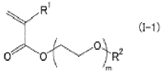
- Procédé de capture de cellules spécifiques selon l'une quelconque des revendications 1 à 7,
dans lequel la couche de polymère hydrophile a une épaisseur de 10 à 500 nm. - Procédé d'analyse de cellules spécifiques, comprenant la capture de cellules spécifiques à partir de sang ou d'un fluide biologique par le procédé de capture de cellules spécifiques selon l'une quelconque des revendications 1 à 8, et l'analyse des cellules spécifiques.
Applications Claiming Priority (1)
| Application Number | Priority Date | Filing Date | Title |
|---|---|---|---|
| JP2018024118A JP7109719B2 (ja) | 2018-02-14 | 2018-02-14 | 特定細胞捕捉方法 |
Publications (2)
| Publication Number | Publication Date |
|---|---|
| EP3553516A1 EP3553516A1 (fr) | 2019-10-16 |
| EP3553516B1 true EP3553516B1 (fr) | 2023-04-12 |
Family
ID=65241115
Family Applications (1)
| Application Number | Title | Priority Date | Filing Date |
|---|---|---|---|
| EP19152149.1A Active EP3553516B1 (fr) | 2018-02-14 | 2019-01-16 | Procédé de capture de cellules spécifiques |
Country Status (4)
| Country | Link |
|---|---|
| US (1) | US11573232B2 (fr) |
| EP (1) | EP3553516B1 (fr) |
| JP (1) | JP7109719B2 (fr) |
| CN (1) | CN110157670A (fr) |
Families Citing this family (5)
| Publication number | Priority date | Publication date | Assignee | Title |
|---|---|---|---|---|
| JP7639275B2 (ja) * | 2020-05-27 | 2025-03-05 | 住友ゴム工業株式会社 | 親水性基材及び親水性基材作製方法 |
| JP2023150438A (ja) | 2022-03-31 | 2023-10-16 | 住友ゴム工業株式会社 | ポリマー被覆基材及び医療用検査装置 |
| JP7816326B2 (ja) * | 2022-12-23 | 2026-02-18 | 住友ゴム工業株式会社 | ポリマー被覆ガラス基材 |
| EP4389714A1 (fr) * | 2022-12-23 | 2024-06-26 | Sumitomo Rubber Industries, Ltd. | Substrat de verre revêtu de polymère |
| US20240409768A1 (en) * | 2023-06-09 | 2024-12-12 | Sumitomo Rubber Industries, Ltd. | Method of producing polymer-coated substrate for capturing cancer cells |
Citations (2)
| Publication number | Priority date | Publication date | Assignee | Title |
|---|---|---|---|---|
| EP3527985B1 (fr) * | 2018-02-14 | 2020-12-30 | Sumitomo Rubber Industries, Ltd. | Procédé de capture de cellules spécifiques |
| EP3527986B1 (fr) * | 2018-02-14 | 2022-08-03 | Sumitomo Rubber Industries, Ltd. | Procédé de capture de cellules spécifiques |
Family Cites Families (96)
| Publication number | Priority date | Publication date | Assignee | Title |
|---|---|---|---|---|
| US4695553A (en) * | 1985-11-01 | 1987-09-22 | Becton Dickinson And Co., Inc. | Method for increasing agglutination of groups of cells to produce improved cell layer interface in centrifuged blood sample using antibodies |
| JP2667447B2 (ja) | 1988-06-28 | 1997-10-27 | オリンパス光学工業株式会社 | 固体表面への細胞の固定化方法 |
| US5202025A (en) | 1989-04-12 | 1993-04-13 | Terumo Kabushiki Kaisha | Porous membrane and method for preparing the same |
| JPH03110473A (ja) | 1989-09-26 | 1991-05-10 | Tokuyama Soda Co Ltd | 血液型自動判定方法及びその装置 |
| US6743878B2 (en) * | 1991-07-05 | 2004-06-01 | Biocompatibles Uk Limited | Polymeric surface coatings |
| JP3443891B2 (ja) | 1993-09-14 | 2003-09-08 | 日本油脂株式会社 | タンパク質吸着防止剤 |
| US6171780B1 (en) | 1997-06-02 | 2001-01-09 | Aurora Biosciences Corporation | Low fluorescence assay platforms and related methods for drug discovery |
| US6517781B1 (en) | 1997-06-02 | 2003-02-11 | Aurora Biosciences Corporation | Low fluorescence assay platforms and related methods for drug discovery |
| AU6293499A (en) | 1998-10-07 | 2000-04-26 | E-Ink Corporation | Capsules for electrophoretic displays and methods for making the same |
| DE19904267A1 (de) | 1999-02-03 | 2000-08-10 | Michael W Dahm | Verfahren zur Anreicherung von Tumorzellen aus einer Körperflüssigkeit und dazu geeigneter Kit |
| CA2375115C (fr) | 1999-05-28 | 2011-05-24 | Stemcell Technologies Inc. | Procede de separation des cellules par utilisation de rosettes immunitaires |
| AU8843701A (en) | 2000-09-09 | 2002-03-22 | Univ New York State Res Found | Method and compositions for isolating metastatic cancer cells, and use in measuring metastatic potential of a cancer thereof |
| US20100261159A1 (en) | 2000-10-10 | 2010-10-14 | Robert Hess | Apparatus for assay, synthesis and storage, and methods of manufacture, use, and manipulation thereof |
| JP2004532756A (ja) | 2001-06-18 | 2004-10-28 | ベクトン・ディキンソン・アンド・カンパニー | 多層容器および多層容器の形成方法 |
| US7008979B2 (en) | 2002-04-30 | 2006-03-07 | Hydromer, Inc. | Coating composition for multiple hydrophilic applications |
| US20060008807A1 (en) | 2002-08-23 | 2006-01-12 | O'hara Shawn M | Multiparameter analysis of comprehensive nucleic acids and morphological features on the same sample |
| JP4518767B2 (ja) | 2003-09-09 | 2010-08-04 | チッソ株式会社 | 刺激応答性ポリマー固定化磁性微粒子及びこれを用いた吸着材 |
| WO2005064347A1 (fr) | 2003-12-22 | 2005-07-14 | Micro Typing Systems, Inc. | Reduction du delai pour le resultat d'essai diagnostique d'une banque de sang |
| JP4567326B2 (ja) | 2003-12-24 | 2010-10-20 | 三菱化学メディエンス株式会社 | 全血を用いる測定法 |
| JP4542409B2 (ja) | 2004-10-14 | 2010-09-15 | オリンパス株式会社 | 骨髄由来間葉系幹細胞の培養方法 |
| EP1655354B1 (fr) | 2004-11-09 | 2013-10-23 | JSR Corporation | Composition de revêtement prevenant l'absorbtion d'une substance biologique, articles revêtus de celle-ci et procédé d'utilisation |
| AU2006206426B2 (en) | 2005-01-20 | 2012-03-15 | The Regents Of The University Of California | Cellular microarrays for screening differentiation factors |
| EP1693109A1 (fr) | 2005-02-21 | 2006-08-23 | Hexal Ag | Container pour la separation des cellulles cancéreuse |
| EP1874920A4 (fr) | 2005-04-05 | 2009-11-04 | Cellpoint Diagnostics | Dispositifs et procédés permettant d'enrichir et de modifier des cellules tumorales circulantes et d'autres particules |
| WO2007092028A2 (fr) * | 2005-04-08 | 2007-08-16 | Medical Discovery Partners Llc | Procédé d'enrichissement de sous-populations cellulaires rares à partir du sang |
| WO2006129735A1 (fr) | 2005-05-31 | 2006-12-07 | Olympus Corporation | Cellule a gene transfere et procede d’analyse cellulaire |
| JP4761448B2 (ja) | 2005-09-16 | 2011-08-31 | 独立行政法人科学技術振興機構 | 血液エンドトキシン測定方法 |
| EP1826563A1 (fr) | 2006-02-22 | 2007-08-29 | Fujifilm Corporation | Biocapteur |
| JP4053579B2 (ja) | 2006-02-22 | 2008-02-27 | 富士フイルム株式会社 | バイオセンサー |
| US20080008736A1 (en) | 2006-07-06 | 2008-01-10 | Thierry Glauser | Random copolymers of methacrylates and acrylates |
| JP2010509570A (ja) | 2006-11-03 | 2010-03-25 | パーデュー・リサーチ・ファウンデーション | エクスビボフローサイトメトリーの方法および装置 |
| AU2009219437B2 (en) | 2008-02-25 | 2014-10-16 | Nestec S.A. | Drug selection for breast cancer therapy using antibody-based arrays |
| AU2010229924B2 (en) | 2009-03-24 | 2016-07-21 | Plus Therapeutics Inc. | Devices and methods of cell capture and analysis |
| WO2010124227A2 (fr) | 2009-04-24 | 2010-10-28 | The Board Of Trustees Of The University Of Illinois | Procédés et dispositifs de capture de cellules tumorales circulantes |
| EP2470914B1 (fr) | 2009-07-27 | 2020-05-13 | Meso Scale Technologies, LLC | Appareils et procédés d'essai |
| GB2472321B (en) | 2009-07-31 | 2014-03-05 | Oxley Hughes Ltd | Means for improved liquid handling in a microplate |
| EP2363501A1 (fr) | 2010-03-02 | 2011-09-07 | Universitätsklinikum Hamburg-Eppendorf | Nanocomposites dotés d'une homogénéité améliorée |
| JP5907957B2 (ja) | 2010-06-16 | 2016-04-26 | ディーエスエム アイピー アセッツ ビー.ブイ. | 親水性コーティングを製造するためのコーティング配合物 |
| GB201010736D0 (en) | 2010-06-25 | 2010-08-11 | Imp Innovations Ltd | IWAP (Interwell assay plate) |
| US9334477B2 (en) | 2010-08-20 | 2016-05-10 | Japan Advanced Institute Of Science And Technology | Method for collecting nucleated red blood cells via density-gradient centrifugation utilizing changes in blood cell density |
| RU2477981C2 (ru) | 2010-10-13 | 2013-03-27 | Иван Васильевич Крайник | Способ хирургического лечения перфорации перегородки носа |
| US9012202B2 (en) | 2010-11-02 | 2015-04-21 | University Of Florida Research Foundation, Inc. | Cell-based arrays, methods of making, and methods of using |
| JP6474540B2 (ja) * | 2010-11-17 | 2019-02-27 | 国立大学法人山形大学 | 溶液から細胞を分離する細胞分離方法、細胞吸着用水和性組成物、および細胞分離システム |
| US9926523B2 (en) | 2010-12-16 | 2018-03-27 | General Electric Company | Cell carriers and methods for culturing cells |
| US9551700B2 (en) | 2010-12-20 | 2017-01-24 | Milagen, Inc. | Device and methods for the detection of cervical disease |
| JPWO2012108087A1 (ja) | 2011-02-10 | 2014-07-03 | エドワーズ株式会社 | 真空ポンプ |
| AU2012256403B2 (en) | 2011-03-17 | 2016-10-20 | Corning Incorporated | Synthetic coating for cell culture |
| JP5975532B2 (ja) * | 2011-06-13 | 2016-08-23 | 日立化成株式会社 | 癌細胞接着性向上剤 |
| EP2781594A4 (fr) | 2011-11-20 | 2015-04-08 | Univ Tokyo Womens Medical | Substrat de culture cellulaire et son procédé de fabrication |
| WO2013099901A1 (fr) | 2011-12-28 | 2013-07-04 | Jsr株式会社 | Inhibiteur de l'adhérence cellulaire |
| CA2862468A1 (fr) | 2012-01-23 | 2013-08-01 | The Ohio State University | Dispositifs et procedes permettant de detecter rapidement et precisement les substances a analyser |
| AU2013229764A1 (en) | 2012-03-09 | 2014-09-25 | Massachusetts Institute Of Technology | Adhesion signatures |
| EP2706357A1 (fr) | 2012-09-07 | 2014-03-12 | Andreas-Claudius Hoffmann | Procédé pour identifier des sous-groupes de cellules tumorales circulantes (CTC) dans un échantillon ou une population CTC |
| WO2014065861A1 (fr) | 2012-10-26 | 2014-05-01 | The Trustees Of The University Of Pennsylvania | Compositions, procédés et dispositif microfluidique à base de télomérase dans des analyses de diagnostic in vitro pour la détection de cellules tumorales circulantes (ctc) |
| JP2014105159A (ja) | 2012-11-22 | 2014-06-09 | Nippon Koden Corp | ヒト由来上皮細胞接着分子に対するモノクローナル抗体、およびこれを用いた循環腫瘍細胞の検出方法 |
| US20140158604A1 (en) | 2012-12-12 | 2014-06-12 | Jacques Chammas | Platelet Storage Container |
| CA2895791A1 (fr) | 2013-01-25 | 2014-07-31 | Xcell Biosciences, Inc. | Procedes, compositions, trousses et systemes pour l'enrichissement selectif de cellules cibles |
| US10161936B2 (en) | 2013-03-08 | 2018-12-25 | University Of Leipzig | Method and kit for cytokine analysis from a human whole blood sample |
| CN105102606A (zh) | 2013-04-04 | 2015-11-25 | 日立化成株式会社 | 生物体物质捕获用的过滤器 |
| US10647829B2 (en) | 2013-06-20 | 2020-05-12 | Sumitomo Rubber Industries, Ltd. | Surface modification method and surface modification body |
| CN104520420B (zh) | 2013-07-24 | 2016-09-07 | 株式会社奥普特尼克斯精密 | 末梢循环肿瘤细胞或稀少细胞分离用装置及末梢循环肿瘤细胞或稀少细胞分离方法 |
| WO2015046557A1 (fr) * | 2013-09-30 | 2015-04-02 | 積水メディカル株式会社 | Dispositif pour concentrer et séparer des cellules tumorales circulantes, et procédé associé |
| JP6157429B2 (ja) | 2013-10-21 | 2017-07-05 | 住友ゴム工業株式会社 | 潤滑性、低タンパク質吸着性および/または低細胞吸着性を有する金属医療用具及びその製造方法 |
| US9699974B2 (en) | 2014-02-03 | 2017-07-11 | Hunter Industries, Inc. | Rotor-type sprinkler with pressure regulator in outer case |
| JP6495241B2 (ja) | 2014-03-11 | 2019-04-03 | テルモ株式会社 | 医療用具の製造方法および医療用具 |
| US10472598B2 (en) | 2014-05-22 | 2019-11-12 | Sumitomo Bakelite Co., Ltd. | Cell mass culture vessel |
| JP2015224332A (ja) | 2014-05-30 | 2015-12-14 | 株式会社日立製作所 | 刺激応答型材料とそれを用いた細胞培養容器 |
| EP3177904A4 (fr) | 2014-08-07 | 2018-01-03 | The General Hospital Corporation | Isolement de cellules microfluidiques de plaquette cible |
| US11167286B2 (en) | 2014-09-04 | 2021-11-09 | The University Of Massachusetts | Sensors and methods for capturing targeted cells |
| US11278884B2 (en) | 2014-10-28 | 2022-03-22 | Arteriocyte Medical Systems, Inc. | Centrifuge tube comprising a floating buoy, and methods for using the same |
| JP7089240B2 (ja) | 2014-12-22 | 2022-06-22 | エコール・ポリテクニーク・フェデラル・ドゥ・ローザンヌ (ウ・ペ・エフ・エル) | 高い収集能力で哺乳動物細胞を操作(マニピュレーション)する装置 |
| WO2016115537A2 (fr) | 2015-01-15 | 2016-07-21 | Massachusetts Institute Of Technology | Systèmes, procédés, et appareil pour l'identification et la récupération in vitro de cellules individuelles |
| JP2016131561A (ja) | 2015-01-22 | 2016-07-25 | 国立大学法人山形大学 | 細胞を回収する方法、及びそれに用いられるポリマー |
| JP6089052B2 (ja) | 2015-02-23 | 2017-03-01 | 株式会社エヌ・ティ・ティ・データ | 携帯端末及びプログラム |
| US10317406B2 (en) | 2015-04-06 | 2019-06-11 | The Regents Of The University Of Michigan | System for detecting rare cells |
| WO2017030196A1 (fr) | 2015-08-20 | 2017-02-23 | 東京エレクトロン株式会社 | Récipient de culture et procédé de culture cellulaire et procédé d'observation de cellules utilisant le récipient de culture |
| US9968929B2 (en) | 2015-10-27 | 2018-05-15 | Apex Biotechnology Corp. | Reaction cassette and assay device |
| JP6617516B2 (ja) * | 2015-10-27 | 2019-12-11 | 東ソー株式会社 | 血液試料中に含まれる目的細胞の検出方法 |
| CA3001718A1 (fr) | 2015-11-16 | 2017-05-26 | Sio2 Medical Products, Inc. | Substrat polymere ayant une surface presentant une adhesion de biomolecule, et articles thermoplastiques constitues de ce substrat |
| JP6639906B2 (ja) | 2015-12-25 | 2020-02-05 | 東ソー株式会社 | 生物試料検出方法 |
| WO2017130802A1 (fr) * | 2016-01-29 | 2017-08-03 | 学校法人東京女子医科大学 | Composition de feuille cellulaire comprenant des cellules souches mésenchymateuses, et procédé de cicatrisation d'organe luminal au moyen de ladite composition |
| JP2017181096A (ja) * | 2016-03-28 | 2017-10-05 | 日立化成株式会社 | 希少細胞を捕捉する方法 |
| FR3050212B1 (fr) | 2016-04-15 | 2020-09-25 | Chu Montpellier | Procede de detection et/ou de caracterisation de cellules tumorales et appareil associe |
| JP6468520B2 (ja) * | 2016-05-09 | 2019-02-13 | 住友ゴム工業株式会社 | 医療用検査装置及び細胞検査方法 |
| EP3244208A1 (fr) * | 2016-05-09 | 2017-11-15 | Sumitomo Rubber Industries, Ltd. | Dispositif d'analyse médicale et procédé d'analyse cellulaire |
| EP3244206A1 (fr) | 2016-05-13 | 2017-11-15 | Linde Aktiengesellschaft | Composant de codage de gaz et gaz code correspondant |
| JP6485783B2 (ja) | 2016-09-29 | 2019-03-20 | 住友ゴム工業株式会社 | 医療用検査装置及び細胞検査方法 |
| JP6779483B2 (ja) | 2016-09-29 | 2020-11-04 | 住友ゴム工業株式会社 | 医療用検査装置及び細胞検査方法 |
| JP6518985B2 (ja) * | 2016-09-29 | 2019-05-29 | 住友ゴム工業株式会社 | がん細胞捕捉方法 |
| US10941374B2 (en) | 2016-09-29 | 2021-03-09 | Sumitomo Rubber Industries, Ltd. | Medical analysis device and cell analysis method |
| CN106635995B (zh) * | 2017-03-10 | 2020-06-02 | 亚能生物技术(深圳)有限公司 | 一种循环肿瘤细胞阴性富集方法 |
| JP7358030B2 (ja) | 2018-01-31 | 2023-10-10 | 住友ゴム工業株式会社 | 親水性基材 |
| EP3830237A1 (fr) | 2018-08-16 | 2021-06-09 | TERUMO Kabushiki Kaisha | Substrat de culture cellulaire |
| JP7315653B2 (ja) | 2018-08-16 | 2023-07-26 | テルモ株式会社 | 細胞培養基材 |
| EP3830239A1 (fr) | 2018-08-16 | 2021-06-09 | TERUMO Kabushiki Kaisha | Substrat de culture cellulaire |
| EP3830238A1 (fr) | 2018-08-16 | 2021-06-09 | TERUMO Kabushiki Kaisha | Substrat de culture cellulaire |
-
2018
- 2018-02-14 JP JP2018024118A patent/JP7109719B2/ja active Active
-
2019
- 2019-01-16 EP EP19152149.1A patent/EP3553516B1/fr active Active
- 2019-01-16 US US16/249,454 patent/US11573232B2/en active Active
- 2019-01-29 CN CN201910088843.4A patent/CN110157670A/zh active Pending
Patent Citations (2)
| Publication number | Priority date | Publication date | Assignee | Title |
|---|---|---|---|---|
| EP3527985B1 (fr) * | 2018-02-14 | 2020-12-30 | Sumitomo Rubber Industries, Ltd. | Procédé de capture de cellules spécifiques |
| EP3527986B1 (fr) * | 2018-02-14 | 2022-08-03 | Sumitomo Rubber Industries, Ltd. | Procédé de capture de cellules spécifiques |
Also Published As
| Publication number | Publication date |
|---|---|
| CN110157670A (zh) | 2019-08-23 |
| EP3553516A1 (fr) | 2019-10-16 |
| US11573232B2 (en) | 2023-02-07 |
| US20190250150A1 (en) | 2019-08-15 |
| JP7109719B2 (ja) | 2022-08-01 |
| JP2019138841A (ja) | 2019-08-22 |
Similar Documents
| Publication | Publication Date | Title |
|---|---|---|
| EP3527985B1 (fr) | Procédé de capture de cellules spécifiques | |
| EP3527986B1 (fr) | Procédé de capture de cellules spécifiques | |
| EP3553516B1 (fr) | Procédé de capture de cellules spécifiques | |
| EP3301444B1 (fr) | Procédé permettant de capturer des cellules cancéreuses | |
| US10941374B2 (en) | Medical analysis device and cell analysis method | |
| US11360078B2 (en) | Medical analysis device and cell analysis method | |
| JP6895662B2 (ja) | 特定細胞の分画方法及び捕捉方法 | |
| JP6485783B2 (ja) | 医療用検査装置及び細胞検査方法 | |
| EP3686598B1 (fr) | Procédés de capture et de fractionnement de cellules spécifiques | |
| JP7580076B2 (ja) | 細胞培養装置及び細胞培養方法 | |
| JP6886667B2 (ja) | がん細胞捕捉方法 |
Legal Events
| Date | Code | Title | Description |
|---|---|---|---|
| PUAI | Public reference made under article 153(3) epc to a published international application that has entered the european phase |
Free format text: ORIGINAL CODE: 0009012 |
|
| STAA | Information on the status of an ep patent application or granted ep patent |
Free format text: STATUS: THE APPLICATION HAS BEEN PUBLISHED |
|
| AK | Designated contracting states |
Kind code of ref document: A1 Designated state(s): AL AT BE BG CH CY CZ DE DK EE ES FI FR GB GR HR HU IE IS IT LI LT LU LV MC MK MT NL NO PL PT RO RS SE SI SK SM TR |
|
| AX | Request for extension of the european patent |
Extension state: BA ME |
|
| STAA | Information on the status of an ep patent application or granted ep patent |
Free format text: STATUS: REQUEST FOR EXAMINATION WAS MADE |
|
| 17P | Request for examination filed |
Effective date: 20200325 |
|
| RBV | Designated contracting states (corrected) |
Designated state(s): AL AT BE BG CH CY CZ DE DK EE ES FI FR GB GR HR HU IE IS IT LI LT LU LV MC MK MT NL NO PL PT RO RS SE SI SK SM TR |
|
| GRAP | Despatch of communication of intention to grant a patent |
Free format text: ORIGINAL CODE: EPIDOSNIGR1 |
|
| STAA | Information on the status of an ep patent application or granted ep patent |
Free format text: STATUS: GRANT OF PATENT IS INTENDED |
|
| INTG | Intention to grant announced |
Effective date: 20221024 |
|
| RAP3 | Party data changed (applicant data changed or rights of an application transferred) |
Owner name: YAMAGATA UNIVERSITY Owner name: SUMITOMO RUBBER INDUSTRIES, LTD. |
|
| RIN1 | Information on inventor provided before grant (corrected) |
Inventor name: EMURA, HARUKA Inventor name: HOSHIBA, TAKASHI Inventor name: TANAKA, MASARU Inventor name: MINAGAWA, YASUHISA |
|
| GRAS | Grant fee paid |
Free format text: ORIGINAL CODE: EPIDOSNIGR3 |
|
| GRAA | (expected) grant |
Free format text: ORIGINAL CODE: 0009210 |
|
| STAA | Information on the status of an ep patent application or granted ep patent |
Free format text: STATUS: THE PATENT HAS BEEN GRANTED |
|
| AK | Designated contracting states |
Kind code of ref document: B1 Designated state(s): AL AT BE BG CH CY CZ DE DK EE ES FI FR GB GR HR HU IE IS IT LI LT LU LV MC MK MT NL NO PL PT RO RS SE SI SK SM TR |
|
| REG | Reference to a national code |
Ref country code: GB Ref legal event code: FG4D |
|
| REG | Reference to a national code |
Ref country code: CH Ref legal event code: EP |
|
| REG | Reference to a national code |
Ref country code: DE Ref legal event code: R096 Ref document number: 602019027366 Country of ref document: DE |
|
| REG | Reference to a national code |
Ref country code: IE Ref legal event code: FG4D |
|
| REG | Reference to a national code |
Ref country code: AT Ref legal event code: REF Ref document number: 1560054 Country of ref document: AT Kind code of ref document: T Effective date: 20230515 |
|
| P01 | Opt-out of the competence of the unified patent court (upc) registered |
Effective date: 20230510 |
|
| REG | Reference to a national code |
Ref country code: LT Ref legal event code: MG9D |
|
| REG | Reference to a national code |
Ref country code: NL Ref legal event code: MP Effective date: 20230412 |
|
| REG | Reference to a national code |
Ref country code: AT Ref legal event code: MK05 Ref document number: 1560054 Country of ref document: AT Kind code of ref document: T Effective date: 20230412 |
|
| PG25 | Lapsed in a contracting state [announced via postgrant information from national office to epo] |
Ref country code: NL Free format text: LAPSE BECAUSE OF FAILURE TO SUBMIT A TRANSLATION OF THE DESCRIPTION OR TO PAY THE FEE WITHIN THE PRESCRIBED TIME-LIMIT Effective date: 20230412 |
|
| PG25 | Lapsed in a contracting state [announced via postgrant information from national office to epo] |
Ref country code: SE Free format text: LAPSE BECAUSE OF FAILURE TO SUBMIT A TRANSLATION OF THE DESCRIPTION OR TO PAY THE FEE WITHIN THE PRESCRIBED TIME-LIMIT Effective date: 20230412 Ref country code: PT Free format text: LAPSE BECAUSE OF FAILURE TO SUBMIT A TRANSLATION OF THE DESCRIPTION OR TO PAY THE FEE WITHIN THE PRESCRIBED TIME-LIMIT Effective date: 20230814 Ref country code: NO Free format text: LAPSE BECAUSE OF FAILURE TO SUBMIT A TRANSLATION OF THE DESCRIPTION OR TO PAY THE FEE WITHIN THE PRESCRIBED TIME-LIMIT Effective date: 20230712 Ref country code: ES Free format text: LAPSE BECAUSE OF FAILURE TO SUBMIT A TRANSLATION OF THE DESCRIPTION OR TO PAY THE FEE WITHIN THE PRESCRIBED TIME-LIMIT Effective date: 20230412 Ref country code: AT Free format text: LAPSE BECAUSE OF FAILURE TO SUBMIT A TRANSLATION OF THE DESCRIPTION OR TO PAY THE FEE WITHIN THE PRESCRIBED TIME-LIMIT Effective date: 20230412 |
|
| PG25 | Lapsed in a contracting state [announced via postgrant information from national office to epo] |
Ref country code: RS Free format text: LAPSE BECAUSE OF FAILURE TO SUBMIT A TRANSLATION OF THE DESCRIPTION OR TO PAY THE FEE WITHIN THE PRESCRIBED TIME-LIMIT Effective date: 20230412 Ref country code: PL Free format text: LAPSE BECAUSE OF FAILURE TO SUBMIT A TRANSLATION OF THE DESCRIPTION OR TO PAY THE FEE WITHIN THE PRESCRIBED TIME-LIMIT Effective date: 20230412 Ref country code: LV Free format text: LAPSE BECAUSE OF FAILURE TO SUBMIT A TRANSLATION OF THE DESCRIPTION OR TO PAY THE FEE WITHIN THE PRESCRIBED TIME-LIMIT Effective date: 20230412 Ref country code: LT Free format text: LAPSE BECAUSE OF FAILURE TO SUBMIT A TRANSLATION OF THE DESCRIPTION OR TO PAY THE FEE WITHIN THE PRESCRIBED TIME-LIMIT Effective date: 20230412 Ref country code: IS Free format text: LAPSE BECAUSE OF FAILURE TO SUBMIT A TRANSLATION OF THE DESCRIPTION OR TO PAY THE FEE WITHIN THE PRESCRIBED TIME-LIMIT Effective date: 20230812 Ref country code: HR Free format text: LAPSE BECAUSE OF FAILURE TO SUBMIT A TRANSLATION OF THE DESCRIPTION OR TO PAY THE FEE WITHIN THE PRESCRIBED TIME-LIMIT Effective date: 20230412 Ref country code: GR Free format text: LAPSE BECAUSE OF FAILURE TO SUBMIT A TRANSLATION OF THE DESCRIPTION OR TO PAY THE FEE WITHIN THE PRESCRIBED TIME-LIMIT Effective date: 20230713 Ref country code: AL Free format text: LAPSE BECAUSE OF FAILURE TO SUBMIT A TRANSLATION OF THE DESCRIPTION OR TO PAY THE FEE WITHIN THE PRESCRIBED TIME-LIMIT Effective date: 20230412 |
|
| PG25 | Lapsed in a contracting state [announced via postgrant information from national office to epo] |
Ref country code: FI Free format text: LAPSE BECAUSE OF FAILURE TO SUBMIT A TRANSLATION OF THE DESCRIPTION OR TO PAY THE FEE WITHIN THE PRESCRIBED TIME-LIMIT Effective date: 20230412 |
|
| REG | Reference to a national code |
Ref country code: DE Ref legal event code: R097 Ref document number: 602019027366 Country of ref document: DE |
|
| PG25 | Lapsed in a contracting state [announced via postgrant information from national office to epo] |
Ref country code: SK Free format text: LAPSE BECAUSE OF FAILURE TO SUBMIT A TRANSLATION OF THE DESCRIPTION OR TO PAY THE FEE WITHIN THE PRESCRIBED TIME-LIMIT Effective date: 20230412 |
|
| PG25 | Lapsed in a contracting state [announced via postgrant information from national office to epo] |
Ref country code: SM Free format text: LAPSE BECAUSE OF FAILURE TO SUBMIT A TRANSLATION OF THE DESCRIPTION OR TO PAY THE FEE WITHIN THE PRESCRIBED TIME-LIMIT Effective date: 20230412 Ref country code: SK Free format text: LAPSE BECAUSE OF FAILURE TO SUBMIT A TRANSLATION OF THE DESCRIPTION OR TO PAY THE FEE WITHIN THE PRESCRIBED TIME-LIMIT Effective date: 20230412 Ref country code: RO Free format text: LAPSE BECAUSE OF FAILURE TO SUBMIT A TRANSLATION OF THE DESCRIPTION OR TO PAY THE FEE WITHIN THE PRESCRIBED TIME-LIMIT Effective date: 20230412 Ref country code: EE Free format text: LAPSE BECAUSE OF FAILURE TO SUBMIT A TRANSLATION OF THE DESCRIPTION OR TO PAY THE FEE WITHIN THE PRESCRIBED TIME-LIMIT Effective date: 20230412 Ref country code: DK Free format text: LAPSE BECAUSE OF FAILURE TO SUBMIT A TRANSLATION OF THE DESCRIPTION OR TO PAY THE FEE WITHIN THE PRESCRIBED TIME-LIMIT Effective date: 20230412 Ref country code: CZ Free format text: LAPSE BECAUSE OF FAILURE TO SUBMIT A TRANSLATION OF THE DESCRIPTION OR TO PAY THE FEE WITHIN THE PRESCRIBED TIME-LIMIT Effective date: 20230412 |
|
| PLBE | No opposition filed within time limit |
Free format text: ORIGINAL CODE: 0009261 |
|
| STAA | Information on the status of an ep patent application or granted ep patent |
Free format text: STATUS: NO OPPOSITION FILED WITHIN TIME LIMIT |
|
| 26N | No opposition filed |
Effective date: 20240115 |
|
| PG25 | Lapsed in a contracting state [announced via postgrant information from national office to epo] |
Ref country code: SI Free format text: LAPSE BECAUSE OF FAILURE TO SUBMIT A TRANSLATION OF THE DESCRIPTION OR TO PAY THE FEE WITHIN THE PRESCRIBED TIME-LIMIT Effective date: 20230412 |
|
| PG25 | Lapsed in a contracting state [announced via postgrant information from national office to epo] |
Ref country code: SI Free format text: LAPSE BECAUSE OF FAILURE TO SUBMIT A TRANSLATION OF THE DESCRIPTION OR TO PAY THE FEE WITHIN THE PRESCRIBED TIME-LIMIT Effective date: 20230412 Ref country code: IT Free format text: LAPSE BECAUSE OF FAILURE TO SUBMIT A TRANSLATION OF THE DESCRIPTION OR TO PAY THE FEE WITHIN THE PRESCRIBED TIME-LIMIT Effective date: 20230412 |
|
| PG25 | Lapsed in a contracting state [announced via postgrant information from national office to epo] |
Ref country code: MC Free format text: LAPSE BECAUSE OF FAILURE TO SUBMIT A TRANSLATION OF THE DESCRIPTION OR TO PAY THE FEE WITHIN THE PRESCRIBED TIME-LIMIT Effective date: 20230412 |
|
| PG25 | Lapsed in a contracting state [announced via postgrant information from national office to epo] |
Ref country code: MC Free format text: LAPSE BECAUSE OF FAILURE TO SUBMIT A TRANSLATION OF THE DESCRIPTION OR TO PAY THE FEE WITHIN THE PRESCRIBED TIME-LIMIT Effective date: 20230412 |
|
| REG | Reference to a national code |
Ref country code: CH Ref legal event code: PL |
|
| PG25 | Lapsed in a contracting state [announced via postgrant information from national office to epo] |
Ref country code: LU Free format text: LAPSE BECAUSE OF NON-PAYMENT OF DUE FEES Effective date: 20240116 |
|
| GBPC | Gb: european patent ceased through non-payment of renewal fee |
Effective date: 20240116 |
|
| PG25 | Lapsed in a contracting state [announced via postgrant information from national office to epo] |
Ref country code: LU Free format text: LAPSE BECAUSE OF NON-PAYMENT OF DUE FEES Effective date: 20240116 |
|
| PG25 | Lapsed in a contracting state [announced via postgrant information from national office to epo] |
Ref country code: GB Free format text: LAPSE BECAUSE OF NON-PAYMENT OF DUE FEES Effective date: 20240116 |
|
| PG25 | Lapsed in a contracting state [announced via postgrant information from national office to epo] |
Ref country code: BE Free format text: LAPSE BECAUSE OF NON-PAYMENT OF DUE FEES Effective date: 20240131 |
|
| PG25 | Lapsed in a contracting state [announced via postgrant information from national office to epo] |
Ref country code: CH Free format text: LAPSE BECAUSE OF NON-PAYMENT OF DUE FEES Effective date: 20240131 |
|
| PG25 | Lapsed in a contracting state [announced via postgrant information from national office to epo] |
Ref country code: GB Free format text: LAPSE BECAUSE OF NON-PAYMENT OF DUE FEES Effective date: 20240116 Ref country code: CH Free format text: LAPSE BECAUSE OF NON-PAYMENT OF DUE FEES Effective date: 20240131 Ref country code: BE Free format text: LAPSE BECAUSE OF NON-PAYMENT OF DUE FEES Effective date: 20240131 |
|
| REG | Reference to a national code |
Ref country code: BE Ref legal event code: MM Effective date: 20240131 |
|
| PG25 | Lapsed in a contracting state [announced via postgrant information from national office to epo] |
Ref country code: BG Free format text: LAPSE BECAUSE OF FAILURE TO SUBMIT A TRANSLATION OF THE DESCRIPTION OR TO PAY THE FEE WITHIN THE PRESCRIBED TIME-LIMIT Effective date: 20230412 |
|
| PG25 | Lapsed in a contracting state [announced via postgrant information from national office to epo] |
Ref country code: BG Free format text: LAPSE BECAUSE OF FAILURE TO SUBMIT A TRANSLATION OF THE DESCRIPTION OR TO PAY THE FEE WITHIN THE PRESCRIBED TIME-LIMIT Effective date: 20230412 |
|
| PG25 | Lapsed in a contracting state [announced via postgrant information from national office to epo] |
Ref country code: IE Free format text: LAPSE BECAUSE OF NON-PAYMENT OF DUE FEES Effective date: 20240116 |
|
| PG25 | Lapsed in a contracting state [announced via postgrant information from national office to epo] |
Ref country code: IE Free format text: LAPSE BECAUSE OF NON-PAYMENT OF DUE FEES Effective date: 20240116 |
|
| PG25 | Lapsed in a contracting state [announced via postgrant information from national office to epo] |
Ref country code: CY Free format text: LAPSE BECAUSE OF FAILURE TO SUBMIT A TRANSLATION OF THE DESCRIPTION OR TO PAY THE FEE WITHIN THE PRESCRIBED TIME-LIMIT; INVALID AB INITIO Effective date: 20190116 |
|
| PG25 | Lapsed in a contracting state [announced via postgrant information from national office to epo] |
Ref country code: HU Free format text: LAPSE BECAUSE OF FAILURE TO SUBMIT A TRANSLATION OF THE DESCRIPTION OR TO PAY THE FEE WITHIN THE PRESCRIBED TIME-LIMIT; INVALID AB INITIO Effective date: 20190116 |
|
| PG25 | Lapsed in a contracting state [announced via postgrant information from national office to epo] |
Ref country code: TR Free format text: LAPSE BECAUSE OF FAILURE TO SUBMIT A TRANSLATION OF THE DESCRIPTION OR TO PAY THE FEE WITHIN THE PRESCRIBED TIME-LIMIT Effective date: 20230412 |
|
| PGFP | Annual fee paid to national office [announced via postgrant information from national office to epo] |
Ref country code: DE Payment date: 20260328 Year of fee payment: 8 |
|
| PGFP | Annual fee paid to national office [announced via postgrant information from national office to epo] |
Ref country code: FR Payment date: 20260123 Year of fee payment: 8 |