EP3314010B1 - Procede de detection d'aberrations chromosomiques - Google Patents
Procede de detection d'aberrations chromosomiques Download PDFInfo
- Publication number
- EP3314010B1 EP3314010B1 EP16731904.5A EP16731904A EP3314010B1 EP 3314010 B1 EP3314010 B1 EP 3314010B1 EP 16731904 A EP16731904 A EP 16731904A EP 3314010 B1 EP3314010 B1 EP 3314010B1
- Authority
- EP
- European Patent Office
- Prior art keywords
- locus
- chromosome
- specific hybridization
- signals
- marked
- Prior art date
- Legal status (The legal status is an assumption and is not a legal conclusion. Google has not performed a legal analysis and makes no representation as to the accuracy of the status listed.)
- Active
Links
Images


Classifications
-
- C—CHEMISTRY; METALLURGY
- C12—BIOCHEMISTRY; BEER; SPIRITS; WINE; VINEGAR; MICROBIOLOGY; ENZYMOLOGY; MUTATION OR GENETIC ENGINEERING
- C12Q—MEASURING OR TESTING PROCESSES INVOLVING ENZYMES, NUCLEIC ACIDS OR MICROORGANISMS; COMPOSITIONS OR TEST PAPERS THEREFOR; PROCESSES OF PREPARING SUCH COMPOSITIONS; CONDITION-RESPONSIVE CONTROL IN MICROBIOLOGICAL OR ENZYMOLOGICAL PROCESSES
- C12Q1/00—Measuring or testing processes involving enzymes, nucleic acids or microorganisms; Compositions therefor; Processes of preparing such compositions
- C12Q1/68—Measuring or testing processes involving enzymes, nucleic acids or microorganisms; Compositions therefor; Processes of preparing such compositions involving nucleic acids
- C12Q1/6813—Hybridisation assays
- C12Q1/6841—In situ hybridisation
-
- C—CHEMISTRY; METALLURGY
- C12—BIOCHEMISTRY; BEER; SPIRITS; WINE; VINEGAR; MICROBIOLOGY; ENZYMOLOGY; MUTATION OR GENETIC ENGINEERING
- C12Q—MEASURING OR TESTING PROCESSES INVOLVING ENZYMES, NUCLEIC ACIDS OR MICROORGANISMS; COMPOSITIONS OR TEST PAPERS THEREFOR; PROCESSES OF PREPARING SUCH COMPOSITIONS; CONDITION-RESPONSIVE CONTROL IN MICROBIOLOGICAL OR ENZYMOLOGICAL PROCESSES
- C12Q1/00—Measuring or testing processes involving enzymes, nucleic acids or microorganisms; Compositions therefor; Processes of preparing such compositions
- C12Q1/68—Measuring or testing processes involving enzymes, nucleic acids or microorganisms; Compositions therefor; Processes of preparing such compositions involving nucleic acids
- C12Q1/6844—Nucleic acid amplification reactions
- C12Q1/6858—Allele-specific amplification
-
- C—CHEMISTRY; METALLURGY
- C12—BIOCHEMISTRY; BEER; SPIRITS; WINE; VINEGAR; MICROBIOLOGY; ENZYMOLOGY; MUTATION OR GENETIC ENGINEERING
- C12Q—MEASURING OR TESTING PROCESSES INVOLVING ENZYMES, NUCLEIC ACIDS OR MICROORGANISMS; COMPOSITIONS OR TEST PAPERS THEREFOR; PROCESSES OF PREPARING SUCH COMPOSITIONS; CONDITION-RESPONSIVE CONTROL IN MICROBIOLOGICAL OR ENZYMOLOGICAL PROCESSES
- C12Q1/00—Measuring or testing processes involving enzymes, nucleic acids or microorganisms; Compositions therefor; Processes of preparing such compositions
- C12Q1/68—Measuring or testing processes involving enzymes, nucleic acids or microorganisms; Compositions therefor; Processes of preparing such compositions involving nucleic acids
- C12Q1/6876—Nucleic acid products used in the analysis of nucleic acids, e.g. primers or probes
- C12Q1/6883—Nucleic acid products used in the analysis of nucleic acids, e.g. primers or probes for diseases caused by alterations of genetic material
- C12Q1/6886—Nucleic acid products used in the analysis of nucleic acids, e.g. primers or probes for diseases caused by alterations of genetic material for cancer
-
- C—CHEMISTRY; METALLURGY
- C12—BIOCHEMISTRY; BEER; SPIRITS; WINE; VINEGAR; MICROBIOLOGY; ENZYMOLOGY; MUTATION OR GENETIC ENGINEERING
- C12Q—MEASURING OR TESTING PROCESSES INVOLVING ENZYMES, NUCLEIC ACIDS OR MICROORGANISMS; COMPOSITIONS OR TEST PAPERS THEREFOR; PROCESSES OF PREPARING SUCH COMPOSITIONS; CONDITION-RESPONSIVE CONTROL IN MICROBIOLOGICAL OR ENZYMOLOGICAL PROCESSES
- C12Q2565/00—Nucleic acid analysis characterised by mode or means of detection
- C12Q2565/10—Detection mode being characterised by the assay principle
- C12Q2565/101—Interaction between at least two labels
-
- C—CHEMISTRY; METALLURGY
- C12—BIOCHEMISTRY; BEER; SPIRITS; WINE; VINEGAR; MICROBIOLOGY; ENZYMOLOGY; MUTATION OR GENETIC ENGINEERING
- C12Q—MEASURING OR TESTING PROCESSES INVOLVING ENZYMES, NUCLEIC ACIDS OR MICROORGANISMS; COMPOSITIONS OR TEST PAPERS THEREFOR; PROCESSES OF PREPARING SUCH COMPOSITIONS; CONDITION-RESPONSIVE CONTROL IN MICROBIOLOGICAL OR ENZYMOLOGICAL PROCESSES
- C12Q2565/00—Nucleic acid analysis characterised by mode or means of detection
- C12Q2565/10—Detection mode being characterised by the assay principle
- C12Q2565/102—Multiple non-interacting labels
Definitions
- the present invention relates to the technical field of detection methods for chromosomal abnormalities or chromosome aberrations.
- the present invention relates to a method for the detection of at least two mutually different chromosome aberrations by means of in situ hybridization.
- the present invention further relates to a composition suitable for detecting at least two different chromosome aberrations and to the uses thereof according to the invention.
- the present invention furthermore relates to the uses of at least three mutually different, locus-specific hybridization probes marked with detection labels.
- the subject of the present invention is a kit for the detection of chromosome aberrations.
- ISH in situ hybridizations
- the in-situ hybridization is based on the hybridization or pairing of complementary bases of nucleic acid single strands, in particular DNA single strands, so that specific nucleic acid sequences can be detected in a sample, in particular in a tissue or cell preparation.
- nucleic acid single strands in particular DNA single strands
- synthetically produced probes are hybridized with nucleic acid single strands of the sample and then detected.
- fluorescence-marked nucleic acid fragments or fluorescence-marked hybridization probes can be used come.
- antigen-labeled probes especially hapten-labeled probes, can be used, which are then made visible with the help of antibodies through color reactions, so that an analysis by light microscopy is possible ((brightfield ISH (BrISH), chromogens ISH (CISH), silver ISH (SISH)).
- FISH FISH-linked immunosorbent assays
- nucleic acid fragments which address different genomic regions or are specific for this are each marked or coupled with different fluorescent dyes which differ from one another in their absorption and / or emission spectrum.
- the individual colors can be determined by using specific microscope filters, which guide precisely defined wavelength ranges of the light to excite the dyes on the preparation as well as precisely defined wavelength ranges of the emitted by the dyes Direct light to the evaluator, shown separately from each other (so-called single bandpass filter set).
- filters or filter sets which allow the simultaneous display of different fluorescent dyes and thus several nucleic acid fragments. In the case of two different fluorescent dyes, for example, one speaks of a dual bandpass filter set.
- FISH usually only analyzes two colors (orange / red and green) or three colors (orange / red and green simultaneously or together with a blue nuclear counterstain (DAPI)).
- DAPI blue nuclear counterstain
- the state of the art is the use of two haptens, mostly selected from the group of biotin, dinitrophenyl (DNP) and digoxigenin, and two antibody-coupled enzymes, mostly alkaline phosphatase and peroxidase.
- DNP dinitrophenyl
- digoxigenin two antibody-coupled enzymes, mostly alkaline phosphatase and peroxidase.
- Locus-specific probes are understood to mean those probes which address selected DNA sections of a chromosome, mostly individual genes or neighboring genes, with a total size of up to about 1,000 kb and are referred to as gene-specific probes or “single copy” probes.
- Repetitive sequence-specific probes are probes that address repetitive sequences and therefore address regions with a size of several 1,000 kb. These probes also include centromere or alpha satellite probes, for example.
- the area of a first breakpoint of the chromosome is flanked proximally and distally by nucleic acid fragments of the same color (e.g. orange)
- the area of a second breakpoint, i.e. H. of the reciprocal translocation partner is flanked proximally and distally by nucleic acid fragments of a second color (e.g. green).
- the normal situation i.e. H. without chromosomal breaks in the area of the two translocation partners, is characterized by a green and a spatially separated orange signal.
- the area of a break point is flanked proximally and distally by nucleic acid fragments marked in different ways or in different colors (e.g. distal orange, proximal green).
- the normal situation i.e. H. without a chromosomal break in this area, is characterized by a fusion signal.
- an aberrant situation i.e. H. if there is a chromosomal break between the probe fragments, the signals separate spatially from one another. The difference between the normal situation and the aberrant situation is thus characterized by the distance between the differently colored signals.
- Statements about involved translocation partners are not possible with this method. It only allows the conclusion that a specific chromosomal rearrangement has taken place.
- the disadvantage of this probe composition is that with the aid of the two colors used, only a single breaking point area and thus only a specific translocation or inversion can be detected.
- locus-specific probes are also supplemented by so-called repetitive sequence-specific probes, such as centromere or alpha satellite probes:
- the principle of detecting gain or loss of signals through the occurrence of deletions, aneuplodies and amplifications usually takes place in so-called dual-color probe approaches. With this principle, which is described below, and the signal patterns derived from it, it should be noted that a normal cell is usually diploid, i.e. H. each allele is present in duplicate.
- two different chromosomal areas are marked with differently marked or color-marked nucleic acid fragments (e.g. genomic region 1 or target region 1 in orange, genomic region 2 or target region 2 in green).
- the normal situation i.e. H. without gain or loss of these areas, is characterized by two orange and two green signals.
- an aberrant situation i.e. H. if genomic regions in which target regions 1 and / or 2 have been gained or lost, fewer green and / or orange signals are visible if they are lost, and more signals are visible if they are gained.
- many additional signals can be visible, which can also be represented as clusters.
- triple FISH approaches which address the detection of different translocation events that may cluster side by side in a chromosomal region (i.e., different genes that are in proximity are affected).
- different genes that are in proximity are affected.
- the signal pattern only two colors are analyzed that detect a single aberration, the third color being irrelevant.
- Three different locus-specific probes are used, each marked with a different label.
- the US 6 576 421 B1 describes a method for the detection of cancer-associated translocations in which three probes are used, two of which hybridize on one side of a breakpoint on a fusion chromosome and the third probe on the other side of the breakpoint. Each of the probes is marked with a marker that differs from those of the other probes.
- the US 6,344,315 B1 discloses methods of staining interphase chromosomal material using a nucleic acid probe greater than 50,000 bases in length and in which, for example, a biotin-labeled ABL probe is used, which is detected indirectly with the fluorochrome Texas red, and a digoxigenin-labeled BCR probe, which is detected indirectly with the fluorochrome FITC, to carry out a FISH method.
- the WO 93/21345 A1 describes methods for detecting a deletion of a gene in which two probes are used which are provided with fluorescent markers which can be distinguished from one another.
- the WO 2005/111235 A2 describes a method which involves the use of three colors for detection purposes.
- the chromosomal region that is marked by the third label of a probe is not directly affected by a change, so that if the chromosome structure changes, the first fusion signal is eliminated, so that a new split signal and a new fusion signal arise.
- This procedure uses probes that are each marked with only one label.
- only one possible translocation, which is specified by the probes, can be detected with this method.
- the WO 02/093130 A3 discloses a method for the detection of chromosomal translocations using two or, alternatively, four labels or dyes which flank the distal and proximal breakpoints of both breakpoints involved in a translocation. This method does not offer any possibilities to detect more than one translocation at the one breakpoint area that is flanked by the probes.
- mFISH multiplex FISH
- SKY-FISH spectral karyotyping
- multicolor FISH COBRA-FISH (Combined Binary Ratio labeling FISH) or 24-color FISH
- a total of twenty-four different dyes can be produced 'Chromosome Painting Probes' can be marked and differentiated.
- the chromosome arm-specific probes of all chromosomes can be marked differently in a so-called 42-color FISH.
- the aforementioned methods are only suitable for cells that are in the metaphase.
- the present invention is therefore based on the object of providing a method or a composition which is suitable for the detection or analysis of chromosome mutations or chromosome aberrations and at least largely avoids or at least mitigates the disadvantages of the prior art described above.
- the present invention is based on the object of providing a method which enables a reliable and simultaneous detection of several mutually different chromosome aberrations, in particular independent (i.e. non-reciprocal) chromosome aberrations, in particular in one approach.
- the present invention is likewise based on the object of providing a method which, in addition, also enables chromosome aberrations to be assigned to a specific chromosome or DNA region.
- the present invention proposes a method for the detection of at least two different chromosome aberrations according to patent claim 1; further advantageous refinements are the subject matter of the dependent claims.
- the present invention also relates to a composition for the detection of at least two different chromosomal aberrations according to the independent patent claim or a composition for use in prophylactic or therapeutic treatment or in the diagnosis or prognosis of diseases associated with chromosomal aberrations.
- the present invention also further provides the use of a composition according to the present invention according to the independent patent claim in this regard.
- the present invention relates to the use of at least three, preferably at least four mutually different locus-specific hybridization probes according to the independent patent claim.
- the subject matter of the present invention is the use of at least one location-specific hybridization probe marked with at least two detection labels according to the independent patent claim.
- the subject matter of the present invention is a kit or kit-of-parts or set for the detection of at least two different chromosome aberrations; further advantageous properties are the subject of the relevant sub-claim.
- the present invention proposes - according to a first aspect of the invention - a method for detecting at least two different chromosome aberrations, in particular structural and / or numerical chromosome aberrations, preferably structural chromosome aberrations, by means of in-situ hybridization by detection of chromosomes - and / or DNA areas in a biological sample, preferably in one or more cell (s) and / or in one or more cell nucleus (s), where the in- situ hybridization is carried out as an interphase in-situ hybridization, wherein the in situ hybridization is carried out with at least four mutually different, each marked with a first detection label, location-specific hybridization probes, wherein, to generate at least one mixed signal by two or more different detection labels located on a location-specific hybridization probe, at least one of the location-specific hybridization probes is marked with at least one further detection label different from the first detection label, based on the respective location-specific hybridization probe, so that
- the present invention is based on the basic principle, through the targeted generation of mixed signals in signal patterns generated by interphase in situ hybridizations, the simultaneous or simultaneous detection of several different chromosome aberrations, in particular independent, ie non-reciprocal chromosome aberrations, in a biological sample and its assignment to a detected chromosome or DNA area.
- the chromosome aberrations are mutually independent chromosome aberrations.
- the chromosome aberrations are not dependent on one another. It can also be provided that the chromosome aberrations are not reciprocal.
- the chromosome aberrations are not related to reciprocal or mutually dependent chromosome aberrations or are associated with such.
- the present invention is associated with numerous advantages and special features, which are discussed below in a non-restrictive manner and are to be assessed as an indication of the patentability of the present invention.
- chromosome aberrations in the context of an interphase in situ hybridization on the one hand a clear detection of several possible structural or numerical chromosome aberrations and on the other hand the clear differentiation of these chromosome aberrations or .
- the chromosome aberrations can be chromosome aberrations which are independent of one another and which are not mutually dependent or which are not reciprocal.
- any Detected chromosome aberrations can be assigned to a defined or specific DNA or chromosome area.
- the amount of sample required for the detection of chromosome aberrations can be significantly reduced with the aid of the method according to the invention. This is particularly advantageous against the background that the removal of tissue for examination purposes, in particular in connection with the diagnosis or detection or further analysis of cancer, is now usually done by means of fine needle biopsy, which only allows a limited amount of sample to be taken, whereas open biopsies, which also allow the removal of larger amounts of tissue, are carried out less and less frequently.
- the required amount of sometimes costly materials, such as enzymes, fluorescent dyes and the like, for performing the in-situ hybridization is reduced, so that the method is also advantageous with regard to economic and ecological aspects.
- chromosome aberrations synonymously also referred to as chromosome anomalies
- chromosome anomalies is understood in particular to be structural and numerical chromosome aberrations.
- structural chromosome aberrations there are changes in the structure of a chromosome, so that these are also referred to as chromosome mutations. In particular, these can be inversions, translocations, deletions, segmental duplications, insertions, duplications or amplifications.
- Numerical chromosome aberrations lead to a change in the number of chromosomes.
- genome mutation is also used synonymously. Numerical chromosome aberrations or genome mutations can in particular be aneuplodia or polyploidy.
- the method according to the invention is particularly suitable for the detection of structural chromosome aberrations.
- the in situ hybridization used according to the invention is based on the hybridization or pairing of complementary bases of nucleic acid single strands, in particular DNA single strands, so that specific nucleic acid sequences can be detected in a sample, such as a tissue or a cell preparation.
- a sample such as a tissue or a cell preparation.
- directly or indirectly labeled, synthetically produced, in particular locus-specific hybridization probes are hybridized with nucleic acid single strands of the sample and then detected.
- in-situ hybridization can take place or be carried out at different stages of the cell cycle of the cells or cell nuclei examined, with it being carried out in the metaphase when the chromosomes are in a condensed state, or in the interphase when the chromosomes decondensed exist, have established.
- the in situ hybridization it is not always possible to perform it on condensed chromosomes in the metaphase, in particular, for example, when examining the cells of solid tumors for chromosome aberrations. According to the invention, it is therefore provided that the in situ hybridization is carried out on cells or cell nuclei which are in the interphase.
- locus-specific hybridization probes are understood to mean probes specific to a certain chromosome area or DNA area or complementary to a certain chromosome area or DNA area of the DNA material or the genetic material in a sample to be examined.
- the hybridization probes used according to the invention are usually based on nucleic acids or nucleic acid fragments and are capable of specifically binding or hybridizing to the chromosome region or DNA region to be detected.
- the chromosome area or DNA area to be detected can have a variable length. In particular, it can be provided that a chromosome region or DNA region to be detected partially or completely comprises a single or a single gene. Equally, it can also be provided that a chromosome area or DNA area to be detected comprises several genes, preferably adjacent genes, preferably two genes, partially or completely.
- a location-specific hybridization probe is based on several, in particular a plurality of nucleic acid fragments (synonymous also probe fragments), which in their entirety are referred to as a location-specific hybridization probe.
- the location-specific hybridization probes are based on only a single nucleic acid fragment or are formed by a single nucleic acid fragment.
- detection labels denote substances or substances which are coupled to nucleic acids or nucleic acid fragments, in particular hybridization probes, for detection or detection purposes.
- the selection of suitable detection labels is within the usual skill of the person skilled in the art and does not require any further explanations at this point.
- the nucleic acid fragments marked with detection labels and bound or hybridized to the DNA or chromosome segment to be detected or detected by means of in situ hybridization can be subjected to the Methods known per se and adapted to the detection label used can be detected directly or indirectly, for example by means of fluorescence microscopy or, in particular, after enzymatic conversion or visualization by means of enzymatically converted dye substrates by bright field microscopy.
- a signal pattern is generated by the detection label on the locus-specific hybridization probes in the context of in-situ hybridization, which is the basis for examining a sample for any chromosome aberrations.
- detection label used in accordance with the invention relates below to the type or type of detection label and not to the numerical number of detection label molecules, i.e. Formulations such as "at least one detection label” refer to a specific type of detection label or the specific selection of a detection label.
- severe detection labels thus also relates to the selection of mutually different detection labels of different types and not to the number of detection label molecules used. It goes without saying that in the context of the labeling of hybridization probes, these are usually coupled to more than one detection label molecule.
- locus-specific hybridization probes used according to the invention thus hybridize specifically to a selected DNA or chromosome region of the genetic material in a sample and generate a signal pattern based on the coupled detection label in the context of in-situ hybridization.
- a signal pattern is understood to mean the entirety of all signals generated by the in-situ hybridization based on the location-specific hybridization probes marked with detection labels.
- a mixed signal within the meaning of the invention is thus a signal which is generated by at least two, but equally also several, different detection labels located on a location-specific hybridization probe.
- mixed signals are generated by the at least two, in particular several, detection labels of a locus-specific hybridization probe, these are also visible in the case of chromosome aberrations in the signal pattern of the in-situ hybridization or remain in place in the case of chromosome aberrations. Possible embodiments and configurations of the location-specific hybridization probes for generating mixed signals are explained in detail below.
- the first detection labels of the location-specific hybridization probes used are each identical.
- the location-specific hybridization probes used are marked with first detection labels that are different from one another.
- the location-specific hybridization probes used - purely by way of example and not by way of limitation - are used as the first detection label, e.g. have the same fluorescent dye or hapten.
- the location-specific hybridization probes used can for example - and likewise not restrictively - have fluorescent dyes that are different from one another, haptens or the like that are different from one another as the first detection label in each case. Marking with mutually different first detection labels has proven to be advantageous in particular with regard to the detection of chromosome aberrations associated with chromosome breaks, such as translocations or inversions.
- chromosome aberrations As far as the detection of chromosome aberrations according to the invention is concerned, it is according to the invention that at least two, in particular several different chromosome aberrations from a large number of possible chromosome aberrations are detected and / or detected in the sample.
- the method according to the invention is carried out as a multiplex method for the simultaneous detection of several mutually different chromosome aberrations.
- a particular advantage - as already stated above - of the method according to the invention over the methods known in the prior art for the detection of chromosome aberrations by means of in-situ hybridization is that samples, in particular based on cells or cells located in the interphase, are now also Cell nuclei, in a single hybridization approach simultaneously or simultaneously to several possible Chromosome aberrations can be examined by assigning them to a specific DNA or chromosome area.
- chromosome aberrations in the signal pattern are identified or assigned to the chromosome and / or DNA regions to be detected by means of the at least one mixed signal, in particular several mixed signals.
- Fig. 7 referenced, which exemplifies the detection according to the invention of chromosome aberrations in the form of amplifications.
- Fig. 7 four different chromosome areas are examined, with four different hybridization probes being used, three of which are marked with at least one further detection label to generate specific mixed signals (cf. Fig. 7 a) ).
- the "cluster" generated by an amplification of a detected chromosome area can be assigned in the signal pattern to a hybridization probe or a detected chromosome area (cf. Fig. 7 b) ).
- the marking of further location-specific hybridization probes with the at least one further detection label takes place in such a way that the location-specific hybridization probes marked with at least one further detection label each generate different mixed signals in the signal pattern.
- each area detected by a locus-specific hybridization probe can be assigned to a specific signal, in particular a mixed signal, within the signal pattern.
- each chromosome aberration to be detected in the signal pattern is assigned a detected chromosome and / or DNA region based on a specific mixed signal.
- locus-specific hybridization probes marked with at least one further detection label this is variable and depends in particular on the number of chromosome aberrations to be detected or examined: According to the invention, it is preferred if at least two, in particular at least three, preferably at least four, preferably at least five, particularly preferably at least six, very particularly preferably at least seven, further location-specific hybridization probes are marked with at least one further detection label different from the first.
- At least two, in particular at least three, preferably at least four, preferably at least five, particularly preferably at least six, very particularly preferably at least seven, further locus-specific hybridization probes in the signal pattern each for a chromosome and / or DNA Area-specific mixed signals can be generated.
- This previously described procedure according to the invention is particularly suitable for the case that chromosome aberrations which do not result from chromosome breaks, such as amplifications or deletions, are to be detected.
- the number of chromosomal aberrations to be detected or detected at the same time can thus also be increased.
- chromosome aberrations that result from chromosome breaks, such as translocations or inversions, and to assign them to a specific chromosome or DNA area:
- two location-specific hybridization probes each flank a chromosome segment, in particular a breakpoint area
- the locus-specific hybridization probes flanking a chromosome segment, in particular a breakpoint area being marked with mutually different detection labels, so that each one chromosome segment, in particular Breakpoint area, flanking locus-specific hybridization probes in the signal pattern a fusion signal is generated, in particular in the event that there is no chromosome aberration.
- “Flanking” in this context preferably means that that end of a hybridization probe which comes closest to the chromosome segment or breakpoint area hybridizes with a base which is at a distance of 0 to 1 Mbp from the chromosome segment or breakpoint area, in particular a distance of 0 to 500 kb, preferably a distance of 0 to 100 kb, preferably a distance of 0 to 10,000 bp and particularly preferably a distance of 0 to 1,000 bp.
- the DNA or chromosome areas to be detected are located distal and proximal of specifically selected chromosome segments, in particular potential breakpoint areas on a chromosome. It can be provided in particular that either the distal or the proximal chromosome segment is detected with a hybridization probe marked with at least one further detection label different from the first detection label.
- a breakpoint area is understood to mean those areas of a chromosome which can be affected by chromosome breaks.
- chromosome aberrations based on structural rearrangements can occur, in particular translocations or inversions.
- disease-specific chromosomal aberrations based on chromosome breaks, in particular translocations or inversions are known.
- breakpoint regions in the genetic material of a sample, in particular a tissue sample can be examined for the presence of chromosome aberrations.
- a fusion signal is generated by the two locus-specific hybridization probes, each flanking a breakpoint region, which is generated on the basis of the two mutually different detection labels of the first and second locus-specific hybridization probes, especially in the event that no chromosome aberration is present.
- fusion signals are generated by mutually different locus-specific hybridization probes, which are hybridized to the genetic material or the DNA in the sample in close proximity.
- a fusion signal is generated in the event that there is no chromosome aberration at a flanked breakpoint and the locus-specific hybridization probes used hybridize in the immediate vicinity with the genetic material or the DNA in the sample. If, on the other hand, there is a chromosome aberration in the area of a breakpoint, the location-specific hybridization probes used can no longer bind in the immediate vicinity due to the structural rearrangement of a DNA segment.
- two individual signals also synonymous with “split signals”
- the hybridization probes marked with at least one further detection label different from the first detection label generate mixed signals based on the first detection label and the at least one further detection label of the respective hybridization probe.
- a second hybridization probe flanking a chromosome segment, in particular a breakpoint area these can thus form a mixed and fusion signal in the signal pattern in the event that no chromosome aberration is present.
- chromosome aberrations based on the hybridization probes flanking a chromosome segment, in particular a breakpoint area, in normal cells or cell nuclei, one of which is marked with at least one additional detection label, an individual signal and an individual signal, which is accompanied by a mixed signal, are displayed in generated by the signal pattern.
- the mixed signals on the basis of the locus-specific hybridization probes marked with at least one further detection label to assign chromosome aberrations to defined breakpoint areas or detected chromosome or DNA areas.
- the hybridization probe marked with at least one further detection label different from the first generates a mixed and fusion signal in the signal pattern with the second locus-specific hybridization probe flanking a chromosome segment, in particular a breakpoint region, in particular in the event that none Chromosomal aberration is present.
- the hybridization probe marked with at least one further detection label different from the first and the second locus-specific hybridization probe flanking a chromosome segment, in particular the breakpoint region each generate an individual signal in the signal pattern, in particular the one marked with at least one further detection label Hybridization probe continues to generate a mixed signal in the signal pattern, in particular in the event that a chromosome aberration is present.
- chromosome aberrations are assigned to a detected chromosome and / or DNA area and / or a chromosome segment, in particular a breakpoint area, in the signal pattern by mixed signals.
- At least six, preferably at least eight, preferably at least ten, particularly preferably at least twelve, even more preferably at least fourteen different location-specific hybridization probes are used, two location-specific hybridization probes each having a chromosome segment, in particular a breakpoint area , flank or that a maximum of twenty-four different locus-specific hybridization probes are used, two locus-specific hybridization probes each flanking a chromosome segment, in particular a breakpoint area.
- each flanked chromosome segment, in particular the breakpoint area, and / or each chromosome to be detected and / or DNA region can be identified and / or assigned.
- a first breakpoint area to be examined is flanked by two hybridization probes, each of which has only one detection label, so that this breakpoint area in the signal pattern is only visible through a fusion signal or two single or split signals is.
- Each further breakpoint area to be examined is assigned a specific or individual mixed signal in the signal pattern by marking at least one of the two flanking hybridization probes with at least one further detection label, so that a large number of chromosome or DNA areas in the signal pattern, in particular breakpoint areas, can be shown distinguishable from one another.
- this can be formed by a single detection label, in particular a detection label X 1 .
- the detection label X is formed by several mutually different detection labels, preferably selected from the group of detection labels X 1 , X 2 ,... And / or X n , the index "n" being a natural one Whole number from 1 to 20, in particular 1 to 10, preferably 1 to 5, represents.
- the detection labels X 1 , X 2 ,... And / or X n are used to generate mutually different, in particular specific mixed signals in different ratios to one another.
- the detection label X is formed by several detection labels that differ from one another, preferably selected from the group of detection labels X 1 , X 2 , X 3 , X 4 , X 5 and / or X 6 .
- the detection label X 1 , X 2 , X 3 , X 4 , X 5 and / or X 6 it is preferred if the detection label X 1 , X 2 , X 3 , X 4 , X 5 and / or X 6 to generate different, in particular specific mixed signals are used in different ratios to one another.
- a location-specific hybridization probe can - in addition to the first detection label - have three further detection labels, the proportion of the first detection label being 20%, the proportion of the second detection label being 60% and the proportion of the third detection label being 20%, based on the three other detection labels.
- the procedure for analyzing chromosome aberrations in the signal pattern generated by means of the in situ hybridization it has proven to be particularly efficient if, in a first step, the fusion signals generated by the first detection label are detected and / or analyzed and in one subsequent step in the case of the occurrence of individual signals a detection and / or analysis of the mixed signals and their assignment to the detected chromosome and / or DNA areas takes place.
- this can be done by initially using a filter when analyzing the signal pattern, through which only the signals generated by the first detection label, i.e. H. the fusion signals or potential individual signals are visible. Since chromosome aberrations are only present in the signal pattern when individual signals occur, which require further analysis, the mixed signals are also displayed by using a different filter, in particular a filter suitable for displaying the mixed signals, based on which - in conjunction with the position of the fusion - or individual signals in the signal pattern - chromosome aberrations can be assigned to a specific chromosome or DNA area.
- the detection of the signal pattern takes place by means of computer-aided analysis.
- hybridization probes are marked with more than at least one further detection label, preferably at least two further, preferably several further, detection labels in different or defined ratios to one another in order to generate mixed signals.
- Computer-aided analyzes on the basis of measurements of the underlying color components or colors, also allow the differentiation of mixed signals, which would not be distinguishable from one another with the naked eye or when viewed in a fluorescence microscope.
- the detection of translocations or inversions is carried out using up to twenty-four location-specific hybridization probes, two location-specific hybridization probes each flanking a respective breakpoint area distally and proximally and individual location-specific hybridization probes of these probe pairs are simultaneously marked with further detection labels .
- up to twelve different breakpoint areas with up to twelve different break-apart approaches for representing up to twelve translocations or inversions can thus preferably be examined.
- a specific translocation is detected by identifying the separations of the probe pairs or the fusion signals of the probe pairs and on the basis of the respective mixed colors or mixed signals.
- translocations and inversions using up to twenty-four locus-specific Detect hybridization probes, with two probes distal and proximal flanking a respective breakpoint area, these probe pairs are each marked with the same labels A and B, one probe each with label A and the other probe each with label B, and individual probes of these Pairs of probes are marked simultaneously with further detection labels X.
- a specific translocation and / or inversion is detected by changing specific fusion and mixed signals AB / X in the case of chromosome aberration to form new and separate mixed signals A / X and / or B / X.
- no further label X can be used for a pair of probes either, so that the usual separate signals A and / or B arise only for this pair of probes in the case of the underlying aberration.
- the individual chromosome or DNA regions to be detected by a single locus-specific hybridization probe are concerned, in the context of the present invention they are preferably less than 5 Mbp, in particular less than 2 Mbp, preferably less than 1 Mbp, preferably less than 750 kbp, particularly preferably less than 500 kbp.
- the chromosome and / or DNA area to be detected by a single locus-specific hybridization probe has at least 500 bp, in particular at least 1 kbp, preferably at least 5 kbp, preferably at least 10 kbp.
- the chromosome and / or DNA area to be detected by a single locus-specific hybridization probe has a length in the range from 500 bp to 5 Mbp, in particular in the range from 1 kbp to 2 Mbp, preferably in the range from 5 kbp to 1 Mbp, preferably in the range from 10 kbp to 750 kbp, particularly preferably in the range from 10 kbp to 500 kbp.
- these are preferably in the form of nucleic acid fragments, in particular in the form of modified polynucleotides Polynucleotides, modified nucleic acid fragments, oligonucleotides and / or oligonucleotides.
- modified nucleic acid fragments can in particular be locked nucleic acids (LNA) or peptide nucleic acids (PNA).
- the locus-specific hybridization probes are each formed by a single nucleic acid fragment which each covers the chromosome and / or DNA area to be detected.
- the location-specific hybridization probes are each formed by a plurality of nucleic acid fragments (“probe fragments”) which each cover the chromosome and / or DNA area to be detected.
- probe fragments the individual nucleic acid fragments (“probe fragments”) of a location-specific hybridization probe have a length in the range from 5 to 2,000 bp, in particular in the range from 10 to 1,500 bp, preferably in the range from 50 to 1,000 bp
- nucleic acid fragments (“probe fragments”) of a location-specific hybridization probe to be marked with a further detection label different from the first detection label in addition to the first detection label to generate mixed signals.
- probe fragments the nucleic acid fragments of a location-specific hybridization probe to be marked with a further detection label different from the first detection label in addition to the first detection label to generate mixed signals.
- Hybridization probes marked in this way thus generate a mixed signal which is based only on two mutually different detection labels.
- nucleic acid fragments a location-specific hybridization probe next to the first detection label with several, in particular two to twenty, preferably two to ten, preferably two to six, different detection labels from the first detection label are marked. It can be provided in particular that the detection labels are used in mutually different amounts.
- a location-specific hybridization probe can - in addition to the first detection label - have three further detection labels, the proportion of the first detection label being 20%, the proportion of the second detection label being 60% and the proportion of the third detection label being 20%, based on the three other detection labels.
- the hybridization probes have at least two, preferably several, different detection labels in addition to the first detection label.
- the different detection labels in mutually different ratios, the total number of specific, in particular distinguishable mixed signals can be increased, which in turn enables the detection of a larger number of detectable chromosome aberrations.
- nucleic acid fragments of a hybridization-specific probe are marked with only one detection label. This can be the first detection label and / or any further detection label (cf. Fig. 2 I) ).
- a first portion of the nucleic acid fragments of a location-specific hybridization probe is marked only with the first detection label and further portions of the nucleic acid fragments of a location-specific hybridization probe are each marked with a further detection label different from the first detection label.
- the generation of mixed signals can thus take place according to one embodiment of the present invention in that the mutually different detection labels forming the mixed signal are present on mutually different nucleic acid fragments of a hybridization-specific probe (cf. Fig. 2 I) ).
- nucleic acid fragments of a hybridization-specific probe are marked with several different detection labels, in particular it can be the first detection label and / or any further detection label (cf. Fig. 2 II)).
- the mutually different detection labels forming the mixed signal are present or jointly on the same nucleic acid fragments of a hybridization-specific probe present on the nucleic acid fragments of a hybridization-specific probe (so-called “mixed probe”) (cf. Fig. 2 II)).
- mixed signals it is also possible for mixed signals to be generated if a spacing of a maximum of 3 Mbp, in particular a maximum of 2.5 Mbp, preferably a maximum of 2 Mbp, preferably a maximum of 1 Mbp, between individual hybridized nucleic acid fragments of a location-specific hybridization probe marked with at least one further detection label, particularly preferably a maximum of 500 kb, even more preferably a maximum of 200 kb.
- a "gap" of a maximum of 3 Mbp in particular a maximum of 2.5 Mbp, preferably a maximum of 2.5 Mbp, is between the individual hybridized nucleic acid fragments of a location-specific hybridization probe marked with at least one additional detection label 2 Mbp, preferably a maximum of 1 Mbp, particularly preferably a maximum of 500 kb, even more preferably a maximum of 200 kb (cf. Fig. 2 III)).
- Mixed signals can thus be generated in particular through the use of "mixed labels" for a single probe.
- mixed labels for a single probe.
- all fragments of a probe or optionally only individual fragments of a probe can be marked with several labels or II) identical fragments can be marked with different labels or III) alternately Fragments must be marked with different labels so that only one mixed signal is visible or detectable here too (cf. Fig. 2 I to III )).
- Mixed labels can also arise in the sense of the method according to the invention if individual fragments or fragment groups of a probe, which are marked with at least one label, and other individual fragments or fragment groups of the probe, which are marked with at least one further label, have a distance of 2 Mbp, optionally 1 Mbp, optionally 500 kb and optionally 200 kb.
- Mixing labels and mixed signals in the sense of the method according to the invention can thus also arise when two or more probes with the same sequence or nearly the same sequence are used, i.e. two or more probes addressing the same specific chromosomal regions or the same genomic sections, but are marked with different labels, whereby the mentioned probes only 95%, optionally 90%, optionally 80%, optionally 70%, optionally 60 %, optionally 50%, where differences arise either from sequence variations of basically similar sequences or from partial overlapping of only individual areas of the probes.
- suitable detection labels as such for carrying out the in situ hybridization is within the customary ability of the person skilled in the art and takes place as a function of the method used for carrying out the in situ hybridization.
- the hybridization probes can usually be labeled directly or indirectly by selecting suitable detection labels.
- the detection labels are selected from the group of dyes; Dye substrates; Chemiluminescent dyes, especially acridinum; Radioisotopes; Spin labels; Enzymes, especially alkaline phosphatase, horseradish peroxidase, soybean peroxidase and / or beta-galactosidase; Haptens, in particular digoxigenin, biotin, 2,4-dinitrophenol, 5 (6) -carboxyfluorescein, rhodamine, bromodeoxyuridine, acetylaminofluorene, trinitrophenol, trinitrophenol derivatives, estradiol and / or 2,4-dinitrophenol; Quantum dots; Beads; Aminohexylene; Pyrenees; and / or fluorescent dyes, in particular fluorescein, fluorescein derivative, 5 (6) -carboxyfluorescein, coumarin, coumarin derivative,
- in-situ hybridization it can be provided that this takes place with direct labeling of the hybridization probes, in particular by means of fluorescence in-situ hybridization (FISH).
- FISH fluorescence in-situ hybridization
- the in-situ hybridization with marking of the hybridization probes with fluorescent dyes in particular for the visible, infrared and / or ultraviolet emission range, preferably for the emission ranges green, orange / red, red, gold and / or blue, he follows.
- in-situ hybridization it can likewise be provided that this takes place with indirect marking of the hybridization probes, in particular by means of brightfield in-situ hybridization (BrISH).
- BrISH brightfield in-situ hybridization
- the in situ hybridization with labeling of the hybridization probes with haptens in particular biotin, digoxigenin and / or DNP, and subsequent detection by means of antibody-coupled alkaline phosphatase, antibody-coupled peroxidase and / or antibody-coupled beta-galactosidase.
- this is preferably carried out using specific single or multiple filter sets, which in particular allow the targeted display of fusion and mixed signals.
- the method according to the invention can be used to detect translocations, inversions, segmental duplications, deletions, insertions, duplications, aneuplodies and amplifications, in particular translocations and / or inversions.
- the chromosome aberrations are related to diseases, in particular malignancies, preferably carcinomas, sarcomas and / or leukemias.
- the genes to be examined for potential chromosome aberrations are preferably selected from the group of ALK, ROS1, RET, NRG1, NTRK1, CARS, EML4, FGFR2, FGFR3, KIF5B, TGF, BCR, ABL, ALK, BCL2, BCL6, BIRC3, CCND1, EGR1, ETV6, FGFR1, FGFR3, IGH, KMT2A, MYC, PML, RARA, RUNX1, RUNX1T1, EWSR1, CHOP, FUS, COL1A1, DDIT3, JAZF1, NR4A3, FOXO1, FUS, PAX3, PAX7, PDGFB USP6, WT1, HER2 / ERBB2, FGFR1, ALK, CCND1, CDK4, CD274, PDCD1 LG2, EGR1, EGFR, ESR1, ETV1, FGF3,4,19, FGFR2, FGFR3, FHIT (RCC), KRAS, MDM2, MDM4, MET
- the method according to the invention is used to detect inversions and / or translocations:
- the method according to the invention is used to detect different translocations and / or Inversions, in particular in lung tumors, where in particular the genes ALK, ROS1, RET, NRG1, NTRK1, CARS, EML4, FGFR2, FGFR3, KIF5B and / or TGF are affected.
- the method according to the invention for the detection of different translocations and / or inversions, in particular in lymphomas and leukemias wherein in particular the genes BCR, ABL, ALK, BCL2, BCL6, BIRC3, CCND1, EGR1, ETV6, FGFR1, FGFR3 , IGH, KMT2A, MYC, PML, RARA, RUNX1 and / or RUNX1T1 are used.
- the method according to the invention is used to detect different translocations and / or inversions, in particular in sarcomas, where in particular the genes EWSR1, CHOP, FUS, COL1A1, DDIT3, JAZF1, NR4A3, FOXO1, FUS, PAX3 , PAX7, PDGFB, SS18, TFE3, USP6 and / or WT1 are affected.
- the method according to the invention is used to detect inversions and / or translocations, the ALK and ROS1 genes in particular being affected.
- the method according to the invention is used for the detection of amplifications and / or deletions:
- amplifications or deletions up to twenty-four different locus-specific probes can be used, one probe addressing a respective genomic region and the different probes being marked with different labels in different combinations and ratios.
- the various locus-specific probes can be clearly distinguished from one another.
- a specific amplification or deletion is detected by counting the various mixed signals or mixed colors.
- the method according to the invention is preferably used to detect different amplifications and deletions, in particular in breast, intestinal and lung tumors, the genes HER2 / ERBB2, FGFR1, ALK, CCND1, CDK4, CD274, PDCD1LG2, EGR1, EGFR, ESR1, ETV1 , FGF3,4,19, FGFR2, FGFR3, FHIT (RCC), KRAS, MDM2, MDM4, MET, MYB, MYC, MYCN, PIK3CA, PTEN, SMARCB1, SOX2, TERT, TOP2A, TP53, TYMS and / or VHL affected are.
- Another object of the present invention is - according to a second aspect of the invention - a composition for the detection of at least two different chromosome aberrations, in particular structural and / or numerical chromosome aberrations, preferably structural chromosome aberrations, by means of in-situ hybridization, in particular by detection of chromosome and / or DNA regions in a biological sample, preferably in one or more cell (s) and / or in one or more cell nucleus (s), in particular by means of a method according to one of the preceding claims, wherein the composition is at least three, preferably at least four comprises location-specific hybridization probes which are different from one another, each marked with a first detection label, and wherein at least one of the location-specific hybridization probes has at least one further detection label different from the first, based on the respective one locus-specific hybridization probe, is labeled, and wherein a mixed signal can be generated by the two or more different detection labels located on a locus-specific hybridization probe.
- the present invention - according to this aspect of the invention a composition for use in prophylactic and / or therapeutic treatment and / or in the diagnosis and / or prognosis of diseases related to chromosomal aberrations, in particular malignancies, preferably carcinomas, sarcomas and / or leukemias, particularly preferably lung tumors, lymphomas, leukemias, sarcomas, breast carcinomas and / or colon cancer, the composition comprising at least three, preferably at least four different, location-specific hybridization probes, each marked with a first detection label, and at least one of the location-specific hybridization probes is marked with at least one further, different from the first detection label, based on the respective locus-specific hybridization probe.
- malignancies preferably carcinomas, sarcomas and / or leukemias
- the composition comprising at least three, preferably at least four different, location-specific hybridization probes, each marked with a first detection label, and at least one of the location-specific
- compositions according to the invention are intended or used for carrying out a method as described above.
- the subject matter of the present invention - according to a third aspect of the invention - is the use of a composition, in particular as described above, for the detection of at least two different chromosome aberrations, in particular structural and / or numerical chromosome aberrations, preferably structural chromosome aberrations, by means of in- Situ hybridization, in particular by detecting chromosome and / or DNA regions in a biological sample, preferably in one or more cell (s) and / or in one or more cell nucleus (s), in particular by means of the method described above and where a mixed signal can be generated by the two or more different detection labels located on a locus-specific hybridization probe.
- a composition in particular as described above, for the detection of at least two different chromosome aberrations, in particular structural and / or numerical chromosome aberrations, preferably structural chromosome aberrations, by means of in- Situ hybridization, in particular by detecting chromosome and / or DNA regions in a biological sample,
- Another object of the present invention is - according to a fourth aspect of the invention - the use of at least three, preferably at least four different, each labeled with a first detection label, locus-specific hybridization probes, at least one of the locus-specific hybridization probes with at least one other, from the first different detection labels, based on the respective locus-specific hybridization probe, is marked, for the detection of chromosome aberrations, in particular structural and / or numerical chromosome aberrations, by means of in-situ hybridization, in particular by detecting chromosome and / or DNA areas in a biological sample, preferably in one or more cell (s) and / or in one or more cell nucleus (s), preferably by means of a method as described above and wherein the two or more hybridize on a locus-specific basis ungssonde located from each other different detection label a mixed signal can be generated.
- the subject of the present invention is the use of at least three, preferably at least four mutually different, each labeled with a first detection label, location-specific hybridization probes, at least one of the location-specific hybridization probes with at least one further, different from the first detection label, based on the respective locus-specific hybridization probe is labeled, preferably in the context of a previously described method according to the invention, in the diagnosis and / or prognosis of diseases associated with chromosomal aberrations, in particular malignancies, preferably carcinomas, sarcomas and / or leukemias, particularly preferably lung tumors, lymphomas, leukemias, sarcomas, breast carcinomas and / or colon cancer.
- diseases associated with chromosomal aberrations in particular malignancies, preferably carcinomas, sarcomas and / or leukemias, particularly preferably lung tumors, lymphomas, leukemias, sarcomas, breast carcinomas and / or colon cancer
- the present invention also relates to the use of at least one location-specific hybridization probe marked with at least two detection labels together with at least one, in particular at least two, preferably at least three further, each marked with at least one first detection label, different from one another locus-specific hybridization probes for the detection of at least two different chromosome aberrations, in particular structural and / or numerical chromosome aberrations, preferably structural chromosome aberrations, by means of in-situ hybridization, in particular by detecting chromosome and / or DNA areas in a biological sample, preferably in a or more cell (s) and / or in one or more cell nucleus (s), preferably by means of a method as described above and wherein the two or more arise a location-specific hybridization probe located different from each other detection label a mixed signal can be generated.
- the subject matter of the present invention - according to a sixth aspect of the invention - is a kit or kit-of-parts or set for the detection of at least two different chromosome aberrations, in particular structural and / or numerical chromosome aberrations, preferably structural chromosome aberrations, by means of in-situ - Hybridization, in particular by detection of chromosome and / or DNA regions in a biological sample, preferably in one or more cells and / or in one or more cell nuclei, comprising at least two, in particular at least three, preferably at least four different location-specific hybridization probes each marked with a first detection label, at least one of the location-specific hybridization probes being marked with at least one further detection label different from the first, based on the respective hybridization probe, in particular where there s kit is intended and / or is used for carrying out the method described above and whereby the two or the more a mixed signal can be generated on a locus-specific hybridization probe that is different from one
- the at least three, preferably at least four mutually different locus-specific hybridization probes are present in a common composition, in particular in a composition as described above. It can likewise be provided that the at least three, preferably at least four mutually different locus-specific hybridization probes are present separately from one another in separate compositions.
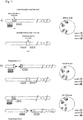
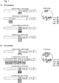
- Fig. 1 shows a schematic representation of a method according to the invention for the detection of two translocations using four probes and three labels, one probe being marked with two labels at the same time. It shows the signal pattern in normal cells and cells with translocation of the ALK gene in 2p23 or the ROS1 gene in 6q22. Both breakpoint regions (ALK and ROS1) are each flanked by label A and B of the quadruple ISH probe and each result in a fusion signal AB. One side of the ALK breakpoint region is also flanked with label C, so that there is a mixed label A / C.
- the ROS1 gene loci are marked by fusion signals A-B and ALK gene loci are marked by fusion signals A-B, which are accompanied by A / C mixed signals.
- the ALK gene affected by the translocation is marked by a separate signal of label B and a separate mixed signal A / C.
- the ROS1 gene affected by the translocation is marked by a separate signal of label A and a separate signal of label B.
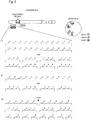
- Fig. 2 shows a schematic representation of a method according to the invention relating to the use of several labels to represent mixed labels and mixed signals. For the sake of clarity, only two labels are listed.
- Mixed signals that are specific for a locus-specific probe and thus for a chromosomal region or a genomic section can arise if I) fragments of a probe are marked with different labels and / or II) all fragments of a probe or, optionally, only individual fragments of a probe are marked with several labels and / or III) alternating fragments are marked with different labels, so that here too only one mixed signal is finally visible or detectable.
- All or only individual fragments according to I) to III) can superimpose or overlap (not shown) and mixed labels can also arise if individual fragments or fragment groups according to I) to III) have a distance of up to 2 Mbp, e.g. in the "gap" shown.
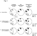
- Fig. 3 shows a diagram of signal patterns when using a corresponding quadruple FISH probe "ZytoLight SPEC ALK & ROS1 Break Apart Dual-Mix NG-FISH Probe” from ZytoVision.
- the probe consists of green-marked polynucleotides (absorption at 503 nm and emission at 528 nm), which in 2p23 are directed against sequences proximal to the ALK breakpoint region and in 6q22 against sequences located proximal to the ROS1 breakpoint region, orange-marked polynucleotides ( Absorption at 547 nm and emission at 572 nm), which in 2p23 are directed against sequences distal to the ALK breakpoint region and in 6q22 against sequences distal to the ROS1 breakpoint region, as well as blue-labeled polynucleotides (absorption at 426 nm and emission at 480 nm), which in the region 2p23 are directed against sequences distal and prox
- the hybridization signals for the non-rearranged ALK gene appear as green-orange fluorescence fusion signals, which consist of green / blue and put together orange / blue fluorescence mixed signals.
- the hybridization signals for the non-rearranged ROS1 gene appear as green-orange fluorescence fusion signals.
- a 2p23 locus affected by an ALK translocation is identified by a separate green / blue mixed signal and a separate orange / blue mixed signal. (see. Figure 3b ).
- a 6q22 locus affected by a ROS1 translocation is identified by a separate green and a separate orange signal (cf. Figure 3c ).
- green and separate orange signals initially only allow the statement that an ALK or ROS1 translocation is present.
- a diagnostically relevant distinction between ALK or ROS1 translocation can then be made by including the blue fluorescence signals. If the separate green signals overlap blue signals (green / blue mixed signals) or if the separate orange signals overlap blue signals (orange / blue mixed signals), this indicates an ALK translocation. If the separate green and orange signals do not overlap with blue signals, this indicates a ROS1 translocation.
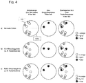
- Fig. 4 shows a diagram of signal patterns when using a corresponding quadruple FISH probe "ZytoLight SPEC ALK & ROS1 Break Apart Single-Mix NG-FISH Probe" from ZytoVision.
- the probe consists of green-marked polynucleotides (absorption at 503 nm and emission at 528 nm), which in 2p23 are directed against sequences proximal to the ALK breakpoint region and in 6q22 against sequences located proximal to the ROS1 breakpoint region, orange-marked polynucleotides ( Absorption at 547 nm and emission at 572 nm), which in 2p23 are directed against sequences distal to the ALK breakpoint region and in 6q22 against sequences distal to the ROS1 breakpoint region, as well as blue-labeled polynucleotides (absorption at 426 nm and emission at 480 nm), which in the region 2p23 are directed against sequences distal to the A
- the hybridization signals for the non-rearranged ALK gene appear as green-orange fluorescence fusion signals, which are composed of green and orange / blue mixed fluorescence signals.
- the hybridization signals for the non-rearranged ROS1 gene appear as green-orange fluorescence fusion signals.
- a 2p23 locus affected by an ALK translocation is identified by a separate green signal and a separate orange / blue mixed signal. (see. Figure 4b ).
- a 6q22 locus affected by a ROS1 translocation is identified by a separate green and a separate orange signal (cf. Figure 4c ).
- green and separate orange signals initially only allow the statement that an ALK or ROS1 translocation is present.
- a diagnostically relevant distinction between ALK or ROS1 translocation can then be made by including the blue fluorescence signals. If the separate orange signals overlap blue signals (orange / blue mixed signals), this indicates an ALK translocation. If the separate orange signals do not overlap with blue signals, this indicates a ROS1 translocation.
- Fig. 5 shows a diagram of signal patterns when using a corresponding six-fold FISH probe "ZytoLight SPEC ALK & ROS1 & RET Break Apart Dual-Mix NG-FISH Probe" from ZytoVision.
- the probe consists of green-marked polynucleotides (absorption at 503 nm and emission at 528 nm), the sequences located proximal to the ALK breakpoint region in 2p23, sequences located proximal to the ROS1 breakpoint region in 6q22 and proximal to the RET region in 10q11 Sequences located at the breakpoint region are directed, orange-marked polynucleotides (absorption at 547 nm and emission at 572 nm), the sequences located distal to the ALK breakpoint region in 2p23, sequences located distal to the ROS1 breakpoint region in 6q22 and sequences distal to the ROS1 breakpoint region in 10q11 Sequences located in the RET breakpoint region
- the hybridization signals for the non-rearranged ALK gene appear as green-orange fluorescence fusion signals, which are composed of green and orange / blue mixed fluorescence signals.
- the hybridization signals for the non-rearranged RET gene appear as green-orange fluorescence fusion signals, which are composed of green / blue mixed signals and orange signals.
- the hybridization signals for the non-rearranged ROS1 gene appear as green-orange fluorescence fusion signals.
- green-orange fusion signals appear when using a suitable green-orange dual bandpass filter set, four blue signals when using a suitable single bandpass filter set and when using one suitable triple bandpass filter sets two green-orange fusion signals, two green-orange / blue fusion and mixed signals and two green / blue-orange fusion and mixed signals (cf. Figure 5a ).
- a 2p23 locus affected by an ALK translocation is identified by a separate green signal and a separate orange / blue mixed signal. (see. Figure 5b ).
- a 6q22 locus affected by a ROS1 translocation is identified by a separate green and a separate orange signal (cf. Figure 5c ).
- a 10q11 locus affected by a RET translocation is identified by a separate orange signal and a separate green / blue mixed signal. (see. Fig. 5d ).
- green and separate orange signals initially only allow the statement that an ALK, ROS1 or RET translocation is present.
- a diagnostically relevant distinction between ALK, ROS1 or RET translocation can then be made by taking into account the blue fluorescence signals. If the separate orange signals overlap blue signals (orange / blue mixed signals), this indicates an ALK translocation. If the separate green signals overlap blue signals (green / blue mixed signals), this indicates a RET translocation. If neither the separate orange nor the separate green signals overlap with blue signals, this indicates a ROS1 translocation.
- Fig. 6 shows a diagram of signal patterns when using a corresponding six-fold FISH probe "ZytoLight SPEC ALK & ROS1 & RET Break Apart Dual-Mix II NG-FISH Probe" from ZytoVision GmbH.
- the probe consists of green-marked polynucleotides (absorption at 503 nm and emission at 528 nm), the sequences located proximal to the ALK breakpoint region in 2p23, sequences located proximal to the ROS1 breakpoint region in 6q22 and proximal to the RET region in 10q11 Sequences located at the breakpoint region are directed, red-marked polynucleotides (absorption at 580 nm and emission at 599 nm), the sequences located distal to the ALK breakpoint region in 2p23, sequences located distal to the ROS1 breakpoint region in 6q22 and sequences distal to the ROS1 breakpoint region in 10q11 Sequences located in the RET break
- the hybridization signals for the non-rearranged ALK gene appear as green-red fluorescence fusion signals which are composed of green and red / blue mixed fluorescence signals.
- the hybridization signals for the non-rearranged RET gene appear as green-red fluorescence fusion signals, which are composed of green / gold-yellow mixed signals and red signals.
- the hybridization signals for the non-rearranged ROS1 gene appear as green-red fluorescence fusion signals.
- a 2p23 locus affected by an ALK translocation is identified by a separate green signal and a separate red / blue mixed signal. (see. Figure 6b ).
- a 6q22 locus affected by a ROS1 translocation is identified by a separate green and a separate red signal (cf. Figure 6c ).
- a 10q11 locus affected by a RET translocation is identified by a separate red signal and a separate green / golden yellow mixed signal. (see. Fig. 6d ).
- green and separate red signals initially only allow the statement that there is basically an ALK, ROS1 or RET translocation.
- a diagnostically relevant distinction between ALK, ROS1 or RET translocation can then be made by taking into account the blue or golden-yellow fluorescence signals. If the separate red signals overlap blue signals (red / blue mixed signals), this indicates an ALK translocation. If the separate green signals overlap golden-yellow signals (green / golden-yellow mixed signals), this indicates a RET translocation. If neither the separate red nor the separate green signals overlap with blue or golden yellow signals, this indicates a ROS1 translocation.
- Fig. 7 shows a schematic representation of a method according to the invention for the detection of four numerical aberrations using four probes and four labels, three probes being marked simultaneously with two labels each, the labels used for the combination differing from one another in these three probes. It shows the signal pattern in normal cells and in cells with amplification of the MET gene in 7q31.
- the region 17q11.2-q12 of the ERBB2 gene is covered with label A, the region 7p12 of the EGFR gene with label A and also label D, so that a mixed label A / D is created, the region 8p11.23- p11.22 of the FGFR1 gene with label A and also label C, so that there is a mixed label A / C as well as the region 7q31 of the MET gene with label A and also label B, so that there is a mixed label A / B.
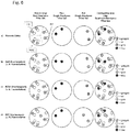
- Fig. 8 shows a schematic representation of a method according to the invention for the detection of seven translocations or amplifications using 14 probes and five labels each, one probe being marked with two labels at the same time and the ratio of the two labels per probe differing.
- the probes each flank a breakpoint region or address an amplification region as well as another region on the same chromosome (eg the centromere region) as on the upper left or upper right part of the Fig. 8 is shown.
- the two probes of a chromosome are marked with two different labels and the same label (e.g. probe 1: 25% green, 75% blue and probe 2: 25% yellow and 75% blue).
- the two probes each flanking a breakpoint region, thus each result in fusion and mixed signals from three labels in a cell that is not affected by a translocation.
- the two probes which each address an amplification region and a further region on the same chromosome, thus each produce separate mixed signals in a cell that is not affected by amplification (unless the distance between the two probes is so small, see above) that fusion and mixed signals arise).
- different mixed signals are generated so that a large number of probes with the same labels can be marked differentially (e.g. 4 probes for the breakpoint regions 1 to 4:25 in the example shown % to 75%; 50% to 50%; 75% to 25% and 100% to 0%).
- the breakpoint regions of a gene are marked by mixed signals AB and CB, which combine to form A / B / C fusion signals.
- the amplification regions of a gene are marked by the mixed signals AB and other regions on the same chromosome, for example centromere regions, are marked by the mixed signals CB.
- the gene affected by the translocation is identified by a separate signal of the label AB and one of these separate mixed signal CB marked.
- the gene affected by the amplification is marked by an amplified mixed signal from a label pair, eg AB.
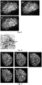
- Fig. 9 shows a FISH analysis to detect a translocation of the ROS1 region in 6q22 using the quadruple FISH probe "ZytoLight SPEC ALK & ROS1 Break Apart Single-Mix NG-FISH Probe" from ZytoVision.
- the probe consists of green-marked polynucleotides (absorption at 503 nm and emission at 528 nm), which in 2p23 are directed against sequences located proximal to the ALK breakpoint region and in 6q22 sequences located proximal to the ROS1 breakpoint region, orange-marked polynucleotides (absorption at 547 nm and emission at 572 nm), which in 2p23 are directed against sequences distal to the ALK breakpoint region and in 6q22 sequences located distal to the ROS1 breakpoint region, as well as blue-marked polynucleotides (absorption at 426 nm and emission at 480 nm) , which are directed in the region 6q22 proximal to the ROS1 breakpoint region located sequences.
- the hybridization signals for non-rearranged ROS1 and / or ALK genes appear as green-orange fluorescence fusion signals and for rearranged ROS1 and / or ALK genes as a separate green and separate orange signal (see Sect. Figure 9A showing the green and orange fluorescent signals).
- ROS1-specific green signals co-localize with blue fluorescence signals (s. Figure 9B , which shows the blue fluorescence signals), so that the unrearranged ROS1 gene is composed of orange and green / blue fluorescence mixed signals.
- the hybridization signals for the non-rearranged ALK gene appear as green-orange fluorescence fusion signals, without mixed signals with blue fluorescence signals.
- the 6q22 locus affected by a ROS1 translocation is indicated by a separate green and a separate orange signal (arrows in Figures 9A and C ).
- the separate green signal overlaps with a blue signal.
- This green / blue mixed signal indicates ROS1, not ALK, as the gene affected by the translocation (s. Figure 9C showing the blue, green, and orange fluorescent signals).
- the signal pattern can be made clearly visible with suitable filter sets.
- Fig. 10 shows a CISH analysis to detect a translocation of the ALK region in 2p23 using the quadruple CISH probe "ZytoDot SPEC ALK & ROS1 Break Apart Single-MIX NG-FISH Probe" from ZytoVision.
- the probe consists of digoxigenin-labeled polynucleotides that are directed against sequences located proximal to the ALK breakpoint region in 2p23 and sequences located proximal to the ROS1 breakpoint region in 6q22, DNP-labeled polynucleotides that are directed against sequences distal to the ALK breakpoint region in 2p23 and Sequences located distal to the ROS1 breakpoint region are directed in 6q22, as well as biotin-labeled polynucleotides which are directed in the region 6q22 distal to the ROS1 breakpoint region sequences.
- the markings were verified via primary (unlabeled) antibodies (Anti-DIG / Anti-DNP / Anti-BIO), which are detected by secondary polymerized enzyme-conjugated antibodies (HRP-Polymer / AP-Polymer / beta-GAL), as well as the enzymatic conversion of the Substrates (AP-RED / HRP-GREEN / beta-GAL-BLUE), which lead to the creation of strong, permanent, red (R), green (G) and blue (B) signals, which are light microscopically eg with a 40x dry lens can be represented leads.
- primary (unlabeled) antibodies Anti-DIG / Anti-DNP / Anti-BIO
- HRP-Polymer / AP-Polymer / beta-GAL secondary polymerized enzyme-conjugated antibodies
- AP-RED / HRP-GREEN / beta-GAL-BLUE the enzymatic conversion of the Substrates
- R red
- G green
- B
- Fig. 11 shows a FISH analysis to detect the amplification of the ERBB2 region using the fivefold FISH probe "ZytoLight SPEC ERBB2, EGFR, FGFR1, MET & SOX2 FiveCheck TM NG-FISH Probe” from ZytoVision.
- the probe consists of green-marked polynucleotides (absorption at 503 nm and emission at 528 nm) that target the region 17q11.2-q12 of the ERBB2 gene, the region 7p12 of the EGFR gene, the region 8p11.23-p11.
- the 7q31 region of the MET gene and the 3q26.3-q27 region of the SOX2 gene are directed as well as blue-marked polynucleotides (absorption at 426 nm and emission at 480 nm) that target the region of the EGFR gene and the SOX2 gene, golden yellow-marked polynucleotides (absorption at 532 nm and emission at 553 nm), which are against the region of the FGFR1 gene and red-marked polynucleotides (absorption at 580 nm and emission at 599 nm), which are directed against the region of the MET gene and the SOX2 gene.
- the single green signal is a non-amplified ERBB2 gene
- the green signal cluster identifies an ERBB2 gene amplification.
- Co-localizing green / blue mixed signals identify two copies of the EGFR gene
- co-localizing green / golden yellow mixed signals identify two copies of the FGFR1 gene
- co-localizing green / red mixed signals identify two copies of the MET gene
- co-localizing green / blue / red mixed signals identify two copies of the SOX2 gene (s. Figure 11E ).
- Example 1 FISH analysis for the detection of multiple numerical aberrations in different cell types using the five-fold FISH probe "SPEC ERBB2, EGFR, FGFR1, MET & SOX2 FiveCheck TM NG-FISH Probe "from ZytoVision GmbH
- the FISH was performed on 3 to 5 ⁇ m thick sections of formalin-fixed paraffin-embedded (FFPE) lung and breast cancer preparations with or without previously diagnosed ERBB2 gene amplification, which were mounted on coated glass slides and kept overnight at 58 ° C were baked.
- FFPE formalin-fixed paraffin-embedded
- the preparations were first heated on a hot plate for 10 minutes at 70 ° C. and then incubated twice in 100% xylene for 10 minutes each time at room temperature (RT).
- the preparations were then rehydrated by means of a descending series of ethanol (5 minutes each time at RT in 96%, 96%, 90%, 70% denatured ethanol) and incubation in ultrapure water (two times each two minutes at RT).
- a descending series of ethanol 5 minutes each time at RT in 96%, 96%, 90%, 70% denatured ethanol
- ultrapure water two times each two minutes at RT.
- the proteolytic pretreatment was carried out by dropping pepsin solution (Pepsin Solution, ZytoVision GmbH) onto the preparations and then incubating them in a humid chamber at 37 ° C. for 25 minutes. After subsequent incubation for 5 min in Wash Buffer SSC (ZytoVision GmbH), the preparations were dehydrated (one minute each at RT in ultrapure water, 70%, 90%, 96% ethanol). After the specimens had air-dried, 10 ⁇ l of the FISH probe ZytoLight SPEC ERBB2, EGFR, FGFR1, MET & SOX2 FiveCheck TM NG-FISH probe (ZytoVision GmbH) were applied directly to the sections using a pipette.
- pepsin solution Pepsin Solution, ZytoVision GmbH
- the probe was a mixture based on five locus-specific hybridization probes, the mixture of green-labeled polynucleotides (absorption at 503 nm and emission at 528 nm) that act against the region 17q11.2-q12 of the ERBB2 gene, the 7p12 region of the EGFR gene, the 8p11.23-p11.22 region of the FGFR1 gene, the 7q31 region of the MET gene and the 3q26.3-q27 region of the SOX2 gene were directed and were composed of blue-labeled polynucleotides ( Absorption at 426 nm and emission at 480 nm), which were directed against the region of the EGFR gene and the SOX2 gene, golden yellow-labeled polynucleotides (absorption at 532 nm and emission at 553 nm), which were directed against the region of the FGFR1- Gene, and red-labeled polynucleotides (absorption at 580 nm and
- the evaluation was then carried out on a fluorescence microscope (Axio Scope.A1 with HXP 120 V lighting unit, Carl Zeiss Microscopy GmbH) using suitable filter sets (Sp. Green HC mFISH filter set; Sp. Red HC mFISH filter set; Sp. Aqua HC mFISH filter set; ZyGold HC mFISH filter set (all AHF Analysentechnik AG)).
- the signal pattern could be interpreted as follows: Two green signals without spatially identical localization of signals of a different color identified the two ERBB2 gene copies of a diploid cell. Two green signals with spatially identical localization of two aqua signals identified the two EGFR gene copies, two green signals with spatially identical localization of two gold-yellow signals identified the two FGFR1 gene copies, two green signals with spatially identical localization of two red signals identified the two MET gene copies and two green signals with spatially identical localization of two red and two aqua signals identified the two SOX2 gene copies.
- the cell nuclei of the preparations with ERBB2 amplification showed a signal pattern comparable to the example described above, with the exception that, in addition to nine green signals, a green signal cluster or signal pattern, consisting of around fifteen signals that were inseparably close together, was observed (cf. Figure 11A ). This green signal pattern did not co-localize with signals of a different color and thus identified an ERBB2 gene amplification.
- Example 2 FISH analysis for the detection of translocations of the ALK or ROS1 regions in different cell types using the quadruple FISH probe "ZytoLight SPEC ALK & ROS1 Break Apart Single-Mix NG-FISH Probe” from ZytoVision GmbH_
- the FISH was performed on 3 to 5 ⁇ m thick sections of formalin-fixed paraffin-embedded (FFPE) cells of the cell lines HeLa (ATCC® CCL-2 TM), HCC78 (provided by Prof. Schildhaus, Göttingen) and H3122 ( provided by Prof. Schildhaus, Göttingen), which were mounted on coated glass slides and baked at 58 ° C overnight.
- FFPE formalin-fixed paraffin-embedded
- the preparations were first heated on a hot plate for 10 min at 70 ° C. and then incubated twice in 100% xylene for 10 min each time at room temperature (RT).
- the preparations were then rehydrated by means of a descending ethanol series (five minutes each at room temperature in 96%, 96%, 90%, 70% denatured ethanol) and incubation in ultrapure water (two times two minutes each at room temperature).
- RT room temperature
- ultrapure water two times two minutes each at room temperature.
- heat pretreatment for 15 minutes at 98 ° C. in Heat Pretration Solution Citric (ZytoVision GmbH), followed by two further incubation steps for two minutes in ultrapure water at room temperature.
- the proteolytic pretreatment was carried out by dripping a pepsin solution (Pepsin-Solution, ZytoVision GmbH) onto the preparations and subsequent incubation in a humid chamber at 37 ° C. for 15 minutes. After subsequent incubation for five minutes in wash buffer (Wash Buffer SSC, ZytoVision GmbH), the preparations were dehydrated (one minute each at RT in ultrapure water, 70%, 90%, 96% ethanol). After the specimens had dried in the air, 10 ⁇ l of the FISH probe ZytoLight SPEC ALK & ROS1 Break Apart Single-Mix NG-FISH Probe (ZytoVision GmbH) were applied directly to the sections using a pipette.
- a pepsin solution Pepsin-Solution, ZytoVision GmbH
- the probe was a mixture based on four locus-specific hybridization probes, the mixture of green-marked polynucleotides (absorption at 503 nm and emission at 528 nm), the sequences located proximal to the ALK breakpoint region in 2p23 and 6q22 were directed against sequences located proximal to the ROS1 breakpoint region, orange-marked polynucleotides (absorption at 547 nm and emission at 572 nm), the sequences located distal to the ALK breakpoint region in 2p23 and sequences located distal to the ROS1 breakpoint region in 6q22 were, as well as blue-marked polynucleotides (absorption at 426 nm and emission at 480 nm), which were directed in region 6q22 against sequences located proximal to the ROS1 breakpoint region.
- Example 3 FISH analysis to detect a translocation of the ROS1 region in 6q22 using the quadruple FISH probe "ZytoLight SPEC ALK & ROS1 Break Apart Single-Mix NG-FISH Probe" from ZytoVision GmbH
- a FISH analysis was carried out to detect a translocation of the ROS1 region in 6q22 using the quadruple FISH probe "ZytoLight SPEC ALK & ROS1 Break Apart Single-Mix NG-FISH Probe" from ZytoVision GmbH.
- the probe was a mixture based on four locus-specific hybridization probes, the mixture of green-marked polynucleotides (absorption at 503 nm and emission at 528 nm), the sequences located proximal to the ALK breakpoint region in 2p23 and 6q22 were directed against sequences located proximal to the ROS1 breakpoint region, orange-marked polynucleotides (absorption at 547 nm and emission at 572 nm), the sequences in 2p23 directed against sequences distal to the ALK breakpoint region and in 6q22 against sequences located distal to the ROS1 breakpoint region were, as well as blue-marked polynucleotides (absorption at 426 nm and emission at 480 nm), which were directed in region 6q22 against sequences located proximal to the ROS1 breakpoint region.
- the hybridization signals for non-rearranged ROS1 and / or ALK genes appeared as green-orange fluorescence fusion signals and for a rearranged ROS1 and / or ALK genes as a separate green and separate orange signal.
- ROS1-specific green signals co-localized with blue fluorescence signals, so that the non-rearranged ROS1 gene was composed of orange and green / blue mixed fluorescence signals.
- the hybridization signals for the non-rearranged ALK gene appeared as green-orange fluorescence fusion signals without mixed signals with blue fluorescence signals ( Figure 9A ).
- the 6q22 locus affected by a ROS1 translocation was indicated by a separate green and a separate orange signal ( Figure 9A Arrows).
- the separate green signal overlapped with a blue signal (blue signals Figure 9B ). This green / blue mixed signal indicated ROS1, not ALK, as the gene affected by the translocation.
- the signal pattern could be made clearly visible with suitable filter sets.
- Example 4 CISH analysis to detect a translocation of the ALK region in 2p23 using the quadruple CISH probe "ZytoDot SPEC ALK & ROS1 Break Apart Single-MIX NG-FISH Probe" from ZytoVision GmbH
- the probe was a mixture based on four locus-specific hybridization probes, the mixture of digoxigenin-labeled polynucleotides directed against sequences located proximal to the ALK breakpoint region in 2p23 and against sequences located proximal to the ROS1 breakpoint region in 6q22, DNP-labeled polynucleotides that in 2p23 against sequences distal to the ALK breakpoint region and in 6q22 against sequences distal to the ROS1 breakpoint region, and biotin-labeled polynucleotides in region 6q22 directed against sequences distal to the ROS1 breakpoint region.
- the markings were detected using primary (non-marked) antibodies (anti-DIG / anti-DNP / anti-BIO), which were detected by secondary polymerized enzyme-conjugated antibodies (HRP polymer / AP polymer / beta-GAL), as well as the enzymatic conversion of the substrates (AP-RED / HRP-GREEN / beta-GAL-BLUE), which led to the development of strong, permanent, red, green and blue signals, which could be visualized with a light microscope, for example with a 40x dry lens .
- Diploid or disome cell nuclei without rearrangements or translocations of the ALK or ROS1 genes showed two signals, each consisting of a red and a green signal, which were inseparably close together or partly overlapped or mixed and specific for the two copies of the ALK -Gens were ( Fig. 10 ).
- Example 5 FISH analysis to detect the amplification of the ERBB2 region using the fivefold FISH probe “ZytoLight SPEC ERBB2, EGFR, FGFR1, MET & SOX2 FiveCheck TM NG-FISH Probe” from ZytoVision
- the probe was a mixture based on five locus-specific hybridization probes, the mixture of green-labeled polynucleotides (absorption at 503 nm and emission at 528 nm) that act against the region 17q11.2-q12 of the ERBB2 gene, the 7p12 region of the EGFR gene, the 8p11.23-p11.22 region of the FGFR1 gene, the 7q31 region of the MET gene, and the region 3q26.3-q27 of the SOX2 gene as well as from blue-marked polynucleotides (absorption at 426 nm and emission at 480 nm) that were directed against the region of the EGFR gene and the SOX2 gene, golden-yellow-marked polynucleotides (absorption at 532 nm and emission at 553 nm), which were directed against the region of the FGFR1 gene and red-marked polynucleotides (absorption at 580 nm and emission at 599
- the single green signal was a non-amplified ERBB2 gene, the green signal cluster identified an ERBB2 gene amplification.
- Co-localizing green / blue mixed signals identified two copies of the EGFR gene, co-localizing green / gold-yellow mixed signals identified two copies of the FGFR1 gene, co-localizing green / red mixed signals identified two copies of the MET gene and co-localizing green / blue / red mixed signals identified two copies of the SOX2 gene.
Landscapes
- Chemical & Material Sciences (AREA)
- Life Sciences & Earth Sciences (AREA)
- Organic Chemistry (AREA)
- Health & Medical Sciences (AREA)
- Proteomics, Peptides & Aminoacids (AREA)
- Wood Science & Technology (AREA)
- Zoology (AREA)
- Engineering & Computer Science (AREA)
- Immunology (AREA)
- Analytical Chemistry (AREA)
- Genetics & Genomics (AREA)
- Molecular Biology (AREA)
- General Engineering & Computer Science (AREA)
- Biotechnology (AREA)
- Biochemistry (AREA)
- Bioinformatics & Cheminformatics (AREA)
- General Health & Medical Sciences (AREA)
- Microbiology (AREA)
- Physics & Mathematics (AREA)
- Biophysics (AREA)
- Pathology (AREA)
- Hospice & Palliative Care (AREA)
- Oncology (AREA)
- Chemical Kinetics & Catalysis (AREA)
- Measuring Or Testing Involving Enzymes Or Micro-Organisms (AREA)
Claims (15)
- Procédé de détection d'au moins deux aberrations chromosomiques différentes l'une de l'autre au moyen d'une hybridation in situ par détection de régions chromosomiques et/ou d'ADN dans un échantillon biologique, caractérisé en ce que l'hybridation in situ est effectuée sous forme d'hybridation d'interphase-in situ, en ce que l'hybridation in situ est effectuée à l'aide d'au moins trois sondes d'hybridation spécifiques à un locus, différentes les unes des autres et marquées chacune au moyen d'un premier marqueur de détection, dans lequel, pour la production d'au moins un signal mixte par au moins deux marqueurs de détection différents les uns des autres se trouvant sur une sonde d'hybridation spécifique à un locus, au moins l'une des sondes d'hybridation spécifique à un locus est marquée à l'aide d'au moins un autre marqueur de détection différent du premier marqueur de détection, par rapport à la sonde d'hybridation spécifique à un locus respective, de sorte qu'un modèle de signal soit produit, et que des aberrations chromosomiques présentes soient identifiées au moyen du modèle de signal et/ou associées à une région chromosomique et/ou à une région d'ADN.
- Procédé selon la revendication 1, dans lequel l'hybridation in situ est effectuée à l'aide d'au moins quatre sondes d'hybridation spécifiques à un locus différentes les unes des autres et marquées chacune à l'aide d'un premier marqueur de détection.
- Procédé selon l'une des revendications 1 à 2, dans lequel les aberrations chromosomiques sont des aberrations chromosomiques indépendantes les unes des autres, et/ou dans lequel les aberrations chromosomiques ne sont pas réciproques.
- Procédé selon l'une des revendications précédentes, dans lequel le marquage d'autres sondes d'hybridation spécifiques à un locus est effectué à l'aide d'au moins un autre marqueur de détection, dans lequel les sondes d'hybridation spécifiques à un locus marquées à l'aide d'autres marqueurs de détection produisent chacune des signaux mixtes dans le modèle de signal différents les uns des autres, et/ou dans lequel d'autres sondes d'hybridation spécifiques à un locus sont marquées à l'aide de l'au moins un autre marqueur de détection, dans lequel un signal mixte spécifique à une région chromosomique et/ou d'ADN est produit par chaque sonde d'hybridation spécifique à un locus marquée à l'aide d'au moins un autre marqueur de détection dans le modèle de signal.
- Procédé selon l'une des revendications précédentes, dans lequel au moins deux autres sondes d'hybridation spécifiques à un locus sont marquées à l'aide d'au moins un autre marqueur de détection différent du premier.
- Procédé selon l'une des revendications précédentes, dans lequel deux sondes d'hybridation spécifiques à un locus flanquent chacune une section chromosomique, dans lequel les sondes d'hybridation spécifiques à un locus flanquant chacune une section chromosomique sont marquées à l'aide de marqueurs de détection différents les uns des autres, de sorte qu'un signal de fusion soit produit dans le modèle de signal par les sondes d'hybridation spécifiques à un locus flanquant chacune une section chromosomique.
- Procédé selon la revendication 6, dans lequel un signal de fusion est produit par les sondes d'hybridation spécifiques à un locus, flanquant chacune une section chromosomique, dans le modèle de signal au cas où aucune aberration chromosomique ne serait présente.
- Procédé selon la revendication 6, dans lequel au moins six sondes d'hybridation différentes spécifiques à un locus sont utilisées, dans lequel, dans chaque cas, deux sondes d'hybridation spécifiques à un locus flanquent chacune une section chromosomique.
- Procédé selon l'une des revendications 6 à 8, dans lequel(a) une première sonde d'hybridation spécifique à un locus marquée à l'aide d'un marqueur de détection A et une seconde sonde d'hybridation spécifique à un locus marquée à l'aide d'un marqueur de détection B flanquent une section chromosomique et produisent un signal de fusion A-B dans le modèle de signal produit au moyen de l'hybridation in situ,(b) 2 à 12 autres sondes d'hybridation spécifiques à un locus flanquent jusqu'à six autres sections chromosomiques, dans lequel, également, chacune d'une des deux sondes d'hybridation spécifiques à un locus flanquant une section chromosomique est marquée à l'aide d'un marqueur de détection A et chacune d'une des deux sondes d'hybridation spécifiques à un locus flanquant une section chromosomique est marquée à l'aide d'un marqueur de détection B, de sorte que les sondes d'hybridation spécifiques à un locus flanquant chacune une section chromosomique produisent un signal de fusion A-B dans le modèle de signal produit au moyen de l'hybridation in situ et(c) au moins l'une des sondes d'hybridation spécifiques à un locus est marquée à l'aide d'au moins un autre marqueur de détection X, de sorte que les sondes d'hybridation spécifiques à un locus marquées à l'aide d'au moins un autre marqueur de détection produisent des signaux de fusion et mixtes A-B/X dans le modèle de signal produit au moyen de l'hybridation in situ, dans lequel, dans le modèle de signal produit au moyen de l'hybridation in situ, les signaux de fusion et mixtes A-B/X se transforment en signaux mixtes A/X et/ou B/X en cas d'aberrations chromosomiques ou dans lequel, dans le modèle de signal produit au moyen de l'hybridation in situ, les signaux de fusion A-B se transforment en signaux individuels A et/ou B en cas d'aberrations chromosomiques, de sorte que les aberrations chromosomiques sont associées à une région chromosomique et/ou d'ADN et/ou à un segment chromosomique flanqué de deux sondes d'hybridation spécifiques à un locus sur la base du modèle de signal produit au moyen de l'hybridation in situ.
- Procédé selon l'une des revendications précédentes, dans lequel les sondes d'hybridation spécifiques à un locus sont chacune formées par une pluralité de fragments d'acides nucléiques couvrant chacun la région chromosomique et/ou d'ADN à détecter.
- Composition destinée à la détection d'au moins deux aberrations chromosomiques différentes l'une de l'autre au moyen d'une hybridation in situ, la composition comprenant au moins trois sondes d'hybridation spécifiques à un locus différentes les unes des autres, chacune d'elles étant marquée à l'aide d'un premier marqueur de détection, et dans laquelle au moins l'une des sondes d'hybridation spécifiques à un locus est marquée à l'aide d'au moins un autre marqueur de détection différent du premier, par rapport à la sonde d'hybridation spécifique à un locus respective, et dans laquelle un signal mixte peut être produit par les au moins deux marqueurs de détection différents les uns des autres se trouvant sur une sonde d'hybridation spécifique à un locus.
- Composition destinée à être utilisée pour le diagnostic et/ou le pronostic de maladies associées à des aberrations chromosomiques, la composition comprenant au moins trois sondes d'hybridation spécifiques à un locus différentes les unes des autres, chacune d'elles étant marquée à l'aide d'un premier marqueur de détection, et dans laquelle au moins l'une des sondes d'hybridation spécifiques à un locus est marquée à l'aide d'au moins un autre marqueur de détection différent du premier, par rapport à la sonde d'hybridation spécifique à un locus respective, et dans laquelle un signal mixte peut être produit par les au moins deux marqueurs de détection différents les uns des autres se trouvant sur une sonde d'hybridation spécifique à un locus.
- Utilisation d'une composition selon l'une des revendications 11 à 12 pour la détection d'au moins deux aberrations chromosomiques différentes l'une de l'autre au moyen d'une hybridation in situ, en particulier au moyen du procédé selon l'une des revendications 1 à 10.
- Trousse (trousse de pièces ou ensemble) destinée à la détection d'au moins deux aberrations chromosomiques différentes l'une de l'autre au moyen d'une hybridation in situ, comprenant au moins trois sondes d'hybridation spécifiques à un locus différentes les unes des autres, chacune d'elles étant marquée à l'aide d'un premier marqueur de détection, dans laquelle au moins l'une des sondes d'hybridation spécifiques à un locus est marquée à l'aide d'au moins un autre marqueur de détection différent du premier, par rapport à la sonde d'hybridation respective, et dans laquelle un signal mixte peut être produit par les au moins deux marqueurs de détection différents les uns des autres se trouvant sur une sonde d'hybridation spécifique à un locus.
- Trousse selon la revendication 14, dans laquelle les au moins trois sondes d'hybridation spécifiques à un locus différentes les unes des autres sont présentes dans une composition commune.
Priority Applications (1)
| Application Number | Priority Date | Filing Date | Title |
|---|---|---|---|
| PL16731904T PL3314010T3 (pl) | 2015-06-23 | 2016-06-23 | Sposób wykrywania aberracji chromosomowych |
Applications Claiming Priority (4)
| Application Number | Priority Date | Filing Date | Title |
|---|---|---|---|
| EP15001845 | 2015-06-23 | ||
| EP15002075 | 2015-07-13 | ||
| EP15002200.2A EP3109324B1 (fr) | 2015-06-23 | 2015-07-24 | Procede de detection d'aberrations chromosomiques |
| PCT/EP2016/064631 WO2016207325A1 (fr) | 2015-06-23 | 2016-06-23 | Procédé de détection d'aberrations chromosomiques |
Publications (2)
| Publication Number | Publication Date |
|---|---|
| EP3314010A1 EP3314010A1 (fr) | 2018-05-02 |
| EP3314010B1 true EP3314010B1 (fr) | 2020-09-09 |
Family
ID=53757951
Family Applications (2)
| Application Number | Title | Priority Date | Filing Date |
|---|---|---|---|
| EP15002200.2A Active EP3109324B1 (fr) | 2015-06-23 | 2015-07-24 | Procede de detection d'aberrations chromosomiques |
| EP16731904.5A Active EP3314010B1 (fr) | 2015-06-23 | 2016-06-23 | Procede de detection d'aberrations chromosomiques |
Family Applications Before (1)
| Application Number | Title | Priority Date | Filing Date |
|---|---|---|---|
| EP15002200.2A Active EP3109324B1 (fr) | 2015-06-23 | 2015-07-24 | Procede de detection d'aberrations chromosomiques |
Country Status (10)
| Country | Link |
|---|---|
| US (2) | US11174508B2 (fr) |
| EP (2) | EP3109324B1 (fr) |
| JP (2) | JP6630823B2 (fr) |
| KR (2) | KR102190401B1 (fr) |
| CN (3) | CN113667722B (fr) |
| DK (2) | DK3109324T5 (fr) |
| ES (2) | ES2702603T3 (fr) |
| PL (1) | PL3314010T3 (fr) |
| PT (1) | PT3314010T (fr) |
| WO (1) | WO2016207325A1 (fr) |
Families Citing this family (13)
| Publication number | Priority date | Publication date | Assignee | Title |
|---|---|---|---|---|
| SG11201807117WA (en) | 2016-02-23 | 2018-09-27 | Dovetail Genomics Llc | Generation of phased read-sets for genome assembly and haplotype phasing |
| US20180044722A1 (en) * | 2016-08-12 | 2018-02-15 | Agilent Technologies, Inc. | Tri-color probes for detecting multiple gene rearrangements in a fish assay |
| US12020493B2 (en) | 2018-07-27 | 2024-06-25 | Ventana Medical Systems, Inc. | Systems for automated in situ hybridization analysis |
| EP3874057A4 (fr) * | 2018-11-01 | 2022-08-17 | Advanced Cell Diagnostics, Inc. | Procédés pour le multiplexage numérique d'acides nucléiques in situ |
| CN109920479B (zh) * | 2019-03-13 | 2023-08-15 | 复旦大学附属妇产科医院 | 一种鉴别胚胎染色体倒位携带状态的方法 |
| KR102452413B1 (ko) * | 2019-08-19 | 2022-10-11 | 주식회사 지씨지놈 | 핵산 단편간 거리 정보를 이용한 염색체 이상 검출 방법 |
| CA3147613A1 (fr) * | 2019-08-19 | 2021-02-25 | Chang-Seok Ki | Methode de detection d'une anomalie chromosomique a l'aide d'informations concernant la distance entre des fragments d'acide nucleique |
| AU2021247767A1 (en) | 2020-03-30 | 2022-10-20 | Oriental Yeast Co., Ltd. | Chromosome-stabilizing agent for stem cells |
| EP4200863A4 (fr) * | 2020-08-24 | 2024-07-10 | Applied Materials, Inc. | Détection de cellules à l'aide d'une segmentation fondée sur une coloration nucléaire et des images d'hybridation fluorescente in situ multiplexée |
| CN111951266A (zh) * | 2020-09-01 | 2020-11-17 | 厦门汉舒捷医疗科技有限公司 | 一种染色体畸变的人工智能识别分析方法 |
| WO2023091592A1 (fr) * | 2021-11-19 | 2023-05-25 | Dovetail Genomics, Llc | Dendrimères pour procédés et compositions d'analyse génomique |
| KR102397822B1 (ko) | 2021-11-19 | 2022-05-13 | 주식회사 클리노믹스 | 염색체 구조의 상태 정보를 이용한 세포 분석 장치 및 방법 |
| CN114512183B (zh) * | 2022-01-27 | 2022-09-20 | 北京吉因加医学检验实验室有限公司 | 一种预测met基因扩增或多倍体的方法及装置 |
Family Cites Families (20)
| Publication number | Priority date | Publication date | Assignee | Title |
|---|---|---|---|---|
| US6344315B1 (en) * | 1986-01-16 | 2002-02-05 | The Regents Of The University Of California | Chromosome-specific staining to detect genetic rearrangements associated with chromosome 3 and/or chromosome 17 |
| ES2018959A6 (es) * | 1988-11-15 | 1991-05-16 | Univ Yale | Metodo de delineacion de cromosomas humanos individuales en celulas metafasicas e interfasicas mediante hibridacion de suspension in situ. |
| CA2091764C (fr) | 1990-09-20 | 2003-04-08 | Michael Louis Bittner | Compositions de sondes pour l'identification de chromosomes et methodes |
| US5784162A (en) * | 1993-08-18 | 1998-07-21 | Applied Spectral Imaging Ltd. | Spectral bio-imaging methods for biological research, medical diagnostics and therapy |
| US6025126A (en) * | 1991-10-28 | 2000-02-15 | Arch Development Corporation | Methods and compositions for the detection of chromosomal aberrations |
| EP1134293A3 (fr) * | 1992-03-04 | 2004-01-07 | The Regents of The University of California | Hybridation génomique comparative |
| AU4292893A (en) * | 1992-04-21 | 1993-11-18 | Regents Of The University Of California, The | Multicolor in situ hybridization methods for genetic testing |
| US5731153A (en) * | 1996-08-26 | 1998-03-24 | The Regents Of The University Of California | Identification of random nucleic acid sequence aberrations using dual capture probes which hybridize to different chromosome regions |
| US6174681B1 (en) | 1999-03-05 | 2001-01-16 | Mayo Foundation For Medical Education And Research | Method and probe set for detecting cancer |
| US7585964B2 (en) | 2001-05-14 | 2009-09-08 | Cancer Genetics, Inc. | Methods of analyzing chromosomal translocations using fluorescence in situ hybridization (FISH) |
| US20030129626A1 (en) * | 2001-09-24 | 2003-07-10 | Nielsen Kirsten Vang | Methods, kits and compositions pertaining to the suppression of detectable probe binding to randomly distributed repeat sequences in genomic nucleic acid |
| US7306916B2 (en) | 2004-05-04 | 2007-12-11 | Dako Denmark A/S | Methods for detecting chromosome aberrations |
| WO2006061994A1 (fr) * | 2004-12-08 | 2006-06-15 | Takeshi Yamamoto | Procede d'etude d'une sequence d'un gene |
| EP2789697B1 (fr) * | 2005-02-18 | 2017-07-19 | Abbott Molecular Inc. | Procédés et sondes permettant de détecter un cancer de l'oesophage |
| JP2009507227A (ja) | 2005-09-02 | 2009-02-19 | ザ リージェンツ オブ ザ ユニバーシティ オブ カリフォルニア | メラノーマを診断するための方法及びプローブ組み合わせ |
| EP1978106A1 (fr) * | 2007-04-07 | 2008-10-08 | Universitätsklinikum Hamburg-Eppendorf | Amplificateur ayant une protection de sortie, en particulier un amplificateur opérationnel pour une application audio |
| WO2012033828A2 (fr) * | 2010-09-08 | 2012-03-15 | Cancer Genetics, Inc. | Méthodes de détection de cancers associés au papillomavirus humain |
| US9562259B2 (en) * | 2011-03-14 | 2017-02-07 | Ventana Medical Systems, Inc. | Method of analyzing chromosomal inversions |
| DE102011100242A1 (de) * | 2011-05-02 | 2012-11-08 | Zytovision Gmbh | Verfahren zur Detektion von Chromosomenaberration |
| JP6200133B2 (ja) * | 2012-07-12 | 2017-09-20 | 花王株式会社 | ケトミウム・グロボーサム関連種群の検出方法 |
-
2015
- 2015-07-24 ES ES15002200T patent/ES2702603T3/es active Active
- 2015-07-24 EP EP15002200.2A patent/EP3109324B1/fr active Active
- 2015-07-24 DK DK15002200.2T patent/DK3109324T5/en active
-
2016
- 2016-06-23 KR KR1020197028974A patent/KR102190401B1/ko active Active
- 2016-06-23 PL PL16731904T patent/PL3314010T3/pl unknown
- 2016-06-23 DK DK16731904.5T patent/DK3314010T3/da active
- 2016-06-23 PT PT167319045T patent/PT3314010T/pt unknown
- 2016-06-23 JP JP2018517489A patent/JP6630823B2/ja active Active
- 2016-06-23 WO PCT/EP2016/064631 patent/WO2016207325A1/fr not_active Ceased
- 2016-06-23 CN CN202110972867.3A patent/CN113667722B/zh active Active
- 2016-06-23 EP EP16731904.5A patent/EP3314010B1/fr active Active
- 2016-06-23 US US15/737,536 patent/US11174508B2/en active Active
- 2016-06-23 CN CN201680034524.XA patent/CN107810276B/zh active Active
- 2016-06-23 CN CN201810147310.4A patent/CN108467884B/zh active Active
- 2016-06-23 KR KR1020177035975A patent/KR20180009763A/ko not_active Ceased
- 2016-06-23 ES ES16731904T patent/ES2836134T3/es active Active
-
2019
- 2019-07-24 JP JP2019135874A patent/JP2019213537A/ja active Pending
-
2021
- 2021-08-11 US US17/399,714 patent/US12312635B2/en active Active
Non-Patent Citations (1)
| Title |
|---|
| None * |
Also Published As
| Publication number | Publication date |
|---|---|
| PT3314010T (pt) | 2020-12-11 |
| US20180282795A1 (en) | 2018-10-04 |
| WO2016207325A1 (fr) | 2016-12-29 |
| US11174508B2 (en) | 2021-11-16 |
| CN113667722B (zh) | 2024-12-20 |
| CN107810276A (zh) | 2018-03-16 |
| DK3109324T5 (en) | 2019-02-11 |
| CN113667722A (zh) | 2021-11-19 |
| EP3109324A1 (fr) | 2016-12-28 |
| JP6630823B2 (ja) | 2020-01-15 |
| US12312635B2 (en) | 2025-05-27 |
| EP3109324B1 (fr) | 2018-09-19 |
| JP2018517436A (ja) | 2018-07-05 |
| CN107810276B (zh) | 2022-09-09 |
| CN108467884B (zh) | 2023-01-17 |
| CN108467884A (zh) | 2018-08-31 |
| ES2836134T3 (es) | 2021-06-24 |
| DK3314010T3 (da) | 2020-12-14 |
| JP2019213537A (ja) | 2019-12-19 |
| KR102190401B1 (ko) | 2020-12-11 |
| KR20190116548A (ko) | 2019-10-14 |
| KR20180009763A (ko) | 2018-01-29 |
| DK3109324T3 (en) | 2019-01-07 |
| US20210371913A1 (en) | 2021-12-02 |
| ES2702603T3 (es) | 2019-03-04 |
| EP3314010A1 (fr) | 2018-05-02 |
| PL3314010T3 (pl) | 2021-05-04 |
Similar Documents
| Publication | Publication Date | Title |
|---|---|---|
| EP3314010B1 (fr) | Procede de detection d'aberrations chromosomiques | |
| EP0530794B1 (fr) | Méthode pour la détection d'acides nucléiques ayant une séquence similaire | |
| DE69821540T2 (de) | Mit einem Adapter versehene kompetitive PCR | |
| EP1370691B1 (fr) | Procede de detection de structures de methylation de la cytosine a haute sensibilite | |
| DE69836885T2 (de) | Nachweis von Nukleinsäuren durch Fluoreszenz quenching | |
| DE3505287A1 (de) | Verbesserte nucleinsaeure-reagenzien, verfahren zu ihrer herstellung und ihre verwendung | |
| EP1463841A2 (fr) | Procede de detection de modeles de methylation de la cytosine par ligation exponentielle d'oligonucleotides sondes hybrides (mla) | |
| EP1019544B1 (fr) | Procede pour comparer le nombre de repliques de sequences d'acide nucleique | |
| EP0894870B1 (fr) | Extraction d'ADN de cellules de champignons d'un materiel clinique | |
| EP3350340B1 (fr) | Test de vérification de produits d'amplification primaires d'acides nucléiques dans un processus réactionnel continu et analyse directe au moyen d'un procédé immunochromatographique | |
| EP2705158A1 (fr) | Procédé pour la détection d'aberrations chromosomiques | |
| DE60210607T2 (de) | Nährboden zum nachweis und/oder zur unterscheidung von enterokokken | |
| DE19616750A1 (de) | Verfahren zum Nachweis von Mikroorganismen in Gemischen | |
| EP3321375A1 (fr) | Procédé de détection de délétions, duplications ou d'inversions de la même région critique | |
| DE10051174A1 (de) | Verfahren zum Nachweis von Staphylokokken und dazu geeignete Mittel | |
| DE10158283A1 (de) | Verfahren zum Nachweis von Cytosin-Methylierungsmustern mit hoher Sensitivität | |
| DE102005023055A1 (de) | Verfahren zur Herstellung einer Mischung von Fragmenten einer Polynukleinsäure |
Legal Events
| Date | Code | Title | Description |
|---|---|---|---|
| STAA | Information on the status of an ep patent application or granted ep patent |
Free format text: STATUS: THE INTERNATIONAL PUBLICATION HAS BEEN MADE |
|
| PUAI | Public reference made under article 153(3) epc to a published international application that has entered the european phase |
Free format text: ORIGINAL CODE: 0009012 |
|
| STAA | Information on the status of an ep patent application or granted ep patent |
Free format text: STATUS: REQUEST FOR EXAMINATION WAS MADE |
|
| 17P | Request for examination filed |
Effective date: 20171206 |
|
| AK | Designated contracting states |
Kind code of ref document: A1 Designated state(s): AL AT BE BG CH CY CZ DE DK EE ES FI FR GB GR HR HU IE IS IT LI LT LU LV MC MK MT NL NO PL PT RO RS SE SI SK SM TR |
|
| AX | Request for extension of the european patent |
Extension state: BA ME |
|
| DAV | Request for validation of the european patent (deleted) | ||
| DAX | Request for extension of the european patent (deleted) | ||
| STAA | Information on the status of an ep patent application or granted ep patent |
Free format text: STATUS: EXAMINATION IS IN PROGRESS |
|
| 17Q | First examination report despatched |
Effective date: 20190121 |
|
| REG | Reference to a national code |
Ref country code: DE Ref legal event code: R079 Ref document number: 502016011114 Country of ref document: DE Free format text: PREVIOUS MAIN CLASS: C12Q0001680000 Ipc: C12Q0001684100 |
|
| RIC1 | Information provided on ipc code assigned before grant |
Ipc: C12Q 1/6841 20180101AFI20200117BHEP |
|
| GRAP | Despatch of communication of intention to grant a patent |
Free format text: ORIGINAL CODE: EPIDOSNIGR1 |
|
| STAA | Information on the status of an ep patent application or granted ep patent |
Free format text: STATUS: GRANT OF PATENT IS INTENDED |
|
| INTG | Intention to grant announced |
Effective date: 20200316 |
|
| GRAS | Grant fee paid |
Free format text: ORIGINAL CODE: EPIDOSNIGR3 |
|
| GRAA | (expected) grant |
Free format text: ORIGINAL CODE: 0009210 |
|
| STAA | Information on the status of an ep patent application or granted ep patent |
Free format text: STATUS: THE PATENT HAS BEEN GRANTED |
|
| AK | Designated contracting states |
Kind code of ref document: B1 Designated state(s): AL AT BE BG CH CY CZ DE DK EE ES FI FR GB GR HR HU IE IS IT LI LT LU LV MC MK MT NL NO PL PT RO RS SE SI SK SM TR |
|
| REG | Reference to a national code |
Ref country code: GB Ref legal event code: FG4D Free format text: NOT ENGLISH |
|
| REG | Reference to a national code |
Ref country code: AT Ref legal event code: REF Ref document number: 1311634 Country of ref document: AT Kind code of ref document: T Effective date: 20200915 Ref country code: CH Ref legal event code: EP |
|
| REG | Reference to a national code |
Ref country code: IE Ref legal event code: FG4D Free format text: LANGUAGE OF EP DOCUMENT: GERMAN |
|
| REG | Reference to a national code |
Ref country code: DE Ref legal event code: R096 Ref document number: 502016011114 Country of ref document: DE |
|
| REG | Reference to a national code |
Ref country code: PT Ref legal event code: SC4A Ref document number: 3314010 Country of ref document: PT Date of ref document: 20201211 Kind code of ref document: T Free format text: AVAILABILITY OF NATIONAL TRANSLATION Effective date: 20201204 |
|
| REG | Reference to a national code |
Ref country code: DK Ref legal event code: T3 Effective date: 20201209 |
|
| REG | Reference to a national code |
Ref country code: LT Ref legal event code: MG4D |
|
| PG25 | Lapsed in a contracting state [announced via postgrant information from national office to epo] |
Ref country code: LT Free format text: LAPSE BECAUSE OF FAILURE TO SUBMIT A TRANSLATION OF THE DESCRIPTION OR TO PAY THE FEE WITHIN THE PRESCRIBED TIME-LIMIT Effective date: 20200909 Ref country code: SE Free format text: LAPSE BECAUSE OF FAILURE TO SUBMIT A TRANSLATION OF THE DESCRIPTION OR TO PAY THE FEE WITHIN THE PRESCRIBED TIME-LIMIT Effective date: 20200909 Ref country code: BG Free format text: LAPSE BECAUSE OF FAILURE TO SUBMIT A TRANSLATION OF THE DESCRIPTION OR TO PAY THE FEE WITHIN THE PRESCRIBED TIME-LIMIT Effective date: 20201209 Ref country code: NO Free format text: LAPSE BECAUSE OF FAILURE TO SUBMIT A TRANSLATION OF THE DESCRIPTION OR TO PAY THE FEE WITHIN THE PRESCRIBED TIME-LIMIT Effective date: 20201209 Ref country code: HR Free format text: LAPSE BECAUSE OF FAILURE TO SUBMIT A TRANSLATION OF THE DESCRIPTION OR TO PAY THE FEE WITHIN THE PRESCRIBED TIME-LIMIT Effective date: 20200909 Ref country code: FI Free format text: LAPSE BECAUSE OF FAILURE TO SUBMIT A TRANSLATION OF THE DESCRIPTION OR TO PAY THE FEE WITHIN THE PRESCRIBED TIME-LIMIT Effective date: 20200909 |
|
| PG25 | Lapsed in a contracting state [announced via postgrant information from national office to epo] |
Ref country code: RS Free format text: LAPSE BECAUSE OF FAILURE TO SUBMIT A TRANSLATION OF THE DESCRIPTION OR TO PAY THE FEE WITHIN THE PRESCRIBED TIME-LIMIT Effective date: 20200909 Ref country code: LV Free format text: LAPSE BECAUSE OF FAILURE TO SUBMIT A TRANSLATION OF THE DESCRIPTION OR TO PAY THE FEE WITHIN THE PRESCRIBED TIME-LIMIT Effective date: 20200909 |
|
| PG25 | Lapsed in a contracting state [announced via postgrant information from national office to epo] |
Ref country code: EE Free format text: LAPSE BECAUSE OF FAILURE TO SUBMIT A TRANSLATION OF THE DESCRIPTION OR TO PAY THE FEE WITHIN THE PRESCRIBED TIME-LIMIT Effective date: 20200909 Ref country code: RO Free format text: LAPSE BECAUSE OF FAILURE TO SUBMIT A TRANSLATION OF THE DESCRIPTION OR TO PAY THE FEE WITHIN THE PRESCRIBED TIME-LIMIT Effective date: 20200909 Ref country code: SM Free format text: LAPSE BECAUSE OF FAILURE TO SUBMIT A TRANSLATION OF THE DESCRIPTION OR TO PAY THE FEE WITHIN THE PRESCRIBED TIME-LIMIT Effective date: 20200909 |
|
| PG25 | Lapsed in a contracting state [announced via postgrant information from national office to epo] |
Ref country code: AL Free format text: LAPSE BECAUSE OF FAILURE TO SUBMIT A TRANSLATION OF THE DESCRIPTION OR TO PAY THE FEE WITHIN THE PRESCRIBED TIME-LIMIT Effective date: 20200909 Ref country code: IS Free format text: LAPSE BECAUSE OF FAILURE TO SUBMIT A TRANSLATION OF THE DESCRIPTION OR TO PAY THE FEE WITHIN THE PRESCRIBED TIME-LIMIT Effective date: 20210109 |
|
| REG | Reference to a national code |
Ref country code: DE Ref legal event code: R097 Ref document number: 502016011114 Country of ref document: DE |
|
| REG | Reference to a national code |
Ref country code: ES Ref legal event code: FG2A Ref document number: 2836134 Country of ref document: ES Kind code of ref document: T3 Effective date: 20210624 |
|
| PG25 | Lapsed in a contracting state [announced via postgrant information from national office to epo] |
Ref country code: SK Free format text: LAPSE BECAUSE OF FAILURE TO SUBMIT A TRANSLATION OF THE DESCRIPTION OR TO PAY THE FEE WITHIN THE PRESCRIBED TIME-LIMIT Effective date: 20200909 |
|
| PLBE | No opposition filed within time limit |
Free format text: ORIGINAL CODE: 0009261 |
|
| STAA | Information on the status of an ep patent application or granted ep patent |
Free format text: STATUS: NO OPPOSITION FILED WITHIN TIME LIMIT |
|
| 26N | No opposition filed |
Effective date: 20210610 |
|
| PG25 | Lapsed in a contracting state [announced via postgrant information from national office to epo] |
Ref country code: SI Free format text: LAPSE BECAUSE OF FAILURE TO SUBMIT A TRANSLATION OF THE DESCRIPTION OR TO PAY THE FEE WITHIN THE PRESCRIBED TIME-LIMIT Effective date: 20200909 |
|
| REG | Reference to a national code |
Ref country code: NL Ref legal event code: FP |
|
| PG25 | Lapsed in a contracting state [announced via postgrant information from national office to epo] |
Ref country code: MC Free format text: LAPSE BECAUSE OF FAILURE TO SUBMIT A TRANSLATION OF THE DESCRIPTION OR TO PAY THE FEE WITHIN THE PRESCRIBED TIME-LIMIT Effective date: 20200909 |
|
| PG25 | Lapsed in a contracting state [announced via postgrant information from national office to epo] |
Ref country code: LU Free format text: LAPSE BECAUSE OF NON-PAYMENT OF DUE FEES Effective date: 20210623 |
|
| PG25 | Lapsed in a contracting state [announced via postgrant information from national office to epo] |
Ref country code: IE Free format text: LAPSE BECAUSE OF NON-PAYMENT OF DUE FEES Effective date: 20210623 |
|
| P01 | Opt-out of the competence of the unified patent court (upc) registered |
Effective date: 20230516 |
|
| PG25 | Lapsed in a contracting state [announced via postgrant information from national office to epo] |
Ref country code: CY Free format text: LAPSE BECAUSE OF FAILURE TO SUBMIT A TRANSLATION OF THE DESCRIPTION OR TO PAY THE FEE WITHIN THE PRESCRIBED TIME-LIMIT Effective date: 20200909 |
|
| PG25 | Lapsed in a contracting state [announced via postgrant information from national office to epo] |
Ref country code: HU Free format text: LAPSE BECAUSE OF FAILURE TO SUBMIT A TRANSLATION OF THE DESCRIPTION OR TO PAY THE FEE WITHIN THE PRESCRIBED TIME-LIMIT; INVALID AB INITIO Effective date: 20160623 |
|
| PG25 | Lapsed in a contracting state [announced via postgrant information from national office to epo] |
Ref country code: MK Free format text: LAPSE BECAUSE OF FAILURE TO SUBMIT A TRANSLATION OF THE DESCRIPTION OR TO PAY THE FEE WITHIN THE PRESCRIBED TIME-LIMIT Effective date: 20200909 |
|
| PG25 | Lapsed in a contracting state [announced via postgrant information from national office to epo] |
Ref country code: MT Free format text: LAPSE BECAUSE OF FAILURE TO SUBMIT A TRANSLATION OF THE DESCRIPTION OR TO PAY THE FEE WITHIN THE PRESCRIBED TIME-LIMIT Effective date: 20200909 |
|
| PGFP | Annual fee paid to national office [announced via postgrant information from national office to epo] |
Ref country code: PL Payment date: 20250616 Year of fee payment: 10 Ref country code: DE Payment date: 20250526 Year of fee payment: 10 |
|
| PGFP | Annual fee paid to national office [announced via postgrant information from national office to epo] |
Ref country code: GB Payment date: 20250620 Year of fee payment: 10 Ref country code: DK Payment date: 20250618 Year of fee payment: 10 |
|
| PGFP | Annual fee paid to national office [announced via postgrant information from national office to epo] |
Ref country code: NL Payment date: 20250618 Year of fee payment: 10 Ref country code: BE Payment date: 20250617 Year of fee payment: 10 |
|
| PGFP | Annual fee paid to national office [announced via postgrant information from national office to epo] |
Ref country code: PT Payment date: 20250612 Year of fee payment: 10 |
|
| PGFP | Annual fee paid to national office [announced via postgrant information from national office to epo] |
Ref country code: FR Payment date: 20250626 Year of fee payment: 10 |
|
| PGFP | Annual fee paid to national office [announced via postgrant information from national office to epo] |
Ref country code: GR Payment date: 20250616 Year of fee payment: 10 |
|
| PGFP | Annual fee paid to national office [announced via postgrant information from national office to epo] |
Ref country code: AT Payment date: 20250616 Year of fee payment: 10 |
|
| PGFP | Annual fee paid to national office [announced via postgrant information from national office to epo] |
Ref country code: TR Payment date: 20250619 Year of fee payment: 10 |
|
| PGFP | Annual fee paid to national office [announced via postgrant information from national office to epo] |
Ref country code: CZ Payment date: 20250612 Year of fee payment: 10 |
|
| PGFP | Annual fee paid to national office [announced via postgrant information from national office to epo] |
Ref country code: ES Payment date: 20250718 Year of fee payment: 10 |
|
| PGFP | Annual fee paid to national office [announced via postgrant information from national office to epo] |
Ref country code: IT Payment date: 20250630 Year of fee payment: 10 |
|
| PGFP | Annual fee paid to national office [announced via postgrant information from national office to epo] |
Ref country code: CH Payment date: 20250701 Year of fee payment: 10 |