EP3334747B1 - Filtration en profondeur chargée de protéines de liaison à un antigène - Google Patents
Filtration en profondeur chargée de protéines de liaison à un antigène Download PDFInfo
- Publication number
- EP3334747B1 EP3334747B1 EP16757780.8A EP16757780A EP3334747B1 EP 3334747 B1 EP3334747 B1 EP 3334747B1 EP 16757780 A EP16757780 A EP 16757780A EP 3334747 B1 EP3334747 B1 EP 3334747B1
- Authority
- EP
- European Patent Office
- Prior art keywords
- antigen
- ion
- binding protein
- charged depth
- protein molecules
- Prior art date
- Legal status (The legal status is an assumption and is not a legal conclusion. Google has not performed a legal analysis and makes no representation as to the accuracy of the status listed.)
- Active
Links
Images






Classifications
-
- C—CHEMISTRY; METALLURGY
- C07—ORGANIC CHEMISTRY
- C07K—PEPTIDES
- C07K1/00—General methods for the preparation of peptides, i.e. processes for the organic chemical preparation of peptides or proteins of any length
- C07K1/14—Extraction; Separation; Purification
- C07K1/36—Extraction; Separation; Purification by a combination of two or more processes of different types
-
- B—PERFORMING OPERATIONS; TRANSPORTING
- B01—PHYSICAL OR CHEMICAL PROCESSES OR APPARATUS IN GENERAL
- B01D—SEPARATION
- B01D15/00—Separating processes involving the treatment of liquids with solid sorbents; Apparatus therefor
- B01D15/08—Selective adsorption, e.g. chromatography
- B01D15/26—Selective adsorption, e.g. chromatography characterised by the separation mechanism
- B01D15/36—Selective adsorption, e.g. chromatography characterised by the separation mechanism involving ionic interaction, e.g. ion-exchange, ion-pair, ion-suppression or ion-exclusion
- B01D15/361—Ion-exchange
- B01D15/362—Cation-exchange
-
- B—PERFORMING OPERATIONS; TRANSPORTING
- B01—PHYSICAL OR CHEMICAL PROCESSES OR APPARATUS IN GENERAL
- B01D—SEPARATION
- B01D15/00—Separating processes involving the treatment of liquids with solid sorbents; Apparatus therefor
- B01D15/08—Selective adsorption, e.g. chromatography
- B01D15/26—Selective adsorption, e.g. chromatography characterised by the separation mechanism
- B01D15/38—Selective adsorption, e.g. chromatography characterised by the separation mechanism involving specific interaction not covered by one or more of groups B01D15/265 and B01D15/30 - B01D15/36, e.g. affinity, ligand exchange or chiral chromatography
- B01D15/3804—Affinity chromatography
- B01D15/3809—Affinity chromatography of the antigen-antibody type, e.g. protein A, G or L chromatography
-
- C—CHEMISTRY; METALLURGY
- C07—ORGANIC CHEMISTRY
- C07K—PEPTIDES
- C07K1/00—General methods for the preparation of peptides, i.e. processes for the organic chemical preparation of peptides or proteins of any length
- C07K1/107—General methods for the preparation of peptides, i.e. processes for the organic chemical preparation of peptides or proteins of any length by chemical modification of precursor peptides
- C07K1/113—General methods for the preparation of peptides, i.e. processes for the organic chemical preparation of peptides or proteins of any length by chemical modification of precursor peptides without change of the primary structure
- C07K1/1133—General methods for the preparation of peptides, i.e. processes for the organic chemical preparation of peptides or proteins of any length by chemical modification of precursor peptides without change of the primary structure by redox-reactions involving cystein/cystin side chains
-
- C—CHEMISTRY; METALLURGY
- C07—ORGANIC CHEMISTRY
- C07K—PEPTIDES
- C07K1/00—General methods for the preparation of peptides, i.e. processes for the organic chemical preparation of peptides or proteins of any length
- C07K1/14—Extraction; Separation; Purification
- C07K1/16—Extraction; Separation; Purification by chromatography
- C07K1/18—Ion-exchange chromatography
-
- C—CHEMISTRY; METALLURGY
- C07—ORGANIC CHEMISTRY
- C07K—PEPTIDES
- C07K1/00—General methods for the preparation of peptides, i.e. processes for the organic chemical preparation of peptides or proteins of any length
- C07K1/14—Extraction; Separation; Purification
- C07K1/16—Extraction; Separation; Purification by chromatography
- C07K1/22—Affinity chromatography or related techniques based upon selective absorption processes
-
- C—CHEMISTRY; METALLURGY
- C07—ORGANIC CHEMISTRY
- C07K—PEPTIDES
- C07K1/00—General methods for the preparation of peptides, i.e. processes for the organic chemical preparation of peptides or proteins of any length
- C07K1/14—Extraction; Separation; Purification
- C07K1/34—Extraction; Separation; Purification by filtration, ultrafiltration or reverse osmosis
-
- C—CHEMISTRY; METALLURGY
- C07—ORGANIC CHEMISTRY
- C07K—PEPTIDES
- C07K16/00—Immunoglobulins [IG], e.g. monoclonal or polyclonal antibodies
- C07K16/06—Immunoglobulins [IG], e.g. monoclonal or polyclonal antibodies from serum
- C07K16/065—Purification, fragmentation
-
- C—CHEMISTRY; METALLURGY
- C07—ORGANIC CHEMISTRY
- C07K—PEPTIDES
- C07K16/00—Immunoglobulins [IG], e.g. monoclonal or polyclonal antibodies
- C07K16/18—Immunoglobulins [IG], e.g. monoclonal or polyclonal antibodies against material from animals or humans
- C07K16/28—Immunoglobulins [IG], e.g. monoclonal or polyclonal antibodies against material from animals or humans against receptors, cell surface antigens or cell surface determinants
- C07K16/2887—Immunoglobulins [IG], e.g. monoclonal or polyclonal antibodies against material from animals or humans against receptors, cell surface antigens or cell surface determinants against CD20
-
- C—CHEMISTRY; METALLURGY
- C07—ORGANIC CHEMISTRY
- C07K—PEPTIDES
- C07K2317/00—Immunoglobulins specific features
- C07K2317/10—Immunoglobulins specific features characterized by their source of isolation or production
- C07K2317/14—Specific host cells or culture conditions, e.g. components, pH or temperature
-
- C—CHEMISTRY; METALLURGY
- C07—ORGANIC CHEMISTRY
- C07K—PEPTIDES
- C07K2317/00—Immunoglobulins specific features
- C07K2317/40—Immunoglobulins specific features characterized by post-translational modification
-
- G—PHYSICS
- G01—MEASURING; TESTING
- G01N—INVESTIGATING OR ANALYSING MATERIALS BY DETERMINING THEIR CHEMICAL OR PHYSICAL PROPERTIES
- G01N27/00—Investigating or analysing materials by the use of electric, electrochemical, or magnetic means
- G01N27/26—Investigating or analysing materials by the use of electric, electrochemical, or magnetic means by investigating electrochemical variables; by using electrolysis or electrophoresis
- G01N27/416—Systems
- G01N27/447—Systems using electrophoresis
- G01N27/44756—Apparatus specially adapted therefor
- G01N27/44791—Microapparatus
Definitions
- the present disclosure relates to methods of producing an aqueous formulation comprising a re-oxidized antigen-binding protein.
- Antigen-binding proteins such as antibodies are currently used to treat millions of patients world-wide.
- Antigen-binding protein molecules are typically produced in mammalian cell culture systems and recovered using a standard series of filtration and chromatography steps (see, e.g., Liu et al., mAbs. 2(5): 480-499(2010 )).
- Methods for isolating and purifying antibodies from a sample using affinity chromatography have been described, for instance, in WO 2015/070068 , US 2014/0010820 .
- Continuous processes for the purification of biological molecules have been described, for example, in US 2015/0133636 .
- US 2010/0056759 and US 2013/0273607 are related to methods for the purification of target proteins using depth filtration.
- WO 2014/100443 discloses methods of preparing active immunoconjugates comprising a refolding and/or column stripping step.
- the structure and stability of antigen-binding protein molecules depend heavily on the disulfide bonds that link the two heavy chains and the heavy and light chains in each antigen-binding protein molecule, however, during the production and purification process, one or more disulfide bonds can be reduced to free thiol groups.
- the present disclosure is directed to methods of producing an aqueous formulation of an antigen-binding protein (such as an antigen-binding protein comprising an Fc region, an antibody, or a peptibody) or enhancing re-oxidation of such an antigen-binding protein and to formulations comprising a re-oxidized antigen-binding protein prepared according to these methods.
- an antigen-binding protein such as an antigen-binding protein comprising an Fc region, an antibody, or a peptibody
- the present invention is directed at a method of producing an aqueous formulation of an antigen-binding protein comprising (a) contacting an aqueous solution comprising antigen-binding protein molecules with a charged depth filter, wherein the charged depth filter comprises a metal ion selected from the group consisting of a sodium ion, calcium ion, magnesium ion, mercury ion, chromium ion, cadmium ion, aluminum ion, potassium ion, lead ion, arsenic ion, cobalt ion, iron ion, manganese ion, titanium ion, zinc ion, nickel ion, copper ion, and combinations thereof; and (b) measuring an amount or relative amount of reduced antigen-binding protein molecules in the solution following contacting the aqueous solution with the charged depth filter, wherein the percentage of reduced antigen-binding protein molecules is decreased by at least 20% compared to the percentage of reduced antigen-binding protein molecules observed prior to
- the method comprises (1) a Protein A chromatography step, (2) a viral inactivation step; (3) a depth filtration step; and (4) an incubation step for at least four hours.
- the method further comprises (5) a cation exchange chromatography step; and one or more of (6) a salt-intolerant interaction chromatography step; (7) a virus filtration step; and (8) ultrafiltration and/or diafiltration.
- the present invention is directed at a method of enhancing re-oxidization of an antigen-binding protein comprising (a) contacting an aqueous solution comprising antigen-binding protein molecules with a charged depth filter, wherein the charged depth filter comprises a metal ion selected from the group consisting of a sodium ion, calcium ion, magnesium ion, mercury ion, chromium ion, cadmium ion, aluminum ion, potassium ion, lead ion, arsenic ion, cobalt ion, iron ion, manganese ion, titanium ion, zinc ion, nickel ion, copper ion, and combinations thereof; and (b) measuring an amount or relative amount of reduced antigen-binding protein molecules, wherein the re-oxidation of the antigen-binding protein molecules is increased at least two-fold following the contacting step.
- a metal ion selected from the group consisting of a sodium ion, calcium ion
- step (a) is preceded by subjecting the solution of antigen-binding protein molecules to Protein A chromatography.
- the method further comprises a step of inactivating one or more viruses in said solution of antigen-binding protein molecules.
- the contacting occurs (a) at room temperature; or (b) at a temperature of 2 degrees to 8 degrees Celsius.
- the method further comprises (a) a step of sparging air or oxygen through the solution of antigen-binding protein molecules; and/or (b) contacting the solution of antigen-binding protein molecules with a positive ion selected from the group consisting of sodium, calcium, magnesium, mercury, molybdenum, chromium, cadmium, aluminum, potassium, cobalt, iron, manganese, titanium, zinc, nickel, copper, and combinations thereof.
- the solution of antigen-binding protein molecules is contacted with more than one charged depth filter.
- the charged depth filter comprises a diatomaceous earth layer, optionally further comprising a cellulose layer and an inorganic layer, wherein the inorganic layer comprises a polyamine resin.
- the charged depth filter comprises a copper ion. In some embodiments, the contacting occurs at a throughput of between 250 L/m 2 and 850 L/m 2 .
- the antigen-binding protein is an IgG antibody, wherein the IgG antibody is optionally an IgG1 antibody (optionally an IgG1 antibody with a Kappa light chain or an IgG1 antibody with a Lambda light chain) or IgG2 antibody.
- the antigen-binding protein (a) binds an antigen selected from the group consisting of RANKL, tumor necrosis factor alpha, epidermal growth factor receptor, CD20, calcitonin gene-related peptide, sclerostin, and platelet glycoprotein IIb/IIIa; (b) is selected from the group consisting of abciximab, adalimumab, alemtuzumab, basiliximab, belimumab, bevacizumab, brentuximab vedotin, canakinumab, cetuximab, certolizumab pegol, daclizumab, denosumab, eculizumab, efalizumab, gemtuzumab, golimumab, ibritumomab tiuxetan, infliximab, ipilimumab, muromonab-CD3, natalizuma
- the amount of reduced antigen-binding protein molecules is measured using non-reduced capillary electrophoresis with sodium dodecyl sulfate. In some embodiments, the method further comprises a step of cation exchange chromatography.
- the method comprises (1) a Protein A chromatography step, optionally followed by charged depth filtration; (2) a viral inactivation step, optionally followed by charged depth filtration; and (3) a cation exchange chromatography step, optionally followed by charged depth filtration; further optionally comprising one or more of (4) a salt-intolerant interaction chromatography step, optionally followed by charged depth filtration; (5) a virus filtration step, optionally followed by charged depth filtration; and (5) ultrafiltration and/or diafiltration, optionally followed by charged depth filtration.
- the filtrate is incubated for at least about 4 hours before being subjected to Protein A chromatograpy.
- the amount or relative amount of reduced antigen-binding protein molecules is determined using non-reduced capillary electrophoresis with sodium dodecyl sulfate (nrCE-SDS). In some embodiments, the amount (such as the total amount) or relative amount of reduced antigen-binding protein molecules is measured using non-reduced capillary electrophoresis with sodium dodecyl sulfate (nrCE-SDS).
- the total amount of reduced antigen-binding protein molecules after contact with the charged depth filter in step (a) may be 10% or less of the total amount of antigen-binding protein molecules and/or may be decreased by at least three-fold compared to before step (a).
- step (a) of the methods described herein is preceded by subjecting the aqueous solution of antigen-binding protein molecules to Protein A chromatography.
- a method according to the present invention further comprises a step of inactivating one or more viruses in the aqueous solution of antigen-binding protein molecules and/or subjecting the aqueous solution of antigen-binding protein molecules to cation exchange chromatography and/or sparging air or oxygen into the aqueous solution of antigen-binding protein molecules.
- the method may further comprises adding an inhibitor of thioredoxin or thioredoxin-like protein to the aqueous solution of antigen-binding protein molecules (see, e.g., U.S. Patent Publication No. 20090053786 ).
- a method of the present invention comprises (1) a Protein A chromatography step, optionally followed by charged depth filtration; (2) a viral inactivation step, optionally followed by charged depth filtration; and (3) a cation exchange chromatography step, optionally followed by charged depth filtration; further optionally comprising one or more of (4) a chromatography step optionally selected from salt-intolerant interaction chromatography, hydrophobic interaction chromatography, and mixed mode chromatography, optionally followed by charged depth filtration; (5) a virus filtration step, optionally followed by charged depth filtration; and (5) ultrafiltration and/or diafiltration, optionally followed by charged depth filtration.
- the filtrate is incubated for at least 4 hours, for example, at least about 4, 5, 6, 7, 8, 9, 10, 12, 24 or more hours, before being subjected to Protein A chromatography.
- the charged depth filter comprises a diatomaceous earth layer.
- the charged depth filter further comprises a cellulose layer and/or an inorganic layer, such as an inorganic layer comprises a polyamine resin.
- the charged depth filter comprises a metal ion selected from the group consisting of sodium, calcium, magnesium, mercury, chromium, aluminum, potassium, lead, arsenic, cadmium, cobalt, iron, manganese, titanium, zinc, nickel, copper, or combinations thereof.
- the charged depth filter may comprise one of the following combinations of positive ions: 1) copper and cobalt, 2) copper and cadmium, 3) cobalt and cadmium, or 4) copper, cobalt, and cadmium.
- the positive ion may be a metal with a + 2 or higher oxidation state (such as + 3 or + 4).
- the method comprises contacting an aqueous solution comprising antigen-binding protein molecules with more than one, charged depth filters, such as two, three, four, five, or more charged depth filter(s).
- the aqueous solution comprises an antigen-binding protein molecule which is an IgG antibody, such as an IgG1 or IgG2 antibody.
- the antibody is an IgG1 antibody with a kappa light chain or an IgG1 antibody with a lambda light chain.
- the antigen-binding protein may bind an antigen selected from the group consisting of CD3, CD4, CD8, CD19, CD20, CD34, HER2, HER3, HER4, the EGF receptor, LFA-1, Mol, p150, p95, VLA-4, ICAM-1, VCAM, alpha v/beta 3 integrin, vascular endothelial growth factor, growth hormone, thyroid stimulating hormone, follicle stimulating hormone, luteinizing hormone, growth hormone releasing factor, parathyroid hormone, mullerian-inhibiting substance, human macrophage inflammatory protein, erythropoietin, NGF-beta, platelet-derived growth factor, aFGF, bFGF, epidermal growth factor, TGF-alpha, TGF-beta1, TGF-beta2, TGF-beta3, TGF-beta4, TGF-beta5, IGF-I, IGF-II, des(1-3)-IGF-I, insulin, insulin A-chain
- the antigen binding protein binds an antigen selected from the group consisting of abciximab, adalimumab, alemtuzumab, basiliximab, belimumab, bevacizumab, brentuximab vedotin, canakinumab, cetuximab, certolizumab pegol, daclizumab, denosumab, eculizumab, efalizumab, gemtuzumab, golimumab, ibritumomab tiuxetan, infliximab, ipilimumab, muromonab-CD3, natalizumab, nivolumab, ofatumumab, omalizumab, palivizumab, panitumumab, ranibizumab, rituximab, tocilizumab, tositumomab, trast
- the aqueous formulation comprises rituximab or an antibody comprising 1, 2, 3, 4, 5, or 6 of the complementarity determining regions (CDRs) of rituximab
- the antibody can comprise (a) a light chain containing all three light chain CDRs of rituximab, (b) a heavy chain containing all three heavy chain CDRs of rituximab, or (c) both.
- the aqueous formulation may comprise infliximab or an antibody comprising 1, 2, 3, 4, 5, or 6 of the complementarity determining regions (CDRs) of infliximab
- the antibody can comprise (a) a light chain containing all three light chain CDRs of infliximab, (b) a heavy chain containing all three heavy chain CDRs of infliximab, or (c) both.
- the aqueous formulation may comprise ofatumumab or an antibody comprising 1, 2, 3, 4, 5, or 6 of the complementarity determining regions (CDRs) of ofatumumab
- the antibody can comprise (a) a light chain containing all three light chain CDRs of ofatumumab, (b) a heavy chain containing all three heavy chain CDRs of ofatumumab, or (c) both.
- the disclosure provides a formulation comprising a re-oxidized antigen-binding protein molecule (such as an antigen-binding protein comprising an Fc region, a fusion protein, an antibody, an antibody fragment or a peptibody) prepared using any of the methods described herein.
- a re-oxidized antigen-binding protein molecule such as an antigen-binding protein comprising an Fc region, a fusion protein, an antibody, an antibody fragment or a peptibody
- the present invention is based, at least in part, on the surprising discovery that material from a charged depth filter promotes the re-oxidation of antigen-binding molecules at least three-fold more than an uncharged depth filter control.
- Use of charged depth filtration to promote re-oxidation is particular desirable for antigen-binding molecules prone to reduction, such as IgG1 antibodies.
- the present disclosure provides methods of producing an aqueous formulation of an antigen-binding protein (such as an antigen-binding protein comprising an Fc region, an antibody, or a peptibody) or enhancing re-oxidation of an antigen-binding protein comprising contacting an aqueous solution comprising antigen-binding protein molecules with a charged depth filter under conditions sufficient to decrease the percentage of reduced antigen-binding protein molecules.
- an antigen-binding protein such as an antigen-binding protein comprising an Fc region, an antibody, or a peptibody
- enhancing re-oxidation of an antigen-binding protein comprising contacting an aqueous solution comprising antigen-binding protein molecules with a charged depth filter under conditions sufficient to decrease the percentage of reduced antigen-binding protein molecules.
- formulations comprising a re-oxidized antigen-binding protein (such as an antigen-binding protein comprising an Fc region, an antibody, or a peptibody) prepared using the
- the methods comprising a charged depth filter according to the present disclosure are more effective than other methods such as sparging with air, chilling, and sterile membrane filtration, for decreasing the amount of partially reduced antigen-binding protein molecules in the aqueous solution and thus remedy the fragmentation and aggregation issues that mar antigen-binding protein production processes and the resulting pharmaceutical formulations.
- any and all of the embodiments described for antibodies may also be used for an antigen binding protein, such as an antigen-binding protein comprising an Fc region (e.g., a peptibody).
- an antigen binding protein such as an antigen-binding protein comprising an Fc region (e.g., a peptibody).
- an antigen binding protein comprising an Fc region (e.g., a peptibody).
- an antigen binding protein such as an antigen-binding protein comprising an Fc region (e.g., a peptibody).
- an antigen binding protein such as an antigen-binding protein comprising an Fc region (e.g., a peptibody).
- any and all of the embodiments described for antigen-binding proteins also specifically apply, in each and every instance, to antibodies as defined herein.
- antigen-binding protein refers to a protein or polypeptide that comprises an antigen-binding region or antigen-binding portion that has a strong affinity for another molecule to which it binds (antigen).
- Antigen-binding proteins encompass antibodies, peptibodies, antibody fragments, antibody derivatives, antibody analogs, fusion proteins (including single-chain variable fragments (scFvs) and double-chain (divalent) scFvs), and antigen receptors including chimeric antigen receptors (CARs).
- antibody is used herein in accordance with its ordinary meaning in the biochemical and biotechnological arts.
- antibodies within the meaning of the term as it is used herein are those isolated from biological sources, including monoclonal and polyclonal antibodies, antibodies made by recombinant DNA techniques (also referred to at times herein as recombinant antibodies), including those made by processes that involve activating an endogenous gene and those that involve expression of an exogenous expression construct, including antibodies made in cell culture and those made in transgenic plants and animals, and antibodies made by methods involving chemical synthesis, including peptide synthesis and semi-synthesis.
- the prototypical IgG antibody is a tetrameric glycoprotein comprised of two identical light chain-heavy chain dimers joined together by disulfide bonds.
- Each heavy chain is comprised of a variable region and a constant region, which usually comprise three domains.
- the five heavy chain types define five classes of vertebrate antibodies (isotypes): IgA, IgD, IgE, IgG, and IgM.
- IgG subclasses There are four human IgG subclasses, IgG1, IgG2, IgG3, and IgG4, and two IgA subclasses, IgA1 and IgA2, for example.
- the antibody is a full-length antibody. All of these and others not specifically described above are included in the meaning of the term "antibody” or "antibodies" as used herein.
- charged depth filter or “depth filter” refers to a filter comprising a) porous matrix (e.g., 2 mm to 5 mm thick matrix) that filters a solution based on physical capture within the matrix channels and/or electrokinetic adsorption, e.g., due to a charge on the matrix.
- porous matrix e.g., 2 mm to 5 mm thick matrix
- electrokinetic adsorption e.g., due to a charge on the matrix.
- a variety of positively charged ions, preferably metal ions, are suitable for use in such a filter.
- Charged depth filters are available commercially from, for example, Cuno, Inc.
- EMD Millipore e.g., D0HC, C0HC, F0HC, A1HC, B1HC, X0HC
- Sartorius AG e.g., STA PLUS S series, ZETA PLUS SP series, ZETA PLUS LP series, ZETA PLUS CP series, ZETA PLUS LP BC series
- EMD Millipore e.g., D0HC, C0HC, F0HC, A1HC, B1HC, X0HC
- Sartorius AG e.g., SEITZ P series, SEITZ K series, SUPRADUR series, STAX series, SUPRACAP Series, SUPRAPAK series, SUPRADISC series.
- CDR complementarity determining region
- the present invention is directed at a method of producing an aqueous formulation of an antigen-binding protein comprising (a) contacting an aqueous solution comprising antigen-binding protein molecules with a charged depth filter, wherein the charged depth filter comprises a metal ion selected from the group consisting of a sodium ion, calcium ion, magnesium ion, mercury ion, chromium ion, cadmium ion, aluminum ion, potassium ion, lead ion, arsenic ion, cobalt ion, iron ion, manganese ion, titanium ion, zinc ion, nickel ion, copper ion, and combinations thereof; and (b) measuring an amount or relative amount of reduced antigen-binding protein molecules in the solution following contacting the aqueous solution with the charged depth filter, wherein the percentage of reduced antigen-binding protein molecules is decreased by at least 20% compared to the percentage of reduced antigen-binding protein molecules observed prior to
- the present invention is directed at a method of enhancing re-oxidization of an antigen-binding protein comprising (a) contacting an aqueous solution comprising antigen-binding protein molecules with a charged depth filter, wherein the charged depth filter comprises a metal ion selected from the group consisting of a sodium ion, calcium ion, magnesium ion, mercury ion, chromium ion, cadmium ion, aluminum ion, potassium ion, lead ion, arsenic ion, cobalt ion, iron ion, manganese ion, titanium ion, zinc ion, nickel ion, copper ion, and combinations thereof; and (b) measuring an amount or relative amount of reduced antigen-binding protein molecules, wherein the re-oxidation of the antigen-binding protein molecules is increased at least two-fold following the contacting step.
- a metal ion selected from the group consisting of a sodium ion, calcium ion
- Re-oxidation of antigen-binding protein molecules can be evidenced by a decrease in the amount (such as the total amount) or relative amount (e.g., percentage) of reduced antigen-binding protein molecules, compared to the amount (such as the total amount) or relative amount (e.g., percentage) of reduced antigen-binding protein molecules observed prior to step (a).
- the decrease in reduced antigen-binding protein molecules can be measured, for example, by quantifying the amount of antigen-binding protein fragments in the aqueous solution before and after contact with the charged depth filter to assess the degree of inter-chain disulfide bond breakage.
- One method of identifying size variants and quantifying the amount of partially reduced antigen-binding protein molecules in a sample comprises using nrCE-SDS to determine the percentage of pre-peak species corresponding to antigen-binding protein fragments (see, e.g., Guo et al., Electrophoresis. 29(12):2550-6 (2008 )).
- non-reducing buffer is added to a sample.
- the samples are injected into a silica capillary.
- the separation is performed using a capillary electrophoresis sodium dodecyl sulfate (CE-SDS) gel, and effective voltage and detection is performed, for example, at 220 nm by UV absorbance.
- CE-SDS capillary electrophoresis sodium dodecyl sulfate
- Other methods for measuring the purity of an aqueous formulation of an antigen-binding protein e.g., size exclusion chromatography (SEC), differentiate between protein aggregates and monomers, but do not distinguish between partially reduced and re-oxidized antigen-binding protein molecules in a sample and thus are not sufficient for use in the methods of the present disclosure.
- SEC size exclusion chromatography
- the at least 20% decrease is determined using nrCE-SDS. In one embodiment, the at least two-fold increase is determined using nrCE-SDS.
- the percentage of reduced antigen-binding protein (such as an antigen-binding protein comprising an Fc region, an antibody, or a peptibody) molecules in the aqueous solution comprising antigen-binding protein molecules may be decreased at least 20%, at least 21%, at least 22%, at least 23%, at least 24%, at least 25%, at least 26%, at least 27%, at least 28%, at least 29%, at least 30%, at least 31%, at least 32%, at least 33%, at least 34%, at least 35%, at least 36%, at least 37%, at least 38%, at least 39%, at least 40%, at least 45%, at least 50%, at least 55%, at least 60%, at least 65%, at least 70%, at least 75%, at least 80%, at least 85%, at least 90%, or more, following contacting the aqueous solution with a changed depth filter according to the disclosure, compared to the percentage of reduced antigen-binding protein molecules or reduced disulfide bonds observed prior to the
- the total amount of reduced antigen-binding protein molecules after contacting an aqueous solution comprising antigen-binding protein molecules with a charged depth filter may be less than 10%, for example, less than 9%, less than 8%, less than 7%, less than 6%, less than 5%, less than 4%, less than 3%, less than 2%, or less than 1%, of the total amount of antigen-binding protein molecules in the solution.
- the percentage of reduced antigen-binding protein molecules in the aqueous solution is decreased by at at least about 2-fold, at least about 2.5-fold, at least about 3-fold, at least about 4-fold, at least about 5-fold, or more, after the aqueous solution is contacted with a charged depth filter as disclosed, compared to before the contacting step.
- a charged depth filter according to the invention comprises a diatomaceous earth layer
- the diatomaceous earth layer may comprises a high percentage (e.g., about 90%) of silica and/or is calcinated to remove organic matter.
- the charged depth filter further comprises a cellulose layer and/or an inorganic layer wherein the inorganic layer comprises a polyamine resin, for example, polyamidoamine-epichlorohydrin (PAAE).
- PAAE polyamidoamine-epichlorohydrin
- the charge depth filter comprises a metal ion selected from the group consisting of sodium, calcium, magnesium, mercury, chromium, cadmium, aluminum, potassium, lead, arsenic, cobalt, iron, manganese, titanium, zinc, nickel, copper, and combinations thereof.
- the charged depth filter may comprise one of the following combinations of metals : 1) copper and cobalt, 2) copper and cadmium, 3) cobalt and cadmium, or 4) copper, cobalt, and cadmium.
- the metal (or one or more or all metals in a combination of metals) may have a + 2 or higher oxidation state (such as + 3 or + 4).
- Charged depth filters suitable for use in the methods of the disclosure include, but are not limited to, the MILLISTAK+ A1HC and X0HC filters (EMD Millipore, Billerica, MA), and the ZETA PLUS (e.g., ZETA PLUS 30SP) filter (Cuno, Inc., Meriden, CT).
- One or more of the metals (such as copper) on the charged depth filter may promote re-oxidation.
- a charged depth filter according to the disclosure may comprises one or more of the following media: HC, CE, DE, IM, CR, ZA, SP, HP, ZC, ELIS, LA, LP, EKS-P, EKM-P, SUPRA EK 1 P, KS 50 P, SUPRA 80 P, K 100 P. K 250 P, K 700 P, and K 900 P.
- Protocols for charged depth filtration are known in the art and are also available from the manufacturers of commercial charged depth filters.
- the charged depth filter may be is flushed with de-ionized water and equilibration buffer prior to loading the aqueous solution comprising antigen-binding protein molecules.
- the aqueous solution comprising antigen-binding protein molecules may be loaded into the charged depth filter system to achieve a throughput between about 10 L/m 2 and about 1000 L/m 2 , for example, between about 350 L/m 2 and about 850 L/m 2 , between about 250 L/m 2 and about 450 L/m 2 , between about 150 L/m 2 and about 450 L/m 2 between about 50 L/m 2 and about 800 L/m 2 , or about 150 L/m 2 , about 200 L/m 2 , about 250 L/m 2 , about 300 L/m 2 , about 350 L/m 2 , about 400 L/m 2 , about 450 L/m 2 , about 500 L/m 2 , about 550 L/m 2 , about 600 L/m 2 , about 650 L/m 2 , about 700 L/m 2 , about 750 L/m 2 , about In some embodiments, the contacting occurs at a throughput of between 250 L/m 2 and 850 L/m 2 .
- the contacting occurs at a throughput of between 250 L/m 2 .
- the flow rate of the aqueous solution through the charged depth filter system may be less than about 500 L/m /h, for example, less than about 400 L/m 2 /h, less than about 300 L/m 2 /h, less than about 200 L/m 2 /h, less than about 100 L/m 2 /h, or less than about 50 L/m 2 , optionally at a pressure less than or equal to about 50 psi, for example, about 50 psi, less than about 50 psi, less than about 40 psi, less than about 30 psi, less than about 20 psi, or less than about 10 psi.
- the total amount of the aqueous solution comprising antigen-binding protein molecules may be filtered through the charged depth filter system over about 5 hours or less, for example, about 5 hours, about 4.5 hours, about 4 hours, about 3.5 hours, about 3 hours, about 2.5 hours, about 2 hours, about 1.5 hours, about 1 hour, about 30 minutes, or less.
- the total amount of the aqueous solution comprising antigen-binding protein molecules may be filtered through the charged depth filter system over a time period of between about 1 and about 3 hours, such as about 1 to 2 hours, or about 1.5 to 2 hours.
- the aqueous solution comprising antigen-binding protein molecules is contacted with a charged depth filter at room temperature, i.e., about 20 °C to about 26 °C, for example, at about 20 °C, about 21 °C, about 22 °C, about 23 °C, about 24 °C, about 25 °C, or about 26 °C.
- the contacting step occurs at a temperature between 2 °C and 8 °C, for example, at about 2 °C, about 3 °C, about 4 °C, about 5 °C, about 6 °C, about 7 °C, or about 8 °C.
- a charged depth filter or material from a charged depth filter may be tested for the ability to re-oxidize an antigen-binding protein using any of the methods described herein.
- the antigen-binding protein may be incubated with a charged depth filter or material from a charged depth filter (such as the diatomaceous earth layer) and then samples of the antigen-binding protein may be taken at various time points (such as every 30 minutes for 1 or 2 hours) to measure the amount of reduced antigen-binding protein.
- Material from a charged depth filter (such as the diatomaceous earth layer) may be placed into a column and the antigen-binding protein may be loaded onto the column and pushed through the column. The amount of reduced antigen-binding protein may be measured for samples collected from the column.
- a method according to the invention further comprises subjecting the aqueous solution comprising antigen-binding protein molecules to Protein A chromatography preceding step (a).
- Techniques for Protein A chromatography are known in the art, and the process is routinely used to remove contaminants such as host cell protein, DNA, and viruses from a solution comprising antigen-binding protein molecules with an Fc region based on the affinity of Protein A for the Fc and/or Fab region of immunoglobulins.
- a neutral or basic loading buffer (such as pH 7 to 8) may be used to bind the antigen-binding protein onto the Protein A resin.
- Low pH may be used to elute the antigen-binding protein from the Protein A resin, such as a pH between 3 and 5, such as 3 to 4, or 4 to 5.
- the aqueous solution comprising antigen-binding protein molecules is subjected to Protein A chromatography before being contacted with a charged depth filter.
- the aqueous solution comprising antigen-binding protein molecules is incubated for at least about four hours, for example, at least 4, 5, 6, 7, 8, 9, 10, 12, 15, 20, 24, 32, 48 or more hours to promote re-oxidation before being subjected to protein A chromatography.
- the aqueous solution comprising antigen-binding protein molecules is incubated for between 2 and 32 hours (such as between 12 to 24 hours, 24 to 48 hours, or 24 to 32 hours) before being subjected to protein A chromatography.
- a method of the invention further comprises a step of inactivating one or more viruses present in the aqueous solution comprising antigen-binding protein molecules.
- the method comprises inactivating one or more viruses in an aqueous solution comprising antigen-binding protein molecules before contacting the solution with a charged depth filter.
- a method comprises inactivating one or more viruses in an aqueous solution comprising antigen-binding protein molecules after contacting the solution with a charged depth filter.
- a virus-inactivated aqueous solution comprising antigen-binding protein molecules is incubated for at least 1, 2, 3, 4, 5, 6, 7, 8, 9, 10, 12, 15, 20, 24, 32, 48 or more hours following charged depth filtration to promote re-oxidation.
- Methods for inactivating viruses are known in the art and generally comprise lowering the pH of an aqueous solution comprising antigen-binding protein molecules, e.g., to a pH between 3.0 and 4.0, for an extended period of time, such as about one hour.
- a method according to the disclosure comprises subjecting an aqueous solution comprising antigen-binding protein molecules to Protein A chromatography and a viral inactivation step, e.g., Protein A chromatography followed by viral inactivation, before being contacted with a charged depth filter.
- a viral inactivation step e.g., Protein A chromatography followed by viral inactivation
- the aqueous solution comprising antigen-binding protein molecules is subjected to Protein A chromatography and a viral activation step, e.g., Protein A chromatography followed by viral inactivation, after being contacted with a charged depth filter.
- the aqueous solution comprising antigen-binding protein molecules is subjected to Protein A chromatography, followed by being contacted with a charged depth filter, followed by viral inactivation.
- the aqueous solution comprising antigen-binding protein molecules is subjected to Protein A chromatography, followed by viral inactivation, and then contacted with a charged depth filter, followed by an incubation hold time of at least about 1, 2, 3, 4, 5, 6, 7, 8, 9, 10, 12, 14, 16, 18, 20, 24 or more hours.
- a method of the invention further comprises subjecting an aqueous solution comprising antigen-binding protein molecules to cation exchange (CEX) chromatography.
- CEX cation exchange
- Techniques for CEX chromatography are known in the art, and the process is routinely used to separate antibodies such as a human or humanized IgG1 and IgG2 antibodies based on the affinity of the antibodies for the negatively charged CEX resin.
- the aqueous solution comprising antigen-binding protein molecules may be subjected to CEX chromatography before being contacted with a charged depth filter.
- the aqueous solution comprising antigen-binding protein molecules may be first contacted with a charged depth filter and then subjected to CEX chromatography.
- An aqueous solution comprising antigen-binding protein molecules may be subjected to Protein A chromatography and a viral inactivation step and then contacted with a charged depth filter, optionally with an incubation hold time of at least about 1, 2, 3, 4, 5, 6, 7, 8, 9, 10, 12, 14, 16, 18, 20, 24 or more hours followed charged depth filtration, before being subjected to CEX chromatography.
- An aqueous solution comprising antigen-binding protein molecules may be subjected to Protein A chromatography followed by CEX chromatography before being contacted with a charged depth filter.
- a method according to the invention comprises contacting an aqueous solution comprising antigen-binding protein molecules with one charged depth filter.
- an aqueous solution comprising antigen-binding protein molecules is contacted with more than one charged depth filter, for example, two, three, four, or more charged depth filters, e.g., in series or in parallel or separated by other process steps such as centrifugation, microfiltration, ultrafiltration, diafiltration, Protein A chromatography, cation exchange chromatography, anion exchange chromatography, and/or viral inactivation/filtration.
- an aqueous solution comprising antigen-binding protein molecules is optionally contacted with a charged depth filter and then subjected to a Protein A chromatography step, optionally followed by a charged depth filtration step, then subjected to a viral inactivation step, optionally followed by a charged depth filtration step, then subjected to a cation exchange chromatography step, optionally followed by a charged depth filtration step, then subjected to another chromatography step, optionally selected from salt-intolerant interaction chromatography with primary amine ligand (STIC PA), hydrophobic interaction chromatography (HIC), and mixed mode chromatography (MMC), optionally followed by a charged depth filtration step, then subjected to a virus filtration step, optionally followed by a charged depth filtration step, then subjected to an ultra/diafiltration step, optionally followed by a charged depth filtration step. Further optionally, there is an incubation hold time of at least about
- the effect of enhancing re-oxidation of the antigen-binding protein molecules may continue for an extended period of time after the aqueous solution of antigen-binding protein molecules is contacted with the charged depth filter.
- the percentage of reduced antigen-binding protein molecules may continue to decrease for at least one hour, for example, at least two hours, at least three hours, at least four hours, at least five hours, or more, following charged depth filtration, eventually reaching a steady state amount of reduced antigen-binding protein molecules that is minimal, e.g., after about 4 to about 24 hours.
- the percentage of reduced antigen-binding protein molecules in the aqueous solution may continue to reduce at a temperature between about 2 °C and room temperature.
- the amount of partially reduced antigen-binding protein molecules in an aqueous solution of antigen-binding protein molecules continues to increase if the solution is not contacted with a charged depth filter (see Examples).
- the aqueous solution of antigen-binding protein molecules is further contacted with a composition comprising diatomaceous earth.
- the composition comprises diatomaceous earth that is acid washed and/or contains about 90% silicon dioxide.
- compositions comprising diatomaceous earth include, but are not limited to, Celite 545 Filter Aid (Fisher Scientific, Pittsburgh, PA) and HYFLO SUPERCEL (Sigma-Aldrich, St. Louis, MO).
- the aqueous solution of antigen-binding protein molecules is further contacted with a positive ion in solution, for example, a metal ion such as sodium, calcium, magnesium, mercury, molybdenum, chromium, cadmium, aluminum, potassium, cobalt, iron, manganese, titanium, zinc, nickel, copper, or combinations thereof.
- a metal ion such as sodium, calcium, magnesium, mercury, molybdenum, chromium, cadmium, aluminum, potassium, cobalt, iron, manganese, titanium, zinc, nickel, copper, or combinations thereof.
- the aqueous solution of antigen-binding protein molecules is further contacted with one of the following combinations of metals: 1) copper and cobalt, 2) copper and cadmium, 3) cobalt and cadmium, or 4) copper, cobalt, and cadmium.
- the metal may have a + 2 or higher oxidation state (such as + 3 or + 4).
- the positive ion e.g., a dissolved metal ion
- copper is optionally added into HCCF.
- the aqueous solution comprising antigen-binding protein molecules is sparged with air or oxygen, i.e., before, during, and/or after contacting the aqueous solution with a charged depth filter.
- the dissolved oxygen level in the bioreactor is increased, the HCCF vessel is prefilled with oxygen saturated buffer before collection of HCCF, and/or aeration in the bioreactor and/or HCCF vessel is increased.
- an antigen-binding protein such as an antigen-binding protein comprising an Fc region, a fusion protein, an antibody, or a peptibody
- An extractable from a charged depth filter can be, for example, a positive ion described herein or other component present in the charged depth filter.
- the extractable is removed from a charged depth filter by contacting the charged depth filter with an acidic solution, e.g., H 2 SO 4 .
- an acidic solution e.g., H 2 SO 4
- the aqueous solution of antigen-binding protein molecules may be contacted with at least one extractable from a charged depth filter in lieu of being contacted with a charged depth filter.
- the aqueous solution of antigen-binding protein molecules may be contacted with at least one extractable from a charged depth filter in addition to being contacted with a charged depth filter.
- Antigen-binding proteins according to the present disclosure may comprise heavy and light chain polypeptides that have the same amino acid sequence as those that occur in and constitute naturally-occurring antibodies, and/or those that are made by hybridoma technologies, by activation of an endogenous gene (by homologous or non-homologous recombination, for instance), by expression of an exogenous gene under the control of an endogenous transcription control region, by expression of an exogenous expression construct, by semi-synthesis and by de novo synthesis, to name some techniques commonly employed for making antigen-binding proteins in accordance with the disclosure.
- antigen-binding proteins include those in whole or part having a de novo amino acid sequence, those having an amino acid sequence that matches in some way that of a naturally occurring antibody, but differs from it in other ways, those that have the same but different amino acid sequences as a naturally occurring counterpart or sequence relating thereto, but differ from the counterpart in one or more post-translational modifications, and those comprised in part of any of the foregoing (in part or in whole) fused to one or more polypeptide regions that can be of or derived from or related to a second, different antigen-binding protein polypeptide, and can be of or derived from any other polypeptide or protein, whether naturally occurring, resembling but differing therefrom, having a semi-de novo amino acid sequence and/or a de novo sequence, among others, as long as the antigen-binding protein structure comprises a disulfide bond that is capable of being reduced.
- antigen-binding proteins are proteins comprising one or more CDRs and/or CDR-derived and/or CDR-related regions of a naturally occurring or commercially available antigen-binding protein.
- antigen-binding proteins as used herein includes "peptibodies” comprising one or more antigen-specific peptides (e.g., two or three peptides in series) fused to an Fc region of an antibody. See, e.g., Shimamoto, MAbs. 4(5):586-91 (2012 ); U.S. Patent Publication 2014/0024111, published January 23, 2014 .
- modified proteins in accordance with all of the foregoing. Included among such modified proteins are proteins modified chemically by a non-covalent bond, covalent bond, or both a covalent and non-covalent bond. Also included are all of the foregoing further comprising one or more post-translational modifications which may be made by cellular modification systems or modifications introduced ex vivo by enzymatic and/or chemical methods, or introduced in other ways.
- antigen-binding proteins in accordance with the foregoing, see, for example, Protein Engineering: Principles and Practice, Jeffrey L. Cleland and Charles S. Craik, eds. Wiley-Liss, Inc., New York (1996 ), particularly therein Kelley, Robert F., "Engineering Therapeutic Antibodies," Chapter 15, pp. 399-434 and Hollinger, P. & Hudson, P., “Engineered antibody fragments and the rise of single domains," Nature Biotechnology, September 2005, 1126-1136 , particularly in parts pertinent to the structure and engineering of antigen-binding proteins, particularly biopharmaceutical antibodies and antibody-related pharmaceutical proteins in accordance with the disclosure.
- the antigen-binding protein may belong to a class particularly sensitive to reduction.
- the antigen-binding protein is an IgG1 or IgG2 antibody.
- the antibody has a lambda light chain.
- the antibody is selected from the group consisting of IgG1 ⁇ , IgG1 ⁇ , IgG2 ⁇ , and IgG2 ⁇ .
- human, humanized, and other antigen-binding proteins such as human and humanized antibodies, that do not engender significantly deleterious immune responses when administered to a human.
- antigen-binding proteins in accordance with all the foregoing that similarly do not cause a significantly deleterious immune responses when administered to non-humans, e.g., domesticated mammals.
- the antibody may be selected from the group consisting of proteins that bind specifically to one or more CD proteins, HER receptor family proteins, cell adhesion molecules, growth factors, nerve growth factors, fibroblast growth factors, transforming growth factors (TGF), insulin-like growth factors, osteoinductive factors, insulin and insulin-related proteins, coagulation and coagulation-related proteins, colony stimulating factors (CSFs), other blood and serum proteins blood group antigens; receptors, receptor-associated proteins, growth hormone receptors, T-cell receptors; neurotrophic factors, neurotrophins, relaxins, interferons, interleukins, viral antigens, lipoproteins, integrins, rheumatoid factors, immunotoxins, surface membrane proteins, transport proteins, homing receptors, addressins, regulatory proteins, and immunoadhesins.
- CD proteins that bind specifically to one or more CD proteins, HER receptor family proteins, cell adhesion molecules, growth factors, nerve growth factors, fibroblast growth factors, transforming growth factors (TGF), insulin
- an antigen-binding protein may bind to one of more of the following, alone or in any combination: (i) CD proteins including but not limited to CD3, CD4, CD8, CD19, CD20, and CD34; (ii) HER receptor family proteins, including, for instance, HER2, HER3, HER4, and the EGF receptor; (iii) cell adhesion molecules, for example, LFA-1, Mol, p150,95, VLA-4, ICAM-1, VCAM, and alpha v/beta 3 integrin; (iv) growth factors, including but not limited to, for example, vascular endothelial growth factor ("VEGF"); growth hormone, thyroid stimulating hormone, follicle stimulating hormone, luteinizing hormone, growth hormone releasing factor, parathyroid hormone, mullerian-inhibiting substance, human macrophage inflammatory protein (MIP-1-alpha), erythropoietin (EPO), nerve growth factor, such as NGF-beta, platelet-derived growth
- VEGF
- the antigen-binding protein may be selected from the group consisting of: antigen-binding proteins that bind any of: OPGL, myostatin, IL-4 receptor, IL1-R1, Ang2, NGF, CD22, IGF-1 receptor, B7RP-1, IFN gamma, TALL-1, stem cell factors, Flt-3, IL-17 or IL-17 receptor.
- an antibody or peptibody according to the disclosure can be characterized as follows: (i) OPGL specific antibodies and peptibodies (also referred to as RANKL specific antibodies, peptibodies), including fully humanized and human OPGL specific antibodies, particularly fully humanized monoclonal antibodies, including, but not limited to, the antibodies described in International Publication No.
- OPGL specific antibodies and peptibodies also referred to as RANKL specific antibodies, peptibodies
- fully humanized and human OPGL specific antibodies particularly fully humanized monoclonal antibodies, including, but not limited to, the antibodies described in International Publication No.
- WO 03/002713 which is incorporated herein in its entirety as to OPGL specific antibodies, particularly those having the sequences set forth therein, particularly, but not limited to, those denoted therein: 9H7; 18B2; 2D8; 2E11; 16E1; and 22B3, including the OPGL specific antibodies having either the light chain of SEQ ID NO: 2 as set forth therein in Figure 2 and/or the heavy chain of SEQ ID NO:4, as set forth therein in Figure 4 of WO 03/002713 ; (ii) Myostatin binding agents or peptibodies, including myostatin specific peptibodies, particularly those described in US Application Publication No.
- IL-4 receptor specific antibodies particularly those that inhibit activities mediated by binding of IL-4 and/or IL-13 to the receptor, including those described in International Publication No. WO 2005/047331 of International Application No.
- NGF specific antibodies including, in particular, but not limited to those described in US Application Publication No.
- Patent 5,789,554 particularly human CD22 specific antibodies, such as, but not limited to, humanized and fully human antibodies, including but not limited to humanized and fully human monoclonal antibodies, particularly including but not limited to human CD22 specific IgG antibodies, such as, for instance, a dimer of a human-mouse monoclonal hLL2 gamma-chain disulfide linked to a human-mouse monoclonal hLL2 kappa-chain, including, but limited to, for example, the human CD22 specific fully humanized antibody in Epratuzumab, CAS registry number 501423-23-0; (viii) IGF-1 receptor specific antibodies such as those described in International Patent Application No.
- human CD22 specific antibodies such as, but not limited to, humanized and fully human antibodies, including but not limited to humanized and fully human monoclonal antibodies, particularly including but not limited to human CD22 specific IgG antibodies, such as, for instance, a dimer of a human-mouse monoclonal hLL2
- CD20 specific antibodies such as those described in U.S. Patent Nos. 5,736,137 and 5,843,439 , particularly human CD20 specific antibodies, such as, but not limited to humanized and fully human antibodies, including but not limited to humanized and fully human monoclonal antibodies, particularly including but not limited to human CD20 specific IgG antibodies, including, but limited to, for example, the chimeric mouse/human CD20 specific antibody rituximab, CAS registry number 174722-31-7 , and ofatumumab, CAS registry number 679818-59-8 ; (xiv) calcitonin gene-related peptide (CGRP) specific antibodies; (xv) platelet specific (e.g., platelet glycoprotein IIb/IIIa (PAC-1) specific) antibodies; (xvi) sclerostin specific antibodies; and (xvii) bispecific antibodies, for example, bispecific T cell engagers (BiTEs), CGRP
- the antibody or peptibody may be selected from the group consisting of proteins that bind specifically to one or more of: CD3, CD4, CD8, CD19, CD20, CD34; HER2, HER3, HER4, the EGF receptor; LFA-1, Mol, p150,95, VLA-4, ICAM-1, VCAM, alpha v/beta 3 integrin; vascular endothelial growth factor (VEGF); growth hormone, thyroid stimulating hormone, follicle stimulating hormone, luteinizing hormone, growth hormone releasing factor, parathyroid hormone, mullerian-inhibiting substance, human macrophage inflammatory protein (MIP-1-alpha), erythropoietin (EPO), NGF-beta, platelet-derived growth factor (PDGF), aFGF, bFGF, epidermal growth factor (EGF), TGF-alpha, TGF-beta1, TGF-beta2, TGF-beta3, TGF-beta4, TGF-beta5, IGF
- the antigen-binding protein is selected from the group consisting of: abciximab, adalimumab, alemtuzumab, basiliximab, belimumab, bevacizumab, brentuximab vedotin, canakinumab, cetuximab, certolizumab pegol, daclizumab, denosumab, eculizumab, efalizumab, gemtuzumab, golimumab, ibritumomab tiuxetan, infliximab, ipilimumab, muromonab-CD3, natalizumab, nivolumab, ofatumumab, omalizumab, palivizumab, panitumumab, ranibizumab, rituximab, tocilizumab, tositumomab, trastuzum
- the antigen-binding protein is rituximab (see, e.g., U.S. Patent 5,843,439 ).
- the antigen-binding protein comprises an antigen-binding protein region comprising an amino acid sequence selected from the group consisting of SEQ ID NOs: 1-8.
- the antigen-binding protein may comprise a heavy chain variable region that is at least 90%, for example, at least 92%, at least 95%, at least 97%, or at least 99%, identical to SEQ ID NO: 1, and a light chain variable region that is at least 90%, for example, at least 92%, at least 95%, at least 97%, or at least 99%, identical to SEQ ID NO: 2; or comprises 1, 2, 3, 4, 5, or 6 of the CDRs of rituximab (SEQ ID NOs: 3-8).
- the antigen-binding protein is infliximab (see, e.g., U.S.
- the antigen-binding protein may comprise a heavy chain variable region that is at least 90%, for example, at least 92%, at least 95%, at least 97%, or at least 99%, identical to SEQ ID NO: 9, and a light chain variable region that is at least 90%, for example, at least 92%, at least 95%, at least 97%, or at least 99%, identical to SEQ ID NO: 10.
- the antigen-binding protein is ofatumumab (see, e.g., U.S.
- the antigen binding protein may comprise a heavy chain variable region that is at least 90%, for example, at least 92%, at least 95%, at least 97%, or at least 99%, identical to SEQ ID NO: 11, and a light chain variable region that is at least 90%, for example, at least 92%, at least 95%, at least 97%, or at least 99%, identical to SEQ ID NO: 12.
- antigen-binding proteins encompass all of the foregoing and further include variants that retain all of the heavy chain CDRs thereof, and/or all of the light chain CDRs thereof, and comprise a region that is 70% or more, especially 80% or more, more especially 90% or more, yet more especially 95% or more, particularly 97% or more, more particularly 98% or more, yet more particularly 99% or more identical in amino acid sequence to a reference amino acid sequence of an antigen-binding protein, as illustrated above, particularly a pharmaceutical binding protein, such as a GenBank or other reference sequence of a reference protein. Identity in this regard can be determined using a variety of well-known and readily available amino acid sequence analysis software.
- Preferred software includes those that implement the Smith-Waterman algorithms, considered a satisfactory solution to the problem of searching and aligning sequences. Other algorithms also may be employed, particularly where speed is an important consideration.
- Commonly employed programs for alignment and homology matching of DNAs, RNAs, and polypeptides that can be used in this regard include FASTA, TFASTA, BLASTN, BLASTP, BLASTX, TBLASTN, PROSRCH, BLAZE, and MPSRCH, the latter being an implementation of the Smith-Waterman algorithm for execution on massively parallel processors made by MasPar.
- Particularly preferred variants in this regard have 50% to 150% of the activity of the aforementioned reference antigen-binding protein, particularly highly preferred embodiments in this regard have 60% to 125% of the activity of the reference antigen-binding protein, yet more highly preferred embodiments have 75% to 110% of the activity of the reference antigen-binding protein, still more highly preferred embodiments have 85% to 125% the activity of the reference, still more highly preferred embodiments have 90% to 110% of the activity of the reference.
- the disclosure provides formulations comprising a re-oxidized antigen-binding protein molecule prepared using any of the methods described herein.
- Many reagents and methods conventionally employed for the formulation of pharmaceutical antigen-binding protein formulations can be used.
- many methods and ingredients for formulating and using pharmaceuticals that are well-known and routine in the pertinent arts can be used in designing, making, and using formulations. Such methods and ingredients are described in, for example, REMINGTON: THE SCIENCE AND PRACTICE OF PHARMACY, 21st Ed.; Beringer et al. Editors, Lippincott, Williams & Wilkins, Philadelphia, Pa.
- formulations comprising a re-oxidized antigen-binding protein in will contain additional ingredients including, but not limited in any way to, excipients and other pharmaceutical agents.
- Formulations may contain, among others, excipients, as described below, including, but not limited to, ingredients for modifying, maintaining, or preserving, for example, osmolality, osmolarity, viscosity, clarity, color, tonicity, odor, sterility, stability, rate of dissolution or release, adsorption or penetration of the formulations and/or antigen-binding protein.
- Formulations will, of course, depend upon, for example, the particular antigen-binding protein being formulated, the other active agents, such as other pharmaceuticals, that will be comprised in the formulation, the intended route of administration, the method of administration to be employed, the dosage, the dosing frequency, and the delivery format, among others.
- formulations according to the present disclsoure may provide compositions comprising a re-oxidized antigen-binding protein and a carrier.
- concentration of the antigen-binding protein may be between approximately, in mg/mL: 10 and 400, or 10 and 300, or 10 and 250, or 10 and 200, or 10 and 150, or 10 and 100, or 10 and 70, or 10 and 50.
- Formulations may provid compositions comprising a re-oxidized antigen-binding protein and a carrier, and further comprising one or more pharmaceutically acceptable salts; osmotic balancing agents (tonicity agents); anti-oxidants; antibiotics; antimycotics; bulking agents; lyoprotectants; anti-foaming agents; chelating agents; preservatives; colorants; analgesics; or additional pharmaceutical agents.
- osmotic balancing agents tonicity agents
- anti-oxidants antibiotics
- antimycotics bulking agents
- lyoprotectants anti-foaming agents
- chelating agents preservatives
- preservatives colorants
- analgesics or additional pharmaceutical agents.
- the carrier may be a solid, such as a powder in which a protein may be dispersed.
- the carrier may be a liquid, particularly a liquid in which the self-buffering protein is highly soluble, particularly at concentrations that provide the desired buffer capacity.
- Liquid carriers may be organic or non-organic. Preferably they are aqueous, most preferably they are largely or entirely comprised of pure water.
- the carrier comprises a pharmaceutically acceptable buffer, e.g., acetate, succinate, citrate, histidine (imidazole), phosphate, Tris, or combinations thereof.
- the carrier may also comprises a biological buffer are biological buffers, such as those described in, among other texts: TEITZ TEXTBOOK OF CLINICAL CHEMISTRY, 3rd Ed., Burtis and Ashwood, eds., W.B. Saunders Company, Philadelphia, Pa. (1999 ), in particular in Tables 50-13 to 50-16; THE TOOLS OF BIOCHEMISTRY, Terrance G. Cooper, John Wiley & Sons, New York, N.Y. (1977), in particular Chapter 1, pages 1-35 , most particularly Tables 1-3, 1-4, and 1-5 and text relating thereto, and PROTEIN PURIFICATION PRINCIPLES AND PRACTICE, 3rd Ed., Robert K.
- the formulation is self-buffering, for example, as described in U.S. Patent Publication No. 20080311078 .
- formulations according to the present disclsoure may provide compositions comprising a re-oxidized antigen-binding protein and a carrier, in a solution that is hypotonic, isotonic, or hypertonic, preferably approximately isotonic, and may comprise one or more polyols including sugars, especially preferably any one or more of sorbitol, mannitol, sucrose, glucose, lactose, trehalose, propylene glycol, or glycerol.
- Formulations may further comprise one or more pharmaceutically acceptable surfactants, preferably one or more of polysorbate 20, polysorbate 80, other fatty acid esters of sorbitan, polyethoxylates, or poloxamer 188.
- Additional excipients can be used in the formulations according to the disclosure for a wide variety of purposes, such as adjusting physical, chemical, or biological properties of formulations, such as adjustment of viscosity, and or processes of the invention to improve effectiveness and or to stabilize such formulations and processes against degradation and spoilage due to, for instance, stresses that occur during manufacturing, shipping, storage, pre-use preparation, administration, and thereafter.
- formulations according to the present disclsoure may comprise one or more salts, for example, to adjust the ionic strength and/or the isotonicity and/or the viscosity of the formulation and/or to improve the solubility and/or physical stability of the re-oxidized antigen-binding protein.
- the salt concentration is less than 150 mM, for example, less than 125 mM, less than 100 mM, less than 75 mM, less than 50 mM, or less than 25 mM
- a formulation according to the disclosure comprise one or more amino acids, for example, lysine, proline, serine, alanine, glycine, arginine, methionine, or combinations thereof, as, for example, a bulking agent, stabilizer, and/or antioxidant.
- formulations according to the present disclsoure may comprise one or more antioxidants, for example, one or more of a reducing agent, an oxygen/free-radical scavenger, or chelating agent, including, but not limited to, EDTA and glutathione.
- the amount of EDTA may be between 0.5 and 15 mM, such as 1 to 10 mM, 2 to 8 mM, 3 to 7, mM, 3 to 4 mM, 4 to 5 mM, or 5 to 6 mM.
- the concentration of EDTA is 5 mM.
- the EDTA may inhibit thioredoxin or thioredoxin-like protein.
- a formulation may comprises one or metal ions, including, but not limited to metals with a + 2 or higher oxidation state (such as + 3 or + 4), such as Mg +2 , Mn +2 , Ca +2 , Zn +2 , Cu +2 , Cd +2 , Co +2 , Sr +2 , or Al +3 .
- the concentration of metal may be between 1 to 900 ppm, such as 10 to 800 ppm, 100 to 700 ppm, 10 to 100 ppm, 100 to 200 ppm, 200 to 300 ppm, 300 to 400 ppm, 400 to 500 ppm, 500 to 600 ppm, or 600 to 700 ppm.
- a formulation may comprises a preservative, to inhibit microbial growth and maintain sterility.
- suitable preservatives include, but are not limited to, benzyl alcohol, phenol, m-cresol, butyl alcohol, parabens, resorcinol, catechol, cyclohexanol, 3-pentanol, quaternary ammonium salts, and combinations thereof.
- Formulations may be administered by a variety of suitable routes, well-known to those skilled in the art of administering therapeutics to a subject.
- routes in a variety of embodiments include but are not limited to administration of the compositions orally, ocularly, mucosally, topically, rectally, pulmonarily, such as by inhalation spray, and epicutaneously.
- the following parenteral routes of administration also are useful in various embodiments of the invention: administration by intravenous, intraarterial, intracardiac, intraspinal, intrathecal, intraosseous, intraarticular, intrasynovial, intracutaneous, intradermal, subcutaneous, peritoneal, and/or intramuscular injection.
- intravenous, intraarterial, intracutaneous, intradermal, subcutaneous and/or intramuscular injection are used.
- subcutaneous, and/or intramuscular injection are used.
- Production and purification of recombinant antibody molecules from host cells grown in a bioreactor can result in the partial reduction of some of the antibody molecules, thereby reducing the therapeutic efficacy of the molecules.
- the partial reduction of the antibody molecules can begin in the bioreactor and continue throughout the production and purification process, even if aerating steps are added in an attempt to re-oxidize the reduced antibody molecules.
- the following Examples investigate the reduction of antibody molecules over the course of the production and purification process and the effects of one or more depth filtration steps on enhancing re-oxidation of the antibody molecules and reducing the percentage of reduced antibody molecules in an aqueous solution.
- Cell culture fluid comprising recombinant antibody molecules was harvested and clarified using cross-flow microfiltration.
- the harvested cell culture fluid (HCCF) was sparged with air and/or chilled and optionally subjected to one or more freeze/thaw cycles.
- Protein A chromatography was carried out in accordance with the resin manufacturer's instructions to form a Protein A pool.
- the amount of partially reduced antibody molecules in the HCCF and Protein A pool were analyzed using nrCE-SDS to determine the percentage of pre-peak species corresponding to partially reduced antibody molecules. Partially reduced antibodies were present in the HCCF, but the percentage of reduced antibodies was significantly higher in the Protein A pool compared to the HCCF.
- host cells producing an anti-CD20 IgG1 antibody with a kappa light chain having a heavy chain variable region of SEQ ID NO: 1 and a light chain variable region of SEQ ID NO: 2 were cultured in a bioreactor for up to 15 days.
- the cell culture fluid comprising the antibody was harvested and clarified using cross-flow microfiltration.
- the harvested cell culture fluid (HCCF) was then sparged with air and/or chilled to maintain 80 ⁇ 15% dissolved oxygen and/or a temperature of 2 °C to 8 °C.
- the sparged/chilled HCCF was brought to room temperature prior to being subjected to Protein A chromatography using an AKTA Explorer and MabSelect SuRE resin (GE Healthcare Life Sciences, Pittsburgh, PA) in accordance with the manufacturer's instructions to form a Protein A pool.
- the amount of partially reduced antibody molecules in the HCCF and Protein A pool were analyzed using nrCE-SDS to determine the percentage of pre-peak species corresponding to partially reduced antibody molecules ( Figure 1 ).
- the reduction of antibody molecules began in the bioreactor. Despite a decrease in the percentage of partially reduced antibodies after air sparging and chilling the cell culture fluid, reduction of the disulfide bonds continued post-harvest, and the percentage of pre-peak species was significantly higher in the Protein A pool compared to the harvested cell culture fluid. Therefore, the controlled oxygen and temperature conditions, i.e., air sparging and chilling of the harvested cell culture fluid, were not sufficient to prevent the partial reduction of the antibody molecules and did not sufficiently enhance re-oxidation.
- aqueous antibody solution comprising Antibody A was subjected to Protein A chromatography as described above and then a viral inactivation step comprising reducing the pH of the Protein A pool to about pH 3.6 for 1 hour and then adjusting to pH 5 was performed to form a neutralized viral inactivation pool (nVIP).
- the nVIP was subjected to charged depth filtration using a MILLISTAK+ A1HC charged depth filter system (EMD Millipore) followed by sterile filtration using a Millipore EXPRESS SHC hydrophilic filter in accordance with the manufacturer's instructions to form a filtered viral inactivation pool (FVIP).
- EMD Millipore MILLISTAK+ A1HC charged depth filter system
- FVIP Millipore EXPRESS SHC hydrophilic filter
- Filtration was performed using a normal flow filtration system (PendoTECH, Princeton, NJ) with the filter connected to a pressure sensor to monitor and control the pressure limit. The flowrate and pressure were maintained at ⁇ about 200 LMH and ⁇ about 50 psi, respectively.
- the filter was first flushed with deionized water for about 100 L/m 2 , followed by a equilibration phase with 30 mM acetate buffer, pH 5.0, for ⁇ about 50L/m 2 prior to loading the solution to be filtered in the range of about 50 L/m 2 to about 800 L/m 2 . At the end, the filter was flushed with about 20 L/m 2 of the equilibration buffer.
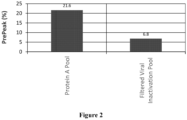
- the level of partial antibody reduction as the percentage of pre-peak species measured by nrCE-SDS in the Protein A pool and the FVIP was determined ( Figure 2 ). Following contact with the charged depth filter, the amount of partially reduced antibody in the Protein A pool was significantly decreased, with a greater than 3-fold decrease in the % pre-peaks observed. All of the Protein A pool product mass was recovered following filtration using the charged depth filter, demonstrating that the decrease in % pre-peak species was due to reoxidation of partially reduced antibody following contact with the charged depth filter according to the disclosure and not loss of antibody molecules.
- the presence of thioredoxin and thioredoxin reductase was determined using mass spectrophotometry. Thioredoxin like proteins were present in the Protein A Pool, but the charged depth filtrate was free of both thioredoxin and thioredoxin reductase.
- a Protein A pool was prepared and contacted with a charged depth filter to form a FVIP as described above or not filtered further and then held at room temperature or a temperature between 2 °C and 8 °C, for up to 8 days.
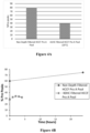
- the level of partial antibody reduction as the percentage of pre-peak species measured by nrCE-SDS in the FVIP ( Figure 3A ) or non-charged depth filtered Protein A pool ( Figure 3B ) was determined.
- the percentage of pre-peak species in the FVIP continued to decrease following contact with the charged depth filter, and the re-oxidation was enhanced at both room temperature and between 2 °C and 8 °C.
- the amount of partially reduced antibody molecules in the FVIP decreased almost three-fold and reached a steady state level in three days or less.
- the percentage of pre-peak species in the non-filtered Protein A pool continued to increase over time at both temperatures, indicating further reduction of the antibody molecules, with faster reduction kinetics at room temperature compared to when chilled.
- the charged depth filtration was also effective in enhancing re-oxidation of antibody molecules when performed prior to Protein A chromatography.
- HCCF was subjected to either charged depth filtration using a MILLISTAK+ X0HC depth filter system (EMD Millipore) in accordance with the manufacturer's instructions and at a throughput of 350 L/m 2 or no filtration and then subjected to Protein A chromatography to form a Protein A pool.
- EMD Millipore MILLISTAK+ X0HC depth filter system
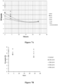
- the level of partial antibody reduction as the percentage of pre-peak species measured by nrCE-SDS in the charged depth filtered Protein A pool or non-charged depth filtered Protein A pool was determined ( Figure 4A and Figure 4B ).
- nVIP was produced as described in Example 2 and subjected to either charged depth filtration using a MILLISTAK+ A1HC depth filter system or sterile membrane filtration using a Millipore EXPRESS SHC hydrophilic filter at a throughput of 350 L/m 2 .
- the level of partial antibody reduction as the percentage of pre-peak species measured by nrCE-SDS in the nVIP loaded into the filter system and following filtration was determined ( Figure 5A and Figure 5B ).
- Charged depth filtration was shown to facilitate re-oxidation of even highly reduced material achieving a decrease in the percentage of pre-peak species from 45% in the load to less than 10% in the depth filtered filtrate ( Figure 5A ).
- An aqueous solution comprising Antibody A was subject to Protein A chromatography followed by (a) a viral inactivation step and then charged depth filtration using a MILLISTAK+ A1HC charged depth filter system to form a FVIP; (b) a viral inactivation step to form a nVIP followed by air sparging to 100% dissolved oxygen; or (c) viral inactivation only to form a nVIP followed up a hold step for up to 50 hours, or (d) the hold step only as a control.
- the level of partial antibody reduction as the percentage of pre-peak species measured by nrCE-SDS in the Protein A Pool, FVIP, nVIP oxygenated with air and nVIP without air was compared ( Figure 6 ).
- the starting percentage of pre-peak species was comparable among the Protein A pool and the nVIPs, regardless of whether the nVIP was oxygenated with air. In contrast, the amount of reduced antibody molecules following charged depth filtration in the FVIP was approximately two-fold lower. The percentage of pre-peak species in the FVIP continued to decrease during the hold, reaching a steady state level more than three-fold lower than the Protein A pool or non-oxygenated nVIP and more than two-fold lower than the oxygenated nVIP. The presence of 100% saturated oxygen had minimal impact, therefore, on re-oxidization of the antibody molecules and was much less effective than the charged depth filtration on decreasing the amount of partially reduced antibody molecules.
- throughput i.e., the total amount of aqueous solution contacted with a charged depth filter per square meter of filter
- throughput i.e., the total amount of aqueous solution contacted with a charged depth filter per square meter of filter
- Aqueous solutions comprising Antibody A were subjected to charged depth filtration using a MILLISTAK+ A1HC filter as described above at a throughput of 350 L/m 2 to 850 L/m 2 , and the level of partial antibody reduction as the percentage of pre-peak species was measured by nrCE-SDS for up to 24 hours post-filtration ( Figure 7A ).
- aqueous solution comprising Antibody A was subjected to Protein A chromatography and then a viral inactivation step to form a nVIP.
- the nVIP was subjected to (a) CEX chromatography only at 50 g/L using a FRACTOGEL SO3 resin (EMD Millipore) to form a CEX pool; (b) charged depth filtration only using a MILLISTAK+ A1HC depth filter system to form a FVIP, or (c) charged depth filtration to form a FVIP, followed by CEX chromatography, as described in (a) and (b), to form a CEX pool.
- the CEX step was designed to resolve high molecular weight (HMW) species in a bind and elute mode.
- Partial antibody molecules can dimerize on the resin via free thiol and elute out as HMW species.
- the level of partial antibody reduction as the percentage of pre-peak species was measured by nrCE-SDS before and after CEX chromatography of the nVIP ( Figure 8A ) and immediately following CEX chromatography of the charged depth filtered FVIP ( Figure 8B ) as well as 4 hours post-CEX chromatography of the charged depth filtered FVIP ( Figure 8C ).
- the percentage of HMW species was also determined as a measure of partial antibody molecules.
- the percentage of reduced pre-peak species in the non-charged depth filtered nVIP before and after CEX chromatography were comparable ( Figure 8A ).
- the level of reduced antibody in the FVIP was more than three-fold lower than in the nVIP following charged depth filtration.
- the percentage of pre-peak species in the charged depth filtered FVIP was further decreased by more than 1.5-fold immediately following CEX chromatography ( Figure 8B ) and was comparable at 0 and 4 hours post-CEX chromatography ( Figures 8B and 8C ).
- the percentage of HMW species in the charged depth filtered FVIP decreased following CEX chromatography, confirming a decrease in partial antibody molecules. The results indicated that re-oxidation of the antibody species following charged depth filtration continued on the CEX column and reached steady state.
- a charged depth filter was flushed with deionized water for 100 L/m 2 , followed by a 2-hour recirculation phase with a 0.1 M H 2 SO 4 solution to strip the filter and remove bound metals, or with a 100 mM acetate solution, pH 5.0, as a control.
- An aqueous solution comprising Antibody A was subjected to Protein A chromatography and then a viral inactivation step followed by a neutralization step to form a nVIP.
- the acetate and sulfuric solutions were spiked into nVIP samples at different volume ratios of 0.5, 1, 2 and 4 parts of buffer to 1 part of nVIP.
- the sulfuric solution contained bound metals and other material stripped from the charged depth filter.
- the sulfuric solution contained about 1500 parts per billion (ppb) Cu 2+ , while the acetate solution compared less than 5 ppb Cu 2+ .
- the level of partial antibody reduction as the percentage of pre-peak species in the spiked samples was measured by nrCE-SDS at 0, 2, 4 and 24 hours post-spike. As expected, unspiked nVIP exhibited no re-oxidation of the antibody molecules. In contrast, the percentage of pre-peak species in the nVIP spiked with the sulfuric solution were significantly lower ( Figure 9A ).
- the percentage of pre-peak species in the nVIP spiked with the sulfuric acid solution at a concentration of 1x or above reached steady state at t 0, indicated that contacting the nVIP with the components stripped from the charged depth filter efficiently enhanced re-oxidation.
- Spiking the nVIP with the acetate solution was not as efficient in facilitating re-oxidization of the antibody molecules as the sulfuric solution or direct contact of antibody with the depth filter ( Figure 9B ).
- Table 1 describes Processes A through N for preparing aqueous solutions comprising re-oxidized antibody molecules. TABLE 1 Process Process Steps A B C D E F G H I J K L M N Depth Filtration X X X X X X X Protein A Chromatography X X X X X X X X X X X X X X X X X X X X X Viral Inactivation X X X X X X X X X X X X X X X Cation Exchange Chromatography X X X X X X X X X X X X X X X X X X Cation Exchange Chromatography X X X X X X X X X X X X X X X X X X X Additional Chromatography (e.g., STIC PA, HIC, MMC
- aqueous solutions comprising reduced antigen-binding protein molecules are obtained without sparging the cell culture fluid with air.
- the aqueous solution is subjected to a charged depth filtration step to form a first filtrate, and the first filtrate is subjected to a Protein A chromatography step to form a first eluate.
- the first eluate is subjected to a charged depth filtration step to form a second filtrate, and the second filtrate is subjected to a low pH viral inactivation step to form a virally inactivated second filtrate.
- the virally inactivated second filtrate is subjected to a charged depth filtration step to form a third filtrate.
- the third filtrate is subjected to a cation exchange chromatography step to form a second eluate, followed by a charged depth filtration step to form a fourth filtrate.
- the fourth filtrate is subjected to one or more additional chromatography steps, for example, STIC PA, HIC, and/or MMC, to form a third (or fourth or fifth) eluate, followed by a charged depth filtration step to form a fifth filtrate.
- the aqueous solution is subjected to a Protein A chromatography step to form a first eluate.
- the first eluate is subjected to a charged depth filtration step to form a first filtrate, and the first filtrate is subjected to a low pH viral inactivation step to form a virally inactivated first filtrate.
- the virally inactivated first filtrate is subjected to a charged depth filtration step to form a second filtrate.
- the second filtrate is subjected to a cation exchange chromatography step to form a second eluate, followed by a charged depth filtration step to form a third filtrate.
- the third filtrate is subjected to one or more additional chromatography steps, for example, STIC PA, HIC, and/or MMC, to form a third (or fourth or fifth) eluate, followed by a charged depth filtration step to form a fourth filtrate.
- additional chromatography steps for example, STIC PA, HIC, and/or MMC
- the Process optionally further includes additional, subsequent purification steps, for example, one or more of ultrafiltration, diafiltration, and viral filtration steps, to achieve an aqueous solution comprising re-oxidized antibody molecules.
- the aqueous solution comprising re-oxidized antibody molecules achieved using the Process has a decreased percentage of reduced antigen-binding protein molecules, compared to the percentage of reduced antigen-binding protein molecules observed prior to the start of the Process.
Landscapes
- Chemical & Material Sciences (AREA)
- Organic Chemistry (AREA)
- Health & Medical Sciences (AREA)
- Molecular Biology (AREA)
- Analytical Chemistry (AREA)
- Life Sciences & Earth Sciences (AREA)
- Proteomics, Peptides & Aminoacids (AREA)
- Genetics & Genomics (AREA)
- Medicinal Chemistry (AREA)
- General Health & Medical Sciences (AREA)
- Biophysics (AREA)
- Biochemistry (AREA)
- Chemical Kinetics & Catalysis (AREA)
- Immunology (AREA)
- Engineering & Computer Science (AREA)
- Water Supply & Treatment (AREA)
- General Chemical & Material Sciences (AREA)
- Peptides Or Proteins (AREA)
- Medicines Containing Antibodies Or Antigens For Use As Internal Diagnostic Agents (AREA)
- Medicinal Preparation (AREA)
Claims (19)
- Procédé de production d'une formulation aqueuse d'une protéine de liaison à un antigène comprenant :(a) la mise en contact d'une solution aqueuse comprenant des molécules de protéine de liaison à un antigène avec un filtre en profondeur chargé, dans lequel le filtre en profondeur chargé comprend un ion métallique sélectionné dans le groupe constitué d'un ion sodium, d'un ion calcium, d'un ion magnésium, d'un ion mercure, d'un ion chrome, d'un ion cadmium, d'un ion aluminium, d'un ion potassium, d'un ion plomb, d'un ion arsenic, d'un ion cobalt, d'un ion fer, d'un ion manganèse, d'un ion titane, d'un ion zinc, d'un ion nickel, d'un ion cuivre, et de combinaisons de ceux-ci ; et(b) la mesure d'une quantité ou quantité relative de molécules réduites de protéine de liaison à un antigène dans la solution après la mise en contact de la solution aqueuse avec le filtre en profondeur chargé, dans lequel le pourcentage de molécules réduites de protéine de liaison à un antigène est réduit d'au moins 20 % comparé au pourcentage de molécules réduites de protéine de liaison à un antigène observées avant l'étape de mise en contact.
- Procédé d'amélioration de la réoxydation d'une protéine de liaison à un antigène comprenant :(a) la mise en contact une solution aqueuse comprenant des molécules de protéine de liaison à un antigène avec un filtre en profondeur chargé, dans lequel le filtre en profondeur chargé comprend un ion métallique sélectionné dans le groupe constitué d'un ion sodium, d'un ion calcium, d'un ion magnésium, d'un ion mercure, d'un ion chrome, d'un ion cadmium, d'un ion aluminium, d'un ion potassium, d'un ion plomb, d'un ion arsenic, d'un ion cobalt, d'un ion fer, d'un ion manganèse, d'un ion titane, d'un ion zinc, d'un ion nickel, d'un ion cuivre, et de combinaisons de ceux-ci ; et(b) la mesure d'une quantité ou quantité relative de molécules réduites de protéine de liaison à un antigène, dans lequel la réoxydation des molécules de liaison à un antigène est accrue au moins deux fois après l'étape de mise en contact.
- Procédé selon l'une quelconque des revendications 1 et 2, dans lequel l'étape (a) est précédée par la soumission de la solution de molécules de protéine de liaison à un antigène à une chromatographie sur protéine A.
- Procédé selon l'une quelconque des revendications 1 à 3, comprenant en outre une étape d'inactivation d'un ou plusieurs virus dans ladite solution de molécules de protéine de liaison à un antigène.
- Procédé selon l'une quelconque des revendications 1 à 4, dans lequel la mise en contact se produit(a) à température ambiante ; ou(b) à une température de 2 °C à 8 °C.
- Procédé selon l'une quelconque des revendications 1 à 5, comprenant en outre(a) une étape de barbotage d'air ou d'oxygène à travers la solution de molécules de protéines de liaison à un antigène ; et/ ou(b) la mise en contact de la solution de molécules de protéine de liaison à un antigène avec un ion positif sélectionné dans le groupe constitué du sodium, du calcium, du magnésium, du mercure, du molybdène, du chrome, du cadmium, de l'aluminium, du potassium, du cobalt, du fer, du manganèse, du titane, du zinc, du nickel, du cuivre, et de combinaisons de ceux-ci.
- Procédé selon l'une quelconque des revendications 1 à 6, dans lequel la solution de molécules de protéine de liaison à un antigène est mise en contact avec plus d'un filtre en profondeur chargé.
- Procédé selon l'une quelconque des revendications 1 à 7, dans lequel le filtre en profondeur chargé comprend une couche de terre de diatomées, comprenant en outre facultativement une couche de cellulose et une couche inorganique, dans lequel la couche inorganique comprend une résine polyamine.
- Procédé selon l'une quelconque des revendications 1 à 8, dans lequel le filtre en profondeur chargé comprend un ion cuivre.
- Procédé selon l'une quelconque des revendications 1 à 9, dans lequel la mise en contact se produit à un débit compris entre 250 L/m2 et 850 L/m2.
- Procédé selon l'une quelconque des revendications 1 à 10, dans lequel la protéine de liaison à un antigène est un anticorps IgG, dans lequel l'anticorps IgG est facultativement un anticorps IgG1 (facultativement un anticorps IgG1 avec une chaîne légère kappa ou un anticorps IgG1 avec une chaîne légère lambda) ou un anticorps IgG2.
- Procédé selon l'une quelconque des revendications 1 à 11, dans lequel la protéine de liaison à un anticorps(a) se lie à un antigène sélectionné dans le groupe constitué de RANKL, du facteur onconécrosant alpha, du récepteur du facteur de croissance épidermique, de CD20, d'un peptide associé au gène de la calcitonine, de la sclérostine, et de la glycoprotéine plaquettaire Ilb/Illa ;(b) est sélectionné dans le groupe constitué de l'abciximab, de l'adalimumab, de l'alemtuzumab, du basiliximab, du bélimumab, du bévacizumab, du brentuximab védotine, du canakinumab, du cétuximab, du certolizumab pégol, du daclizumab, du dénosumab, de l'éculizumab, de l'éfalizumab, du gemtuzumab, du golimumab, de l'ibritumomab tiuxétan, de l'infliximab, de l'ipilimumab, du muromonab-CD3, du natalizumab, du nivolumab, de l'ofatumumab, de l'omalizumab, du palivizumab, du panitumumab, du ranibizumab, du rituximab, du tocilizumab, du tositumomab, du trastuzumab, de l'ustékinumab, du védolizumab, et d'un biosimilaire de l'un quelconque des éléments précédents ; ou(c) comprend une région de liaison à un antigène comprenant une séquence d'acides aminés sélectionnée dans le groupe constitué de SEQ ID NO : 1 à 8.
- Procédé selon l'une quelconque des revendications 1 à 12, dans lequel la quantité de molécules réduites de protéine de liaison à un antigène est mesurée en utilisant une électrophorèse capillaire non réduite avec du sodium dodécyl sulfate
- Procédé selon l'une quelconque des revendications 1 à 13, comprenant en outre une étape de chromatographie échangeuse de cations.
- Procédé selon l'une quelconque des revendications 1 à 14, comprenant (a) une étape de chromatographie sur protéine A, facultativement suivie par une filtration en profondeur chargée ; (2) une étape d'inactivation virale, facultativement suivie par une filtration en profondeur chargée ; et (3) une étape de chromatographie échangeuse de cations, facultativement suivie par une filtration en profondeur chargée ; comprenant en outre facultativement une ou plusieurs de (4) une étape de chromatographie d'interaction intolérante au sel, facultativement suivie par une filtration en profondeur chargée ; (5) une étape de filtration virale, facultativement suivie par une filtration en profondeur chargée ; et (5) une ultrafiltration et/ou une diafiltration, facultativement suivies par une filtration en profondeur chargée.
- Procédé selon l'une quelconque des revendications 1 à 15, dans lequel après la mise en contact avec un filtre en profondeur chargé, le filtrat est mis à incuber pendant au moins 4 heures avant d'être soumis à une chromatographie sur protéine A.
- Procédé selon l'une quelconque des revendications 1 à 16, dans lequel la quantité ou quantité relative de molécules réduites de protéine de liaison à un antigène est déterminée en utilisant une électrophorèse capillaire non réduite avec du sodium dodécyl sulfate (nrCE-SDS).
- Procédé selon la revendication 1, comprenant (1) une étape de chromatographie sur protéine A, (2) une étape d'inactivation virale ; (3) une étape de filtration en profondeur ; et (4) une étape d'incubation pendant au moins 4 heures.
- Procédé selon la revendication 18, comprenant en outre (5) une étape de chromatographie échangeuse de cations ; et une ou plusieurs de (6) une étape de chromatographie d'interaction intolérante au sel ; (7) une étape de filtration virale ; et (8) une ultrafiltration et/ou une diafiltration.
Priority Applications (5)
| Application Number | Priority Date | Filing Date | Title |
|---|---|---|---|
| DK22211542.0T DK4209499T3 (da) | 2015-08-13 | 2016-08-12 | Ladet dybdefiltrering af antigenbindende proteiner |
| HRP20231544TT HRP20231544T1 (hr) | 2015-08-13 | 2016-08-12 | Nabijena dubinska filtracija proteina koji vežu antigene |
| EP24193226.8A EP4470648A3 (fr) | 2015-08-13 | 2016-08-12 | Filtration sur profondeur chargée de protéines de liaison à l'antigène |
| EP22211542.0A EP4209499B1 (fr) | 2015-08-13 | 2016-08-12 | Filtration sur profondeur chargée de protéines de liaison à l'antigène |
| SI201631774T SI3334747T1 (sl) | 2015-08-13 | 2016-08-12 | Globinsko nabito filtriranje proteinov, ki vežejo antigen |
Applications Claiming Priority (2)
| Application Number | Priority Date | Filing Date | Title |
|---|---|---|---|
| US201562204831P | 2015-08-13 | 2015-08-13 | |
| PCT/US2016/046929 WO2017027861A1 (fr) | 2015-08-13 | 2016-08-12 | Filtration en profondeur chargée de protéines de liaison à un antigène |
Related Child Applications (3)
| Application Number | Title | Priority Date | Filing Date |
|---|---|---|---|
| EP24193226.8A Division EP4470648A3 (fr) | 2015-08-13 | 2016-08-12 | Filtration sur profondeur chargée de protéines de liaison à l'antigène |
| EP22211542.0A Division EP4209499B1 (fr) | 2015-08-13 | 2016-08-12 | Filtration sur profondeur chargée de protéines de liaison à l'antigène |
| EP22211542.0A Division-Into EP4209499B1 (fr) | 2015-08-13 | 2016-08-12 | Filtration sur profondeur chargée de protéines de liaison à l'antigène |
Publications (2)
| Publication Number | Publication Date |
|---|---|
| EP3334747A1 EP3334747A1 (fr) | 2018-06-20 |
| EP3334747B1 true EP3334747B1 (fr) | 2023-09-27 |
Family
ID=56802689
Family Applications (3)
| Application Number | Title | Priority Date | Filing Date |
|---|---|---|---|
| EP24193226.8A Pending EP4470648A3 (fr) | 2015-08-13 | 2016-08-12 | Filtration sur profondeur chargée de protéines de liaison à l'antigène |
| EP16757780.8A Active EP3334747B1 (fr) | 2015-08-13 | 2016-08-12 | Filtration en profondeur chargée de protéines de liaison à un antigène |
| EP22211542.0A Active EP4209499B1 (fr) | 2015-08-13 | 2016-08-12 | Filtration sur profondeur chargée de protéines de liaison à l'antigène |
Family Applications Before (1)
| Application Number | Title | Priority Date | Filing Date |
|---|---|---|---|
| EP24193226.8A Pending EP4470648A3 (fr) | 2015-08-13 | 2016-08-12 | Filtration sur profondeur chargée de protéines de liaison à l'antigène |
Family Applications After (1)
| Application Number | Title | Priority Date | Filing Date |
|---|---|---|---|
| EP22211542.0A Active EP4209499B1 (fr) | 2015-08-13 | 2016-08-12 | Filtration sur profondeur chargée de protéines de liaison à l'antigène |
Country Status (15)
| Country | Link |
|---|---|
| US (3) | US11098079B2 (fr) |
| EP (3) | EP4470648A3 (fr) |
| JP (1) | JP6889149B2 (fr) |
| DK (2) | DK3334747T5 (fr) |
| ES (2) | ES2964640T3 (fr) |
| FI (2) | FI3334747T3 (fr) |
| HR (2) | HRP20241510T1 (fr) |
| HU (2) | HUE064252T2 (fr) |
| LT (2) | LT3334747T (fr) |
| MA (1) | MA42613A (fr) |
| PL (1) | PL3334747T3 (fr) |
| PT (2) | PT3334747T (fr) |
| SG (1) | SG10202101105XA (fr) |
| SI (2) | SI4209499T1 (fr) |
| WO (1) | WO2017027861A1 (fr) |
Families Citing this family (14)
| Publication number | Priority date | Publication date | Assignee | Title |
|---|---|---|---|---|
| EP2877589B1 (fr) | 2012-07-26 | 2024-03-06 | Momenta Pharmaceuticals, Inc. | Glycoprotéines ayant des propriétés anti-inflammatoires |
| US10464996B2 (en) | 2013-05-13 | 2019-11-05 | Momenta Pharmaceuticals, Inc. | Methods for the treatment of neurodegeneration |
| WO2015057622A1 (fr) | 2013-10-16 | 2015-04-23 | Momenta Pharmaceuticals, Inc. | Glycoprotéines sialylées |
| MA42613A (fr) | 2015-08-13 | 2018-06-20 | Amgen Inc | Filtration en profondeur chargée de protéines de liaison à un antigène |
| US10961305B2 (en) | 2016-12-21 | 2021-03-30 | Mereo Biopharma 3 Limited | Use of anti-sclerostin antibodies in the treatment of osteogenesis imperfecta |
| CN107973487B (zh) * | 2017-11-14 | 2021-03-02 | 东华大学 | 一种染整废水层级处理工艺及装置 |
| CN114981286B (zh) | 2019-05-03 | 2026-02-13 | 豪夫迈·罗氏有限公司 | 降低从纯化平台获得的组合物中酶水解活性速率的方法 |
| BR112021022682A2 (pt) | 2019-05-14 | 2022-02-22 | Provention Bio Inc | Métodos e composições para prevenir diabetes do tipo 1 |
| EP4090666A1 (fr) | 2020-01-15 | 2022-11-23 | F. Hoffmann-La Roche AG | Procédés de réduction d'impuretés à partir de procédés de fabrication de protéines recombinantes |
| WO2021158469A1 (fr) * | 2020-02-03 | 2021-08-12 | Amgen Inc. | Approche d'encadrement à plusieurs variables pour la validation d'un filtre stérile |
| CN111410688A (zh) * | 2020-03-13 | 2020-07-14 | 苏州智享众创孵化管理有限公司 | 一种提高双特异性抗体纯度的分离纯化方法 |
| US12006366B2 (en) | 2020-06-11 | 2024-06-11 | Provention Bio, Inc. | Methods and compositions for preventing type 1 diabetes |
| WO2022251253A1 (fr) | 2021-05-24 | 2022-12-01 | Provention Bio, Inc. | Méthodes de traitement du diabète de type 1 |
| WO2025252480A1 (fr) | 2024-06-07 | 2025-12-11 | F. Hoffmann-La Roche Ag | Procédé de purification d'adn plasmidique |
Citations (16)
| Publication number | Priority date | Publication date | Assignee | Title |
|---|---|---|---|---|
| WO2003066669A2 (fr) | 2002-02-04 | 2003-08-14 | Millipore Corporation | Procede visant a extraire des agregats de proteines et des virus d'une solution proteinique |
| WO2008036899A2 (fr) | 2006-09-22 | 2008-03-27 | Amgen Inc. | Procédés d'élimination des contaminants viraux durant la purification des protéines |
| WO2009009523A2 (fr) | 2007-07-09 | 2009-01-15 | Genentech, Inc. | Prévention de la réduction des liaisons disulfure au cours de la production recombinante de polypeptides |
| WO2010017964A1 (fr) | 2008-08-14 | 2010-02-18 | Sartorius Stedim Biotech Gmbh | Couche épaisse de filtration avec hydroxyde minéral double stratifié |
| US20100056759A1 (en) | 2006-10-19 | 2010-03-04 | Tolerx, Inc. | Methods and Compositions for Efficient Removal of Protein A from Binding Molecule Preparations |
| WO2010125187A2 (fr) | 2009-04-30 | 2010-11-04 | Ablynx Nv | Procédé de production d'anticorps à domaines |
| WO2011146179A2 (fr) | 2010-05-18 | 2011-11-24 | Abbott Laboratories | Appareil et procédé de purification des protéines |
| WO2012051147A1 (fr) | 2010-10-11 | 2012-04-19 | Abbott Laboratories | Procédés de purification de protéines |
| WO2012054679A1 (fr) | 2010-10-20 | 2012-04-26 | Medimmune, Llc | Méthodes de traitement de corps d'inclusion |
| US20140010820A1 (en) | 2012-05-21 | 2014-01-09 | Abbvie, Inc. | Novel purification of non-human antibodies using protein a affinity chromatography |
| WO2014100443A2 (fr) | 2012-12-20 | 2014-06-26 | Medimmune, Llc | Procédés de production d'immunoconjugués |
| WO2014145744A1 (fr) | 2013-03-15 | 2014-09-18 | Alder Biopharmaceuticals, Inc. | Purification d'anticorps et contrôle de pureté |
| WO2015070068A1 (fr) | 2013-11-07 | 2015-05-14 | Abbvie Inc. | Isolement et purification d'anticorps |
| US20150133636A1 (en) | 2012-06-29 | 2015-05-14 | Emd Millipore Corporation | Purification of Biological Molecules |
| WO2016106291A1 (fr) | 2014-12-22 | 2016-06-30 | Alexion Pharmaceuticals, Inc. | Procédés permettant de purifier des protéines recombinantes |
| WO2017027861A1 (fr) | 2015-08-13 | 2017-02-16 | Amgen Inc. | Filtration en profondeur chargée de protéines de liaison à un antigène |
Family Cites Families (70)
| Publication number | Priority date | Publication date | Assignee | Title |
|---|---|---|---|---|
| US4597966A (en) | 1985-01-09 | 1986-07-01 | Ortho Diagnostic Systems, Inc. | Histidine stabilized immunoglobulin and method of preparation |
| US5237054A (en) | 1987-02-20 | 1993-08-17 | Akzo Pharma | Stabilized aqueous composition containing antibodies |
| US5945098A (en) | 1990-02-01 | 1999-08-31 | Baxter International Inc. | Stable intravenously-administrable immune globulin preparation |
| JPH0565233A (ja) | 1991-03-08 | 1993-03-19 | Mitsui Toatsu Chem Inc | モノクローナル抗体含有凍結乾燥製剤 |
| US6284471B1 (en) | 1991-03-18 | 2001-09-04 | New York University Medical Center | Anti-TNFa antibodies and assays employing anti-TNFa antibodies |
| GB9122820D0 (en) | 1991-10-28 | 1991-12-11 | Wellcome Found | Stabilised antibodies |
| US5736137A (en) | 1992-11-13 | 1998-04-07 | Idec Pharmaceuticals Corporation | Therapeutic application of chimeric and radiolabeled antibodies to human B lymphocyte restricted differentiation antigen for treatment of B cell lymphoma |
| DE69303494T2 (de) | 1992-11-13 | 1997-01-16 | Idec Pharma Corp | Therapeutische verwendung von chimerischen und markierten antikörper gegen menschlichen b lymphozyt beschränkter differenzierung antigen für die behandlung von b-zell-lymphoma |
| US6288030B1 (en) | 1993-12-22 | 2001-09-11 | Amgen Inc. | Stem cell factor formulations and methods |
| DE4344824C1 (de) | 1993-12-28 | 1995-08-31 | Immuno Ag | Hochkonzentriertes Immunglobulin-Präparat und Verfahren zu seiner Herstellung |
| US5955448A (en) | 1994-08-19 | 1999-09-21 | Quadrant Holdings Cambridge Limited | Method for stabilization of biological substances during drying and subsequent storage and compositions thereof |
| AU3272695A (en) | 1994-08-12 | 1996-03-07 | Immunomedics Inc. | Immunoconjugates and humanized antibodies specific for b-cell lymphoma and leukemia cells |
| US6267958B1 (en) | 1995-07-27 | 2001-07-31 | Genentech, Inc. | Protein formulation |
| GB9610992D0 (en) | 1996-05-24 | 1996-07-31 | Glaxo Group Ltd | Concentrated antibody preparation |
| GB9705810D0 (en) | 1997-03-20 | 1997-05-07 | Common Services Agency | Intravenous immune globulin |
| US6171586B1 (en) | 1997-06-13 | 2001-01-09 | Genentech, Inc. | Antibody formulation |
| US6214221B1 (en) | 1999-02-22 | 2001-04-10 | Henry B. Kopf | Method and apparatus for purification of biological substances |
| AUPQ599700A0 (en) | 2000-03-03 | 2000-03-23 | Super Internet Site System Pty Ltd | On-line geographical directory |
| JP4340062B2 (ja) | 2000-10-12 | 2009-10-07 | ジェネンテック・インコーポレーテッド | 粘度の減少した濃縮タンパク質製剤 |
| HU229910B1 (hu) | 2001-05-11 | 2014-12-29 | Amgen Inc Thousand Oaks | A TALL-1-hez kötõdõ peptidek és rokon molekulák |
| GB0113179D0 (en) | 2001-05-31 | 2001-07-25 | Novartis Ag | Organic compounds |
| CA2451955C (fr) | 2001-06-26 | 2015-09-29 | Abgenix, Inc. | Anticorps opgl |
| IL160358A0 (en) | 2001-08-23 | 2004-07-25 | Genmab As | Human antibodies specific for interleukin 15 (il-15) |
| US7247304B2 (en) | 2001-08-23 | 2007-07-24 | Genmab A/S | Methods of treating using anti-IL-15 antibodies |
| US7521053B2 (en) | 2001-10-11 | 2009-04-21 | Amgen Inc. | Angiopoietin-2 specific binding agents |
| US7138370B2 (en) | 2001-10-11 | 2006-11-21 | Amgen Inc. | Specific binding agents of human angiopoietin-2 |
| ES2392073T3 (es) | 2001-11-08 | 2012-12-04 | Abbott Biotherapeutics Corp. | Formulación farmacéutica líquida estable de anticuerpos IGG |
| US8188231B2 (en) * | 2002-09-27 | 2012-05-29 | Xencor, Inc. | Optimized FC variants |
| CN104178491B (zh) | 2002-09-06 | 2018-07-03 | 安姆根有限公司 | 治疗性人抗-il-1r1单克隆抗体 |
| WO2004034988A2 (fr) | 2002-10-16 | 2004-04-29 | Amgen Inc. | Anticorps humains neutralisants anti-ifn-$g(g) utilises comme inhibiteurs selectifs des voies de ifn-$g(g) |
| KR20110017020A (ko) | 2002-10-17 | 2011-02-18 | 젠맵 에이/에스 | Cd20에 대한 인간 모노클로날 항체 |
| US20040191243A1 (en) | 2002-12-13 | 2004-09-30 | Bei Chen | System and method for stabilizing antibodies with histidine |
| KR20110086881A (ko) | 2002-12-20 | 2011-08-01 | 암겐 인코포레이티드 | 미오스타틴을 저해하는 결합제 |
| AU2004229335C1 (en) | 2003-04-04 | 2010-06-17 | Genentech, Inc. | High concentration antibody and protein formulations |
| US7157276B2 (en) | 2003-06-20 | 2007-01-02 | Biogen Idec Inc. | Use of depth filtration in series with continuous centrifugation to clarify mammalian cell cultures |
| MEP31508A (hr) | 2003-07-15 | 2010-10-10 | Amgen Inc | Humana anti-ngf neutrališuća antitijela kao selektivni ihibitori ngf signalne kaskade |
| JP4746552B2 (ja) | 2003-11-04 | 2011-08-10 | ノバルティス バクシンズ アンド ダイアグノスティックス,インコーポレーテッド | 自己免疫疾患および炎症性疾患ならびに臓器移植拒絶の処置のためのアンタゴニスト抗cd40抗体の使用 |
| NZ547588A (en) | 2003-11-07 | 2009-05-31 | Immunex Corp | Antibodies that bind interleukin-4 receptor |
| EP1712240B1 (fr) | 2003-12-25 | 2015-09-09 | Kyowa Hakko Kirin Co., Ltd. | Preparation medicamenteuse aqueuse stable contenant un anticorps |
| ES2407380T5 (es) | 2004-02-27 | 2017-07-07 | Octapharma Ag | Procedimiento para proporcionar una preparación de anticuerpos purificada, sin riesgo respecto a virus |
| ES2579805T3 (es) | 2004-09-23 | 2016-08-16 | Genentech, Inc. | Anticuerpos y conjugados modificados por ingeniería genética con cisteína |
| SG156672A1 (en) | 2004-10-22 | 2009-11-26 | Amgen Inc | Methods for refolding of recombinant antibodies |
| WO2006138181A2 (fr) | 2005-06-14 | 2006-12-28 | Amgen Inc. | Preparations de proteines a tamponnage spontane |
| WO2007035283A1 (fr) | 2005-09-15 | 2007-03-29 | Wyeth | Floculation de proteines au moyen de sels |
| JP2010510963A (ja) | 2006-06-14 | 2010-04-08 | グラクソスミスクライン・リミテッド・ライアビリティ・カンパニー | セラミックヒドロキシアパタイトを使用する抗体の精製方法 |
| EP3663318A1 (fr) | 2008-01-07 | 2020-06-10 | Amgen Inc. | Procédé de fabrication de molécules hétérodimères fc d'anticorps utilisant les effets de conduite électrostatique |
| TW201024318A (en) | 2008-10-20 | 2010-07-01 | Abbott Lab | Isolation and purification of antibodies using protein A affinity chromatography |
| EP2490780A4 (fr) | 2009-10-20 | 2014-04-09 | Merck Sharp & Dohme | Utilisation de la chromatographie en mode mixte pour la capture et la purification de produits d'anticorps de base |
| ES2813398T3 (es) | 2009-10-20 | 2021-03-23 | Abbvie Inc | Aislamiento y purificación de anticuerpos anti-IL-13 usando cromatografía de afinidad a Proteína A |
| GB201006753D0 (en) | 2010-04-22 | 2010-06-09 | Biotest Ag | Process for preparing an immunolobulin composition |
| WO2011140405A1 (fr) * | 2010-05-07 | 2011-11-10 | Millipore Corporation | Milieu de clarification amélioré |
| US20120238730A1 (en) | 2011-03-15 | 2012-09-20 | Abbott Laboratories | Integrated approach to the isolation and purification of antibodies |
| ES2809211T3 (es) | 2011-07-08 | 2021-03-03 | Emd Millipore Corp | Filtros de profundidad mejorados para procesos biotecnológicos desechables |
| FR2977893B1 (fr) | 2011-07-11 | 2015-02-20 | Lab Francais Du Fractionnement | Procede de preparation d'un concentre d'immunoglobulines polyvalentes |
| US9534037B2 (en) | 2011-08-02 | 2017-01-03 | Baxalta GmbH | Systems and methods to increase protein yield from recombinant manufacturing processes |
| US9096648B2 (en) | 2011-08-19 | 2015-08-04 | Emd Millipore Corporation | Methods of reducing level of one or more impurities in a sample during protein purification |
| DK2771364T3 (da) * | 2011-10-27 | 2019-08-19 | Genmab As | Fremstilling af heterodimere proteiner |
| US10364268B2 (en) | 2011-12-22 | 2019-07-30 | Genentech, Inc. | Ion exchange membrane chromatography |
| US20140154270A1 (en) | 2012-05-21 | 2014-06-05 | Chen Wang | Purification of non-human antibodies using kosmotropic salt enhanced protein a affinity chromatography |
| US9249182B2 (en) | 2012-05-24 | 2016-02-02 | Abbvie, Inc. | Purification of antibodies using hydrophobic interaction chromatography |
| CA2899449A1 (fr) | 2013-03-14 | 2014-10-02 | Abbvie Inc. | Composition d'especes faiblement acides et leurs methodes de production utilisant la chromatographie de deplacement |
| KR20150129033A (ko) | 2013-03-14 | 2015-11-18 | 애브비 인코포레이티드 | 저 산성 종 조성물 및 이의 제조 및 사용 방법 |
| WO2014196926A1 (fr) | 2013-06-04 | 2014-12-11 | Agency For Science, Technology And Research | Procédé de purification de protéine |
| US10513555B2 (en) | 2013-07-04 | 2019-12-24 | Prothena Biosciences Limited | Antibody formulations and methods |
| SG11201509390TA (en) | 2013-08-30 | 2015-12-30 | Emd Millipore Corp | High capacity composite depth filter media with low extractables |
| US8946395B1 (en) | 2013-10-18 | 2015-02-03 | Abbvie Inc. | Purification of proteins using hydrophobic interaction chromatography |
| GB201318840D0 (en) | 2013-10-24 | 2013-12-11 | Univ Leeds | Method and device for protein preparation |
| CN106164086A (zh) | 2014-02-27 | 2016-11-23 | 新加坡科技研究局 | 抗体纯化方法 |
| EP3119871A4 (fr) | 2014-03-17 | 2018-01-03 | Regeneron Pharmaceuticals, Inc. | Clarification de culture cellulaire de mammifère |
| EP3145951A1 (fr) | 2014-06-24 | 2017-03-29 | InSight Biopharmaceuticals Ltd. | Procédés de purification d'anticorps |
-
2016
- 2016-08-12 MA MA042613A patent/MA42613A/fr unknown
- 2016-08-12 DK DK16757780.8T patent/DK3334747T5/da active
- 2016-08-12 JP JP2018506988A patent/JP6889149B2/ja active Active
- 2016-08-12 LT LTEPPCT/US2016/046929T patent/LT3334747T/lt unknown
- 2016-08-12 ES ES16757780T patent/ES2964640T3/es active Active
- 2016-08-12 HR HRP20241510TT patent/HRP20241510T1/hr unknown
- 2016-08-12 SI SI201631860T patent/SI4209499T1/sl unknown
- 2016-08-12 ES ES22211542T patent/ES2995985T3/es active Active
- 2016-08-12 EP EP24193226.8A patent/EP4470648A3/fr active Pending
- 2016-08-12 EP EP16757780.8A patent/EP3334747B1/fr active Active
- 2016-08-12 SG SG10202101105XA patent/SG10202101105XA/en unknown
- 2016-08-12 HR HRP20231544TT patent/HRP20231544T1/hr unknown
- 2016-08-12 PT PT167577808T patent/PT3334747T/pt unknown
- 2016-08-12 HU HUE16757780A patent/HUE064252T2/hu unknown
- 2016-08-12 SI SI201631774T patent/SI3334747T1/sl unknown
- 2016-08-12 FI FIEP16757780.8T patent/FI3334747T3/fi active
- 2016-08-12 LT LTEP22211542.0T patent/LT4209499T/lt unknown
- 2016-08-12 EP EP22211542.0A patent/EP4209499B1/fr active Active
- 2016-08-12 FI FIEP22211542.0T patent/FI4209499T3/fi active
- 2016-08-12 PT PT222115420T patent/PT4209499T/pt unknown
- 2016-08-12 US US15/751,231 patent/US11098079B2/en active Active
- 2016-08-12 PL PL16757780.8T patent/PL3334747T3/pl unknown
- 2016-08-12 HU HUE22211542A patent/HUE069195T2/hu unknown
- 2016-08-12 WO PCT/US2016/046929 patent/WO2017027861A1/fr not_active Ceased
- 2016-08-12 DK DK22211542.0T patent/DK4209499T3/da active
-
2021
- 2021-07-28 US US17/386,692 patent/US20210363180A1/en active Pending
-
2025
- 2025-01-15 US US19/022,213 patent/US20250230187A1/en active Pending
Patent Citations (20)
| Publication number | Priority date | Publication date | Assignee | Title |
|---|---|---|---|---|
| WO2003066669A2 (fr) | 2002-02-04 | 2003-08-14 | Millipore Corporation | Procede visant a extraire des agregats de proteines et des virus d'une solution proteinique |
| WO2008036899A2 (fr) | 2006-09-22 | 2008-03-27 | Amgen Inc. | Procédés d'élimination des contaminants viraux durant la purification des protéines |
| US20100056759A1 (en) | 2006-10-19 | 2010-03-04 | Tolerx, Inc. | Methods and Compositions for Efficient Removal of Protein A from Binding Molecule Preparations |
| WO2009009523A2 (fr) | 2007-07-09 | 2009-01-15 | Genentech, Inc. | Prévention de la réduction des liaisons disulfure au cours de la production recombinante de polypeptides |
| US20110207196A1 (en) | 2008-08-14 | 2011-08-25 | Sartorius Stedim Iotech Gmbh | Depth filter layer with inorganic layer double hydroxide |
| WO2010017964A1 (fr) | 2008-08-14 | 2010-02-18 | Sartorius Stedim Biotech Gmbh | Couche épaisse de filtration avec hydroxyde minéral double stratifié |
| US20120157664A1 (en) | 2009-04-30 | 2012-06-21 | Ablynx N.V. | Method for the production of domain antibodies |
| WO2010125187A2 (fr) | 2009-04-30 | 2010-11-04 | Ablynx Nv | Procédé de production d'anticorps à domaines |
| WO2011146179A2 (fr) | 2010-05-18 | 2011-11-24 | Abbott Laboratories | Appareil et procédé de purification des protéines |
| WO2012051147A1 (fr) | 2010-10-11 | 2012-04-19 | Abbott Laboratories | Procédés de purification de protéines |
| US20120264920A1 (en) | 2010-10-11 | 2012-10-18 | Abbott Laboratories | Processes for purification of proteins |
| WO2012054679A1 (fr) | 2010-10-20 | 2012-04-26 | Medimmune, Llc | Méthodes de traitement de corps d'inclusion |
| US20140010820A1 (en) | 2012-05-21 | 2014-01-09 | Abbvie, Inc. | Novel purification of non-human antibodies using protein a affinity chromatography |
| US20150133636A1 (en) | 2012-06-29 | 2015-05-14 | Emd Millipore Corporation | Purification of Biological Molecules |
| WO2014100443A2 (fr) | 2012-12-20 | 2014-06-26 | Medimmune, Llc | Procédés de production d'immunoconjugués |
| WO2014145744A1 (fr) | 2013-03-15 | 2014-09-18 | Alder Biopharmaceuticals, Inc. | Purification d'anticorps et contrôle de pureté |
| WO2015070068A1 (fr) | 2013-11-07 | 2015-05-14 | Abbvie Inc. | Isolement et purification d'anticorps |
| WO2016106291A1 (fr) | 2014-12-22 | 2016-06-30 | Alexion Pharmaceuticals, Inc. | Procédés permettant de purifier des protéines recombinantes |
| WO2017027861A1 (fr) | 2015-08-13 | 2017-02-16 | Amgen Inc. | Filtration en profondeur chargée de protéines de liaison à un antigène |
| US11098079B2 (en) | 2015-08-13 | 2021-08-24 | Amgen Inc. | Charged depth filtration of antigen-binding proteins |
Non-Patent Citations (51)
| Title |
|---|
| "Industrial Minerals and Rocks", 1 January 1983, article KADEY, JR. FREDERIC L.: "Diatomite", pages: 677 - 708, XP093195613 |
| "KIRK-OTHMER ENCYCLOPEDIA OF CHEMICAL TECHNOLOGY (4th edition)", 1 January 2014, WILEY-INTERSCIENCE , Hoboken, NJ, USA , ISBN: 978-0-471-48494-3, article ENGH KENNETH R.: "Diatomite", pages: 1 - 11, XP093195600, DOI: 10.1002/0471238961.0409012005140708.a01.pub2 |
| "Mammalian Cell Cultures for Biologics Manufacturing", vol. 139, 1 January 2013, SPRINGER BERLIN HEIDELBERG, Berlin, Heidelberg, ISBN: 978-3-642-54050-9, ISSN: 0724-6145, article GRAMER MICHAEL J.: "Product Quality Considerations for Mammalian Cell Culture Process Development and Manufacturing", pages: 123 - 166, XP093039744, DOI: 10.1007/10_2013_214 |
| "Millistak+® Depth Filter in µPod® format, A1HC media series, 23 cm2 surface area, Luer fitting connection", Retrieved from the Internet <URL:https://www.merckmillipore.com/GB/en/product/Millistak-Depth-Filter-in-Pod-format-A1HC-media-series-23cm2-surface-area-Luer-fitting-connection,MM_NF-MA1HC23CL3> [retrieved on 20240409] |
| "Millistak+® HC Pod Depth Filter, A1HC media series, 1.1 m2 surface area, 27/pack shipped on a North American standard sized pallet", Retrieved from the Internet <URL:https://www.merckmillipore.com/GB/en/product/Millistak-HC-Pod-Depth-Filter-A1HC-media-series-1.1m2-surface-area-27-pack-shipped-on-a-North-American-standard-sized-pallet,MM_NF-MA1HC10FS27NA> [retrieved on 20240409] |
| "The Chemistry of the Thiol Group", 1 January 1974, article CAPOZZI G., MODENA G.: "Chapter 17 - Oxidation of thiols", pages: 785 - 839, XP093195623 |
| 3M: "Zeta Plus ZA Series - Depth Filter Cartridges & Capsules", PRODUCT DATA SHEET, 1 February 2009 (2009-02-01), pages 1 - 4, XP093195626 |
| 3M: "Zeta Plus® CP & SP Series", DATA SHEET, 1 January 2007 (2007-01-01), pages 1 - 5, XP093195629 |
| 3M: "Zeta Plus®BioCap™ - Disposable Filter Capsules for the Biotechnology & Pharmaceutical Industries", CUNO PRODUCT BROCHURE, 1 January 2007 (2007-01-01), pages 1 - 8, XP093195632 |
| ANONYMOUS: "Diatomaceous earth", WIKIPEDIA, 9 August 2024 (2024-08-09), XP093195634, Retrieved from the Internet <URL:https://en.wikipedia.org/w/index.php?title=Diatomaceous_earth&oldid=1227216614%27> |
| ANONYMOUS: "Millistak+® Pod depth filter system", MERCK KGAA - DATA SHEET, 1 January 2013 (2013-01-01), pages 1 - 8, XP093195094, Retrieved from the Internet <URL:https://www.sigmaaldrich.com/deepweb/assets/sigmaaldrich/marketing/global/documents/151/827/millistak-datasheet-ds0217en-mk.pdf> |
| ANONYMOUS: "Zeta Plus® BioCap™ Disposable Filter Capsules", PRODUCT DATA SHEET, 1 January 1997 (1997-01-01), pages FP, 1 - 7, XP093195612 |
| BRIAN O'MARA; ZHONG-HUA GAO; MANJU KURUGANTI; ROBERT MALLETT; GAUTAM NAYAR; LAURA SMITH; JEFFREY D. MEYER; JON THERRIAULT; CAMERON: "Impact of depth filtration on disulfide bond reduction during downstream processing of monoclonal antibodies from CHO cell cultures", BIOTECHNOLOGY AND BIOENGINEERING, JOHN WILEY, HOBOKEN, USA, vol. 116, no. 7, 4 April 2019 (2019-04-04), Hoboken, USA, pages 1669 - 1683, XP071108839, ISSN: 0006-3592, DOI: 10.1002/bit.26964 |
| C. NATTRASS;C. J. HORWELL;D. E. DAMBY;A. KERMANIZADEH;D. M. BROWN;V. STONE: "The global variability of diatomaceous earth toxicity: a physicochemical and in vitro investigation", JOURNAL OF OCCUPATIONAL MEDICINE AND TOXICOLOGY, BIOMED CENTRAL LTD, LO, vol. 10, no. 1, 10 July 2015 (2015-07-10), Lo , pages 23, XP021228690, ISSN: 1745-6673, DOI: 10.1186/s12995-015-0064-7 |
| CATALIN DONEANU, ALEX XENOPOULOS, KEITH FADGEN, JIM MURPHY, ST. JOHN SKILTON, HOLLY PRENTICE, MARTHA STAPELS, WEIBIN CHEN: "Analysis of host-cell proteins in biotherapeutic proteins by comprehensive online two-dimensional liquid chromatography/mass spectrometry", MABS, LANDES BIOSCIENCE, US, vol. 4, no. 1, 1 January 2012 (2012-01-01), US , pages 24 - 44, XP055297897, ISSN: 1942-0862, DOI: 10.4161/mabs.4.1.18748 |
| CHADERJIAN WENDY B, ET AL: "Effect of copper sulfate on performance of a serum-free CHO cell culture process and the level of free thiol in the recombinant antibody expressed", BIOTECHNOLOGY PROGRESS, AMERICAN CHEMICAL SOCIETY, HOBOKEN, USA, vol. 21, no. 2, 1 March 2005 (2005-03-01), Hoboken, USA, pages 550 - 553, XP002500111, ISSN: 8756-7938, DOI: 10.1021/BP0497029 |
| D.J. ROUSH, Y. LU: "Advances in Primary Recovery: Centrifugation and Membrane Technology", BIOTECHNOLOGY PROGRESS, AMERICAN CHEMICAL SOCIETY, HOBOKEN, USA, vol. 24, no. 3, 6 June 2008 (2008-06-06), Hoboken, USA, pages 488 - 495, XP055090116, ISSN: 8756-7938, DOI: 10.1021/bp070414x |
| EMDMILLIPORE: "Millistak+® Pod Disposable Depth Filter ", PERFORMANCE GUIDE, 1 June 2013 (2013-06-01), pages 1 - 28, XP093195614 |
| EMEA: "Guideline On Development, Production, Characterisation and Specifications for Monoclonal Antibodies and Related Products", EMEA/CHMP/BWP/157653/2007, 18 December 2008 (2008-12-18), pages 1 - 11, XP093195606 |
| FABER RENE, YANG YUJING, GOTTSCHALK UWE: "Salt Tolerant Interaction Chromatography for Large-Scale Polishing with Convective Media", BIOPHARM INTERNATIONAL, ADVANSTAR COMMUNICATIONS, DULUTH, MN, US, vol. 2009suppl., no. 7, 2 October 2009 (2009-10-02), US , XP093195615, ISSN: 1542-166X |
| GENNARO LYNN, FELTEN CHANTAL: "Optimization Approaches in the Routine Analysis of Monoclonal Anitbodies by Capillary Electrophoresis", LCGC EUROPE, 1 December 2008 (2008-12-01), XP093195079, Retrieved from the Internet <URL:https://www.chromatographyonline.com/view/optimization-approaches-routine-analysis-monoclonal-anitbodies-capillary-electrophoresis-0> |
| HONGCHENG LIU, KIMBERLY MAY: "Disulfide bond structures of IgG molecules: Structural variations, chemical modifications and possible impacts to stability and biological function", MABS, vol. 4, no. 1, 1 January 2012 (2012-01-01), pages 17 - 23, XP055134217, ISSN: 19420862, DOI: 10.4161/mabs.4.1.18347 |
| HUI F. LIU, MA JUNFEN, WINTER CHARLES, BAYER ROBERT: "Recovery and purification process development for monoclonal antibody production", MABS, vol. 2, no. 5, 1 September 2010 (2010-09-01), pages 480 - 499, XP055027612, ISSN: 19420862, DOI: 10.4161/mabs.2.5.12645 |
| JORNITZ MAIK W., MELTZER THEODORE H.: "Filtration and Purification in the Biopharmaceutical Industry, 2nd ed.", 1 January 2008, pages: 540, XP093195625 |
| KAO Y H, HEWITT D P, TREXLER-SCHMIDT M, LAIRD M W.: "Mechanism of Antibody Reduction in Cell Culture Production Processes", BIOTECHNOLOGY AND BIOENGINEERING, JOHN WILEY, HOBOKEN, USA, vol. 107, no. 4, 1 November 2010 (2010-11-01), Hoboken, USA, pages 622 - 632, XP002694207, ISSN: 0006-3592, DOI: 10.1002/bit.22848 |
| KATARIINA M HUTTERER, ROBERT W HONG, JONATHON LULL, XIAOYANG ZHAO, TIAN WANG, REX PEI, M ELEANOR LE, OLEG BORISOV, ROB PIPER, YAOQ: "Monoclonal antibody disulfide reduction during manufacturing: Untangling process effects from product effects.", MABS, LANDES BIOSCIENCE, US, vol. 5, no. 4, 1 January 2013 (2013-01-01), US , pages 608 - 613, XP055514733, ISSN: 1942-0862, DOI: 10.4161/mabs.2475 |
| KRISTEN L. KOTERBA; TRISSA BORGSCHULTE; MICHAEL W. LAIRD;: "Thioredoxin 1 is responsible for antibody disulfide reduction in CHO cell culture", JOURNAL OF BIOTECHNOLOGY, ELSEVIER, AMSTERDAM NL, vol. 157, no. 1, 16 November 2011 (2011-11-16), Amsterdam NL , pages 261 - 267, XP028395757, ISSN: 0168-1656, DOI: 10.1016/j.jbiotec.2011.11.009 |
| LIU H F, MA J, WINTER C, BAYER R.: "Recovery and purification process development for monoclonal antibody production", MABS, LANDES BIOSCIENCE, US, vol. 2, no. 5, 1 September 2010 (2010-09-01), US , pages 480 - 499, XP002691388, ISSN: 1942-0862, DOI: 10.4161/mabs.2.5.12645 |
| MAGNUSSON C G , BJOERNSTEDT M, HOLMGREN A: "Human IgG is substrate for the thioredoxin system: differential cleavage pattern of interchain disulfide bridges in IgG subclasses.", MOLECULAR IMMUNOLOGY, PERGAMON, GB, vol. 34, no. 10, 1 July 1997 (1997-07-01), GB , pages 709 - 717, XP002497450, ISSN: 0161-5890, DOI: 10.1016/S0161-5890(97)00092-8 |
| MARTINEZ ET AL.: "Disulfide Connectivity of Human Immunoglobulin G2 Structural Isoforms", BIOCHEMISTRY , vol. 47, 2008, XP002512403, DOI: 10.1021/BI800576C |
| MELISSA MUN, STEFANIE KHOO, ALINE DO MINH, JAMES DVORNICKY, MELODY TREXLER-SCHMIDT, YUNG-HSIANG KAO, MICHAEL W. LAIRD: "Air sparging for prevention of antibody disulfide bond reduction in harvested CHO cell culture fluid : Air Sparging for Prevention of Antibody Reduction", BIOTECHNOLOGY AND BIOENGINEERING, JOHN WILEY, HOBOKEN, USA, vol. 112, no. 4, 1 April 2015 (2015-04-01), Hoboken, USA, pages 734 - 742, XP055487175, ISSN: 0006-3592, DOI: 10.1002/bit.25495 |
| MILLIPORE SIGMA: "Pod Depth Filters", USER GUIDE, 1 May 2021 (2021-05-01), pages 1 - 12, XP093195630 |
| MILLIPORE: "Millistak+® Pod Disposable Depth Filter System", DATA SHEET, 1 April 2009 (2009-04-01), pages 1 - 4, XP093195627 |
| MILLIPORE: "Millistak+® Pods", VALIDATION GUIDE, 1 February 2015 (2015-02-01), pages FP, 1 - 105, XP093195636 |
| NRIPEN SINGH, ABHIRAM ARUNKUMAR, SRINIVAS CHOLLANGI, ZHIJUN GEORGE TAN, MICHAEL BORYS, ZHENG JIAN LI: "Clarification technologies for monoclonal antibody manufacturing processes: Current state and future perspectives", BIOTECHNOLOGY AND BIOENGINEERING, JOHN WILEY, HOBOKEN, USA, vol. 113, no. 4, 1 April 2016 (2016-04-01), Hoboken, USA, pages 698 - 716, XP055315431, ISSN: 0006-3592, DOI: 10.1002/bit.25810 |
| O'BRIEN THOMAS P, BROWN LAURA A, BATTERSBY DENNIS G, RUDOLPH AIMEE S, RAMAN L P: "Large-Scale, Single-Use Depth Filtration Systems for Mammalian Cell Culture Clarification", BIOPROCESS INTERNATIONAL, vol. 10, no. 5s, 1 May 2012 (2012-05-01), pages 50 - 57, XP093195594 |
| P. S. ROMMER; A. DUDESEK; O. STUVE; U.K. ZETTL: "Monoclonal antibodies in treatment of multiple sclerosis", CLINICAL AND EXPERIMENTAL IMMUNOLOGY, WILEY-BLACKWELL PUBLISHING LTD., GB, vol. 175, no. 3, 4 February 2014 (2014-02-04), GB , pages 373 - 384, XP071084519, ISSN: 0009-9104, DOI: 10.1111/cei.12197 |
| REKA ARIANIT A., PAVLOVSKI BLAGOJ, FAZLIJA EMIRA, BERISHA AVNI, PACARIZI MUSAJ, DAGHMEHCHI MARIA, SACALIS CARMEN, JOVANOVSKI GLIGO: "Diatomaceous Earth: Characterization, thermal modification, and application", OPEN CHEM., vol. 19, no. 1, 1 January 2021 (2021-01-01), pages 451 - 461, XP093195604, ISSN: 2391-5420, DOI: 10.1515/chem-2020-0049 |
| Retrieved from the Internet <URL:https://www.merckmillipore.com/GB/en/product/Millistak-HC-Pod-Depth-Filter-X0HC-media-series-0.55m2-surface-area,MM_NF-MX0HC05FS1> [retrieved on 2024] |
| RICHARD R. RUSTANDI; MICHAEL W. WASHABAUGH; YANG WANG: "Applications of CE SDS gel in development of biopharmaceutical antibody‐based products", ELECTROPHORESIS, VERLAG CHEMIE, HOBOKEN, USA, vol. 29, no. 17, 19 September 2008 (2008-09-19), Hoboken, USA, pages 3612 - 3620, XP071498944, ISSN: 0173-0835, DOI: 10.1002/elps.200700958 |
| S. MENDIOROZ, M. J. BELZUNCE, J. A. PAJARES: "Thermogravimetric study of diatomites", JOURNAL OF THERMAL ANALYSIS., HEYDEN AND SON, LONDON., GB, vol. 35, no. 7, 1 November 1989 (1989-11-01), GB , pages 2097 - 2104, XP055410300, ISSN: 0368-4466, DOI: 10.1007/BF01911874 |
| SCHREFFLER JOHN, BAILLEY MATTHEW, KLIMEK TOM, AGNETA PETER, WILTSIE W ERICK, FELO MICHAEL, MAISEY PAM, ZUO XUN, ROUTHIER ERIC: "Characterization of Postcapture Impurity Removal Across an Adsorptive Depth Filter", BIOPROCESS TECHNICAL, vol. 13, no. 3, 1 March 2015 (2015-03-01), pages 36 - 45, XP093195617 |
| SHUKLA, A.A. HUBBARD, B. TRESSEL, T. GUHAN, S. LOW, D.: "Downstream processing of monoclonal antibodies-Application of platform approaches", JOURNAL OF CHROMATOGRAPHY B, ELSEVIER, AMSTERDAM., NL, vol. 848, no. 1, 12 March 2007 (2007-03-12), NL , pages 28 - 39, XP005922825, ISSN: 1570-0232, DOI: 10.1016/j.jchromb.2006.09.026 |
| TREXLER-SCHMIDT M, SARGIS S, CHIU J, SZE-KHOO S, MUN M, KAO YH, LAIRD M W: "Identification and prevention of antibody disulfide bond reduction during cell culture manufacturing", BIOTECHNOLOGY AND BIOENGINEERING, JOHN WILEY, HOBOKEN, USA, vol. 106, no. 3, 15 June 2010 (2010-06-15), Hoboken, USA, pages 452 - 461, XP002694206, ISSN: 0006-3592, DOI: 10.1002/BIT.22699 |
| WAI KEEN CHUNG; BRIAN RUSSELL; YANHONG YANG; MICHAEL HANDLOGTEN; SUZANNE HUDAK; MINGYAN CAO; JIHONG WANG; DAVID ROBBINS; SANJEEV A: "Effects of antibody disulfide bond reduction on purification process performance and final drug substance stability", BIOTECHNOLOGY AND BIOENGINEERING, JOHN WILEY, HOBOKEN, USA, vol. 114, no. 6, 6 March 2017 (2017-03-06), Hoboken, USA, pages 1264 - 1274, XP071153334, ISSN: 0006-3592, DOI: 10.1002/bit.26265 |
| WANG TIAN, LIU YAOQING DIANA, CAI BING, HUANG GANG, FLYNN GREGORY C.: "Investigation of antibody disulfide reduction and re-oxidation and impact to biological activities", JOURNAL OF PHARMACEUTICAL AND BIOMEDICAL ANALYSIS, vol. 102, 1 January 2015 (2015-01-01), pages 519 - 528, XP093195607 |
| WEHR TIM, WEHR TIM: "Capillary electrophoresis in the biopharmaceutical industry: Part I", LC GC NORTH AMERICA, ADVANSTAR COMMUNICATIONS, DULUTH, MN, US, vol. 23, no. 7, 1 July 2005 (2005-07-01), US , pages 676 - 681, XP093195074, ISSN: 1527-5949 |
| WENZ CHRISTIAN: "Performance of commercially available gels for protein characterization by capillary gel electrophoresis with UV detection on the Agilent 7100 CE System", AGILENT TECHNOLOGIES - APPLICATION NOTE, 1 July 2011 (2011-07-01), XP093195077, Retrieved from the Internet <URL:https://www.agilent.com/cs/library/applications/5990-7976EN.pdf> |
| YAVORSKY DAV, ROBERT BLANCK, CHARLES LAMBALOT, ROGER BRUNKOW: "The clarification of bioreactor cell cultures for biopharmaceuticals", PHARMACEUTICAL TECHNOLOG, 1 March 2003 (2003-03-01), pages 62 - 76, XP093195067 |
| YINGES YIGZAW; ROBERT PIPER; MINH TRAN; ABHINAV A. SHUKLA: "Exploitation of the Adsorptive Properties of Depth Filters for Host Cell Protein Removal during Monoclonal Antibody Purification", BIOTECHNOLOGY PROGRESS, AMERICAN CHEMICAL SOCIETY, HOBOKEN, USA, vol. 22, no. 1, 16 September 2008 (2008-09-16), Hoboken, USA, pages 288 - 296, XP072299095, ISSN: 8756-7938, DOI: 10.1021/bp050274w |
| YUNG‐HSIANG KAO; DANIEL P. HEWITT; MELODY TREXLER‐SCHMIDT; MICHAEL W. LAIRD: "Mechanism of antibody reduction in cell culture production processes", BIOTECHNOLOGY AND BIOENGINEERING, JOHN WILEY, HOBOKEN, USA, vol. 107, no. 4, 29 June 2010 (2010-06-29), Hoboken, USA, pages 622 - 632, XP071152739, ISSN: 0006-3592, DOI: 10.1002/bit.22848 |
Also Published As
| Publication number | Publication date |
|---|---|
| JP6889149B2 (ja) | 2021-06-18 |
| EP4470648A3 (fr) | 2025-03-05 |
| PT3334747T (pt) | 2023-12-12 |
| FI3334747T3 (fi) | 2023-11-30 |
| DK4209499T3 (da) | 2024-11-11 |
| MA42613A (fr) | 2018-06-20 |
| EP4209499A1 (fr) | 2023-07-12 |
| HRP20231544T1 (hr) | 2024-03-15 |
| PL3334747T3 (pl) | 2024-04-02 |
| HUE064252T2 (hu) | 2024-02-28 |
| EP4209499B1 (fr) | 2024-08-07 |
| FI4209499T3 (fi) | 2024-11-05 |
| US20250230187A1 (en) | 2025-07-17 |
| US11098079B2 (en) | 2021-08-24 |
| DK3334747T3 (da) | 2023-12-11 |
| SG10202101105XA (en) | 2021-03-30 |
| WO2017027861A1 (fr) | 2017-02-16 |
| HUE069195T2 (hu) | 2025-02-28 |
| US20210363180A1 (en) | 2021-11-25 |
| PT4209499T (pt) | 2024-11-08 |
| DK3334747T5 (da) | 2024-10-07 |
| LT4209499T (lt) | 2024-11-25 |
| SI3334747T1 (sl) | 2024-02-29 |
| HRP20241510T1 (hr) | 2025-01-03 |
| ES2964640T3 (es) | 2024-04-08 |
| SI4209499T1 (sl) | 2025-01-31 |
| ES2995985T3 (en) | 2025-02-11 |
| EP3334747A1 (fr) | 2018-06-20 |
| JP2018526357A (ja) | 2018-09-13 |
| US20180230180A1 (en) | 2018-08-16 |
| EP4470648A2 (fr) | 2024-12-04 |
| LT3334747T (lt) | 2023-12-27 |
Similar Documents
| Publication | Publication Date | Title |
|---|---|---|
| US20250230187A1 (en) | Charged depth filtration of antigen-binding proteins | |
| US12545703B2 (en) | Method to produce an immunoglobulin preparation with improved yield | |
| EP3765856B1 (fr) | Procédés de préparation de polypeptides résistant à la trypsine pour analyse par spectrométrie de masse | |
| CN1671741A (zh) | 浓缩抗体的缓冲剂制剂及其使用方法 | |
| TW202043253A (zh) | 藉由將藥物物質和藥物產品過程整體化的生物製劑製造之連續製造過程 | |
| JP2013527832A (ja) | タンパク質含有製剤の安定化に有用な組成物及び方法 | |
| JP2021515893A (ja) | 質量分光測定法による分析のためのポリペプチドの連続消化 | |
| AU2021343495A1 (en) | Methods of processing a sample for peptide mapping analysis | |
| US8796430B2 (en) | Method to produce an immunoglobulin preparation with improved yield | |
| HK40096795B (en) | Charged depth filtration of antigen-binding proteins | |
| HK40096795A (en) | Charged depth filtration of antigen-binding proteins | |
| HK40120510A (en) | Charged depth filtration of antigen-binding proteins | |
| AU2024351533A1 (en) | IMMUNOGLOBULIN FORMULATIONS DEPLETED IN IgA | |
| EA049616B1 (ru) | Непрерывный способ изготовления биопрепаратов посредством интеграции процессов получения лекарственного вещества и лекарственного препарата | |
| HK1181682B (en) | Method for preparing an enriched igg composition from plasma | |
| HK1203838B (en) | Method for preparing an enriched igg composition from plasma | |
| HK1170168B (en) | Method for preparing an enriched igg composition from plasma | |
| HK1119939A (en) | Self-buffering protein formulations |
Legal Events
| Date | Code | Title | Description |
|---|---|---|---|
| REG | Reference to a national code |
Ref country code: HR Ref legal event code: TUEP Ref document number: P20231544T Country of ref document: HR |
|
| STAA | Information on the status of an ep patent application or granted ep patent |
Free format text: STATUS: UNKNOWN |
|
| STAA | Information on the status of an ep patent application or granted ep patent |
Free format text: STATUS: THE INTERNATIONAL PUBLICATION HAS BEEN MADE |
|
| PUAI | Public reference made under article 153(3) epc to a published international application that has entered the european phase |
Free format text: ORIGINAL CODE: 0009012 |
|
| STAA | Information on the status of an ep patent application or granted ep patent |
Free format text: STATUS: REQUEST FOR EXAMINATION WAS MADE |
|
| 17P | Request for examination filed |
Effective date: 20180307 |
|
| AK | Designated contracting states |
Kind code of ref document: A1 Designated state(s): AL AT BE BG CH CY CZ DE DK EE ES FI FR GB GR HR HU IE IS IT LI LT LU LV MC MK MT NL NO PL PT RO RS SE SI SK SM TR |
|
| AX | Request for extension of the european patent |
Extension state: BA ME |
|
| RAV | Requested validation state of the european patent: fee paid |
Extension state: MA Effective date: 20180307 |
|
| STAA | Information on the status of an ep patent application or granted ep patent |
Free format text: STATUS: EXAMINATION IS IN PROGRESS |
|
| 17Q | First examination report despatched |
Effective date: 20190402 |
|
| GRAP | Despatch of communication of intention to grant a patent |
Free format text: ORIGINAL CODE: EPIDOSNIGR1 |
|
| STAA | Information on the status of an ep patent application or granted ep patent |
Free format text: STATUS: GRANT OF PATENT IS INTENDED |
|
| RIC1 | Information provided on ipc code assigned before grant |
Ipc: B01D 15/38 20060101ALI20230530BHEP Ipc: B01D 15/36 20060101ALI20230530BHEP Ipc: C07K 1/34 20060101ALI20230530BHEP Ipc: C07K 1/113 20060101AFI20230530BHEP |
|
| INTG | Intention to grant announced |
Effective date: 20230627 |
|
| GRAS | Grant fee paid |
Free format text: ORIGINAL CODE: EPIDOSNIGR3 |
|
| GRAA | (expected) grant |
Free format text: ORIGINAL CODE: 0009210 |
|
| STAA | Information on the status of an ep patent application or granted ep patent |
Free format text: STATUS: THE PATENT HAS BEEN GRANTED |
|
| AK | Designated contracting states |
Kind code of ref document: B1 Designated state(s): AL AT BE BG CH CY CZ DE DK EE ES FI FR GB GR HR HU IE IS IT LI LT LU LV MC MK MT NL NO PL PT RO RS SE SI SK SM TR |
|
| REG | Reference to a national code |
Ref country code: GB Ref legal event code: FG4D |
|
| REG | Reference to a national code |
Ref country code: DE Ref legal event code: R026 Ref document number: 602016083026 Country of ref document: DE |
|
| PLBI | Opposition filed |
Free format text: ORIGINAL CODE: 0009260 |
|
| REG | Reference to a national code |
Ref country code: CH Ref legal event code: EP |
|
| P01 | Opt-out of the competence of the unified patent court (upc) registered |
Effective date: 20230831 |
|
| REG | Reference to a national code |
Ref country code: DE Ref legal event code: R096 Ref document number: 602016083026 Country of ref document: DE |
|
| REG | Reference to a national code |
Ref country code: IE Ref legal event code: FG4D |
|
| 26 | Opposition filed |
Opponent name: ROTH, CARLA Effective date: 20230928 |
|
| REG | Reference to a national code |
Ref country code: FI Ref legal event code: FGE |
|
| REG | Reference to a national code |
Ref country code: DK Ref legal event code: T3 Effective date: 20231207 Ref country code: NO Ref legal event code: T2 Effective date: 20230927 |
|
| REG | Reference to a national code |
Ref country code: PT Ref legal event code: SC4A Ref document number: 3334747 Country of ref document: PT Date of ref document: 20231212 Kind code of ref document: T Free format text: AVAILABILITY OF NATIONAL TRANSLATION Effective date: 20231206 |
|
| REG | Reference to a national code |
Ref country code: NL Ref legal event code: FP |
|
| REG | Reference to a national code |
Ref country code: SE Ref legal event code: TRGR |
|
| REG | Reference to a national code |
Ref country code: GR Ref legal event code: EP Ref document number: 20230402403 Country of ref document: GR Effective date: 20240110 |
|
| PG25 | Lapsed in a contracting state [announced via postgrant information from national office to epo] |
Ref country code: RS Free format text: LAPSE BECAUSE OF FAILURE TO SUBMIT A TRANSLATION OF THE DESCRIPTION OR TO PAY THE FEE WITHIN THE PRESCRIBED TIME-LIMIT Effective date: 20230927 |
|
| REG | Reference to a national code |
Ref country code: HU Ref legal event code: AG4A Ref document number: E064252 Country of ref document: HU Ref country code: SK Ref legal event code: T3 Ref document number: E 43237 Country of ref document: SK |
|
| REG | Reference to a national code |
Ref country code: HR Ref legal event code: T1PR Ref document number: P20231544 Country of ref document: HR Ref country code: EE Ref legal event code: FG4A Ref document number: E023911 Country of ref document: EE Effective date: 20231205 |
|
| REG | Reference to a national code |
Ref country code: ES Ref legal event code: FG2A Ref document number: 2964640 Country of ref document: ES Kind code of ref document: T3 Effective date: 20240408 |
|
| PG25 | Lapsed in a contracting state [announced via postgrant information from national office to epo] |
Ref country code: SM Free format text: LAPSE BECAUSE OF FAILURE TO SUBMIT A TRANSLATION OF THE DESCRIPTION OR TO PAY THE FEE WITHIN THE PRESCRIBED TIME-LIMIT Effective date: 20230927 |
|
| REG | Reference to a national code |
Ref country code: AT Ref legal event code: UEP Ref document number: 1615359 Country of ref document: AT Kind code of ref document: T Effective date: 20230927 |
|
| PLBI | Opposition filed |
Free format text: ORIGINAL CODE: 0009260 |
|
| PLAB | Opposition data, opponent's data or that of the opponent's representative modified |
Free format text: ORIGINAL CODE: 0009299OPPO |
|
| PLBI | Opposition filed |
Free format text: ORIGINAL CODE: 0009260 |
|
| PLAX | Notice of opposition and request to file observation + time limit sent |
Free format text: ORIGINAL CODE: EPIDOSNOBS2 |
|
| PLAF | Information modified related to communication of a notice of opposition and request to file observations + time limit |
Free format text: ORIGINAL CODE: EPIDOSCOBS2 |
|
| 26 | Opposition filed |
Opponent name: HAMPTON KNOWLES LIMITED Effective date: 20240627 Opponent name: MAIWALD GMBH Effective date: 20240627 Opponent name: WITHERS & ROGERS LLP Effective date: 20240626 |
|
| 26 | Opposition filed |
Opponent name: GRAF VON STOSCH PATENTANWALTSGESELLSCHAFT MBH Effective date: 20240627 Opponent name: MARGARET DIXON LIMITED Effective date: 20240607 Opponent name: HEMSATH, LARS Effective date: 20240627 |
|
| R26 | Opposition filed (corrected) |
Opponent name: HAMPTON KNOWLES LIMITED Effective date: 20240627 Opponent name: MAIWALD GMBH Effective date: 20240627 Opponent name: ROTH, CARLA Effective date: 20230928 |
|
| REG | Reference to a national code |
Ref country code: HR Ref legal event code: ODRP Ref document number: P20231544 Country of ref document: HR Payment date: 20240731 Year of fee payment: 9 |
|
| REG | Reference to a national code |
Ref country code: DK Ref legal event code: T5 Effective date: 20241001 |
|
| PLBB | Reply of patent proprietor to notice(s) of opposition received |
Free format text: ORIGINAL CODE: EPIDOSNOBS3 |
|
| PLAB | Opposition data, opponent's data or that of the opponent's representative modified |
Free format text: ORIGINAL CODE: 0009299OPPO |
|
| R26 | Opposition filed (corrected) |
Opponent name: MARGARET DIXON LIMITED Effective date: 20240627 |
|
| PGFP | Annual fee paid to national office [announced via postgrant information from national office to epo] |
Ref country code: NL Payment date: 20250723 Year of fee payment: 10 Ref country code: LU Payment date: 20250723 Year of fee payment: 10 |
|
| REG | Reference to a national code |
Ref country code: HR Ref legal event code: ODRP Ref document number: P20231544 Country of ref document: HR Payment date: 20250728 Year of fee payment: 10 |
|
| PGFP | Annual fee paid to national office [announced via postgrant information from national office to epo] |
Ref country code: HU Payment date: 20250815 Year of fee payment: 10 |
|
| PGFP | Annual fee paid to national office [announced via postgrant information from national office to epo] |
Ref country code: FI Payment date: 20250723 Year of fee payment: 10 Ref country code: ES Payment date: 20250901 Year of fee payment: 10 Ref country code: PT Payment date: 20250724 Year of fee payment: 10 |
|
| PGFP | Annual fee paid to national office [announced via postgrant information from national office to epo] |
Ref country code: LT Payment date: 20250723 Year of fee payment: 10 Ref country code: DK Payment date: 20250723 Year of fee payment: 10 Ref country code: DE Payment date: 20250724 Year of fee payment: 10 |
|
| PLBP | Opposition withdrawn |
Free format text: ORIGINAL CODE: 0009264 |
|
| PGFP | Annual fee paid to national office [announced via postgrant information from national office to epo] |
Ref country code: MC Payment date: 20250728 Year of fee payment: 10 Ref country code: GR Payment date: 20250725 Year of fee payment: 10 Ref country code: NO Payment date: 20250725 Year of fee payment: 10 |
|
| PGFP | Annual fee paid to national office [announced via postgrant information from national office to epo] |
Ref country code: TR Payment date: 20250728 Year of fee payment: 10 Ref country code: IT Payment date: 20250723 Year of fee payment: 10 Ref country code: PL Payment date: 20250725 Year of fee payment: 10 |
|
| PGFP | Annual fee paid to national office [announced via postgrant information from national office to epo] |
Ref country code: BE Payment date: 20250723 Year of fee payment: 10 Ref country code: BG Payment date: 20250724 Year of fee payment: 10 Ref country code: GB Payment date: 20250725 Year of fee payment: 10 |
|
| PGFP | Annual fee paid to national office [announced via postgrant information from national office to epo] |
Ref country code: HR Payment date: 20250728 Year of fee payment: 10 |
|
| PGFP | Annual fee paid to national office [announced via postgrant information from national office to epo] |
Ref country code: FR Payment date: 20250723 Year of fee payment: 10 Ref country code: AT Payment date: 20250725 Year of fee payment: 10 |
|
| PGFP | Annual fee paid to national office [announced via postgrant information from national office to epo] |
Ref country code: MT Payment date: 20250730 Year of fee payment: 10 Ref country code: SE Payment date: 20250723 Year of fee payment: 10 Ref country code: CH Payment date: 20250901 Year of fee payment: 10 |
|
| PGFP | Annual fee paid to national office [announced via postgrant information from national office to epo] |
Ref country code: EE Payment date: 20250806 Year of fee payment: 10 Ref country code: CZ Payment date: 20250730 Year of fee payment: 10 Ref country code: IE Payment date: 20250725 Year of fee payment: 10 |
|
| PGFP | Annual fee paid to national office [announced via postgrant information from national office to epo] |
Ref country code: RO Payment date: 20250804 Year of fee payment: 10 |
|
| PGFP | Annual fee paid to national office [announced via postgrant information from national office to epo] |
Ref country code: SK Payment date: 20250729 Year of fee payment: 10 |
|
| PGFP | Annual fee paid to national office [announced via postgrant information from national office to epo] |
Ref country code: IS Payment date: 20250811 Year of fee payment: 10 |
|
| PGFP | Annual fee paid to national office [announced via postgrant information from national office to epo] |
Ref country code: LV Payment date: 20250723 Year of fee payment: 10 |
|
| PGFP | Annual fee paid to national office [announced via postgrant information from national office to epo] |
Ref country code: SI Payment date: 20250731 Year of fee payment: 10 |
|
| PLAB | Opposition data, opponent's data or that of the opponent's representative modified |
Free format text: ORIGINAL CODE: 0009299OPPO |
|
| PLBP | Opposition withdrawn |
Free format text: ORIGINAL CODE: 0009264 |
|
| PLBP | Opposition withdrawn |
Free format text: ORIGINAL CODE: 0009264 |
|
| R26 | Opposition filed (corrected) |
Opponent name: ROTH, CARLA Effective date: 20230928 Opponent name: HEMSATH, LARS Effective date: 20240627 |
|
| PGFP | Annual fee paid to national office [announced via postgrant information from national office to epo] |
Ref country code: CY Payment date: 20250724 Year of fee payment: 10 |
|
| VS25 | Lapsed in a validation state [announced via postgrant information from nat. office to epo] |
Ref country code: MA Free format text: FAILURE TO ELECT DOMICILE IN THE NATIONAL COUNTRY Effective date: 20231228 |
|
| VS25 | Lapsed in a validation state [announced via postgrant information from nat. office to epo] |
Ref country code: MA Free format text: LAPSE BECAUSE OF FAILURE TO SUBMIT A TRANSLATION OF THE DESCRIPTION OR TO PAY THE FEE WITHIN THE PRESCRIBED TIME-LIMIT Effective date: 20230927 |