EP3345535A1 - Fenstervorrichtung zur gewinnung eines mikroskopischen bildes von in-vivo-brustgewebe und verfahren zur gewinnung eines bildes damit - Google Patents
Fenstervorrichtung zur gewinnung eines mikroskopischen bildes von in-vivo-brustgewebe und verfahren zur gewinnung eines bildes damit Download PDFInfo
- Publication number
- EP3345535A1 EP3345535A1 EP16842281.4A EP16842281A EP3345535A1 EP 3345535 A1 EP3345535 A1 EP 3345535A1 EP 16842281 A EP16842281 A EP 16842281A EP 3345535 A1 EP3345535 A1 EP 3345535A1
- Authority
- EP
- European Patent Office
- Prior art keywords
- chamber
- window apparatus
- breast tissue
- window
- obtaining
- Prior art date
- Legal status (The legal status is an assumption and is not a legal conclusion. Google has not performed a legal analysis and makes no representation as to the accuracy of the status listed.)
- Granted
Links
Images


Classifications
-
- G—PHYSICS
- G02—OPTICS
- G02B—OPTICAL ELEMENTS, SYSTEMS OR APPARATUS
- G02B21/00—Microscopes
- G02B21/0004—Microscopes specially adapted for specific applications
- G02B21/002—Scanning microscopes
- G02B21/0024—Confocal scanning microscopes (CSOMs) or confocal "macroscopes"; Accessories which are not restricted to use with CSOMs, e.g. sample holders
- G02B21/0028—Confocal scanning microscopes (CSOMs) or confocal "macroscopes"; Accessories which are not restricted to use with CSOMs, e.g. sample holders specially adapted for specific applications, e.g. for endoscopes, ophthalmoscopes, attachments to conventional microscopes
-
- A—HUMAN NECESSITIES
- A61—MEDICAL OR VETERINARY SCIENCE; HYGIENE
- A61B—DIAGNOSIS; SURGERY; IDENTIFICATION
- A61B5/00—Measuring for diagnostic purposes; Identification of persons
-
- A—HUMAN NECESSITIES
- A61—MEDICAL OR VETERINARY SCIENCE; HYGIENE
- A61B—DIAGNOSIS; SURGERY; IDENTIFICATION
- A61B90/00—Instruments, implements or accessories specially adapted for surgery or diagnosis and not covered by any of the groups A61B1/00 - A61B50/00, e.g. for luxation treatment or for protecting wound edges
- A61B90/20—Surgical microscopes characterised by non-optical aspects
- A61B90/25—Supports therefor
-
- G—PHYSICS
- G02—OPTICS
- G02B—OPTICAL ELEMENTS, SYSTEMS OR APPARATUS
- G02B21/00—Microscopes
- G02B21/0004—Microscopes specially adapted for specific applications
- G02B21/002—Scanning microscopes
- G02B21/0024—Confocal scanning microscopes (CSOMs) or confocal "macroscopes"; Accessories which are not restricted to use with CSOMs, e.g. sample holders
- G02B21/0032—Optical details of illumination, e.g. light-sources, pinholes, beam splitters, slits, fibers
-
- G—PHYSICS
- G02—OPTICS
- G02B—OPTICAL ELEMENTS, SYSTEMS OR APPARATUS
- G02B21/00—Microscopes
- G02B21/34—Microscope slides, e.g. mounting specimens on microscope slides
-
- A—HUMAN NECESSITIES
- A61—MEDICAL OR VETERINARY SCIENCE; HYGIENE
- A61B—DIAGNOSIS; SURGERY; IDENTIFICATION
- A61B2503/00—Evaluating a particular growth phase or type of persons or animals
- A61B2503/40—Animals
-
- A—HUMAN NECESSITIES
- A61—MEDICAL OR VETERINARY SCIENCE; HYGIENE
- A61B—DIAGNOSIS; SURGERY; IDENTIFICATION
- A61B2562/00—Details of sensors; Constructional details of sensor housings or probes; Accessories for sensors
- A61B2562/02—Details of sensors specially adapted for in-vivo measurements
- A61B2562/0233—Special features of optical sensors or probes classified in A61B5/00
-
- A—HUMAN NECESSITIES
- A61—MEDICAL OR VETERINARY SCIENCE; HYGIENE
- A61B—DIAGNOSIS; SURGERY; IDENTIFICATION
- A61B5/00—Measuring for diagnostic purposes; Identification of persons
- A61B5/0059—Measuring for diagnostic purposes; Identification of persons using light, e.g. diagnosis by transillumination, diascopy, fluorescence
- A61B5/0082—Measuring for diagnostic purposes; Identification of persons using light, e.g. diagnosis by transillumination, diascopy, fluorescence adapted for particular medical purposes
- A61B5/0091—Measuring for diagnostic purposes; Identification of persons using light, e.g. diagnosis by transillumination, diascopy, fluorescence adapted for particular medical purposes for mammography
-
- G—PHYSICS
- G02—OPTICS
- G02B—OPTICAL ELEMENTS, SYSTEMS OR APPARATUS
- G02B21/00—Microscopes
- G02B21/0004—Microscopes specially adapted for specific applications
- G02B21/002—Scanning microscopes
- G02B21/0024—Confocal scanning microscopes (CSOMs) or confocal "macroscopes"; Accessories which are not restricted to use with CSOMs, e.g. sample holders
- G02B21/0052—Optical details of the image generation
- G02B21/0076—Optical details of the image generation arrangements using fluorescence or luminescence
-
- G—PHYSICS
- G02—OPTICS
- G02B—OPTICAL ELEMENTS, SYSTEMS OR APPARATUS
- G02B21/00—Microscopes
- G02B21/24—Base structure
- G02B21/26—Stages; Adjusting means therefor
Definitions
- the present disclosure relates to a window apparatus for obtaining a microscopic image of an in vivo breast tissue and a method for obtaining image using the same.
- a confocal laser scanning microscope using fluorescent signals is used to observe cellular-level and molecular-level phenomenon.
- Evaporation of moisture and inflammation may be generated on the breast tissues existing under the skin, unlike other tissues, due to cutting of the skin for taking images of the breast tissues, so there is a limit in a repetitive and long-term imaging technology.
- lactiferous ducts which are a characteristic of breast tissues, and vascular tissues around the lactiferous ducts.
- the present disclosure provides a window apparatus for obtaining microscopic image in vivo of a breast tissue
- the apparatus can obtain real-time cellular-level and molecular-level microscopic images of a lactiferous duct and a blood vessel of a breast tissue stably and for a long period of time without extracting the breast tissue while maintaining an in vivo environment, and a method of obtaining an image using the apparatus.
- a window apparatus for obtaining an in vivo microscopic image of a breast tissue
- the window apparatus comprising: a first chamber configured to have a ring-shaped structure with an open window at the center of the first chamber wherein a cover glass is placed on the upper part of the first chamber and a breast tissue is placed on the lower part of the first chamber; a second chamber configured to have an open window at the center of the second chamber wherein the second chamber is combined with the first chamber to support the breast tissue; and a chamber holder configured to fix the first chamber and the second chamber and have a tilting mount seat where a tilting mount is placed to maintain the cover glass and an objective lens of a confocal microscope system in parallel with each other.
- a window apparatus for obtaining an in vivo microscopic image of a breast tissue
- the window apparatus consisting of: a first chamber configured to have a ring-shaped structure with an open window at the center of the first chamber wherein a cover glass is placed on the upper part of the first chamber and a breast tissue is placed on the lower part of the first chamber; a second chamber configured to have an open window at the center of the second chamber wherein the second chamber is combined with the first chamber to support the breast tissue; and a chamber holder configured to fix the first chamber and the second chamber and have a tilting mount seat where a tilting mount is placed to maintain the cover glass and an objective lens of a confocal microscope system in parallel with each other.
- a window apparatus for obtaining an in vivo microscopic image of a breast tissue
- the window apparatus essentially consisting of: a first chamber configured to have a ring-shaped structure with an open window at the center of the first chamber wherein a cover glass is placed on the upper part of the first chamber and a breast tissue is placed on the lower part of the first chamber; a second chamber configured to have an open window at the center of the second chamber wherein the second chamber is combined with the first chamber to support the breast tissue; and a chamber holder configured to fix the first chamber and the second chamber and have a tilting mount seat where a tilting mount is placed to maintain the cover glass and an objective lens of a confocal microscope system in parallel with each other.
- a plurality of holes that communicate with each other may be formed along the outer circumference surface of the first chamber and the outer side of the second chamber.
- the first chamber and the second chamber may be combined with each other by one or more bolts or threads.
- a first protrusion and a second protrusion that are stepped on sides facing each other may be formed on a side of the chamber holder, and the first chamber and the second chamber may be fixed between the first protrusion and the second protrusion.
- a first chamber seat where the first chamber is placed may be formed on the first protrusion and the second protrusion.
- the first chamber may be smaller in outer diameter than the second chamber, the outer diameter of the first chamber may correspond to the length of first facing sides at the upper portion of the first chamber seat, and the first chamber may be tightly fitted on the first facing sides.
- the outer diameter of the second chamber may correspond to the length between second facing sides at the lower part of the first chamber seat.
- a method for obtaining an image using a confocal microscope system and a window apparatus wherein the window apparatus comprises a first chamber, a second chamber, and a chamber holder which is configured to fix the first chamber and the second chamber combined with each other and have a tilting mount seat, the method comprising: adjusting the angle of the window apparatus by using a tilting mount placed on the tilting mount seat of the window apparatus; radiating laser beams having a plurality of wavelengths to a breast tissue through an open window of the first chamber and the second chamber, wherein a cover glass is placed on the upper part of the first chamber and a breast tissue is placed between the first chamber and the second chamber; and detecting a fluorescent signal excited in the breast tissue, wherein the cover glass and an objective lens of the confocal microscope system are maintained in parallel with each other during a process of obtaining the image by adjusting the angle of the window apparatus using the tilting mount.
- a method for obtaining an image using a confocal microscope system and a window apparatus wherein the window apparatus comprises a first chamber, a second chamber, and a chamber holder which is configured to fix the first chamber and the second chamber combined with each other and have a tilting mount seat, the method consisting of: adjusting the angle of the window apparatus by using a tilting mount placed on the tilting mount seat of the window apparatus; radiating laser beams having a plurality of wavelengths to a breast tissue through an open window of the first chamber and the second chamber, wherein a cover glass is placed on the upper part of the first chamber and a breast tissue is placed between the first chamber and the second chamber; and detecting a fluorescent signal excited in the breast tissue, wherein the cover glass and an objective lens of the confocal microscope system are maintained in parallel with each other during a process of obtaining the image by adjusting the angle of the window apparatus using the tilting mount.
- a method for obtaining an image using a confocal microscope system and a window apparatus wherein the window apparatus comprises a first chamber, a second chamber, and a chamber holder which is configured to fix the first chamber and the second chamber combined with each other and have a tilting mount seat, the method essentially consisting of: adjusting the angle of the window apparatus by using a tilting mount placed on the tilting mount seat of the window apparatus; radiating laser beams having a plurality of wavelengths to a breast tissue through an open window of the first chamber and the second chamber, wherein a cover glass is placed on the upper part of the first chamber and a breast tissue is placed between the first chamber and the second chamber; and detecting a fluorescent signal excited in the breast tissue, wherein the cover glass and an objective lens of the confocal microscope system are maintained in parallel with each other during a process of obtaining the image by adjusting the angle of the window apparatus using the tilting mount.
- FIG. 1 is a perspective view showing a window apparatus according to an embodiment
- FIG. 2 is a vertical cross-sectional view of the window apparatus according to an embodiment
- FIG. 3 is a view showing the use state of the window apparatus according to an embodiment.
- a window apparatus may include a first chamber 100, a second chamber 102, and a chamber holder 104.
- the first chamber 100 and second chamber 102 have a ring-shaped structure with an open window at the center.
- the first chamber 100 is disposed close to an objective lens 140 of a confocal microscope system and has a cover glass seat 116 where a cover glass 114 is placed.
- a plurality of holes 112 and 122 that can communicate with each other are formed respectively outside the first chamber 100 and the second chamber 102.
- the skin of a breast of the animal is cut and a cut breast tissue is placed between the first chamber 100 and the second chamber 102.
- the tissue placed between the first chamber 100 and the second chamber 102 is observed through the open window 110 of the first chamber 100 and the second chamber 102.
- the first chamber 100 and the second chamber 102 are combined by a plurality of bolts 130, nuts 132, and threads (not shown).
- a chamber holder 104 is provided to fix the first chamber 100 and the second chamber 102 and keep the cover glass 114 placed on the first chamber 100 and the objective lens 140 in parallel.
- the chamber holder 104 may have a tilting mount seat 152 where a tilting mount is placed and two protrusions of a first protrusion 154-1 and a second protrusion 154-2 that extending from a side of the tilting mount seat 152.
- the tilting mount seat 152 has a plurality of fastening holes 156 and the tilting mount is coupled to at least some of the fastening holes 156.
- the first protrusion 154-1 and the second protrusion 154-2 extend in parallel with each other and each have a stepped portion.
- a first chamber seat 158 is formed on the sides facing each other of the first protrusion 154-1 and the second protrusion 154-2.
- the first chamber 100 is larger in outer diameter than the second chamber 102.
- the outer diameter of the first chamber 100 corresponds to the length d1 between first facing sides 155 of the first protrusion 154-1 and the second protrusion 154-2.
- the outer diameter of the second chamber 102 corresponds to the length d2 between second facing sides 157 of the first protrusion 154-1 and the second protrusion 154-2.
- first chamber 100 and the second chamber 102 are tightly fitted on the first facing sides 155 and the second facing sides 157.
- the first chamber 100 and the second chamber 102 fitted to each other, as described above, are fixed to the chamber holder 104 and then the angle of the window apparatus is adjusted through the tilting mount placed on the tilting mount seat 152.
- the tilting mount (not shown) according to the embodiment may be a kinematic tilting mount.
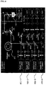
- FIG. 4 is a view showing the configuration of a confocal microscope system according to an embodiment.
- a process of obtaining an image according to the embodiment is as follows.
- the angle of the window apparatus is adjusted using the tilting mount placed on the tilting mount seat 152 of the window apparatus.
- laser beams having a plurality of wavelengths are radiated to a breast tissue through the open window 110 of the window apparatus.
- a fluorescent signal excited in the cancer tissue is detected through a detector.
- the confocal microscope system includes four laser sources 400-1 to 400-4 respectively four wavelengths of 405nm, 488nm, 561nm, and 640nm within the visible light band, a polygonal rotation mirror 402, and a galvanometer mirror 404, and generates s XY raster scanning pattern, using these components.
- the confocal microscope system may include a plurality of neutral density filters ND, mirrors M, and Dichroic beam splitters DBS, and beam pass filters BPF and photomultiplier tubes(PMT) for detecting a fluorescent signal excited in a breast tissue.
- Images of a breast tissue were obtained from an actual animal model, using the confocal optical microscope using the window apparatus of the present disclosure.
- An optical system was designed to have an observation view of 250 ⁇ 250 ⁇ m 2 at the focus when using a ⁇ 40 objective lens (LUCPlanFL, NA0.6; Olympus), and a fluorescent signal was detected and processed by photomultiplier tubes and frame grabbers (Matrox, SOLIOS) that are provided for respective wavelengths such that 2D images having cellular-level resolution and being able to be sectioned in the Z-axial direction could be obtained at a speed of 30 sheets per second.
- LOCPlanFL ⁇ 40 objective lens
- SOLIOS frame grabbers
- FIG. 5 is a view showing the cells and structure of an imaged lactiferous duct of an Actin-GFP mouse, using the window apparatus and the confocal microscope system according to an embodiment.
- FIG. 6 is a view obtained by observing a lactiferous duct formation process at a cellular-level, using the window apparatus and the confocal microscope system according to an embodiment.
- FIG. 7 shows pictures obtained by observing movement of a cell at intervals of 30 minutes after implant and culture a breast cancer cell, MDA-MB-231-GFP Cell in a breast tissue.
- FIG. 8 shows pictures obtained by observing blood vessels formed around a cancer cell, using the window apparatus and the confocal microscope system according to an embodiment.
Landscapes
- Physics & Mathematics (AREA)
- Health & Medical Sciences (AREA)
- Analytical Chemistry (AREA)
- General Physics & Mathematics (AREA)
- Optics & Photonics (AREA)
- Chemical & Material Sciences (AREA)
- Life Sciences & Earth Sciences (AREA)
- Surgery (AREA)
- General Health & Medical Sciences (AREA)
- Engineering & Computer Science (AREA)
- Pathology (AREA)
- Heart & Thoracic Surgery (AREA)
- Medical Informatics (AREA)
- Molecular Biology (AREA)
- Biomedical Technology (AREA)
- Animal Behavior & Ethology (AREA)
- Public Health (AREA)
- Veterinary Medicine (AREA)
- Radiology & Medical Imaging (AREA)
- Ophthalmology & Optometry (AREA)
- Biophysics (AREA)
- Nuclear Medicine, Radiotherapy & Molecular Imaging (AREA)
- Oral & Maxillofacial Surgery (AREA)
- Microscoopes, Condenser (AREA)
- Investigating, Analyzing Materials By Fluorescence Or Luminescence (AREA)
- Multimedia (AREA)
- Gynecology & Obstetrics (AREA)
- Reproductive Health (AREA)
- Apparatus Associated With Microorganisms And Enzymes (AREA)
Applications Claiming Priority (2)
| Application Number | Priority Date | Filing Date | Title |
|---|---|---|---|
| KR1020150123212A KR101689879B1 (ko) | 2015-08-31 | 2015-08-31 | 생체 내 유방조직 미세영상 획득을 위한 윈도우 장치 및 이를 이용한 영상 획득 방법 |
| PCT/KR2016/009719 WO2017039315A1 (ko) | 2015-08-31 | 2016-08-03 | 생체 내 유방조직 미세영상 획득을 위한 원도우 장치 및 이를 이용한 영상 획득 방법 |
Publications (3)
| Publication Number | Publication Date |
|---|---|
| EP3345535A1 true EP3345535A1 (de) | 2018-07-11 |
| EP3345535A4 EP3345535A4 (de) | 2019-04-24 |
| EP3345535B1 EP3345535B1 (de) | 2022-10-05 |
Family
ID=57733653
Family Applications (1)
| Application Number | Title | Priority Date | Filing Date |
|---|---|---|---|
| EP16842281.4A Active EP3345535B1 (de) | 2015-08-31 | 2016-08-31 | Fenstervorrichtung zur gewinnung eines mikroskopischen bildes von in-vivo-brustgewebe und verfahren zur gewinnung eines bildes damit |
Country Status (6)
| Country | Link |
|---|---|
| US (1) | US11029504B2 (de) |
| EP (1) | EP3345535B1 (de) |
| JP (1) | JP6670385B2 (de) |
| KR (1) | KR101689879B1 (de) |
| CN (1) | CN108366727B (de) |
| WO (1) | WO2017039315A1 (de) |
Families Citing this family (3)
| Publication number | Priority date | Publication date | Assignee | Title |
|---|---|---|---|---|
| KR101849706B1 (ko) | 2017-08-24 | 2018-04-18 | 한국화학연구원 | 생체 조직 이미지 관찰용 장치, 이의 제조방법, 및 이를 이용한 생체 조직 이미지를 관찰하는 방법 |
| KR102186327B1 (ko) * | 2018-10-17 | 2020-12-03 | 한국과학기술원 | 생체 심부 조직의 미세 영상 획득 시스템 및 이의 미세 영상 제공 방법 |
| WO2020080721A1 (ko) * | 2018-10-17 | 2020-04-23 | 한국과학기술원 | 생체 심부 조직의 미세 영상 획득 시스템 및 이의 미세 영상 제공 방법 |
Family Cites Families (26)
| Publication number | Priority date | Publication date | Assignee | Title |
|---|---|---|---|---|
| JPS5612598Y2 (de) * | 1975-07-15 | 1981-03-24 | ||
| JPS5421752A (en) * | 1977-07-19 | 1979-02-19 | Toshiba Corp | Stage device for microscopes |
| US4974952A (en) * | 1988-03-31 | 1990-12-04 | Focht Daniel C | Live cell chamber for microscopes |
| JPH0618949U (ja) * | 1992-08-12 | 1994-03-11 | 東ソー株式会社 | 採取試料の連続計量装置 |
| JP3214805B2 (ja) * | 1995-07-17 | 2001-10-02 | シャープ株式会社 | 対物レンズ調整機構 |
| JPH10165403A (ja) * | 1996-12-13 | 1998-06-23 | Toshiba Corp | マンモ用バイオプシー装置 |
| WO2000049392A1 (en) * | 1999-02-17 | 2000-08-24 | Lucid, Inc. | Cassette for facilitating optical sectioning of a retained tissue specimen |
| DE602004014697D1 (de) * | 2003-10-17 | 2008-08-14 | Olympus Co | Objektiveinführvorrichtung, Befestigungsvorrichtung für ein Objektivsystem |
| JP4579563B2 (ja) * | 2004-03-15 | 2010-11-10 | オリンパス株式会社 | 対物光学系の固定装置 |
| KR100537070B1 (ko) * | 2004-03-06 | 2005-12-16 | 이용진 | 현미경용 챔버형 시료 장착기 |
| EP1748724A1 (de) * | 2004-05-11 | 2007-02-07 | Koninklijke Philips Electronics N.V. | Messkopf für die nichtinvasive blutanalyse |
| JP4624725B2 (ja) * | 2004-05-28 | 2011-02-02 | オリンパス株式会社 | 顕微鏡観察システムおよび顕微鏡観察方法 |
| US20050280892A1 (en) * | 2004-05-28 | 2005-12-22 | Nobuyuki Nagasawa | Examination method and examination apparatus |
| WO2006124672A2 (en) | 2005-05-12 | 2006-11-23 | University Of Alabama In Huntsville | Apparatus and method for incubating cell cultures |
| JP4885545B2 (ja) * | 2006-01-12 | 2012-02-29 | オリンパス株式会社 | 観察装置 |
| JP4996977B2 (ja) * | 2007-05-25 | 2012-08-08 | オリンパス株式会社 | スタビライザおよび生体観察装置 |
| US20090185980A1 (en) * | 2008-01-23 | 2009-07-23 | National Taiwan University | In vivo drug screening system |
| CA2731956A1 (en) * | 2008-07-25 | 2010-01-28 | Daniel S. Gareau | Rapid confocal microscopy to support surgical procedures |
| KR101042505B1 (ko) * | 2008-11-27 | 2011-06-16 | 현대제철 주식회사 | 주사 전자 현미경의 시편 홀더장치 |
| US8259170B2 (en) * | 2009-08-24 | 2012-09-04 | Cellomics, Inc. | Integrated calibration sample bay for fluorescence readers |
| JP5700950B2 (ja) * | 2010-04-21 | 2015-04-15 | キヤノン株式会社 | 生体情報取得装置 |
| JP5779963B2 (ja) * | 2011-04-28 | 2015-09-16 | ナノフォトン株式会社 | 観察試料密閉容器 |
| JP2013090867A (ja) * | 2011-10-27 | 2013-05-16 | Canon Inc | 被検体情報取得装置およびその制御方法 |
| JP5930364B2 (ja) * | 2011-11-28 | 2016-06-08 | 国立大学法人京都大学 | 生体試料固定器 |
| JP6037732B2 (ja) * | 2012-09-03 | 2016-12-07 | オリンパス株式会社 | 浸液保持具、観察部位固定装置、及び、顕微鏡 |
| EP3149533A4 (de) * | 2014-05-29 | 2017-06-07 | Rarecyte, Inc. | Vorrichtung zum halten eines substrats innerhalb einer sekundären vorrichtung |
-
2015
- 2015-08-31 KR KR1020150123212A patent/KR101689879B1/ko active Active
-
2016
- 2016-08-03 WO PCT/KR2016/009719 patent/WO2017039315A1/ko not_active Ceased
- 2016-08-03 JP JP2018530458A patent/JP6670385B2/ja active Active
- 2016-08-31 EP EP16842281.4A patent/EP3345535B1/de active Active
- 2016-08-31 CN CN201680062598.4A patent/CN108366727B/zh active Active
- 2016-08-31 US US15/756,394 patent/US11029504B2/en active Active
Also Published As
| Publication number | Publication date |
|---|---|
| JP6670385B2 (ja) | 2020-03-18 |
| EP3345535A4 (de) | 2019-04-24 |
| KR101689879B1 (ko) | 2016-12-26 |
| US20180235476A1 (en) | 2018-08-23 |
| CN108366727A (zh) | 2018-08-03 |
| WO2017039315A8 (ko) | 2018-03-22 |
| JP2018533078A (ja) | 2018-11-08 |
| WO2017039315A1 (ko) | 2017-03-09 |
| EP3345535B1 (de) | 2022-10-05 |
| CN108366727B (zh) | 2021-03-23 |
| US11029504B2 (en) | 2021-06-08 |
Similar Documents
| Publication | Publication Date | Title |
|---|---|---|
| Thériault et al. | Extended two-photon microscopy in live samples with Bessel beams: steadier focus, faster volume scans, and simpler stereoscopic imaging | |
| CN110161668B (zh) | 用于样品成像的显微镜模块 | |
| Gómez-Gaviro et al. | Optimized CUBIC protocol for three-dimensional imaging of chicken embryos at single-cell resolution | |
| US20130335817A1 (en) | Multiple light source microscope | |
| EP3345535B1 (de) | Fenstervorrichtung zur gewinnung eines mikroskopischen bildes von in-vivo-brustgewebe und verfahren zur gewinnung eines bildes damit | |
| JP2012014066A5 (de) | ||
| US20210231942A1 (en) | System for in vivo microscopic imaging of deep tissue, and microscopic imaging method | |
| KR101767339B1 (ko) | 생체 내 췌장조직 미세영상 획득을 위한 윈도우 장치 및 이를 이용한 영상 획득 방법 | |
| Conci et al. | In vivo label-free tissue histology through a microstructured imaging window | |
| EP3345546A1 (de) | Auf mikroaspiration basierende lungenfenstervorrichtung zum erhalt eines mikroskopischen bildes von in-vivo-lungengewebe und verfahren zum erhalt des bildes damit | |
| Bell | Imaging morphogenesis | |
| JP7113645B2 (ja) | 試料保持容器及びライトシート顕微鏡 | |
| Yuan et al. | Calcium imaging of inner ear hair cells within the cochlear epithelium of mice using two-photon microscopy | |
| JP6722620B2 (ja) | 細胞状態の解析装置および解析方法 | |
| Schade-Mann et al. | Calcium signaling in interdental cells during the critical developmental period of the mouse cochlea | |
| Bernardello | Development of novel multimodal light-sheet fluorescence microscopes for in-vivo imaging of vertebrate organisms | |
| Morozov et al. | Light Sheet Microscopy Comes of Age | |
| JP5302063B2 (ja) | 微弱光および高強度光の画像を撮像可能な顕微鏡撮像装置 | |
| Lee et al. | Observation of cell division in a fertilized egg of a zebrafish by using a multimodal nonlinear optical microscope | |
| Carmichael | Opening a New Window for Observing Embryogenesis | |
| JP2021114957A (ja) | 培養組織の観察方法、培養方法、評価方法及び培養器具 | |
| De Mauro et al. | Detection of calcium waves in mice heart tissue with multispot two-photon imaging | |
| KR20170110940A (ko) | 세포 배양 장치 및 이를 포함하는 세포 배양 시스템 |
Legal Events
| Date | Code | Title | Description |
|---|---|---|---|
| STAA | Information on the status of an ep patent application or granted ep patent |
Free format text: STATUS: THE INTERNATIONAL PUBLICATION HAS BEEN MADE |
|
| PUAI | Public reference made under article 153(3) epc to a published international application that has entered the european phase |
Free format text: ORIGINAL CODE: 0009012 |
|
| STAA | Information on the status of an ep patent application or granted ep patent |
Free format text: STATUS: REQUEST FOR EXAMINATION WAS MADE |
|
| 17P | Request for examination filed |
Effective date: 20180322 |
|
| AK | Designated contracting states |
Kind code of ref document: A1 Designated state(s): AL AT BE BG CH CY CZ DE DK EE ES FI FR GB GR HR HU IE IS IT LI LT LU LV MC MK MT NL NO PL PT RO RS SE SI SK SM TR |
|
| AX | Request for extension of the european patent |
Extension state: BA ME |
|
| DAV | Request for validation of the european patent (deleted) | ||
| DAX | Request for extension of the european patent (deleted) | ||
| A4 | Supplementary search report drawn up and despatched |
Effective date: 20190325 |
|
| RIC1 | Information provided on ipc code assigned before grant |
Ipc: A61B 90/25 20160101ALI20190319BHEP Ipc: A61B 5/00 20060101AFI20190319BHEP Ipc: G02B 21/34 20060101ALI20190319BHEP Ipc: G02B 21/36 20060101ALI20190319BHEP Ipc: G02B 21/00 20060101ALI20190319BHEP |
|
| GRAP | Despatch of communication of intention to grant a patent |
Free format text: ORIGINAL CODE: EPIDOSNIGR1 |
|
| STAA | Information on the status of an ep patent application or granted ep patent |
Free format text: STATUS: GRANT OF PATENT IS INTENDED |
|
| INTG | Intention to grant announced |
Effective date: 20220630 |
|
| GRAS | Grant fee paid |
Free format text: ORIGINAL CODE: EPIDOSNIGR3 |
|
| GRAA | (expected) grant |
Free format text: ORIGINAL CODE: 0009210 |
|
| STAA | Information on the status of an ep patent application or granted ep patent |
Free format text: STATUS: THE PATENT HAS BEEN GRANTED |
|
| AK | Designated contracting states |
Kind code of ref document: B1 Designated state(s): AL AT BE BG CH CY CZ DE DK EE ES FI FR GB GR HR HU IE IS IT LI LT LU LV MC MK MT NL NO PL PT RO RS SE SI SK SM TR |
|
| REG | Reference to a national code |
Ref country code: GB Ref legal event code: FG4D |
|
| REG | Reference to a national code |
Ref country code: CH Ref legal event code: EP |
|
| REG | Reference to a national code |
Ref country code: AT Ref legal event code: REF Ref document number: 1522231 Country of ref document: AT Kind code of ref document: T Effective date: 20221015 |
|
| REG | Reference to a national code |
Ref country code: IE Ref legal event code: FG4D |
|
| REG | Reference to a national code |
Ref country code: DE Ref legal event code: R096 Ref document number: 602016075507 Country of ref document: DE |
|
| REG | Reference to a national code |
Ref country code: LT Ref legal event code: MG9D |
|
| REG | Reference to a national code |
Ref country code: NL Ref legal event code: MP Effective date: 20221005 |
|
| REG | Reference to a national code |
Ref country code: AT Ref legal event code: MK05 Ref document number: 1522231 Country of ref document: AT Kind code of ref document: T Effective date: 20221005 |
|
| PG25 | Lapsed in a contracting state [announced via postgrant information from national office to epo] |
Ref country code: NL Free format text: LAPSE BECAUSE OF FAILURE TO SUBMIT A TRANSLATION OF THE DESCRIPTION OR TO PAY THE FEE WITHIN THE PRESCRIBED TIME-LIMIT Effective date: 20221005 |
|
| PG25 | Lapsed in a contracting state [announced via postgrant information from national office to epo] |
Ref country code: SE Free format text: LAPSE BECAUSE OF FAILURE TO SUBMIT A TRANSLATION OF THE DESCRIPTION OR TO PAY THE FEE WITHIN THE PRESCRIBED TIME-LIMIT Effective date: 20221005 Ref country code: PT Free format text: LAPSE BECAUSE OF FAILURE TO SUBMIT A TRANSLATION OF THE DESCRIPTION OR TO PAY THE FEE WITHIN THE PRESCRIBED TIME-LIMIT Effective date: 20230206 Ref country code: NO Free format text: LAPSE BECAUSE OF FAILURE TO SUBMIT A TRANSLATION OF THE DESCRIPTION OR TO PAY THE FEE WITHIN THE PRESCRIBED TIME-LIMIT Effective date: 20230105 Ref country code: LT Free format text: LAPSE BECAUSE OF FAILURE TO SUBMIT A TRANSLATION OF THE DESCRIPTION OR TO PAY THE FEE WITHIN THE PRESCRIBED TIME-LIMIT Effective date: 20221005 Ref country code: FI Free format text: LAPSE BECAUSE OF FAILURE TO SUBMIT A TRANSLATION OF THE DESCRIPTION OR TO PAY THE FEE WITHIN THE PRESCRIBED TIME-LIMIT Effective date: 20221005 Ref country code: ES Free format text: LAPSE BECAUSE OF FAILURE TO SUBMIT A TRANSLATION OF THE DESCRIPTION OR TO PAY THE FEE WITHIN THE PRESCRIBED TIME-LIMIT Effective date: 20221005 Ref country code: AT Free format text: LAPSE BECAUSE OF FAILURE TO SUBMIT A TRANSLATION OF THE DESCRIPTION OR TO PAY THE FEE WITHIN THE PRESCRIBED TIME-LIMIT Effective date: 20221005 |
|
| PG25 | Lapsed in a contracting state [announced via postgrant information from national office to epo] |
Ref country code: RS Free format text: LAPSE BECAUSE OF FAILURE TO SUBMIT A TRANSLATION OF THE DESCRIPTION OR TO PAY THE FEE WITHIN THE PRESCRIBED TIME-LIMIT Effective date: 20221005 Ref country code: PL Free format text: LAPSE BECAUSE OF FAILURE TO SUBMIT A TRANSLATION OF THE DESCRIPTION OR TO PAY THE FEE WITHIN THE PRESCRIBED TIME-LIMIT Effective date: 20221005 Ref country code: LV Free format text: LAPSE BECAUSE OF FAILURE TO SUBMIT A TRANSLATION OF THE DESCRIPTION OR TO PAY THE FEE WITHIN THE PRESCRIBED TIME-LIMIT Effective date: 20221005 Ref country code: IS Free format text: LAPSE BECAUSE OF FAILURE TO SUBMIT A TRANSLATION OF THE DESCRIPTION OR TO PAY THE FEE WITHIN THE PRESCRIBED TIME-LIMIT Effective date: 20230205 Ref country code: HR Free format text: LAPSE BECAUSE OF FAILURE TO SUBMIT A TRANSLATION OF THE DESCRIPTION OR TO PAY THE FEE WITHIN THE PRESCRIBED TIME-LIMIT Effective date: 20221005 Ref country code: GR Free format text: LAPSE BECAUSE OF FAILURE TO SUBMIT A TRANSLATION OF THE DESCRIPTION OR TO PAY THE FEE WITHIN THE PRESCRIBED TIME-LIMIT Effective date: 20230106 |
|
| REG | Reference to a national code |
Ref country code: DE Ref legal event code: R097 Ref document number: 602016075507 Country of ref document: DE |
|
| P01 | Opt-out of the competence of the unified patent court (upc) registered |
Effective date: 20230607 |
|
| PG25 | Lapsed in a contracting state [announced via postgrant information from national office to epo] |
Ref country code: SM Free format text: LAPSE BECAUSE OF FAILURE TO SUBMIT A TRANSLATION OF THE DESCRIPTION OR TO PAY THE FEE WITHIN THE PRESCRIBED TIME-LIMIT Effective date: 20221005 Ref country code: RO Free format text: LAPSE BECAUSE OF FAILURE TO SUBMIT A TRANSLATION OF THE DESCRIPTION OR TO PAY THE FEE WITHIN THE PRESCRIBED TIME-LIMIT Effective date: 20221005 Ref country code: EE Free format text: LAPSE BECAUSE OF FAILURE TO SUBMIT A TRANSLATION OF THE DESCRIPTION OR TO PAY THE FEE WITHIN THE PRESCRIBED TIME-LIMIT Effective date: 20221005 Ref country code: DK Free format text: LAPSE BECAUSE OF FAILURE TO SUBMIT A TRANSLATION OF THE DESCRIPTION OR TO PAY THE FEE WITHIN THE PRESCRIBED TIME-LIMIT Effective date: 20221005 Ref country code: CZ Free format text: LAPSE BECAUSE OF FAILURE TO SUBMIT A TRANSLATION OF THE DESCRIPTION OR TO PAY THE FEE WITHIN THE PRESCRIBED TIME-LIMIT Effective date: 20221005 |
|
| PLBE | No opposition filed within time limit |
Free format text: ORIGINAL CODE: 0009261 |
|
| STAA | Information on the status of an ep patent application or granted ep patent |
Free format text: STATUS: NO OPPOSITION FILED WITHIN TIME LIMIT |
|
| PG25 | Lapsed in a contracting state [announced via postgrant information from national office to epo] |
Ref country code: SK Free format text: LAPSE BECAUSE OF FAILURE TO SUBMIT A TRANSLATION OF THE DESCRIPTION OR TO PAY THE FEE WITHIN THE PRESCRIBED TIME-LIMIT Effective date: 20221005 Ref country code: AL Free format text: LAPSE BECAUSE OF FAILURE TO SUBMIT A TRANSLATION OF THE DESCRIPTION OR TO PAY THE FEE WITHIN THE PRESCRIBED TIME-LIMIT Effective date: 20221005 |
|
| 26N | No opposition filed |
Effective date: 20230706 |
|
| PG25 | Lapsed in a contracting state [announced via postgrant information from national office to epo] |
Ref country code: SI Free format text: LAPSE BECAUSE OF FAILURE TO SUBMIT A TRANSLATION OF THE DESCRIPTION OR TO PAY THE FEE WITHIN THE PRESCRIBED TIME-LIMIT Effective date: 20221005 |
|
| PG25 | Lapsed in a contracting state [announced via postgrant information from national office to epo] |
Ref country code: MC Free format text: LAPSE BECAUSE OF FAILURE TO SUBMIT A TRANSLATION OF THE DESCRIPTION OR TO PAY THE FEE WITHIN THE PRESCRIBED TIME-LIMIT Effective date: 20221005 |
|
| REG | Reference to a national code |
Ref country code: CH Ref legal event code: PL |
|
| PG25 | Lapsed in a contracting state [announced via postgrant information from national office to epo] |
Ref country code: MC Free format text: LAPSE BECAUSE OF FAILURE TO SUBMIT A TRANSLATION OF THE DESCRIPTION OR TO PAY THE FEE WITHIN THE PRESCRIBED TIME-LIMIT Effective date: 20221005 |
|
| PG25 | Lapsed in a contracting state [announced via postgrant information from national office to epo] |
Ref country code: LU Free format text: LAPSE BECAUSE OF NON-PAYMENT OF DUE FEES Effective date: 20230831 |
|
| PG25 | Lapsed in a contracting state [announced via postgrant information from national office to epo] |
Ref country code: LU Free format text: LAPSE BECAUSE OF NON-PAYMENT OF DUE FEES Effective date: 20230831 Ref country code: CH Free format text: LAPSE BECAUSE OF NON-PAYMENT OF DUE FEES Effective date: 20230831 |
|
| REG | Reference to a national code |
Ref country code: BE Ref legal event code: MM Effective date: 20230831 |
|
| REG | Reference to a national code |
Ref country code: IE Ref legal event code: MM4A |
|
| PG25 | Lapsed in a contracting state [announced via postgrant information from national office to epo] |
Ref country code: IT Free format text: LAPSE BECAUSE OF FAILURE TO SUBMIT A TRANSLATION OF THE DESCRIPTION OR TO PAY THE FEE WITHIN THE PRESCRIBED TIME-LIMIT Effective date: 20221005 |
|
| PG25 | Lapsed in a contracting state [announced via postgrant information from national office to epo] |
Ref country code: IE Free format text: LAPSE BECAUSE OF NON-PAYMENT OF DUE FEES Effective date: 20230831 |
|
| PG25 | Lapsed in a contracting state [announced via postgrant information from national office to epo] |
Ref country code: IE Free format text: LAPSE BECAUSE OF NON-PAYMENT OF DUE FEES Effective date: 20230831 |
|
| PG25 | Lapsed in a contracting state [announced via postgrant information from national office to epo] |
Ref country code: BE Free format text: LAPSE BECAUSE OF NON-PAYMENT OF DUE FEES Effective date: 20230831 |
|
| PG25 | Lapsed in a contracting state [announced via postgrant information from national office to epo] |
Ref country code: BG Free format text: LAPSE BECAUSE OF FAILURE TO SUBMIT A TRANSLATION OF THE DESCRIPTION OR TO PAY THE FEE WITHIN THE PRESCRIBED TIME-LIMIT Effective date: 20221005 |
|
| PG25 | Lapsed in a contracting state [announced via postgrant information from national office to epo] |
Ref country code: BG Free format text: LAPSE BECAUSE OF FAILURE TO SUBMIT A TRANSLATION OF THE DESCRIPTION OR TO PAY THE FEE WITHIN THE PRESCRIBED TIME-LIMIT Effective date: 20221005 |
|
| PG25 | Lapsed in a contracting state [announced via postgrant information from national office to epo] |
Ref country code: CY Free format text: LAPSE BECAUSE OF FAILURE TO SUBMIT A TRANSLATION OF THE DESCRIPTION OR TO PAY THE FEE WITHIN THE PRESCRIBED TIME-LIMIT; INVALID AB INITIO Effective date: 20160831 |
|
| PG25 | Lapsed in a contracting state [announced via postgrant information from national office to epo] |
Ref country code: HU Free format text: LAPSE BECAUSE OF FAILURE TO SUBMIT A TRANSLATION OF THE DESCRIPTION OR TO PAY THE FEE WITHIN THE PRESCRIBED TIME-LIMIT; INVALID AB INITIO Effective date: 20160831 |
|
| PGFP | Annual fee paid to national office [announced via postgrant information from national office to epo] |
Ref country code: DE Payment date: 20250805 Year of fee payment: 10 |
|
| PGFP | Annual fee paid to national office [announced via postgrant information from national office to epo] |
Ref country code: GB Payment date: 20250805 Year of fee payment: 10 |
|
| PGFP | Annual fee paid to national office [announced via postgrant information from national office to epo] |
Ref country code: FR Payment date: 20250807 Year of fee payment: 10 |
|
| PG25 | Lapsed in a contracting state [announced via postgrant information from national office to epo] |
Ref country code: TR Free format text: LAPSE BECAUSE OF FAILURE TO SUBMIT A TRANSLATION OF THE DESCRIPTION OR TO PAY THE FEE WITHIN THE PRESCRIBED TIME-LIMIT Effective date: 20221005 |