EP3349010B1 - Anti-vla-4-assays - Google Patents
Anti-vla-4-assays Download PDFInfo
- Publication number
- EP3349010B1 EP3349010B1 EP17205670.7A EP17205670A EP3349010B1 EP 3349010 B1 EP3349010 B1 EP 3349010B1 EP 17205670 A EP17205670 A EP 17205670A EP 3349010 B1 EP3349010 B1 EP 3349010B1
- Authority
- EP
- European Patent Office
- Prior art keywords
- antibody
- days
- test
- capture agent
- vla
- Prior art date
- Legal status (The legal status is an assumption and is not a legal conclusion. Google has not performed a legal analysis and makes no representation as to the accuracy of the status listed.)
- Active
Links
Images

Classifications
-
- G—PHYSICS
- G01—MEASURING; TESTING
- G01N—INVESTIGATING OR ANALYSING MATERIALS BY DETERMINING THEIR CHEMICAL OR PHYSICAL PROPERTIES
- G01N33/00—Investigating or analysing materials by specific methods not covered by groups G01N1/00 - G01N31/00
- G01N33/48—Biological material, e.g. blood, urine; Haemocytometers
- G01N33/50—Chemical analysis of biological material, e.g. blood, urine; Testing involving biospecific ligand binding methods; Immunological testing
- G01N33/68—Chemical analysis of biological material, e.g. blood, urine; Testing involving biospecific ligand binding methods; Immunological testing involving proteins, peptides or amino acids
- G01N33/6854—Immunoglobulins
-
- A—HUMAN NECESSITIES
- A61—MEDICAL OR VETERINARY SCIENCE; HYGIENE
- A61P—SPECIFIC THERAPEUTIC ACTIVITY OF CHEMICAL COMPOUNDS OR MEDICINAL PREPARATIONS
- A61P31/00—Antiinfectives, i.e. antibiotics, antiseptics, chemotherapeutics
- A61P31/12—Antivirals
-
- A—HUMAN NECESSITIES
- A61—MEDICAL OR VETERINARY SCIENCE; HYGIENE
- A61P—SPECIFIC THERAPEUTIC ACTIVITY OF CHEMICAL COMPOUNDS OR MEDICINAL PREPARATIONS
- A61P37/00—Drugs for immunological or allergic disorders
- A61P37/02—Immunomodulators
-
- G—PHYSICS
- G01—MEASURING; TESTING
- G01N—INVESTIGATING OR ANALYSING MATERIALS BY DETERMINING THEIR CHEMICAL OR PHYSICAL PROPERTIES
- G01N33/00—Investigating or analysing materials by specific methods not covered by groups G01N1/00 - G01N31/00
- G01N33/48—Biological material, e.g. blood, urine; Haemocytometers
- G01N33/50—Chemical analysis of biological material, e.g. blood, urine; Testing involving biospecific ligand binding methods; Immunological testing
- G01N33/53—Immunoassay; Biospecific binding assay; Materials therefor
- G01N33/543—Immunoassay; Biospecific binding assay; Materials therefor with an insoluble carrier for immobilising immunochemicals
- G01N33/54366—Apparatus specially adapted for solid-phase testing
- G01N33/54386—Analytical elements
- G01N33/54387—Immunochromatographic test strips
- G01N33/54388—Immunochromatographic test strips based on lateral flow
-
- G—PHYSICS
- G01—MEASURING; TESTING
- G01N—INVESTIGATING OR ANALYSING MATERIALS BY DETERMINING THEIR CHEMICAL OR PHYSICAL PROPERTIES
- G01N33/00—Investigating or analysing materials by specific methods not covered by groups G01N1/00 - G01N31/00
- G01N33/48—Biological material, e.g. blood, urine; Haemocytometers
- G01N33/50—Chemical analysis of biological material, e.g. blood, urine; Testing involving biospecific ligand binding methods; Immunological testing
- G01N33/53—Immunoassay; Biospecific binding assay; Materials therefor
- G01N33/566—Immunoassay; Biospecific binding assay; Materials therefor using specific carrier or receptor proteins as ligand binding reagents where possible specific carrier or receptor proteins are classified with their target compounds
-
- G—PHYSICS
- G01—MEASURING; TESTING
- G01N—INVESTIGATING OR ANALYSING MATERIALS BY DETERMINING THEIR CHEMICAL OR PHYSICAL PROPERTIES
- G01N33/00—Investigating or analysing materials by specific methods not covered by groups G01N1/00 - G01N31/00
- G01N33/48—Biological material, e.g. blood, urine; Haemocytometers
- G01N33/50—Chemical analysis of biological material, e.g. blood, urine; Testing involving biospecific ligand binding methods; Immunological testing
- G01N33/94—Chemical analysis of biological material, e.g. blood, urine; Testing involving biospecific ligand binding methods; Immunological testing involving narcotics or drugs or pharmaceuticals, neurotransmitters or associated receptors
-
- G—PHYSICS
- G01—MEASURING; TESTING
- G01N—INVESTIGATING OR ANALYSING MATERIALS BY DETERMINING THEIR CHEMICAL OR PHYSICAL PROPERTIES
- G01N2333/00—Assays involving biological materials from specific organisms or of a specific nature
- G01N2333/435—Assays involving biological materials from specific organisms or of a specific nature from animals; from humans
- G01N2333/705—Assays involving receptors, cell surface antigens or cell surface determinants
- G01N2333/70546—Integrin superfamily, e.g. VLAs, leuCAM, GPIIb/GPIIIa, LPAM
-
- G—PHYSICS
- G01—MEASURING; TESTING
- G01N—INVESTIGATING OR ANALYSING MATERIALS BY DETERMINING THEIR CHEMICAL OR PHYSICAL PROPERTIES
- G01N2333/00—Assays involving biological materials from specific organisms or of a specific nature
- G01N2333/435—Assays involving biological materials from specific organisms or of a specific nature from animals; from humans
- G01N2333/705—Assays involving receptors, cell surface antigens or cell surface determinants
- G01N2333/70546—Integrin superfamily, e.g. VLAs, leuCAM, GPIIb/GPIIIa, LPAM
- G01N2333/7055—Integrin beta1-subunit-containing molecules, e.g. CD29, CD49
-
- G—PHYSICS
- G01—MEASURING; TESTING
- G01N—INVESTIGATING OR ANALYSING MATERIALS BY DETERMINING THEIR CHEMICAL OR PHYSICAL PROPERTIES
- G01N2800/00—Detection or diagnosis of diseases
- G01N2800/26—Infectious diseases, e.g. generalised sepsis
-
- G—PHYSICS
- G01—MEASURING; TESTING
- G01N—INVESTIGATING OR ANALYSING MATERIALS BY DETERMINING THEIR CHEMICAL OR PHYSICAL PROPERTIES
- G01N2800/00—Detection or diagnosis of diseases
- G01N2800/52—Predicting or monitoring the response to treatment, e.g. for selection of therapy based on assay results in personalised medicine; Prognosis
-
- Y—GENERAL TAGGING OF NEW TECHNOLOGICAL DEVELOPMENTS; GENERAL TAGGING OF CROSS-SECTIONAL TECHNOLOGIES SPANNING OVER SEVERAL SECTIONS OF THE IPC; TECHNICAL SUBJECTS COVERED BY FORMER USPC CROSS-REFERENCE ART COLLECTIONS [XRACs] AND DIGESTS
- Y02—TECHNOLOGIES OR APPLICATIONS FOR MITIGATION OR ADAPTATION AGAINST CLIMATE CHANGE
- Y02A—TECHNOLOGIES FOR ADAPTATION TO CLIMATE CHANGE
- Y02A90/00—Technologies having an indirect contribution to adaptation to climate change
- Y02A90/10—Information and communication technologies [ICT] supporting adaptation to climate change, e.g. for weather forecasting or climate simulation
Definitions
- the invention relates to assays associated with immuno-modulatory therapies.
- the invention is based, at least in part, on the development of a test to assay the level of an anti-integrin antibody and the anti-(anti-integrin antibody) antibody, e.g., anti-drug antibody, in a sample.
- the present invention provides a method of assaying the efficacy of a treatment, the method comprising:
- the invention further provides a kit for assaying the level of an anti-anti-alpha4 integrin antibody in a biological sample in a subject comprising:
- Contacting the sample with a detectable capture agent and detecting and correlating the binding of the capture agent can include assaying in a lateral flow system.
- the binding of the capture agent to the antibody can produce a detectable signal.
- Binding of the capture agent to the antibody can be detected by a secondary agent.
- the biological sample can be obtained from the subject prior to treatment or one day, two days, three days, four days, five days, six days, one week, two weeks, three weeks, or four weeks after the treatment. Obtaining the biological sample can be repeated once a day, or once every two days, three days, four days, five days, six days, one week, two weeks, three weeks, or four weeks.
- the biological sample can be blood, plasma, serum, urine, saliva, cerebrospinal fluid, sputum, ocular lens fluid, sweat, milk, ascites fluid, mucous, synovial fluid, peritoneal fluid, transdermal exudates, pharyngeal exudates, bronchoalveolar lavage, tracheal aspirations, semen, cervical mucus, vaginal secretion, urethral secretion, or amniotic fluid.
- the anti-anti-integrin antibody is an anti-anti-alpha4 integrin chain antibody.
- the capture agent can be an antibody.
- an element means one element or more than one element.
- the term “purify,” “purified,” or “purification” means the removal or isolation of a molecule from its environment by, for example, isolation or separation.
- “Substantially purified” molecules are at least about 60% free, for example, at least about 75% free, or at least about 90% free from other components with which they are associated. In some cases, a substantially purified molecule is at least 60% free, 75% free, 90%, or 95% free from other components. As used herein, these terms also refer to the removal of contaminants from a sample.
- sample refers to anything that may contain an analyte for which an analyte assay is desired.
- the sample may be a biological sample, such as a biological fluid or a biological tissue.
- biological fluids include blood, serum, plasma, saliva, sputum, ocular lens fluid, sweat, urine, milk, ascites fluid, mucous, synovial fluid, peritoneal fluid, transdermal exudates, pharyngeal exudates, bronchoalveolar lavage, tracheal aspirations, cerebrospinal fluid, semen, cervical mucus, vaginal or urethral secretions, amniotic fluid or the like.
- Biological tissues are aggregate of cells, usually of a particular kind together with their intercellular substance that form one of the structural materials of a human, animal, plant, bacterial, fungal or viral structure, including connective, epithelium, muscle and nerve tissues. Examples of biological tissues also include organs, tumors, lymph nodes, arteries and individual cell(s).
- fluid sample refers to a material suspected of containing an analyte(s) of interest, which material has sufficient fluidity to flow through a device or assay described herein.
- a fluid sample can be obtained from a source and used directly in an assay described herein or can be pretreated so as to modify its character. Such samples can include human, animal or man-made samples.
- the sample can be prepared in any medium that does not interfere with an assay described herein.
- the sample is an aqueous solution or biological fluid described herein.
- a fluid sample can be derived from any source, such as a physiological fluid, including blood, serum, plasma, saliva, sputum, ocular lens fluid, sweat, urine, milk, ascites fluid, mucous, synovial fluid, peritoneal fluid, transdermal exudates, pharyngeal exudates, bronchoalveolar lavage, tracheal aspirations, cerebrospinal fluid, semen, cervical mucus, vaginal or urethral secretions, amniotic fluid, and the like. Fluid homogenates of cellular tissues such as, for example, hair, skin and nail scrapings.
- a solid material to be assayed for the presence of an analyte can be used as a test sample once it is modified to form a liquid medium, e.g., and extract, or to release the analyte.
- antigen means any compound capable of binding to an antibody, or against which antibodies can be raised.
- antibody refers to a polypeptide substantially encoded by an immunoglobulin gene or immunoglobulin genes, or fragments thereof. Recognized immunoglobulin genes include the kappa, lambda, alpha, gamma, delta, epsilon, and mu constant regions, as well as myriad immunoglobulin variable region genes. Light chains are classified as either kappa or lambda. Heavy chains are classified as gamma, mu, alpha, delta, or epsilon, which in turn define the immunoglobulin classes IgG, IgM, IgA, IgD, and IgE, respectively.
- an antibody is an immunoglobulin having an area on its surface or in a cavity that specifically binds to and is thereby defined as complementary with a particular spatial and polar organization of another molecule.
- the antibody can be polyclonal or monoclonal.
- Antibodies may include a complete immunoglobulin or fragments thereof. Fragments thereof can include, for example, Fab, Fab', F(ab') 2 , Fv, single chain Fv (scFv), and the like.
- Antibodies may also include chimeric antibodies or fragment thereof made by recombinant methods.
- the detected antibody is an IgG4 antibody. In an assay as described herein, IgG4 half antibodies can be detected, complete IgG4 antibodies can be detected, or both IgG4 half antibodies and IgG4 complete antibodies can be detected.
- analyte refers to a known or unknown component of a sample, which will specifically bind to a capture agent if the analyte and the capture agent are members of a specific binding pair.
- analytes are biopolymers, e.g., an oligomer or polymer such as an oligonucleotide, a polypeptide, (e.g., an antibody), or the like.
- An analyte can exist in a mobile phase (such as in a fluid) to be detected by a capture agent.
- the term "labeled reagent” refers to a substance comprising a detectable label attached to a specific binding agent.
- the attachment may be covalent or non-covalent binding, but the method of attachment is not critical.
- the label allows the labeled reagent to produce a detectable signal that is related to the presence of an analyte in a sample described herein.
- the specific binding agent component of the labeled reagent is selected to directly or indirectly bind to an analyte.
- the labeled reagent can be associated with a substrate described herein, it can be combined with a fluid sample to form a fluid solution, it can be added to a substrate separately from a sample, or it can be predeposited or reversibly immobilized on the substrate.
- the specific binding agent may be labeled before or during the performance of an assay described herein by means of any suitable attachment method known in the art.
- the detectable label can be a directly detectable label or a label that is detected using indirect methods.
- detectable signal refers to a signal produced by the binding of a capture agent with an analyte.
- a detectable signal may be detectable by visual inspection. Without limitation, the type of signal produced depends on the label used.
- detectable signals indicating the presence or absence of an analyte in a sample may be evident of their own accord, e.g., detectable as lines, plus or minus signs, or particularly shaped symbols, or may be evident through comparison with a reference, such as a color indicator reference.
- the term "capture agent” refers to an agent that binds an analyte through an interaction that is sufficient to permit the agent to specifically bind an analyte.
- the capture agent can specifically bind an analyte with a dissociation constant (K d ) of less than about 10 -6 M without binding to other targets.
- K d dissociation constant
- the binding interaction can be mediated by a binding site of the capture agent.
- Capture agents can include, e.g., any polypeptide, e.g., an antibody. Capture agents can be bound to a substrate or can be present in solution.
- specific binding refers to preferential binding of an agent to a particular analyte that is present in a mixture of different analytes. Typically, specific binding differentiates between a desirable and undesirable analytes in a sample.
- a specific binding agent typically differentiates between a desirable and undesirable analyte by binding more than about 10 to 100-fold or more to a desirable analyte in preference to other analytes. In certain circumstances, such preferential binding can be at least 1000- to 10,000-fold.
- the affinity between a capture agent and analyte when they are specifically bound in a capture agent/analyte complex is at least about 10 -7 M, at least about 10 -8 M, at least about 10 -9 M, at least about 10 -10 M, or about at least 10 -11 e.g., at least 10 -7 M, at least 10 -8 M, at least 10 -9 M, at least 10 -10 M, or at least 10 -11 .
- capture agent/analyte complex is a complex that results from the specific binding of a capture agent with an analyte.
- binding partners refers to pairs of molecules that can be found in a capture agent/analyte complex, i.e., exhibit specific binding with each other.
- assessing refers to any form of measurement, and includes determining if an element is present or not.
- determining means determining if an element is present or not.
- evaluating means determining if an element is present or not.
- assessing means determining if an element is present or not.
- a "detectable label” refers to an atom (e.g., radionuclide), molecule (e.g., fluorescein), or complex, that is detected or can be used to detect (e.g., due to a physical or chemical property) the presence of an analyte, or to enable binding of another molecule to which it is covalently bound or otherwise associated.
- the term also refers to covalently bound or otherwise associated molecules (e.g., a biomolecule such as an enzyme) that act on a substrate to produce a detectable atom, molecule or complex.
- Detectable labels include any composition detectable by spectroscopic, photochemical, biochemical, immunochemical, electrical, optical or chemical means.
- labels include biotin for staining with labeled streptavidin conjugate, magnetic beads (e.g., Dynabeads TM .), fluorescent dyes (e.g., fluorescein, Texas red, rhodamine, green fluorescent protein, enhanced green fluorescent protein, and the like), radiolabels (e.g., 3 H, 125 I, 35 S, 14 C, or 32 P), enzymes (e.g., hydrolases, particularly phosphatases such as alkaline phosphatase, esterases and glycosidases, or oxidoreductases, particularly peroxidases such as horse radish peroxidase, and others commonly used in ELISAs), substrates, cofactors, inhibitors, chemiluminescent groups, chromogenic agents, and colorimetric labels such as colloidal metallic particles (such as gold), colloidal non-metallic particles (such as selenium or tellurium), or colored glass or plastic (e.g., polystyrene
- Non-limiting examples of patents disclosing such labels include U.S. Pat. Nos. 3,817,837 ; 3,850,752 ; 3,939,350 ; 3,996,345 ; 4,277,437 ; 4,275,149 ; and 4,366,241 . Means of detecting such labels are known to those of skill in the art.
- sandwich As used herein, the terms “sandwich”, “sandwich ELISA”, “sandwich diagnostic”, and “capture ELISA” refer to the concept of detecting an analyte with two or more different test agents.
- a capture agent can be directly or indirectly attached to a substrate, and a test sample can be passed over the surface of the substrate allowing the capture agent to specifically bind its cognate analyte.
- a labeled reagent e.g., a labeled antibody or alternative detection reagent (which can bind the analyte), can then be used to determine whether a capture agent specifically bound the analyte.
- the term "substrate” means any support capable of binding or being associated with a capture agent.
- substrates include glass, polystyrene, polypropylene, polyethylene, dextran, nylon, amylases, natural and modified celluloses, polyacrylamides, agarose, and magnetite.
- the nature of the substrate can be either soluble to some extent or insoluble.
- the structure of the substrate is not limiting, and the substrate can have any structural configuration so long as the capture agent is capable of binding to an analyte.
- the structural configuration can be spherical, as in a bead, or cylindrical, as in the inside surface of a test tube, or the external surface of a rod.
- the surface can be flat, such as a sheet, culture dish, test strip, etc.
- the term "subject” refers to any animal, e.g., a mammal, human or non-human.
- exemplary subjects include, but are not limited to, humans, non-human primates, mice, rats, guinea pigs, cattle, sheep, goats, pigs, dogs, cats, birds, deer, elk, rabbit, reindeer, deer, and horses.
- the term “measuring the level of” refers to detection both quantitatively and qualitatively.
- PML progressive multifocal leukoencephalopathy
- natalizumab a progressive multifocal leukoencephalopathy
- one method of treating such subjects is to reduce the amount (e.g., plasma concentration) of the immuno-modulatory therapeutic agent (e.g., natalizumab) in the patient.
- the amount e.g., plasma concentration
- the immuno-modulatory therapeutic agent e.g., natalizumab
- the test Because of the rapid progression of the disease and the wide geographic distribution of patients, as well as other issues, there is a need for a rapid, reliable test that can be used to determine the level of the drug in the patient. It is an advantage for the test to be formatted so it can be administered and accurately interpreted within a short period of time.
- Assays provided herein can detect anti-integrin antibodies in a subject who was treated with a natalizumab and has been diagnosed with or is suspected of having a JC virus infection.
- the JC virus is a type of human polyomavirus which can cause PML and other diseases in patients with immunodeficiency or in patients treated with drug intended to induce a state of immnosuppression (e.g. organ transplant patients).
- Assays are provided herein to detect anti-drug antibodies.
- the apparatus is a solid substrate associated with a capture agent that can be used to specifically detect the immuno-modulatory therapeutic agent (e.g., natalizumab) or antibodies against an immuno-modulatory agent (e.g., anti-drug antibody such as antibodies against natalizumab).
- the detection is calibrated to provide a method of determining whether the amount of immuno-modulatory therapeutic agent or anti-drug antibody is above or below a specified concentration or titer.
- additional therapy e.g., plasmapheresis
- kits suitable for testing for the presence of an anti-anti-alpha4 integrin antibody will generally include at least one reagent or apparatus as described herein for detecting the immuno-modulatory therapeutic agent or antibodies against an immuno-modulatory agent.
- the kit can also include instructions for use of the reagents and/or apparatus. Such instructions can also include use of the kit for determining an amount of an analyte and interpretation of assay results. For example, the instructions can provide guidance as to whether detection of a specified level of a natalizumab is sufficient to treat a subject having or suspected of having PML.
- Various analytical devices are known to those of skill in the art, and such analytical devices can be adapted to utilize one or more capture agents, e.g., an antibody described herein, to assay one or more analytes, e.g., an anti-integrin antibody such as an anti-VLA-4 (e.g., a natalizumab), e.g., an anti-drug antibody (e.g., anti-natalizumab) described herein.
- capture agents e.g., an antibody described herein
- an anti-integrin antibody such as an anti-VLA-4 (e.g., a natalizumab)
- an anti-drug antibody e.g., anti-natalizumab
- Nonlimiting examples include dipstick or tester, lateral flow, dual flow and flow-through devices, particularly those that are immunoassays.
- Non-limiting examples of lateral flow devices include those described in U.S. Pat. Nos.
- dipstick devices include those described in U.S. Pat. Nos: 4,235,601 , 5,559,041 , 5,712,172 and 6,790,611 .
- An apparatus for assaying the level of an analyte can include a substrate, e.g., a substrate with which a capture agent is associated.
- substrates include glass, polystyrene, polypropylene, polyethylene, dextran, nylon, amylases, natural and modified celluloses, polyacrylamides, agarose, and magnetite.
- the substrate can have any structural configuration so long as the capture agent is capable of binding to an analyte.
- the substrate is housed in a support, e.g., a support that is spherical, as in a bead, or cylindrical, as in the inside surface of a test tube, or the external surface of a rod.
- a support e.g., a support that is spherical, as in a bead, or cylindrical, as in the inside surface of a test tube, or the external surface of a rod.
- the substrate or support can be flat, such as a sheet, culture dish, test strip, and the like.
- the apparatus is a lateral flow apparatus that includes a sample receiving zone, a label zone, a test zone and a control zone.
- a test region comprises the test and control zones.
- the test region, which comprises the test and control zones, is observable.
- the sample receiving zone accepts a fluid sample that may contain one or more analytes of interest.
- the sample receiving zone is dipped into a fluid sample.
- a label zone contains one or more mobile labeled reagents that recognize, or are capable of binding, one or more analytes of interest.
- a test region is disposed downstream from the label zone, and contains test and control zones.
- the test zone(s) generally contain a capture agent associated with the substrate at the test zone.
- the capture agent is immobilized on the substrate at the test zone.
- the immobilized capture agent specifically binds to the analyte of interest.
- the test region is comprised of a material that is opaque in a dry state and transparent in a moist state.
- this mark is positioned about the test region such that it becomes visible within the test region when the test region is in a moist state.
- the fluid sample flows along a flow path running from the sample receiving zone (upstream), to the test and control zones (together comprised in a test region) (downstream).
- the fluid sample may thereafter continue to the absorbent zone.
- the sample receiving zone is composed of an absorbent application pad.
- Suitable materials for manufacturing absorbent application pads include, but are not limited to, hydrophilic polyethylene materials or pads, acrylic fiber, glass fiber, filter paper or pads, desiccated paper, paper pulp, fabric, and the like.
- the sample receiving zone may be composed of a material such as a nonwoven spunlaced acrylic fiber (e.g., available from DuPont Nonwovens) or HDK material (available from HDK Industries, Inc., Rogersville, TN).
- the sample receiving zone is constructed from any material that is capable of absorbing water.
- the test device is configured to perform an immunological assay.
- the liquid transport along the substrate is based upon capillary action.
- the liquid transport along the substrate is based on non-bibulous lateral flow, wherein all of the dissolved or dispersed components of the liquid sample are carried at substantially equal rates and with relatively unimpaired flow laterally through the substrate, as opposed to preferential retention of one or more components as would occur, e.g., in materials that interact, chemically, physically, ionically or otherwise with one or more components (see, for example, U.S. Pat. No. 4,943,522 ).
- the labeling zone of immunoassay devices can also include control-type reagents.
- These labeled control reagents often comprise detectable moieties that will not become restrained in the test zones and that are carried through to the test region and control zone(s) by fluid sample flow through the device.
- these detectable moieties are coupled to a member of a specific binding pair to form a control conjugate that can then be restrained in a separate control zone of the test region by a corresponding member of the specific binding pair to verify that the flow of liquid is as expected.
- the visible moieties used in the labeled control reagents can be the same or different color, or of the same or different type, as those used in the analyte of interest specific labeled reagents.
- the test region generally includes a control zone that is useful to verify that the sample flow is as expected.
- Each of the control zones can comprise a spatially distinct region that can include an immobilized member of a specific binding pair that reacts with a labeled control reagent.
- the procedural control zone contains an authentic sample of the analyte of interest, or a fragment thereof.
- one type of labeled reagent can be utilized, wherein fluid sample transports the labeled reagent to the test and control zones; and the labeled reagent not bound to an analyte of interest will then bind to the authentic sample of the analyte of interest positioned in the control zone.
- control line contains antibody that is specific for, or otherwise provides for, the immobilization of the labeled reagent.
- a labeled reagent is restrained in each of the one or more control zones, even when any or all the analytes of interest are absent from the test sample.
- capture agents can be used in the methods and apparatus described herein, provided the capture agent specifically binds an anti-anti-alpha4 integrin antibody to form a complex.
- the capture agent can be an antigen, e.g., an integrin.
- the capture agent is an antibody that binds to an anti-anti-alpha4 integrin antibody.
- Any antibody that specifically binds to an anti-anti-alpha4 integrin antibody can be utilized as a capture agent.
- the antibody can be a polyclonal antibody; a monoclonal antibody or antigen binding fragment thereof; a modified antibody such as a chimeric antibody, reshaped antibody, humanized antibody, or fragment thereof (e.g., Fab', Fab, F(ab') 2 ); or a biosynthetic antibody, e.g., a single chain antibody, single domain antibody (DAB), Fv, single chain Fv (scFv), or the like.
- DAB single domain antibody
- scFv single chain Fv
- the capture agent is an antibody that can specifically bind an anti-integrin antibody (e.g., an anti anti-VLA-4 antibody such as an anti-natalizumab), such as the rat 12C2 antibody, murine 12C4 antibody, or a suitable rabbit antibody.
- an anti-integrin antibody e.g., an anti anti-VLA-4 antibody such as an anti-natalizumab
- the capture agent is an anti-VLA-4 antibody, such as natalizumab, that can specifically bind to an anti-(anti-VLA-4 antibody) antibody.
- analyte of interest e.g., in a biological sample from a subject.
- exemplary analytes include, without limitation, toxins, organic compounds, polypeptides, microorganisms, bacteria, viruses, amino acids, nucleic acids, carbohydrates, hormones, steroids, vitamins, drugs (including those administered for therapeutic purposes as well as those administered for illicit purposes), pollutants, pesticides, and metabolites of or antibodies to any of the above substances.
- Any antigenic substance, hapten, antibody, macromolecule, and combinations thereof can also be an analyte assayed using a method or an apparatus described herein.
- the analyte is an antibody, such as an anti-integrin antibody.
- Integrins are known in the art (see, e.g., Takeda et al., Genome Biol. 8:215 (2007 )), and the level of any anti-integrin antibody can be assayed using a method or an apparatus described herein.
- An antibody can be, e.g., a polyclonal antibody; a monoclonal antibody or antigen binding fragment thereof; a modified antibody such as a chimeric antibody, reshaped antibody, humanized antibody, or fragment thereof (e.g., Fab', Fab, F(ab') 2 ); or a biosynthetic antibody, e.g., a single chain antibody, single domain antibody (DAB), Fv, single chain Fv (scFv), or the like.
- the antibody can be naturally produced by the subject or one that is administered to the subject.
- the analyte according to the invention is an anti-anti-alpha4 integrin antibody.
- the analyte may be an anti-VLA-4 antibody previously administered to the subject, e.g., as a therapeutic antibody.
- the analyte can be, for example, a natalizumab.
- the analyte is an anti-(anti-VLA-4 antibody) antibody.
- the analyte can be, for example, anti-natalizumab antibody.
- Biological samples to be assayed for the presence of analyte using the methods and apparatus described herein can be obtained from any subject, e.g., a human or a non-human subject.
- the biological sample is obtained from a living human subject.
- the subject from whom the sample is obtained can be apparently healthy, where the assay is performed as a part of routine screening.
- the subject can have been provisionally diagnosed as having a disease or disorder and has undergone treatment, e.g., with an anti-integrin antibody.
- the biological sample can be from a subject who has been treated to decrease the amount of analyte.
- a sample from a subject can be tested using a method described herein before the subject undergoes a treatment, e.g., to modulate the amount of analyte in the subject or to establish the baseline level of analyte in the subject or to establish the suitability of the treatment, and after the subject has been treated, e.g., to monitor and/or to modulate the amount of analyte in the subject.
- the biological sample can be derived from any tissue, organ or group of cells of the subject.
- the biological sample is a cervical scrape, biopsy, or lavage obtained from a subject.
- the sample is a blood, plasma, serum, or urine sample.
- the biological sample can be processed, e.g., to remove certain components that may interfere with a method described herein, using methods that are known in the art.
- the biological sample can be processed to be enriched for proteins, e.g., by salt precipitation, and the like.
- the level of an analyte in a sample can be quantified and/or compared to controls.
- Suitable control samples can be, e.g., from individuals who have not received a treatment.
- Control samples can be from individuals genetically related to the subject being assayed, but can also be from genetically unrelated individuals. In some cases, the control is an established reference.
- the level of an analyte can be quantified using known methods. In yet other situations, the presence or absence of an analyte within a sample can be determined by visual inspection of the assay.
- a sample can also be contacted to a solid support-bound capture agent under conditions suitable for binding of the capture agent to analytes in the sample, and, after separation of unbound sample analytes from the bound analytes, the bound analytes are detected as described herein.
- An analyte can be assayed using a method selected from a variety of immunoassay methods, both qualitatively and quantitatively.
- Immunological and immunoassay procedures are known and described in, e.g., Stites et al. (eds.) Basic and Clinical Immunology (4th ed.) Lange Medical Publications, Los Altos, CA .
- Immunoassays can be performed in any of several configurations known in the art and described in, e.g., Maggio Enzyme Immunoassay, CRC Press, Boca Raton, Fla.
- An analyte can be detected and/or quantified using any of a number of known immunological binding assays (see, e.g., U.S. Pat. Nos. 4,366,241 , 4,376,110 ; 4,517,288 ; 4,837,168 ; and Asai, Methods in Cell Biology Volume 37: Antibodies in Cell Biology, Academic Press, Inc. NY (1993 ).
- Immunoassays can also utilize a labeled reagent to specifically bind to and label the capture agent/analyte complex.
- the labeled reagent may itself be one of the moieties comprising the capture agent/analyte complex.
- the labeled reagent may be a third moiety, such as an antibody, that specifically binds to the analyte or the capture agent/analyte complex.
- the analyte can be an antibody, e.g., an anti-integrin antibody, or e.g., an anti-drug antibody, and the labeled reagent can be a second antibody bearing a label.
- the second antibody may lack a label, but it may, in turn, be bound by a labeled reagent that includes a third antibody specific to antibodies of the species from which the second antibody is derived.
- the second antibody can be modified with a detectable moiety, such as biotin, to which a labeled reagent can specifically bind, such as enzyme-labeled streptavidin.
- proteins that can specifically bind immunoglobulin constant regions can also be used as the labeled reagent. These proteins are normal constituents of the cell walls of streptococcal bacteria. They exhibit a strong non-immunogenic reactivity with immunoglobulin constant regions from a variety of species (see, e.g., Kronval et al., J. Immunol. 111:1401-1406 (1973 ); and Akerstrom et al., J. Immunol. 135:2589-2542 (1985 ))
- incubation and/or washing steps may be required after one or more combination of reagents.
- Incubations carried out during the assay can vary from about 5 seconds to several hours, in general, from about 5 minutes to about 24 hours. The incubation time will depend upon the assay format, analyte, volume of solution, concentrations, and the like.
- an assay is carried out at ambient temperature, although an assay can be conducted over a range of temperatures, such as about 4°C to about 40°C, e.g., 4 °C-10 °C, or 4 °C-25 °C.
- Immunoassays for detecting an analyte of interest can be competitive or noncompetitive.
- Noncompetitive immunoassays are assays in which the amount of analyte in a capture agent/analyte complex is directly measured.
- the capture agent can be bound directly to a solid substrate where it is immobilized. These immobilized capture agents can then specifically bind an analyte present in a test sample.
- the analyte e.g., anti-integrin antibody, or e.g., an anti-drug antibody
- a labeled reagent such as a second antibody bearing a label.
- the second antibody may lack a label, but it may, in turn, be bound by a labeled reagent that is a third antibody specific to antibodies of the species from which the second antibody is derived.
- the second antibody can be modified with a detectable moiety, such as biotin, to which a third labeled reagent can specifically bind, such as enzyme-labeled streptavidin.
- the amount of analyte (such as an anti-integrin antibody or an anti-drug antibody) present in a sample is measured indirectly by measuring the amount of an added (exogenous) analyte displaced (or competed away) from a capture agent (e.g., an antibody specific for the analyte) by the analyte present in the sample.
- a capture agent e.g., an antibody specific for the analyte
- an analyte e.g., anti-integrin antibody, or e.g., an anti-drug antibody
- a capture agent e.g., an antibody that specifically binds to an anti-integrin antibody
- the amount of analyte bound to the capture agent, e.g., antibody is inversely proportional to the concentration of analyte present in the sample.
- the capture agent e.g., an antibody that binds to an anti-integrin antibody
- the amount of the analyte, e.g., anti-integrin antibody, or e.g., an anti-drug antibody, bound to the capture agent, e.g., antibody that binds an anti-integrin antibody, or e.g., drug that anti-drug antibody binds to may be determined either by measuring the amount of analyte present in a capture agent/analyte complex or, alternatively, by measuring the amount of remaining uncomplexed analyte. The amount of analyte may be detected by providing a labeled reagent.
- the technique generally comprises separating sample proteins by gel electrophoresis on the basis of molecular weight, transferring the separated proteins to a suitable solid support (such as, e.g., a nitrocellulose filter, a nylon filter, or a derivatized nylon filter), and incubating the sample with the antibodies that specifically bind the polypeptide of interest.
- a suitable solid support such as, e.g., a nitrocellulose filter, a nylon filter, or a derivatized nylon filter
- the antibodies specifically bind to a polypeptide of interest on the solid support.
- These antibodies may be directly labeled or alternatively may be subsequently detected using labeled antibodies (e.g., labeled sheep anti-mouse antibodies) that specifically bind to the antibodies against the polypeptide of interest.
- LOA liposome immunoassays
- the detectable label used in a method or apparatus described herein is not limiting as long as it does not significantly interfere with the specific binding of the labeled reagent used in the method or apparatus.
- the detectable label can be any material having a detectable physical or chemical property. Such detectable labels have been well developed in the field of immunoassays and, in general, most labels useful in such methods can be used in the methods and apparatus described herein.
- a label can be, e.g., any composition detectable by spectroscopic, photochemical, biochemical, immunochemical, electrical, optical or chemical means.
- Useful labels in the methods and apparatus described herein include, without limitation, magnetic beads (e.g., Dynabeads TM ), fluorescent dyes (e.g., fluorescein isothiocyanate, Texas red, rhodamine, and the like), radiolabels (e.g., 3 H, 125 I, 35 S, 14 C, or 32 P), enzymes (e.g., horseradish peroxidase, alkaline phosphatase and others that can be used in an ELISA), and colorimetric or particulate labels such as colloidal gold or colored glass or plastic (e.g., polystyrene, polypropylene, latex, etc.) beads.
- magnetic beads e.g., Dynabeads TM
- fluorescent dyes e.g., fluorescein isothiocyanate, Texas red, rhodamine, and the like
- radiolabels e.g., 3 H, 125 I, 35 S, 14 C, or
- the label can be coupled directly or indirectly to the desired component of the assay according to methods known in the art. As described herein, a wide variety of labels can be used, with the choice of label depending on the sensitivity required, the ease of conjugation, stability requirements, available instrumentation, and disposal provisions.
- Non-radioactive labels can be attached by indirect means.
- Molecules can also be conjugated directly to signal generating compounds, e.g., by conjugation with an enzyme or fluorescent compound.
- an enzyme or fluorescent compound e.g., A variety of enzymes and fluorescent compounds can be used with the methods and apparatus described herein and are well-known to those of skill in the art (see, e.g., U.S. Pat. No. 4,391,904 ).
- Means of detecting labels are known to those of skill in the art.
- means for detection include a scintillation counter or photographic film as in autoradiography.
- the label is a fluorescent label, it can be detected by exciting the fluorochrome with the appropriate wavelength of light and detecting the resulting fluorescence. The fluorescence can be detected visually, by means of photographic film, by the use of electronic detectors such as charge-coupled devices (CCDs) or photomultipliers and the like.
- CCDs charge-coupled devices
- enzymatic labels can be detected by providing the appropriate substrates for the enzyme and detecting the resulting reaction product.
- simple colorimetric labels can be detected directly by observing the color associated with the label. Thus, in various dipstick assays, conjugated gold often appears pink, while various conjugated beads appear the color of the bead.
- agglutination assays can be used to detect the presence of target antibodies.
- antigen-coated particles are agglutinated by samples comprising target antibodies.
- none of the assay components need to be labeled, and the presence of the target antibody is detected by simple visual inspection.
- the signal can be compared to a reference and/or correlated with a predetermined level corresponding to treatment recommendations. For example, in an assay to detect the level of an anti-VLA-4 antibody in a subject being treated with plasmapheresis to reduce the amount of the antibody, a level of signal below a predetermined level may be correlated with a recommendation that further plasmapheresis is not required. A signal correlated with a higher level may indicate that additional plasmapheresis is advisable.
- Detection of signal in an assay described herein can be visual, but may also be performed using a reader to detect a signal.
- readers include, for example, automated plate readers, EIA readers, and the like. Readers can be used for semi-quantitative or quantitative determination of the concentration for tested analytes. Semi-quantitative or quantitative determination of concentration can be depicted using a colorimetric system.
- the secondary agents used in a method or apparatus described herein can be used to detect binding of the capture agent to the antibody
- the secondary agent can be an antibody that has a detectable label attached, either covalently or non-covalently.
- the detectable label attached to the secondary agent can be a detectable label as previously described.
- the secondary agent can be a non-specific antibody, or antibodies that recognize the analyte or fragments thereof or polypeptides that recognize the analyte.
- the secondary agent can include anti-IgG4 antibody, for example, anti-human IgG4 or anti-human IgG4 that reacts with the Fc portion of the heavy chain of human IgG4.
- Binding of the capture agent to the antibody can be detectable within about 30 seconds, 1 minute, 5 minutes, 10 minutes, 20 minutes, 30 minutes, 40 minutes, 50 minutes, 1 hour, 2 hours, 5 hours, or 10 hours after the binding.
- the chase buffer can include a surfactant. Any suitable surfactant can be used.
- a surfactant can be a polysorbate surfactant such as TWEEN 20 ® , TWEEN 40 ® , TWEEN 60 ® , TWEEN 80 ® , SPAN 20 ® , SPAN 40 ® , SPAN 60 ® , SPAN 65 ® , and SPAN 80 ® .
- the buffer can also include a buffered saline, such as Tris buffered saline (TBS) or phosphate buffered saline (PBS).
- TBS Tris buffered saline
- PBS phosphate buffered saline
- the buffer can also include a blocking agent, for example a protein such as bovine serum albumin (BSA), milk or gelatin.
- BSA bovine serum albumin
- the assays described herein can be performed before and/or after administering a treatment to a subject.
- the treatment is a treatment to remove an analyte or component from the subject, such as from the blood of a subject.
- the treatment is blood apheresis, such as plasma exchange, to remove one or more antibodies from the subject.
- Blood apheresis is a common medical use of continuous fluid separation. Apheresis has many clinical uses, including multiple therapies that involve removing blood from a subject's body, separating the blood into components, altering one or more of the components, and putting some mixture or selection from the removed and/or altered fluid back into the subject's body.
- Some exemplary therapeutic apheresis procedures include therapeutic plasma exchange (TPE or PLEX) (a procedure by which cell-free plasma is removed and replaced with colloid/saline solution); cytoreduction (a process by which platelets and white blood cells are removed); photopheresis (a procedure by which mononuclear cells collected by therapeutic apheresis are exposed to ultraviolet-A light and psoralen, and reinfused into the subject); and selective adsorption (a process by which plasma is adsorbed on a column and returned to the subject).
- TPE or PLEX therapeutic plasma exchange
- cytoreduction a process by which platelets and white blood cells are removed
- photopheresis a procedure by which mononuclear cells collected by therapeutic apheresis are exposed to ultraviolet-A light and psoralen, and reinfused into the subject
- selective adsorption a process by which plasma is adsorbed on a column and returned to the subject.
- blood is withdrawn from a subject through a needle inserted into the vein of the subject.
- the needle is attached to one end of a plastic tube that provides a flow path for the blood.
- the other end of the tube leads to a separator, such as a centrifuge, for separating the blood into its components.
- a separator such as a centrifuge
- Flow-through centrifuges that allow for the continuous inflow and outflow of materials to and from the centrifuge are well known in the art (see, for example, U.S. Pat. No. 4,425,112 ).
- the blood that is separated into plasma and cells can be returned to the subject through the other arm.
- a plasma substitute or an altered plasma can be recombined with the blood elements to be returned to the subject. Since the rate of blood being withdrawn from the subject and the blood returning to the subject can be at the same rate, only a small amount of the subject's blood can be outside of the body at any time.
- Another apheresis procedure utilizes an automated system that uses disposable, pre-sterilized fluid circuits through which the blood flows.
- the fluid circuits are mounted on reusable machines that may have pumps, valves, sensors, and the like.
- These automated systems further include an internal computer and associated software programs that control many of the processing functions.
- One exemplary automated system is described in U.S. Pat. No. 6,706,008 .
- Other methods and apparatus for apheresis are described in, e.g., U.S. Pat. Nos. 7,267,771 ; 6,849,183 ; 5,200,090 ; and 4,954,128 .
- the level of an anti-integrin antibody or an anti-drug antibody described herein can be assayed before administering a treatment to a subject.
- the level of an anti-integrin antibody or an anti-drug antibody can also be assayed after administering a treatment to a subject.
- the level of an anti-integrin antibody or anti-drug antibody is assayed one day, two days, three days, four days, five days, six days, one week, two weeks, three weeks, or four weeks after the administration of the treatment.
- Such assays can be administered as part of a treatment regime, for example to periodically monitor drug levels or levels of anti-drug antibodies.
- the treatment can be administered and the level of an anti-integrin antibody or anti-drug antibody assayed in repeated cycles of treatment and assay, for example, until the level of the anti-integrin antibody in the sample reaches a predetermined level.
- the predetermined level can be about 1 ⁇ g/ml, about 2 ⁇ g/ml, about 3 ⁇ g/ml, about 4 ⁇ g/ml, about 5 ⁇ g/ml, about 6 ⁇ g/ml, about 7 ⁇ g/ml, about 8 ⁇ g/ml, about 9 ⁇ g/ml, about 10 ⁇ g/ml, about 15 ⁇ g/ml, or about 20 ⁇ g/ml.
- the predetermined level is 1 ⁇ g/ml, 2 ⁇ g/ml, 3 ⁇ g/ml, 4 ⁇ g/ml, 5 ⁇ g/ml, 6 ⁇ g/ml, 7 ⁇ g/ml, 8 ⁇ g/ml, 9 ⁇ g/ml, 10 ⁇ g/ml, 15 ⁇ g/ml, or 20 ⁇ g/ml.
- the treatments can be administered, e.g., once a day, or once every two days, three days, four days, five days, six days, one week, two weeks, three weeks, four weeks, every three months, every four months, every six months, or annually.
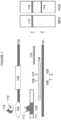
- a lateral flow immunochromatographic test system was used to develop an anti-VLA-4 specific test.
- the lateral flow test system was composed of a two part plastic cassette (top 100 and bottom 101).
- the top part contained two openings - one opening 102 for the application of samples 110 and chase buffer 118 and a window 103 for visual detection of the results.
- the lower part accommodated a nitrocellulose test strip 104 on which the rat 12C2 mAb or mouse 12C4 mAb 105 was immobilized at a "test site" ("T") 106 and a universal gold capture reagent 107 at a "control site” (“C”) 108.
- T test site
- C universal gold capture reagent
- the complex then traveled through the nitrocellulose 104 where it encountered the mouse 12C4 mAb or rat 12C2 mAb 105 at the test site 106.
- the sample 110 reacted with the mouse 12C4 mAb or rat 12C2 mAb 105, forming a complex of 112 and 110 that was arrested at the test site 106 and was visualized as a red line 114.
- the sample 110 can also be added downstream of the gold conjugate.
- the specificity of the test was due to the fact that the complex at the test site only formed when the sample molecule contains a human or humanized IgG4 moiety to capture the mouse anti-human IgG4 gold conjugate and form the first complex and the anti-VLA-4 moiety to react with the anti-(anti-VLA-4 antibody) antibody at the test site.
- the anti-VLA-4 molecule is sandwiched between the gold labeled mouse anti-human IgG4 and the membrane immobilized anti-(anti-VLA-4 antibody) antibody.
- the approach for developing the anti-VLA-4 antibody test was to target the antigen binding site with a capture antibody specific for the anti-VLA-4 antibody (mAb 12C4 or 12C2) immobilized on the membrane, and a gold labeled detector antibody (anti-human IgG4) directed against the constant region of the anti-VLA-4 antibody, which comprises a human IgG4 molecule.
- Anti-VLA-1 is a humanized monoclonal antibody to VLA-1.
- the constant region of the antibody is human IgG1, while the variable antigen binding region is of mouse origin. Because VLA-1 and VLA-4 are closely related, anti-VLA-1 can be used as an additional control of the test system. Such an inclusion can be used to confirm the specificity of the test system.
- a lateral flow immunochromatographic system was developed with the mAb 12C4 or 12C2 directed against the humanized anti-VLA-4 antibody.
- 12C4 or 12C2 was utilized, which was titrated between 0.25 and 1.0 mg/mL on separate test strips and tested against the humanized anti-VLA-4 antibody at 80 ⁇ g/mL.
- the 12C4 or 12C2 antibody was then immobilized at the test site of the membrane at 1.0 mg/mL.
- a mouse monoclonal antibody anti-human IgG4 (gamma chain specific) at 30 ⁇ g/mL was conjugated to gold particles.
- the mouse anti-human IgG4 immunogold conjugate was then lyophilized in the conjugate pad and utilized as a detector molecule.
- universal gold reagent at 3.0 mg/mL was utilized.
- the humanized anti-VLA-4 antibody, humanized anti-VLA-1, IgG1 (myeloma), IgG4 (myeloma), anti-IgG1 and the formulation buffer are tested on a flow system with 12C4 or 12C2 immobilized.
- the humanized anti-VLA-4 antibody was tested at the concentration range of 0.015 and 500 ⁇ g/mL.
- the 12C4 or 12C2 antibody was able to detect the humanized anti-VLA-4 antibody to a concentration of 0.08 ⁇ g/mL.
- Figure 1 illustrates the specificity of the test utilizing the test cassettes. All tests showed a single band 115, indicating a valid test, but only the cassette for the humanized anti-VLA-4 antibody test showed a second band 114 indicating a positive test.
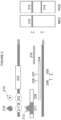
- the top part 200 contained three openings - opening 202 is for the application of samples 210, opening 217 - for chase buffer 218 and a window 203 for visual detection of the results.
- the chase buffer can be added downstream of the sample or added at the same opening as the one for application of samples.
- the lower part 201 accommodated a nitrocellulose test strip 204 on which the mouse 12C4 mAb or rat 12C2 mAb 205 was immobilized at a "test site” (T) 206 and a goat anti-mouse antibody 207 at a "control site” (C) 208.
- Antibody against serum-specific analytes e.g., serum albumin, may be utilized as control antibody.
- sample pad 209 Upstream from the test and control sites, there was a fibrous pad ("sample pad") 209 that received the sample 210. Further upstream from the test and control sites there was a "gold pad” 211 of fibrous material where a mouse anti-human IgG4 was conjugated to gold particles, the complex being 212.
- the two-step assay was utilized. In the first step, serum sample 210 was applied to the sample opening 202 and migrated toward the capture zone 205 ahead of the gold conjugate to form a complex of a sample 210 (anti-VLA4) with the mouse 12C4 or rat 12C2 on test line 205.
- the chase buffer 218 was applied to the chase buffer port 217 rehydrated the monoclonal anti-human IgG4 gold conjugate 212.
- the conjugate 212 then traveled toward the capture test line 205 to form a complex with anti-VLA-4 bound to capture line 205, and the reaction at site 206 was visualized as a red line 214.
- the specificity of the test was due to the fact that the complex at the test site only formed when the sample contains anti-VLA-4 moiety to react with the anti-(anti-VLA-4 antibody) antibody at the test site, and to be detected by a mouse anti-human IgG4 moiety conjugated to gold.
- the anti-VLA-4 molecule is sandwiched between the gold labeled mouse anti-human IgG4 and the membrane immobilized anti-(anti-VLA-4 antibody) antibody.
- the success of this format is reliant upon any endogenous interfering moiety (e.g., IgG4) in the sample traveling ahead of the anti- IgG4 gold conjugate reducing its interference with the detection of the anti-VLA-4 at the test line.
- a reader can be used for semi-quantitative determination of the concentration of the tested analytes.
- a reader based on visual positive/negative scoring was used.
- the visual threshold on the reader corresponds to values within the 20-40 unit range. If the signal was recorded visually positive within this range then it should be considered a very weak pink line that may not be visible to all operators.
- the approach for developing the test was to target the antigen binding site with a capture antibody (mAb 12C4 or mAb 12C2) immobilized on the membrane, specific for the anti-VLA-4 antibody, and a gold labeled detector antibody (mouse anti-human IgG4) directed against the constant region of the anti-VLA-4 antibody, which comprises a human IgG4 molecule.
- a capture antibody mAb 12C4 or mAb 12C2
- mAb 12C2 gold labeled detector antibody directed against the constant region of the anti-VLA-4 antibody, which comprises a human IgG4 molecule.
- a monoclonal antibody used in the assay such as 12C4 or 12C2 is highly specific, no other analyte is captured on the membrane and therefore the test is negative.
- a lateral flow immunochromatographic system was developed with the mAb 12C4 or 12C2 directed against the humanized anti-VLA-4 antibody.
- 12C4 was utilized, which was titrated on separate test strips and tested against the humanized anti-VLA-4 antibody at a variety of concentrations ranging from 100 ⁇ g/mL to 0.25 ⁇ g/mL.
- the 12C4 antibody was then immobilized at the test site of the membrane at 2.1 mg/mL.
- a mouse monoclonal antibody anti-human IgG4 (gamma chain specific) was conjugated to 40 nm gold particles.
- the mouse anti-human IgG4 immunogold conjugate was then lyophilized in the conjugate pad and utilized as a detector molecule.
- a goat anti-mouse antibody at 3.0 mg/mL was utilized.
- a positive reaction was indicated by the presence of a band on the top portion of each strip.
- the 2.1 mg/mL antibody concentration was selected, as it resulted in an intense signal with minimum background, and was immobilized at the test site of the membrane.
- the protocol consisted of adding 20 ⁇ L serum (pre-diluted 1:10 in chase buffer or neat) at port 202 immediately ( ⁇ 1 minute) followed by 150 ⁇ L chase buffer 218 at port 217. The sample was left to incubate in device for 30 minutes at room temperature and ambient humidity.
- the humanized anti-VLA-4 antibody, in sample matrix, IgG4 (myeloma), anti-VLA-4 together with IgG4, and the formulation buffer are tested on a flow system with 12C4 immobilized.
- the humanized anti-VLA-4 antibody was tested at the concentration range of 0.25 and 100 ⁇ g/mL.
- the 12C4 antibody was able to detect the humanized anti-VLA-4 antibody to a concentration of 0.25 ⁇ g/mL.
- Figure 2 illustrates the specificity of the test utilizing the test cassettes. All tests showed a single band 215, indicating a valid test, but only the cassettes for the humanized anti-VLA-4 antibody test showed a second band 214 indicating a positive test.
- the test may include additional components.
- Anti-VLA-1 is a humanized monoclonal antibody to VLA-1.
- the constant region of some anti-VLA-1 antibodies is IgG1, while the variable antigen binding region is of mouse origin.
- Anti-VLA-1 may also be used as an additional control to assess specificity of the test system. Additional controls that can be included in the test system include assessing tolerance of the test to excess levels of analytes that may be found in the serum, e.g., IgG4, drug, or anti-drug antibodies.
- a reader can be used for semi-quantitative or quantitative measurement of anti-VLA4 concentrations.
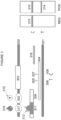
- the top part 300 contained three openings - one 302 is for the application of samples 310, opening 317 - for application of chase buffer 318 and a window 303 for visual detection of the results.
- the lower part 301 accommodated a nitrocellulose test strip 304 on which the mouse 12C4 mAb 305 was immobilized at a "test site" (T) 306 and a goat anti-mouse antibody 307 at a "control site” (C) 308.
- sample pad 309 Upstream from the test and control sites, there was a fibrous pad (“sample pad”) 309 that received the sample 310. Further upstream from the test and control sites there was a "gold pad” 311 of fibrous material where a mouse anti-anti-VLA4 antibody 12C4 was conjugated to gold particles, the complex being 312.
- the two-step assay was utilized. In the first step, sample 310 was applied to the sample opening 302 and migrated toward the capture zone 305 ahead of the gold conjugate to form a complex of a sample 310 (anti-VLA4) with the mouse 12C4 or rat 12C2 on test line 305.
- the chase buffer 318 was applied to the chase buffer port 317 and rehydrated the monoclonal mouse anti-anti-VLA4 antibody 12C4 gold conjugate 312.
- the conjugate 312 then traveled toward the capture test line 305 to form a complex with anti-VLA-4 bound to capture line 305, and the reaction at site 306 was visualized as a red line 314.
- this test format is more restricted in its range of detection than the test conducted with the anti-human IgG4 gold conjugate because the complex at the test site only formed when the sample molecule can react with both the anti-(anti-VLA-4 antibody)/gold conjugate and the anti-(anti-VLA-4 antibody) antibody at the test site.
- the anti-VLA-4 molecule is bridging between the gold labeled anti-(anti-VLA-4 antibody) antibody and the membrane immobilized anti-(anti-VLA-4 antibody) antibody. Therefore, this test is less sensitive for overall detection of the anti-VLA-4 antibody when using actual samples from patients.
- This format results in a more restricted test in that it detects only intact natalizumab, which is an IgG4-based antibody.
- This format does not detect half-antibody exchanged molecules, which is observed in patients treated with natalizumab and other IgG4 antibodies. Therefore, the uses of this format are generally restricted to detection of intact drug.
- a lateral flow immunochromatographic system was developed with the mAb 12C4 directed against the humanized anti-VLA-4 antibody.
- 12C4 was titrated on separate test strips and tested against the humanized anti-VLA-4 antibody at the concentrations ranging from 0.25 to 100 ⁇ g/mL.
- the 12C4 antibody was then immobilized at the test site of the membrane at 2.1 mg/mL.
- 12C4 antibody was conjugated to 40 nm gold particles, lyophilized in the conjugate pad and utilized as a detector molecule.
- a goat anti-mouse antibody at 3.0 mg/mL was utilized.
- a positive reaction was indicated by the presence of a band on the top portion of each strip.
- the 2.1 mg/mL antibody concentration was selected, as it resulted in an intense signal with minimum background, and was immobilized at the test site of the membrane.
- the protocol consisted of adding 20 ⁇ L serum (pre-diluted 1:10 in chase buffer or neat) at port 302 immediately ( ⁇ 1 minute) followed by 150 ⁇ L chase buffer 318 at port 317. The sample was left to incubate in device for 30 minutes at room temperature and ambient humidity.
- the humanized anti-VLA-4 antibody in matrix, anti-VLA-4 antibody with additional IgG4 added and the formulation buffer were tested on a flow system with 12C4 immobilized.
- the humanized anti-VLA-4 antibody was tested at the concentration range of 1 and 100 ⁇ g/mL.
- the 12C4 antibody was able to detect the humanized anti-VLA-4 antibody to a concentration of 1 ⁇ g/mL.
- Figure 3 illustrates the specificity of the test utilizing the test cassettes. All tests showed a single band 315, indicating a valid test, but only the cassette for the humanized anti-VLA-4 antibody test showed a second band 314, indicating a positive test.
- the two-part plastic cassette of this lateral flow test system was the same as that for Examples 1-3 with a few alterations.
- the top part 400 contained three openings - opening 402 for the application of samples 410, opening 417 - for chase buffer 418 and a window 403 for visual detection of the results.
- the lower part 401 accommodated a nitrocellulose test strip 404 on which the anti-VLA-4 antibody 405 was immobilized at a "test site" (T) 406 and a control antibody 407 at a "control site” (C) 408.
- sample pad Upstream from the test and control sites, there was a fibrous pad (“sample pad”) 409 that received the sample 410. Further upstream from the test and control sites there was a "gold pad” 411 of fibrous material where anti-VLA-4 was conjugated to gold particles, the complex being 412.
- the two-step assay was utilized. In the first step, sample 410 was applied to the sample opening 402 and migrated toward the capture zone 405 ahead of the gold conjugate to form a complex of a sample 410 (anti-anti-VLA4) with the anti-VLA-4 on test line 405. In the second step, the chase buffer 418 was applied to the chase buffer port 417 and rehydrated the anti-VLA-4 gold conjugate 412. The conjugate 412 then traveled toward the capture test line 405 to form a complex with anti-(anti-VLA-4) bound to capture line 405, and the reaction at test site 406 was visualized as a red line 414.
- the specificity of the test was due to the fact that the complex at the test site only formed when the sample molecule must bind to both the anti-VLA antibody on the test line and anti-VLA-4 gold conjugate.
- the anti-(anti-VLA-4 antibody) antibody is bridging between the gold labeled anti-VLA-4 antibody and the membrane immobilized anti-VLA-4 antibody, thus, in some cases, this type of assay is referred to as a "bridging assay".
- Excess gold-labeled and free anti-VLA-4 antibody can travel further to the control site 408 where a control antibody 407 is available to capture the matrix components, giving rise to an additional visible red line 415.
- a reader can be used for semi-quantitative determination of the concentration of the tested analytes.
- a reader based on visual positive/negative scoring was used.
- the visual threshold on the reader corresponds to values within the 20-40 unit range. If the signal was recorded visually positive within this range then it should be considered a very weak pink line that may not be visible to all operators.
- a lateral flow immunochromatographic system was developed with the anti-VLA-4 antibody directed against the anti-(anti-VLA-4 antibody) antibody.
- anti-VLA-4 antibody was utilized, which was titrated on separate test strips and tested against anti-(anti-VLA-4 antibody) antibody at a range of concentrations from 25 ⁇ g/mL to 0.5 ⁇ g/mL.
- the anti-VLA-4 antibody was then immobilized at the test site of the membrane at 0.5 mg/mL.
- An anti-VLA-4 was conjugated to 40 nm gold particles.
- the anti-VLA-4 immunogold conjugate was then lyophilized in the conjugate pad and utilized as a detector molecule.
- control antibody was utilized.
- the 0.5 mg/mL antibody concentration was selected for immobilization based on titration experiments, as it resulted in an intense signal with minimum background.
- the protocol consisted of adding 20 ⁇ L serum (pre-diluted 1:10 in chase buffer or neat) at port 402 immediately ( ⁇ 1 minute) followed by 150 ⁇ L chase buffer 418 at port 417. The sample was left to incubate in device for 30 minutes at room temperature and ambient humidity.
- the mouse and rabbit anti-(anti-VLA-4 antibody) antibody, humanized anti-VLA-4 antibody, the combination of the above, and the formulation buffer were tested on a flow system with anti-VLA-4 antibody immobilized.
- the anti-(anti-VLA-4 antibody) antibody was tested at the concentration range of 25 ⁇ g/mL to 0.5 ⁇ g/mL.
- the humanized anti-VLA-4 antibody was able to detect the human anti-(anti-VLA-4 antibody) antibody to a concentration of 0.5 ⁇ g/mL.
- Figure 4 illustrates the specificity of the test utilizing the test cassettes. All tests showed a single band 415, indicating a valid test, but only the cassette for the anti-drug antibody test showed a second band 414, indicating a positive test.
- a control line or additional normalization line may be used for the purpose of data normalization in the lateral flow assay.
- This normalization line may be an immobilized antibody against one of the matrix (e.g., serum) components that have similar concentrations across individual matrix samples.
- a normalization line may be an immobilized antibody against a protein from an irrelevant species that is not present in the tested matrix, but added at the same concentrations to each matrix sample prior to placing it on a lateral flow device.
- This protein can be added to each sample directly, or, in case when samples are being diluted with a chase buffer, may be a component of the chase buffer.
- a reader can be used for semi-quantitative or quantitative measurement of anti anti-VLA4 concentrations.
Landscapes
- Health & Medical Sciences (AREA)
- Life Sciences & Earth Sciences (AREA)
- Immunology (AREA)
- Engineering & Computer Science (AREA)
- Molecular Biology (AREA)
- Chemical & Material Sciences (AREA)
- Biomedical Technology (AREA)
- Urology & Nephrology (AREA)
- Hematology (AREA)
- Medicinal Chemistry (AREA)
- General Health & Medical Sciences (AREA)
- Food Science & Technology (AREA)
- Analytical Chemistry (AREA)
- Physics & Mathematics (AREA)
- Biochemistry (AREA)
- General Physics & Mathematics (AREA)
- Pathology (AREA)
- Microbiology (AREA)
- Biotechnology (AREA)
- Cell Biology (AREA)
- Pharmacology & Pharmacy (AREA)
- Bioinformatics & Cheminformatics (AREA)
- Proteomics, Peptides & Aminoacids (AREA)
- Organic Chemistry (AREA)
- Nuclear Medicine, Radiotherapy & Molecular Imaging (AREA)
- Chemical Kinetics & Catalysis (AREA)
- General Chemical & Material Sciences (AREA)
- Veterinary Medicine (AREA)
- Public Health (AREA)
- Animal Behavior & Ethology (AREA)
- Oncology (AREA)
- Virology (AREA)
- Communicable Diseases (AREA)
- Investigating Or Analysing Biological Materials (AREA)
- Peptides Or Proteins (AREA)
- Measuring Or Testing Involving Enzymes Or Micro-Organisms (AREA)
- Medicines Containing Antibodies Or Antigens For Use As Internal Diagnostic Agents (AREA)
Claims (14)
- Verfahren zum Untersuchen der Wirksamkeit einer Behandlung, wobei das Verfahren Folgendes umfasst:a) Inkontaktbringen einer biologischen Probe, die von einem Patienten entnommen wurde, der sich einer Behandlung mit einem Anti-alpha4-Integrin-Antikörper unterzieht, mit einem mit einem Substrat assoziierten Einfangmittel, wobei das Einfangmittel einen Anti-anti-alpha4-Integrin-Antikörper in der Probe unter Bilden eines Komplexes binden kann und wobei das Substrat Teil eines Lateral-Flow-Immunochromatographiesystems ist; undb) Nachweisen der Bindung des Einfangmittels an den Anti-anti-alpha4-Integrin-Antikörper mit einem Sekundärmittel, das an den Komplex in dem Lateral-Flow-Immunochromatographiesystem bindet, und Korrelieren der Bindung des Einfangmittels mit der Konzentration des Anti-anti-alpha4-Integrin-Antikörpers zum Bestimmen der Wirksamkeit der Behandlung, wobei das Einfangmittel ein Antikörper oder ein Antigen-Bindungsfragment davon ist.
- Verfahren nach Anspruch 1, wobei eine Konzentration des Anti-anti-alpha4-Integrin-Antikörpers über oder unter einer vorbestimmten Konzentration die Wirksamkeit der Behandlung angibt.
- Verfahren nach Anspruch 1 oder Anspruch 2, wobei das Sekundärmittel ein Anti-alpha4-Integrin-Antikörper, gegebenenfalls ein markierter Anti-alpha4-Integrin-Antikörper, ist.
- Verfahren nach einem der Ansprüche 1-3, wobei die Wirksamkeit einen Tag, zwei Tage, drei Tage, vier Tage, fünf Tage, sechs Tage, eine Woche, zwei Wochen, drei Wochen oder vier Wochen nach der Verabreichung der Behandlung untersucht wird.
- Verfahren nach einem der Ansprüche 1-4, wobei die Behandlung Natalizumab ist.
- Verfahren nach einem der Ansprüche 1-5, wobei die Wirksamkeit als Teil eines Behandlungsregimes, gegebenenfalls eines Natalizumab-Regimes, untersucht wird.
- Verfahren nach Anspruch 6, wobei das Behandlungsregime einmal pro Tag oder einmal alle zwei Tage, drei Tage, vier Tage, fünf Tage, sechs Tage, jede Woche, alle zwei Wochen, drei Wochen, vier Wochen, alle drei Monate, alle vier Monate, alle sechs Monate oder jährlich verabreicht wird.
- Verfahren nach Anspruch 1, wobei:(a) die biologische Probe von dem Patienten einen Tag, zwei Tage, drei Tage, vier Tage, fünf Tage, sechs Tage, eine Woche, zwei Wochen, drei Wochen oder vier Wochen nach der Behandlung entnommen wurde; oder(b) die biologische Probe wiederholt einmal täglich oder einmal alle zwei Tage, drei Tage, vier Tage, fünf Tage, sechs Tage, jede Woche, alle zwei Wochen, drei Wochen oder vier Wochen entnommen wurde.
- Verfahren nach Anspruch 8(a), wobei eine biologische Probe auch vor der Behandlung von dem Patienten entnommen wurde.
- Kit zur Untersuchung der Konzentration eines Anti-anti-alpha4-Integrin-Antikörpers in einer biologischen Probe bei einem Patienten, umfassend:(a) eine Testvorrichtung, umfassend:ein Lateral-Flow-Immunochromatographiesystem, umfassend eine Probenaufnahmezone, eine Markerzone und eine Testzone, wobei die Testzone der Probenaufnahmezone nachgeordnet ist und ein mit einem Substrat assoziiertes Einfangmittel enthält, wobei das Einfangmittel einen Anti-anti-alpha4-Integrin-Antikörper in der biologischen Probe unter Bilden eines Komplexes binden kann, und sich die Markerzone der Probenaufnahmezone vorgeordnet befindet und ein oder mehrere Sekundärmittel enthält, wobei das eine oder die mehreren Sekundärmittel in der Lage sind, den Komplex, umfassend das Einfangmittel und den Anti-anti-alpha4-Integrin-Antikörper, zu binden,wobei das Einfangmittel ein Antikörper oder ein Antigen-Bindungsfragment davon ist und das Sekundärmittel ein Anti-alpha4-Integrin-Antikörper, gegebenenfalls ein markierter anti-alpha4-Integrin-Antikörper, ist; und(b) einen Chase-Puffer.
- Kit nach Anspruch 10, wobei:(a) das Binden des Einfangmittels an den Anti-anti-alpha4-Integrin-Antikörper ein nachweisbares Signal erzeugt; oder(b) das Binden des Einfangmittels an den Anti-anti-alpha4-Integrin-Antikörper durch das Sekundärmittel nachgewiesen wird.
- Kit nach Anspruch 10, wobei:(a) das Einfangmittel ein Anti-alpha4-Integrin-Antikörper oder ein Integrin ist; oder(b) das Substrat Nitrocellulose, Cellulose, Acetat, Filterpapier, Tuch oder Glasfaserpapier ist.
- Kit nach Anspruch 10, wobei die biologische Probe Blut, Plasma, Serum, Urin, Speichel, Liquor, Sputum, Augenlinsenflüssigkeit, Schweiß, Milch, Aszitesflüssigkeit, Schleim, Synovialflüssigkeit, Peritonealflüssigkeit, transdermale Exsudate, pharyngeale Exsudate, bronchoalveoläre Lavage, tracheale Aspirationen, Sperma, Zervixschleim, Vaginalsekret, Urethrasekret oder Fruchtwasser ist.
- Kit nach Anspruch 10, wobei das Binden des Einfangmittels an den Anti-anti-alpha4-Integrin-Antikörper innerhalb von etwa 30 Sekunden bis etwa 60 Minuten oder länger nach der Bindung nachweisbar ist.
Applications Claiming Priority (3)
| Application Number | Priority Date | Filing Date | Title |
|---|---|---|---|
| US25055309P | 2009-10-11 | 2009-10-11 | |
| PCT/US2010/052172 WO2011044553A1 (en) | 2009-10-11 | 2010-10-11 | Anti-vla-4 related assays |
| EP10822820.6A EP2485762B1 (de) | 2009-10-11 | 2010-10-11 | Anti-vla-4-assays |
Related Parent Applications (1)
| Application Number | Title | Priority Date | Filing Date |
|---|---|---|---|
| EP10822820.6A Division EP2485762B1 (de) | 2009-10-11 | 2010-10-11 | Anti-vla-4-assays |
Publications (2)
| Publication Number | Publication Date |
|---|---|
| EP3349010A1 EP3349010A1 (de) | 2018-07-18 |
| EP3349010B1 true EP3349010B1 (de) | 2024-08-28 |
Family
ID=43857181
Family Applications (2)
| Application Number | Title | Priority Date | Filing Date |
|---|---|---|---|
| EP17205670.7A Active EP3349010B1 (de) | 2009-10-11 | 2010-10-11 | Anti-vla-4-assays |
| EP10822820.6A Active EP2485762B1 (de) | 2009-10-11 | 2010-10-11 | Anti-vla-4-assays |
Family Applications After (1)
| Application Number | Title | Priority Date | Filing Date |
|---|---|---|---|
| EP10822820.6A Active EP2485762B1 (de) | 2009-10-11 | 2010-10-11 | Anti-vla-4-assays |
Country Status (7)
| Country | Link |
|---|---|
| US (5) | US9377458B2 (de) |
| EP (2) | EP3349010B1 (de) |
| JP (1) | JP2013507618A (de) |
| AU (1) | AU2010303156B2 (de) |
| CA (1) | CA2776756A1 (de) |
| ES (2) | ES2666372T3 (de) |
| WO (1) | WO2011044553A1 (de) |
Families Citing this family (8)
| Publication number | Priority date | Publication date | Assignee | Title |
|---|---|---|---|---|
| EP2758780A4 (de) * | 2011-09-22 | 2015-09-16 | Marv Entpr Llc | Verfahren zur behandlung von multipler sklerose |
| KR101515020B1 (ko) * | 2013-11-13 | 2015-04-24 | 에스케이텔레콤 주식회사 | 햅텐 및 이에 결합하는 항체를 레퍼런스 항체로 이용하는 면역학적 측정 방법 및 상기 레퍼런스 항체를 이용한 면역학적 측정장치 |
| US20180106799A1 (en) * | 2015-04-06 | 2018-04-19 | Bludiagnostics, Inc. | A test device for detecting an analyte in a saliva sample and method of use |
| US9927443B2 (en) * | 2015-04-10 | 2018-03-27 | Conquerab Inc. | Risk assessment for therapeutic drugs |
| US20170003284A1 (en) * | 2015-06-30 | 2017-01-05 | Alpha Diagnostic International, Inc. | Western blot process validation and quantification system |
| US9903866B2 (en) * | 2016-04-05 | 2018-02-27 | Conquerab Inc. | Portable devices for detection of antibodies against therapeutic drugs |
| WO2018031887A1 (en) | 2016-08-12 | 2018-02-15 | Abreos Biosciences, Inc. | Detection and quantification of natalizumab |
| AT527879A1 (de) * | 2023-12-18 | 2025-07-15 | Tagtron Gmbh | Vorrichtung für einen Lateral-Flow-Test |
Family Cites Families (112)
| Publication number | Priority date | Publication date | Assignee | Title |
|---|---|---|---|---|
| US3639350A (en) | 1970-04-15 | 1972-02-01 | Allied Chem | Esterification of terephthalic acid with an alkylene glycol in the presence of guanidine or an alkyl guanidine |
| NL154598B (nl) | 1970-11-10 | 1977-09-15 | Organon Nv | Werkwijze voor het aantonen en bepalen van laagmoleculire verbindingen en van eiwitten die deze verbindingen specifiek kunnen binden, alsmede testverpakking. |
| US3817837A (en) | 1971-05-14 | 1974-06-18 | Syva Corp | Enzyme amplification assay |
| US3939350A (en) | 1974-04-29 | 1976-02-17 | Board Of Trustees Of The Leland Stanford Junior University | Fluorescent immunoassay employing total reflection for activation |
| US3996345A (en) | 1974-08-12 | 1976-12-07 | Syva Company | Fluorescence quenching with immunological pairs in immunoassays |
| US4425112A (en) | 1976-02-25 | 1984-01-10 | The United States Of America As Represented By The Department Of Health And Human Services | Flow-through centrifuge |
| US4277437A (en) | 1978-04-05 | 1981-07-07 | Syva Company | Kit for carrying out chemically induced fluorescence immunoassay |
| US4275149A (en) | 1978-11-24 | 1981-06-23 | Syva Company | Macromolecular environment control in specific receptor assays |
| US4235601A (en) | 1979-01-12 | 1980-11-25 | Thyroid Diagnostics, Inc. | Test device and method for its use |
| US4391904A (en) | 1979-12-26 | 1983-07-05 | Syva Company | Test strip kits in immunoassays and compositions therein |
| US5179017A (en) | 1980-02-25 | 1993-01-12 | The Trustees Of Columbia University In The City Of New York | Processes for inserting DNA into eucaryotic cells and for producing proteinaceous materials |
| US4399216A (en) | 1980-02-25 | 1983-08-16 | The Trustees Of Columbia University | Processes for inserting DNA into eucaryotic cells and for producing proteinaceous materials |
| US4634665A (en) | 1980-02-25 | 1987-01-06 | The Trustees Of Columbia University In The City Of New York | Processes for inserting DNA into eucaryotic cells and for producing proteinaceous materials |
| US4376110A (en) | 1980-08-04 | 1983-03-08 | Hybritech, Incorporated | Immunometric assays using monoclonal antibodies |
| US4366241A (en) | 1980-08-07 | 1982-12-28 | Syva Company | Concentrating zone method in heterogeneous immunoassays |
| US4517288A (en) | 1981-01-23 | 1985-05-14 | American Hospital Supply Corp. | Solid phase system for ligand assay |
| US4472509A (en) | 1982-06-07 | 1984-09-18 | Gansow Otto A | Metal chelate conjugated monoclonal antibodies |
| US4703017C1 (en) | 1984-02-14 | 2001-12-04 | Becton Dickinson Co | Solid phase assay with visual readout |
| US4938948A (en) | 1985-10-07 | 1990-07-03 | Cetus Corporation | Method for imaging breast tumors using labeled monoclonal anti-human breast cancer antibodies |
| CA1291031C (en) | 1985-12-23 | 1991-10-22 | Nikolaas C.J. De Jaeger | Method for the detection of specific binding agents and their correspondingbindable substances |
| GB8607679D0 (en) | 1986-03-27 | 1986-04-30 | Winter G P | Recombinant dna product |
| US5763262A (en) | 1986-09-18 | 1998-06-09 | Quidel Corporation | Immunodiagnostic device |
| EP0307434B2 (de) | 1987-03-18 | 1998-07-29 | Scotgen Biopharmaceuticals, Inc. | Geänderte antikörper |
| ES2150428T3 (es) | 1987-04-27 | 2000-12-01 | Unilever Nv | Ensayos de union especifica. |
| US4943522A (en) | 1987-06-01 | 1990-07-24 | Quidel | Lateral flow, non-bibulous membrane assay protocols |
| US5120643A (en) | 1987-07-13 | 1992-06-09 | Abbott Laboratories | Process for immunochromatography with colloidal particles |
| US4818677A (en) | 1987-12-03 | 1989-04-04 | Monoclonal Antibodies, Inc. | Membrane assay using focused sample application |
| AU2684488A (en) | 1988-06-27 | 1990-01-04 | Carter-Wallace, Inc. | Test device and method for colored particle immunoassay |
| US5221616A (en) | 1988-07-15 | 1993-06-22 | Quidel Corporation | Prevention of spontaneous complement activation in mammalian biological fluids |
| US5096809A (en) | 1988-07-25 | 1992-03-17 | Pacific Biotech, Inc. | Whole blood assays using porous membrane support devices |
| US4954128A (en) | 1988-08-23 | 1990-09-04 | Baxter International Inc. | Therapeutics plasma exchange system |
| US5118630A (en) | 1988-11-04 | 1992-06-02 | Quidel Corporation | Method for determining periodic infertility in females |
| US5530101A (en) | 1988-12-28 | 1996-06-25 | Protein Design Labs, Inc. | Humanized immunoglobulins |
| IL162181A (en) | 1988-12-28 | 2006-04-10 | Pdl Biopharma Inc | A method of producing humanized immunoglubulin, and polynucleotides encoding the same |
| US6352862B1 (en) | 1989-02-17 | 2002-03-05 | Unilever Patent Holdings B.V. | Analytical test device for imuno assays and methods of using same |
| US5084828A (en) | 1989-09-29 | 1992-01-28 | Healthtech Services Corp. | Interactive medication delivery system |
| US5252496A (en) | 1989-12-18 | 1993-10-12 | Princeton Biomeditech Corporation | Carbon black immunochemical label |
| US5096837A (en) | 1990-02-08 | 1992-03-17 | Pacific Biotech, Inc. | Immunochromatographic assay and method of using same |
| US5223220A (en) | 1990-03-27 | 1993-06-29 | Pacific Biotech, Inc. | Solid phase immunoassay device and method of making same |
| US5200090A (en) | 1990-03-30 | 1993-04-06 | Baxter International Inc. | Multiple fluid source isolation, metering and alarm system and method |
| US5789650A (en) | 1990-08-29 | 1998-08-04 | Genpharm International, Inc. | Transgenic non-human animals for producing heterologous antibodies |
| US5118428A (en) | 1990-11-13 | 1992-06-02 | Quidel | Method to remove red blood cells from whole blood samples |
| DE4139840B4 (de) | 1990-12-04 | 2005-06-02 | Quidel Corp., San Diego | Antigen-Zubereitung zum Nachweis von H. pylori |
| EP0566695B1 (de) | 1991-01-11 | 1999-06-02 | Quidel Corporation | Einstufiges querfluss analysenverfahren und ein darin verwendeter nichtsaugfähiger träger |
| US5225328A (en) | 1991-05-30 | 1993-07-06 | Quidel Corporation | Stable alkaline phosphatase compositions with color enhancement and their use in assays |
| ATE255131T1 (de) | 1991-06-14 | 2003-12-15 | Genentech Inc | Humanisierter heregulin antikörper |
| US5686315A (en) | 1991-06-14 | 1997-11-11 | Quidel Corporation | Assay device for one step detection of analyte |
| WO1993015217A1 (en) | 1992-02-04 | 1993-08-05 | Quidel Corporation | Simplified extraction method for bacterial antigens using dried reagents |
| US5541069A (en) | 1992-02-28 | 1996-07-30 | Quidel Corporation | Assay having improved dose response curve |
| JP2948318B2 (ja) | 1992-03-10 | 1999-09-13 | クイデル コーポレイション | 特異的結合アッセイ用の赤血球の分離方法 |
| NZ261259A (en) | 1993-01-12 | 1996-12-20 | Biogen Inc | Humanised recombinant anti-vla4 antibody and diagnostic compositions and medicaments |
| JP3593343B2 (ja) | 1993-02-09 | 2004-11-24 | バイオジェン インコーポレイテッド | インシュリン依存型糖尿病の治療 |
| US5415994A (en) | 1993-08-02 | 1995-05-16 | Quidel Corporation | Lateral flow medical diagnostic assay device with sample extraction means |
| US5827690A (en) | 1993-12-20 | 1998-10-27 | Genzyme Transgenics Corporatiion | Transgenic production of antibodies in milk |
| US5840299A (en) | 1994-01-25 | 1998-11-24 | Athena Neurosciences, Inc. | Humanized antibodies against leukocyte adhesion molecule VLA-4 |
| US5434057A (en) | 1994-02-02 | 1995-07-18 | Quidel Corporation | Sperm motility assay and devices |
| US5521102A (en) | 1994-08-08 | 1996-05-28 | Quidel Corporation | Controlled sensitivity immunochromatographic assay |
| US5845255A (en) | 1994-10-28 | 1998-12-01 | Advanced Health Med-E-Systems Corporation | Prescription management system |
| US5712172A (en) | 1995-05-18 | 1998-01-27 | Wyntek Diagnostics, Inc. | One step immunochromatographic device and method of use |
| US5804452A (en) | 1995-04-27 | 1998-09-08 | Quidel Corporation | One step urine creatinine assays |
| US5786220A (en) | 1995-04-28 | 1998-07-28 | Quidel Corporation | Assays and devices for distinguishing between normal and abnormal pregnancy |
| WO1996034096A1 (en) | 1995-04-28 | 1996-10-31 | Abgenix, Inc. | Human antibodies derived from immunized xenomice |
| US20010051350A1 (en) | 1995-05-02 | 2001-12-13 | Albert Nazareth | Diagnostic detection device and method |
| US5773234A (en) | 1995-08-07 | 1998-06-30 | Quidel Corporation | Method and device for chlamydia detection |
| DE19541844C1 (de) | 1995-11-09 | 1997-07-24 | Gsf Forschungszentrum Umwelt | Verfahren zur Herstellung von menschlichen Antikörpern und deren Verwendung |
| DE19543553B4 (de) | 1995-11-22 | 2009-04-09 | Deutsches Primatenzentrum Gmbh | VP-Antigene des JC-Virus |
| ATE549918T1 (de) | 1996-12-03 | 2012-04-15 | Amgen Fremont Inc | Menschliche antikörper, die ausdrücklich menschliches tnf alpha binden |
| US6305377B1 (en) | 1996-12-12 | 2001-10-23 | Michael T. Portwood | System and method for improving compliance of a medical regimen |
| US6306642B1 (en) | 1997-11-24 | 2001-10-23 | Quidel Corporation | Enzyme substrate delivery and product registration in one step enzyme immunoassays |
| US6623981B2 (en) * | 1998-01-27 | 2003-09-23 | Bristol-Myers Squibb Company | Detection of patients at risk for developing integrin antagonist/agonist mediated disease states |
| US6014631A (en) | 1998-04-02 | 2000-01-11 | Merck-Medco Managed Care, Llc | Computer implemented patient medication review system and process for the managed care, health care and/or pharmacy industry |
| US6045501A (en) | 1998-08-28 | 2000-04-04 | Celgene Corporation | Methods for delivering a drug to a patient while preventing the exposure of a foetus or other contraindicated individual to the drug |
| US7171371B2 (en) | 1999-09-03 | 2007-01-30 | Smg Trust | Method and system for providing pre and post operative support and care |
| EP1232392B2 (de) | 1999-10-12 | 2014-04-02 | Oxoid (Ely) Limited | Verbessertes verfahren zum nachweis von säure-resistenten bakterien der gattung helicobacter im stuhl |
| AU4465001A (en) * | 2000-03-30 | 2001-10-15 | Nippon Kayaku Kabushiki Kaisha | Method of examining cancer by assaying autoantibody against mdm2 and reagent therefor |
| US8288322B2 (en) | 2000-04-17 | 2012-10-16 | Dyax Corp. | Methods of constructing libraries comprising displayed and/or expressed members of a diverse family of peptides, polypeptides or proteins and the novel libraries |
| US7018847B2 (en) * | 2000-05-05 | 2006-03-28 | Pharmacia Diagnostics Ab | Assay device with timer function |
| US6620626B1 (en) * | 2000-08-09 | 2003-09-16 | Mission Research Corp. | Antigen detection device and method |
| JP2004511807A (ja) | 2000-10-17 | 2004-04-15 | ベッスト−テスト アンパーツゼルスカブ | 体液サンプル中のrsウイルスに関連した生物学的細胞の直接的検出のためのアッセイ |
| US6315720B1 (en) | 2000-10-23 | 2001-11-13 | Celgene Corporation | Methods for delivering a drug to a patient while avoiding the occurrence of an adverse side effect known or suspected of being caused by the drug |
| US6485460B2 (en) | 2001-01-12 | 2002-11-26 | Bracco Diagnostics, Inc. | Tamper evident syringe barrel |
| US6706008B2 (en) | 2001-03-06 | 2004-03-16 | Baxter International Inc. | Automated system and method for withdrawing compounds from blood |
| US7291477B2 (en) * | 2001-07-03 | 2007-11-06 | Xenotope Diagnostics, Inc. | Method and device for trichomonas detection |
| WO2003016909A1 (en) | 2001-08-16 | 2003-02-27 | Ludwig Institute For Cancer Research | Method for determining protein component in a biological sample |
| US7205159B2 (en) | 2001-08-20 | 2007-04-17 | Proteome Systems Intellectual Property Pty Ltd. | Diagnostic testing process and apparatus |
| WO2003016902A1 (en) | 2001-08-20 | 2003-02-27 | Proteome Systems Intellectual Property Pty Ltd | Diagnostic testing process and apparatus |
| US6849183B2 (en) | 2002-08-13 | 2005-02-01 | Transvivo, Inc. | Method and apparatus for therapeutic apheresis |
| SI1485127T1 (sl) | 2002-02-25 | 2011-09-30 | Elan Pharm Inc | Dajanje aktivne snovi za zdravljenje vnetja |
| WO2004001539A2 (en) | 2002-06-21 | 2003-12-31 | Mckesson Information Solutions Llc | Closed loop medication use system and method |
| CA2502926A1 (en) | 2002-11-08 | 2004-05-27 | Linda J. Sandell | Uncoupled collagen synthesis and degradation assays |
| US7238538B2 (en) * | 2003-09-19 | 2007-07-03 | Freitag Helmut E | Chromatographic assay device and methods |
| US7378285B2 (en) * | 2003-09-22 | 2008-05-27 | Paul Lambotte | Devices for the detection of multiple analytes in a sample |
| JP4672263B2 (ja) | 2004-01-27 | 2011-04-20 | デンカ生研株式会社 | 簡便な検出法、検出装置及び検出キットとその製法 |
| US7419666B1 (en) | 2004-02-23 | 2008-09-02 | Massachusetts Eye And Ear Infirmary | Treatment of ocular disorders |
| US20050283385A1 (en) | 2004-06-21 | 2005-12-22 | The Permanente Medical Group, Inc. | Individualized healthcare management system |
| US20060027599A1 (en) | 2004-07-21 | 2006-02-09 | Lancer Partnership, Ltd. | Method and apparatus for dispensing compressed ice |
| US8445293B2 (en) * | 2005-02-09 | 2013-05-21 | Rapid Pathogen Screening, Inc. | Method to increase specificity and/or accuracy of lateral flow immunoassays |
| ES2387317T3 (es) | 2005-03-03 | 2012-09-20 | Seedlings Life Science Ventures, Llc. | Procedimiento de control de riesgos de pacientes en tratamiento con natalizumab |
| ES2640295T5 (en) | 2005-04-04 | 2025-02-13 | Biogen Ma Inc | Methods for evaluating an immune response to a therapeutic agent |
| US20060246513A1 (en) * | 2005-05-02 | 2006-11-02 | Bohannon Robert C | Method and device to detect the presence of analytes in a sample |
| CA2641160A1 (en) | 2006-02-28 | 2007-09-07 | Elan Pharmaceuticals, Inc. | Methods of treating inflammatory and autoimmune diseases with alpha-4 inhibitory compounds |
| HUE047230T2 (hu) | 2006-02-28 | 2020-04-28 | Biogen Ma Inc | Gyulladásos és autoimmun betegségek natalizumabbal való kezelésének módszerei |
| AU2007224228B2 (en) | 2006-03-03 | 2013-09-19 | Biogen Ma Inc. | Methods of treating inflammatory and autoimmune diseases with natalizumab |
| CA2658795A1 (en) * | 2006-07-26 | 2008-02-07 | Abon Biopharm (Hangzhou) Co., Ltd. | A test device for detecting an analyte in a liquid sample |
| US20110184747A1 (en) * | 2006-08-09 | 2011-07-28 | Carmen Bozic | Method for distribution of a drug |
| US7704702B2 (en) * | 2006-08-10 | 2010-04-27 | Inverness Medical Switzerland Gmbh | Test strip for lateral flow assays |
| US20080233150A1 (en) | 2006-11-16 | 2008-09-25 | Gale Smith | Respiratory syncytial virus-virus like particle (vlps) |
| EP1933140B1 (de) | 2006-12-11 | 2011-08-17 | AraGen Biotechnology Co. Ltd. | Verfahren zur Detektion von Antikörpern mittels eines durch Oligonukleotide verstärkten kolloidalen Gold Signals |
| WO2010129302A1 (en) * | 2009-04-28 | 2010-11-11 | Innovative Laboratory Technologies, Inc. | Lateral-flow immuno-chromatographic assay devices |
| WO2011005357A2 (en) * | 2009-07-08 | 2011-01-13 | Anp Technologies, Inc. | Immunogenicity assay |
| PT2524060T (pt) | 2010-01-11 | 2018-03-28 | Biogen Ma Inc | Ensaio para a deteção de anticorpos contra o vírus jc |
| PT3575792T (pt) | 2011-05-31 | 2023-02-28 | Biogen Ma Inc | Método de avaliação do risco de lmp |
-
2010
- 2010-10-11 WO PCT/US2010/052172 patent/WO2011044553A1/en not_active Ceased
- 2010-10-11 US US13/501,111 patent/US9377458B2/en active Active
- 2010-10-11 EP EP17205670.7A patent/EP3349010B1/de active Active
- 2010-10-11 CA CA2776756A patent/CA2776756A1/en not_active Abandoned
- 2010-10-11 JP JP2012533379A patent/JP2013507618A/ja active Pending
- 2010-10-11 ES ES10822820.6T patent/ES2666372T3/es active Active
- 2010-10-11 AU AU2010303156A patent/AU2010303156B2/en active Active
- 2010-10-11 ES ES17205670T patent/ES2989929T3/es active Active
- 2010-10-11 EP EP10822820.6A patent/EP2485762B1/de active Active
-
2016
- 2016-05-27 US US15/166,514 patent/US9726672B2/en active Active
-
2017
- 2017-06-29 US US15/637,446 patent/US10302654B2/en active Active
-
2019
- 2019-05-24 US US16/422,822 patent/US11280793B2/en active Active
-
2022
- 2022-03-21 US US17/700,242 patent/US12385925B2/en active Active
Also Published As
| Publication number | Publication date |
|---|---|
| US20200088738A1 (en) | 2020-03-19 |
| EP2485762B1 (de) | 2017-12-13 |
| HK1256154A1 (en) | 2019-09-13 |
| CA2776756A1 (en) | 2011-04-14 |
| ES2666372T3 (es) | 2018-05-04 |
| US20120263796A1 (en) | 2012-10-18 |
| EP3349010A1 (de) | 2018-07-18 |
| JP2013507618A (ja) | 2013-03-04 |
| US9726672B2 (en) | 2017-08-08 |
| WO2011044553A1 (en) | 2011-04-14 |
| US12385925B2 (en) | 2025-08-12 |
| US10302654B2 (en) | 2019-05-28 |
| AU2010303156B2 (en) | 2016-02-04 |
| AU2010303156A1 (en) | 2012-05-03 |
| US20220357338A1 (en) | 2022-11-10 |
| EP2485762A4 (de) | 2013-05-15 |
| US11280793B2 (en) | 2022-03-22 |
| EP2485762A1 (de) | 2012-08-15 |
| ES2989929T3 (es) | 2024-11-28 |
| US20180143204A1 (en) | 2018-05-24 |
| US20160274125A1 (en) | 2016-09-22 |
| US9377458B2 (en) | 2016-06-28 |
Similar Documents
| Publication | Publication Date | Title |
|---|---|---|
| US12385925B2 (en) | Anti-VLA-4 related assays | |
| US8445293B2 (en) | Method to increase specificity and/or accuracy of lateral flow immunoassays | |
| KR102322094B1 (ko) | 바이러스 및 박테리아 감염의 복합 검출을 위한 방법 및 장치 | |
| US9372192B2 (en) | Method and device for combined detection of viral and bacterial infections | |
| US20190376970A1 (en) | Method and Device for Combined Detection of Viral and Bacterial Infections | |
| CA2755486A1 (en) | Detection of fibrin and fibrinogen degradation products and associated methods of production and use for the detection and monitoring of cancer | |
| US20250283880A1 (en) | Sensitizer for immunochromatographic assays, and assay | |
| HK1256154B (en) | Anti-vla-4 related assays | |
| HK1173967B (en) | Anti-vla-4 related assays | |
| HK1173967A (en) | Anti-vla-4 related assays | |
| US20110300529A1 (en) | Detection of ifi16 in body fluids |
Legal Events
| Date | Code | Title | Description |
|---|---|---|---|
| PUAI | Public reference made under article 153(3) epc to a published international application that has entered the european phase |
Free format text: ORIGINAL CODE: 0009012 |
|
| STAA | Information on the status of an ep patent application or granted ep patent |
Free format text: STATUS: THE APPLICATION HAS BEEN PUBLISHED |
|
| AC | Divisional application: reference to earlier application |
Ref document number: 2485762 Country of ref document: EP Kind code of ref document: P |
|
| AK | Designated contracting states |
Kind code of ref document: A1 Designated state(s): AL AT BE BG CH CY CZ DE DK EE ES FI FR GB GR HR HU IE IS IT LI LT LU LV MC MK MT NL NO PL PT RO RS SE SI SK SM TR |
|
| STAA | Information on the status of an ep patent application or granted ep patent |
Free format text: STATUS: REQUEST FOR EXAMINATION WAS MADE |
|
| 17P | Request for examination filed |
Effective date: 20190114 |
|
| RBV | Designated contracting states (corrected) |
Designated state(s): AL AT BE BG CH CY CZ DE DK EE ES FI FR GB GR HR HU IE IS IT LI LT LU LV MC MK MT NL NO PL PT RO RS SE SI SK SM TR |
|
| REG | Reference to a national code |
Ref country code: HK Ref legal event code: DE Ref document number: 1256154 Country of ref document: HK |
|
| STAA | Information on the status of an ep patent application or granted ep patent |
Free format text: STATUS: EXAMINATION IS IN PROGRESS |
|
| 17Q | First examination report despatched |
Effective date: 20200225 |
|
| RIC1 | Information provided on ipc code assigned before grant |
Ipc: A61P 31/12 20060101ALI20230203BHEP Ipc: A61P 37/02 20060101ALI20230203BHEP Ipc: G01N 33/94 20060101ALI20230203BHEP Ipc: G01N 33/68 20060101ALI20230203BHEP Ipc: G01N 33/558 20060101AFI20230203BHEP |
|
| GRAP | Despatch of communication of intention to grant a patent |
Free format text: ORIGINAL CODE: EPIDOSNIGR1 |
|
| STAA | Information on the status of an ep patent application or granted ep patent |
Free format text: STATUS: GRANT OF PATENT IS INTENDED |
|
| INTG | Intention to grant announced |
Effective date: 20230515 |
|
| GRAJ | Information related to disapproval of communication of intention to grant by the applicant or resumption of examination proceedings by the epo deleted |
Free format text: ORIGINAL CODE: EPIDOSDIGR1 |
|
| STAA | Information on the status of an ep patent application or granted ep patent |
Free format text: STATUS: EXAMINATION IS IN PROGRESS |
|
| INTC | Intention to grant announced (deleted) | ||
| GRAP | Despatch of communication of intention to grant a patent |
Free format text: ORIGINAL CODE: EPIDOSNIGR1 |
|
| STAA | Information on the status of an ep patent application or granted ep patent |
Free format text: STATUS: GRANT OF PATENT IS INTENDED |
|
| INTG | Intention to grant announced |
Effective date: 20231013 |
|
| GRAJ | Information related to disapproval of communication of intention to grant by the applicant or resumption of examination proceedings by the epo deleted |
Free format text: ORIGINAL CODE: EPIDOSDIGR1 |
|
| STAA | Information on the status of an ep patent application or granted ep patent |
Free format text: STATUS: EXAMINATION IS IN PROGRESS |
|
| GRAP | Despatch of communication of intention to grant a patent |
Free format text: ORIGINAL CODE: EPIDOSNIGR1 |
|
| STAA | Information on the status of an ep patent application or granted ep patent |
Free format text: STATUS: GRANT OF PATENT IS INTENDED |
|
| INTC | Intention to grant announced (deleted) | ||
| INTG | Intention to grant announced |
Effective date: 20240319 |
|
| GRAS | Grant fee paid |
Free format text: ORIGINAL CODE: EPIDOSNIGR3 |
|
| GRAA | (expected) grant |
Free format text: ORIGINAL CODE: 0009210 |
|
| STAA | Information on the status of an ep patent application or granted ep patent |
Free format text: STATUS: THE PATENT HAS BEEN GRANTED |
|
| AC | Divisional application: reference to earlier application |
Ref document number: 2485762 Country of ref document: EP Kind code of ref document: P |
|
| AK | Designated contracting states |
Kind code of ref document: B1 Designated state(s): AL AT BE BG CH CY CZ DE DK EE ES FI FR GB GR HR HU IE IS IT LI LT LU LV MC MK MT NL NO PL PT RO RS SE SI SK SM TR |
|
| REG | Reference to a national code |
Ref country code: GB Ref legal event code: FG4D |
|
| REG | Reference to a national code |
Ref country code: CH Ref legal event code: EP |
|
| REG | Reference to a national code |
Ref country code: DE Ref legal event code: R096 Ref document number: 602010069497 Country of ref document: DE |
|
| REG | Reference to a national code |
Ref country code: IE Ref legal event code: FG4D |
|
| REG | Reference to a national code |
Ref country code: ES Ref legal event code: FG2A Ref document number: 2989929 Country of ref document: ES Kind code of ref document: T3 Effective date: 20241128 |
|
| REG | Reference to a national code |
Ref country code: LT Ref legal event code: MG9D |
|
| PG25 | Lapsed in a contracting state [announced via postgrant information from national office to epo] |
Ref country code: NO Free format text: LAPSE BECAUSE OF FAILURE TO SUBMIT A TRANSLATION OF THE DESCRIPTION OR TO PAY THE FEE WITHIN THE PRESCRIBED TIME-LIMIT Effective date: 20241128 |
|
| REG | Reference to a national code |
Ref country code: AT Ref legal event code: MK05 Ref document number: 1718495 Country of ref document: AT Kind code of ref document: T Effective date: 20240828 |
|
| PG25 | Lapsed in a contracting state [announced via postgrant information from national office to epo] |
Ref country code: NL Free format text: LAPSE BECAUSE OF FAILURE TO SUBMIT A TRANSLATION OF THE DESCRIPTION OR TO PAY THE FEE WITHIN THE PRESCRIBED TIME-LIMIT Effective date: 20240828 Ref country code: PL Free format text: LAPSE BECAUSE OF FAILURE TO SUBMIT A TRANSLATION OF THE DESCRIPTION OR TO PAY THE FEE WITHIN THE PRESCRIBED TIME-LIMIT Effective date: 20240828 Ref country code: PT Free format text: LAPSE BECAUSE OF FAILURE TO SUBMIT A TRANSLATION OF THE DESCRIPTION OR TO PAY THE FEE WITHIN THE PRESCRIBED TIME-LIMIT Effective date: 20241230 Ref country code: FI Free format text: LAPSE BECAUSE OF FAILURE TO SUBMIT A TRANSLATION OF THE DESCRIPTION OR TO PAY THE FEE WITHIN THE PRESCRIBED TIME-LIMIT Effective date: 20240828 Ref country code: GR Free format text: LAPSE BECAUSE OF FAILURE TO SUBMIT A TRANSLATION OF THE DESCRIPTION OR TO PAY THE FEE WITHIN THE PRESCRIBED TIME-LIMIT Effective date: 20241129 |
|
| PG25 | Lapsed in a contracting state [announced via postgrant information from national office to epo] |
Ref country code: BG Free format text: LAPSE BECAUSE OF FAILURE TO SUBMIT A TRANSLATION OF THE DESCRIPTION OR TO PAY THE FEE WITHIN THE PRESCRIBED TIME-LIMIT Effective date: 20240828 |
|
| PG25 | Lapsed in a contracting state [announced via postgrant information from national office to epo] |
Ref country code: LV Free format text: LAPSE BECAUSE OF FAILURE TO SUBMIT A TRANSLATION OF THE DESCRIPTION OR TO PAY THE FEE WITHIN THE PRESCRIBED TIME-LIMIT Effective date: 20240828 |
|
| REG | Reference to a national code |
Ref country code: NL Ref legal event code: MP Effective date: 20240828 |
|
| PG25 | Lapsed in a contracting state [announced via postgrant information from national office to epo] |
Ref country code: AT Free format text: LAPSE BECAUSE OF FAILURE TO SUBMIT A TRANSLATION OF THE DESCRIPTION OR TO PAY THE FEE WITHIN THE PRESCRIBED TIME-LIMIT Effective date: 20240828 Ref country code: IS Free format text: LAPSE BECAUSE OF FAILURE TO SUBMIT A TRANSLATION OF THE DESCRIPTION OR TO PAY THE FEE WITHIN THE PRESCRIBED TIME-LIMIT Effective date: 20241228 |
|
| PG25 | Lapsed in a contracting state [announced via postgrant information from national office to epo] |
Ref country code: HR Free format text: LAPSE BECAUSE OF FAILURE TO SUBMIT A TRANSLATION OF THE DESCRIPTION OR TO PAY THE FEE WITHIN THE PRESCRIBED TIME-LIMIT Effective date: 20240828 |
|
| PG25 | Lapsed in a contracting state [announced via postgrant information from national office to epo] |
Ref country code: RS Free format text: LAPSE BECAUSE OF FAILURE TO SUBMIT A TRANSLATION OF THE DESCRIPTION OR TO PAY THE FEE WITHIN THE PRESCRIBED TIME-LIMIT Effective date: 20241128 |
|
| PG25 | Lapsed in a contracting state [announced via postgrant information from national office to epo] |
Ref country code: RS Free format text: LAPSE BECAUSE OF FAILURE TO SUBMIT A TRANSLATION OF THE DESCRIPTION OR TO PAY THE FEE WITHIN THE PRESCRIBED TIME-LIMIT Effective date: 20241128 Ref country code: PT Free format text: LAPSE BECAUSE OF FAILURE TO SUBMIT A TRANSLATION OF THE DESCRIPTION OR TO PAY THE FEE WITHIN THE PRESCRIBED TIME-LIMIT Effective date: 20241230 Ref country code: PL Free format text: LAPSE BECAUSE OF FAILURE TO SUBMIT A TRANSLATION OF THE DESCRIPTION OR TO PAY THE FEE WITHIN THE PRESCRIBED TIME-LIMIT Effective date: 20240828 Ref country code: NO Free format text: LAPSE BECAUSE OF FAILURE TO SUBMIT A TRANSLATION OF THE DESCRIPTION OR TO PAY THE FEE WITHIN THE PRESCRIBED TIME-LIMIT Effective date: 20241128 Ref country code: NL Free format text: LAPSE BECAUSE OF FAILURE TO SUBMIT A TRANSLATION OF THE DESCRIPTION OR TO PAY THE FEE WITHIN THE PRESCRIBED TIME-LIMIT Effective date: 20240828 Ref country code: LV Free format text: LAPSE BECAUSE OF FAILURE TO SUBMIT A TRANSLATION OF THE DESCRIPTION OR TO PAY THE FEE WITHIN THE PRESCRIBED TIME-LIMIT Effective date: 20240828 Ref country code: IS Free format text: LAPSE BECAUSE OF FAILURE TO SUBMIT A TRANSLATION OF THE DESCRIPTION OR TO PAY THE FEE WITHIN THE PRESCRIBED TIME-LIMIT Effective date: 20241228 Ref country code: HR Free format text: LAPSE BECAUSE OF FAILURE TO SUBMIT A TRANSLATION OF THE DESCRIPTION OR TO PAY THE FEE WITHIN THE PRESCRIBED TIME-LIMIT Effective date: 20240828 Ref country code: GR Free format text: LAPSE BECAUSE OF FAILURE TO SUBMIT A TRANSLATION OF THE DESCRIPTION OR TO PAY THE FEE WITHIN THE PRESCRIBED TIME-LIMIT Effective date: 20241129 Ref country code: FI Free format text: LAPSE BECAUSE OF FAILURE TO SUBMIT A TRANSLATION OF THE DESCRIPTION OR TO PAY THE FEE WITHIN THE PRESCRIBED TIME-LIMIT Effective date: 20240828 Ref country code: BG Free format text: LAPSE BECAUSE OF FAILURE TO SUBMIT A TRANSLATION OF THE DESCRIPTION OR TO PAY THE FEE WITHIN THE PRESCRIBED TIME-LIMIT Effective date: 20240828 Ref country code: AT Free format text: LAPSE BECAUSE OF FAILURE TO SUBMIT A TRANSLATION OF THE DESCRIPTION OR TO PAY THE FEE WITHIN THE PRESCRIBED TIME-LIMIT Effective date: 20240828 |
|
| PG25 | Lapsed in a contracting state [announced via postgrant information from national office to epo] |
Ref country code: DK Free format text: LAPSE BECAUSE OF FAILURE TO SUBMIT A TRANSLATION OF THE DESCRIPTION OR TO PAY THE FEE WITHIN THE PRESCRIBED TIME-LIMIT Effective date: 20240828 Ref country code: RO Free format text: LAPSE BECAUSE OF FAILURE TO SUBMIT A TRANSLATION OF THE DESCRIPTION OR TO PAY THE FEE WITHIN THE PRESCRIBED TIME-LIMIT Effective date: 20240828 Ref country code: SM Free format text: LAPSE BECAUSE OF FAILURE TO SUBMIT A TRANSLATION OF THE DESCRIPTION OR TO PAY THE FEE WITHIN THE PRESCRIBED TIME-LIMIT Effective date: 20240828 |
|
| PG25 | Lapsed in a contracting state [announced via postgrant information from national office to epo] |
Ref country code: EE Free format text: LAPSE BECAUSE OF FAILURE TO SUBMIT A TRANSLATION OF THE DESCRIPTION OR TO PAY THE FEE WITHIN THE PRESCRIBED TIME-LIMIT Effective date: 20240828 |
|
| PG25 | Lapsed in a contracting state [announced via postgrant information from national office to epo] |
Ref country code: CZ Free format text: LAPSE BECAUSE OF FAILURE TO SUBMIT A TRANSLATION OF THE DESCRIPTION OR TO PAY THE FEE WITHIN THE PRESCRIBED TIME-LIMIT Effective date: 20240828 |
|
| PG25 | Lapsed in a contracting state [announced via postgrant information from national office to epo] |
Ref country code: SK Free format text: LAPSE BECAUSE OF FAILURE TO SUBMIT A TRANSLATION OF THE DESCRIPTION OR TO PAY THE FEE WITHIN THE PRESCRIBED TIME-LIMIT Effective date: 20240828 |
|
| REG | Reference to a national code |
Ref country code: CH Ref legal event code: PL Ref country code: DE Ref legal event code: R097 Ref document number: 602010069497 Country of ref document: DE |
|
| PLBE | No opposition filed within time limit |
Free format text: ORIGINAL CODE: 0009261 |
|
| STAA | Information on the status of an ep patent application or granted ep patent |
Free format text: STATUS: NO OPPOSITION FILED WITHIN TIME LIMIT |
|
| PG25 | Lapsed in a contracting state [announced via postgrant information from national office to epo] |
Ref country code: MC Free format text: LAPSE BECAUSE OF FAILURE TO SUBMIT A TRANSLATION OF THE DESCRIPTION OR TO PAY THE FEE WITHIN THE PRESCRIBED TIME-LIMIT Effective date: 20240828 |
|
| PG25 | Lapsed in a contracting state [announced via postgrant information from national office to epo] |
Ref country code: LU Free format text: LAPSE BECAUSE OF NON-PAYMENT OF DUE FEES Effective date: 20241011 Ref country code: BE Free format text: LAPSE BECAUSE OF NON-PAYMENT OF DUE FEES Effective date: 20241031 |
|
| PG25 | Lapsed in a contracting state [announced via postgrant information from national office to epo] |
Ref country code: CH Free format text: LAPSE BECAUSE OF NON-PAYMENT OF DUE FEES Effective date: 20241031 |
|
| 26N | No opposition filed |
Effective date: 20250530 |
|
| REG | Reference to a national code |
Ref country code: BE Ref legal event code: MM Effective date: 20241031 |
|
| PG25 | Lapsed in a contracting state [announced via postgrant information from national office to epo] |
Ref country code: SE Free format text: LAPSE BECAUSE OF FAILURE TO SUBMIT A TRANSLATION OF THE DESCRIPTION OR TO PAY THE FEE WITHIN THE PRESCRIBED TIME-LIMIT Effective date: 20240828 |
|
| PGFP | Annual fee paid to national office [announced via postgrant information from national office to epo] |
Ref country code: IT Payment date: 20250923 Year of fee payment: 16 |
|
| PGFP | Annual fee paid to national office [announced via postgrant information from national office to epo] |
Ref country code: GB Payment date: 20250923 Year of fee payment: 16 |
|
| PGFP | Annual fee paid to national office [announced via postgrant information from national office to epo] |
Ref country code: FR Payment date: 20250923 Year of fee payment: 16 |
|
| PG25 | Lapsed in a contracting state [announced via postgrant information from national office to epo] |
Ref country code: IE Free format text: LAPSE BECAUSE OF NON-PAYMENT OF DUE FEES Effective date: 20241011 |
|
| PGFP | Annual fee paid to national office [announced via postgrant information from national office to epo] |
Ref country code: DE Payment date: 20250923 Year of fee payment: 16 |
|
| PG25 | Lapsed in a contracting state [announced via postgrant information from national office to epo] |
Ref country code: CY Free format text: LAPSE BECAUSE OF FAILURE TO SUBMIT A TRANSLATION OF THE DESCRIPTION OR TO PAY THE FEE WITHIN THE PRESCRIBED TIME-LIMIT; INVALID AB INITIO Effective date: 20101011 |
|
| PGFP | Annual fee paid to national office [announced via postgrant information from national office to epo] |
Ref country code: ES Payment date: 20251103 Year of fee payment: 16 |
|
| PG25 | Lapsed in a contracting state [announced via postgrant information from national office to epo] |
Ref country code: HU Free format text: LAPSE BECAUSE OF FAILURE TO SUBMIT A TRANSLATION OF THE DESCRIPTION OR TO PAY THE FEE WITHIN THE PRESCRIBED TIME-LIMIT; INVALID AB INITIO Effective date: 20101011 |